Note : Les descriptions sont présentées dans la langue officielle dans laquelle elles ont été soumises.
CA 02581444 2007-03-22
WO 2006/032686 PCT/EP2005/054747
CATHETER SYSTEM FOR PROTECTED ANGIOPLASTY
AND STENTING AT A CAROTID BIFURCATION
FIELD OF THE INVENTION
The present invention relates generally to catheter based treatments for
vascular disease. More particularly, it relates to an improved apparatus for
performing
angioplasty and stenting utilizing embolic protection to capture any potential
embolic
debris. The method is particularly applicable for treatment of vascular
disease at a
carotid bifurcation.
14
BACKGROUND OF THE INVENTION
Catheter based treatments, including angioplasty and stenting, represent a
tremendous advancement in the treatment of obstructive vascular disease.
Percutaneous transluminal angioplasty (PTA) of stenotic lesions in peripheral
arteries
using a balloon dilatation catheter was first reported by Gruentzig et al in
1974
(Percutaneous recanalization after chronic arterial occlusion with a new
dilator-
catheter modification of the Dotter technique; Dtsch Med Wochenschr 1974 Dec
6;99(49):2502-10, 2511). The first cases of percutaneous transluminal
angioplasty of
coronary arteries (PTCA) in humans were reported by Gruentzig et al in 1978
(Percutaneous transluminal dilatation of chronic coronary stenosis; First
experiences,
Schweiz Med Wochenschr 1978 Nov 4;108(44):1721-3). (See also Gruentzig et al,
U.S. Patent 4,195,637, Catheter arrangement, method of catheterization, and
method
of manufacturing a dilatation element.) The use of a self-expanding vascular
stent or
endovascular prosthesis to prevent acute reclosure after coronary angioplasty
in
humans was reported by Sigwart et al. in 1987 (Intravascular stents to prevent
occlusion and restenosis after transluminal angioplasty; N Engl J Med 1987 Mar
19;316(12):701-6). The first angioplasty of the carotid artery in humans was
reported
by Kerber et al in 1980 (Catheter dilatation of proximal carotid stenosis
during distal
bifurcation endarterectomy; Am J Neuroradiol 1980;1:348-9). Multiple centers
reported results for stent-supported angioplasty of the carotid artery
beginning in
1996 (Yadav et al, Angioplasty and stenting for restenosis after carotid
endarterectomy. Initial experience. Stroke 1996;27:2075-2079; Wholey et al,
Percutaneous transluminal angioplasty and stents in the treatment of
extracranial
CA 02581444 2007-03-22
WO 2006/032686 PCT/EP2005/054747
2
circulation. J Invasive Cardiol 1996;9:225-31; Dorros, Carotid arterial
obliterative
disease: Should endovascular revascularization (stent supported angioplasty)
today
supplant carotid endarterectomy. J Intervent Cardiol 1996;9:193-196; Bergeron
et al,
Recurrent carotid disease: will stents be an alternative to surgery? J
Endovasc Surg
1996;3:76-9; 21; Amor et al, Endovascular treatment of atherosclerotic
internal
carotid artery stenosis. J Endovasc Surg 1997;4(Suppl 1):1-14.)
Despite this tremendous progress, problems and difficulties remain in the
treatment of carotid artery disease by angioplasty and stenting. In
particular, the
manipulation of catheters in the carotid arteries can dislodge embolic
materials, such
as thrombotic material and atherosclerotic plaque, which have the potential of
being
carried distally by the bloodstream into the cerebral vasculature and causing
ischemic
damage in the brain. (Naylor et al, Randomized study of carotid angioplasty
and
stenting versus carotid endarterectomy: a stopped trial. J Vasc Surg
1998;28:326-34;
DeMonte et al, Carotid transluminal angioplasty with evidence of distal
embolisation.
J Neurosurg 1989;70:138-41.)
Methods and devices for embolic protection have been devised to reduce the
potential risks of embolization and ischemic damage during carotid angioplasty
(Theron et al, New triple coaxial catheter system for carotid angioplasty with
cerebral
protection. AJNR 1990; 11:869-874) and during carotid stenting (Theron et al,
Carotid artery stenosis: treatment with protected balloon angioplasty and
stent
placement. Radiology. 1996 Dec;201(3):627-36). (See also Theron, U.S. Patent
5,
423,742, Method for the widening of strictures in vessels carrying body fluid,
and
Theron, U.S. Patent 6,156,005 Ballon catheter for stent implantation. The
disclosures
of these and all patents and patent applications referred to herein are
incorporated by
reference.)
Distal embolic protection devices currently available for use in performing
protected angioplasty and stenting of carotid arteries include filter devices
to capture
potential emboli and occlusion balloon catheters combined with aspiration to
remove
potential emboli. The commercially available systems tend to be costly and
somewhat
cumbersome to use. Another disadvantage of using distal embolic protection
devices
is that placement of the device distal to the treatment site tends to cause a
spasm of
the distal cervical internal carotid artery, which can sometimes lead to
serious
complications. Other approaches, such as retrograde blood flow or proximal
CA 02581444 2007-03-22
WO 2006/032686 PCT/EP2005/054747
3
occlusion of the carotid artery, have not yet been shown to be effective at
reducing
embolic complications.
What is desired therefore is an improved catheter system for performing
protected angioplasty and stenting of carotid arteries, which is simple to
operate, that
effectively reduces embolic complications and which is free from complications
due
to spasm of the distal cervical internal carotid artery.
SUMMARY OF THE INVENTION
In keeping with the foregoing discussion, the present invention provides a
catheter system for performing angioplasty and stenting that utilizes an
embolic
protection device combined with aspiration to capture and remove any potential
embolic debris. The embolic protection device is deployed within the treatment
area,
rather than downstream or distal to the treatment site, to avoid any
complications due
to spasm of the vessel distal to the treatment site. The catheter system is
particularly
applicable to the treatment of vascular disease at a carotid bifurcation.
In its main aspect, the invention is directed to a catheter system comprising
a rapid exchange angioplasty catheter having a catheter shaft with a proximal
end and
a distal end, an inflatable angioplasty balloon mounted near the distal end of
the shaft
and a guidewire lumen that extends through the shaft from the distal end to a
proximal guidewire port located on the shaft intermediate the angioplasty
balloon and
the proximal end of the shaft;
an embolic protection device having a shaft with a proximal end and a distal
end, the
shaft of the embolic protection device extending through the guidewire lumen
of the
rapid exchange angioplasty catheter; and
a linking device for releasably linking the rapid exchange angioplasty
catheter and the
embolic protection device together as a unit.
According to a preferred embodiment, the invention is directed to a catheter
system for protected angioplasty and stenting of a patient's carotid artery,
comprising:
a guiding catheter having an internal lumen and a precurved distal portion
with a
curve configured to engage the patient's carotid artery;
CA 02581444 2007-03-22
WO 2006/032686 PCT/EP2005/054747
4
a stent delivery catheter insertable through the internal lumen of the guiding
catheter
and a self-expanding stent sized and configured for deployment in the
patient's
carotid artery;
a rapid exchange angioplasty catheter having a catheter shaft with a proximal
end and
a distal end, an inflatable angioplasty balloon mounted near the distal end of
the shaft
and a guidewire lumen that extends through the shaft from the distal end to a
proximal guidewire port located on the shaft intermediate the angioplasty
balloon and
the proximal end of the shaft;
an occlusion balloon catheter with a tubular shaft and an inflatable occlusion
balloon
mounted near a distal end of the tubular shaft, the tubular shaft of the
occlusion
balloon catheter extending through the guidewire lumen of the rapid exchange
angioplasty catheter; and
a linking device having a split-tube for releasably linking the rapid exchange
angioplasty catheter and the occlusion balloon catheter together as a unit for
insertion
through the internal lumen of the guiding catheter, and a tab located near a
distal end
of the split-tube for initiating release of the catheter shaft of the rapid
exchange
angioplasty catheter and the shaft of the occlusion balloon catheter through a
slit in a
side wall of the split-tube.
Among the three standard technical steps in the technique of carotid
angioplasty and stenting, (A) prestenting angioplasty, (B) deployment of the
stent,
and (C) poststenting angioplasty, the most dangerous, by far, is the
poststenting
angioplasty step in terms of the embolic risk from detachment of cholesterol
particles
in the cerebral circulation. Results have been reported from a series of
patients
confirming this and cerebral protection is routinely used only at the
poststenting
angioplasty step without any complication. The technical evolution in stent
devices
has made this possibility even more favorable because the lower profile and
flexibility of most new stents allows them to be positioned without performing
a
prestenting angioplasty in most cases.
With the new catheter system, the embolic protection device is deployed only
after initial stent placement, with the occlusion balloon inflated within the
lumen of
the deployed stent, rather than downstream or distally from the stent. This
technique
has significant advantages over prior methods in that (a) inflation of the
occlusion
balloon inside the stent provides a full and reliable occlusion of the carotid
artery;
CA 02581444 2007-03-22
WO 2006/032686 PCT/EP2005/054747
(b) inflation within the stent provides a more positive fixation of the
balloon without
migration of the balloon or movement of the balloon during catheter exchanges;
(c)
the volume to purge is significantly less than with occlusion balloons
positioned more
distally, which will increase the efficacy of the aspiration of potential
embolic
5 particles after angioplasty; and (d) spasm of the distal carotid artery is
effectively
eliminated.
Another significant step in the new technique is the introduction of the
guiding catheter into the lumen of the stent after its deployment. This step
provides
additional advantages by: (e) simplifying catheter manipulations in the
subsequent
steps by providing a positive pathway for advancing the catheters into the
lumen of
the stent; and (f) further reducing the volume that must be purged of
potential emboli.
These and other advantages will be apparent upon reading the following
detailed description of the invention.
BRIEF DESCRIPTION OF THE DRAWINGS
FIG 1 illustrates a patient's carotid arteries with an atherosclerotic plaque
at
the carotid bifurcation.
FIG 2 shows a guiding catheter positioned in the patient's common carotid
artery and a guidewire advanced across the stenosis.
FIG 3 illustrates the optional step of dilating the stenosis prior to stenting
with
a small diameter angioplasty balloon.
FIG 4 shows a stent delivery catheter advanced across the stenosis and
deploying a self-expanding stent within the lesion.
FIG 5 illustrates the self-expanding stent deployed within the lesion.
FIG 6 shows the distal end of the guiding catheter advanced into the lumen of
the deployed self-expanding stent.
FIG 7 shows the guiding catheter with the guidewire withdrawn.
FIG 8 shows a rapid exchange balloon angioplasty catheter with an occlusion
balloon catheter positioned through the guidewire lumen being advanced
together
through the guiding catheter.
FIG 9 shows the occlusion balloon inflated within the lumen of the self-
expanding stent and the angioplasty catheter positioned with dilatation
balloon across
the lesion.
CA 02581444 2007-03-22
WO 2006/032686 PCT/EP2005/054747
6
FIG 10 shows an angiography study performed to confirm occlusion of the
internal carotid artery prior to dilatation of the lesion.
FIG 11 shows the angioplasty balloon inflated to dilate the stenosis and
complete the deployment of the self-expanding stent.
FIG 12 illustrates potential embolic material being aspirated through the
lumen of the guiding catheter.
FIG 13 illustrates the patient's carotid bifurcation after completion of the
protected angioplasty and stenting procedure.
FIG 14 shows an embodiment of a catheter system for protected angioplasty
and stenting at the carotid bifurcation.
FIG 15 shows a cross section of a split-tube linking device for the catheter
system of FIG 14.
FIG 16 shows the catheter system of FIG 14 in use.
DETAILED DESCRIPTION OF THE INVENTION
FIG I illustrates a patient's carotid arteries with an atherosclerotic plaque
50
at the carotid bifurcation. The carotid bifurcation is a unique anatomical
spot of the
human body because of the carotid sinus. This dilatation at the origin of the
internal
carotid artery and the external carotid artery creates an area of turbulent
flow that
represents a kind of filter for the cerebral vasculature: the particles of
cholesterol that
circulate in the artery deposit on the arterial wall, mainly the posterior
wall. There is
usually no deposit of cholesterol above the site of the bifurcation. One of
the goals of
the present invention is to concentrate the whole procedure on the actual
pathological
area, which is limited in length and volume.
The procedure begins by establishing arterial access, typically with a needle
puncture of the femoral artery or radial artery. A 7 or 8 French introducer
sheath is
positioned in the artery at the puncture site using a standard Seldinger
technique or
other known insertion technique. The common carotid artery is catheterized
with a 5
French diagnostic catheter and an exchange guidewire is advanced through the
diagnostic catheter into the common carotid artery.
The diagnostic catheter is withdrawn and a 7 or 8 French guiding catheter 52,
with a vertebral curve or other suitable distal curve, is advanced over the
exchange
guidewire into the common carotid artery. The exchange guidewire is withdrawn
and
CA 02581444 2007-03-22
WO 2006/032686 PCT/EP2005/054747
7
angiography is performed by injecting radiopaque dye through the lumen of the
guiding catheter 52.
Next, a guidewire 54 is advanced through the guiding catheter 52 and across
the stenosis 50 in the carotid artery. FIG 2 shows a guiding catheter 52
positioned in
the patient's common carotid artery and a guidewire 54 advanced across the
stenosis.
Preferably, a coronary style steerable guidewire with a diameter of 0.014 to
0.018
inches is used. Alternatively, the catheter system can be modified to use
other
diameters of guidewire such as 0.035 to 0.038 inches.
When necessary (in less than 5% of the cases), a prestenting angioplasty
(typically using a rapid exchange style angioplasty catheter 56 with a 2 mm
diameter
dilatation balloon 58) is performed without embolic protection to facilitate
stent
crossing. Recent experience has shown that this step is usually unnecessary
because
recent advances in stent technology have resulted in lower profile, more
flexible
stents that can cross most lesions without predilatation. FIG 3 illustrates
this optional
step of dilating the stenosis prior to stenting. After the stenosis has been
dilated, the
balloon 58 is deflated and the angioplasty catheter 56 is withdrawn.
FIG 4 shows a stent delivery catheter 60 advanced across the stenosis 50 and
deploying a self-expanding stent 70 within the lesion. The stent 70 is
deployed
without embolic protection as this step presents very low risk for release of
embolic
material.
In our experience, it is very important to cover the whole atherosclerotic
plaque with the stent from a normal arterial wall to a normal arterial wall.
This
implies the use of long stents. Because of the strong flow in the carotid
artery there is
no evidence, contrary to the experience in other arteries, that a long stent
produces
more restenosis than short stents at the carotid bifurcation.
The recommended characteristics of the stent 70 for use in carotid
bifurcations
are: (a) the stent should be self-expanding, (b) preferably a minimum of 5 cm
length
should be used, (c) an expanded diameter of 7 to 9 mm is typically necessary
to fit
with the common carotid artery, (d) a good radial expansion force is mandatory
to
rule out secondary complications due to aggregation on poorly deployed stents,
(e)
continuous, not segmented, framework of the stent is recommended to get a
straightening of the carotid artery that facilitates the stenting technique,
(f) longer and
CA 02581444 2007-03-22
WO 2006/032686 PCT/EP2005/054747
8
conic stents might be considered in the future. These characteristics may be
varied for
adapting the stenting technique to other parts of the vasculature.
FIG 5 illustrates the self-expanding stent 70 deployed within the lesion. A
residual stenosis 50 may remain at the site of the original stenosis, but the
entire
length of the lesion is effectively covered by the expanded stent 70.
FIG 6 shows the distal end of the guiding catheter 52 advanced into the lumen
of the deployed self-expanding stent 70.
FIG 7 shows the guiding catheter 52 with the guidewire withdrawn. The
guiding catheter 52 is firmly positioned into the lumen of the deployed self-
expanding stent 70 leaving an open road for the following steps of the
technique.
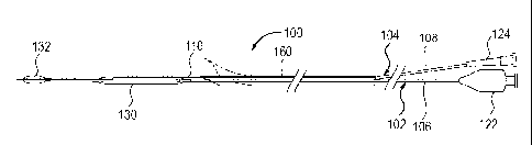
FIG 8 shows a catheter system 100 that includes a rapid exchange balloon
angioplasty catheter 102 with an occlusion balloon catheter 104 positioned
through
the guidewire lumen 110 being advanced together through the guiding catheter
52.
The angioplasty catheter 102 and the occlusion balloon catheter 104 are
effectively
coupled together and are advanced together as a unit into the guiding catheter
52 and
then up to the distal extremity of the stent 70. The rapid exchange balloon
angioplasty
catheter 102, which is intended for post stenting angioplasty, will typically
have a 6
to 9 mm diameter dilatation balloon 130.
The occlusion balloon catheter 104 has an occlusion balloon 132 made of
latex, silicone, polyurethane or another material, with a 6 to 9 mm inflated
diameter,
attached or glued on a simple metallic tube 108 (e.g. superelastic Nitinol or
spring
temper stainless steel tubing) with a diameter of 0.014 to 0.018 inches and a
length
that is preferably approximately 120-140 cm or longer. The short length of the
tube
108 is possible because of the short guidewire lumen 110 on the rapid exchange
or
monorail-type angioplasty catheter 102. The elimination of the dead space in
the
occlusion balloon catheter 104 before the procedure will be performed by
aspiration
using a simple 3 way stopcock and a 50 cc syringe.
The occlusion balloon catheter 104 will preferably be made with a luer lock
fitting permanently attached to the proximal end of the tubing 108, for
example by
insert molding or gluing the fitting onto the tubing. Alternatively, a
removable fitting,
such as a Touhy-Borst adapter or a compression fitting, may be used to
facilitate
catheter exchanges over the tubing 108 of the occlusion balloon catheter 104.
In this
case, an internal sealing member, such as described in U.S. Patent 6,156,005,
may be
CA 02581444 2007-03-22
WO 2006/032686 PCT/EP2005/054747
9
used to maintain the occlusion balloon 132 in the inflated state when the
fitting is
removed.
Preferably, the occlusion balloon 132 has a deflated profile that is small
enough to fit through the guidewire lumen I10 of the angioplasty catheter 102
for
assembly of the catheter system 100. Alternatively, the catheter system 100
can be
assembled by inserting the bare tubular shaft 108 of the occlusion balloon
catheter
104 through the guidewire lumen 110 of the angioplasty catheter 102 in an
anterograde or retrograde fashion and then attaching the occlusion balloon 132
and/or
the proximal fitting to the tubular shaft 108. If a permanently attached
proximal
fitting is used, the two catheters 102, 104 will be permanently coupled
together.
The tubular shaft 108 of the occlusion balloon catheter described with a
diameter of 0.014 to 0.018 inches could alternatively be made in other
diameters,
such as 0.035 to 0.038 inches. The rapid exchange angioplasty catheter 102
would
have to be modified with a guidewire lumen I 10 corresponding to the diameter
of the
shaft 108 of the occlusion balloon catheter 104. The positioning of the
guiding
catheter 52 inside of the stent 70 leaves an open avenue that could be used
with other
instruments (angioplasty, echography, fibroscopy, etc.) as long as they fit
into it.
These various instruments could be used to perform diagnostic or therapeutic
procedures that require isolation of the carotid bifurcation space.
Preferably, the catheter system 100 includes a releasable linking device 160
that holds the rapid exchange angioplasty catheter 102 and the occlusion
balloon
catheter 104 together so that the catheter system 100 can be easily advanced
as a unit.
Various configurations of releasable linking devices 160 that can be used in
the
catheter system 100 have been described in U.S. Patent Application, serial
number
10/833,494, which is incorporated by reference.
The releasable linking device of the catheter system may comprise a body
with a first channel and a second channel arranged in a side-by-side
configuration, the
first channel being configured to releasably hold the shaft of the first
catheter and the
second channel being configured to releasably hold the shaft of the second
catheter.
In a further embodiment, the releasable linking device of the catheter system
may comprise a body with a first channel and a second channel arranged in a
side-by-
side configuration, a first locking device associated with the first channel
configured
to releasably hold the shaft of the first catheter, and a second locking
device
CA 02581444 2007-03-22
WO 2006/032686 PCT/EP2005/054747
associated with the second channel configured to releasably hold the shaft of
the
second catheter.
In a further embodiment, the releasable linking device of the catheter system
may comprise a first linking member attached to the shaft of the first
catheter and a
5 second linking member attached to the shaft of the second catheter, the
first linking
member and the second linking member have interlocking features so that the
first
linking member and the second linking member can be releasably attached to one
another.
In a further embodiment, the releasable linking device of the catheter system
10 may comprise a peel-away sheath releasably attaching the shaft of the first
catheter
and the shaft of the second catheter together.
In a further preferred embodiment, the releasable linking device of the
catheter system may comprise a split-tube releasably attaching the shaft of
the first
catheter and the shaft of the second catheter together.
The originality of the linking device resides in the fact that it is
configured so
that one or both of the catheters can be advanced as a unit and when it is
desired can
be released from the linking device and maneuvered separately from the rest of
the
catheter system.
The linking device may be self-releasing in the sense that the linking device
demounts itself from the first and second catheters as the catheter system is
advanced
into the patient's body. On account of the fact that the linking device is
attachable
near the proximal ends of the catheters, it may be released by the physician
in a very
convenient manner, thereby allowing the two catheters to be maneuvered
separately.
One preferred embodiment of the catheter system 100 is shown in FIG 14 for
protected angioplasty and stenting at the carotid bifurcation utilizing a
linking device
160 constructed of an elongated split-tube 200. The split-tube 200 of the
linking
device 160 is configured to hold the proximal portion 106 of the rapid
exchange
angioplasty catheter 102 and the tubular shaft 108 of the occlusion balloon
catheter
104 arranged in a side-by-side configuration and aligned with one another
along a
longitudinal axis. A longitudinal split 202 extends the length of the split-
tube 200.
The longitudinal split 202 allows the split-tube 200 to be placed over the
proximal
sections 106, 108 of the catheters 102, 104 during assembly of the catheter
system
100 and to be removed from the catheters 102, 104 at the appropriate time
during the
CA 02581444 2007-03-22
WO 2006/032686 PCT/EP2005/054747
11
protected angioplasty and stenting procedure. The length of the split-tube 200
can
vary. Good results have been obtained with a catheter system 100 having a
split-tube
200 that extends along most of the proximal section 106, 108 of the balloon
catheters
102, 104 between the proximal hubs 122, 124 and at least to the proximal
guidewire
port of the rapid exchange angioplasty catheter 102. Preferably, the split-
tube 200 of
the linking device 160 is configured with a distal pull-tab 210 or other
feature to
facilitate lifting the distal part of the split-tube 200 to remove the linking
device 160
and release the balloon catheters 102, 104 so that they can be maneuvered
separately
from one another. The pull-tab 210 is preferably located on a side of the
split-tube
200 opposite to the longitudinal split 202. The pull-tab 210 can be formed by
skiving
or cutting away part of the tube 200 as shown.
FIG 15 shows a cross section of one embodiment of the split-tube 200 of the
linking device 160 for the catheter system 100 of FIG 14. The split-tube 200
has an
inner lumen 204 that is sized and configured to hold the proximal sections
106, 108
of the rapid exchange angioplasty catheter 102 and the occlusion balloon
catheter 104
together with sufficient friction that the catheter system 100 can be advanced
as a unit
without any relative movement of the two catheters. In one particularly
preferred
embodiment, the split-tube 200 is manufactured as an extruded profile with an
approximately circular outer profile and an approximately oval inner lumen
204. The
longitudinal split 202 connects the inner lumen 204 with the exterior of the
split-tube
200 at a thin part of the wall that coincides with the major axis of the oval
inner
lumen 204. The longitudinal split 202 is preferably formed during the
extrusion of the
split-tube 200. Alternatively, the tube 200 can be extruded without the
longitudinal
split 202 and then slitted along the length to form the longitudinal split 202
in a
secondary operation. Suitable materials for the split-tube 200 include
polyamide
copolymers (e.g. PEBAX 6333 or PA 8020 from ATOFINA), polypropylene, and any
extrudable medical grade polymer with a suitable combination of strength,
flexibility
and friction characteristics.
The split-tube 200 of the linking device 160 can be made with many other
possible configurations, including single-lumen and multiple-lumen
configurations,
and may include one or more longitudinal splits 202.
FIG 16 shows the catheter system 100 of FIG 14 in use. The linking device
160 with the split-tube 200 has the advantage that, once it is started, the
split-tube 200
CA 02581444 2007-03-22
WO 2006/032686 PCT/EP2005/054747
12
will demount itself as the catheter system 100 is advanced so that the
physician does
not need to unpeel, remove or displace a linking member that would otherwise
require a "third hand". The catheter system 100 is prepared for use by
aligning the
rapid exchange angioplasty catheter 102 and the occlusion balloon catheter 104
in the
desired longitudinal alignment and then pressing the longitudinal split 202 of
the
split-tube 200 against the proximal sections 106, 108 of the catheters until
they are
enclosed within the inner lumen 204 of the split-tube 200, as shown in FIGS 14
and
15. This preparation if preferably carried out at the manufacturing facility
or,
alternatively, it may be performed at the point of use by a medical
practitioner. The
distal ends of the rapid exchange angioplasty catheter 102 and the occlusion
balloon
catheter 104 are inserted into the patient in the usual manner through a
guiding
catheter with a Y-fitting 220 or other hemostasis adapter on the proximal end
of the
guiding catheter. The distal pull-tab 210 is pulled toward the side to start
demounting
the split-tube 200 from the rapid exchange angioplasty catheter 102 and the
occlusion
balloon catheter 104, and then the catheter system 100 is advanced as a unit.
As
shown in FIG 16, when the split-tube 200 encounters the Y-fitting 220, the
split-tube
200 will peel away or demount itself from the proximal sections 106, 108 of
the rapid
exchange angioplasty catheter 102 and the occlusion balloon catheter 104. Once
the
rapid exchange angioplasty catheter 102 and the occlusion balloon catheter 104
have
been advanced into the distal part of the self-expanding stent, the split-tube
linking
device 160 can be set aside and discarded.
FIG 9 shows the occlusion balloon 132 inflated within the lumen of the self-
expanding stent 70 and the angioplasty catheter 102 positioned with dilatation
balloon 130 across the lesion 50. The occlusion balloon 132 is inflated in the
distal
part of the stent 70 to occlude the carotid artery and to prevent any embolic
debris
from traveling downstream from the treatment site. Then, the angioplasty
balloon 130
is withdrawn to the site of the remaining narrowing of the stent 70 to be
dilated.
FIG 10 shows an angiography study performed to confirm occlusion of the
internal carotid artery prior to dilatation of the lesion 50. The patient is
clinically
tested. An angiography series is performed to confirm the effective temporary
occlusion of the internal carotid. The contrast 90 should remain close to the
bifurcation site and usually does not reach the occlusion balloon 1 32.
CA 02581444 2007-03-22
WO 2006/032686 PCT/EP2005/054747
13
FIG 11 shows the angioplasty balloon 130 inflated to dilate the stenosis 50
and to complete the deployment or expansion of the self-expanding stent 70. It
is
recommended that atropine be injected at least 5 minutes previously to rule
out the
bradycardia induced by the compression of the carotid glomus.
After completion of the poststenting angioplasty, the angioplasty balloon 130
is deflated and the angioplasty catheter is withdrawn from the guiding
catheter 52.
The tubular shaft 108 of the occlusion balloon catheter 104 has sufficient
length that
the short guidewire lumen 110 of the angioplasty catheter can be "parked" on
the
shaft 108 near the proximal end of the occlusion balloon catheter 104 so that
it will
not interfere with the aspiration step, which is to follow. Alternatively, if
the
occlusion balloon catheter 104 is made with a removable proximal fitting, the
fitting
may be removed at this point so that the angioplasty catheter 102 can be
removed
completely. The internal sealing member described above will maintain the
occlusion
balloon 132 in the inflated state.
With the occlusion balloon 132 still inflated, blood is aspirated back through
the lumen of the guiding catheter 52. FIG 12 illustrates potential embolic
material 92
being aspirated through the lumen of the guiding catheter 52.
The occlusion balloon 132 is then deflated and the angioplasty catheter 102
and occlusion balloon catheter 104 are withdrawn. An angiography series is
performed through the guiding catheter 52 to verify patency of the lumen and
full
deployment of the self-expanding stent 70. Then, the guiding catheter 52 and
introducer are withdrawn and the puncture site is closed.
FIG 13 illustrates the patient's carotid bifurcation with the fully deployed
stent 70 after completion of the protected angioplasty and stenting procedure.
Although it has been described in relation to treatment of obstructive carotid
artery disease, the method of the present invention can be adapted for
performing
protected angioplasty and stenting in other parts of the vasculature, for
example in the
coronary arteries.
While the present invention has been described herein with respect to the
exemplary embodiments and the best mode for practicing the invention, it will
be
apparent to one of ordinary skill in the art that many modifications,
improvements
and subcombinations of the various embodiments, adaptations and variations can
be
made to the invention without departing from the spirit and scope thereof.