Note : Les descriptions sont présentées dans la langue officielle dans laquelle elles ont été soumises.
CA 02585309 2012-10-16
1
VIROSOME PARTICLES COMPRISING ANTIGENS FROM INFLUENZA
VIRUS AND HEPATITIS B VIRUS
The present invention relates to a virosome comprising a virosomal membrane
comprising at least one lipid and envelope proteins of an enveloped virus and
nucleocapsid particles of said enveloped virus located on the inside and the
outside of the virosome and attached to said envelope proteins. Furthermore,
the
invention relates to a vaccine comprising the virosome of the invention and a
method for the production of a virosome of the invention. Moreover, the
invention
relates to a use of a virosome of the invention for the preparation of a
vaccine,
e.g. for the prevention or alleviation of a disease related to an HBV
infection, and
a method for the vaccination of a subject.
The development of novel and increasingly safer vaccines frequently makes use
of well-characterized antigens, in particular highly purified recombinant
proteins
or synthetic peptides. In spite of some achievements, this approach is impeded
by the fact that such antigens are often poor immunogens when administered
alone. This fact has necessitated the development of suitable adjuvant and
carrier systems that possess the ability to enhance the immunogenicity of a
given
antigen. One possible approach is the integration of antigens into a higher
structure, e.g. a virus-like particle. The physical association of all vaccine
components in a single particle ensures their simultaneous interaction with
individual immune cells, and thereby, maximal exploitation of synergistic
potentials. This is of particular relevance if immuno-stimulatory or immuno-
modulatory components (adjuvants) are included in the formulation.
Furthermore,
the particle structure itself can have immuno-stimulatory effects and increase
both, the stability and the immunogenicity of the individual components.
CA 02585309 2007-04-25
WO 2006/045532
PCT/EP2005/011297
2
It is thus a problem to find a suitable approach of combining the relevant
antigens in
an industrial applicable formulation, which leads to an efficient prophylactic
and/ or
therapeutic application.
During a virus replication in a host cell, copies of the viral gnome are
generated
and viral proteins are expressed and processed before they assemble to mature
virions while taking advantage of the cellular infrastructure. The common
basis is
that virus replication and assembly of progeny requires the environment of a
living
host cell and an ordered series of specific interactions between viral nucleic
acids,
viral and host cell proteins, and lipid membranes, that leads to segregation
and
assembly of the n-iacromolecular virion structure. The large number of
different
molecules required and in addition, the cellular structures involved
illustrate the high
complexity of any virion assembly. There are major differences between
different
virus classes, and in particular, between enveloped and non-enveloped viruses.
Common to all enveloped viruses is the outeH shell of the virus, composed of a
lipid
membrane with integrated viral proteins, and, as a consequence, the necessary
interaction between membrane-associated and., 5aluble viral proteins or
protein-
based structures, e.g. the nucleocapsids in order to assemble a mature
enveloped
virus. Non-enveloped viruses lack a lipid-based membrane and assemble from
protein and nucleic acid molecules only.
Numerous approaches to reconstitute viral particles in vitro and in vivo have
been
described in the literature and can be divided into distinct categories:
(a) In vitro reconstitution of viral envelopes
Virus-derived or recombinant envelope proteins can be purified and formulated
with or without additional lipids into proteoliposomes. This pure in vitro
approach
achieves the generation of the outer shell of enveloped viruses, the envelope,
but does not include the core of the virus, the nucleocapsid. There are
examples
of chimerical virosomal structures, which integrate envelope proteins from
different viruses. Reconstituted viral envelopes have also been used
successfully for gene transfer (DNA or RNA) but these methods did not depend
on packaging of a functional, protein-based nucleocapsid but rather an
association of the nucleic acids directly with the reconstituted envelope.
(b) Heterologous expression of one or more viral proteins
CA 02585309 2007-04-25
WO 2006/045532
PCT/EP2005/011297
3
Isolated recombinant viral proteins can self-assemble into virus-likp
structures
(VL,Ps): HPV (yeast, baculo), HCV (baculo), Hs antigen (yeast, CHO), HBc (E.
coif). Common to all these pproaches is that the self-assembly tales place in
the heterolOgous cellular Qxpression system and the virus-like p?rticles are
subsequently purified. Therfore, the assembly does not take place in vitro but
relies on a cellular system. 'VLPs have been used as vaccines and as vaccine
carriers. (Pumpens, P.; Grans, E. (2(01) Intervirology 44 (2-3); 98-114; Noad
R,
Roy P. (2003) Trends IVIicrolpiol. 11(9): 438-44)
(c) Reconstitution of non-enveloped (icosahedral) viruses or virus-like
particles in
vitro
This approach is based on separately purified components. Due to the absence
of a lipid membrane-based envelope, non-enveloped viruses are simpler in their
structure and can self-assemble under certain conditions in vitro if all
necessary
components are present in the correct stoechiometry. Similarly, the inner core
of
enveloped viruses, the lipid-free nucleooapsids, or subunits thereof have been
reconstituted in vitro from purified recombinant components, e.g. of influenza
virus (Martin-Benito J. at al. (2001) EMBO Rep. 2(4): 313-7).
(d) Purification of viral nucleocapsids
Nucleocapsids have been ?xtracted and purified from many different virus types
in order to characterise their composition. These preparations can also be
used
for transfection of susceptible cells aiming at virus rescue. Sucoessful virus
rescue implies that a functional nucleocapsid was isolated and delivered to
the
cytoplasm of a host cell. However, this does not imply the successful
reconstitution of a functional enveloped virus, because the natural way of
infection, which depends on a functional envelope, is bypassed by the use of a
transfectant, the latter mediating direct delivery of the nucleocapsid into
the
cytoplasm of a host cell.
(e) Pseudotyping of enveloped viruses and viral vectors in a cell culture
ystern.
This in vivo approach has been widely and successfully used fOr the production
of Chimerical viruses or vectors (e.g. Retroviruses, Lentiviruses, and AAV) at
lab
scale. The key element 'for the production of pseudotyped Viruses is a helper
cell
that Co-expresses all the proteins to be integrated into the virion and
mediates
assembly of the virions. In contrast, an in vitro assembly of an envelOped
virus is
CA 02585309 2007-04-25
WO 2006/045532
PCT/EP2005/011297
4
based on defined, separately produced and purified components, and the
physical association is performed under controlled conditions in vitro.
(Sandrin
V. et al. (2003) Curr Top Microbiol Immunol.; 281:137-78)
As a specific form of virus-like particles, virosomes are a clinically proven
vaccine
carrier/adjuvant system with an excellent safety and tolerability profile in
human s.
The capability of the virosomal carrier to mediate antigen processing through
both
the exogenous and the endogenous pathway makes this system a good candidate
for a therapeutic vaccine.
The basic concept of Virosomes comprises the reconstitution in vitro of empty
viral
enveloped, or more general, of viral envelope proteins integrated into a
spherical
lipid bilayer. Virosomes have been generated from a number of viruses (Y Kaned
a.
(2000) Adv, Drug Delivery Rev. 43, 197-205; Drummond DC, et al. (2000) Frog
Lipid Res. 39(5): 409-60). The possibility of producing chimerical virosores
containing envelope proteins from two different viruses has been demonstrated
(Bagai S, Sarkar DP. (1994) FEBS Lett. 353(3): 332-6).
In all cases, the viral protein of interest is a transmembrane or membrane-
anchored
structure, which is prerequisite for spontaneous integration.
The virosomal formulation of molecules that do not directly interact with the
virosomal lipid membrane Js far more difficult to achieve. Although the idea
of
linking molecules to viroSomal structures has been proposed earlier (WO
95/327)6,
INEX), the technical hurdles to achieve stable and efficient formulations can
be
enormous, depending on the biochemical properties of the molecule of interest.
Nucleic acids can be associated to the virosomal structure via the use of
positively
charged lipids (WO 98/52603, Berna). Small molecules (peptides, drugs) lacking
a
secondary and tertiary Structure for their function can be Modified
biochemically in
order to enable association, integration or encapsulation. A number of Methods
have been described for virosomal formulation, in particular, encapsulation of
sm all
molecules (Walti et al, (2002) Canc. Res) or synthetic particles (Jana et al.
(20(2)2)
FEBS Lett.; 515(1-3: 184-188). These Methods only work under chemical
conditions that would affect the authentic conformation of larger proteins and
even
more so, the integrity of multimeric protein complexes such as viral
nudeocapsi ds
(e.g. the HBc antigen particle). The methods described so far to associate one
or
more large proteins lacking exposed lipophilic domains into the virosorrial
CA 02585309 2007-04-25
WO 2006/045532
PCT/EP2005/011297
membrane required biochemical modifications of the protein, e.g. covalent
linkage
to lipid molecules (Hunziker IF. et al. (2002) Int Immunol, 14(6): 615-26), in
order to
anchor the respective protein in the lipid membrane. This method has also
proven
efficacious for retargeting virosomes to specific cell types via crosslinked
antibodies
5
(Mastrobattista E. et al, (2001) FEBS Lett. 509(1): 71-6. , VValti et al,
Canc. Res.
= 2002). However, biochemical modifications require conditions (e.g,
oxidative
conditions for activation of reactive side groups) which are likely to
dissociate non-
covalently linked multimeric structures (e.g. a viral nucleocapsid). In
addition, such
conditions can also alter the conformation of the protein molecule in
question, and,
as a consequence, impact on its immunogenicity, and ultimately, on the
efficacy of
the vaccine, Furthermore, the crosslinking procedure increases both the number
of
steps required for the formulation and the loss of antigen. Only one exarmple
exists
for a multimeric protein structure successfully associated with virosornes
without
biochemical modification, namely the Hepatitis A vaccine Epaxal (GILIck. R.,
1995,
J. of Liposome Research 1995, 5(3), 487-479). However, in this vaccine the
antigen
is associated to the outer surface only after formulation of influenza
virosernes, due
to electrostatic interaction between virosomal membrane and virus particle. As
a
consequence, no antigen is located in the aqueous interior of the virosome,
which is
the preferred location for efficient cytoplasmatic delivery and induction of a
CD8-
based cellular response as it will be required for a therapeutic vaccine.
(Bungener L.
et al. (2002) J Liposome Res. 12(1-2): 155-63; Bungener L. at al. (2002)
Vaccine.
20(17-18): 2287-95.)
The potential and the limitation of approaches for the prevention and the
treatment
of infectious diseases are discussed herein below exemplarily for HBV. HBV
infection represents a huge health problem world-wide, in particular because
of life
threatening late complications. The World Health Organisation (WHO) estimates
that currently approximately 400 million individuals are chronic HBN carriers.
Patients suffering from chronic HBV infection show a wide range of symptoms,
from
clinically inapperent to severe chronic liver disease, yet the long-term risk
of liver
disease (chronic hepatitis, cirrhosis and hepatocarcinoma) is dramatically
increased
for all chronic HBV carriers (25% incidence within 20 to 30 years after
infection).
Common to all chronic patients is also a poor immune response to the causative
CA 02585309 2007-04-25
WO 2006/045532
PCT/EP2005/011297
6
agent, HBV, and, in particular, against the HBV core protein (HBc), despite of
The
fact that large amounts of antigen are circulating throughout the chronic
infection. In
contrast, resolution of acute Hepatitis B, as well as the spontaneous or
treatment-
induced resolutions of chronic Hepatitis B, is strictly associated to the
development
of a broad and vigorous immune response against HBV antigens. Conventional
therapeutic approaches, such as therapies with interferon or antiviral drugs
to
control chronic hepatitis are only partially successful, yet cost-intensive
and
associated with significant side-effects. Patients with HBV-associated chronic
hepatitis would thus greatly benefit from a therapeutic vaccine that can
control this
persistent virus infection.
According to the current understanding of HBV pathogenesis and immunology, the
key to a successful therapeutic vaccination is to overcome the HBV-specific
immunological non-responsiveness of chronic carriers. To that end, the
relevant
antigens (HBc and HBs) must be presented to the patient's immune system in a
way that the existing inefficient Th2 type (humoral) immunity is skewed into a
strong
and sustained Thl type (cellular) of response, and at the same time, boost the
Th2
type response.
The immune response against the relevant antigens should be broad and directed
simultaneously against many different epitopes in order to prevent immune-
escape
mutants of the virus. Such variants have been shown to evolve under selective
pressure directed against single epitopes. Furthermore, the use of full-length
proteins as antigens of the vaccine takes in account the genetic diversity of
the
patients with regard to antigen processing and MHC genotype-dependent epitope
selection.
Significant efforts have been dedicated to the development of therapeutic I-I
BV
vaccines in the past, as reviewed by M. Hilleman (Vaccine 21(2003): 4626-
4649). In
a number of clinical trials, conventional HBs-based prophylactic vaccines have
been
used in chronic HBV patients, but no sustained positive effects were observed
so
far. Peptide-based vaccines intended to focus the immune response to few
relevant
= epitopes (reviewed by Engler et al., Mol Immunol. 2001 .Dec; 38(6): 457-
65). This
approach yielded promising results in preciinical research but not in humans.
More
retently, recombinant HBc particles were produced which carried single
epitopes of
CA 02585309 2007-04-25
WO 2006/045532
PCT/EP2005/011297
7
HBs on the surface in an attempt to combine the two relevant HBV antigens in
one
vaccine (Chen et al, Vaccine. 2004 Jan 2; 22(3-4): 439-46).
These approaches mainly aim at humoral response of the immune system. Th e
more advanced approaches follow the concept that a cellular response (Thl
type)
against HBV is a key element of a successful therapeutic immunisation.
The induction of a Thl-type immune response against the HBV antigens,
especially
against HBc, is the ultimate goal of a therapeutic HBV vaccine. While HBs alon
e
can elicit a Thl response to some degree, HBc alone does not, Therefore, these
antigens alone cannot induce the adequate immune response required for a
therapeutic effect. It can only be achieved by combination of the HBV antigens
with
a Thl-supporting adjuvant or carrier system. In contrast, aluminium salts, the
most
widely used adjuvant in current human vaccines, are well known to abolish Thu
responses in favour of a Th2 response. This property of aluminium salts makes
it a
very attractive adjuvant for prophylactic vaccines, which are primarily aiming
at the
induction of high titre of protective antibodies. In a therapeutic setting, a
sustained
Thl response plays a crucial role since Thl effector cells mediate control of
virus
replication and elimination of virus-infected cells:
Attempts were made with DNA vaccines (plasmid DNA encoding the HBV core or S
genes) known to promote primarily a Cellular response. Despite the fact that
DMA
vaccines work very well in the mouse model, numerous clinical trials have
failed to= ,
provide proof of principle in n-ian, not only in the HBV field. Similarly, the
use of viral
vectors expressing HBV antigens (e.g. vaccinia) aiming at enhancing cellular
response failed to induce significant and sustained responses in humans.
Although various approaches have been tested flon.- therapeutic HBV vaccines
none
of those lead to a sufficient therapeutic vaccine.
The two major structural HBV proteins, HBs and HBc can be expressed individua
Ily
in several heterologous systems: E.coli, yeast, and mammalian cell lines, Both
antigens form typical virus-like particle structures (HBs particles and HBc
particles,
= respectively) which are clearly distinct from the infectious, enveloped, and
nucleocapsid-containing HBV virions.
Recombinant Hepatitis B core antigen (HBc) can be produced in a bacterial or a
CA 02585309 2007-04-25
WO 2006/045532
PCT/EP2005/011297
8
yeast-based expression system, since this protein is not glycosylated. HBV
core
monomers self-assemble into virus-like particles with a diameter of about 30nm
and
can be purified in this form from the producer cells. Very similar to
authentic l-11E3V
nucleocapsids, HBc particles are composed of either 180 or 240 monomeric core
=
molecules which self-assemble into the particle structure. HBc particles do
not
contain lipids. The HBV core monomer consists of 183 to 185 amino acid (aa)
residues (length is isolate-dependent). The C-terminal 30 aa feature a nucleic
acid
binding domain, which results in the presence of significant amounts of
nucleic
acids (predominantly RNA derived from the expressing cell in the absence of
HIEN
genomes) in purified preparations of HBc particles, an unwanted contamination.
HBc can be truncated at the C-terminus to a length of 144 aa, which reduces
the
nucleic acid content by 99% while retaining the particle structure. Shorter
constru cts
than 144 aa do not form particles any more.
The production of recombinant HBc particles has been described in many
variants.
Engineered variants of HBo have been used as a carrier system for heterologous
antigens (Pumpens, P.; Grens, E. (2001) Intervirology 44 (2-3); 98-114). In
:this
approach the foreign aa sequence is inserted in the region (aa 70-90) of the
protein
chain which is exposed to the outer surface in the context of the multimeric
particle.
However, the size of the genetically inserted foreign antigen sequence is very
limited due to the necessity that the monomers retain their capability to self-
assemble into particles. When used as a vaccine in animal models, HBc
particles
alone induce a significant humoral response but lack the ability to produce a
H Bc-
specific CD8-tyPe cellular response which is considered essential for a
therapeutic
effect.
WO 00/32625 (Biogen) describes Hepatitis= B core particles comprising
Immunogens, epitopes resulting potentially in multivalent hepatitis B core
particles.
An approach already entering clinical trials is the construction of a modified
Hepatitis B core particle containing multiple epitopes of Plasmodium
falciparurn for
prevention of Malaria (Birkett A., et al, Infection and Immunity 2002, p 686-
6870)
The authentic HBV envelope protein (HBs) exists in three forms L (large), M
(middle), S (small) which are expressed from three staggered translation start
std.
CA 02585309 2007-04-25
WO 2006/045532
PCT/EP2005/011297
=
9
All three forms of HBs in multin-leric form are present in the envelope of HBV
virions. The 0-terminal preS1 (L) and preS2, (M) domains are involved in the
binding
of HBV to cells during infection, and antibodies against the preS1 domain are
capable of neutralising HBV. When expressed as recombinant protein in yeast or
mammalian cells, HBs is secreted in the form of micelle particle structures
with a
diameter 35-45nm, which contain also significant amounts (60% w/vv) cellular
membrane lipids (Satoh O. et al. (2000) J Eiochem; 127(4): 543-50). Depending
on
the expression construct, the recombinant particles contain either S alone or
include
the C-terminal pre S1 and/or preS2 domains.
All current prophylactic vaccines against HBV are based on recombinant HBV
envelope (HBs) proteins formulated with Aluminium salts. Most products are
based
on yeast expression. More recent products, so-called 3rd generation HBV
vaccines
are derived from mammalian cells. These vaccines contain the preS domains and,
in addition; the authentic mammalian glycosylation pattern and a mammalian
lipid
composition, both of which are thought to be beneficial for a imm une
response.
The use of HBs particles as a vaccine carrier is claimed in WO 99/39736
(Yissum)
but, the system is limited to monomeric antigens thereby excluding a co-
formulation
with HBc particles or other nucleocapSid-type structures. In addition, the
said
system does not foresee a destruction and re-assembly procedure in vitro of
the
carrier particle. =
A recent publication (Ponsel and Bruss, (2003) JV .77 416-422) describes the
formation: and secretion of HBV .particles containing both HBs and HBc from
= mammalian cells co-transfected with expression =plasmids for both
antigens.
However, there are no reports on the reconstitution of in vitro envelope.d
particles
containing both HBs and HBc antigen.
,US 6,020,167 (Medeva) claims a method of treating Hepatitis B by
administering a
composition comprising one or more T cell activating epitopes from pre S1 or
HBV
core and a carrier capable of presenting the polypeptide. The carrier
according to
the invention can be a HBsAg particle.
As a specific form of virus-like particles, virosomes are a clinically proven
vaccine
carrier/adjuvant system with an excellent safety and tolerability profile in
humans.
CA 02585309 2007-04-25
WO 2006/045532
PCT/EP2005/011297
= The capability of the virosomal carrier to mediate antigen processing
through both
the exogenous and the endogenous pathway makes this system a good candidate
for a therapeutic vaccine.
The basic concept of virosomes comprises the in vitro reconstitution of empty
viral
5
envelopes, or more general, of viral envelope proteins integrated into a
spherical
lipid bilayer. Virosomes have been generated from a number of viruses (Y
Kaneda.
(2000) Adv. Drug Delivery Rev. 43, 197-205; Drummond DC. et al. (200C) Frog
Lipid Res. 39(5): 409-60). The Possibility of producing chimerical virosomes
containing envelope proteins from two different viruses has been demonstrated
10 (Bagel S, Sarkar DP. (1994) FEES Lett. 353(3): 332-6)
In all cases, the viral protein of interest is a transmembrane or membrane-
anchored
structure, which is prerequisite for spontaneous integration.
The virosomal formulation of molecules that do not directly interact with the
virosomal lipid membrane is far more difficult to achieve. Although the idea
of
linking molecules to virosomal structures has been proposed earlier (WO
95/32706,
INEX), the technical hurdles to achieve stable and efficient formulations can
be
enormous, depending on the biochemical properties of the molecule of interest.
Nucleic acids can be associated to the virosomal structure via the use of
positively
charged lipids (VVO 98/52603, Berna). Small molecules (peptides, drugs)
lacking a
secondary and tertiary structure for their function can be modified
biochemically in
order to enable association, integration or encapsulation. A number of
rnethods
have been described for virosomal formulation, in particular, encapsulation of
small
molecules (VValti et al, (2002) Canc. Res) or synthetic particles (Jana et al.
(2002)
FEES Lett.; 515(1-3: 184-188). These methods only work under chemical.
conditions that would affect the authentic conformation Of larger proteins and
even
more so, the integrity of multimeric protein complexes such as viral
nucleocapsids
(e.g. the HI3c antigen particle). The methods described so far to associate
one or
more large proteins lacking , exposed lipophilic domains into the viroscrnal
membrane required blochemital modifications of the protein, e.g. covalerit
linkage
to lipid molecules (Hunziker IF. et al. (2002) Int Immunol. 14(6): 615-26), la
order to
anchor the respective protein in the lipid membrane. This method has also
proven
efficacious for retargeting virbsomes to specific cell types via crosslinked
antibodies
(Mastrobattista E. et al. (2001) FEES Lett. 509(1): 71-6. , Vialti et al,
Canc. Res.
CA 02585309 2007-04-25
WO 2006/045532
PCT/EP2005/011297
11
2002), However, biochemical modifications require conditions (e.g. oxidative
conditions for activation of reactive side groups) which are likely to
dissociate non-
covalently linked multin-ieric structures (e.g. a viral nucleocapsid). In
addition, such
conditions can also alter the conformation of the protein molecule in
question, and,
as a consequence, impact on its immunogenicity, and ultimately, on the
efficacy of
the vaccine. Furthermore, the crosslinking procedure increases both the number
of
steps required for the formulation and the loss of antigen. Only one example
exists
for a multimeric protein strUcture successfully associated with virosomes
without
biochemical modification, namely the Hepatitis A vaccine Epaxal (Gluck R,
1995,
0 J. of Liposome Research 1995, 5(3), 467-479). However, in this vaccine
the antigen
is associated to the outer surface only after formulation of influenza
virosomes, due
to electrostatic interaction between virosomal membrane and virus particle, As
a
consequence, no antigen is located in the aqueous interior of the virosome,
which is
the preferred location for = efficient cytoplasmatic delivery and induction of
a 0D8-
.5 based cellular response as it will be required for a therapeutic
vaccine. (Bungener L.
et al. (2002) J Liposome Res. 12(1-2): 155-63; Bungener L. et al. (2002)
Vaccine.
20(17-18):2287-95.)
A number of patents have been applied/granted for influenza virosomes
2D (W092/19267 WO 98/52603, Berna) and virosome-like structures derived
from
other enveloped viruses (e.g. Sendai Virus). These methods comprise the
solubilisation of the viral envelope, removal of the nucleocapsid containing
the viral
genome, followed by reconstitution of an "empty" viral envelope. Furthermore,
additional antigens are either adhered to ready-made virosomes (Epaxal ) or
2,5 crosslinked to lipid molecules in order to anchor them in the virosomal
membrane.
In view of the above described limitations of vaccination against virus
infections, the
technical problem underlying the present invention was to provide improved
means
and methods for the vaccination of subjects for the prevention, alleviation or
30 treatment of virus infections.
The solution to said technical problem is achieved by providing the
embodiments
characterized in the claims.
CA 02585309 2012-10-16
12
Accordingly, the present invention provides a virosome comprising
(a) a virosomal membrane comprising at least one lipid and envelope proteins
of an enveloped virus; and
(b) nucleocapsid particles of said enveloped virus located on the inside and
the
outside of the virosome and attached to said envelope proteins.
There is described herein a virosome comprising
(a) a virosomal membrane comprising at least one lipid and envelope
proteins
of influenza virus and hepatitis B virus (HBV); and
(b) nucleocapsid particles comprising the hepatitis B core (HBc) protein of
the
HBV located on the inside and the outside of the virosome and attached to
said envelope proteins.
There is also described herein a method of producing a virosome comprising the
steps of:
(a) solubilizing envelope proteins of the enveloped virus influenza virus
and
HBV in the presence of a lipid in a detergent solution;
(b) decreasing the concentration of the detergent in the solution;
(c) adding nucleocapside particles of HBV comprising the hepatitis B core
(HBc) protein to the solution obtained in step (b); and
(d) removing the detergent so that virosomes are produced.
The term "virosome" defines a specific form of virus-like particles (VLPs).
Virosomes are semi-synthetic complexes derived from viral particles and
produced by an in vitro procedure. They are essentially reconstituted viral
coats,
while the viral nucleocapsid is replaced by a compound of choice. Virosomes
retain their fusogenic activity and thus deliver the incorporated compound
(antigens, drugs, genes) inside the target cell. They can be used for
vaccines,
drug delivery, or gene transfer.
CA 02585309 2012-10-16
,
12a
VLPs are particle structures that are in size and shape reminiscent of or even
indistinguishable from their parental virus but are lacking the capability to
infect
and replicate in host cells. VLPs are multimeric structures composed of viral
proteins (authentic or modified variants of it). In addition, VLPs may or may
not
contain nucleic acids, lipids, and include lipid membrane structures or not.
Two
typical but very distinct examples for VLPs derived from a single Virus (HBV)
are
HBs and HBc particles.
The term "virosomal membrane" defines in the context of the present invention
a
spherical membrane structure that is reconstituted in vitro and that is
composed
of a lipid bilayer with integrated viral envelope proteins.
The term "envelope proteins" is intended to mean in the context of the present
invention a protein encoded by an enveloped virus that in its nature form
interacts directly with the virus lipid membrane.
In line with the present invention a broad range of lipids can be
comprised in said virosomal membrane. The group of lipids comprises
neutral and charged phospholipids, steroid-derived lipids, neutral and
charged synthetic lipids. In addition to the purified lipids added to
the formulation, the lipids contained in the viral components are also
included in the final formulation, e.g. lipids derived from Influenza Virus
or any other enveloped virus included or from lipid containing VLPs
included in the formulation ( e.g. HBs particles ). These virus-derived lipids
are
CA 02585309 2007-04-25
WO 2006/045532
PCT/EP2005/011297
13
heterogenous and reflect the lipid composition of the producer cell of the
virus or
the recombinant expression cell. The preferred formulations are based on
phospholipids only in order to minimise the complexity of the formulation. The
phospholipids used for HB-virosomes which are described in the appended
examples are usually GMP-grade and preferably identical to the material used
for
the registered vaccines lnflexal and Epaxal .
The term "enveloped virus" defines in the context of the present invention a
virus
that includes a host-cell derived lipid membrane in the mature virion
structure.
Classes of enveloped viruses are listed in table 1.
The term 'nucleocapsid particles" is intended to mean in the context of the
present
invention a particle structure composed of viral capsid proteins. This
particle
structure can be a VLP (composed of one or more recombinant viral capsid
proteins), or a nucleocapsid complex purified from the parental virus. Whether
or
not the nucleocapsid particle contains nucleic acids is not relevant for
formation of
the particle.
Virosomes of the invention (chimerical virus-like particles) comprise the
above
characterized molecules physically associated in a single particle. The
envelope
protein in virosomes of the invention may be integrated in the surface of the
virosome in the natural orientation with the interaction side for
corresponding
nucleocapsid particles to the inside of the virosome, as well as in an
artificial
orientation with the interaction side for corresponding nucleocapsid particles
to the
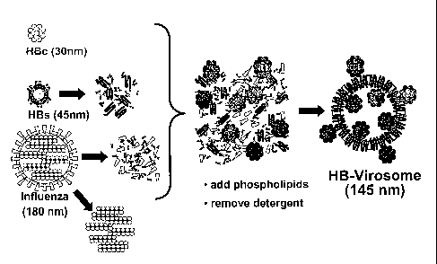
outside of the virosome. One example of such virosome is depicted in figure 1.
The
structure of this novel class of virosomes is distinct from the particulate
structures of
the individual components described above, or from the original viruses. This
type
of particle does not exist in nature and has neither been described nor
suggested in
the state of the art so far as a structure generated in vitro. Thus, the
particle
structure represents the first enveloped virus-like particle re-assembled
completely
in vitro from isolated components.
Virosomes known in the art and described herein above are produced in cell-
based
systems. In such cell-based system, all components must be produced in the
same
cell simultaneously, which restricts the choice of the expression system
dramatically
and forces compromises with respect to yield and scalabi lity. The biological
CA 02585309 2007-04-25
WO 2006/045532
PCT/EP2005/011297
14
expression systems and metabolic processes that are difficult to control
define the
composition of the resulting virus-like particles, e.g. the ratio between the
components. Furthermore; virus-like particles produced in cellular system must
be
extracted and purified subsequently without affecting the particle structure
or
composition in order to obtain a useful preparation.
In contrast and as described herein below, the composition of the in vitro
formulation of virosomes of the invention is controllable via the input
material and
the chosen biochemical parameters. The simplicity of the process ensures its
robustness. The resulting formulation does not require further purification. =
The
individual components may be produced beforehand in separate cell-based
systems (e.g. E. coli, mammalian cells, and yeast), and for each component,
the
optimal system with regard to yield, scalability and purity can be chosen.
The formulation process for virosomes of the present invention takes advantage
of =
the interaction between viral proteins which are easily integrated into a
virosome-
type of structure (membrane-associated proteins, envelope proteins) and
proteins
that do not associate with membranes by themselves. Although this interaction
is
essential and efficient during the assembly of most enveloped viruses in the
course
of their natural replication inside a host cell, the use of this property for
an in vitro
formulation process of a pharmaceutical product is novel. Surprisingly, the
intracellular virus assembly process can indeed be mimicked in vitro, although
under completely different conditions.
Preferably the virosome is a virosome, wherein said at least one lipid
comprises at
least one phospholipid. More prefereably, said phospholipids comprise
phosphatidylcholine, phosphatidylethanolamine and phosphatitylserine.
=
=
Also envisaged by the present invention are, in a further preferred
embodiment,
virosomes, wherein said envelope proteins are the envelope proteins of a first
and a
. second enveloped virus and the nucleocapsid particles are the nucleocapsid
particles of said second enveloped virus,
Said first enveloped virus may be selected from any enveloped virus.
Particularly
'preferred for the present invention are influenza viruses. .
= =
CA 02585309 2007-04-25
WO 2006/045532
PCT/EP2005/011297
In a more preferred embodiment of the invention the envelope proteins of said
first
enveloped virus are hemagglutinin (HA) and/or neuraminidase (NA).
The influenza components of virosomes of the invention, hemagglutinin (HA) and
neuraminidase (NA), may be purified from inactivated influenza virus (e.g.
strain
5
A/Singapore) in analogy to the established and patented formulation of
influenza
virosomes (Epaxal WO 92/19267, WO 92/19268, Gluck R., 1995, Journal of
Liposome Research 5(3), 467-479). The influenza-derived proteins/proteins of
said
first enveloped virus may be included for functional rather than
structural/mechanical reasons, =since virosomes of the invention may also be
10
formulated in the absence of the influenza proteins/proteins of said first
enveloped
virus. The influenza component may be included in order to strengthen the
aspect
of the virosome-like carrier immunological properties of the virosomes of the
invention.
15
Also in line with the present invention said second enveloped virus is
preferably
selected from a group of enveloped virus consisting of hepatitis B virus (HBV)
(which is preferred), hepatitis C virus (HCV) or any other Flavivirus, and
human
immunodeficiency virus (HIV).
In a further preferred embodiment of the invention the nucleocapsid particle
comprises HBc protein.
HBc particles may be produced in Emil, either containing the full-length amino
acid
sequence or truncated forms. Both the full-length as \vell as a truncated 144
amino
acid construct were successfullY formulated into HB-virosomes. Alternatively,
it is
contemplated that shorter (non-particular) HBV core are incorporated into HB-
virosomes. Corresponding techniques are known in the art and described in the
appended examples.
It is also preferred, that the envelope protein of said second enveloped virus
is HBs
protein.
HBs particles may contain S alone or pre S and S combined. Methods for the
production of said particles are known in the art. The particles may be e.g.
produced in yeast or mammalian cells. The presen ce of the preS domain in a
vaccine comprising the virosome of the invention is likely to contribute to a
broader
and a more efficient immune response but has no impact on the formulation
CA 02585309 2007-04-25
WO 2006/045532
PCT/EP2005/011297
16
process. HB-virosomes can be produced from HBs of either source. Even the
combination of different HBs types from different sources or of different
serotypes
into a single HB-virosome has been demonstrated using the method described
here.
The current invention falls in the above preferred embodiment into the class
of
influenza virosomes. However, the incorporation of an envelope protein (HBsAg)
of
a cpmpletely unrelated virus (HBV) which is in turn used to link the
nucleocapsid
protein (HBc) of the same virus to the virosomal structure is completely
novel.
An alternative embodiment of the invention relates to a vaccine comprising the
virosome of the invention.
The term "vaccine" understood in the context of the present invention to
define a
prophylactic composition which is administered to a subject in the prevention
of a
virus disease. Alternatively or additionally, the term is intended to mean a
pharmaceutical composition which is administered to a subject in the
alleviation or
the treatment of a virus disease.
In accordance with this invention, the terms "prophylactic composition" and
"pharmaceutical composition" relate to a compositions for ad ministration to a
patient, preferably a human patient. In a preferred embodiment, said
compositions
corn prise compositions for parenteral, transdermal, intralumi nal,
intraarterial,
Intrathecal administration or by direct injection into tissue. It is in
particular
envisaged that said compositions are administered to a patient via infusion or
injection. Administration of the suitable compositions may be effected by
different
ways, e.g., by intravenous, intraperitoneal, subcutaneous, intramuscular,
topical or
intradermal administration. The vaccines/compositions of the present invention
may
further comprise a pharmaceutically acceptable exipient. Excipients used
according
to the invention comprise carriers, additives and dilutens such as e.g.
capsules,
vehicles, conservants, colourants, disintegrating agents, binders,
emulsifiers,
solubilisers, wetting agents, solvents, buffering agents, gal-forming agents,
thickeners, film-forming agents, lubricants, glidants, form-separating agents,
flow-
regulating agents, sorbents and additives such as antioxidants, taste- and
smell-
correcting agents. Examples of suitable pharmaceutical carriers are well known
in
the art and include phosphate buffered saline solutions, water, emulsions,
such as
CA 02585309 2007-04-25
WO 2006/045532
PCT/EP2005/011297
17
oil/water emulsions, various types of wetting agents, sterile solutions, etc.
Compositions comprising such carriers can be formulated by well known
conventional methods. These compositions can be administered to the subject at
a
suitable dose. The dosage regiment will be determined by the attending
physician
and clinical factors. As is well known in the medical arts, dosages for any
one
patient depend upon many factors, including the patient's size, body surface
area,
age, the particular compound to be administered, sex, time and route of
administration, general health, and other drugs being administered
concurrently. A
preferred dosage for administration might be in the range of 1 rig to 1 mg per
application.
The vaccines/compositions of the invention may be administered locally or
systematically. Administration will preferably be parenterally, e.g., by
biolistic
delivery to an internal or external target site . Preparations for parenteral
administration include sterile aqueous or non-aqueous solutions, suspensions,
and
emulsions, Aqueous carriers include water, emulsions or suspensions, including
saline and buffered media. Parenteral vehicles include sodium chloride
solution,
Ringer's dextrose, dextrose and sodium chloride, or lactated Ringer's.
Intravenous
vehicles include fluid and nutrient replenishers, electrolyte replenishers
(such as
those based on Ringer's dextrose), and the like. Preservatives and other
additives
may also be present such as, for example, antimicrobials, anti-oxidants,
chelating
agents, inert gases and the like. It is envisaged that the
vaccines/compositions of
the invention might comprise, in addition to the virosome of the invention,
further
biologically active agents, depending on the intended use of the compositions.
Such
agents might be adjuvants. An adjuvant is a substance added to a vaccine
formulation to enhance or modulate the immune response against the antigens
included in the vaccine. A wide variety of different adjuvants are known in
the art,
which are composed of lipids, proteins, carbohydrates, detergents, salts or
combinations thereof.
CA 02585309 2007-04-25
WO 2006/045532
PCT/EP2005/011297
18
As described in the appended examples, it has been shown that the virosomal
formulation indeed improve the cellular response against the antigens. This
has
been particularly demonstrated by the detection of a cellular response against
HBc
after vaccination of mice with HB-virosomes. A sustained induction of a
cellular
response against a viral core antigen, e.g. HBc, and in particular, a 0D8/Th1
type
response, is a of advantage and particularly preferred result of the
vaccination of
subjects with the vaccine of the invention.
Optionally, vaccines of the invention may further comprise a pharmaceutically
acceptable carrier or diluent and/or an adjuvant.
Pharmaceutically acceptable carrier or diluents are described herein below.
Immuno-stimulating substances, so-called adjuvants may be added to the
formulation of a vaccine of the invention in order to further increase or
modulate the
immune response against the antigens contained in said vaccine. A large number
of
compounds with adjuvant properties are known in the art. The group of such
compounds comprise proteins, lipids, carbohydrates, nucleic acids and
combinations thereof. The compounds may be synthetic or biologically produced.
The adjuvant can be added to previously formulated virosomes, or co-formulated
and integrated into the virosome structure. The latter is possible if the
biochemical
properties of the adjuvant allow an interaction with any of the virosome
components.
It is particularly preferred, that the adjuvant is RC529 (Corixa).
Preferred HB-virosomes of the invention are, as characterized herein above, a
stable, homogenous virosomal co-formulation with the influenza and the HBV
antigens physically associated in a single particle. The association of the
antigen
with the virosomal carrier is a well-documented prerequisite for the full
exploration
of the immuno-stimulating effects of the virosomal antigen carrier/adjuvant
system
(reviewed in Moser C et at. (2003) Expert Rev Vaccines, 2(2): 189-96).
9 HA-mediated MHC-I presentation of and Thl immune response against antigen
= $ Presentation of the antigen in a repetitive, virus-like structure
O Targeting of antigen-presenting cells
$ Protection from extracellular degradation
CA 02585309 2007-04-25
WO 2006/045532
PCT/EP2005/011297
19
With respect to a therapeutic HBV vaccine, the virosome (HA)-mediated MHC-I
presentation and targeting of dendritic cells are the most relevant features
of the
virosomal vaccine carrier. Therefore, it is preferred that at least part of
the HBc
antigen should be encapsulated into the virosome in order to be delivered to
the
cytoplasm of antigen-presenting cells, resulting in the induction of antigen-
specific
CD8 T-cells (Bungener L et al. (2002) J Liposome Res. 12(1-2) 155-63). A more
recent study has demonstrated that virosornes enhanced MHC class I restricted
CTL through CD4 T cell activation (Schumacher et. al, Vaccine 22(2004): 714-
723).
The physical integration and incorporation, respectively, of unmodified Hi3s
and
HBc antigens represented a major technical hurdle, at the level of
experimental
prototype formulations, and even more so at industrial cGMP scale. Said hurdle
has
been overcome by the present invention. The HBV and influenza antigens used in
the formulation are produced and purified separately in different recombinant
expression systems (mammalian cells, yeast, or E,coli), and these purified
antigens
form characteristic particles by themselves.
In a further embodiment, the invention provides a method of producing a
virosome
comprising the steps of:
(a) solubilizing envelope proteins of an enveloped virus in the presence of a
lipid in
a detergent solution;
(D) decreasing the concentration of the detergent in the solution;
(c) adding nucleocapside particles of said enveloped virus to the solution
obtained
in step (b); and
(d) removing the detergent or the lecithin so that virosomes are produced.
The term "decreasing the concentration" is understood in the context of the
present
invention to include the addition of a solution without the recited detergent
or, the
addition of a solution with a reduced concentration of the recited detergent
compared to the solution obtained in step (a).
The term "removing the detergent" is understood in the context of the present
invention to include processes such as dialysis, diafiltration, or
chromatography,
The latter, chromatography, is preferred where detergent is eliminiated by
adsorbance to a matrix (e.g. beads, resin).
The formulation procedure of the virosom es of the invention can be adapted to
a
CA 02585309 2007-04-25
WO 2006/045532
PCT/EP2005/011297
method for GMP production without further ado. As described for the example of
HB-virosomes, modifications with regard to the biochemical and stoechiometric
conditions are necessary in order to obtain homogenous and efficacious
formulations of the novel multi-component particle structure including the HBc
5
protein (figure 2) and can be carried out without an undue burden on the basis
of
the teachings of this specification.
The concept underlying the method of the present invention is a complete in
vitro
assembly of virosomes by taking advantage of the specific interaction between
a
10
viral envelope protein and the corresponding nucleocapsid or a nucleocapsid-
like
particle, as it occurs during intracellular virus assembly in the course of
virus
replication. In the examples, HBs and HBc represent a typical and preferred
envelope protein and a nucleocapsid complex, respectively, but it is
understood,
that the invention is not limited thereto but can be applied to any enveloped
virus.
15
As a general principle, the chosen biochemical conditions during the
formulation in
vitro have to allow the interaction between envelope protein (env) and
nucleocapsid
(nc) component. The biochemical key parameters in the formulation comprise the
detergent concentration, the pH-value, the osmolarity and the presence of
chelators, specific salts, buffer molecules.
20
The presence of a detergent is required in order to dissolve the starting
material
(the envelope membrane of Virions or VLPs) and the lipid components. The
detergent types and concentration ranges are described herein below.
The pH and the osmolarity are preferably kept as close to physiological
conditions
(pH 7.4, 150 mEq) as technically possible in order to reduce the risk of poor
interaction or unspecific interactions with other components. For other
enveloped
viruses a pH range from 5 to 10, and osmolarities from 10 to 400 mEq are
preferred.
.Salts, chelators and buffers also influence the interaction between proteins
and
thus, the efficiency of a env/nc formulation. In a protocol described in the
appended
examples, phosphate-buffered saline solution (PBS) is used which mimics a the
physiological salt composition, and comprises NaCl and the physiological
buffer
system Phosphate. However, for the formulation of other enveloped viruses the
use
of modified buffer systems (eg. Tris-based buffers or Carbonate-based buffers)
in
CA 02585309 2007-04-25
WO 2006/045532
PCT/EP2005/011297
21
combination with NaCI, MgCI, KCI, and CaCI salts may be required. In a
preferred
embodiment of the invention the inclusion of chelators (eg. EDTA, EGTA) may be
envisaged in order to inactivate unwanted enzymatic activities.
A successful formulation is best described as single, homogenous popul ation
of
virus-like particles which are distinct in size, Content, or biochemical
properties from
each of the individual starting materials (purified virus or VLPs).
Differences in size
are easily detected by photon correlation spectroscopy (PCS) analysis, as
illustrated in figure 4 for HB-Virosomes in figure 4. The analytical !method
is
described in example 4 (p.33).
Whether the nucleocapsid is composed of a single recombinant protein subunit
(as
it is the case or HBc) or contains several different viral proteins associated
with
nucleic acids, is not relevant with regard to the principle of the method. The
viral
envelope protein component, e.g. the HBs, acts as the linker between the
reconstituted virosomal membrane and the nucleocapsid particle (e.g. the HBc),
which, by itself, does not interact or associate with membrane structures
efficiently.
Although fundamentally different, the in vitro formulation process must to
some
extent mimic the conditions inside an HBV-infected cell to allow efficient
interaction
between HBs and HBc. In the present in vitro method, the assembly occurs
during
removal Of the detergent, in the absence of any macromolecular cellular str-
uctures.
in contrast, during HBV replication the HBs molecules are anchored in cellular
membranes while interacting with the HBV nucleocapsid. Surprisingly and
despite
of the fundamentally different conditions, virus assembly ¨ the packagi rg of
a
nucleocapsid particle into the viral envelope - occurs at high efficiency in
our
formulation procedure. Two processes have to occur simultaneously in order to
link
both antigens to the virosornal structure:
(i) the transrnembrane proteins, influenza envelope (HA and NA) and HBs
(the
HBV envelope) have to integrate into the virosomal membrane, and
(ii) the HBc particles have to associate efficiently, with HBs anchored in
the
membrane.
Accordingly, efficient HB-virosome aSsembly can only occur under optimised
biochemical conditions and the correct stoechiometry of the individual corn
ponents,
The same holds true for the assembly virosomes comprising proteins from virus
other than influenza-Virus and HBV. The integrity of the complex nucleocapsid
CA 02585309 2007-04-25
WO 2006/045532
PCT/EP2005/011297
22
structure is regarcied as a prerequisite for its interaction with the membrane-
anchored envelope protein.
In the present example, three distinOt biological particle structures from
independent
sources are transformed in vitro into one novel type of synthetic virus-like
particle,
which is clearly distinct from any of the starting structures, and which does
not exist
in nature.
It is preferred, that the lipid recited in the above embodiment of the method
of the
invention comprises at least one phospholipid. More preferably, said
phospholipids
comprise phosphatidylcholine, phosphatidyletanolamine and phosphatidylserine.
It is also preferred in the method of the invention that said envelope
proteins are the
envelope proteins of a first and a second enveloped virus and the nucleocapsid
particles are the nucleocapsid particles of said second enveloped virus.
In a further preferred embodiment of the method said first enveloped virus is
influenza virus. More preferably, said envelope proteins are hemagglutinin
(HA)
and/or neuraminidase (NA).
It is further preferred in the !method of the invention that said second
enveloped
virus is hepatitis B virus (HBV). More preferably, the nucleocapsid particle
comprises HBc protein, Also preferably, the envelope protein is HBs protein.
According to the method of the invention it is preferred that prior to the
step of
solubilizing in step (a) a dilution of envelope proteins is centrifuged and
the obtained
pellet is solubilized in the detergent or lecithin solution in the presence of
the lipid.
Lipids which may be used in the method of the invention have been defined in
more
detail herein above.
It is further preferred that step (a) further comprises a sonication of the
dilution prior
to the centrifugation:
More preferably the centrifugation is performed for at least 2 h at 100,000g
and 4'
C and/or the sonication is performed for at least 2 min in a water bath at 37
C.
=
CA 02585309 2007-04-25
WO 2006/045532
PCT/EP2005/011297
23
In one alternative of the method of the invention, said method may further
comprise
the step (b') performed subsequent of the step (b):
(b') sterile filtration of the dilution obtained in step (b).
Means and methods for the sterile filtration of a solution are known in the
art. The
sterile filtration e.g. may comprise the filtration of the dilution obtained
in step (b)
through a 0.22 pm filter as described in the appended examples.
The removal of the detergent in step (d) of the above described method of the
invention may be achieved by;
(i) addition of Bio-Beads SM-2 and incubation of the dilution under
rotation; and
(ii) removal of the Bio-Beads SM-2 from the dilution.
It is preferred that said steps (I) and (ii) are repeated with fresh Bio-Beads
SM-2 for
at least one time, preferably for at least two times.
It is also preferred, that steps (i) and (ii) are performed at room
temperature.
Furthermore, also preferably, the incubation in step (1) is at least for 30
min.
It is also envisaged, that the method of the invention may comprise the step
(e):
(e) sterile filtration of the dilution obtained in step (d).
Preferably, the detergent recited herein above is a non-ionic detergent.
Examples
for non-ionic detergents according to the invention cormprise detergents e.g.
such as
octaethylene glycol mono(N-dodecyl)ether (OEG), Triton X-100, Triton X-114, NP
40, Tvveen 20/80 and lecithin. The detergents may be preferably used in a
concentration range of 0.1 to 15 A (v/v). Due to the nature of the detergents
the
preferred concentrations are generally given in (v/v). However, CEO is
obtained in
powder form and therefore, its concentration is preferably given in mM units.
For
example the concentration of 100 mM OEG corresponds to roughly 5.5% (v/v)
OEG.
It is particularly preferred, that the non-ionic detergent is octaethylene
glycol
mono(N-dodecyl)ether (CEO).
CA 02585309 2007-04-25
WO 2006/045532
PCT/EP2005/011297
24
It is also preferred for the method of the invention that CEO is adapted in
step (b) to
a concentration of in a rage of 20 mM to 100 rnM, Most preferably said
concentration is 50 mM (corresponding to about 2.75% CEO (v/v).
In a preferred embodiment of the method Of the invention the adapted
lipid:protein
ratio is in the range between 1:10 and 20:1, More preferably said
lipid:protein ratio
is about 5:1.
It is preferred in the method of the invention that the portion of
phosphatidylcholine
in the virosome is 22 %.
It is also preferred in the method of the invention that the ration of
HA:viral envelope
protein:viral capside protein is 1:1:1.
Moreover, it is preferred that the method of the invention comprises the
addition of
an adjuvant prior to the production of the virosomes in step (d).
=
A further alternative embodiment of the invention relates to the use of a
virosome of
the invention or a virosome produced by a method of the invention, for the
preparation of a vaccine.
A further alternative embodiment of the invention concerns the use of a
virosome of
the invention or a virosome produced by a method of the invention, for the
preparation of a vaccine for the prevention, alleviation or treatment of a HBV
infection.
The invention also relates to a method for the vaccination of a subject
comprising
the step of administering a virosome of the invention or a virosome produced
by a
method of the invention to a subject in the need thereof. The virosome may be
administered as deScribed in general for pharmaceutical compositions herein
above.
A further embodiment of the invention concerns a method for the vaccination of
a
subject for the prevention, alleviation or treatment of a HBV infection
comprising the
step of administering a virosome of the invention or a virosoma produced by a
method of the invention to a subject in the need thereof. Optionally the
virosome
CA 02585309 2012-10-16
may be administered in combination with a pharmaceutically acceptable carrier
or diluent and/or an adjuvant.
It is particularly preferred that the above recited subjects are human.
5 Table 1: List of enveloped virus
Virus family Examples for human pathogens
Arenaviridae Lymphocytic choriomeningitis virus (LCMV)
Bunyaviridae Hantaan virus
Coronaviridae SARS virus
Filoviridae Ebola virus
Flaviviridae Hepatitis C virus (HCV), Yellow Fever virus, Dengue
virus, Tick-borne
encelphalitis virus, West Nile virus
Hepadnaviridae Hepatitis B virus (HBV)
Herpesviridae Human herpes virus 1 -5
Orthomyxoviridae Influenza A, B, C
Paramyxovirida Respiratory syncytial virus (RSV), human parainfluenza
virus (hPIV)
Poxviridae Smallpox virus
Retroviridae Human immunodeficiency virus (HIV)
Rhabdoviridae Rabies virus
Togaviridae Rubella virus
The figures show:
Figure 1:
Schematic drawings depicting the structure of the individual components and of
the resulting HB-virosome are shown in figure 1. The analytical data are
10 consistent with this proposed structure.
Figure 2:
Flow chart of the formulation process is shown in figure 2. The detailed
formulation protocol for HB-virosomes is provided in the appended example.
CA 02585309 2007-04-25
WO 2006/045532
PCT/EP2005/011297
28
Figure 3:
Figure 3 shows the results of a SOS-PAGE analysis of HB-virosomes. The protein
composition of HB-virosomes and of the starting materials (influenza, HBs, and
HBc) is shown in figure 3 on a SDS-PAGE analysis.
The predicted physical structure of the HB-virosomes was confirmed by the
following analytical results (Fig 4-7)
Figure 4:
Figure 4 shows the results of size analysis of HB-virosomes in comparison to
the
size of its individual components. HB-virosomes consists of one single type of
particles distinct from individual components, represented as a single narrow
peak
in particle size distribution in Photon correlation spectroscopy
Figure 5:
= Figure 5 shows the results of a gradient fraction analysis of HB-
virosomes. All
antigens are found in the same gradient fraction after ultracentrifugation of
HB-
virosomes on a sucrose gradient, indicating their physical association.
Figure 6:
= Figure 6 shows the results of a SOS-PAGE/Western blot analysis of HB-
virosomes.
lmmunoprecipitation of HB-virosomes using either anti-HA= or anti-HBs antibody
results in both cases in co-precipitation of all antigens (HBs, HBc and HA).
If the
HB-virosome structure is destroyed before immunop recipitation by addition of
detergent, only the antigen recognised directly by the antibody is
precipitated (HA or
HBs, respectively). This finding confirms that all antigens are associated in
a single
structure
Figure 7:
Figure 7 shows the results of a SOS-PAGE/Western blat analysis of HB-
virosomes.
When HB-virosomes are subjected to trypsin digest, both HBV antigens are
partially
protected (50%, according to Western blot). If a the vim, some structure is
destroyed
= by addition of detergent before the incubation in trypsin, the HBV
antigens are
CA 02585309 2007-04-25
WO 2006/045532
PCT/EP2005/011297
27
completely degraded within a short time
Figure 8:
Figure Q, shows the results of a test of the antibody immune response to HB-
virosomes after immunization of mice. Only preliminary data is available at
this point
on the immunogenicity of HB-virosomes. Experiments were performed in mice
using
HB-virosomes without an additional adjuvant. Mice were immunised via
intramuscular injection with different amounts of HB-virosornes and high
antibody
titres against all three antigens were detected
I0
Figure 9:
Figure 9 shows the results of a test of the cellular immune response to HB-
virosomes after immunization of mice, After a third boost, spleen cells were
purified
from immunised animals and, after re-stimulation in vitro with the respective
L5 antigens, the cellular response was determined by ELISPOT for Interferon-
gamma.
It has to be noted that this method does not differentiate between CD4 and 0D8-
type response.
= Figure 10:
20 The summary table shows the immunological data obtained so far after
immunisation with HB-virosomes. A humoral and a cellular response against both
HBs and HBc were detectable in immunised animals.
The invention is now described by reference to the followi-ng examples which
are
25 merely illustrative and are not to be construed as a limitatiorl of
scope of the present
= invention.
Example 1: Expression and purification of HEsAg in yeast
Schaefer S. et al., in Hansenula polymorpha: Biology and Applications, WILEY-
VCH
30 Verlag, Weinhelm, 2002, Recombinant hepatitis B vaccines- disease
characterization and vaccine production ( p 187-p 193)
The process comprises the following steps: expression cassette and vector
construction; transformation of yeast Hansenula polymorpha, strain selection
and
CA 02585309 2007-04-25
WO 2006/045532
PCT/EP2005/011297
28
characterization, fermentation, cell harvest, cell disruption, clarification,
adsorption,
Ion exchange chromatography, ultrafiltration, ultracentrifugation, gel
filtration
chromatography, sterile filtration of final aqueous bulk.
Quality of final bulk material is specified by various biochemical assays such
as
Lowry, SOS/FAG E, Western Blot analysis, or AUSZYME.
Example 2: Expression of HBc in E.coli
Zheng et al, Journal of Biological Chemistry 1992, 13: 9422-9429
The structure of Hepadnaviral core antigens
Preikschat eta]., Journal of General Virology 1999, 80: 1777-1788
Expression, assembly competence and antigenic properties of hepatitis B virus
core
gene deletion variants from infected liver cells.
The process comprises the following steps: expression cassette and vector
construction; transformation of bacteria Escherichia coli, strain selection
and
characterization, fermentation, cell harvest, cell disruption, clarification,
adsorption,
chromatography, gel filtration, sterile filtration of final aqueous bulk.
Quality of final bulk material is specified by various biochemical assays such
as
Lowry, SOS/PAGE, Western Blot analysis.
Example 3: Reconstitution of influenza virosomes
Gluck R, 1995, Journal of LipoSome Research 1995, 5(3), 467-479:
Liposomal Hepatitis A Vaccine and Liposomal multiantigen combination vaccines
Influenza virosor'nes are produced from phospholipids and from Influenza
virus,
either grown in embryonated chicken eggs or in cell culture. The virus is
harvested,
purified and concentrated by one or more centrifugation steps, and
subsequently
inactivated by treatment with beta-propiolactone (BPL).
The inactivated virus is pelleted by ultracentrifugation and resuspended in a
detergent (PBS containing 100 mM CEO) thereby dissolving the cuter shell of
the
virus, the viral envelope membrane whereas the inner part of the virus, the
nucleocapsid remains a complex of proteins and residual nucleic acids. In
parallel,
the lipids (Lecithin and others) are dissolved in the same detergent (PBS
containing
100 mM CEO). Lipids and dissolved influenza virus are then mixed and,
optionally,
CA 02585309 2007-04-25
WO 2006/045532
PCT/EP2005/011297
= =
29
=
treated with ultrasound pulses to complete the dissociation. Subsequently, a
second
ultracentrifugation step is performed in = order to pellet and remove all
insoluble
material in the mixture. This insoluble material predominately includes the
viral
nucleocapsid complexes. The supernatant after ultracentrifugation contains all
components of the future virosomes in solution: the viral envelope proteins
and
lipids and the phospholipids added separately. In a last step, the detergent
is
removed from the supernatant by batch-chromatography using SM-2 Bio-Beads.
This sequential elimination of the detergent leads to the spontaneous
formation of a
homogeneous population of virosomal vesicles with a. mean diameter of 100 to
200
nm, depending on the exact composition and the lipid:protein ratio.
Example 4: Formulation and analytics of HB-virosomes
In order to achieve quantitative integration of the HBs and HBc components
into
HB-virosomes, the formulation process established for influenza-virosomes was
modified significantly and optimised with respect to a number of variables.
Using the
standard influenza virosome protocol, the incorporation rate of the HBV
antigens as
well as the reproducibility of the process proved to be unsatisfactory.
= =
4.1 Detailed basic formulation protocol for HB-virosomes as shown in figures 3-
11.
Inactivated influenza virus (strain A/Singapore) containing 2mg HA and 2mg of
purified recombinant HBs antigen, both in phosphate buffered saline (PBS) were
mixed and centrifuged for 2 hours at 100000 g, 4 C. The resulting pellet was
solubilised in 1 ml of PBS containing 100 mM PBS-OEG.
Egg-derived phospholipids in powder form (18,5-mg phosphatidylcholine and 4.5
mg phosphatidyletanolamine) were dissolved in 1 ml 100 mM PBS-0Ea
Phospholipids and HA-HBs antigen solutions were then mixed and sonicated 2
minutes in a water bath at 370 to complete dissolution. Insoluble residual
material
= was eliminated by centrifugation for 2 h at 100000g, 4 C. The resulting
supernatant
(21-n1) was collected and diluted with PBS to a final volume of 3.5m1,
'The HBc antigen was diluted in PBS to 4mg/m1 and 0.5 ml of the dilution was
added
to the solution containing HA, HBs antigen and phospholipids in PBS-OEG,
resulting in a final OEG concentration of 50mM.
CA 02585309 2007-04-25
WO 2006/045532
PCT/EP2005/011297
The formulation mix was filtrated through a 0.22-microrneter filter
(Millipore) and
subjected to the detergent removal procedure. The mixture was added to 1.2 g
(dry
weight) Bio-Beads SM-2 (BioRad) and incubated under rotation for 30 min at
room
temperature. Subsequently, the suspension was transferred to 0.8 g fresh Bio-
5 Beads for 30 min incubation under the same conditions, followed by a
third
incubation with 0.8 g fresh Bio-Beads under identical conditions. The
resulting HB-
virosomes were then sterile filtered (0.22 micrometer) and stored in glass
ampoules
at 4 C until use.
10 4.2 Possible modifications of the composition of HB-virosomes
HB-virosomes containing HBV antigens from different sources (CHO-derived HBs,
yeast-derived HBs), sub-types (ayw and adw HBV core particles) or variants
(full
size and truncated HBV core) were prepared in a similar manner. The
preparation
of reconstituted HB-virosomes, described above, resulted from series of
formulation
15 performed to identify critical parameters for particle size and antigen
incorporation
rate, and allowed a good antigen incorporation in particles of homogeneous
size
compatible with a final sterile filtration. Formulations with different
phospholipid
compositions, (phosphatidyl-ethanolamine and phosphatidylcholine in different
ratios) showed an inversely proportional relationship between particle size
and
20 phosphatidylethanolamine content. The effect of lipid to protein ratio
(2.5, 5, 6, 7.5)
on size and antigen incorporation has been investigated in a series of
formulations.
The relative amount of the different antigens was shown to influence
incorporation,
increasing concentration of HA in formulations (without HBc antigen), lead to
an
increase of HBs antigen incorporation, reaching 80% for a 1 to 1 ratio. The
following
25 parameters were tested and optimised systematically:
Lipid: Protein ratio
The optimal lipid:protein ratio is 5:1 in our hands, but a range of 20:1 to
1:10
for maximal antigen incorporation is conceivable if phospholipids are used.
The lipid:protein ratio may vary even more if other lipids (synthetic lipids,
30 steroid-type lipids) or combinations of different lipids are used.
Phospholipid composition (PC, PE, other lipids)
CA 02585309 2007-04-25
WO 2006/045532
PCT/EP2005/011297
31
HB-virosomes with PC only can be produced, and, in our hands, 22% PE is
optimal with regard to size and homogeneity. Again, if other lipids are used,
these ratios may vary considerably.
Ratio between antigens (HA: HBs: HBc)
A ratio of 1:1:1 proved optimal in our hand. However, with modified lipid
compositions and lipid: protein ratios, the optimal amounts for maximal
antigen incorporation are likely to vary. In addition, formulations without
any
HA also yielded the desired particle structure
Detergent
The detergent of choice is CEO at a concentration of 50 mM. However a
range between 20 to 100 mM, may be applicable when modifying
compositions and ratios. Other detergents of non-ionic, ionic, or zwitterionic
nature may be used instead of OEG for the formulation process.
Other non-ionic detergent candidates:
Detergent concentration range
Triton X-100 0.1 to 15% (v/v)
Triton X-114 0.1 to 15% (v/v)
NP 40 0.1 to 15% (v/v)
Tween 20/80 0:1 to 15% (v/v)
4.3 Analytics of virosomal formulation
A thorough physico-chemical analysis of HB-virosomes represents a crucial
element for the optimisation of the formulation process and quality control of
the
future product. Thus, significant efforts were dedicated to the developm ent
of
assays to investigate the content and the structure of HB-virosomes. Since the
adjuvant effect (MHC-1 presentation) depends directly on the physical
structure of
the HB-virosomes, particular emphasis was given to the demonstration of a
single
particle type, which physically associates the HBV components of the vaccine
with
the virosomal carrier.
Quantification of components:
CA 02585309 2007-04-25
WO 2006/045532
PCT/EP2005/011297
32
= Proteins (SDS-PAGE)
HBs (ELISA, Western blot)
HBc (ELISA, Western blot)
HA (S SD)
.Phospholipids (Enzymatic assay)
Virosorne Structure:
= Photon correlation spectroscopy
Co-Immunoprecipitation
Density gradient ultracentrifugation
Trypsin digestion
Electron microscopy (planned)
Determination of total protein concentration
Protein concentrations were measured by UV light absorption at wavelengths of
260, 280, and 320 nm and calculated according to .the following formula: 1.55
x
(A930 - A390) - 0.76 x (A260 - A320). Results were expressed as milligrams per
millilitre.
HBs and HBc antigen quantification
The amounts of HBs and HBc antigens incorporated in HB-virosome were
determined by quantitative .Elisa assays. To yield maximum access to antigen,
the
antigen standard and HB-virosome samples were dissolved in PBS-OEG during the
first dilution, while successive dilutions were performed in PBS. HBs assay
was
performed using a commercial HBs Elise detection kit (DADE Behring), serial
dilutions of purified HBs antigen tested in the same assay allowed for a
quantitative
determination of antigens in the HB-Virosorne samples. For the quantitative
HBc
VVLISA, microtitre plates were coated with a monoclonal antibody (mAb)
directed
against NBC (clone 7E6, Biogenesis, dilution 1:1000) in Na2003, 50 mM, pH 9.6.
The plates were then blocked with BSA 1%, sucrose 5%, 0.05% NaN3 in PBS at
'room temperature for at least 1 hour, and washed with PBS containing 0.05%
(v/v)=
Tween 20 (washing buffer). Samples (0.1m1) were loaded and incubated for 1
hour
at room temperature. A second., biotinylated mAb directed against HBc (clone
4H5,
Biogenesis) was diluted 1:1000 in PBS with 0.05% Tween 20 and BSA 0.1% =
(dilution .buffer), was added (0.1m1 per well) and incubated for 1 hour at
'room . .
temperature. After four washes, plates .were incubated in presence of
streptavidin =
=
= =
CA 02585309 2007-04-25
WO 2006/045532
PCT/EP2005/011297
33
(1:5000 in dilution buffer) for 1 h at room temperature. After four additional
washes,
TMB (0.075 ml per well) was incubated 30 min and colour reaction was stopped
with 1-12SO4, 1 M (75 pi per well) and the optical density at 450 nm was
measured.
HA quantification
HA contents of HB-virosomes were determined using a standard radial diffusion
(SRD) assay. This test is a validated assay for analysis viroson-ial vaccines
and was
performed by the Berna Biotech Ltd. QC department in accordance with the
respective SOP for Epaxal .
Ge/ electrophoresis, Western blot, Silver staining
In order to demonstrate the presence of HA, HBs or HBc antigens in HB-
virosomes ,
samples from the formulation, gradient fractions, protease digestions, or co¨
immunoprecipitations, were separated on a NuPage Bis-Tris SDS-Page pre-cast
gel
(Invitrogen) with MES buffer and then transferred to a nitrocellulose membrane
following manufacturer's instructions, Membranes were blocked with 5%milk in
PBST (1% Tween20 in PBS) and incubated 1 h at room temperature with a 1:1000
dilution of the antigen specific antibody. Membranes were washed and then
incubated 1 h with a 1:10.000 dilution of peroxidase-conjugated anti-rabbit or
anti-
sheep immunoglobulin. Proteins were visualised with ECL Plus substrate
reagerat =
(Amersham Pharmacia, Piscataway, N _J.).
Silver staining of gels was performed according to supplier's instruction
(Invitrogen) 7
Particle sizing: Photon Correlation Spa ctroscopy.
The hydrodynamic diameter, the polydispersity index, and the statistical
particle size
distribution of starting materials and formulated HB-virosomes was determined
by
Photon Correlation Spectroscopy or dynamic light scattering. This method
relies Du
the size-dependent speed of Brovvri's movements, which is measured as the,
variation of light scattering over time. A Malvern Zetasizer 1000HS (Malvern
Ltd,
. Malvern, UK) was used for this purpose, including the software for the
calculation of
the parameters from the raw data, change of light intensity. The samples were
diluted adequately in PBS for measurement and 1 ml of the dilution was
analysecl
under standard conditions at 25 C.
Sucrose gradient
An ultracentrifugetion through a discontinuous sucrose gradient was applied as
analytical method to assess antigen incorporation in HB-virosomes structure,
based
CA 02585309 2007-04-25
WO 2006/045532
PCT/EP2005/011297
34
on the distinct densities of the individual components. Aliquots of
HB¨virosome
formulations in PBS were applied on the top of a 20-60% (w/v) discontinuous
sucrose gradient in PBS and centrifuged at 100,000 g for 24h at 4 C. The
collected
fractions were subsequently analysed for density, protein amount and by
western
blot for the presence of the different antigens.
Trypsin digestion
Antigen incorporation and eventual encapsulation in HB-virosome particles was
investigated via limited trypsin digestion. The processed samples were
subsequently analysed by western blot for proteolysis-resistant fragments or
full
size protected protein. HB-virosomes were diluted either in PBS buffer (intact
particles = native condition), or in 0.5% Na-deoxycholate and 1% Triton X100
in
PBS (virosome structure destroyed = denaturing conditions). Protease digestion
was performed with trypsin 5% (w/w protein) for 0, 2, 5 and 10h at room
temperature, As a control for effective accessibility of individual components
to
trypsin digestion, a mixture of the different antigens at the same
concentration was
digested in the same conditions. The reaction was stopped by adding 4x SOS-
PAGE sample buffer. After 10-min denaturation at 95 C digestion products were
subjected to SOS-PAGE electrophoresis and immunoblot analysis for HBs and
HBc,
Co-immuno precipitation
= Physical association of HA, HBs and HBc antigens in HB-virosome structure
was
demonstrated by separate (individual) immuno-precipitation for each antigen
under
native and denaturing conditions (as described for trypsin digestion), and
= successive identification of co-immunoprecipitated antigens by western
blot
analysis, Immunoprecipitation of HB-virosome formulations was performed in
parallel assays in the presence of specific antibodies for HA, HBc or HBs.
innmune
complexes were subsequently 'incubated 4 h with protein G-Sepharose coated
beads (Promega) and centrifuged. The resulting pellet was washed five times
with
PBS. The immuno-precipitated proteins were resuspended by boiling 10 min in
sample buffer and analysed by SOS-PAGE and Western blot. The presence of
each antigen was investigated by incubation with the respective specific anti
bodies,
CA 02585309 2007-04-25
WO 2006/045532
PCT/EP2005/011297
Virosomal formulation: irnmunoganicity in mouse model
Only preliminary data is available at this point on the immunogenicity of HB-
virosomes. Experiments were performed in mice using HB-virosomes with or
without . an additional adjuvant, RC529 (Corixa). Mice were immunised via
5
intramuscular injection with different amounts of HB-virosomes and high
antibody
titres against all three antigens vvere detected (Figure 8). After a third
boost, spleen
cells were purified from immunised animals and, after re-stimulation in vitro
with -the
respective antigens, the cellular response was determined by ELISPOT for
Interferon-gamma(Figure 9). It has to be noted that this method does not
10
differentiate between CD4 and 008-type response. In addition splenocytes from
immunised animals were analysed by FACS (Figure 10.). Fresh splenocytes were
directly stained with HBc-specific pentamers in combination with anti-0D8
antibody.
Alternatively, splenocytes were stimulated with 008-specific peptides or whole
protein and subsequently stained for intracellular interferon-gamma in
combination
15 with either anti 004 or anti-CD8 antibodies.
Ecan-iple 5: Applications of the formulation principle to other viral systems.
The interaction between different viral proteins is crucial for virion
assembly and is a
general principle found for all viruses. Enveloped virus particles depend on
cellular
20
membrane structures for their assen-ibly. The nucleocapsid, a complex of
nucleic
acids and proteins, associates with imArnhrPnP-hnunri viral proteins in order
to form
the mature virus structure. We have shown for hepatitis B virus that this
process
can be mimicked in vitro, and in the absence of a cellular membrane structure
and
using viral proteins from different recombinant sources. Although a
recombinant
25
source of the viral antigens is preferable with regard to quantity and purity,
the viral
proteins may also be derived from the original virus, as applied here for
influenza
HA. The flexibility of the vitro formulation allows the inclusion of Multiple
antig ens
from different sources. Multivalent formulations containing protein's from
several
viruses (as shown for HBV and influenza) may improve the physical stability,
the
30 immunological properties, or the spectrum of protection of vaccines
based on this
, principle.
It is conceivable that the same principle can be applied to any other
enveloped
virus, if the .conditions are adapted to the respective pathogen and the
necessary
CA 02585309 2007-04-25
WO 2006/045532
PCT/EP2005/011297
36
components are available in sufficient amounts. A number of candidates are
listed ,
below to which the in vitro formulation principle could be applied and which
also
repl'esent attractive and urgent vaccine targets. However, whether or not the
immune response induced by such hypothetical virus-like particles will have
protective or even therapeutic effect cannot be predicted.
5.1 Hepatitis C virus
HCV as the causative agent of hepatitis C represents a global health problem.
It is
estimated that % of the world population is infected this virus. No vaccine is
currently available against HCV, neither for prophylactic nor for therapeutic
use. The
assembly of virus-like particles in cellular systems has been demonstrated
(Baumert
et al, Journal of Virology 1998, 75: 3827-3838; Hepatitis C virus structural
proteins
assemble into viruslike particles in insect cells). As in HBV, core protein
monomers
associate to an icosahedral nu cleocapsid that interacts with membrane-
anchored
envelope proteins (El, E2).
5.2 Other Flaviviruses
Aside from HCV, a number of relevant human pathogens are included in the
family
of flavidiridae: (West Nile virus, Kunjin virus, Japanese encephalitis virus,
dengue
virus, yellow fever virus, and tick-borne encephalitis virus). The similarity
with HCV
(and HBV) at the structural level is high and, therefore, an in vitro re-
assembly of
= virus-like particles seems possible.
5.3 HIV
HIV is the most prominent member of the family of RetrOviridae. Again, no
effective
vaccines are available despite of an urgent medical need. Although these
viruses
form a more complex nucleocapsid, an in vitro formulation of a multi-antigen
vaccine based on the same principle as HB-Virosomes appears feasible.