Note : Les descriptions sont présentées dans la langue officielle dans laquelle elles ont été soumises.
CA 02600899 2007-08-30
WO 2006/007701 PCT/CA2005/001117
MICROFLUIDIC DEVICE AND METHOD OF USING SAME
Related Applications
[0001]
This application claims priority from US Provisional Patent
Application No. 60/588,317, filed 16 July 2004.
Technical Field
[0002]
This invention relates to microfluidic devices and methods of
using the devices.
Background
[0003]
In recent years, microfluidic "chip" technology has been widely
applied for biochemical analysis". In particular, various microfluidic chip
techniques for cellular biochemical analysis have been recently developed 4-
19. For on-chip experiments, transport and selection of cells has been mainly
achieved by liquid flow4-7, 9, 11, 20-22. The main technical issues for
successful
cell biochemical studies include methods of retaining the cell and
maintaining cell integrity during reagent delivery. To date, the major methods
for cell immobilization include (1) cell adhesion8' 23' 24, (2) physical
retention
within slit-type fi1ters25-28, weir-type filters9' 11,
29' 30, or polymeric materials31'
32, and (3) dielectrophoresis33-35. Adhesion or blocking of the cell usually
generates a local force on a small part of the cell's surface rather than
uniformly on the whole cell surface. Even if these particle retention
strategies do not have any negative effect on a stationary cell, the liquid
flow
which is essential for transport of buffer and reagents to the cell might
damage the cell. This is because the liquid flow always exerts a force on the
cell. Therefore, a strong flow might damage the cell. On the other hand, the
flow should not be too weak to ensure a sufficient flow for reagent delivery.
CA 02600899 2007-08-30
WO 2006/007701 PCT/CA2005/001117
2
To balance the force of the liquid flow, an opposite force needs to be applied
to the cell.
[0004] Recently, biochemical studies have benefited from microfluidic
chip techniques". In particular, studies have been conducted on biological
cells retained within microfluidic chips449. Most studies have been performed
on groups of cells, and only a few studies have been performed on single
cells" 6' 14' 19. Moreover, microfluidic chip single-cell experiments
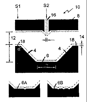
generally
have been limited to only one type of stimulus, or the experiments are only
conducted once or over short periods of time. This provides insufficient
information regarding single-cell biochemistry. In many cases useful
information regarding single cells is unattainable by measurements
performed on an ensemble of cells. Although there is a need to study groups
of cells (e.g. to understand cell-cell interactions), it is also useful to
conduct
genuine single-cell microfluidic experiments.
Summary
[0005] The invention relates to a microfluidic device comprising at
least
one first channel for introducing a first fluid into the device and a
generally
V-shaped particle retention structure for retaining a particle in the device,
the
particle retention structure having opposed wall portions and a central wall
portion disposed between said opposed wall portions, wherein the particle
retention structure is located generally opposite the first channel, and
wherein
the opposed and central wall portions have sloped side walls. One or more
fluid ports are disposed between the first channel and the particle retention
structure for delivering a second fluid to the microfluidic device or for
allowing fluids to flow out of the device.
CA 02600899 2007-08-30
WO 2006/007701 PCT/CA2005/001117
3
[0006] The sloped side walls can be curved, and they can be arcuately
curved. When the side walls are arcuately curved, they can have an arc with
a radius of curvature which is two or more times the width of the cell or
particle to be retained in the microfluidic device. The first channel has a
width greater than the width of the cell or particle to be retained in the
microfluidic device. The central wall portion can have a width 2 or more
times the width of the cell or particle. The V-shaped particle retention
structure can have a height 2 or more times the width of the cell or particle.
The width of the one or more fluid ports can be 2 or more times the width of
the cell or particle and can be 4 times the width of the cell or particle.
Lateral
end portions of the particle retention structure can be angled between 00 and
180 relative to the opposed wall portions, and the angle can be 135 .
[0007] When fluid is delivered through the first channel, the fluid
can
form a zero speed point on the V-shaped particle retention structure. The
zero speed point can be laterally shifted by an increase in delivery of a
second fluid from one of the one or more fluid ports, due to an increase in
electric potential or fluid potential in one of the one or more fluid ports.
[0008] The microfluidic device of the invention can also comprise a
detection window proximate to the V-shaped particle retention structure for
detecting biological parameters of the particle. The central wall portion can
also comprise one or more grooves.
[0009] The invention also relates to a microfluidic device comprising
two or more particle retention structures, two or more fluid ports, and two or
more first fluid channels.
CA 02600899 2007-08-30
WO 2006/007701 PCT/CA2005/001117
4
[0010] The invention also relates to a method of monitoring,
observing,
measuring, or recording a biological parameter of a particle using the
microfluidic device of the invention. The biological parameter can be any
parameter, including size, morphology, growth rate, biomarkers, influx of a
substance, efflux of a substance, reaction of the particle to one or more
stimuli, or reaction of the particle to changes in the environment of the
particle. The substance can be a coloured substance, a chromogenic
substance, a fluorescent substance, a fluorescent-labelled substance, and a
radio-labeled substance, or any other substance. Kinetic or thermodynamic
parameters can be mathematically extracted from the biological parameters of
the cell or particle. The biological parameter can be monitored, observed,
measured, or recorded in real-time and over extended periods of time.
[0011] The invention also relates to a method of culturing a cell
comprising growing the cell in a microfluidic device of the invention, a
method of treating a particle with a fluid in the microfluidic device, and a
method of separating a particle from a group of particles using the
microfluidic device. 1
[0012] The invention also relates to a method of moving a particle in
a
microfluidic device comprising isolating the particle in a zero speed point
and
moving the zero speed point in the microfluidic device.
[0013] The invention also relates to methods of monitoring the
synthesis and growth of proteins, protein crystals, nanoparticles or other
particles.
[0014] The microfluidic device and methods can also be used with any
type of particle, such as cells, beads, viral particles, proteins, protein
crystals,
CA 02600899 2007-08-30
WO 2006/007701 PCT/CA2005/001117
nanoparticles or other particles. The cells can be prokaryotic cells or
eukaryotic cells, such as yeast cells, fungal cells, plant cells, animal cells
or
other cells.
Brief Description of Drawings,
5 [0015] Figure 1 illustrates the design of an embodiment of the
microfluidic device and cell selection mechanism. (Fig. 1A) The microfluidic
device contains ports 12 and 14 for cell introduction (from either port 12 or
14) and a channel 16 (40 um wide) for delivery of buffer or reagent solutions.
The V-shaped particle retention structure, which is opposite to the reagent
channel 16, consists of opposed wall portions with a central wall portion in
between. Fluorescent signal was detected within the detection window (white
rectangle shown in the inset) by a photomultiplier tube (PMT). A single
yeast cell lies freely on the sloped wall of 15 gm radius (see inset) balanced
by the liquid flow. (B) Cell introduction: The liquid flow from the left
carries
a group of cells to the particle retention structure. (C) Cell selection: The
liquid flow from channel 16 separates the cells and sends the desired cell
downward to the detection window. Liquid flow can be driven by either fluid
potential (<1mm) or electric potential difference (0.01-1.5kV). "+" shows
the high potential. (D) illustrates one embodiment of the microfluidice
device. (E) is a cross-sectional view of one embodiment of the microfluidic
device taken at line Si as indicated in Fig. 1D. (F) is a cross-sectional view
taken at line S2. (G) is a magnified view of a portion of Fig. 1E. (H) is a
perspective view of an embodiment of a microfluidic device.
[0016] Figure 2 illustrates the 3-dimensional flow control achieved
by
an embodiment of the microfluidic device. (A) A two-dimensional channel
CA 02600899 2007-08-30
WO 2006/007701 PCT/CA2005/001117
6
flow field was created by the flow from channel 16. There is a zero speed
point (ZSP) where the flow speed decreases nearly to zero. When there is no
flow from ports 12 and 14, the ZSP is in the middle, directly opposite to
channel 16 (the notations of 12, 14, 16 have been described in Fig.1). The
third dimensional flow field is along the sloped side walls of the particle
retention structure as shown in the cross section diagram in the inset. (B-E)
As the fluid potential from the right is increased, the shape of the flow
field
changes and the ZSP moves to the right. (F) As shown when there is no
reagent flow from channel 16, the flow field can be driven by the fluid
potential from the right. (G-J) The flow field shape also changes when fluid
potential from the left is increased. (K) As shown when there is no reagent
flow from channel 16, the flow field can be driven by the fluid potential from
the left. (L) The third dimensional flow field along the sloped side walls of
the particle retention structure 'results in the cell balancing on the sloped
side
walls. The forces between the upward force exerted by the liquid flow (A
downward gravitational force (g) on the cell, and the reaction force from the
sloping wall (P) are balanced on the cell. (M-0) The position of the cell on
the sloped side walls changes as the reagent flow from channel 16 increases.
(P) The position of the cell when there was no flow.
[0017] Figure 3 illustrates the forces exerted on a cell contained within
the microfluidic device of the invention. (A) The different directions and
strengths of fluid flowing near the sloped side walls of the particle
retention
structure are shown. (B) The forces exerted on a cell balanced on the sloped
side walls of the particle retention structure; g: The cell's gravity
(buoyancy
subtracted);fa: The force exerted by the flow at an angle (a); f11: The force
exerted by horizontal fluid flow (i.e. a=0); Pa: The reaction force of the
CA 02600899 2007-08-30
WO 2006/007701 PCT/CA2005/001117
7
sloped side walls to the cell for a flow directed at an angle (a); PH: The
reaction force of the sloped side walls to the cell for a horizontal flow. (C)
The direction and strength of liquid flowing near a vertical wall. (D) The
forces exerted on a cell balanced against perpendicular walls. Pi,: The
reaction force of the bottom wall to gravity; PH: The reaction force of the
vertical wall to the cell due to horizontal flow. (E)The force relationship
between g, fa and Pa as given in (B). (F) The force relationship between g,TH
and PH when a cell is balanced on arcuately slopped side walls with an
increased angle of the slope (fl).
[0018] Figure 4 is a schematic diagram of an optical measurement
arrangement. The setup includes an inverted microscope and the associated
optics. 20: dichroic filter 1 (495 nm); 22: dichroic filter 2 (540 nm); 24:
band-
pass filter (470 nm/40 nm); 26: long pass filter (645 nm); 28: band pass
filter
(525 nm/50 nm); 30: microscope objective (ELWD, 40X/0.60); 32: mirror.
The first optical path (red light, to 26, to microfluidic device, to 30, to
20, to
32, to 22, and to CCD camera) was used for bright-field optical observation.
The second optical path (excitation light, to 24, to 20, to 30, to
microfluidic
device, to 30, to 32, to 22, to 28, and to PMT) was used for fluorescent
measurement. The embodiment of the microfluidic device as shown was been
used in single-cell experiments. The width of the microfluidic device is
16mm. In the photograph of the microfluidic device, vial a is connected to
port 12, vial b is connected to port 14, and vial c is connected to channel
16.
[0019] Figure 5 contains a series of images demonstrating the 3-
dimensional fluid flow in the microfluidic device. (A) Buffer with FDA
(12HM) was injected from channel 16 toward the particle retention structure.
The solution front expanded downward as observed by the inverted
CA 02600899 2007-08-30
WO 2006/007701 PCT/CA2005/001117
8
microscope in the phase-contrast mode. (B) The beads travelled from
channel 16 towards the particle retention structure. Images were captured
every 0.24s and overlaid. Therefore, in each of the frames, four beads
represent the travel path of one bead in each of the images over 0.72 s. The
distance between any two closest beads illustrates that bead's length of
travel
within 0.24s. Beads travelled quickly in channel 16 but their rate of travel
slowed when they approached the particle retention structure. The beads
demonstrated the flow fields as depicted in Fig.2H. (C) Selection, retention
and immobilization of a bead using fluid flow. The desired bead to be
selected is circled. Again, bead images were captured every 0.24s, and
images are overlaid to show their positions every 0.24s.
[0020] Figure 6 contains images of beads and yeast cells balanced on
the sloped side walls of the particle retention structure. (A) A bead balanced
on the sloped side walls in the 'presence of a weak reagent flow. Trails of
the
beads represent movement of the beads in 0.08s intervals. (B) When flow
was increased, the bead moved higher up on the sloped side walls to a new
balanced forced position. (C) A budding yeast cell moving towards the
sloped side wall until balanced against the side wall (0-8s). The three dots
represent images of the same single cell at different times, which demonstrate
how the fluorescent cell is scanned. The cell was scanned to the right during
fluorescent detection (10-16s). In a stronger reagent flow, the cell moved
further up the sloped side wall to a new balanced force position (39-46s). The
cell was scanned at its new force balance position during fluorescent
detection (59-60s).
[0021] Figure 7 depicts a series of on-chip cell culture images captured
from video recordings (time in seconds). (A) A yeast cell (cell 1) grown in
CA 02600899 2007-08-30
WO 2006/007701 PCT/CA2005/001117
9
the microfluidic device (at 24 C). The cell divided twice before experiments
on FDA metabolism were performed on it. (B) Another yeast cell (cell 2)
picked directly from a cell colony was cultured in the microfluidic device at
24 C. It was grown for 17000s. (C) Another yeast cell (cell 7) had its cell
wall removed on-chip cell. The process had a duration of 3.84s. Each photo is
accompanied by a schematic diagram to illustrate the various steps during the
cell wall removal process.
[0022] Figure 8 depicts the fluoresence signal generated by a yeast
cell
detected through cell scanning and noise filtering. (A) A fluorescent yeast
cell travelling back and forth through the detection window generated peaks
over the background. (B) The peak signal became clearer after filtering the
noise (>2.5Hz). (C) The use of a narrower detection window allowed a
mother yeast cell to be distinguished from its daughter yeast cell, as
fluorescent signal was depicted by a peak (generated by the larger mother
cell) and a shoulder (generated by the smaller daughter cell).
[0023] Figure 9 depicts the background fluorescence of buffer
solutions
(without cells) stored in a microfluid device of the invention. The gradual
increase in fluorescence is due to the slow hydrolysis of FDA to produce
fluorescein in the aqueous buffers. (A) Buffer G7; (B) Buffer H4; (C)
Changing between G7 and H4.
[0024] Figure 10 depicts background fluorescence signals which are
used to correct data signals. Background correction was applied to an
experiment with a yeast cell (cell 5). (A) Peaks due to cell fluorescence plus
background. (B) Background baseline extracted from (A). (C) Cell
fluorescence peaks after background subtraction, (D) Peak envelope of all
CA 02600899 2007-08-30
WO 2006/007701 PCT/CA2005/001117
fluorescence peaks. The two reagent scales indicate the buffer types and the
FDA concentrations. The excitation light scale indicates when the excitation
light was turned off or on.
[0025] Figure 11 consists of a series of images of a yeast cell in
cell
5 culture in a microfluidic device and subsequent to on-chip cell selection
(cell
1 referred to in Fig.12A, time in seconds). (A) The microfluidic device. (B-
G) The cell was selected from a group of cells. (H-K) The daughter cells
escaped from the mother yeast cell in the reagent flow. This cell (cell 1) has
also been described in Fig.7A.
10 [0026] Figure 12 depicts fluorescence signals produced by yeast
cells
during FDA metabolism. (A) On-chip cell culture (Cell 1), medium and
reagent change, fluorescence detection and data processing. (B) (C) (D) (E)
FDA experiments on other single budding yeast cells. (F) FDA experiment
on a spheroplast after on-chip cell wall removal. (G) (H) FDA experiments
on single dormant yeast cells. Three scales of buffer types, FDA
concentrations (0 or 12mM) and cell fluorescence intensity (103 counts per
second) were the same as those in (A). Y: Yeast cell culture medium (YPD).
H4 and H7: 285 mM HEPES, and at pH = 4.3 and pH=7.3, respectively. G7
and G4: 28.5 mM HEPES plus 256mM D-glucose, and at pH 7.3 and
pH=4.3, respectively. Beads: fluorescent beads were used for calibration at
8ks. All results are shown after background correction, as depicted in (A).
[0027] Figure 13 depicts fluorescence signals detected in single
yeast
cells during Ca2+ mobilization tracking tests. (A) (B) (C) Experiments after
on-chip cell selection, followed by on-chip cell wall removal and Fluo-4-AM
loading. (D) Experiments after off-chip cell wall removal and Fluo-4 AM
CA 02600899 2007-08-30
WO 2006/007701 PCT/CA2005/001117
11
loading, followed by on-chip cell selection. (E) Experiments after on-chip
cell selection, followed by direct on-chip high-concentration Fluo-4 AM (in
DMSO) loading. (F-0) Experiments after off-chip high-concentration (1mM
in DMSO) Fluo-4 AM loading, followed by on-chip cell selection. All three
scales of buffer types, Ca2+ concentration and fluorescent intensity were the
same as in Figure 30. Y: culture medium (YPD); E: EDTA. All results
shown are after background correction (shown in Fig.2A) except (E).
[0028] Figure 14 depicts calcium mobilizations in three kinds of
single
yeast cells (dormant, budding and treated budding) in response to glucose and
pH changes. Arrows show the changes of buffer types and the associated
changes of intracellular fluorescence (Ca2+-Fluo-4). The line widths of the
arrows indicate the fluorescence changes, which are given as percentages, in
the legend.
[0029] Figure 15 is a comparison of yeast cell images which
illustrate
the differences in fluorescence of cells grown in different conditions. (A-B)
Fluorescence due to fluorescein formed after G7 (12p,M FDA) incubation for
1.5ks; (A) dormant cells, (B) budding cells after H4 incubation for lks. (C-E)
Fluorescence due to Ca2+-Fluo-4 formed after 4s off-chip loading of high-
concentration Fluo-4-AM/DMS0 (1mM), followed by 0.5ks treatment of
Ca2+ (10mM in G7); (C) dormant cells, (D,E) budding cells. (F) Fluorescence
due to a 6-pm fluorescent bead.
[0030] Figure 16 illustrates a mathematical model for FDA metabolism
in a single cell (Cell 3). Curve:fitting and sensitivity tests: (I) there are
3
cellular processes, namely influx, hydrolysis and efflux. The yeast cell
exerts
control over the influx of FDA (A), hydrolysis of FDA (B), to form
CA 02600899 2007-08-30
WO 2006/007701 PCT/CA2005/001117
12
fluorescein (C) and efflux of fluorescein in response to the stimuli of pH and
glucose. (II) Intracellular concentrations of FDA (B) and fluorescein (C) .
Only C was experimentally measured. To, T1 and T2 represented the time (s)
when buffer change, peak increase and peak decrease occurred, respectively.
(III) Curve fitting: in the graph, dashed and solid lines represent the
modelled
amount of intracellular FDA and fluorescein respectively, and striped areas
underneath the solid lines represent the signal peaks for the measured amount
of fluorescein, which were calibrated with a fluorescent bead of known
intensity. Note that this experiment has been previously represented in
Fig.2B. (IV, V, VI, VII) Sensitivity tests of the model: The effects on the
model lines are depicted as a series of black lines. When one parameter is
changed in each of the following cases, (IV) T2-T1: 2300 ¨ 2700s, (V) Vmo:
0.001 ¨ 0.005 M s-1, (VI) k: (4 ¨ 8) x 10-6 M s-2 (VII) Ve: 1-4 M pm s-1.
(RFI: relative fluorescent intensity in which 1% represents the fluorescence
resulted from full hydrolysis products from 6x10-19 mol of FDA).
[0031] Figure 17 illustrates fluorescence of cells under various
stimuli.
The curve fittings were performed on (A) cell 4 and (B) cell 5 which
underwent a series of changes due to pH and glucose stimuli. Dashed and
solid lines represent the modelled amount of intracellular FDA and
fluorescein respectively, and striped areas underneath the solid lines
represent
the signal peaks for the measured amount of fluorescein. Changes of buffer
type or FDA concentration are, indicated by arrows with numbers, and are
described in the text. Note that (A) has also been described in Fig.12D, and
(B) has been described in Fig.12C.
[0032] Figure 18 illustrates different types of cell scanning. The left
series of illustrations of A-D show the different scanning paths (the arrows
CA 02600899 2007-08-30
WO 2006/007701 PCT/CA2005/001117
13
indicates the moving cells in A and B or the moving detection windows in C
and D) in the cell retention structure. The right series of illustrations of A-
D
show the measurement results.
[0033] Figure 19 illustrates the scanning results of a budding yeast
cell
using a narrow detection window and 2 different scanning speeds. The left
five peaks were generated by 500 V, resulting in a faster scanning speed, and
the right five peaks were generated by 200V, resulting in a slower scanning
speed. The inset shows the 2 mirrored peaks depicting the fluorescent
intensities of the mother cell and its bud.
[0034] Figure 20 illustrates an advantage of cell scanning in an open
region. The left series of illustrations of A-E show different scanning paths
(the arrows indicate the moving detection windows) in different structures.
The right series of illustrations of A-E show the expected results from the
scanning (the dashed lines in C to E indicate the possible cellular signals).
[0035] Figure 21 illustrates he parameters of the photobleaching model
to separate FDA hydrolysis (which increases the fluorescent intensity) and
the photobleaching effect (which decreases the fluorescent intensity). FO, Fl,
F2 and F3 are the fluorescence when t=0, T, 21 and 3T, respectively. When
0<t<T and 2T<t<3T, the excitation light is on. When T<t<2T, the excitation
light is off. (B) Fluorescent intensity of fluorescein resulted from FDA
hydrolysis in G7 without liquid flow. The shutter for the excitation light was
opened and shut for an interval of 100 s. (C) the whole experiment which
lasted for 20000 s from which the data of (B) is derived. (D) The
photobleaching rate constant kp, as determined at each level of relative
fluorescent intensity (RFI), is plotted against RFI.
CA 02600899 2007-08-30
WO 2006/007701 PCT/CA2005/001117
14
[0036] Figure 22 depicts the fluorescent measurement of a yeast cell
embedded on a normal slide: the raw data (A), its separated background (B)
and extracted cell fluorescence (C). FDA was used to generate the cellular
fluorescence after hydrolysis.
[0037] Figure 23 depicts the fluorescent measurement of a yeast cell
under a flow within a microchip: the raw data (A), its separated background
(B) and extracted cell fluorescence (C). FDA was used to generate the
cellular fluorescence after hydrolysis.
Description
[0038] Throughout the following description, specific details are set
forth in order to provide a more thorough understanding of the invention.
However, the invention may be practiced without these particulars. In other
instances, well known elements have not been shown or described in detail to
avoid unnecessarily obscuring the invention. Accordingly, the specification
and drawings are to be regarded in an illustrative, rather than a restrictive,
sense.
[0039] The inventors have developed a microfluidic device which
utilizes 3-dimensional flow control. This flow control combines cell
balancing capabilities in a first dimension (1-D) as well as cell scanning
capabilities in channel dimensions (2-D).
[0040] Although the invention is described herein in the context of
cells, it will be appreciated by a person skilled in the art that the
invention
may be used to retain and manipulate other particles, such as beads, viral
particles, proteins, protein crystals and nanoparticles.
CA 02600899 2007-08-30
WO 2006/007701
PCT/CA2005/001117
[0041] To balance cells or particles within the microfluidic device,
the
inventors make use of the downward residual gravitational force of a cell
residing on a sloped wall to balance the upward force exerted on the cell by
liquid flow through channels or ports in the microfluidic device. The sloped
5 side walls of the microfluidic device can be created, for example, by
isotropic
etching of a microfluidic device made of materials, such as glass or silicon.
[0042] To scan cells and obtain data on biological parameters, the
inventors exploit the zero-speed point (ZSP) created by a liquid flow field
against a specially shaped particle retention structure in the microfluidic
10 device.
[0043] With 3-dimensional flow control, the inventors have
successfully carried out cell balancing, cell scanning, measurement of
physiological parameters, and observations on a single cell. Yeast cells were
chosen for the examples because of their availability and short life cycle
(for
15 cell culture). However, the microfluidic device and methods of using the
microfluidic device can be used on any type of cell, including prokaryotic
cells and eukaryotic cells, such as fungal cells, yeast cells, plant cells and
animal cells. As mentioned above, the invention may also be used to study
any type of particle, including beads, proteins, protein crystals,
nanoparticles
and the like.
[0044] Furthermore, with the techniques of cell balancing and cell
scanning, culturing of a single cell has been accomplished "on-chip."
Throughout this application, the term "on-chip" refers to activities which
occur within the microfluidic device. Current on-chip culture methods are
carried out only in batch mode without keeping track of a single cell, and
CA 02600899 2007-08-30
WO 2006/007701 PCT/CA2005/001117
16
only for adherent ce11s36-41. The microfluidic device of the invention allows
experiments and methods to be carried out with single cells.
3-Dimensional Flow Control
[0045] Fig. 1D illustrates an embodiment of the microfluidic device.
Referring to Fig. 1D, the microfluidic device 10 consists of a channel
defining portion 8 which contains a fluid channel 16, a generally V-shaped
particle retention structure 2 spaced apart from channel defining portion 8
which comprises opposed wall portions 4, a central wall portion 6 disposed
between opposed wall portions 4, and lateral end portions 18, wherein each
opposed wall portion 4 is disposed between central wall portion 6 and one
lateral end portion 18. Particle retention structure 2 is generally opposite
fluid channel 16. Fluid ports 12 and 14 are defined between channel defining
portion 8 and lateral end portions 18. The microfluidic device can comprise
more than one fluid channel 16. Alternatively, microfluidic device 10 can
also comprise a detection window 30 for detecting cells retained in
microfluidic device 10 (see Fig. 1A).
[0046] The side walls of opposed wall portions 4 and central wall
portion 6 are inwardly sloped. In some embodiments, the inwardly sloped
side walls can be inwardly curved, and can be inwardly arcuately sloped (see
Fig.1A, 1F, and 1G). Fig. lE is a cross-sectional view of one embodiment of
the microfluidic device taken at line Si as indicated in Fig. 1D. Fig. 1F is a
cross-sectional view taken at line S2. Fig. 1G is a magnified view of a
portion of Fig. 1F. Fig. 1H is a bisected perspective view of an embodiment
of microfluidic device 10.
CA 02600899 2007-08-30
WO 2006/007701 PCT/CA2005/001117
17
[0047] Angle 0 is the angle formed between lateral end portions 18
and
opposed wall portions 4. Angle 0 can be between 0-180 , such as 135 , or
any other suitable angle. For a cell or particle with a diameter of X, the
length of central wall portion 6 should be equal to or greater than 2X. The
width across fluid port 12 or 14 should be greater than 2X, and can be 4X, for
easy particle washing and particle delivery. The depth of the V-shaped
particle retention structure, which is the distance from lateral end portions
18
to central wall portion 6, should be 2X or more to keep cells away from fluid
which flows across the microfluidic device. In some embodiments, channel
16 can be used to deliver cells or particles to the microfluidic device. In
these embodiments, the width of channel 16 can be more than X, which
allows cells to be delivered from channel 16 into the microfluidic device.
Central wall portion 6 can be flat, or it can comprise one or more grooves, as
shown in the embodiments 6A and 6B of the central wall portion in Fig. 1D,
to help keep a cell centred over a detection window in the central wall
portion. In embodiments where the inwardly sloping side walls of the
particle retention structure are arcuately curved, the radius of curvature e
of
the side walls can be equal to or greater than 2X. However, the inwardly
sloping side walls can comprise any curve shape. Moreover, the slope angle
of the inwardly sloping side walls can vary or it can be constant.
[0048] Referring to Fig. 1B, for cell selection, horizontal liquid
flow
(from port 12 in this case, although either port 12 or 14 can be used) can
carry a group of cells close to the V-shaped particle retention structure.
Another flow from channel 16, which is perpendicular to the direction of
flow from port 12 or 14, separates the cells and sends a desired cell towards
CA 02600899 2007-08-30
WO 2006/007701
PCT/CA2005/001117
18
the V-shaped particle retention structure where the detection window is
located (Fig.1C).
[0049] For cell balancing and cell scanning, the concept of three-
dimensional flow control is exploited (see Fig.2A). When liquid flows out
from channel 16 at a high speed into the more open area of the microfluidic
device and towards particle retention structure 2, some fluid will escape
sideways and the speed of the flow of the liquid slows. Since particle
retention structure 2 is opposite to channel 16, liquid flow will generally
follow the contour of particle retention structure 2 and then divide in the
centre of particle retention structure 2. Therefore, there exists a zero-speed
point (ZSP) in the centre of particle retention structure 2, provided that the
two left and right lateral flows are the same. If the two lateral flows are
not
the same, the ZSP will be displaced. For instance, in Fig.2B, the ZSP is
displaced to the right when the lateral flow toward the left is stronger, due
to
a higher potential being applied on the right. Stronger lateral flow to the
left
will shift the ZSP further to the right (see Fig.2C-D), until the ZSP is no
longer within the particle retention structure region or disappears (see
Fig.2E). In the case when there is only lateral flow but no reagent flow (from
channel 16), the flow is represented in Fig.2F. Similarly, situations in which
the lateral flow is equal, stronger to the right (due to higher potential
applied
to the left), or there is no reagent flow are depicted in Figs.2G, 2H-J and
2K,
respectively. These flows are 2-dimensional in nature. It will be appreciated
by persons skilled in the art tha flow into channel 16 or lateral ports 12 and
14 can be controlled by electrical, pressure or other suitable means.
[0050] Along the third dimension, which is the depth dimension, the
liquid flow is not uniform. This situation is depicted in the cross-sectional
CA 02600899 2007-08-30
WO 2006/007701 PCT/CA2005/001117
19
diagram of Fig.2A (shown as an inset). Here, even though the flow speed
from channel 16 is constant, the flow speed along the sloped side wall of the
particle retention structure wall is gradually decreased to zero. Therefore,
the
ZSP is actually at the upper end of the sloped wall (see the inset of Fig.2A).
This situation is still valid even if the ZSP is displaced sideways due to the
differential lateral flows as previously described.
[0051] These liquid flows, which are in the channel dimension
(lateral
and horizontal flows) and the depth dimension (upward flow along the sloped
side wall) are therefore 3-dimensional in nature.
[0052] For cell scanning, the cell will be stationary and thus retained
around the ZSP. Lateral displacement of the ZSP caused by differential
lateral flow causes lateral displacement of the cell within the microfluidic
device. Periodic lateral displacement of the ZSP therefore causes the cell to
be scanned back and forth in the microfluidic device.
[0053] Cell balancing is achieved by the balance of forces exerted on a
cell (Fig.2L). First, the cell is pushed upward along the sloped side wall due
to the force (f) exerted by the reagent flow. Second, there is resultant force
(f') due to the cell's residual gravitational force (g) (after deducting the
cell's
buoyancy) or sedimentation force and the reaction force (P) acting by the
slope on the cell. When the two forces, f and f' , are balanced, the cell
becomes stationary, and particle retention is achieved.
[0054] If the reagent flow is stronger, f increases and the cell is
retained
at a location higher on the sloped side wall, see Fig.2M-0. If there is no
reagent flow, the cell will not travel up the sloped side wall at all, and
will
rest on the flat channel bottom, see Fig.2P.
CA 02600899 2007-08-30
WO 2006/007701 PCT/CA2005/001117
[0055] Accordingly, the strength of the reagent flow is not a great
concern. The flow will not crush the cell or flush away the cell because the
position of the cell will adjust with the strength of the flow, by moving
upwards along the sloped side wall. Furthermore, if the cell lying against the
5 sloped side wall is very near to the top of wall (at the ZSP), the flow
speed of
the liquid will be very slow coinpared to that in channel 16. The flow speed
will be greater if the cell is fariher away from the ZSP. Therefore, a high-
speed flow can carry reagents very rapidly and proximately to the cell, and
then a low-speed flow will rely those reagents to the cell. All these flow
10 controls can be achieved without any harmful localized force being
exerted
on the cell. In addition, the position of the cell on the sloped side wall, or
the
distance of the cell away from the ZSP (Fig.2M-0), reveals the speed of the
flow exerted on the cell, and therefore allows users of the microfluidic
device
to easily adjust the flow speed by observing the position of the cell within
the
15 microfluidic device. In particular, when the cell is scanned back and
forth
horizontally across the detection window so that signals or biological
parameters of cell can be detected, adhesion of the cell, if any, will be
minimized, and the cell position will be even more sensitive to assist in
adjusting flow rates of liquids from reagent channels or flow ports.
20 [0056] Figure 3 analyzesi the forces balanced on a cell in
greater detail.
When liquid is further away from the side wall, the liquid flows faster
(Fig.3A). As the liquid approaches the sloped side wall, the liquid follows
the
shape of the side wall and the flow rate slows. This will cause forces of
different directions and strengths to be exerted on a cell of a finite size.
Fig.3B depicts a force fa exerted at an angle a (0 <a <900) to the horizontal.
It
is balanced by f' a (or the resultant force of Pa and g) . In a special case,
a
CA 02600899 2007-08-30
WO 2006/007701
PCT/CA2005/001117
21
horizontal forcefll (i.e. a =0) is balanced by f'H (or the force resultant PH
and
g). For comparison, in the case of a vertical wall (see Fig.3C, D),fH and g
are
balanced by PH and Pv, respectively, and there is no angular dependence of
the liquid force.
[0057] The force relationship between, fa, Pa and g is also shown in
Fig.3E. When a=0,f, and Pa attain their maximal values offH and PH,
respectively, see Fig.3F. In addition,fH = g tan /3, and PH= g I cos /3, where
fi
(8<900) represents the slope angle. For example, if a cell stays at an angle
of
45 on the sloped side wall, the reaction force from the wall cannot exceed
Vig, and the flow-induced force cannot exceed g. The reaction force would
have a greater limit if /3>45 ; for instance, if fl>60 , the reaction force
from
the wall cannot exceed 2g, which is still a small force on the cell. However,
users of the microfluidic device can limit the reaction force on the cell by
noting the position of the cell on the slope and adjusting liquid flow rates
accordingly. On the other hand, if the wall were vertical (Fig 3. C, D), the
cell could not adjust its position and a strong flow could cause a very high
reaction force from the vertical wall (p H)
[0058] It is worthwhile to mention that either a sloped side wall or
a
vertical side wall will give rise to a ZSP due to the splitting of fluid flow.
However, only the sloped side wall allows the cell's position to adjust to
prevent damage to the cell by a strong flow. The sloped side wall actually
serves as a buffer zone. When a cell recedes to a point near the ZSP, the cell
can escape from the strong flow. So the sloped side wall is very effective for
protecting the cell.
CA 02600899 2007-08-30
WO 2006/007701
PCT/CA2005/001117
22
[0059] In another embodiment of the invention, the inventors disclose
methods of using the microfluidic device of the invention to measure
biological parameters of a cell over time, including monitoring changes in
biological parameters of a cell in response to various stimuli over time, and
culturing a cell in the microfluidic device over one or more life cycles.
[0060] It will be understood by a person skilled in the art that the
microfluidic device of the invention can also be used with materials other
than cells, such as particles, including beads, viral particles, proteins,
protein
crystals, nanoparticles, and other particles that are capable of being studied
with the microfluidic device of the invention. Throughout this application,
methods of using the microfluidic device with cells can be applied to
particles.
[0061] The biological parameters that can be observed and measured
include cell morphologies, cell size, growth rate, surface or intracellular
biomarkers (e.g. calcium or other minerals or ions, messengers, proteins,
carbohydrates, or other suitable biomarkers), influx and efflux of substrates
and metabolites, including coloured, chromogenic, fluorescent or
radiolabeled substrates and metabolites, reaction to stimuli, reaction to
changes in reagent conditions, or any other parameter that would be useful to
observe.
[0062] In one embodiment, the inventors initiated the influx of a
substrate into a single yeast cell, and observed the formation and efflux of a
metabolite in response to multiple stimuli over a period of a few hours. In
addition, the inventors studied calcium mobilization in a single cell in
response to multiple stimuli, in multiple trials.
CA 02600899 2007-08-30
WO 2006/007701
PCT/CA2005/001117
23
[0063] In another embodiment, using 3-dimensional flow control, a single
yeast cell was selected from a group of cells, retained, cultured, and scanned
back and forth across a detection window to monitor biological activity
within an embodiment of the microfluidic device of the invention.
[0064] The microfluidic device of the invention provides a non-
disturbing environment to study cells and conduct single-cell experiments.
Within the microfluidic device,, culture medium can be continually refreshed
and cells can freely grow. During experiments, the concentrations of reagents
can be changed at any time, and excretion or efflux products are continually
flushed away by the flow. Data from single-cell experiments can provide data
on real-time changes of the concentration of a metabolic product.
[0065] For example, in a conventional solution enzyme model, kinetic
parameters of influx, efflux and enzymatic reaction are normally taken as
constants. Without single-cell biochemical experiments, it is not possible to
test if a cell varies the kinetic parameters or has a strong ability to keep
the
enzymes under control.
[0066] In one of the embodiments of the invention, to study a model of a
yeast metabolic process using the microfluidic device of the invention, the
inventors selected a cell-permeable fluorogenic substrate, fluorescein
diacetate (FDA), which is normally used to determine cell viability42. After
influx of FDA into a yeast cell', the intracellular enzyme carboxylesterase43
hydrolyzes FDA to fluorescein, which will then be excreted from the cell
(through efflux). Efflux is particularly strong with FDA as compared to other
FDA derivatives45. Dynamic studies of FDA metabolism in yeast have been
performed by flow cytometry44' 45, but these studies could not completely
CA 02600899 2007-08-30
WO 2006/007701 PCT/CA2005/001117
24
reveal the complexity of this complex influx- hydrolysis (by esterase) -efflux
process.
[0067] Accordingly, the inventors introduced FDA to cultured, dormant
or treated single yeast cells and obtained kinetic data of the above metabolic
process as stimulated by changes in pH and glucose. The inventors achieved
these by measuring cellular fluorescent signal due to fluorescein formed in
one single yeast cell. These data were then used in a mathematical model to
extract the Michaelis-Menten parameters.
[0068] In one example discussed below, in response to one type of
external stimuli, a yeast cell started to metabolize FDA, and in response to
other external stimuli, the yeast cell started to excrete fluorescein. As a
result, the inventors identified three modes of cellular control, namely 'self-
control', 'lost-control,' and 'death' to describe the metabolic process modes
of the cell. The 'self-control' mode describes a cell that can control
enzymatic activity. The `lost-control' mode describes a cell that does not
alter
enzymatic activity but enzymes may still be working. The 'death' mode
describes a cell that does not respond to any changes in its environment.
Moreover, these metabolic processes were found to correlate with calcium
mobilization.
[0069] In another embodiment of the invention, the inventors studied
FDA metabolism due to carboXylesterase in response to pH or glucose
stimuli. Other enzymes, which act on other subtrates, can also be activated by
stimuli. In another example, the inventors measured intracellular calcium
within a single yeast cell upon various stimuli to study the mobilization of
calcium ions.
CA 02600899 2007-08-30
WO 2006/007701 PCT/CA2005/001117
[0070] It will be appreciated by persons skilled in the art that
other
biological parameters in other Fells can also be analyzed through the use of
the microfluidic device of the invention. Analysis of other metabolites, in
other cells, in response to other stimuli, can also be monitored in the
5 microfluidic device of the invention. On-chip single-cell experiments may
be
used to elucidate complex biological systems.
EXAMPLES
[0071] In examples which are intended to illustrate embodiments of
the
invention and which are not intended to limit the scope of the invention:
10 Example 1: Microfluidic Device
[0072] Figure 1 illustrates the design of an embodiment of the
microfluidic device and cell selection mechanism. The glass microfluidic
device was fabricated through the Protochip Program of Canadian
Microelectronic Corporation. Borofloat glass wafers were used to fabricate
15 the channel plate and over plate (16 mm x 95 mm). Then, the two glass
plates
were thermally bonded together to form the finished chip. The layout of one
embodiment of the particle retention structure has been depicted in Fig.1D. In
this particular example, the microfluidic device used contained 15 'Am deep
channels. The side walls of the particle retention structure are inwardly,
20 arcuately sloped in this embodiment. The radius of curvature of the
arcuately
sloped wide wall should be greater than the diameter of the cell. The central
wall portion 6 is normally flat for uniformity in scanning the cell to measure
cell parameters. The same microfluidic device was easily washed and reused,
and has survived many hours (-200h) of experiments.
CA 02600899 2013-03-25
=
26
[0073] For optical measurements, the microfluidic device was placed
on
the translation stage of an inverted microscope (NikonTM TE 300) with a
dual-image module (Nikon) which was coupled to both a CCD video camera
(JVCTM TKC 1380) and a photomultiplier tube (PMT) (Photon Technology
Intl, PTI) (Fig.4). Simultaneous optical observation and fluorescent
measurement of the single cell was achieved using this special optical
measurement set up. Specifically, red light (> 645 nm) was used to observe
the cells using the video camera. The motions of any cells were continually
displayed on a television monitor and recorded by a video-tape recorder
(JVC-rm HR-S7500U). A xenon arc lamp (PTI) was employed to excite the
fluorophore. Green fluorescent signals due to intracellular fluorescein formed
(520nm) were not able to reach the camera and could only be detected by the
PMT. Fluorescence signals from the PMT were recorded by a computer
using the FelixTM software (PTI). The PMT only recorded the fluorescent
signal within the detection window (Fig.1A). If the yeast cell was within the
window, the signal represented the cellular fluorescence plus the fluorescent
background. If not, only the fluorescent background was detected.
[0074] The 3-dimensional liquid flows could be driven by electric
potentials. To create a downward flow of reagents, a high voltage (50-500V)
was applied to channel 16, and both ports 12 and 14 were at ground. To
create a lateral flow to the right, a high voltage was applied to port 12 with
port 14 at ground, and vice versa.
[0075] When a high electrolyte buffer was required, e.g. in cell
culture
experiments, voltage control could not be used, and only fluid potential (by
liquid head difference <imm) was used. For instance, highly conducting
liquid, such as culture medium, was directly introduced in the microfluidic
CA 02600899 2007-08-30
WO 2006/007701
PCT/CA2005/001117
27
device to the cells (cell 1, 2, 7) used in Fig. 7. By adding, for example, a
drop of fluid in only one of the fluid ports, a fluid potential is created and
fluid flows through the microfluidic device due to hydrostatic pressure.
Alternatively, pumps and valves at fluid ports 12 and 14, and channel 16 may
also be used to control liquid flow.
[0076] To examine the direction and speed of liquid flow in the
microfluidic device, polystyrene beads (6 m diameter, InSpeckTM Green,
Molecular Probes) were used. This bead size was selected because it is
similar in size to a yeast cell.
Example 2: Composition of Buffers
[0077] In single-cell experiments, the microfluidic device allowed
a
flow of reagents to be directly delivered to the cell surface. Unlike
conventional experiments on normal slides, the inventors could be sure that
the reagents or buffers reached the cell at the desired concentration in real-
time. FDA stock solution (5 mg/mL in DMSO) was diluted to 12 M. This
concentration of FDA was used because of its limited solubility in aqueous
= buffers. Two buffers were used for dilution and they were G7 (28.5 mM
HEPES, 256 mM D-glucose, pH = 7.3) and H4 (285 mM HEPES, pH = 4.3).
Experiments were performed at room temperature (24 C) (DMSO: dimethyl
sulfoxide; HEPES: N-[2-hydrOxyethyl] piperazine-N'-[2-ethanesulfonic
acid]).
Example 3: Yeast Strains and Growth Conditions
= [0078] The yeast (Sacchomyces cereviase) strain (wild type,
CBY858)
was first grown on YPD-agar plates, and were then stored in a refrigerator.
CA 02600899 2007-08-30
WO 2006/007701 PCT/CA2005/001117
28
Off-chip cell culture was carried out by growing a cell colony aerobically in
2
mL of YPD culture medium (2% glucose, 2% yeast extract and 1% peptone)
to the exponential phase (0D600õn, ¨ 0.5 ¨1.0). The size of the yeast cell was
2-5 [tm.
[0079] On-chip cell culture was performed with or without off-chip pre-
culture. To initiate off-chip pre-culture, yeast cells were first picked from
a
colony on an agar plate, and then they were put into 2-ml YPD culture
medium for about 7000s at room temperature (24 C). Thereafter, the yeast
cells in its culture medium were introduced into the microfluidic device.
Using 3-dimensional flow control, one budding cell was selected out of a
group of cells. Then the inventors provided the cell with more culture
medium under a constant flow from the reagent channel 16 to carry out on-
chip cell culture. The microfluidic device was maintained at room
temperature all the time. The fresh medium flowing from vial c refreshed the
cell continually. The cell continued its budding process within the
microfluidic device. In the case of direct on-chip culture (i.e. without pre-
culture), a cell colony was directly introduced in the microfluidic device.
Then a single yeast cell was selected on-chip. Thereafter, YPD culture
medium was delivered from the reagent channel to initiate cell growth and
budding, as previously described. Removal of the yeast cell wall was
achieved by an enzyme, zymolase.
Example 4: Flow Fields in the Microfluidic Device
[0080] To image the 3-dimensional flow fields in the microfluidic
device, the inventors used both the reagent liquid and polystyrene beads. The
inventors used a solution containing FDA to image the flow. After FDA was
CA 02600899 2007-08-30
WO 2006/007701 PCT/CA2005/001117
29
introduced into the microfluidic device, the FDA flowed out from channel 16
at a high speed and dispersed sideways at a slower speed in the wider
portions of the microfluidic device. Movement of the liquid front was
recorded by the microscope in the phase-contrast mode (Fig.5A). It was
observed that the speeds of the liquid fronts were not the same in all
directions, and the liquid front was not in the shape of a semicircle. Faster
lateral flow and slower flow perpendicular to the lateral flow rendered the
liquid front to resemble a semi-ellipse. If desired, the flow field lines
could be
obtained by drawing lines at right angle to the liquid fronts.
[0081] As the liquid front did not clearly show how the different flow
speeds vary at various locations, the inventors added beads into the reagent
channel to indicate the flow field directly (Fig.5B). Multiple exposures were
used in the images to show the paths of beads at their 4 consecutive
locations.
Therefore, not only can the flow fields be visualized, but also the speeds of
the moving beads can be determined. It is demonstrated that the travel speed
of the beads slowed as they approached the particle retention device. For
instance, in Fig.5B (0-1s), a bead rushed out of channel 16 at a speed of
about
200 m/s. Then the bead (as circled) slowed down to a speed of about 60 m/s
(1-2s). Thereafter, its speed was about 301imis near the sloped wall (2-3s).
Finally, the bead was close to the wall and rested on the sloped wall (3-4s)
because the force balance had been achieved. In the meantime, liquid
continued to flow from the reagent channel and other beads from the reagent
channel demonstrated their speeds as driven by the liquid flow. The
immobilization of the bead (as circled) near the ZSP could last for a very
long time even in the presence of a fast reagent flow (5-8s). Since the fluid
potential is greater on the left, causing a greater lateral flow to the right,
the
CA 02600899 2007-08-30
WO 2006/007701 PCT/CA2005/001117
ZSP near which the bead was retained was displaced to the left side of the
particle retention structure. This observation can be compared to Fig.2H, as
discussed above.
Example 5: Selection, Retention, and Scanning of Particles and Cells
5 [0082] Selection, retention and scanning of particles could be
easily
accomplished, as shown in theimulti-exposure images in Fig.5C. During 0-
0.72s, a bead (as circled) was ejected from channel 16, and moved to the left
in the microflu-idic device. Meanwhile, a second bead closer to the particle
retention structure also moved to the left side of the particle retention
10 structure. When the inventors increased the left fluid potential, both
beads
turned and moved towards the right (0.72-1.68s). Meanwhile, the downward
reagent flow pushed the first bead further towards the central wall portion of
the particle retention structure, though the second bead did not move as
much. When the inventors scanned the first bead that they selected, it
15 zigzagged down to the bottom (1.92-2.64s). The second bead flowed out of
the particle retention structure because of the dispersed flow field (2.88-
3.60s) and only the first bead remained (3.84-4.56s). While the inventors
scanned the position of the ZSP in order to retain the bead (as circled), many
other beads continually rushed, out of the reagent channel, demonstrating the
20 flow directions in the microfluidic device(4.80-25.20s). These
experiments
demonstrate that the selection, scanning and retention of the first bead were
accomplished by scanning the position of the ZSP. Finally, it should be noted
that the bead could be preserved for as long as desired in the particle
retention structure (210s).
CA 02600899 2007-08-30
WO 2006/007701 PCT/CA2005/001117
31
[0083] Balancing of a bead or cell was also achieved, as shown in
Fig.6.
In Fig.6A, a bead (as circled) was selected in the middle of central wall
portion 6 of the particle retention structure (0-1s). Through reagent flow
from
channel 16 (shown by the one-second movements of other beads), the
retention and scanning of the bead was achieved (7-32s). Fig.6B depicts
another bead retained at a position closer to the wall outline (0-1s), which
was retained because the reagent flow was stronger, as shown by the longer
path traced by the multi-exposure images of the beads. As described in the
theory, a stronger reagent flow would cause the particle to be balanced at a
higher position on the sloped wall (see Fig.2M-0), and was therefore seen to
be closer to the top of the sloped wall or the wall outline. When the reagent
flow was increased even more (4-5s), the bead started to move up higher on
the arc-slope wall, and was balanced at a position even closer to the wall
outline (14-15s). Again, even in the presence of a strong reagent flow, the
bead could be retained for a long period of time (24-25s).
[0084] Retention of a fluorescent yeast cell by force balance is
depicted
in Fig.6C. First, the cell was pushed upwards and towards the sloped wall
close to the wall outline by the reagent flow (0-8s). Second, the cell was
scanned to the right by adjusting the ZSP position using a greater lateral
flow
from the left to the right (10-16s). When the reagent flow was increased, the
cell appeared to move further up to the wall outline (39-46s). The scanning of
the yeast cell could last for a very long time in the experiment (59-60s).
Occasionally, cell scanning was achieved simply by moving back and forth
the microfluidic chip across the detection window, without shifting the ZSP
position of the cell. This was achieved by moving the translation stage where
the chip was mounted.
CA 02600899 2007-08-30
WO 2006/007701 PCT/CA2005/001117
32
Example 7: On-chip yeast cell culture and cell-wall removal
[0085] Based on the 3-dimensional flow control, the cell will not
only
experience little flow-induced forces, but also experience them uniformly.
Accordingly, the inventors consider this flow field as a non-disturbing system
for biological cells, which means that the cells could sense little difference
between the liquid environment in the microfluidic device and that they
normally lived in. Therefore, to take advantage of the non-disturbing system,
the inventors cultured a single yeast cell in the microfluidic device using
the
YPD culture medium. Here, the inventors made use of the short cell cycle of
the yeast cell to carry out on-chip cell culture experiments.
[0086] After off-chip pre-culture, a single yeast cell (cell 1) was
selected using the 3-dimensional flow control. The cell 1 continued its
budding process in the microfluidic device, as shown in Fig.7A. For about
5000s of on-chip cell culture, cell 1 was larger than its daughter cell. At
about
10000s, the daughter cell began to bud again. At about 15000s, cell 1
produced its second daughter cell, and additionally cell l's first daughter
cell
had borne its own daughter cell. These processes resembled the exponential
growth phase in normal off-chip cell culture. At 17039s, cell l's daughter and
granddaughter flowed out of the microfluidic device, and cell 1 (and its
second daughter cell) were selected for subsequent experiments.
[0087] In a second on-chip cell culture experiment, a yeast cell
colony
was directly introduced into the microfluidic device without any off-chip pre-
culture. A yeast cell (cell 2) was selected on-chip. With culture medium
continually provided from channel 16, a yeast cell started to bud at about
CA 02600899 2007-08-30
WO 2006/007701 PCT/CA2005/001117
33
7700s and continued to grow until 17000s (Fig.7B). With this experiment, the
inventors were convinced that the yeast cell in the microfluidic device under
the 3-dimensional flow control had provided the optimal condition for single-
cell culture. These on-chip cell culture experiments were performed at room
temperature.
[0088] The inventors also attempted on-chip removal of a yeast cell
wall by an enzyme (zymolyase). Again, a solution of zymolyase was
introduced continually from channel 16 to a selected yeast cell (cell 7). The
yeast cell wall was permeabilized and became dark from 480s to 1200s
(Fig.7c). Thereafter, the yeast wall collapsed abruptly and it was taken away
by the reagent flow. This process lasted for 3.84s as shown in Fig.7C (i)-
(vii).
Cell-wall removal appeared to be necessary for Ca2+ fluorescent dye loading
in the calcium mobilization experiments.
Example 8: Cell Scanning, Signal Detection And Noise Filtering
[0089] Normally, fluorescence was monitored continually on a
stationary single cell within a fixed detection window. This method was
effective when the cellular fluorescence was very strong, and both the noise
and the background fluorescence were very low (i.e. high signal-to-noise and
signal-to-background ratios). In addition, the background was assumed to be
unchanged over the course of the experiments. In single-cell experiments,
detection should start before the cell generates strong fluorescent signals.
In
this case, the low signal-to-background ratio of the cell did not produce any
useful information from a stationary cell within a fixed detection window.
The situation is worse when background is high.
CA 02600899 2007-08-30
WO 2006/007701 PCT/CA2005/001117
34
[0090] By using 3-dimensional flow control, scanning a cell back and
forth through a fixed detection window generates a series of peaks
representing the cell fluorescent signal (Fig.8A). When the cell was moved
out of the detection window, the PMT measured the background
fluorescence. When the cell entered the window, the PMT measured the
signal together with the background fluorescence. It is shown that the
fluorescent intensity of a yeast cell as given by the peak height began to
rise
due to increased FDA metabolism. Because of the noise, fluorescent intensity
was clearly seen only after 75s (Fig.8A). Since the data collection rate was
50Hz and the inventors normally control the peak width from 1s to over 10s
(i.e. 0.1-1Hz), the inventors performed filtering of noise in the frequency
range of 2.5-50Hz. After filtering the noise in the data represented in
Fig.8A,
the results are shown in Fig.813. After noise filtering, even the weak
cellular
signal became very clear, especially during the time of 25-75s (Fig.8B). If
the
measurement had been performed on a stationary cell within a fixed detection
window, such low signals would have been missed.
[0091] If the detection window was larger than the cell, the peak
height
represented the total fluorescence of the whole cell regardless of the
scanning
rate. If the inventors wanted to know the fluorescent distribution of the
cell,
the inventors could narrow the detection window. This strategy allows
differentiation between a larger mother cell and its smaller budding daughter
cell. This is illustrated in an experiment involving another yeast cell, as
shown in the noise-filtered fluorescent data (Fig.8C). The high peak came
from the mother cell and the shoulder peak was produced by the daughter
cell. Scanning the cell back and forth across the detection window generated
pairs of mirror peaks.
CA 02600899 2007-08-30
WO 2006/007701
PCT/CA2005/001117
[0092] In some cases, the cell may become adherent to the
microchannel bottom in a weak flow, and cannot readily be moved by the cell
scanning procedure. In this case, scanning can be performed by moving the
detection window, instead of moving the cell. Fig. 18 illustrates different
5 methods of cell scanning. The left series of illustrations of A-D show
the
different scanning paths (the arrows indicates the moving cells in A and B or
the moving detection windows in C and D) in the cell retention structure. The
right series of illustrations of A-D show the measurement results. When the
detection window is scanned first from right to left, and then from left to
10 right, the double-peaks are obtained (see Fig. 18C), similar to those
obtained
by cell scanning. Background correction will still be performed, but this is
based on the assumption that the background fluorescence near the cell is the
same as where the cell lies.
[0093] In cell scanning shown in Fig 18A, the detection window, as
15 depicted as a rectangle, remains stationary, and the cell is scanned, as
shown
at 2 locations. When the cell passes through the window, strong total
fluorescence is detected and a fluorescent peak is generated. When the cell is
out of the window, only background is measured. The double peak shape is
caused by the yeast mother cell and its small bud, which has a weaker
20 fluorescence than its mother cell. When the bud first enters the window,
the
small fluorescent peak due to the bud appears at first, followed by a higher
peak of the mother cell (see the left double-peak of Fig.18A). On the other
hand, when the cell returns from right to left, the mother cell first enters
the
window, and the higher peak appears first (see the right double-peak of
25 Fig.! 8A). As discussed before; the use of a narrow window during cell
scanning provides a means to measure the difference in cellular fluorescence
CA 02600899 2007-08-30
WO 2006/007701 PCT/CA2005/001117
36
of the yeast cell and its bud. The continual cell scanning process generates a
pair of mirror peaks.
[0094] When a wider detection window is used, the difference in the
fluorescence of the mother cell and its bud cannot be distinguished, thus
resulting in only a single peak (Fig.18B). Since this wider window detects the
combined fluorescent intensity from both the mother cell and its bud, the
peak height is higher than the double peak obtained from the narrower
window. The background is also higher when a larger region is measured
using a wider window. However, if the window is too wide, the background
increases without the increase in the cellular fluorescence, and there is no
advantage in achieving the best signal-to-background ratio. In both cases,
background correction is performed by subtracting the background from the
total fluorescence.
[0095] When using a narrow window for cell scanning, the scanning
speed can be adjusted to reveal more details about the difference in the
fluorescent intensities of the mother cell and its bud. Figure 19 shows the
results of the cell being scanned at 2 different speeds. For the left set of
peaks, a faster scanning speed is obtained because a higher differential
voltage (500 V) is applied across the device. These 5 peaks are spaced closer
to each other, and the peak widths are smaller, as compared to the right set
of
peaks which are obtained using a lower differential voltage of 200 V. Fig 19
inset shows the details about the difference in the fluorescent intensities of
the mother cell and its bud, obtainable only at a slower scan speed. These
results are obtained from another budding yeast cell, which has a smaller bud,
and lower fluorescent intensity, than the cell depicted in Fig 18.
CA 02600899 2007-08-30
WO 2006/007701 PCT/CA2005/001117
37
Advantages of scanning in an open cell retention structure
[0096] In the scanning procedure, the detection window does not
include the walls of the cell retention structure, and this results in a
similar
background fluorescence over the entire scanning region (Fig 20A). This
procedure is dubbed as equal-background scanning. If the scanning window
is moved, for example, to partially include central wall portion 6, the
background fluorescence is lower because of less reagent volume detected,
although the background is still equal over the scanning region (Fig.20B).
The unchanging background is also essential to detect weak cellular
fluorescent signal above the noise of the background. On the other hand,
Fig.20C shows the results of scanning the microfluidic device from top to
bottom, in which the background is higher in the reagent region, and lower in
the chip region, resulting in a valley-like signal. Any cellular fluorescent
signal will only be superimposed on the sloping region of the valley, making
it difficult to discern and extract pure cellular fluorescence.
[0097] Therefore, an open cell retention structure in the microchip,
not
only provides the selection and retention of single cells of a wide range of
sizes and shapes, but can also provide an open space for equal-background
scanning. In addition, reagent switching can take place quickly in the open
region to create a homogeneous background around the cell. In contrast, if
the cell retention structure is similar in size and shape to the cell, as
shown in
Fig.20D or Fig.20E, the background signal appears as a peak. Also, the
background peak occurs at the same location as the cell peak. Therefore, this
scanning method would not be useful for background correction with a
confined cell retention structure. Moreover, a confined cell retention
structure
generates complex light scattering, and makes the extraction of the cell
CA 02600899 2007-08-30
WO 2006/007701 PCT/CA2005/001117
38
fluorescent signal even more difficult. It is also more difficult to
distribute
reagents to the cell because of the small size of the structure. Any
substances excreted by the cell are not easily flushed away in the confined
cell retention structure.
Example 9: Background Correction of Detected Signal
[0098] In fluorescent measurements, it is feasible to correct for
background when the background is measured at the same time as the total
signal. The measurement of these 2 parameters has been carried out by cell
scanning. Background correction was performed by the following procedure.
First, the baseline due to the background fluorescence was determined.
Second, the signal peaks were generated by subtraction from the baseline.
With the signal peak generated, a peak envelope was created for use in curve
fitting to an enzyme model.
[0099] In single-cell experiments using different reagents to
stimulate
the cell, the background fluorescence was not a constant due to the different
fluorescent backgrounds of the reagents or buffers. Figures 9A and 9B depict
the gradual increase in the fluorescent intensity of G7 and H4 buffers,
respectively, over 8000s (or about 2h). This increase was caused by the slow
hydrolysis of FDA in aqueous ,solutions. By continually switching the buffer
between G7 and H4, it was apparent that G7 had a higher fluorescent
background (Fig.9C), presumably due to a greater FDA hydrolysis rate at a
higher pH in the G7 buffer.
[0100] Using the cell scanning technique, the fluorescent background
was recorded as a baseline and the cell fluorescent signal as peaks. In a
complex experiment using various reagents at different time points, the
CA 02600899 2007-08-30
WO 2006/007701
PCT/CA2005/001117
39
fluorescent data appeared to be very strange and were hard to interpret
(Fig.10A). However, the baseline was easily separated (Fig.10B). After
background correction was performed using these baseline data, the peak-
only signals were obtained (Fig.10C). This background correction method
enabled the inventors to grasp the real dynamic information from the cell,
thus assisting data interpretation.
[0101] Furthermore, the baseline provided the inventors with
additional
information. The inventors could know whether the switching of different
buffers, such as between G7 and H4, occurred successfully by examining the
baseline (Fig. 10B atl, 2, 12, 13ks). Moreover, the inventors shut off the
excitation light three times (Fig.10A-C, 10-12ks) to determine if
photobleaching had any significant effect on cell fluorescence. As discussed
below, the fast-decaying baseline showed that the fluorescent background
was indeed affected by the photobleaching effect (Fig.10B, 10-12ks).
Nevertheless, after background correction, the cell showed no apparent
decrease in signal (Fig.10C, 10-12ks). Finally, the peak envelope (Fig.10D)
was generated, which was significant for curve-fitting to the proposed model
of FDA metabolism.
Photob leaching
[0102] The inventors determined whether or not photobleaching had an
effect on the background. Photobleaching is present when an excitation
radiation is used to excite a fluorophore for its emission detection and
measurement. Although a Xenon arc lamp was used in these experiments
instead of a high-power laser, the photobleaching effect is still present,
albeit
to a less extent. Since there was no photobleaching when the excitation light
CA 02600899 2007-08-30
WO 2006/007701
PCT/CA2005/001117
shutter was shut off, and photobleaching resumed when the shutter was
opened again, this open-shut procedure was used to study the photobleaching
effect in the fluorescent measurement system. In this study, the liquid flow
was stopped in the microchip, and so there was no replenishment of FDA-
5 containing G7 buffer from the 'flow. Therefore, the measured fluorescence
was dictated only by the processes of fluorescein formation (from FDA
hydrolysis) and fluorescein photobleaching as follows.
[0103] First, the photobleaching effect is defined as follows,
dC õ
pU
(1) dt
10 where C is the concentration of fluorescein and kp is the photobleaching
rate
constant.
[0104] By integrating equation (1), we have
In -L = ¨kpT
(2) Co
where Co and CT are the concentrations of fluorescein when t=0 and t=T,
15 respectively.
[0105] Rearranging equation (2), kp can be obtained as follows,
1 co
kp=¨In¨
(3) T Cr
[0106] Fig.21A is a schematic diagram showing fluorescent intensity
when the excitation light is open and shut. Since there is no photobleaching
20 before t=0, Co is F0/m, where Fo is the initial fluorescent intensity
and m
CA 02600899 2007-08-30
WO 2006/007701 PCT/CA2005/001117
41
represents the instrumental factor relating the measured fluorescent intensity
to fluorescein concentration. During 0<t<T, there are both photobleaching
and formation of fluorescein from FDA hydrolysis. So the measured
fluorescent intensity should be subtracted by an amount due to fluorescein
formation, i.e. (F2-F 1), by assuming that the same extent of fluorescein
formation occurred during 0<t<T and T<t<2T. This gives CT IF] (F2-
Fi)Vm. Together with Co and T, kp can be determined using equation (3).
[0107] Fig. 21B shows the changes in the fluorescent background (i.e.
no cell) during the open- and shut-cycles (in 100-s intervals) of the shutter
for
the excitation light. During each open-cycle, the fluorescence decreased due
to photobleaching. However, after 100-s of excitation shut-off, the
fluorescence became high again due to fluorescein formation, and so the
overall fluorescence remained increasing due to on-going FDA hydrolysis.
Fig. 21B is actually an expanded region of the experiment over a much
longer period, as shown in Fig. 21C. With this data set, more than 100 kp
values were calculated, and plotted in Fig 21D. This results in an average kp
value of 0.00138 0.00022 s-1, as compared to a value of 0.038 s-1 reported for
free fluorescein in an 0.01 M aqueous solution.56
Photobleaching effect on cellular signal
[0108] To study the photobleaching effect on the cellular signal,
experiments were performed on yeast cells using either a normal slide
without liquid flow, or the microchip under a liquid flow.
[0109] In the normal slide experiment, there was no liquid flow, and
so
any free fluorescein was continually photobleached without replenishment.
Fig.22A shows the raw data. After data extraction, Fig.22B depicts the
CA 02600899 2007-08-30
WO 2006/007701 PCT/CA2005/001117
42
background, and Fig 22C shows the extracted cellular signal. It was clear that
both the cellular and background fluorescein had photobleaching, as
indicated by the gradual drop in fluorescent intensities during each
excitation-on period, see Fig 22B and 22C, respectively. In Fig 22B, the
fluorescent levels at the start and end of the shut-off period are compared.
It
is found that there is a slight recovery in the fluorescent level, though the
general trend is decreasing. This generally decreasing trend is indicative of
the absence of fluorescein replenishment. The recovery after the shut-off
period is not obvious in the cellular fluorescence, as shown in Fig 22C.
Moreover, the cellular fluorescence is overwhelmed by the general
decreasing trend, possibly caused by the efflux of fluorescein. Since the
efflux of fluorescein and its photobleaching cannot be separated in an
experiment without flow as conducted in a normal slide, the inventors
conducted a microfluidic device flow experiment. It was found as follows
that there is no fluorescein efflux and no photobleaching on the cellular
fluorescence.
[0110] In the microfluidic device experiment, FDA-containing buffer
was continually delivered to the cell, and the background level of fluorescein
kept increasing because of FDA hydrolysis. The excitation shut-off period
was set to 3 values, i.e. 100s, 200s, and 300s, to evaluate the effect of shut-
off
time on photobleaching. Fig.23A shows the raw data. After data extraction,
Fig.23B depicts the background, and Fig 23C shows the extracted cellular
signal. It is clear that there is photobleaching of fluorescein in the
background, as shown by the sharp drop in the fluorescent intensity (Fig
23B). Subsequently, the intensity became flat, mainly caused by the
replenishment of FDA-containing buffer (and fluorescein) from the flow.
CA 02600899 2007-08-30
WO 2006/007701
PCT/CA2005/001117
43
After the excitation light was shut off, photobleaching no longer occurred,
and the fluorescent intensity recovered to a higher value due to FDA
hydrolysis. When the shut-off period is longer, the fluorescent intensity
became higher because of more replenishment due to FDA hydrolysis for a
longer time.
[0111] If the cellular fluorescence had a similar photobleaching
effect,
the signal should give rise to a characteristic drop during the excitation-on
period. But no such drop was observed on the peak envelope after the shut-
off period, and the intensity of the series of peaks remained roughly the same
(series of peaks in Fig 23C). This non-photobleaching effect in the yeast cell
may be explained by the restricted environment of the fluorescein molecules
inside the cell, reducing the photobleaching effect.55
Example 10: Single-cell experiments on FDA metabolism
[0112] To examine the cells in an embodiment of the microfluidic
device, the device was placed on the translation stage of an inverted
microscope and simultaneous optical observation and fluorescent
measurement were carried out. To carry out fluorescent intensity calibration
in the microfluidic device, polystyrene beads (6 m diameter, InSpeckTM
Green, Molecular Probes) were used. Three-dimensional liquid flow control
was accomplished by either electric or fluid potential.
[0113] With electric potential control, only low-conducting liquids,
such as G7, H4 could be delivered to the cells (cell 3, 4, 5). The two buffers
were G7 (28.5 mM HEPES, 256 mM D-glucose, pH = 7.3) and H4 (285 mM
HEPES, pH = 4.3). 12 M FDA was used, where appropriate. With fluid
CA 02600899 2007-08-30
WO 2006/007701 PCT/CA2005/001117
44
potential (<1mm) control, highly conducting liquids such as YPD, NaC1,
KC1, EDTA could also be used.
[0114] Three kinds of yeast cells have been used. The dormant cell
was
a starved cell taken directly from a cell colony on an agar plate stored in a
[0115] On-chip yeast cell culture and particle retention were carried out
for subsequent fluorescent measurement, see Fig.11. Figure 11A depicts the
particle retention particle retention structure. Fig.11B-G shows how a yeast
cell (cell 1) was selected from a group, and on-chip cell culture of this cell
continued. Fig.11H-K shows how cell 1 was retained and its daughter cells
[0116] Prior to this experiment, the inventors attempted to introduce
FDA (12RM) in G7 directly to !a cultured cell, according to the conventional
protocol for testing cell viability45. However, the cultured cell (cell 3)
CA 02600899 2007-08-30
WO 2006/007701 PCT/CA2005/001117
questions. Which cell was viable, the budding cell or dormant cell? When
will the metabolism of FDA start in the yeast cell? These questions were not
previously asked because of cell heterogeneity in an ensemble. It was the
observation from these single-cell experiments that have prompted the
5 inventors to ask these question's.
[0117] It is known that the changes of pH and glucose had strong
relations to cell growth and metabolism"' 47. Accordingly, the inventors
incubated the cell for lks in a low pH buffer (i.e. H4). It was found that the
yeast cells were more sensitive to initiate FDA metabolism and produce
10 strong cell fluorescence (Fig.12B). In Fig.12C, FDA metabolism started
at
3ks, and in Fig.12D, at 2.5ks. A dramatic increase in fluorescent intensity
was also obtained at 18.5ks in Fig.12A. Note that background correction was
applied in data processing to generate the results, see Example 9 for details.
It
was concluded that the FDA metabolism was started only after the low-pH
15 incubation.
= [0118] Another observation was that the efflux of fluorescein
spontaneously started without any stimulus at 1.5-2 ks and finished after 4 ks
(Fig.12B). Similar observations of spontaneous fluorescein efflux were also
obtained in Fig.12A (19.2ks), Fig.12C (4ks, 7ks) and Fig.12D (4.5ks). It
20 should be noted that this efflux process could only be observed when the
extracellular fluorescein was removed for genuine cellular fluorescein
detection, which was very easily achieved in the microfluidic device under a
continuous liquid flow.
CA 02600899 2007-08-30
WO 2006/007701 PCT/CA2005/001117
46
[0119] Furthermore, one single cell could start FDA metabolism again
and again by the pH-glucose stimulus (Fig.12C). The data of Fig.12B, C and
D were further employed for mathematical modeling, see Example 13.
[0120] More experiment S showed details of pH and glucose stimuli
separately. In Fig.12E, after on-chip cell selection in YPD medium,
experiments on one single yeast cell (cell 6) started. First, FDA in H4 was
introduced to the cell. During the period 0-0.3ks, the cell produced only a
small amount of fluorescent signal. However, after this incubation of H4 for
0.3ks, addition of glucose (G7) resulted in a fluorescent signal increase
(0.3ks-lks). Replacement of YPD medium (with no FDA) did not cause any
great change on the cell (at lks). Instead, the fluorescent intensity
gradually
decreased which would be caused by efflux of the fluorescein from the cell
(1-2.5ks). This process caused efflux of fluorescein and resulted in the
recovery of the cell for subsequent FDA metabolism studies.
[0121] FDA metabolism can be stimulated by the pH stimulus alone. At
2.5ks, incubation in H4 (with FDA) was performed again (Fig.12E, 2.5ks-
3.3ks). This time the fluorescent intensity began to slowly increase. At
3.3ks,
as the low pH was changed to high pH, the cell responded greatly to the sole
pH stimulus (3.3ks-4ks). To examine the effect of a sole-glucose stimulus,
glucose in pH 7.3 (G7) was added at 4ks (Fig.12E). It was observed that a
small peak appeared on the top of the large fluorescent peak or "mountain",
and this demonstrated the cell's response to glucose. This time, the stimulus
was caused by glucose alone. Subsequently, the cell spontaneously started its
efflux of fluorescein (4.1ks). The addition of the YPD culture medium
without FDA accelerated the efflux of fluorescein (4.25ks).
CA 02600899 2007-08-30
WO 2006/007701 PCT/CA2005/001117
47
[0122] The experiment on the same cell (cell 6) continued. At 5.85ks,
a
third H4 incubation was performed when H4 with FDA was applied to the
cell. During this period, the cell fluorescence remained low. At 6.4ks, even
the glucose in H4 (G4) had no effect on the cell. At 6.7ks, when the pH of the
glucose solution was changed to 7.3, the cell was stimulated again and the
fluorescent signal increased continually from 6.7ks to 7.4ks. This time,
spontaneous efflux was not observed. But the addition of YPD culture
medium at 7.4 ks resulted in immediate efflux, even though FDA was in the
medium.
[0123] After lOks (or 3h) of multiple-stimuli multiple-time experiments
on the cell (cell 6), the cell's response became weaker and weaker. Similar
experiments conducted on the cell after lOks showed low signals until 12ks.
All of these observations revealed that the pH stimulus provided a greater
response in initiating the FDA metabolism than the glucose stimulus.
However, the presence of glucose was essential to the spontaneous
fluorescein efflux, and efflux was stronger in the culture medium, see Fig
12E at 4.3ks and 7.4ks.
[0124] In the FDA experiment performed with the on-chip cultured
yeast cell (cell 1), the inventors also observed that the efflux started very
early and strong (Fig.! 2A, 18.5ks). Note that before the cell's daughter
escaped at 16.7ks, the fluorescent signal included the contributions from the
mother cell and its daughter and granddaughter cells. After that, the
fluorescent signal only showed the mother cell and its second daughter. So
the peak height before 16.7ks should be reduced.
CA 02600899 2007-08-30
WO 2006/007701 PCT/CA2005/001117
48
[0125] The inventors have also performed an experiment on a yeast
cell
after on-chip cell-wall removal. This spheroplast (cell 7) also showed FDA
metabolism as soon as FDA (in H7) was added (Fig.! 2F). Apparently, the
zymolase treatment not only destroyed the cell-wall barrier resulting in more
effective influx of FDA, but also created a situation similar to the no-
glucose
H4 incubation. At 1.3ks, a glucose stimulus caused immediate efflux. After a
while (¨ 0.1ks), the cell burst, leading to disappearance of fluorescent
signals.
[0126] Further FDA experiments (Fig.12G, H) on dormant yeast cells
were performed after experiments on Ca2+, discussed below.
Example 11: Single-Cell Experiments On Ca2+ Mobilization
[0127] Eukaryotic cells can respond (in altering the intracellular
Ca2+
level) to a wide variety of environmental stress, including changes of pH, and
availability of nutrient (e.g. glucose) 48' 49. Therefore, the effects of
these
stress factors on the yeast cell can be studied by monitoring mobilization of
intracellular Ca2+ ions. Intracellular Ca2+ is usually measured by fluorescent
probes like Indo-1, Fluo-3 or Fluo-450. Loading these probes (as their
acetoxylmethyl ester precursors) through the yeast cell wall barrier was slow
and difficult, and various methods such as low pH" or electroporation51 have
been attempted.
[0128] Calcium mobilization experiments were carried out using a
calcium-sensitive dye, Fluo-4. First, Fluo-4 acetoxylmethyl (AM) esters were
loaded into the yeast cell either on-chip (cell 10-14) or off-chip (cell 15-
24).
Formation of Fluo-4 due to hydrolysis of the AM ester within the cell
occurred subsequently.
CA 02600899 2007-08-30
WO 2006/007701 PCT/CA2005/001117
49
[0129] The inventors tried another loading method which involved on-
chip cell wall removal using zymolase. After cell wall removal, the yeast cell
formed a spheroplast. Then, the loading of the Fluo-4 AM ester into the
spheroplast became more effective. Fluo-4 AM ester would then be
hydrolyzed by the cellular carboxylesterase to form Fluo-4, which allowed
intracellular Ca2+ concentration to be detected (Fig 13 A, B, C, D).
[0130] In Fig.13A, after incubation in H4, the spheroplast (cell 10)
was
stimulated by G7 with Ca2+ (10mM) and the fluorescent signal due to Ca2+
influx or mobilization increased (0.7ks). The level of fluorescent intensity
or
intracellular Ca2+ was maintained as long as the cell was bathed in external
Ca2+. At 3.8 ks, removal of external Ca24- caused a slight decrease in
fluorescent intensity or intracellular Ca2+. At 4.3ks, switching the buffer to
Y
(YPD culture medium containing rich nutrients like glucose) caused a sharp
decrease in the fluorescent intensity. Based on the observation of spontaneous
efflux of the fluorescent FDA metabolite (fluorescein) in the presence of
glucose in previous FDA experiments, the inventors believe that this drop in
fluorescent intensity due to Fluo-4-Ca2+ was caused by the efflux of Fluo-4.
More evidence will strengthen this point as described later in this
application.
Further incubation of H4 (4.5-4.8 ks) and application of G7 with Ca2+ at 4.8
ks caused a second rise in fluorescent intensity, albeit low, presumably
caused by the continual loss of Fluo-4 due to efflux.
[0131] In Fig.13B, the Ca2+ mobilization experiment was repeated.
Again, the cell (cell 11) produced fluorescent intensity upon application of
G7 with external Ca2+ at 0.2 ks. Removal of external Ca2+ at lks caused a
decrease in fluorescent intensity, in similar manner to Fig.13A. The decrease
continued in either H7 or H4 (1-1.8ks).
CA 02600899 2007-08-30
WO 2006/007701 PCT/CA2005/001117
[0132] In Fig.13C, the application of external Ca2+ was performed on
cell 12 at the same time when H4 was switched to H7 (0.2ks), instead of
switching from H4 to G7 in Fig.13B. Since the cell produced a fluorescent
increase in the absence of glucose, the inventors believe that Ca2+
5 mobilization was caused by the pH stimulus. When switching to G7 (0.8
ks),
the cell produced another increase over the high fluorescent intensity, as a
response to the glucose stimulus in this time. In the process of fluorescent
decrease after removal of external Ca2+ at 1.2ks, application of EDTA (5mM)
in G7 during 1.5-1.7ks did not produce additional decrease, presumably
10 because the liquid flow had already removed all external Ca2+ away from
the
cell. The re-application of external Ca2+ (1.7 ks) did not stimulate another
fluorescent increase. Finally, the application of a high concentration of KC1
(0.9M) at 1.8ks caused the fluorescent signal to abruptly drop to a very low
level. It is because the spheroplast (without the protection from the yeast
cell
15 wall) collapsed in the presence of the hypertonic KC1 solution.
[0133] The inventors also tried off-chip yeast cell wall removal and
on-
chip selection for the brightest spheroplast (cell 13) which had already shown
cellular fluorescence in G7 containing Ca2+ (Fig.13D). Switching to H4
without Ca2+ at 5.2ks caused a decrease in the fluorescent intensity of the
pre-
20 stimulated cell. Since different yeast cells may have different cellular
activity, this experiment was performed to choose the cell with the greatest
cell activity by on-chip selection of the brightest spheroplast, and then to
examine its response to external stimuli. In this manner, the highest signal-
to-
background ratio of the cell could be obtained, and constant cell scanning
25 was not necessary. However, occasional cell scanning might still be
needed if
there was any change in background fluorescence due to switching of
CA 02600899 2007-08-30
WO 2006/007701 PCT/CA2005/001117
51
reagents. Accordingly, such a cell-selection technique was used in subsequent
experiments (cells 15-24) to achieve the best signal level. This demonstrates
the power of the use of three-dimensional flow control for on-chip single-cell
experiments.
[0134] It was observed that the increases of Ca2+ levels in the
spheroplasts (cell 10, 11, 12) in response to pH or glucose change were not
very high, as compared with the responses of the brightest spheroplast (cell
13). The inventors suspected the cell membrane of some spheroplasts were
compromised in the process of cell-wall removal (even though the dye loaded
into the spheroplast should be greater in amount after the cell-wall removal).
Therefore, the inventors wanted to try another dye-loading method without
the removal of the cell wall. In Fluo-4 AM loading, the concentration of
DMSO was usually controlled under 10% (v/v) because of its toxicity52.
However, the inventors found that the use of a high concentration of Fluo-4
AM in DMSO could generate effective loading into the yeast cells even
without cell wall removal as long as a short loading time was used to avoid
killing the cells.
[0135] In Fig.13E, on-chip Fluo-4 AM (1mM) loading was performed
on a selected budding cell (cell 14). In this experiment, background
correction was not performed to indicate the substantial background
fluorescent fluctuations due to the change of reagents (Y, Fluo-4-AM/DMSO,
H7, Ca2+/H7). Application of external Ca2+ at 0.18 ks produced fluorescence
(see also the inset). Since the fluorescent intensity was still not very high,
the
inventors attributed this to the fact that the loading time could not be
controlled to be too short when on-chip loading was used. Therefore, the
inventors decided to perform off-chip loading.
CA 02600899 2007-08-30
WO 2006/007701 PCT/CA2005/001117
52
[0136] Subsequently, the' inventors applied off-chip high-
concentration
Fluo-4 AM loading, followed by Ca2+ incubation, and on-chip selection of the
brightest cells for Ca2+ mobilization experiments (Fig.13F-0). In Fig.13F,
after short-time (4s) off-chip dye loading, followed by Ca2+ incubation (in
G7) and on-chip cell selection, a budding cell (cell 15) already produced very
strong fluorescence (Os). Since the fluorescence was so strong, cell scanning
was only performed at the beginning. The background of H4, H7, G7, G4
were very low compared to the signal and they were easy to be corrected (0-1
ks). YPD medium had a higher background, so the inventors used the cell
scanning technique again for background correction (after lks). In Fig.13F,
the first stimulus on cell 15 was the pH change from pH 7.3 to pH 4.3 (0.09
ks). This pH stimulus produced a small but obvious increase (see the inset)
which could only be explained by Ca2+ influx. Afterward, the signal steadily
decreased, the inventors re-introduced H7 to the cell as a second stimulus
(0.26 ks). This time the budding cell hardly had any response. The
subsequent glucose stimulus (0.42 ks) did produce an increase. Up to 13
stimuli were applied to cell 15, including different changes of pH, glucose
and even NaC1 or KC1. The budding cell had no responses for many stimuli
but it produced strong efflux of Fluo-4 in YPD medium which contained
glucose (lks). Another decrease in fluorescence occurred when H7-E was
applied (2ks) because EDTA (5m1V1) could sequester Ca2+ and decrease
intracellular Ca2+.
[0137] In Fig.13G, the budding cell (cell 16) had even less
fluorescent
change as stimulated by various stimuli, except when external Ca2+ was
removed at 1.4ks. In Fig.3H, the cell (cell 17) was first treated by H4 for
1000s as performed in previous FDA experiments. The cell then strongly
CA 02600899 2007-08-30
WO 2006/007701 PCT/CA2005/001117
53
responded to the glucose stimulus (from H7 to G7) at 0.18 ks. The second
response at 0.52 ks was a pure pH stimulus when pH changed from 4.3 to 7.3
in the presence of glucose (G4-G7). Adding YPD culture medium at 0.9ks
accelerated the efflux of the dye.
[0138] In contrast to budding cells, starving dormant cells always had
strong Ca2+ responses to pH or, glucose stimuli as long as there was external
Ca2+. For instance, Fig.13I showed that a dormant cell (cell 18) increased its
fluorescent intensity as soon as external Ca2+ reached the cell (0.4 ks). In
the
presence of external Ca2+, the presence of glucose stimulated a peak due to
the Ca2+ increase (0.9 ks). Without external Ca2+, the pH change could not
stimulate a Ca2 peak (1.4 ks).
[0139] Fig.! 3J showed again that when there was no external Ca2+, pH
and glucose stimuli could not result in detectable change of fluorescent
signal
in a dormant cell (cell 19). In Fig.13K, addition of EDTA to a dormant cell
(cell 20) caused removal of Ca2+ and fluorescent decrease at 0.2ks. Note the
cell had been treated with Ca2+ (in G7) and then selected for experiments.
The cell could recover its response to pH/glucose stimuli after external Ca2+
was re-applied (0.7ks).
[0140] The inventors performed glucose stimulus two times on another
dormant cell (cell 21) (Fig.13L) at 0.1ks and 1 ks. The cell responded to the
glucose stimulus immediately. After adding YPD medium, fluorescent
intensity dropped abruptly because fluo-4 was quickly removed by the cell's
efflux. It is concluded that culture medium (YPD) generated quick efflux of
Fluo-4, causing the decrease of cellular fluorescent signal. This observation
is
CA 02600899 2007-08-30
WO 2006/007701
PCT/CA2005/001117
54
similar to the efflux of fluorescein in the FDA metabolism experiments, as
discussed in Example 10.
[0141] More experiments were performed to reveal details about how a
cell responded to different stimuli. Fig.13M showed the responses of a
dormant yeast cell (cell 22) to glucose at the same pH 7.3 (0.21 ks), to the
pH
change (from pH7.3 to 4.3) both in the absence of glucose (0.97 ks) and in
the presence of glucose (to thei YPD culture medium at 2.1 ks).
[0142] Fig.13N showed the resporfses of dormant cell (cell 23) to the
glucose change at the same pH 7.3 (0.1 ks), and to the YPD culture medium
(with glucose) after lengthy incubation in G7 (4 ks).
[0143] Fig.130 showed the responses of another dormant cell (cell 24)
to the pH change (no glucose) from pH 7.3 to 4.3 (0.15 ks), to the pH change
(no glucose) from 4.3 to 7.3 (0.4 ks), to the glucose change at pH 7.3 (0.7
ks),
to the pH change (together with glucose) from 7.3 to 4.3 (1.1 ks), to the pH
change from 4.3 to 7.3 in the presence of glucose (1.26 ks), to the YPD
culture medium (with glucose) at pH 7.3 (1.75 ks), and to the removal of
external Ca2+ (2.4 ks to 3.1 ks).
[0144] In summary, for budding cells, the Ca2+ increase due to the pH
and glucose stimuli were very weak. For instance, in Fig.13F, the fluorescent
intensity (proportional to the Ca2+ mobilization) changes are 5% for the H7-
to-H4 change (or H7-H4), 3% for H7-G7, but undetectable for others. In
Fig.13G, the Ca2+ change was undetectable at all.
[0145] But after incubation in H4 for 1 ks, the budding cell was
starved
and could have greater Ca2+ increase (Fig.13H: 24% H7-G7, 6% G7-G4, 13%
CA 02600899 2007-08-30
WO 2006/007701
PCT/CA2005/001117
G4-G7, 3% G7-YPD). For the starved dormant cell, the Ca2+ changes were
very sensitive to the pH or glucose stimuli (Fig.13I: 80% H7-G7). Similar
observations were obtained in Fig.13K (22% H7-G7).
[0146] It was apparent that Ca2+ increase occurred when extracellular
5 [Ca2+] was high (see the scale which represents the concentrations of
Ca2+ in
Fig.13 I, J, K). In addition, the same stimulus could induce the Ca2 peak
more than once on the same cell (Fig.13L: 12% at 0.1ks 8% at 0.9ks for H7-
G7).
[0147] Multi-stimuli single-cell experiments on the dormant cells
10 (Fig.13M, N, 0) showed more'about the Ca2+ increase in response to the
pH
and glucose stimuli [H7-G7: 25% (cell 22), 69% (cell 23), 14% (cell 24); H7-
H4: 19% (cell 22), 27% (cell 24); H4-H7: 8% (cell 24); G7-G4: 6% (cell 24);
G4-G7: 29% (cell 24); G7-YPD: 30% (cell 22), 13% (cell 23), 15% (cell
24)]. These experiments show that the changes are reasonable and the cell
15 can respond many times to consecutive multiple stimuli.
[0148] In all experiments, (Fig.13F-0; cells 15-24), the fluorescent
intensity had a general decreasing trend. The inventors thought that .a cell
would always have efflux of the dye to some extent. In the experiment,
complex experiments by various stimuli could last over 6ks (see Fig.13N)
20 with detectable fluorescent signals.
[0149] Apparently, the Ca2+ mobilization results showed strong
correlation to the FDA metabolism results. In FDA experiments, the dormant
cell had the strongest response to the pH or glucose stimuli. The budding
cells or cells in the exponential growth phase had the weakest response to pH
CA 02600899 2007-08-30
WO 2006/007701 PCT/CA2005/001117
56
or glucose stimuli. However, after the 1000s-incubation in H4, the budding
cell became more sensitive to the pH or glucose stimuli.
[0150] The experiments of Ca2+ showed similar results. The pH and
glucose stimuli which started FDA metabolism also caused Ca2+
mobilization. Accordingly, the inventors summarized all Ca2+ results
obtained from Fig.13 to show the various extents of intracellular Ca2+ signal
in response to changes in pH, glucose and culture medium for the 3 types of
single yeast cells (dormant cell, budding cell and treated budding cell).
Since
only the relative fluorescent changes (in percentages) were presented,
determination of absolute concentration of Ca2+ was not needed. As shown in
Fig.14, the percentage changes in Ca2+ mobilization are the greatest in
dormant cells (up to 25% or above), and the smallest in budding cells (5% or
lower). After incubating the budding cells in H4 for 1000s, the percentage
Ca2+ changes became greater than those obtained in budding cells without H4
treatment.
[0151] In all types of single cells, great Ca2+ changes were observed
in
response to the following stimulation: (1) glucose increase at pH7 (i.e. H7-
G7, but not H4-G4); (2) pH change from 4.3 to 7.3 in the presence of glucose
(i.e. G4-G7) or from 7.3 to 4.3 without glucose (i.e. H7-H4); (3) the
combined stimuli of both changes from pH4.3 to pH 7.3 and from no glucose
to glucose (H4-G7). Since the H7-G4 change had only little response, it was
not shown in Fig.14.
[0152] After observing that the dormant cell had very strong increase
of
intracellular Ca2+ in response to the glucose stimulus, the inventors then
appended two FDA experiments using single dormant cells (Fig.12G, H). In
CA 02600899 2007-08-30
WO 2006/007701 PCT/CA2005/001117
57
Fig.12G, a dormant cell (cell 8) in YPD culture medium started FDA
metabolism as soon as the FDA was applied. Again, the inventors observed
spontaneous fluorescein efflux at 1.5ks, because of the presence of glucose in
the YPD medium.
[0153] In Fig.12H, for a dormant cell (cell 9) in H7 (no glucose), FDA
metabolism started as soon as FDA was applied. More importantly, a glucose
stimulus increased the FDA metabolism to a greater extent, and resulted in a
second peak on top of the first fluorescent peak (Fig.12H, 3.7 ks). Again,
spontaneous efflux occurred subsequently at 4.5ks because of the presence of
glucose. This efflux process was accelerated when the buffer was switched to
YPD at 6.5ks.
Example 12: Comparison With Experiments On Normal Slides
[0154] For comparison, the inventors have performed some imaging
experiments on normal microscope slides. Figures 15A, B depict the
fluorescent images of yeast cells after FDA addition. In Fig.15A, most
dormant cells could form fluorescein from FDA and become fluorescent. On
the other hand, the yeast cells grown to the exponential phase did not show
any fluorescence after FDA addition and so the inventors never had a
fluorescent image of these budding cells. Only after the incubation of the
budding cells in H4 for 1000s, the treatment of FDA (in G7) to the cells
would make most of them become fluorescent due to fluorescein formation
(Fig.! 5B).
[0155] Fig.15C, D, E showed the fluorescent images of yeast cells for
Ca2+ mobilization experiments. Since the cell-permeable Fluo-4 AM in low
concentration (10 M) could not be easily loaded with the yeast cells with cell
CA 02600899 2007-08-30
WO 2006/007701 PCT/CA2005/001117
58
walls, the inventors applied high-concentration Fluo-4 AM (1mM) in DMSO
for loading, but in very short loading time (4s) to reduce the toxicity of
DMSO. Upon adding external Ca2+, some dormant cells (Fig.15C) and
budding cells (Fig.15D, E) started to show fluorescence due to F1uo-4-Ca2+.
For comparison, Fig.15F represented the image of a fluorescent bead.
[0156] In these imaging experiments performed on microscope slides,
reagent addition was, and could only be, applied once. However, continuous
fluorescent measurements on a single cell with multiple stimuli for dynamic
studies have to be performed in the microfluidic device, and only by using
the three-dimensional flow control for cell manipulations and reagent
delivery.
Example 13: Mathematical Model For FDA Metabolism
[0157] So far, the FDA metabolism and calcium mobilization
experiments have been qualitative, or semi-quantitative. In order to
demonstrate the utility of the single-cell measurement, the inventors
developed a mathematical model to describe the kinetics of FDA metabolism.
Fig.16I depicts the schematic diagram of a yeast cell with a daughter bud,
showing the relationship between the concentrations of extracellular FDA
(A) , intracellular FDA (B) and fluorescein (C) as a function of time (t) .
The
kinetic model can be described by several equations. Equation (1) is the
material balance of FDA (B) in the cell. Equation (2) is the material balance
of fluorescein (C) in the cell. Equations (1) and (2) were derived from first
principles as discussed below.
¨dB=¨Skb(A Võ,B
(1)
dt V Kõ,+B
CA 02600899 2007-08-30
WO 2006/007701 PCT/CA2005/001117
59
dC V õ,B S V ,C
¨ = (2)
di K õ, +B V Ke+C
The notations are A: extracellular FDA (RM); B: intracellular FDA (pM); C:
intracellular fluorescein (pM);1 S: cell surface area (pm2) and V: cell volume
(pm3), as calculated from the diameter (pm) of the cell (D1) and its bud (D2);
kb: transport coefficient (pm s-1); Vn, ( ,M s-1) and Kõ, ( M): Michaelis-
Menten
kinetic parameters for the carboxylesterase; Ve ( M pm s-1) and lc ( M):
Michaelis-Menten kinetic parameters for the efflux process.
[0158] In order to account for the rising part of the fluorescent
peak
being concave upward, the inventors included an additional equation. More
explanations for this equation will be given later.
- k
dt (3)
Equation (3) represents the increasing rate of the carboxylesterase activity
as
indicated by the Michaelis-Menten parameter Võõ where k is the rate constant
( M s-2).
Derivation for equation (1): material balance of FDA in the cell
[0159] If the total amount of FDA in a cell is TB, then
TB=B V (4)
where B is the intracellular concentration of FDA and V is the cell volume.
Since V is a constant for the cell in the experiment, then
dT dB
20(5)
-cit3 = V c-it
CA 02600899 2007-08-30
WO 2006/007701 PCT/CA2005/001117
dT
can be determined by the influx rate (Fin) of FDA and its loss due to the
dt
hydrolysis rate (Fh), = Fõ, - F;, (6)
dt
[0160] First, Fin can be given by Fick's first diffusion law as
follows,
dB
F,,, = -Sk ¨ (7)
dx
5 where S is the surface area of the cell, which is a constant for the cell
in the
experiment; kD is the diffusion coefficient of FDA through the cell membrane,
and ¨is the concentration gradient of FDA across the cell membrane.
dx
[0161] Because of the continuous delivery of buffer and reagent by a
liquid flow in the microfluidic chip, the extracellular FDA concentration (A)
10 can be considered as a constant. Assume ¨to be a constant value, 'then
it
dx
can be calculated from the membrane thickness (4) to give
dB B - A
(8)
dx d,,,
Combining equations (7) and (8), we have
F. = SkD A - B (9)
d,
15 where dõõ which is a constant for the yeast cell, can be combined with
kh to
give kb, and equation (9) becomes
F = Sk b(A - B) (10)
CA 02600899 2007-08-30
WO 2006/007701 PCT/CA2005/001117
61
[0162] Now, F11, which is the enzymatic hydrolysis rate of intracellular
FDA, is given by
F, =V '"
KV+BB (11)
It is the Michaelis-Menten kinetic model and Võ, and K,,, are the usual
kinetic
parameters.
[0163] Combining equations (5), (6), (10) and (11), we obtain equation
(1) as follows,
¨VdB =Skb(A¨B) V _____________
dt K.+ B
(1)
dB S
or ¨=¨Kbviõ
) D ________________________
di' V K.+B
Derivation for equation (2): material balance of fluorescein in the cell
[0164] Intracellular hydrolysis of FDA in the cell increases the amount
of fluorescein in the cell, but fluorescein will also be lost due to an efflux
process.
[0165] In a similar manner to obtain equations (4), (5) and (6), we now
have
V ¨dC = Fb¨ Fe (12)
dt
where C is the intracellular fluorescein concentration, and F, is the efflux
rate
of fluorescein after its formation.
F, can also be described by a second Michaelis-Menten model,
CA 02600899 2007-08-30
WO 2006/007701 PCT/CA2005/001117
62
,
Fe = S VC (13)
Ke+C
where Ve and Ke are the usual kinetic parameters; S has been previously
defined as the surface area of the cell.
[0166] Combining equations (11); (12) and (13), we obtain equation
(2)
as follows,
v.dC= V _______ Võ,B
V
dt K+B Ke +C
e (2)
dC Võ,B S VeC
Or -=
dt K,n+B V Ke+C,
[0167] After numerical calculation by a computer on equations (1),
(2)
and (3), the model provided the time-course variations of B and C were
generated, as given in Fig.16II. Here, To, T1 and 12 are the time at which
influx, hydrolysis and efflux start, respectively. Using this model, excellent
fitting of C to the experimentally measured peak height envelope is obtained
(Fig.16 III).
[0168] There are four reasons why the inventors developed this model:
(1) Even though the external FDA concentration was high, the FDA
metabolism in the cell did not always occur. The cell started the FDA
metabolism only after incubation at low pH and after some stimuli such as
changes in pH or glucose; (2) The rate of fluorescein formation increased
with time, as shown by the upward curvature; (3) In constant external FDA
concentration, a cell could start the FDA metabolism many times by multiple
stimuli; (4) Even though no stimulus was applied to the cell, the cell
abruptly
started its efflux, i.e. the efflux process was spontaneous.
CA 02600899 2007-08-30
WO 2006/007701 PCT/CA2005/001117
63
[0169] Usually, the Michaelis-Menten kinetics of enzymes in solutions
is studied by the initial-rate method using different substrate
concentrations.
However, Michaelis-Menten kinetics of enzymes in cells cannot be studied in
the same manner because of the additional processes such as substrate influx
and product efflux. Therefore, the inventors adopt to study the kinetics by
monitoring the progress curve of the internal product concentration over a
long period of time.
[0170] This new model is different from a previous model developed
for cytometry studies". First, the assumptions required for the previous
model were no long needed in the on-chip single-cell experiments because (a)
The external substrate concentration is indeed time-invariant as the single
cell
is continually bathed in 1211M FDA solution in the microfluidic device, (b)
External product concentration is indeed much less than the internal product
concentration because the efflux product is continually flushed away by the
flow in the microfluidic device, (c) Cell-to-cell variation in surface area
and
volume do not exist in the on-chip experiments performed on a single cell, as
compared to many single cells in flow cytometry experiments.
[0171] Second, Võ, is not a constant because a constant V, can not
explain why the rising part of the peak is concave upwards. The parameter k
is included to account for the increasing rate of enzymatic activity, which
can
be caused by an increasing amount of enzyme. With these considerations, the
inventors believe the cell can exert self-control and the inventors adopt 3
different modes in the model to account for the influx, hydrolysis and efflux
processes occurring in the single cell. These 3 modes are denoted as the "self-
control", "lost control" and "death" modes.
CA 02600899 2007-08-30
WO 2006/007701 PCT/CA2005/001117
64
[0172] In the self-control mode, when influx is not allowed, kb = 0.
When influx is allowed, apparently after priming with a low-pH buffer, influx
(To to TO occurs in the absence of hydrolysis and efflux, i.e. kb>0, Võ,=0,
and
During hydrolysis (T1 to T2), there is no influx and efflux, i.e. kb=0,
Vni>0, and Ve=0. Finally, during efflux (after T2), there is no influx and
hydrolysis, i.e. kb=0, V,,,=0, and V,>0. In the lost-control mode, kb>0,
Võ,>0,
and V,>0. In the "death" mode, kb=0, Vm=0, and Ve=0. In this case, the cell
appears dead when there is no change in the concentration of cellular
fluorescein.
[0173] To illustrate the robustness of the model, various sensitivity tests
for the parameters: T2-T1, Vnio, k and V, (Fig.16 IV-VII) were performed.
Note the Vmo is the initial value of Võ,. In Fig.16IV, the parameter T2-T1
changes, and this affects the starting time of the efflux. In Fig.16V, the
parameter Vmochanges, which affects the hydrolysis rate at the beginning. In
Fig.16VI, the parameter k changes, which affects the concavity of the
increasing curve. In Fig.16VII,' the parameter V, changes, which affects the
starting rate of the efflux.
[0174] Curve-fitting experiments were also carried out for two other
single yeast cells under various stimuli, see Fig.17. In these experiments,
the
inventors found cellular fluorescence fluctuated many times and there were
many maximum peaks. These observations led the inventors to suggest the
self-control mode for the FDA metabolism in the yeast cell. Curve fittings
were carried out at various time intervals to obtain the various parameters in
the 3 model equations depending on whether the cell was in the mode of self-
control or lost-control. For instance, for cell 5 (Fig.! 7A), during intervals
1-2,
CA 02600899 2007-08-30
WO 2006/007701
PCT/CA2005/001117
2-3, 3-4, 4-5, the self-control mode was invoked; during intervals 5-8, the
lost-control mode was used.
[0175] When the cell lost control (i.e. point 5), the inventors
observed
signal saturation as in the conventional model when there was no change in
5 extracellular FDA. In addition, there was a drop in cellular fluorescence
when
extracellular FDA was removed (i.e. point 8). Therefore, the conventional
model is considered to resemble the lost-control mode in this model. For cell
4 (Fig.17B), during intervals 11-13, 13-14, 14-15, the self-control mode was
invoked; during interval 16-17, the lost-control mode was used. During the
10 self-control mode, the fluorescein efflux was not affected by any change
of
'extracellular FDA (point 12). When the cell lost control (i.e. point 16),
signal
saturation was again observed. On the other hand, after point 17, it did not
respond to any change in buffer and extracellular FDA, and the cellular
fluorescent level remained high, so the inventors believed that the cell died.
15 [0176] In this model, the inventors applied three different
modes to
describe the whole process using the data obtained from single-cell
experiments. Therefore, the inventors did not use steady-state calculations.
The various parameters of these and other cells, which were obtained from
curve fitting, were tabulated in Table 1. Unfortunately, these parameters
20 under similar conditions were not found in the literature for
comparison.
CA 02600899 2007-08-30
WO 2006/007701
PCT/CA2005/001117
66
,D2 To Ti-To T2-Ti V ma* k#
Cell Mode
(1m1) 0) 0) (0 ( M (10-6pM s-2) (AM pm s)
3 5,5 2140 70 2300 0.0012 4 1 Self-
control
208 1.6 150 0.02 0 0.1
1030 0.3 150 0.005 0 0.1 Self-
4,2 2026 4.5 1270 0.0018 2 control
3300 17 1500 0.004 15 1
5600 * 0.015 0 1 Lost-
control
2560 9 1250 0.002 25 0.65
4950 8 310 0.01 25 0.3 Self-
4 3.5,2
control
6590 17 700 0.04 0 0.8
8627 * 0.009 0 1 Lost-
control
t 5,5 2200 100 1300 0.007 4 0.6 Self-
control
# When k = 0, this represents constant V,õ
f V, and Ke were not needed because cell 5 did not enter the efflux process
* T1 and 12 were not needed in the lost-control mode
I Another single yeast cell similar to cell 3, which was not described in the
text
* Võ,0 is the initial value of Võ,
Table 1. Curve-fitting parameters in the enzymatic model for yeast cell
influx/hydrolysis/efflux
study. (In all cases, 1(6=0.04 im s-1; Kõ,=0.3 M; Ke=-7001.LM)
[0177] The 3-dimensional flow control concept for single-cell
experiments using the microfluidic device of the invention provides a
5 platform for the study of complex biochemical systems in single cells.
Experimental results revealed the cellular control on a yeast cell metabolic
process using FDA as a model substrate. Further experiments on Ca2+
mobilization at the single-cell level suggested correlation with the FDA
metabolism experiments. Even though a metabolic process is well understood
in the conventional way, experiments with a single cell can reveal further
CA 02600899 2013-03-25
. =
67
information. The data from the FDA metabolism experiments have been used
to fit an enzyme model and obtain relevant parameters.
[0178] In this work, the three-dimensional flow control concept
has
been used to examine the metabolism of a model substrate by an intracellular
esterase as well as the mobilization of calcium in a single yeast cell.
However, the microfluidic device of the invention can be widely used to
study other biochemical processes and to study them on other biological
entities, such as mammalian cells, plants cells, bacteria, viruses, and other
types of cells.
[0179] While a number of exemplary aspects and embodiments have
been discussed above, those of skill in the art will recognize certain
modifications, permutations, additions and sub-combinations thereof. The
scope of the claims should not be limited by the preferred embodiments set
forth in the examples, but should be given the broadest interpretation
consistent with the description as a whole.
CA 02600899 2007-08-30
WO 2006/007701
PCT/CA2005/001117
68
REFERENCES
1. P.A. Auroux, D. Lossifidis, D.R. Reyes and A. Manz, Anal. Chem. 2002,
74, 2637-2652.
2. Landers, J.P. Anal. Chem. 2003, 75, 2919.
3. M.A. Northrup, K.F. Jensen and D.J. Harrison eds. Proc. of 7th Intl.
Symp. Micro Total Analysis System, Oct 2003, Squaw Valley, CA, USA,
Transducer Research Foundation, 2003.
4. Wheeler, A.R.; Throndset, W.R.; Whelan, R.J.; Leach,A.M.; Zare, R.N.;
Liao, Y.H.; Farrell, K.; Manger, I.D.; Daridon, A. Anal. Chem. 2003, 75,
3581-3586
5. Fu, A. Y.; Spence, C.; Scherer, A.; Arnold, F. H.; Quake, S.R. Nature
Biotechnol. 1999, 17, 1109-1111
6. Li, P.C.H.; Harrison, D.J. Anal. Chem. 1997, 69, 1564-1568
7. Voldman, J.; Gray, M.L.; Toner, M.; Schmidt, M.A. Anal. Chem. 74,
2002, 3984-3990
8. Takayama, S.; Mcdonald, J.C.; Ostuni, E.; Liang, M.N.; Kenis, P.J.A.;
Smagilov, R.F.; Whiteside, G.M. Proc. Natl. Acad. Sci. 1999,96, 5545-
5548
9. Li, P.C.H.; Camprieu, L.; Cai. J.; Sangar, M.; Labchip 2004, 4, 174-180
CA 02600899 2007-08-30
WO 2006/007701 PCT/CA2005/001117
69
10. Schilling, E.A.; Kamholz, A.E.; Yager, P.; Anal. Chem. 2002, 74, 1798-
1804
11. Yang, M.; Li, C.W.; Yang, J.; Anal. Chem. 2002, 74, 3991-4001
12. Fu, A.Y.; Spence, C.; Scherer, A.; Arnold, F.H., Quake, S.R. Nat.
Biotechnol. 1999, 17, 1109-1111.
13. Lin, Y.C.; Jen, C.M.; Huang, M.Y.; Wu, C.Y.; Lin, X.Z. Sens. Actuators
B. 2001, 79, 137-143.
14. Roper, M.G.; Shackman, J.G.; Dahlgren G.M.; Kennedy, R.T. Anal.
Chem. 2003, 75, 4711-4717.
15. McClain, M.A.; Culbertson, C.T.; Jacobson, S.C.; Allbritton, N.L.; Sims,
C.E.; Ramsey, R.M. Anal. Chem. 2003, 75, 5646-5655.
16. Sohn, L. L.; Saleh, 0. A.; Facer, G. R.; Beavis, A. J.; Allan, R. S.;
Notterman, D. A. Proc. Nat. Acad Sci, 2000, 97, 10687-10690.
17. Fu, A.Y.; Chou, H.; Spence, C.; Arnold, F.H.; Quake, S.R. Anal. Chem.
2002, 74, 2451-2457
18. Salimi-Moosavi, H.; Szarka, R.; Andersson, P.; Smith, R.; Harrision, D.J.
Proc. Micro Total Analysis Systems '98, Banff, Canada, Oct.1998, 1998,
69-72
19. Tamaki, E.; Sato, K.; Tokeshi, M.; Sato, K.; Aihara, M.; Kitamori, T.
Anal. Chem. 2002, 74, 1560-1564
CA 02600899 2007-08-30
WO 2006/007701
PCT/CA2005/001117
42. Jones, K. H.; Senft, J. A.; J. Histochem. Cytochem. 1985, 33, 77-79
43. Degrassi, G.; Uotila, L.; Klima, R.; Venturi, V. App!. Environ. Microbiol.
1999, 65, 3470-3472
44. Wittrup, K. D.; Bailey, J. E. Biotechnol. Bioeng. 1990, 35, 525-532
5 45. Breeuwer, P.; Drocourt, J.L.; Bunschoten, N.; Zwietering, M.H.,
Rombouts,F.M.; Abee, T. Appl. Environ. Microbiol. 1995, 61, 1614-
1619
46. Palkova, Z. et al. Mol. Biol. Cell. 2002, 13, 3901-3914
47. Ramos, S.; Balbin, M.; Raposo, M.; Valle, E.; Pardo, L.A. J Gm.
10 Microbiol. 1989,135, 2413-2422.
48. Denis, V.; Cyert, M.S. J. Cell Biol. 2002,156, 29-34
49. Halachmi, D.; Eilam, Y.; FEBS Lett. 1989, 256(1-2), 55-61
50. Gee, K.R.; Brown, K.A.; Chen, W-N. U.; Bishop-Stewart, J.; Gray, D.;
Johnson, I. Cell Calcium 2000, 27(2), 97-106
15 51. Iida, H.; Yagawa, Y.; Anraku, Y. J. Biol. Chem. 1990, 256(22), 13391-
13399
52. Murata, Y.; Momose, Y.; Hasegawa, M.; Iwahashi, H.; Komatsu, Y.
Chem-Bio Informatics Journal 2002, 2(1), 18-31
CA 02600899 2007-08-30
WO 2006/007701
PCT/CA2005/001117
71
53. Kell, D.B.; Mendes, P., Technological and Medical Implications of
Metabolic Control Analysis (ed. Cornish-Bowden, A. & Cardenas,M.L.
eds.), Dordrecht, Kluwer Academic Publishers, pp. 3-25. (2000)
54. Arias, A.M.; Stewart, A. Cell, 2003, 113, 8-10
55. Song, L.; Varma, C.A.GØ; Verhoeven, J.W.; Tanke, H.J., Biophy. J.,
1996, 70, 2959.
56. L.Song,; E.J. Hennink.; I.T.Young,; H.J.Tanke, Biophy. J, 1995, 68,
2588.