Note : Les descriptions sont présentées dans la langue officielle dans laquelle elles ont été soumises.
CA 02600981 2012-11-30
SYSTEM AND METHOD FOR PERFORMING A
BIOPSY OF A TARGET VOLUIVIE AND A
COMPUTiNG DEVICE FOR PLANNING THE
SAME
Field of the Invention
[0001-0002] The present invention relates generally to imaging systems and,
specifically, to a system and method for performing a biopsy on a target
volume
and a computing device for planning the same,
Background of the Invention
[0003] Prostate Cancer (PCa) is the most commonly diagnosed
malignancy in men, and is found at autopsy in 30% of men at the age of 50,
40% of men at age 60, and almost 90% of men at age 90. Worldwide, it is the
second leading cause of death due to cancer in men,, accounting for between
2.1% and 15.2% of all cancer deaths. In Canada, 18,800 new PCa cases were
diagnosed (2a% of all new cancers in men) and 4,200 men died from this
disease in 2003. In the United States, 189,000 new cases were diagnosed and
30,200 died from PCa in 2002. When diagnosed at an early stage, the disease
is curable, and even at later stages treatment can be effective. Once the
tumor
has extended beyond the prostate, however, the risk of metastases increases.
In managing patients with possible PCa, the challenges facing physicians are
to:
(a) diagnose clinically relevant cancers at a curable stage, (b) stage the
disease
accurately, (c) apply the appropriate therapy accurately to destroy cancer
cells
while preserving normal tissues, and (d) follow patients to assess side
effects
and therapy effectiveness.
[0004] Definitive diagnosis of PCa involves the detection of
cancerous
tissue obtained from the prostate during biopsy_ Ultrasound-guided biopsy
methodologies such as for the detection of prostate cancer are well-known and
require needles to be inserted into the body to obtain a biopsy sample of one
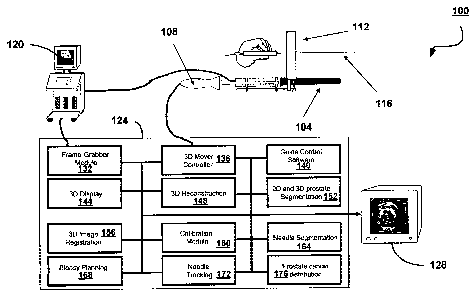
or
More target tissue areas. Historically, these biopsy methodologies have been
CA 02600981 2007-08-28
WO 2006/089426
PCT/CA2006/000282
- 2 -
inaccurate. The introduction of trans-rectal ultrasound ("TRUS") has
revolutionized prostate biopsy techniques and has greatly increased the
accuracy of biopsy. Widespread screening for PCa using the prostate-specific
antigen ("PSA") test has greatly increased the numbers of TRUS-guided biopsy.
[0005] The controversies related to the decision of how best to
manage
early-stage PCa are among the most intensely debated in all of clinical
medicine
by medical professionals as well as concerned patients. Management options
for early-stage PCa are: "watchful-waiting", hormone therapy, surgery and
[0006] Various reports have shown that the detection rate on repeat
biopsy ranges between 10% to 25% (after the first biopsy was negative).
Although advances in technology and understanding of the disease have
technical challenges clearly remain. For example, if an initial biopsy fails
to
detect cancer, who should undergo a repeat biopsy? How should a repeat
CA 02600981 2007-08-28
WO 2006/089426 PCT/CA2006/000282
- 3 -
biopsy be directed? Should the repeat (and initial) biopsy be lesion-directed,
random, or based on the details of the patient's anatomy (e.g., prostate
regions,
volume, shape).
[0007] Worldwide, the most common indication for prostate biopsy is
the
presence of serum PSA levels greater than 4.1 ng/ml. Because a significant
proportion of men with PSA in the 2.5 to 4.0 ng/ml range have PCa, some
investigators have advocated decreasing the PSA threshold to enhance PCa
detection. While early detection may increase the probability that the disease
is
confined to the prostate and that such patients are more likely to be free of
PSA
failure with improved disease-free survival after treatment, lowering the
threshold significantly increases the numbers of patients treated for non-
lethal
PCa. Despite the ongoing debate and lack of a general consensus at this time,
some centers have lowered the threshold for younger men, significantly
increasing the numbers of prostate biopsies performed. As lowering this
threshold results in biopsies of prostates with a small volume of cancer,
improved biopsy techniques are clearly required to increase the yield on the
first
biopsy and improve the planning of the potentially increasing numbers of
repeated biopsies.
[0008] In many cases, significant discomfort is reported during the
biopsy
procedure. After biopsy, common side-effects include hematuria,
hematospermia and hematochezia in about a third to a half of patients.
Although these are relatively minor, there is a potential for other less
frequent
post-biopsy morbidity including sepsis (0.2%-0.6%), urinary tract infection
(0.1%-4.5%) and urinary retention (0.2%-1.2%). As a result, it is desirable to
reduce the frequency of such procedures.
[0009] The optimal distribution of cores within the prostate has been
studied extensively, and it has been shown that uniform biopsy approaches
such as sextant methods are subject to sampling limitations in view of the
wide
variations in gland sizes. This issue has been explored using computer
simulations of the biopsy procedure and prostate anatomy, with probability
distribution of location, frequency and volume of prostate carcinoma obtained
from radical prostatectomy specimens. Results from computer simulations and
CA 02600981 2007-08-28
WO 2006/089426 PCT/CA2006/000282
- 4 -
clinical studies, which explored different systematically distributed cores,
have
demonstrated that the positive biopsy yield depends on the magnitude of gland
sampling. Increasing the number of biopsy cores increases the biopsy yield, .
and this effect is most pronounced in larger prostates. Using the same number
of cores regardless of individual prostate characteristics may lead to over-
sampling of small glands, and less extensive and potentially inadequate
sampling of large glands.
[0010] With more men undergoing PSA testing and the potentially
lowered PSA threshold for prostate biopsy, physicians commonly face the
dilemma of the patient with a negative prostate biopsy who still has
suspicious
clinical exam or serum PSA results. With the limited informational value of a
negative biopsy, and that no evidence of cancer on biopsy does not preclude
the possibility of a missed cancer, patients are often required to undergo
repeat
biopsies when clinical suspicion exists and in cases when a positive biopsy
for
cancer would have therapeutic consequences. Since there are an appreciable
number of men with false-negative biopsy who in fact harbor curable PCa, the
medical science is faced with a difficult challenge.
[0011] Many investigators have examined the positive yields on
repeated
biopsies of men with elevated PSA or suspicious digital rectal exam ("DRE") or
TRUS finding. The results demonstrated that on the first biopsy, about 15% to
40% of men had PCa, about 15% to 23% of men had PCa on the second biopsy
and 8% to 10% of men had PCa on the third biopsy. In some of the patients
with false-negative biopsy, the cancer might be clinically insignificant,
warranting
no therapy, but some of these patients might benefit from detection and
subsequent treatment.
[0012] Another important challenge facing physicians is in men
diagnosed on biopsy to have pre-malignant lesions, i.e. high-grade prostatic
intraepithelial neoplasia ("PIN"), and particularly atypical small acinar
proliferation ("ASAP"). These are challenging to manage as there is a 40% to
50% chance of finding cancer on repeat biopsy with ASAP. Since co-existing
cancer might be present, especially with ASAP, where the pathologist finds
only
a small amount of histologic "atypia" but not enough material to confidently
CA 02600981 2012-11-30
- 5 -
diagnose cancer, these patients typically undergo a repeat biopsy soon after
the first.
In these situations, it is important to re-biopsy the same area to increase
the yield.
Currently, only a vague location of the abnormal findings is available, and it
is not
possible to be certain that the same area has been sampled on the repeat
biopsy.
[0013] As a result of the increasing number of younger men with potentially
early and curable PCa undergoing repeated prostate biopsy, it is therefore
important
not to re-biopsy the same area if the original biopsy was negative, and it is
particularly important to re-biopsy the exact area if a possible abnormal area
was
detected on first biopsy as ASAP. Thus, improved guidance to suspicious
regions of
the prostate using information from other modalities is desired, as well as
the
locations of the cores obtained from the prostate must be known accurately to
help
guide the physician during the repeat biopsy, in order to help in correlating
any
imaging evidence of the disease and provide improved planning for the
subsequent
therapy.
[0014] It is, therefore, an object of the present invention to provide a
novel
system and method for performing a biopsy on a target volume and a computing
device for planning the same.
Summary of the Invention
[0015] In an aspect of the invention, there is provided a system for
performing a
prostate biopsy, comprising:
a three-dimensional (3D) trans-rectal ultrasound (TRUS) transducer for
generating 3D ultrasound images of target volume including said prostate;
a prostate segmentation module for identifying a segmented boundary of
said prostate in said 3D ultrasound images;
a three-dimensional registration module for registering said 3D
ultrasound images with a prior acquired 3D image of said prostate, wherein
said prior
acquired 3D image includes previous core locations of biopsy cores registered
to the
prior acquired image, wherein said registration is based on at least one of:
image intensity features of corresponding anatomical structures in
said 3D ultrasound images and said prior acquired 3D image; and
the segmented boundary of said prostate in said 3D ultrasound
images and a segmented boundary of said prostate in said prior acquired 3D
image;
CA 02600981 2012-11-30
- 6 -
a display for displaying said 3D ultrasound images and said previous
core locations of said prior acquired 3D image superimposed in the frame of
reference of said prostate of said 3D ultrasound images;
a biopsy planning module for use in developing a biopsy plan for said
prostate; and
a biopsy needle for biopsying said target volume in accordance with said
biopsy plan.
[0016-00171 In another aspect of the invention, there is provided a method for
developing a biopsy plan with respect to a target volume in a prostate,
comprising:
obtaining ultrasound volume data of said target volume including said
prostate using a three dimensional (3D) trans-rectal ultrasound transducer;
segmenting a boundary of said prostate in said ultrasound volume;
registering said prostate with a prior acquired image of said prostate,
wherein said
prior acquired 3D image includes previous core locations of biopsy cores
registered
to the prior acquired image, wherein said registering comprises one of:
registering based on image intensity features of corresponding
anatomical structures in said 3D ultrasound images and said prior acquired 3D
image; and
registering the segmented boundary of said prostate in said 3D
ultrasound images and a segmented boundary of said prostate in said prior
acquired
30 image;
displaying said 3D ultrasound images and said previous core locations of
said prior acquired 3D image superimposed in the frame of reference of said
prostate of said 3D ultrasound images; and
processing said 3D ultrasound images and said previous core locations
in combination in order to develop a biopsy plan for said prostate.
CA 02600981 2012-11-30
- -
[0018] By combining 2D/3D TRUS imaging with supplementary volume data
such as functional imaging from another imaging modality, information from
multiple
sources, modalities and/or times can be cross-correlated to enhance the
guidance of
prostate biopsy or, in fact, biopsy of other organs such as the breast.
Brief Description of the Drawings
10019] Embodiments will now be described, by way of example only, with
reference to the attached Figures, wherein:
Figure 1 is a schematic diagram of a three-dimensional (3D) transrectal
ultrasound ("TRUS") transducer and needle guide;
Figure 2 shows the 3D TRUS transducer positioned inside a patient;
Figure 3 is a flowchart of the conventional method of performing a
prostate biopsy;
CA 02600981 2007-08-28
WO 2006/089426 PCT/CA2006/000282
- 7 -
Figure 4 is a schematic diagram of one embodiment of a system
for performing a biopsy;
Figure 5 shows the imaging of a biopsy needle by the 3D TRUS
transducer of Figure 1;
Figure 6 is a flowchart that illustrates the method of performing a
biopsy using the system of Figure 4;
Figure 7 is a diagram showing five two-dimensional ("2D")
ultrasound ("US") images and their relative orientation;
Figure 8 shows a volume reconstructed from a series of 2D US
images captured using the 3D TRUS transducer of Figure 1;
Figure 9 is a flowchart of the steps performed during needle
insertion and guidance in the method of Figure 6;
Figures 10A and 10B illustrate the segmentation of the prostate in
a 2D US image; and
Figure 11 illustrates a 3D image of a prostate reconstructed from a
set of 2D US images.
Detailed Description of the Embodiments
[0020] PCa diagnosis is established by histological examination of
prostate tissue obtained most commonly by TRUS-guided biopsy and
sometimes by trans-urethral resection procedures ("TURP"). Needle biopsy of
the prostate represents the only definitive diagnostic modality capable of
confirming malignancy in men with palpable and ultrasonically undetected
lesions, and is now always performed under ultrasound guidance. Indications
for initial prostate needle biopsy are well established, consisting of any
abnormality on DRE or an abnormal PSA result, although the PSA threshold for
biopsy is being re-examined. Typically, prostate lesions detected with DRE and
TRUS have two or three cores taken through them. Since many small prostate
cancers are not detected with TRUS or DRE, needle samples are obtained from
predetermined regions of the prostate that are known to have high probability
of
harboring cancer. These are typically in the peripheral zone (PZ) (which
harbors
80% of all prostate cancers and a higher proportion of clinically significant
ones),
CA 02600981 2007-08-28
WO 2006/089426 PCT/CA2006/000282
- 8 -
and close to the capsule, as most cancers are thought to start within 5mm of
the
prostate capsule. Traditionally, the predetermined biopsy pattern has included
6
core biopsies, but as this has been shown to miss approximately 20% of
cancers. As a result, most centers are now taking 8 or more PZ tissue core
samples as part of their routine assessment.
[0021] Biopsies are typically performed with a thin, 18-guage
needle
mounted on a spring-loaded gun connected to the ultrasound ("US") probe,
forcing the needle to stay in the imaging plane so that it is always visible
in the
US image. The location of each core is registered, so that the pathologist can
report the extent and grade of the cancer. This is especially important if the
histological result is equivocal and the pathologist requests a repeat biopsy.
It
is, therefore, important to know from what exact location the = sample was
obtained in order to target more relevant tissue if a repeat biopsy is
performed.
,
[0022] Figure 1 shows a TRUS with an attached biopsy guide that
holds
a needle. The needle extends into the plane of the TRUS image so that it is
continuously visible therein.
[0023] Figure 2 illustrates the TRUS of Figure 1 during the
performance
of a prostate biopsy.
[0024] Figure 3 shows the steps performed during a conventional
biopsy.
The two most common reasons that people are referred for prostate biopsy are
the detection of an abnormality in their serum PSA or during an evaluation of
their prostate with a DRE.
[0025] During administration of DRE, the patient is subjected to
pre-
treatment involving a course of prophylactic antibiotics, and the
administration of
a rectal / lower large bowel cleaning protocol. The patient is then positioned
on
a bed, lying on their left side with their hips and knees fully flexed. The
. examiner introduces a copious amount of gel into the rectum with their
finger
and subsequently introduces an "end-fire" TRUS into the rectum of the patient.
The US image parameters are immediately adjusted to ensure optimal image
quality and the prostate is evaluated in two planes using B Mode (always) and
Color or Power Doppler (in some cases).
CA 02600981 2007-08-28
WO 2006/089426 PCT/CA2006/000282
- 9 -
[0026] The prostate is measured during a survey TRUS examination
(step 40). A variety of measurement protocols are used; the most common
presumes the prostate is an ellipse and its volume is calculated by
multiplying its
longest length by its longest width by its longest AP diameter by 0.52.
[0027] After completing the survey TRUS examination, the operator
decides if there is any "nodule" in the prostate that is suspicious for a
focal
cancer (step 50). The operator may feel a focal anomaly prior to imaging,
detect
a non-compressible hypoechoic mass, detect architectural distortion, focal
hypervascularity or marked asymmetry. Alternatively, a prior biopsy may have
identified an "area suspicious for malignancy" and the pathologist may have
recommended re-biopsy of this. This area would also be treated like a "nodule"
for biopsy protocol reasons.
[0028] If a "nodule" is detected at step 50, then a nodule biopsy
protocol
is followed (step 60). Otherwise a "no nodule" protocol is adhered to.
[0029] During the nodule biopsy protocol, the operator plans to obtain a
pre-determined number of samples from the nodule ¨ typically either two or
three 18-gauge core samples. Once the sampling of the nodule is planned, the
sampling of the rest of the prostate is also planned in a systematic way (step
70). This typically would consist of obtaining about another eight samples
from
pre-determined locations in the peripheral zone and possibly a small number
from the central gland.
[0030] During the no nodule biopsy protocol, the operator plans to
obtain
a pre-determined number of samples from the peripheral zone of the prostate.
The original pattern (the sextant pattern) called for six peripheral zone
samples;
one from the base, one from the middle and one from the prostate apex on
either side. Newer protocols typically have an increased number of samples
being obtained from the peripheral zone with increased emphasis of sampling
from the lateral part of the gland. Most protocols nowadays involve ten or
twelve
samples.
[0031] If it is determined at step 80 that this is a repeat biopsy,
additional
systematic biopsies are planned (step 90). The "No Nodule Biopsy Protocol" is
typically altered if the patient has had a prior negative biopsy and yet
clinically
CA 02600981 2007-08-28
WO 2006/089426 PCT/CA2006/000282
- 10 -
and biochemically remains suspicious for disease. These protocols typically
call
for increasing the number of peripheral zone biopsy, possibly altering the
location of some of these biopsies and also taking some samples from the
transition zone.
[0033] With the planned biopsy template mentally "worked out" in the
operator's mind, local anesthesia may be administered. This usually consists
of
Xylocaine 1cY0 or 0.5% administered either about the neurovascular bundles,
into
the periprostatic space or directly into the prostate. The operator then
surveys
the prostate in either the sagittal or transverse plane using a transducer
such as
that shown in Figure 1 and applies the mental template to the prostate and
acquires the samples. This is an inexact process but typically the "mid
peripheral zone" is assigned to that portion of the gland that is at the level
of the
veru montanum. The apex is typically the region of the prostate within 1.5 cm
of
the external sphincter and the base that portion of the gland within 1.5 cm of
the
superior border of the gland. No record of where the samples were obtained is
typically recorded.
[0034] Most operators obtain samples from the base of the gland
initially,
the mid and apical regions subsequently and finally from the transition zones,
if
they need to be sampled. Any periprostatic bleeding, rectal wall bleeding or
rectal luminal bleeding is typically treated with direct compression for
periods of
three to ten minutes. Following a brief period of bed rest the patient is
allowed
to resume light activity in the hospital or office for periods between twenty
to
sixty minutes. If the patient is well following this, he is discharged.
[0035] While it may be theoretically obvious to the operator where
the
protocol requires biopsies to be obtained, applying the mental template to the
actual prostate is challenging as "end-firing" images are not orthogonal to
the
long axis or transverse axis of the prostate. After samples have been
obtained,
it is often not clear exactly where they were taken from and that locations
are
often not recorded. This poses an especially challenging problem in cases
where the pathologist may have recommended re-biopsy of a specific region.
CA 02600981 2007-08-28
WO 2006/089426
PCT/CA2006/000282
- 11 -
[0036] The conventional approach for performing biopsies can result
in a
lower than desired level of care for many patients as much clinical,
biochemical
and imaging information is not being considered during the sampling process.
[0037] A system for performing biopsies that helps to alleviate the
above
disadvantages is shown generally at 100 in Figure 4. The system 100 includes
a TRUS transducer 104 coupled to a motor assembly 108 that operates to
control the longitudinal movement and rotation of the TRUS transducer 104.
The TRUS transducer 104 is operable to continuously capture radial 2D US
images over a radial operational scan range. A needle guide 112 is coupled to
the TRUS transducer 104. The needle guide 112 is a multiple-holed template
used to stabilize lateral movement of a needle 116. The needle 116 is used to
extract biopsy cores from the prostate of a patient. The TRUS transducer 104
is
also coupled to a conventional US machine 120 for displaying image data as it
is captured by the TRUS transducer 104. The motor assembly 108 and US
machine 120 are in communication with a computer 124. A display 128 is
coupled to the computer 124 for presenting images generated by the computer
124.
[0038] The computer 124 is a personal computer having a processor
that
executes software for performing 3D image acquisition, reconstruction and
display. The processor also executes software for performing biopsy planning,
and for controlling the TRUS transducer 104. The computer 124 includes a
video frame-grabber 132, a 3D mover controller module ("MCM") 136, a guide
control module 140, a 3D display module 144, a 3D reconstruction module 148,
a 2D and 3D prostate segmentation module 152, a 3D image registration
module 156, a calibration module 160, a needle segmentation module 164, a
biopsy planning module 168, a needle tracking module 172 and a prostate
cancer distribution module 176.
[0039] The video-frame grabber 132 captures image data from the US
machine 120 and preferably operates at 30Hz or greater to provide rapidly
updated ultrasound images. Such a module for acquiring and storing 2D US
images is described in U.S. Patent Nos. 5,457,371 and 5,562,095. The MCM
136 is coupled to and controls the motor assembly 108. In turn, the motor
CA 02600981 2007-08-28
WO 2006/089426 PCT/CA2006/000282
- 12 -
assembly 108 controls the longitudinal and rotational movement and the image
data acquisition timing of the TRUS transducer 104. The guide control module
140 controls movement of the needle guide 112 perpendicular to the axis of the
TRUS transducer 104 in order to accurately align holes of the needle guide 112
with a desired needle path. The user interface for the 3D display module 144
is
described in U.S. Patent Nos. 5,842,473 and 6,334,847.
[0040] The 3D display module 144 renders 3D images to be presented
on the display 128 using the image data captured and processed by the imaging
software. In particular, the 3D display module 144 generates three orthogonal
views of the target volume: two that are co-planar to the needle 116 and a
third
that generally bisects the trajectory of the needle 116.
[0041] The 3D reconstruction module 148 receives 2D US images from
the video frame-grabber 132 and, using a priori knowledge of the
characteristics
of the TRUS transducer 104, generates 3D images of the volume scanned. The
2D and 3D prostate segmentation module 152 analyzes the 2D images
captured by the video frame-grabber 132 and the 3D images generated by the
3D reconstruction module 148 and distinguishes between the prostate and other
tissue. The 3D image registration module 156 registers the 3D images received
from the video frame-grabber 132 or from other sources with one another. The
calibration module 160 calibrates the position of the needle guide 112 with
the
position of the TRUS transducer 104. The needle segmentation module 164
identifies needles in 2D and 3D US images.
[0042] The biopsy planning module 168 selects a biopsy plan based on
a
number of factors. These factors include the shape of the prostate determined
by the 2D and 3D segmentation module 152, the location of any nodules in the
3D US images generated by the 3D construction module 148, the 3D images of
other modalities received from other sources and general PCa probability
distribution information. In response, the biopsy planning module 168
generates
and provides planned needle trajectory information to the 3D visualization
software so that the planned needle trajectory can be overlaid atop the US
images on the display. The actual needle trajectory can then be viewed in
relation to the planned needle trajectory. The biopsy planning module 168 can
CA 02600981 2007-08-28
WO 2006/089426 PCT/CA2006/000282
- 13 -
also receive and process the US images from the 3D reconstruction module 148
and dynamically re-determine the biopsy plan based on the actual needle
trajectory and previous biopsy locations.
[0043] The needle tracking module 172 registers the location of the
needle 116 and, thus, the location from which biopsies are being taken. The
prostate cancer distribution module 176 determines a probability distribution
for
PCa based on parameters provided about the patient.
[0044] Figure 5 shows the TRUS transducer 104 capturing a set of 2D
US images. As the TRUS transducer 104 is rotated by the MCM 136, it
captures image data to generate a series of 2D images 180. The 2D images
180 are captured at generally regular intervals during rotation of the TRUS
transducer 104. Initially, the TRUS transducer 104 captures a 2D image 180
every one degree of rotation and rotates through 100 degrees, thereby
capturing
one hundred and one 2D images 180. The captured 2D images 180 are fanned
radially in relation to the TRUS transducer 104. As will be understood,
insertion
of the needle 116 along an oblique trajectory results in the intersection of
the 2D
TRUS image planes. As a result, the needle 116 only appears as a point in the
captured 2D US images.
[0045] A method 200 for performing a biopsy that makes use of the
system 100 will now be described with reference to Figure 6. The method 200
enables the presentation of 3D images from multiple modalities, prostate
cancer
probability distributions, previous biopsies, etc.
[0046] The method 200 commences by obtaining supplementary volume
data for the patient (step 210). A 3D TRUS survey image is captured (step
220). The 3D TRUS survey image is registered with the pre-biopsy image and a
probability distribution of PCa (step 230). Biopsy planning is then performed
using the images (step 240). Next, biopsy needle insertion is performed, with
guidance from the system 20 and tissue is sampled (step 250). Using the
detected location of the needle 116, the biopsy location is registered for
future
use (step 260).
[0047] During the obtaining of the supplementary volume data for the
patient at step 210, at least one pre-biopsy functional 3D image of the
patient's
CA 02600981 2007-08-28
WO 2006/089426 PCT/CA2006/000282
- 14 -
prostate generated using at least one other modality is received. These images
can be received from another computing device via a communications interface
(not shown) of the computer 124. The functional image may be based on a
photon emitting radiopharmaceutical as in single photon emission computerized
tomography ("SPECT") or with a gamma camera, or a positron emitting
radiopharmaceutical as in positron emission tomography ("PET"). The
radiopharmaceutical based images show abnormal uptake of the injected drug
in regions of the prostate that may harbor malignant cells. In all cases, the
functional image is obtained together with an anatomical image for use in
registration as described later. The combined functional image and anatomical
image is now routinely obtained with conventional imaging systems such as
PET/ computed tomography ("CT") scanners.
[0048] In addition, the supplementary volume data also includes a
PCa
probability distribution generated using information about the patient. This
PCa
probability distribution is provided as a color overlay on the 3D prostate
image.
The probability distribution of PCa is modified using the following non-
imaging
parameters:
[0049] Demographics ¨ Race: The incidence and prevalence of prostate
cancer is highest in African Americans. They typically have a higher
occurrence
of cancer, an acute Gleason grade in particular, and it typically occurs at a
younger age than evidenced in other races. In contrast, orientals have the
lowest incidence and prevalence of PCa.
[0050] Demographics ¨ Age: Prostate cancer increases in incidence
and
prevalence with age.
[0051] Demographics ¨ Family History: Having a first-degree relative
(brother, father) with PCa almost doubles one's risk of getting the disease.
Having a first-degree relative with breast cancer also increases the risk. A
small
minority of patients with specific genetic mutations is at a particularly high
risk of
prostate cancer.
[0052] Demographics ¨ Geography: There is some evidence that living in
particular geographic location may increase the risk of prostate cancer though
this research is incomplete. Preliminary data suggests for example that there
is
CA 02600981 2007-08-28
WO 2006/089426 PCT/CA2006/000282
- 15 -
a higher incidence and prevalence of PCa in the northern United States and
Canada compared with the southern United States.
[0053] Demographics ¨ Diet and Body Mass Index: A positive
correlation
between the risk of cancer and the body mass Index has been postulated and
there is some evidence to support it. Specific foods are also being
investigated
to see if they have a protective effect against PCa. These include vitamin E,
Selenium and soy products.
[0054] Biochemical Tests ¨ PSA: PSA arguably has had the biggest
influence on the survival rate of PCa patients in the last decade. It has
enabled
cancer to be detected at an earlier stage and has also been used to evaluate
the impact specific treatments have had on individual patients. While the
"absolute" level of serum PSA has some utility, integrating this number with
other information is proving much more useful clinically. Below are some of
these "integrated" PSA parameters that have proved useful.
[0055] Age-Specific PSA: Initially, a serum PSA of 4 ng/ml or less was
considered normal. In recent years, most centers have adopted an age specific
PSA which takes into account that the "normal" serum PSA increases with age.
So, for example, a 45 year old with a PSA of greater than 2.5 ng/ml would be
considered abnormal and a 75 year old with a PSA of less than 5.5 ng /ml would
be considered normal.
[0056] PSA Density: That is, serum PSA / Prostate volume. Most people
with a serum PSA greater than 4 ng/ml do not have prostate cancer. One of the
commonest non-cancerous reasons for an elevated PSA is due to the presence
of benign prostatic hyperplasia (BPH). By correcting the serum PSA for
prostate
volume many unnecessary biopsies could be avoided without clinically
significant cancers being missed. Some authors have contended that
correlating the serum PSA with the volume of the TZ on the prostate would be
even more helpful clinically.
[0057] PSA Velocity: This is the rate of increase in serum PSA year
over
year. In recent years, increased emphasis has been put on detecting cancers at
an earlier and earlier time, often before the PSA is greater than 4 ng/ml. PSA
velocity is one of the tools that has shown some utility in this cohort, with
several
CA 02600981 2007-08-28
WO 2006/089426 PCT/CA2006/000282
- 16 -
authorities recommending that patients be biopsied if the year over year
increase in serum PSA is greater than 25%.
[0058] "Free to Total PSA ratio": PSA exists in different forms in
the
blood. Researchers have found that in patients with PCa most of the PSA is
bound with carrier molecules and very little of it is "free" or unbound. In
contrast,
patients with BPH changes have a higher proportion of their PSA in "free" or
unbound format. Several groups have found this useful in decreasing the
number of unnecessary biopsies.
[0059] Biochemical Tests ¨ Other: Serum Alkaline phostatase in often
elevated in cases of metastatic prostate cancer. There are several other
biochemical markers currently under evaluation.
[0060] Physical Exam ¨ DRE: Signs of prostate cancer on DRE include a
palpable mass, gross asymmetry and a "fixed" prostate.
[0061] Physical Exam ¨ Other: Typically there are no other signs of
prostate cancer until late in the disease.. Then evidence of local spread and
metastases such as bone pain, pathological fractures, hematuria and bladder
dysfunction may be evident.
[0062] Pathology Findings ¨ High-Grade PIN or ASAP: High-grade
prostate intra-epithelial neoplasia (PIN) or atypical small acinar
proliferation
(ASAP) are precancerous conditions that almost always lead to a follow up
series of biopsies. While controversy currently exists about how and when to
re-
biopsy these patients, protocols are under construction. The risk is real with
some studies reporting as high as 50% chance of malignancy in patients with
ASAP at the time of the first re-biopsy.
[0063] Still further, the supplementary volume data includes 2D and 3D
images of the locations of any previous biopsies performed on the patient.
These images are registered and stored during previous biopsy procedures and
are retrieved in order to assist biopsy planning.
[0064] During the performance of the 3D US survey image at step 220,
the TRUS transducer 104 is inserted into the patient as shown in Figure 2. The
MCM 136 directs the motor assembly 108 to cause the TRUS transducer 104 to
rotate about its long axis over about 100 degrees while image data
CA 02600981 2007-08-28
WO 2006/089426 PCT/CA2006/000282
- 17 -
corresponding to 2D US images is captured at one degree intervals. The image
data corresponding to the 2D US images is then transmitted to the computer 40
to be digitized by the video frame-grabber 132 and registered.
[0065] The acquired 2D US images are processed by the 3D
reconstruction module as they are collected. The 2D US images correspond to
planes radially extending from the central axis of rotation of the TRUS
transducer 104. Accordingly, the 3D volume is reconstructed by translating and
rotating the 2D US images with respect to one another. The reconstructed 3D
volume consists of an array of voxels, or 3D pixels. The voxels are typically
cubic (but can also be rhomboidal) and are arranged according to a 3D
Cartesian system. Each voxel is assigned a greyscale-level value based on the
greyscale-level values of the pixels in the translated 2D images adjacent to
it.
[0066] Figure 7 illustrates a set of five 2D US images that have been
translated to their relative positions. As shown, the 2D US images are
radially
fanned.
[0067] Figure 8 illustrates a 3D US image reconstructed from the set
of
2D US images. As can be seen, the 3D US image has a fan profile
corresponding to the volume imaged by the TRUS transducer 104. The
acquired 2D US images are reconstructed into a 3D US image by the 3D
reconstruction module 148. The 3D display module 144 then generates a view
of the 3D US image, and provides a multi-planar 3D display and volume
rendering, as well as an extensive set of measurement tools. The 3D US image
is then presented for viewing on the display 128. As each new 2D US image is
acquired by the TRUS transducer 104 during its rotation, the 3D reconstruction
module 148 and 3D display module 144 dynamically update the 3D image
presented on the display 128.
[0068] The biopsy procedure progresses as follows. After the patient
is
prepared for the biopsy procedure, a 3D TRUS image is obtained and surveyed
by the physician and any observed nodules (suspicious regions) are identified
and whether the procedure is a repeat biopsy is noted.
[0069] The approach used by the system 100 for registration of the 3D
TRUS image with the functional images makes use of an automatic registration
CA 02600981 2007-08-28
WO 2006/089426 PCT/CA2006/000282
- 18 -
method that is based on image intensity features; that is, the normalized
mutual
information is the image similarity index (NMI). The 3D functional image, such
as PET, SPECT, magnetic resonance spectroscopy ("MRS") or optical, acquired
at a separate patient imaging session is color coded by converting the grey-
scale to a color scale using well-known techniques. The color-coded functional
information is then registered automatically to the 3D TRUS image using the
following procedure.
[0070] For use of the PET or SPECT image, the user loads the 3D CT
image, which was acquired together with the PET image using the PET/CT
system. Since the 3D CT and PET images are already registered, the user uses
the 3D CT image to register with the 3D TRUS using an intensity-based
registration procedure (e.g. NMI). This results in registration of the PET
image
to the 3D TRUS image. For use of an MRS image, the user loads the 3D MRI
image, which was acquired together with the MRS image using the MRI/MRS
system. Since the 3D MRI and MRS images are already registered, the user
uses the 3D MRI image to register with the 3D TRUS image using an intensity-
based registration procedure (e.g., NMI). This results in registration of the
MRS
image with the 3D TRUS image.
[0071] All the images are viewed in separate areas of the display, so
that
they can be examined separately or together. After examining the images,
automated alignment and registration of the 3D images is performed by the 3D
image registration module 156 to complete the registration.
[0072] During biopsy planning at step 240, the prostate is segmented
in
the images. The segmentation of the prostate is described with reference to
Figures 10A and 10B. The system uses model-based initialization and the
discrete dynamic contour to match the initial boundary to the actual prostate
boundary, as described in U.S. Patent No. 6,252,072. An initial prostate
boundary is generated by fitting a pre-defined model to four fiducial marks
selected by the user along the prostate's edge, as shown in Figure 10A. The
model is then deformed automatically to fit the prostate, as shown in Figure
10B.
[0073] The 3D TRUS image is sliced into contiguous 2D images, in a
parallel or rotational manner. The prostate boundary is segmented in a single
CA 02600981 2007-08-28
WO 2006/089426 PCT/CA2006/000282
- 19 -
2D image and the result propagated to an adjacent slice repeatedly until the
complete 3D prostate is segmented, as shown in Figure 11.
[0074] In addition to the use of the registered 3D functional and
TRUS
images for planning and targeting the biopsy, additional samples may be
required to sample prostate tissue without apparent visible abnormalities, as
cancer may be present, but at an early stage not yet visible in the functional
or
TRUS images.
[0075] As discussed above, optimal sampling of the prostate requires
knowledge of the prostate size, as using the same number of cores regardless
of individual prostate characteristics may lead to over-sampling of small
glands,
and under-sampling of large glands. In addition, in cases of repeat biopsy,
knowledge of the locations of the previous cores helps to plan and guide the
repeat biopsy, and help correlate any imaging evidence of the disease. Thus,
three important components are required: (1) a method to provide efficient 3D
visualization of the prostate, locations of previous cores, and probability
distribution of prostate carcinoma; (2) a method to allow planning of the
location
of the biopsies; and (3) a method to help guide the needles to their targeted
locations.
[0076] The objective is to provide the radiologist with a: (a) 3D
display of
the prostate with previous core locations, probability distribution of
prostate
carcinoma, biopsy target locations, and biopsy planning tools; and (b) a
continuously updated needle trajectory in 3D to allow biopsy needle guidance
to
the targeted locations.
[0077] These objectives are met in the guidance and planning phase of
the procedure with: (a) a 3D display of all relevant information with
superimposed planned needle trajectory before needle insertion; and (b) real-
time needle trajectory tracking as the needle is being inserted.
[0078] The locations of prior core locations is superimposed on the
3D
TRUS image together with the probability distribution of PCa after the current
and prior 3D TRUS images have been segmented and registered. To
superimpose planned needle trajectories, the known transducer position is
used,
which is obtained in the same manner as for 3D TRUS imaging. For the free-
CA 02600981 2007-08-28
WO 2006/089426 PCT/CA2006/000282
- 20 -
hand trans-rectal approach, the calibrated geometry of the transducer and its
biopsy needle attachment are used to determine the needle trajectory. In the
transperineal approach, the needle trajectory is calculated based on the 3D
location of the target.
[0079] The display provides three orthogonal planes intersecting on the
planned needle trajectory. One plane is orthogonal to the needle in an
approximate transverse orientation, a second is. parallel to the needle in an
approximate coronal plane and the third is in a longitudinal plane. As the
transducer is moved, the display is updated in real-time showing the
appropriate
new planned trajectory and prostate planes with the prior core locations and
carcinoma probability superimposed.
[0080] Nodules, or suspicious regions, are identified using all the
images
and, using the clinical and biochemical information, are assigned a
probability
weighting. The probability weighting and recommended biopsy pattern is then
color-coded and superimposed on the 3D TRUS image. This image is referred
to as a 3D multi-modality probability (3D MMP) image. The 3D MMP image is
then used by the physician to select, plan and guide the biopsy procedure.
[0081] Figure 9 illustrates the steps performed during needle
insertion,
guidance and tissue sampling during step 250. It is noted that, as steps 230
and 240 are performed in real-time, the 3D TRUS transducer 104 remains
positioned inside the patient from the time of capture of the survey image at
step
220. A biopsy target is selected in the 3D MMP image (step 252). The system
identifies the appropriate template hole and displays the needle path in the
3D
MMP image (step 254). The motor assembly 108 causes the 3D TRUS to move
so that a 2D TRUS image lines up with the target tissue and needle path (step
256). The optimal 2D US imaging plane for directing the needle 116 into the
prostate is chosen by interactively manipulating the US transducer and using
the
3D MMP image. This is done by scanning the prostate in real-time with a
standard 2D curved array transducer, which has an attached magnetic tracking
device. The 2D US image is updated in real-time and displayed in the 30 MMP
image for viewing. The operator manipulates the TRUS transducer 104 until the
real-time 2D US image intersects the biopsy target. The needle 116 is inserted
CA 02600981 2007-08-28
WO 2006/089426 PCT/CA2006/000282
-21 -
manually or via a computer-controlled mechanism, and is automatically tracked
as it is being inserted (step 258). The lesion is then biopsied using the
needle
116.
[0082] The image of the needle in the lesion is recorded immediately
after the tissue is sampled (e.g., after the needle is fired) and the needle
is
segmented. The location of the needle tip and the location of the tissue
. sampling is automatically found, recorded and displayed in the 30 MMP
image.
[0083] The needle segmentation module 164 is used to segment the
needle and its tip in real-time or near real-time and display the result.
Using the
segmented location of the needle tip, the approximate transverse plane that
passes through the needle is displayed. The other two planes are as described
above. Thus, with this approach, the radiologist has the usual real-time US
image available on the US system monitor, as well as 3D guidance information
updated in real-time.
[0084] The needle segmentation module 164 and the needle tracking
module 172 are used for identifying the needle trajectory and its tip position
in
2D and 3D TRUS images. To segment the needle, capture of two 2D or 3D US
images are captured. The first (pre-scan) 2D or 3D image is obtained by
scanning the prostate (tissue) before the needle is inserted, and the second
(post-scan) is acquired by scanning only the region containing the needle
during
needle insertion. The second 2D or 3D image is compared against the first and
the needle position within the post-scan image, including entry point and
needle
tip location, is determined using a grey-level change detection technique.
This
approach can segment the needle accurately in real-time for 2D TRUS imaging
and in near real-time (i.e., about 5 segmentations per second) for 3D TRUS
imaging.
[0085] If the targeted lesion was not sampled the procedure is
repeated.
This process is repeated until all suspicious regions and any other regions of
the
prostate as identified in the 3D MMP image are biopsied and the biopsy
locations are recorded.
[0086] While the method of performing biopsy has been described with
specificity to manual biopsy needle insertion using a template, other types of
CA 02600981 2007-08-28
WO 2006/089426 PCT/CA2006/000282
- 22 -
biopsy needle insertion methods will occur to those of skill in the art. For
example, insertion and/or alignment of the biopsy needle can be performed in a
number of manners. In one embodiment, a robotic assembly is used to control
the alignment and insertion of the biopsy needle. In another embodiment, a
computer is used to control the needle guide in order to control the alignment
of
the biopsy needle, but still permits manual control of its insertion. In still
another
embodiment, via a robot or can be computer-controlled.
[0087] In a further embodiment, an end-firing US transducer can be
coupled to a magnetic tracking device that provides position information to
the
computer. In this manner, 2D images with position and orientation
measurements are simultaneously acquired using a free-hand magnetically
tracked approach and are then reconstructed into 3D TRUS images in real-time.
A free-hand magnetically or optically tracked scanning approach is used to
allow
the user to manipulate the transducer freely, and record the position and
orientation of the transducer in space. The magnetic tracking approach is
based
on a small 6 degree-of-freedom magnetic field sensor (receiver) mounted on the
TRUS transducer, and a transmitter is placed near the patient to produces a
spatially varying magnetic field. The small sensor measures the three
components of the local magnetic field strength, and these are used to
calculate
the TRUS transducer's position and orientation, which are then used in the 3D
reconstruction algorithm.
[0088] In still yet another embodiment, markers can be attached to
the
TRUS transducer and a camera tracks movement of the markers in order to
determine the position and orientation of the TRUS transducer.
[0089] The supplementary volume data can include data from only one
source or can include data from a number of sources.
[0090] The above-described embodiments are intended to be examples
of the present invention and alterations and modifications may be effected
thereto, by those of skill in the art, without departing from the scope of the
invention which is defined solely by the claims appended hereto.