Sélection de la langue

Search

Sommaire du brevet 2604101 

Énoncé de désistement de responsabilité concernant l'information provenant de tiers

Une partie des informations de ce site Web a été fournie par des sources externes. Le gouvernement du Canada n'assume aucune responsabilité concernant la précision, l'actualité ou la fiabilité des informations fournies par les sources externes. Les utilisateurs qui désirent employer cette information devraient consulter directement la source des informations. Le contenu fourni par les sources externes n'est pas assujetti aux exigences sur les langues officielles, la protection des renseignements personnels et l'accessibilité.

Disponibilité de l'Abrégé et des Revendications

L'apparition de différences dans le texte et l'image des Revendications et de l'Abrégé dépend du moment auquel le document est publié. Les textes des Revendications et de l'Abrégé sont affichés :

  • lorsque la demande peut être examinée par le public;
  • lorsque le brevet est émis (délivrance).
(12) Brevet: (11) CA 2604101
(54) Titre français: PROCEDE D'ANALYSE DE STRUCTURES CELLULAIRES ET DE LEURS COMPOSANTS
(54) Titre anglais: METHOD OF ANALYZING CELL STRUCTURES AND THEIR COMPONENTS
Statut: Accordé et délivré
Données bibliographiques
(51) Classification internationale des brevets (CIB):
  • G01N 33/48 (2006.01)
(72) Inventeurs :
  • HOMMAN, MOHAMMED (Suède)
  • SINTORN, IDA-MARIA (Suède)
(73) Titulaires :
  • INTELLIGENT VIRUS IMAGING INC.
(71) Demandeurs :
  • INTELLIGENT VIRUS IMAGING INC. (Etats-Unis d'Amérique)
(74) Agent: BLANEY MCMURTRY LLP
(74) Co-agent:
(45) Délivré: 2014-01-21
(86) Date de dépôt PCT: 2006-02-17
(87) Mise à la disponibilité du public: 2006-10-26
Requête d'examen: 2011-01-10
Licence disponible: S.O.
Cédé au domaine public: S.O.
(25) Langue des documents déposés: Anglais

Traité de coopération en matière de brevets (PCT): Oui
(86) Numéro de la demande PCT: PCT/US2006/005571
(87) Numéro de publication internationale PCT: US2006005571
(85) Entrée nationale: 2007-10-10

(30) Données de priorité de la demande:
Numéro de la demande Pays / territoire Date
60/594,526 (Etats-Unis d'Amérique) 2005-04-15

Abrégés

Abrégé français

Dans cette invention, on fournit une cellule (14) qui contient plusieurs particules virales (16). Une première image (20) d'une première particule virale (22) et une seconde image (32) d'une seconde particule virale (34) sont prises par la technique de la microscopie électronique. La première particule virale se caractérise en ce qu'elle se trouve dans une première phase de maturité et la seconde particule virale se caractérise en ce qu'elle se trouve dans une seconde phase de maturité. La première image (20) et la seconde image (32) sont transformées en des premier et second profils d'échelle de gris (24, 46), respectivement, sur la base de données de pixels. Les premier et second profils d'échelle de gris (24, 36) sont ensuite sauvegardés sous la forme de premier et second gabarits, respectivement. Une troisième particule virale dans une troisième image est alors identifiée. La troisième image est transformée en un troisième profil d'échelle de gris. Le troisième profil d'échelle de gris est comparé au premier et au second gabarit pour déterminer une phase de maturité de la troisième particule virale.


Abrégé anglais


A cell (14) is provided that contains a plurality of virus particles (16). A
first image (20) of a first virus particle (22) and a second image (32) of a
second virus particle (34) are taken by electron microscopy technology. The
first virus particle is characterized as being in a first maturity stage and
the second virus particle as being in a second maturity stage. The first image
(20) and the second image (32) are transformed to first and second gray scale
profiles (24, 46), respectively, based on pixel data. The first and second
gray scale profiles (24, 36) are then saved as first and second templates,
respectively. A third virus particle in a third image is identified. The third
image is transformed into a third gray scale profile. The third gray scale is
compared to the first and second template to determine a maturity stage of the
third virus particle.

Revendications

Note : Les revendications sont présentées dans la langue officielle dans laquelle elles ont été soumises.


CLAIMS
1. A method of analyzing viral particles, comprising:
providing a cell containing a plurality of viral particles;
taking a first image of a first viral particle and a second
image of a second particle; characterizing the first
particle as being in a first maturity stage and the second
particle as being in a second maturity stage; transforming
the first image to a first linear gray scale profile
representing a radial density profile of the first viral
particle going from a center of the first viral particle
radially outwardly towards a periphery of the viral
particle; transforming the second image to a second linear
gray scale profile; saving the first gray scale profile as
a first template; saving the second gray scale profile as a
second template; identifying a third particle in a third
image; transforming the third image into a third gray scale
profile; and comparing the third gray scale to the first
and second template to determine a maturity stage of the
third particle.
2. The method according to claim 1 wherein the method
further comprises subjecting the third particle to an
active chemical substance.
3. The method according to claim 1 wherein the method
further comprises creating a fourth image of the third
particle at a time later than a time of the creation of the
third image.
4. The method according to claim 2 wherein the method
further comprises determining an effect of the chemical
11

substance on the third particle based on the determined
maturity stage.
5. The method according to claim 4 wherein the method
further comprises transforming the fourth image to a fourth
gray scale profile to objectively determine the effect of
the chemical substance on the third particle.
6. A method of analyzing virus particles, comprising:
providing a plurality of virus particles; taking a first
image of a first virus particle and a second image of a
second virus particle; characterizing the first virus
particle as being in a first maturity stage and the second
virus particle as being in a second maturity stage;
transforming the first image to a first gray scale profile;
transforming the second image to a second gray scale
profile, saving the first gray scale profile as a first
template; saving the second gray scale profile as a second
template; identifying a third virus particle in a third
image; transforming the third image into a third gray scale
profile; comparing the third gray scale to the first and
second template to determine a maturity stage of the third
virus particle; and transforming elliptical shaped virus
particles to circular shaped virus particles.
7. The method according to claim 6 wherein the method
further comprises determining a beginning and an end of a
virus particle wall by taking derivatives of the first,
second and third gray scale profiles.
8. The method according to claim 6 wherein the method
further comprises determining a beginning and an end of a
12

protein layer by taking derivatives of the first, second
and third gray scale profiles.
9. The method according to claim 6 wherein the method
further comprises removing a protein from the first virus
particle and generating a modified gray scale profile.
13

Description

Note : Les descriptions sont présentées dans la langue officielle dans laquelle elles ont été soumises.

CA 02604101 2007-10-10
WO 2006/112928 PCT/US2006/005571
METHOD OF ANALYZING CELL STRUCTURES AND THEIR COMPONENTS
Technical Field
The present invention relates to a method of analyzing
cell structure that includes virus morphologies.
Background of Invention
Many efforts have been made in the past to classify and
analyze cell structures that include viruses and other
components. Various image analysis methods have been
developed to describe, segment and classify viruses by using
available image technologies. For example, cryo-electron
microscopy has been used but the structures of the cells and
the virus particles are not shown very well. It has also been
difficult to objectively, repeatedly and reliably describe the
cell components to accurately determine the maturity stages of
the cell components. This partly explains why the previous
analysis methods have not been very effective and there is a
need for more effective methods for analyzing cell and virus
particle structures.
Summary of Invention
The method of the present invention provides a solution
to the above-outlined problems. More particularly, the method
of the present invention is for analyzing cell structures and
their components. A cell structure may be provided that

CA 02604101 2007-10-10
WO 2006/112928 PCT/US2006/005571
contains a plurality of virus particles. A first image of a
first virus particle and a second image of a second virus
particle may be taken by using an electron microscopy
technology. The first virus particle is characterized as
being in a first maturity stage and the second virus particle
as being in a second maturity stage. The first and second
images are transformed to first and second gray scale
profiles, respectively, based on pixel data or other
information from the images. The first and second gray scale
profiles are saved as first and second templates,
respectively. A third virus particle in a third image may
then be identified. The third image is transformed into a
third gray scale profile. The third gray scale is compared to
the first and second template to determine a maturity stage of
the third virus particle.
Brief Description of Drawing
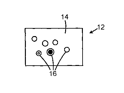
Fig. 1 is a schematic view of a cell nucleus containing
virus particles;
Fig. 2A is a microscope image of a virus virus particle
in a first maturity stage;
Fig. 2B is a schematic view of a gray scale profile of
the virus particle shown in Fig. 2A;
Fig. 3A is a microscope image of a virus virus particle
in a second maturity stage;
Fig. 3B is a schematic view of a gray scale profile of
2

CA 02604101 2007-10-10
WO 2006/112928 PCT/US2006/005571
the virus particle shown in Fig. 3A;
Fig. 4A is a microscope image of a virus virus particle
in a third maturity stage;
Fig. 45 is a schematic view of a gray scale profile of
the virus particle shown in Fig. 4A.
Detailed Description
With reference to Figs. 1-4, the method of the present
invention relates to steps for analyzing cell structures that
include components such as virus particles. The analysis of
virus particles is only used as an illustrative example of the
method of the present invention and the invention is not
limited to virus particles. Any suitable component in a
structure such as a cell may be analyzed using the method. An
important feature of the present invention is that different
virus particles produce unique graphs or gray level profiles.
Also, the various virus particles may be in different
maturity stages so that the present method may not only be
used to determine the types of virus particles but also the
maturity stage of the particular virus particle.
In general, an image 12 may first be taken by a suitable
technique, such as transmission electron microscopy (TEM), of
a cell structure 14. The image may include viral virus
particles 16 that are at various maturity stages. For
example, the virus particles may be categorized as being in
3

CA 02604101 2007-10-10
WO 2006/112928 PCT/US2006/005571
three or more maturity stages including empty or quite
immature virus particles, as shown in Fig. 2A, intermediary
virus particles, as shown in Fig. 3A, and more mature virus
particles, as shown in Fig. 4A.
The virus particles 16 are often linearly deformed making
the virus particles look slightly like ellipses rather than
circles. To be able to make an accurate radial density
profile it is desirable that the virus particles are radially
symmetrical. Thus, the deformation has to be inverted in
order to achieve the best results when the analysis of the
present invention is performed. This may be done by fitting
an ellipse to the viral virus particle which is assumed to be
approximately circular. The principal radii and the
orientation of the ellipse may be used to determine the
deformation and to make the inverse deformation.
Every pixel position in the old elliptic image is then
transformed into the new image to produce a picture where the
virus particle structures are circular. All virus particle
structures are preferably subject to this deformation
adjustment. As described below, the radially symmetric virus
particle structure may be transformed to a gray-level profile.
It may be used to describe the structure by calculating the
mean gray-level at each distance from the center going from
the center and out towards a periphery or shell of the virus
particle structure. The transformation to circular structures
is particularly useful when measuring the particle radius and
4

CA 02604101 2007-10-10
WO 2006/112928 PCT/US2006/005571
the thickness of the virus particle walls.
More particularly, Fig. 2A shows an image 20 of a
relatively immature or empty viral virus particle 22 in a cell
nucleus and Fig. 2B shows its corresponding gray level profile
24 in a graph 25. It is also possible to develop mathematical
algorithm to describe the virus particle structures instead of
relying on gray scale profiles. Viral DNA may be inserted
into the virus particle 22 that may eventually develop into a
mature virus particle with a dense DNA core, as shown in Fig.
4A and discussed below.
The virus particle 22 has a radius 26 that extends
from the center 28 radially outwardly to a center of the shell
wall 30 of the virus particle. Since the virus particle 22 is
virtually empty of virus components, the center portion is
almost white which shows as a high gray level value of the
profile 24 on the left side of the graph 25. The shell 30 is
darker so the profile 24 has a gradually lower gray level
value on the right side of the graph 25 since the shell area
corresponds to the right side of the graph 25.
Fig. 3A shows an image 32 of a virus particle 34 that is
not fully mature. The virus particle 34 contains an
additional viral protein that helps in the packaging of viral
DNA into the virus particles. The virus particle walls may
include protein layers so that the size of the virus particle
and the thickness of the layers may be determined. Fig. 3B
shows a corresponding gray level profile 36 in a graph 38.
5

CA 02604101 2007-10-10
WO 2006/112928 PCT/US2006/005571
The virus particle 34 has a radius 48 that extends from a
center 50 to a center of a peripheral shell 52 of the virus
particle. The black or dark ring 40 is shown as a local
minimum 42 in the profile 36 while the white ring 44 is shown
as a local maximum 46 in the profile 36. The beginning and
the end of the rings and shell wall may be determined by
analyzing the second derivative of the gray scale profiles-
Similarly, Fig. 4A shows an image 54 of a more mature
virus particle 56 and Fig. 4B shows the corresponding gray
level profile 58 in a graph 60. The virus particle 56 has a
radius 62 that extends from a center 64 to a center of a
peripheral shell wall 66 of the virus particle 56. The white
ring 68 is shown as a local maximum 70 in the profile 58.
An important feature of the method of the present
invention is that it is possible to create templates based on
the gray scale profiles to objectively describe the virus
virus particles. The templates may be created by using
mathematical methods also. For example, a template may be
based on an average of the characteristics of many virus
particles that are in the same maturity stage. The templates
may then be saved in a database and later be retrieved when it
is time to analyze and characterize new images containing
virus particles. When thousands of images are taken, the
templates may be used to calculate the number of virus
particles at the various maturity stages. In this way, the
virus production in a cell may be quantified by taking
6

CA 02604101 2007-10-10
WO 2006/112928 PCT/US2006/005571
additional TEM images.
More particularly, template gray level profiles of
intermediate virus particles may be created and used to
identify these particles in electron micrographs. To create
the template gray profile, the center point together with the
approximate size of the virus particle must be known. The
approximate size (radius) in pixels of the virus particle can
be deduced from knowing the magnification of the image and the
actual true size of the virus particle, or it can be marked in
the image. The center point of an object can be marked
manually or found through template matching. Preferably, the
center point is automatically chosen as the center of the
ellipse after deformation adjustment. As described above, the
gray-level profile can be visualized as a curve profile where
peaks in the curve represent bright radial regions and valleys
represent dark radial regions.
The width of certain radial features, such as the
virus particle shell, can give information regarding the
extent of tegumentation of the virus particle when
measurements are applied to cytoplasmic virus particle forms.
The problem with measuring this thickness directly from the
image or the profile is the difficulty of determining where
the virus particle begins and ends. Gradient information
about the profile can be used to solve this problem. The
first gradient measures how the gray-levels change along the
curve. If the gray-levels move from dark to bright along the
7

CA 02604101 2007-10-10
WO 2006/112928 PCT/US2006/005571
profile the gradient will have positive values and if the
gray-levels move from bright to dark the gradient will have
negative values. Where there is no change at all or where the
profile has a local maximum or minimum, the gradient will be
0. The thickness of the shell could, hence, be described as
the number of pixels between the local minimum to the local
maximum in the gradient. This is an objective way of
measuring where the shell begins and ends. To find these
extreme points, the second gradient (the gradient of the
gradient curve) is calculated since a local maximum or minimum
will be zero in the gradient curve.
In this way, gray level profiles are generated for the
many virus particles. The generated gray level profile curves
may be smoothed by convolution with a standard Gauss function.
The curves behavior at the virus particle wall or virus
particle shell may be depicted as a valley relative to the
linear behavior representing the overall virus particle
structure.
The derivative of the function reaches a local minimum
where the downward curve is the steepest which represent the
start of the virus particle wall. There is a local maximum
where the upward curve is the steepest which represents the
end or outside of the virus particle wall. The locations of
these local extremes are found as the zero-crossings of the
second derivative of the profile and the distance between the
extremes serve as the measurement of the width of the virus
8

CA 02604101 2007-10-10
WO 2006/112928 PCT/US2006/005571
particle wall.
As discussed above, the radius of a virus particle is
calculated as the distance between the center of the virus
particle and the center of the virus particle wall. The
center of the virus particle wall may be predicted as the
minimum value in the Gauss smoothed profile which is the zero
crossing of the 15t gradient of the smoothed curve.
Another important feature is that the images may be taken
again at a later time and new statistics of the maturity
stages of the virus particles may be compared to the earlier
statistics of the number of virus particles in the various
maturity stages. This is of particular interest when the
virus particles have been subjected to an active chemical
substance such as a suitable pharmaceutical substance to
better understand how the substance affects the virus
production in the virus particles. For example, an existing
or new substance may be used to determine which part of the
virus production or which maturity stage is affected by or
stopped by the substance. If, for example, the virus goes
through six maturity stages and there is no virus in the
fourth maturity stage it may be concluded that the
pharmaceutical substance prevents the virus from maturing
beyond the fourth maturity stage. The method may also be used
to determine which maturity stage a new pharmaceutical should
be designed to affect.
It is also possible to identify new different virus
9

CA 02604101 2013-04-09
WO 2006/112928 PCT/US2006/005571
morphologies and describe how the new virus differs from
earlier identified virus morphologies.
Another feature of the method of the present invention is
that it is possible to remove certain proteins from the virus
by using bio technical methods, such as siRNA, and then
objectively determine how the removal of the protein or
proteins affect the virus morphology and its corresponding
gray scale profile. For example, a pharmaceutical substance
may be designed to prevent the formation of a certain protein
in the virus particle and the effects of such prevention on
the gray scale profile can be studied. The removal of a
certain protein may prevent the virus from advancing from one
maturity stage to a later maturity stage. The method makes it
possible to determine which maturity stage is affected by the
removal of the particular protein.
While the present invention has been described in
accordance with preferred compositions and
embodiments, those of ordinary skill will understand
and appreciate the existence of variations,
combinations, and equivalents of the specific
embodiment, method, and examples herein.

Dessin représentatif
Une figure unique qui représente un dessin illustrant l'invention.
États administratifs

2024-08-01 : Dans le cadre de la transition vers les Brevets de nouvelle génération (BNG), la base de données sur les brevets canadiens (BDBC) contient désormais un Historique d'événement plus détaillé, qui reproduit le Journal des événements de notre nouvelle solution interne.

Veuillez noter que les événements débutant par « Inactive : » se réfèrent à des événements qui ne sont plus utilisés dans notre nouvelle solution interne.

Pour une meilleure compréhension de l'état de la demande ou brevet qui figure sur cette page, la rubrique Mise en garde , et les descriptions de Brevet , Historique d'événement , Taxes périodiques et Historique des paiements devraient être consultées.

Historique d'événement

Description Date
Inactive : CIB expirée 2024-01-01
Représentant commun nommé 2019-10-30
Inactive : TME en retard traitée 2018-02-20
Lettre envoyée 2018-02-19
Requête visant le maintien en état reçue 2017-01-19
Requête visant le maintien en état reçue 2016-02-11
Requête visant le maintien en état reçue 2015-01-28
Requête visant le maintien en état reçue 2014-01-29
Accordé par délivrance 2014-01-21
Inactive : Page couverture publiée 2014-01-20
Préoctroi 2013-11-07
Inactive : Taxe finale reçue 2013-11-07
Un avis d'acceptation est envoyé 2013-05-29
Lettre envoyée 2013-05-29
Un avis d'acceptation est envoyé 2013-05-29
Inactive : Approuvée aux fins d'acceptation (AFA) 2013-05-27
Modification reçue - modification volontaire 2013-04-09
Inactive : Dem. de l'examinateur par.30(2) Règles 2013-03-13
Requête visant le maintien en état reçue 2013-01-24
Lettre envoyée 2011-01-19
Requête d'examen reçue 2011-01-10
Exigences pour une requête d'examen - jugée conforme 2011-01-10
Toutes les exigences pour l'examen - jugée conforme 2011-01-10
Inactive : CIB enlevée 2010-04-29
Inactive : CIB en 1re position 2010-04-29
Inactive : CIB attribuée 2010-04-29
Inactive : CIB attribuée 2010-03-24
Lettre envoyée 2009-10-30
Exigences de rétablissement - réputé conforme pour tous les motifs d'abandon 2009-10-19
Réputée abandonnée - omission de répondre à un avis sur les taxes pour le maintien en état 2009-02-17
Inactive : IPRP reçu 2008-03-12
Déclaration du statut de petite entité jugée conforme 2008-02-12
Requête visant une déclaration du statut de petite entité reçue 2008-02-12
Inactive : Page couverture publiée 2008-01-10
Inactive : Notice - Entrée phase nat. - Pas de RE 2008-01-08
Inactive : CIB en 1re position 2007-11-07
Demande reçue - PCT 2007-11-06
Exigences pour l'entrée dans la phase nationale - jugée conforme 2007-10-10
Déclaration du statut de petite entité jugée conforme 2007-10-10
Demande publiée (accessible au public) 2006-10-26

Historique d'abandonnement

Date d'abandonnement Raison Date de rétablissement
2009-02-17

Taxes périodiques

Le dernier paiement a été reçu le 2013-01-24

Avis : Si le paiement en totalité n'a pas été reçu au plus tard à la date indiquée, une taxe supplémentaire peut être imposée, soit une des taxes suivantes :

  • taxe de rétablissement ;
  • taxe pour paiement en souffrance ; ou
  • taxe additionnelle pour le renversement d'une péremption réputée.

Les taxes sur les brevets sont ajustées au 1er janvier de chaque année. Les montants ci-dessus sont les montants actuels s'ils sont reçus au plus tard le 31 décembre de l'année en cours.
Veuillez vous référer à la page web des taxes sur les brevets de l'OPIC pour voir tous les montants actuels des taxes.

Historique des taxes

Type de taxes Anniversaire Échéance Date payée
Taxe nationale de base - petite 2007-10-10
TM (demande, 2e anniv.) - petite 02 2008-02-18 2008-02-12
TM (demande, 3e anniv.) - petite 03 2009-02-17 2009-10-19
Rétablissement 2009-10-19
TM (demande, 4e anniv.) - petite 04 2010-02-17 2010-01-07
Requête d'examen - petite 2011-01-10
TM (demande, 5e anniv.) - petite 05 2011-02-17 2011-01-10
TM (demande, 6e anniv.) - petite 06 2012-02-17 2012-01-06
TM (demande, 7e anniv.) - petite 07 2013-02-18 2013-01-24
Taxe finale - petite 2013-11-07
TM (brevet, 8e anniv.) - petite 2014-02-17 2014-01-29
TM (brevet, 9e anniv.) - petite 2015-02-17 2015-01-28
TM (brevet, 10e anniv.) - petite 2016-02-17 2016-02-11
TM (brevet, 11e anniv.) - petite 2017-02-17 2017-01-19
Annulation de la péremption réputée 2018-02-19 2018-02-20
TM (brevet, 12e anniv.) - petite 2018-02-19 2018-02-20
TM (brevet, 13e anniv.) - petite 2019-02-18 2018-11-30
TM (brevet, 14e anniv.) - petite 2020-02-17 2019-11-29
TM (brevet, 15e anniv.) - petite 2021-02-17 2020-12-28
TM (brevet, 16e anniv.) - petite 2022-02-17 2022-02-07
TM (brevet, 17e anniv.) - petite 2023-02-17 2023-02-06
TM (brevet, 18e anniv.) - petite 2024-02-19 2023-12-13
Titulaires au dossier

Les titulaires actuels et antérieures au dossier sont affichés en ordre alphabétique.

Titulaires actuels au dossier
INTELLIGENT VIRUS IMAGING INC.
Titulaires antérieures au dossier
IDA-MARIA SINTORN
MOHAMMED HOMMAN
Les propriétaires antérieurs qui ne figurent pas dans la liste des « Propriétaires au dossier » apparaîtront dans d'autres documents au dossier.
Documents

Pour visionner les fichiers sélectionnés, entrer le code reCAPTCHA :


Pour visualiser une image, cliquer sur un lien dans la colonne description du document. Pour télécharger l'image (les images), cliquer l'une ou plusieurs cases à cocher dans la première colonne et ensuite cliquer sur le bouton "Télécharger sélection en format PDF (archive Zip)" ou le bouton "Télécharger sélection (en un fichier PDF fusionné)".

Liste des documents de brevet publiés et non publiés sur la BDBC .

Si vous avez des difficultés à accéder au contenu, veuillez communiquer avec le Centre de services à la clientèle au 1-866-997-1936, ou envoyer un courriel au Centre de service à la clientèle de l'OPIC.


Description du
Document 
Date
(aaaa-mm-jj) 
Nombre de pages   Taille de l'image (Ko) 
Description 2007-10-09 10 387
Dessins 2007-10-09 1 36
Revendications 2007-10-09 4 93
Abrégé 2007-10-09 1 63
Dessin représentatif 2007-10-09 1 2
Description 2013-04-08 10 384
Revendications 2013-04-08 3 79
Dessin représentatif 2013-12-17 1 3
Rappel de taxe de maintien due 2008-01-07 1 112
Avis d'entree dans la phase nationale 2008-01-07 1 194
Courtoisie - Lettre d'abandon (taxe de maintien en état) 2009-04-13 1 172
Avis de retablissement 2009-10-29 1 162
Rappel - requête d'examen 2010-10-18 1 126
Accusé de réception de la requête d'examen 2011-01-18 1 176
Avis du commissaire - Demande jugée acceptable 2013-05-28 1 163
Avis concernant la taxe de maintien 2018-02-19 1 178
Quittance d'un paiement en retard 2018-02-19 1 163
Paiement de taxe périodique 2018-11-29 1 26
PCT 2007-10-09 5 153
Taxes 2008-02-11 2 52
Correspondance 2008-02-11 2 52
PCT 2007-10-10 3 158
Taxes 2009-10-18 1 29
Taxes 2010-01-06 1 27
Taxes 2011-01-09 1 34
Taxes 2012-01-05 1 29
Taxes 2013-01-23 1 27
Correspondance 2013-11-06 1 35
Taxes 2014-01-28 1 32
Taxes 2015-01-27 1 33
Paiement de taxe périodique 2016-02-10 1 33
Paiement de taxe périodique 2017-01-18 1 28
Paiement de taxe périodique 2018-02-19 1 27
Paiement de taxe périodique 2019-11-28 1 26