Note : Les descriptions sont présentées dans la langue officielle dans laquelle elles ont été soumises.
CA 02606803 2007-11-01
WO 2006/117786 PCT/IL2006/000527
DISEASE THERAPY USING DYING OR DEAD CELLS
FIELD AND BACKGROUND OF THE INVENTION
The present invention relates to methods of using dying cells for treating
diseases characterized by pathological immune responses, and to devices for
preparing such dying cells. More particularly, the present invention relates
to
methods of using apoptotic leukocytes, which do not secret pro-inflammatory
mediators, for treating diseases characterized by pathological immune
responses, sucli
as autoimmune diseases and transplantation-related diseases, and to devices
for
preparing such apoptotic leukocytes.
Diseases characterized by pathological immune responses include a large
number of diseases which are associated with significant mortality and
morbidity, and
for which no satisfactory/optimal treatnients are available. Such diseases
particularly
include autoiminune diseases, such as systemic lupus erythematosus (SLE),
transplantation-related diseases such as graft-versus-host disease (GVHD).
The immune system is a complex networlc comprising cells, antibodies,
tissues, and chemical messenger molecules which allow for conununication
between
these structures. A hallmark of a healtliy immune system is the ability to
recognize
bacteria, viruses, and other foreign bodies and to effectively attack such
pathogens
while continuing to distinguish between the foreign bodies and the molecules,
cells,
tissues and organs of the body. In addition to fighting infections, the immune
system
has other roles in maintaining the normal state of health and function of the
body.
Throughout the life span of an organism, tissues become reshaped with areas of
cells
being removed. This is accomplished by a process termed programmed cell death
or
apoptosis, the apoptotic cells disintegrating in an orderly and harmless
fashion and
being phagocytosed. In many organs, for example, a certain percentage of the
cells
die off every day while different branches of the immune system are typically
called
in to remove the dead cells and parts thereof to make room for the new cells
which
arise to replace them. The process of apoptosis is furthermore considered to
be
particularly important in the development and maintenance of the immune system
itself, where the immune cells which recognize or attack normal cells of the
body are
destroyed and removed by this process.
CA 02606803 2007-11-01
WO 2006/117786 PCT/IL2006/000527
2
The number of monocytes, neutrophils, and lymphocytes that are produced,
circulating, dying, and extravasating in the body is controlled at various
levels,
including via apoptosis.
In the case of monocytes, CFU-GM, the earliest identified cell coinmitted to
differentiate along the myeloid patliway, develops into inoiiocyte in the bone
marrow,
mainly in the presence of M-CSF, IL-3, and low levels of GM-CSF. No bone
marrow
reserve exists for inonocytes, which spend 1-3 days in transit tlirough the
inaiTow and
are then released to spend from 8 to 72 hours in the blood, with subsequent
further
possible differentiation, maturation, and proliferation in tissues [1].
Monocytes
comprise 1-6 percent of peripheral leulcocytes, and it is estimated that 5.7 x
106
monocytes/lcg are produced every day. Monocytes can survive in tissues as
macrophages for long periods, but a substantial portion of monocytes are
constantly
undergoing apoptosis, either in the absence of anti-apoptotic factors or
following
infection or activation.
Monocytes express Fas and Fas ligand irrespective of their state of activation
[2, 3], and were shown to undergo Fas-dependent apoptosis upon culture [3],
activation [4], or infection [5]. Monocytes can be rescued from apoptosis upon
exposure to growth factors, differentiating factors (GM-CSF and IL-4), or
activation
factors [3, 6-8]. Upon differentiation to macrophages, monocytes are rescued
from
Fas-dependent apoptosis by the expression of Fas-associated death domain-like
IL-1-
beta-converting enzyme-inhibitory protein (FLIP) [3, 9].
Neutrophils constitute the most abundant population of leukocytes. In
humans, the daily turnover of neutrophils is about 1.6 x 10⁹ cells/lcg body
weight
(Klebanoff SJ, Clark RX: The Neutrophil: Function and Clinical Disorders.
Amsterdam, North-Holland Publishing, 1978, p 313), which keeps the number of
mature neutrophils within defined limits despite the tremendous proliferative
potential
of the bone marrow precursor cells. This large turnover is mediated by the
continuous
egress of neutrophils from the circulation. Neutrophils do not return to the
circulation
but are eliminated by secretion in mucosa or die in the tissues within 1-2
days
(Klebanoff SJ, Clark RX: The Neutrophil: Function and Clinical Disorders.
Amsterdam, North-Holland Publishing, 1978, p 313). Under normal non-
inflammatory conditions neutrophil turnover takes place without harmful
effects,
despite the large bioagressive and destructive potential of these cells
displayed under
CA 02606803 2007-11-01
WO 2006/117786 PCT/IL2006/000527
3
various inflammatory conditions [Weiss SJ: Tissue destruction by neutrophils.
N Engl
J Med 1989; 320:365-376]. A special mechanism of harmless neutrophil
destruction
is provided by apoptosis, genetically programmed cell suicide.
While apoptosis is a process used by the immune system in protecting the
body, it is also used to maintain tolerance to self-antigens and therefore
allowing the
immune system to fulfill its role in distinguishing the body's own cells from
foreign
bodies.
Cellular apoptosis plays an iniportant role in antigen-presentation. Immature
dendritic cells have the capacity to engulf apoptotic cells and to acquire and
immunologically present their antigens. Immature dendritic cells that capture
apoptotic macrophages exposed to killed influenza-virus, mature and activate
lyinphocytes to mount virus-specific CTL responses in the presence of
conditioned
media. However, in the absence of infection and conditioned media, immature
dendritic cells do not mature following uptake of apoptotic cells and as a
consequence
are less able to efficiently present acquired antigens. Furthermore, it has
been
suggested that following interaction with apoptotic material, iinmature
dendritic cells
may have a role in maintaining peripheral tolerance to self-antigens that are
permanently created at different sites. In support of this, autoimmunity or
SLE-lilce
disease has been observed in mice and humans deficient in receptors important
for
uptalce of apoptotic cells such as ABC1 cassette transporter, Mer, and
coniplement
deficiencies, as further described hereinbelow. Clearance via specific
receptors may
dictate specific immune response or tolerance as demonstrated by TGF-beta and
IL- 10
secretion by macrophages following uptake of apoptotic cells by macrophages.
Thus,
cytokines, chemokines, eicosanoids, and additional mediators present in the
milieu of
the interaction, may polarize the immune response.
When the immune system is deficient in recognition between self- and non-
self-antigens, the result is a state of disease, this may result in the immune
system
attacking one or more specific self molecules or cells leading to tissue and
organ
damage, resulting in autoimmune disease. Immunopathology of non-targeted
tissues
also may be indirectly caused non-specifically as a consequence of
inflammation
resulting from immune rejection of neighboring cells and tissues. Other than
classical
autoimmune diseases such as those mentioned hereinabove, it is becoming
increasingly apparent that many vascular disorders, including atherosclerotic
forins of
CA 02606803 2007-11-01
WO 2006/117786 PCT/IL2006/000527
4
such disorders, have an autoimmune component, and a number of patients with
vascular disease have circulating autoantibodies. Autoimmune diseases may be
divided into two general types, namely systemic autoiinmune diseases, such as
SLE
and scleroderma, and organ specific autoimmune diseases, such as inultiple
sclerosis,
and diabetes. Many clinically different types and subtypes of autoiminune
disease
occur. Although eac11 type of autoimmune disease is associated witll a
spectrum of
clinical symptoms and aberrant laboratory parameters, signs and symptoms of
autoimmune diseases frequently overlap so that one or more are diagnosed in
the
same patient. The vast majority cases in wliich one or more autoimmune disease
has
been diagnosed are characterized by the presence in the affected subject of
antibodies
directed against self-antigens, terined autoantibodies. Such autoantibodies
are often
present in tissues at ten to one hundred times the normal level in healthy
individuals
and give rise to a significant proportion of the organ and tissue damage
associated
with the particular autoimmune disease. For example, in the autoimmune disease
myasthenia gravis, autoantibodies against a receptor in neuromuscular junction
are
associated with muscle weakness, while in SLE, anti-dsDNA antibodies are
associated with nephritis in human patients and can cause nephritis upon
injection to
normal mice. In such diseases, the tissue and organ damage is attributed to
the
presence of autoantibodies and to the inflammation, which arises due
inflammatory
immune responses set off by autoantibodies.
Systemic lupus erythematosus is a model disease for understanding and
developing inventive treatments for autoiminune disease in general. While it
has long
been appreciated that DNA and histones are major autoantigens SLE, only
recently
has evidence been provided that the DNA-histone complex, i.e., nucleosomes,
are the
preferred targets of autoantibodies in SLE. During apoptosis, the membrane of
cells
undergoing apoptosis form cytoplasmic blebs, some of which are shed as
apoptotic
bodies. It was recently demonstrated that exposure of keratinocytes to high
frequency
light induces apoptosis, and that the cell surface expression of the
ribonucleoproteins
Ro and La, but also of nucleosomes and ribosomes, can be explained by
translocation
of certain intracellular particles to the apoptotic surface blebs.
Significantly, another
translocation which occurs during apoptosis is that of phosphatidylserine
(PS), an
acidic phospholipid that normally resides on the inside of the cell, but flips
to the
outside of the cell membrane when the cell undergoes apoptosis.
Phosphatidylserine,
CA 02606803 2007-11-01
WO 2006/117786 PCT/IL2006/000527
like cardiolipin, is a major autoantigen for anti-phospholipid antibodies in
SLE.
Taken togetlier, these findings suggest that SLE involves autoimmunity
directed
against intracellular proteins translocated to the cell surface during
apoptosis, and
hence that SLE patieiits forin an immune response to apoptotic material. This
5 hypothesis is supported by the observation that brief, limited
administration of
syngeneic apoptotic cells to normal strains of mice leads to induction of
autoantibodies and glomerular depositions. The immunopatliology of SLE appears
to
further involve defective uptalce of apoptotic material by macrophages, as
observed in
the reduced uptake/clearance of apoptotic cells by macrophages from SLE
patients in-
vitro, and by the high incidence of SLE in patients deficient in the C l q and
C4
components of the complement system, which is involved in uptalce of targeted
antigens.
Lymphocytes, i.e. T-cells and B-cells, are relatively resistant to apoptosis.
Upon antigenic stimulation, B-cells and T-cells proliferate and some will
differentiate
into effector cells. Plasma cells secrete antibodies that immobilize pathogens
and
promote their complement-mediated destruction and Fc (Ig constant region)-
receptor-
mediated ingestion by certain myeloid cells. Activated T-cells produce
cytokines,
some of which promote proliferation and functional activation of B-cells aud T-
cells
themselves, whereas others provide feedback signals to cells of the innate
immune
system. Immune effector mechanisms are highly potent weapons designed for the
killing of free pathogens and also pathogen-infected host cells. This armory
has the
potential to destroy healthy cells and tissues because many of the effector
molecules,
such as pro-inflammatory cytokines, act in a non-antigen-specific maimer and
also
because certain pathogen-specific receptors, such as B-cell receptors (BCRs)
and T-
cell receptors (TCRs) may cross-react with host antigens.
Immune responses to pathogens therefore pose a potential danger to the host
and immunopathology occurs with many types of infection. In addition,
chronically
activated lymphocytes that are rapidly proliferating, particularly B-cells in
germinal
centers undergoing Ig-variable gene hyper-mutation, are at risk of sustaining
mutations in proto-oncogenes or tumor suppressor genes that could lead to the
developmeiit of lymphoma and/or leukaemia. Multiple regulatory mechanisms have
evolved to prevent immunopathology. These include functional inactivation of
cells
of the immune system, a process that is potentially reversible and therefore
does not
CA 02606803 2007-11-01
WO 2006/117786 PCT/IL2006/000527
6
eliminate the danger, and killing of no-longer needed and/or potentially
dangerous
cells by apoptosis [Marsden and A. Strasser, 2003. Annu. Rev. Iinmunol. 21:71-
105].
Cells undergoing apoptosis signal neighboring cells, professional phagocytes,
and/or antigen presenting cells to rapidly engulf them, without triggering an
inflammatory or autoimmune response [10-12]. This process seems to play an
important role in homeostasis, resolution of inflammation, and tolerance
induction
[13-15]. However disregulation of this process may represent a inechanism of
escape
from immune surveillance against infections and tumors and, if inefficient, it
may
support persistent inflanlmation and autoimmunity [16, 17].
Another issue that remains unclear is the role of apoptotic cell-derived
antigens in cross-priming of immune responses. It has been shown that human
dendritic cells, but not macrophages, efficiently present antigen that is
derived from
influenza-infected apoptotic monocytes, wliich stimulates class I-restricted
CD8+
CTLs [18]. It remains unclear how dendritic cells derive a pro-inflammatory
presentation of antigens from influenza, since these antigens are acquired
from
apoptotic cells that are usually considered anti-inflainmatory, and that were
shown to
prevent maturation of dendritic cells [15, 19]. While in the former study
conditioned
media was employed as an adjuvant, the physiological adjuvants enabling cross-
priming nevertheless remain unknown. Thus, antigens derived from apoptotic
cells of
given lineage may result in activation or suppression of immunity due to
mechanisms
which remain to be resolved.
Manipulation of the immune system to treat immunopathology associated with
autoimmune diseases, such as SLE, and transplantation-related diseases, such
as
GVHD, have been major goals of immunologists for many years. Traditionally,
such
manipulation has involved use of imi.nunosuppressive drugs, such as
corticosteroids,
azathioprine, cyclophosphamide, and cyclosporine. While such drug-induced
immunosuppression has resulted, for example, in improvement of the 5-year
survival
rate of SLE patients in the last three decades, it is far from being an ideal
treatment
since no cure is achieved, since such treatment is associated with very
serious side-
effects, including general immune suppression, leading to high rates of
morbidity, and
is the primary cause of premature mortality. Administration of biological
agents such
as anti-CD40 ligand, and CTLA-41g has also been advocated. However, the
toxicity
CA 02606803 2007-11-01
WO 2006/117786 PCT/IL2006/000527
7
and efficacy of such treatments is suboptimal, being potentially associated,
for
example, with general immune suppression similarly to the above-mentioned
imxnunosuppressive drugs.
Thus, in view of the tolerizing/non-inflammatory properties of dying
leukocytes described hereinabove, a potentially optimal strategy for treatment
of
diseases characterized by patliological immune responses, such as autoimmune
diseases asid transplantation-related diseases, involves adininistration of
dying
leulcocytes having immunosuppressive/non-inflammatory properties. Such a
strategy
would inherently circumvent the aforementioned significant disadvantages of
prior art
1o immunosuppressive drug-based treatment approaches.
Several prior art approaches involving administration of dying leukocytes have
been employed or suggested for treatment of diseases characterized by
pathological
immune responses.
One approach suggests administration of apoptotic donor cells, such as
apoptotic donor leulcocytes, to facilitate engraftment of donor hematopoietic
grafts
transplanted into an allogeneic recipient [Perruche S. et al., 2004. Am J
Transplant.
4:1361-5; Kleinclauss F. et al., 2003. Transplantation 75(9 Suppl):43S-45S].
Such an
approach, however, suffers from various drawbacks, including requirement for
administration of allogeneic leukocytes, which inherently are associated with
risk of
GVHD as well as of their own rejection, suboptimal efficacy, failure to
demonstrate
adequate safety with respect to potential for inflammatory side-effects,
and/or of
never having been attempted in human patients, and hence of never having
demonstrated any therapeutic efficacy in human patients.
Another, apheresis-based, approach, termed "extracorporeal photopheresis",
involves administering to a patient a photoactivatable pigment which can be
specifically taken up by specific hematopoietic cells, such as T-cells, and
following
such uptalce harvesting blood, isolating the specific hematopoietic cells,
triggering
their apoptosis via UV irradiation, and infusing them back into the patient
(United
States Patent 6,219,584; Crovetti G, et al., 2000, Int. J. Artif. Organs.
23(1):55-62;
3o THERAKOS, Inc. PA, USA). This approach has been advocated for treatment of
hypersensitivity, graft rejection, or SLE (United States Patent 4,838,852); or
for
amelioration of GVHD
(http://www.clinicaltrials.gov/ct/show/NCT00054613?order=2). Prior art
approaches
CA 02606803 2007-11-01
WO 2006/117786 PCT/IL2006/000527
8
involving apheresis, however, are often suboptimally effective, and may be
associated
with undesired side-effects of unluiown origin, such as inflammatory side-
effects
(refer, for example, to: Siaini GA. et aL, 1997. Cryofiltration apheresis and
plasma
fractionation causing anaphylactoid reactions in patients receiving
angiotensin
converting enzyme inhibitors. Ther Apher. 1:325-9; Schwarzbeck A. et al.,
1997.
Anaphylactoid reactions during dextrail apheresis may occur even in the
absence of
ACE-inhibitor administration. Nephrol Dial Transplant. 12:1083-4; Koga N. et
al.,
1993. Anaphylactoid reactions and bradykinin generation in patients treated
witll
LDL-apheresis and an ACE inhibitor. ASAIO J. 39:M288-91; Strauss RG., 1996.
Mechanisms of adverse effects during hemapheresis. J Clin Apheresis 11:160-4;
Rossi
PL. et al., 1991. Comparison of the side effects of therapeutic cytapheresis
and those
of other types of hemapheresis. Haematologica. 76 Suppl 1:75-80; Huestis DW.,
1989. Risks and safety practices in hemapheresis procedures. Arch Pathol Lab
Med.
113:273-8; Hocker P, Wagner A., 1987. Side-effects of cytapheresis with cell
separators. Infusionsther Klin Ernahr. 14 Suppl 4:31-5). Extracorporeal
photopheresis, in particular, involves generation and administration of
harmful
necrotic/pro-inflammatory cells (Caricchio R. et al., 2003. Ultraviolet B
Radiation-
Induced Cell Death: Critical Role of Ultraviolet Dose in Inflammation and
Lupus
Autoantigen Redistribution. The Journal of Immunology 171:5778-5786).
Thus, all prior art approaches have failed to provide an adequate solution for
using dying leukocytes for treatment of diseases characterized by pathological
immune responses.
There is thus a widely recognized need for, and it would be highly
advantageous to have, a disease treatment method devoid of the above
limitation.
SUMMARY OF THE INVENTION
The present invention discloses the use of dying or dead leulcocytes which are
substantially free of pro-inflammatory mediators such as IL-1(3. IL-8 and MIP
1 a, for
treatment of diseases associated with pathological immune responses, and
discloses
devices for generating such dying or dead leukocytes. This use can be effected
in a
variety of ways, and these devices can be configured in a variety of ways, as
further
described and exemplified hereinbelow.
CA 02606803 2007-11-01
WO 2006/117786 PCT/IL2006/000527
9
According to one aspect of the present invention there is provided a use of a
cell preparation which comprises dying or dead leukocytes for the manufacture
of a
medicament identified for the treatment of a disease characterized by a
pathological
immune response, the dying or dead leukocytes are obtained by inducing live
leulcocytes to adhere to a surface and are being capable of suppressing the
pathological iminune response.
According to another aspect of the present invention there is provided a use
of
a cell preparation which comprises dying or dead leulcocytes for the
manufacture of a
medicament identified for the treatment of a disease characterized by a
pathological
immune response, the dying or dead leulcocytes do not secrete a pro-
inflanunatory
mediator selected from the group consisting of IL-1(3, IL-8 MIP-la MIP-1(3, IL-
6 and
IL-la, and are capable of suppressing said pathological immune response.
According to yet another aspect of the present invention there is provided a
method of treating a disease characterized by a patlZological immune response
in a
subject in need thereof, the method comprising administering to the subject a
therapeutically effective amount of a cell preparation which comprises dying
or dead
leulcocytes, the dying or dead leulcocytes being capable of suppressing the
pathological immune response, thereby treating the disease in the subject.
According to further features in preferred embodiments of the invention
described below, the method further comprises subjecting live leukocytes to a
cytocidal treatment selected from the group consisting of in-vitro serum
deprivation,
treatment with a steroid or steroid derivative, irradiation, and a pro-
apoptotic
treatment, thereby generating the dying or dead leulcocytes.
According to still further features in the described preferred embodiments,
the
method of treating the disease further comprises inducing live leukocytes to
adhere to
a surface, thereby generating the dying or dead leukocytes.
According to still further features in the described preferred embodiments,
the
pathological immune response is directed against at least one antigen, and the
dying
or dead leulcocytes comprise the at least one antigen.
According to still further features in the described preferred embodiments,
the
dying or dead leukocytes are derived from the subject.
According to still further features in the described preferred embodiments,
the
dying or dead leukocytes comprise dying or dead splenocytes and/or dying or
dead
CA 02606803 2007-11-01
WO 2006/117786 PCT/IL2006/000527
tliymocytes.
According to still further features in the described preferred embodiments,
the
dying or dead leulcocytes coinprise dying or dead lyinphocytes.
According to still further features in the described preferred embodiments,
the
5 dying or dead leulcocytes comprise dying or dead monocytes.
According to still further features in the described preferred embodiments,
the
dying or dead leulcocytes comprise dying or dead neutrophils.
According to still further features in the described preferred embodiments,
the
dying or dead leukocytes comprise apoptotic leukocytes.
10 According to still further features in the described preferred embodiments,
the
disease is a systemic autoimmune disease.
According to still further features in the described preferred embodiments,
the
disease is an antibody-mediated autoimmune disease.
According to still fiu-ther features in the described preferred embodiments,
the
disease is lupus erythematosus.
According to still further features in the described preferred embodiments,
the
disease is a transplantation-related disease.
According to still fiu-ther features in the described preferred embodiments,
the
disease is graft-versus-host disease.
According to still further features in the described preferred embodiments,
administering the cell preparation comprises administering to the subject a
total
number of the dying or dead leukocytes selected from a range of about 20
million to
about 2 billion cells per kilogram body weight of the subject.
According to still further features in the described preferred embodiments,
adnlinistering the cell preparation comprises administering to the subject at
least one
unit dose of the dying or dead leulcocytes, wherein the unit dose comprises a
number
of the dying or dead leulcocytes selected from a range of about 4 million to
about 2
billion cells per kilogram body weight of the subject.
According to still further features in the described preferred embodiments,
the
dying or dead leukocytes are obtained by subjecting live leulcocytes to a
cytocidal
treatment selected from the group consisting of in-vitro serum deprivation,
treatment
with a steroid or steroid derivative, irradiation, and a pro-apoptotic
treatment, thereby
generating said dying or dead leukocytes.
CA 02606803 2007-11-01
WO 2006/117786 PCT/IL2006/000527
11
According to still further features in the described preferred embodiments,
the
medicament comprises a therapeutic effective amount of the dying or dead
leukocytes
cells wliich is selected from a range of about 20 million to about 2 billion
per
kilogram body weight of a subject.
According to still further features in the described prefeiTed embodiments,
the
medicament comprises a therapeutic effective amount of the cell preparation
which
coniprises a number of the dying or dead leukocytes selected from a range of
about 4
million to about 2 billion cells per kilogram body weight of a subject.
According to still another aspect of the present invention there is provided a
device for treating a disease characterized by a pathological immune response,
the
device comprising: (a) a pump for pumping blood from a subject into the device
and
retunZing blood to the subject from the device; (b) a leukocytes separator in
communication with the pump for separating circulating leulcocytes from whole
blood; and (c) an apoptosis-inducing chamber or chambers in communication with
the
leukocytes separator for inducing apoptosis of the leukocytes to thereby
obtain
apoptotic leukocytes, and further in communication with the pump for
administering
the apoptotic leukocytes to the subject.
According to further features in preferred embodiments of the invention
described below, the apoptosis-inducing chambers comprise a first chamber for
inducing apoptosis of monocytes, a second chamber for inducing apoptosis of
neutrophils, and a third chamber for inducing apoptosis of lymphocytes.
According to yet another aspect of the present invention there is provided a
device for inducing apoptosis of leukocytes, wherein the device comprises an
apoptosis-inducing chamber or chambers for inducing apoptosis of leukocytes to
thereby obtain apoptotic leulcocytes, wherein the apoptosis-inducing chamber
or
chambers is selected from the group consisting of a first chamber for inducing
apoptosis of monocytes, a second chamber for inducing apoptosis of
neutrophils, and
a third chamber for inducing apoptosis of lyinphocytes.
According to further features in preferred embodiments of the invention
described below, the first chamber comprises a surface for enhancing adherence
of
monocytes thereto.
According to still further features in the described preferred embodiments,
the
device further comprises a first reservoir for containing a monocyte medium,
wherein
CA 02606803 2007-11-01
WO 2006/117786 PCT/IL2006/000527
12
the monocyte medium is for inducing apoptosis of monocytes.
According to still further features in the described preferred embodiments,
the
device further coinprises a second reservoir for containing a neutrophil
medium,
wherein the neutrophil medium is for inducing apoptosis of neutrophils.
According to still further features in the described preferred embodiments,
the
device furtlier comprises a third reservoir for containing a lymphocyte
medium,
wherein the lymphocyte medium is for inducing apoptosis of lyinphocytes.
According to still further features in the described preferred embodiments,
the
device further coinprises a mechanism for resuspending surface-adherent
monocytes.
According to still further features in the described preferred embodiments,
the
mechanism for resuspending the surface-adherent inonocytes is selected from
the
group consisting of: a reservoir for containing a protease a.nd a mechanism
for
introducing the protease into the first chamber; a flow-generating mechanisin
for
generating in the first chamber a flow of sufficient force and direction for
resuspending the surface-adherent monocytes; and a scraping mechanism for
scraping
the surface-adherent monocytes off the surface of the first chamber.
According to still further features in the described preferred embodiments,
the
apoptosis-inducing chamber or chambers comprises an apoptosis-inducing
mechanism
selected from the group consisting of: an irradiating mechanism for inducing
apoptosis; a mechanical mechanism for inducing apoptosis; and a chemical or
biochemical substance or environtnent for inducing apoptosis.
The present invention successfully addresses the shortcomings of the presently
lcnown configurations by providing a method of treating with improved safety
and
effectiveness diseases associated with pathological immune responses, such as
autoimmune diseases and GVHD, by administration of dying or dead leukocytes,
by
providing a device for generating such leulcocytes, and by providing a device
for
practicing such methods.
Unless otherwise defined, all technical and scientific terms used herein have
the same meaning as commonly understood by one of ordinary skill in the art to
which this invention belongs. Although methods and materials similar or
equivalent
to those described herein can be used in the practice or testing of the
present
invention, suitable methods and materials are described below. All
pitblications,
patent applications, patents, and other references mentioned herein are
incorporated
CA 02606803 2007-11-01
WO 2006/117786 PCT/IL2006/000527
13
by reference in their entirety. In case of conflict, the patent specification,
including
definitions, will control. In addition, the materials, methods, and examples
are
illustrative only and not intended to be limiting.
BRIEF DESCRIPTION OF THE DR.AWINGS
The invention is herein described, by way of example only, wit11 reference to
the accompanying drawings. With specific reference now to the drawings in
detail, it
is stressed that the particulars shown are by way of example and for purposes
of
illustrative discussion of the preferred embodiments of the present invention
only, and
are presented in the cause of providing what is believed to be the most useful
and
readily understood description of the principles and conceptual aspects of the
invention. In this regard, no atteinpt is made to show structural details of
the
invention in more detail than is necessary for a fundamental understanding of
the
invention, the description talcen with the drawings making apparent to those
skilled in
the art how the several forms of the invention may be embodied in practice.
In the drawings:
FIG. 1 is a histogram depicting reduction of serum anti-single-stranded DNA
antibodies in MRL/MpJ-FaslP' mice following treatment with syngeneic apoptotic
cells. Filled circles, control group of 6 week-old MRL/lpr/lpr mice immunized
with
vehicle only; open circles, experimental group of 6 week-old MRL/lpr/lpr mice
immunized with syngeneic apoptotic cells; filled triangles, control group
after 10
weeks of treatment; open triangles, experimental group after 10 weeks of
treatment.
FIG. 2 is a histogram depicting reduction of serum anti-double-stranded DNA
antibodies in MRL/MpJ-Faslp' mice following treatment with syngeneic apoptotic
cells. Filled circles, control group of 6 week-old MRL/lpr/lpr mice immunized
with
vehicle only; open circles, experimental group of 6 week-old MRL/lpr/lpr mice
immunized with syngeneic apoptotic cells; filled triangles, control group
after 10
weeks of treatment; open triangles, experimental group after 10 weeks of
treatment.
FIG. 3. is a set of fluorescence activated cell sorting (FACS) dot plots
depicting induction of monocyte apoptosis by serum withdrawal and substrate-
adherence. More than 70 percent of monocytes were annexin V-positive PI-
negative
by 12 hours indicating early apoptosis. Secondary necrotic cells represented
less than
5 percent of the cells as indicated by annexin V-positive, propidium iodide
(PI)-
CA 02606803 2007-11-01
WO 2006/117786 PCT/IL2006/000527
14
positive cells. The specificity of the apoptotic process was further shown by
marked
inhibition in the presence of 20 mM zVAD-fiiik. The percentage of early
apoptotic
and secondary necrotic cells is indicated within eacli histogram. Data is
representative of six different experiments.
FIG. 4. is a set of fluorescence activated cell sorting (FACS) dot plots
depicting that suspension + serum-withdrawal-induced death of monocytes is non-
apoptotic and shows features of necrosis. Prevention of contact in addition to
serum
witlzdrawal switched the mechanism of death. Cell niasnbers were reduced
progressively whereas the percentage of annexin+PI- remain constant and low.
Cells
were becoming directly annexin+PI+ and 20 mM zVAD-fmk did not reduce the rate
of death (not shown).
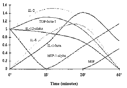
FIGs. 5a-c depict de-novo transcription of pro-inflammatory
cytolcine/chemokine mRNAs by monocytes subjected to suspension + serum
deprivation. Figures 5a-b are gene expression array analyses depicting de-novo
transcription of pro-inflammatory cytokine/chemokine mRNAs by monocytes
subjected to suspension + serum deprivation at 0 time and 30 minutes,
respectively.
Coordinates (A2, B2), which represent IL-1-beta and coordinates (E3, F3),
representing IL-8, show no visible fluorescence at time zero and a marked
fluorescence at 30 minutes following apoptosis induction. Some augmentation of
basal levels is seen for cDNA of IL-6 (C3-D3) and IL-1-alpha (E1-Fl). Other
cDNAs
that are present with viable cells and did not change much upon death
induction are
TGF-beta-1 (A7-B7), IL-2 (C2-D2), and TNF-alpha (A8-B8). MIF (E6-F6) shows
downregulation. Other wells in this membrane that did not show fluorescence
are
(Al-B1) for G-CSF, (C1-Dl) for GM-CSF, (E2-F2) for IL-4, (A3-B3) for IL-5, (A4-
B4) for IL-10, (C4-D4) for IL-12-alpha (E4-F4) for IL-12-beta, (A5-B5) for IL-
16,
(C5-D5) for IL-17, (A6-B6) for LT-beta, (C6-D6) for MCP-1, (C7-D7) for TGF-
beta-
2, (E7-F7) for TGF-beta-3, and (C8-D8) for TNF-beta. Coordinates that
represent
negative controls are (GI-G2, PUC18); and as positive controls (G3-G4, beta-
actin)
and (G5-G6, G7-G8, E8-F8, GAPDH). Chemokine membrane screening showed only
IL-8, MIP-1-alpha and MIP-1-beta upregulation (not shown). Membranes contained
eotaxin, fractalkine, GROa/MGSA, HCC-4, MCP-3, SDF2, PF-4, MDC, HCC-1, I-
309, I-TAC, lymphotactin, MCP-1, MCP-4, MIG, MIP-2, MIP-3-alpha, P10, SDF-1,
RANTES. Figure 5c is a data plot depicting representative cytokine and
chemokine
CA 02606803 2007-11-01
WO 2006/117786 PCT/IL2006/000527
eDNA level (in arbitrary units) changes as a function of time following
induction of
cell death. Note that only IL-1-beta, IL-8, and MIP-1-alpha are produced de-
novo.
FIG. 6a. is a data plot depicting that the pro-inflammatory cytokine IL-1-beta
is produced by monocytes subjected to suspension + serum withdrawal. Control
5 PBMCs (that contain 20 % monocytes) exhibit elevation of IL-1-beta secretion
following suspension + serum withdrawal (open triangles). Magiietically
isolated
monocyes (closed triangles) exhibit even higher elevation of IL-1-beta
secretion
following suspension + serum withdrawal (indicating that dying monocytes are
the
source of IL-1-beta). On the other hand, magnetically isolated monocytes
subjected
10 to serum withdrawal but not to suspension (closed squares) (i.e., were
allowed to
adhere and not be in suspension) did not secrete IL-1-beta (or any pro-
inflannnatory
cytokines) and were capable of inducing tolerance in dendritic cells (not
shown). B-
and T-lymphocytes (closed circles), and polymorphonuclear cells (open
circles),
shows that IL-1-beta secretion is specific to monocytes that were not allowed
to
15 adhere (i.e., monocytes that were subjected to suspension and serum
withdrawal).
FIG. 6b is an ELISA data histogram depicting that pro-inflammatory
cytokine/chemokine mRNA and protein are transcribed and translated de-novo by
monocytes subjected to suspension + serum withdrawal. Inhibition of
transcription
activity with actinomycin D and translational activity with cycloheximide
shows
marked inhibition in IL-1-beta cytokine secretion as measured by ELISA in
picograin/ml (pg/ml). Monocytes that were allowed to adhere during serum
withdrawal did not show any production of pro-inflammatory cytokines (not
shown).
Furthermore, monocytes that were allowed to adhere and were triggered to die
by
other methods of induction of apoptosis such as staurosporine,
cyclophosphamide, Fas
ligand, and more, always kept their non-inflammatory mode of death (not
shown).
FIG. 7a is an ELISA data plot depicting the secretion of IL-1-beta by
monocytes that were subjected to suspension-induced death (and serum
withdrawal)
but not from viable monocytes, monocytes subjected to hyperthermia-induced
necrosis, or apoptotic monocytes that were induced to apoptosis by adherence
and
serum withdrawal. Levels of IL-1-beta (shown in pg/ml) were measured by ELISA
at
0, 1, 4, and 24 hours following incubation of viable monocytes (closed
circles),
monocytes rendered necrotic via hyperthermia (open circles), or monocytes
subjected
to suspension-induced death (closed triangles). The results indicate that the
pro-
CA 02606803 2007-11-01
WO 2006/117786 PCT/IL2006/000527
16
inflammatory characteristics of suspension-induced monocyetes are not seen in
heat-
induced necrosis (accidental cell death) and are innate property of monocytes
that
were not allowed to adhere.
FIG. 7b is an ELISA data plot depicting IL-1p secretion (shown in pg/ml)
from monocytes which were subjected to suspension-serum withdrawal - induced
death and demonstrating that monocyte death via suspension (non adherence) is
neither caspase 3- nor caspase 1-dependent. Secretion of IL-1-beta by
monocytes
subjected to suspension-induced death (closed triangles, in 20 micromolar
DMSO)
was neither inhibited with the caspase 1 inhibitor, Z-WEHD (20 micromolar,
closed
circles), nor with the pan-caspase inhibitor ZVAD-fmk (20 micromolar, open
circles).
FIGs. 8a-c depict that pro-inflammatory cytokine secretion during monocyte
apoptosis is not NFkappaB-dependent. Figure 8a is a photograph of a Western
immunoblotting assay depicting that pro-inflammatory cytokine secretion during
monocyte apoptosis is not NFkappaB-dependent. Shown is 37 kDa IkappaB and
phosphorylated IkappaB (blaclc arrow). Viable monocytes (lanes a and b), were
incubated for 2 hours in the presence of 1 mg/ml zymosan (which triggers TOLL
receptors 2 on macrophagesor dendritic cells to mature and produce
inflammation)
with (lane a) or without (lane b) MG132 (a proteasome inhibitor). Lanes c and
d are
monocytes undergoing apoptosis [by the serum withdrawal and adherence
avoidance
(or suspension) method] at 2 hours (lane c) and 10 minutes (lane d) following
zymosan. As can be seen, viable monocytes exposed to zymosan show
phosphorylation of IkappaB (lane b, black arrow) that does not appear in the
presence
of MG132 (lane a). No phospliorylation is seen at 10 minutes (lane d) or 2
hours
(lane c) when monocytes undergo apoptosis. Additional sainples at 5, 20, 30,
40, 60,
and 90 minutes (not shown), following apoptosis induction did not show
IlcappaB
phosphorylation (representative of 5 experiments). Figure 8b is a bar-graph
depicting
that IL-1-beta secretion in the presence of MG132 is slightly elevated (3
experiments).
Figure 8c is a histogram depicting transcriptional activity in the presence of
MG132
(representative of 3 experiments). Note that fold increases in the levels of
mRNA
(filled bars) are not changed in the presence of MG132 (empty bars).
FIGs. 9a-e depict that pro-inflammatory cytokine secretion during monocyte
apoptosis is p38-dependent. Figure 9a is a bar-graph depicting that after 24
hours in
the presence of anti-Fas inhibitory antibodies (BD29 or ZB4), monocyte
apoptosis
CA 02606803 2007-11-01
WO 2006/117786 PCT/IL2006/000527
17
was only slightly decreased (BD29 is shown) compared to the significant * (p <
0.001) decrease in apoptosis seen in the presence of p38 inhibitor (p38INH) or
p38
and anti-fas (ZB4). Figure 9b is a Western immunoblotting assay depicting that
P38
is expressed at coinparable levels in monocytes exposed to LPS or induced to
undergo
apoptosis. Figure 9c is a Western immunoblotting assay depicting that
phosphorylated p38 is transiently increased upon LPS stimulation but shows
sustained
increase upon apoptosis. No phosphorylation of JNK was found (not shown).
Results
are representative of six experiments. Figure 9d is a bar-graph depicting that
IL-1-
beta secretion by apoptotic inonocytes (which were induced by suspension and
seruin
withdrawal), is conlpletely abrogated by specific p38 inliibitor (p38IN) but
not in p38
control (DMSO). No inhibition is seen in the presence of JNK inhibitor (JNKIN)
or
its control (LTAT). Figure 9e is a bar-graph depicting the marked decrease in
IL-8
secretion from apoptotic monocytes in the presence of p38 inhibitor (p381N)
but not
in control (DMSO) or JNK inhibitor (JNKIN).
FIG. 10 is a schematic diagram depicting a device for inducing apoptosis of
leukocytes. Arrows indicate direction of fluid flow.
FIG. 11 is a schematic diagram depicting a device for treating a disease
characterized by a pathological immune response. Arrows indicate direction of
fluid
flow.
FIGs. 12a-d are FACS analyses depicting that apoptotic monocytes
engulfment down-regulates the expression of maturation-related molecules on
dendritic cells (DCs). Adhered apoptotic monocytes (which were generated by
adherence and serum withdrawal were stained with DiI ( a lypophilic dye that
stains
dying cells) and were added to immature dendritic cells (iDCs), at a 4:1
ratio. The
iDCs acquired apoptotic cell-derived DiI and were exposed (bolded line) or not
(black
line) to LPS. Shown are FACS analyses of the treated DCs in the presence
(Figures
12b and d) or absence (Figures 12a and c) of apoptotic monocytes using anti-DR-
FITC (Figures 12a-b) and anti-CD86-FITC (Figures 12c-d) antibodies. Numbers
indicate the median fluorescence of the LPS-treated iDCs present in each
histogram.
Isotype controls are shown as dotted curves. Note that in the presence of
apoptotic
monocytes, a marked downregulation of CD86 and DR is observed (p < 0.0001).
Non-adhered dying monocytes do not downregulate and even upregulate DR and
CD86 (not shown). Similar results were obtained using CD40 (not shown). These
CA 02606803 2007-11-01
WO 2006/117786 PCT/IL2006/000527
18
results demonstrate that immature dendritic cells (iDCs) maturation is
inhibited by
adhered apoptotic monocytes.
DESCRIPTION OF THE PREFERRED EMBODIMENTS
The present invention is of methods of treating diseases associated with
pathological immune responses using dying or dead leulcocytes which are devoid
of
pro-inflainmatory mediators such as IL-1(3, IL-8 and MIP 1 a,, and of devices
for
generating such cells and practicing such methods.
The principles and operation of the present invention may be better understood
with reference to the drawings and accompanying descriptions.
Before explaining at least one embodiment of the invention in detail, it is to
be
understood that the invention is not limited in its application to the details
set forth in
the following description or exeinplified by the Examples. The inventiari is
capable
of other embodiments or of being practiced or carried out in various ways.
Also, it is
to be understood that the phraseology and terminology employed herein is for
the
purpose of description and should not be regarded as limiting.
Various methods of using administration of dying leukocytes for treatment of
diseases characterized by pathological immune responses have been described by
the
prior art.
One approach involves using administration of apoptotic allogeneic donor
leulcocytes, in an attempt to facilitate engraftment of allogeneic donor
hematopoietic
grafts [Perruche S. et al., 2004. Am J Transplant. 4:1361-5; Kleinclauss F. et
al.,
2003. Transplantation 75(9 Suppl):43S-45S]. Another, apheresis-based, approach
for
treatment of hypersensitivity, graft rejection, or systemic lupus
erythematosus (United
States Patent 4,838,852); or for amelioration of GVHD
(http://www.clinicaltrials.gov/ct/show/NCT00054613?order=2), termed
"extracorporeal photopheresis", involves administering to a patient a
photoactivatable
pigment which can be specifically taken up by specific hematopoietic cells,
such as T-
cells, and subsequently harvesting blood, isolating the specific hematopoietic
cells,
UV-irradiating the isolated cells, and re-infusing them into the patient
(United States
Patent 6,219,584).
However, all such prior art approaches suffer from various drawbacks. For
example, approaches involving administration of allogeneic leukocytes are
associated
CA 02606803 2007-11-01
WO 2006/117786 PCT/IL2006/000527
19
with risk of GVHD and rejection of the administered leulcocytes, and/or have
never
demonstrated any therapeutic efficacy in llumans. Prior art approaches
involving
apheresis are often suboptimally effective, and may be associated with
undesired
and/or unexplained side-effects, such as inflammatoiy side-effects (refer, for
example,
to: Siami GA. et al., 1997. Cryofiltration apheresis and plasma fractionation
causing
anaphylactoid reactions in patients receiving angiotensin converting enzyme
inhibitors. Ther Apher. 1:325-9; Schwarzbeck A. et al., 1997. Anaphylactoid
reactions during dextran apheresis may occur even in the absence of ACE-
inhibitor
administration. Nephrol Dial Transplant. 12:1083-4; Koga N. et al., 1993.
Anaphylactoid reactions and bradykinin generation in patients treated with LDL-
apheresis and an ACE inhibitor. ASAIO J. 39:M288-91; Strauss RG., 1996.
Mechanisms of adverse effects during hemapheresis. J Clin Apheresis 11:160-4;
Rossi
PL. et al., 1991. Comparison of the side effects of therapeutic cytapheresis
and those
of otlier types of hemapheresis. Haematologica. 76 Suppl 1:75-80; Huestis DW.,
1989. Risks and safety practices in hemapheresis procedures. Arch Pathol Lab
Med.
113:273-8; Hoclcer P, Wagner A., 1987. Side-effects of cytapheresis with cell
separators. Infusionsther Klin Ernahr. 14 Suppl 4:31-5). Extracorporeal
photopheresis, in particular, involves generation and administration of harmf-
ul
necrotic/pro-inflammatory cells (Caricchio R. et al., 2003. Ultraviolet B
Radiation-
Induced Cell Death: Critical Role of Ultraviolet Dose in Inflammation and
Lupus
Autoantigen Redistribution. The Journal of Immunology 171:5778-5786).
Thus, the prior art fails to provide satisfactory methods of using dying
leukocytes for treating diseases characterized by pathological immune
responses.
PCT publication WO 02/060376 to the present inventors discloses effective
treatment of a systemic autoimmune disease in mammalian subjects by
administration
of autologous apoptotic lymphocytes. Such apoptotic cells can be used to treat
autoimmune diseases with no or minimal administration of harmful and
suboptimally
effective anti-inflammatory drugs, as is standard practice in the art.
While reducing the present invention to practice and as is described in
Example 2 of the Examples section which follows, primary monocytes subjected
to
suspension conditions ex-vivo were found, for the first time, to undergo
necrosis and
to produce and secrete pro-inflammatory mediators such as IL-1 p, IL-8 and MIP
1 a,,
whereas, in sharp contrast, such cells subjected to substrate-adherent
conditions were
CA 02606803 2007-11-01
WO 2006/117786 PCT/IL2006/000527
found, for the first time, to undergo apoptosis in the absence of production
and/or
secreteion of such pro-inflaimnatory mediators. In addition, as is shown in
Figures
12a-d and as is illustrated in Example 6 of the Examples section which
follows,
apoptotic monocytes which were obtained by inducing live leulcocytes to adhere
to a
5 surface (i.e., the adherence method) are capable of inhibiting the
maturation of
dendritic cells and can tllerefore be used to maintain the peripheral
tolerance to self
antigens such as in autoimmunue diseases. As such, the present invention
teaches for
the first time that prior art procedures involving ex-vivo manipulation of
blood, such
as apheresis procedures, which inlierently involve subjecting primary
monocytes to
10 suspension conditions, in fact involve induction of monocyte necrosis and
concomitant secretion of pro-inflammatory mediators by such necrotic cells,
and
hence in fact involve introduction of potentially harmful pro-inflammatory
mediators
into recipients of therapeutic blood fractions obtained by apheresis. As
described
hereinabove, prior art apheresis procedures, which are employed in numerous
15 therapeutic applications, including treatment of diseases associated with
pathological
inunune responses, such as GVHD and autoimmune diseases, may be associated
with
suboptimal efficacy, and hannful side-effects, such as inflammatory side-
effects.
Thus, the present invention can be used to practice apheresis to as to produce
blood
fractions which are depleted of pro-inflammatory mediators relative to blood
fractions
20 produced via prior art apheresis methods. Therefore, the present invention
can be
used to treat, via apheresis-based methods, diseases associated with
pathological
immune responses, such as GVHD and autoimmune diseases, with improved safety
and effectiveness relative to the prior art.
Thus, according to one aspect of the present invention there is provided a
method of treating a disease characterized by a pathological immune response
in a
subject in need thereof. The method is effected by administering to the
subject a
therapeutically effective amount of a cell preparation which comprises dying
or dead
leukocytes which are capable of suppressing the pathological immune response
and
which are obtained by inducing live leukocytes to adhere to a surface.
The method of the present invention can be used to treat in any of various
types of subject, any of various diseases associated with a pathological
immune
response. Such diseases particularly include autoimmune diseases,
transplantation-
related diseases, and inflammation-associated diseases. Examples of diseases
CA 02606803 2007-11-01
WO 2006/117786 PCT/IL2006/000527
21
characterized by patliological immune responses wliich can be effectively
treated
according to embodiments of the present invention are described hereinbelow.
As used herein, the term "treating" when relating to a disease of the present
invention refers to preventing onset of the disease, alleviating, attenuating,
palliating
or eliminating the symptoms of a disease, slowing, reversing or arresting the
progression of the disease, or curing the disease.
As used herein, the term "disease" refers to any medical disease, disorder,
condition, or syndrome, or to any undesired and/or abnormal physiological
morphological, cosmetic and/or physical state and/or condition.
Preferably, the method of the present invention is used to treat the disease
in a
maiiunalian subject, such as a human subject. It will be readily appreciated
that the
metliod can be used to treat a human subject in view of its successful use in
treating a
systemic autoimmune disease in mice, as is described and illustrated in
Exainple 2 of
the following Examples section, and in view of the well-established extensive
and
relevant homologies between the human and the murine immune systems.
While the dying or dead leulcocytes (hereinafter referred to as "therapeutic
leulcocytes") may be dying or dead as a result of any of various types of
suitable cell
death processes, according to this aspect of the present invention, the
therapeutic
leukocytes are preferably undergoing apoptosis. Leukocytes undergoing
apoptosis are
referred to herein as "apoptotic" leukocytes. Preferably, since the dying or
dead
leukocytes prepared according to the teachings of the present invention do not
produce and/or secrete pro-inflammatory mediators, a cell preparation
containing such
cells (apoptotic leulcocytes, e.g., apoptotic or therapeutic monocytes) is
devoid of pro-
inflammatory mediators such as IL-1(3, IL-8 and MIP l a. Such a cell
preparation can
be used for the manufacturing of a medicament identified for treating a
disease
associated with a patliological immune response disease.
Apoptosis, which is a distinct cell death process from necrosis, is the
programmed and orderly physiological elimination of cells, occurring, for
example,
during normal cell and tissue development, T-lymphocyte killing of pathogen-
infected
cells, and self-elimination of mutationally damaged cells. Apoptotic cells are
characterized by distinct morphologic alterations in the cytoplasm and
nucleus,
chromatin cleavage at regularly spaced sites, and endonucleolytic cleavage of
genomic DNA at internucleosomal sites. Necrosis, on the other hand, is an
inherently
CA 02606803 2007-11-01
WO 2006/117786 PCT/IL2006/000527
22
pathological and pro-inflammatoiy process of cell deatli caused, typically but
not
exclusively, by the uncontrolled, progressive degradative action of enzymes
following
lethal cellular injury. Necrotic cells are typically characterized by
mitochondrial
swelling, nuclear flocculation, cell lysis, loss of inembrane integrity, and
ultimately
cell death. Necrosis can be identified, by light, fluorescence or electron
microscopy
techniques, or via uptake of the dye trypan blue.
Without being bound to a paradigm, the present inventors are of the opinion
that cell death may be suitably induced, as in apoptosis, so as to provide
signals for
suppressing immune responses, and that the method of the present invention
harziesses
such properties of processes to achieve effective treatment of a disease of
the present
invention by suppressing the pathological immune response associated
therewith. In
particular, still witlzout being bound to a paradigm, the present inventors
are of the
opinion that tllerapeutic leulcocytes of the present invention can suppress
immune
responses directed against antigens which are included in the therapeutic
leukocytes.
The aforementioned properties of apoptotic cells stand in sharp contrast to
those of
necrotic cells, since necrosis is inherently a pathological process that is
associated
with generation of pro-inflammatory "danger" signals serving to stimulate --
as
opposed to suppress -- immune responses for the body's defense.
As used herein, the term "suppressing" when relating to an immune response,
such as a pathological immune response of the present invention, refers to
preventing
or reducing the immune response.
Thus, according to teachings of the present invention, by vii-tue of providing
non-antigen-specific immune suppressive signals, the method of the present
invention
can be used to treat diseases which are characterized by pathological non-
antigen-
specific immune responses, such as non-antigen-specific inflammation in
general.
According to further teachings of the present invention, for treating a
disease
characterized by a pathological immune response which is directed against at
least
one antigen (referred to hereinafter as "targeted antigen"), the therapeutic
leukocytes
may advantageously include one or more of the targeted antigens. Thus,
therapeutic
leukocytes which include such targeted antigens, can be administered so as to
suppress such a pathological immune response, to thereby achieve treatment of
such a
disease of the present invention.
While suitable therapeutic leukocytes which include targeted antigens are
CA 02606803 2007-11-01
WO 2006/117786 PCT/IL2006/000527
23
preferably derived from leukocytes selected endogenously expressing such
targeted
antigens, depending on the application and purpose, these may be alternately
derived
from leulcocytes genetically modified to express such targeted antigens. It is
well
within the purview of the ordinarily skilled artisan to genetically modify
leulcocytes so
as to induce these to include a polypeptide or nucleic acid targeted antigen.
Ainple
guidance for genetically modifying leulcocytes so as to induce such cells to
include
desired polypeptides or nucleic acids is provided in the literature of the art
(refer, for
example, to: Rossig C, Brenner MK., 2004. Genetic modification of T
lymphocytes
for adoptive immunotherapy. Mol Ther. 10:5-18; Grassmann R. et al., 1994.
Viral
transformation of huinan T lymphocytes. Adv Cancer Res. 63:211-44; Havemarui
K.
et al., 2003. In-vitro transformation of monocytes and dendritic cells into
endotlielial
like cells. Adv Exp Med Biol. 522:47-57; Mayne GC. et al., 2003.
Centrifugation
facilitates transduction of green fluorescent protein in human monocytes and
macrophages by adenovirus at low multiplicity of infection. J Iminunol
Methods.
278:45-56).
The therapeutic leukocytes may have any one of various genotypes, depending
on the application and purpose.
Preferably, for treating a disease characterized by pathological immune
responses against antigens of the subject or a disease characterized by non-
antigen-
specific pathological immune responses, the therapeutic leulcocytes are
syngeneic
with the subject, more preferably are derived from the subject. It will be
appreciated
that subject-derived/syngeneic leukocytes will be optimal for treating a
disease
characterized by immune responses directed against particular subject-specific
variants, or a combination of variants, of targeted autoantigens (e.g.
allelic,
glycosylation, and/or splice variants of polypeptide autoantigens; or sequence
variants
of nucleic acid autoantigens; etc.), since such combinations may be highly
specific to
the individual.
In general, the use of syngeneic therapeutic leukocytes will avoid the risk of
pro-inflammatory immune alloreactivity or xenoreactivity and concomitant
stimulation of pathological immune responses inherent to the, use of non-
syngeneic
therapeutic leulcocytes, such as allogeneic or xenogeneic therapeutic
leukocytes,
respectively.
Alternately, the therapeutic leukocytes may be advantageously non-syngeneic
CA 02606803 2007-11-01
WO 2006/117786 PCT/IL2006/000527
24
witli the subject, for example, where sufficieiit quantities of autologous
therapeutic
leukocytes are not available, or for treating a disease, such as allograft
rejection, or
alloiminune spontaneous abortion (Pandey MK. et aL, 2004. Arch Gynecol Obstet.
269:161-72), involving pathological immune responses against allogeneic
antigens
from an allogeneic individual. According to the teachings of the present
invention, in
order to induce therapeutic immune tolerance in such diseases, the therapeutic
leukocytes are preferably derived from the allogeneic individual, i.e. the
graft donor
or the father of the fetus for treatment of allograft rejection or alloimmune
spontaneous abortion, respectively.
Preferably, non-syngeneic therapeutic leukocytes are allogeneic leulcocytes,
most preferably allogeneic leukocytes which are haplotype-matched with the
subject.
Haplotype-matching of human subjects with allogeneic cells is routinely
practiced in
the art in the context of therapeutic transplantation, and usually involves
matching of
HLA-A, HLA-B, aiid HLA-DR alleles.
The therapeutic leukocytes used to practice the method of the present
invention may be derived from leulcocytes of any one of various lineages,
depending
on the application and purpose.
According to a preferred embodiment, the therapeutic leulcocytes are dying or
dead lymphocytes (referred to hereinafter as "therapeutic lymphocytes").
As is further described hereinbelow, and as is described and illustrated in
Examples 1 of the Examples section which follows therapeutic lymphocytes can
be
used according to the present teachings to effectively treat, without or with
minimal
requirement for harmful prior art administration of toxic immunosuppressive
agents, a
disease characterized by a pathological immune response, such as an
autoiminune
disease, such as a systemic autoimmune disease, such as systemic lupus
erythematosus.
According to a preferred embodiment, the therapeutic leukocytes are dying or
dead monocytes (referred to hereinafter as "therapeutic monocytes").
On the basis of the novel and unexpected experimental results set forth in
Example 2 of the following Examples section, and as is further described
hereinbelow, the method of the present invention can employ administration of
a cell
preparation comprising therapeutic monocytes generated via an apheresis
procedure
involving adherence of monocytes to treat the disease of the present invention
with
CA 02606803 2007-11-01
WO 2006/117786 PCT/IL2006/000527
enhanced safety and effectiveness relative to the prior art methods which
involve
prearing the monocytes via suspension conditions.
The phrase "adherence of monocytes" refers to culturing conditions which
allow the adhesion of the cultured inonocyte cells to a surface (e.g., a
tissue culture
5 dish, a matrix, a sac or bag with the appropriate type of nylon or plastic).
As used herein, the phrase "suspension conditions" refers to any culturing
conditions which do not involve adhesion of cultured cells to a surface, such
as static
culturing conditions in a culture recipient having an underlying substrate
with a non-
cell adherent surface (e.g. non-tissue culture-treated petri, dishes), or
dynamic
10 culturiiig conditions, such as those involving shaking, which do not allow
for static
contact of cultured cells with a surface/substrate.
Preferably, the therapeutic monocytes of the present invention do not secrete
pro-inflammatory mediators (see for exaniple Figures 6a, 7a and 12a-d and
Examples
2 and 6 of the Examples section which follows) and as such can be used to
treat the
15 disease characterized by a pathological immune response.
According to another embodiment, the therapeutic leukocytes are dying or
dead neutrophils (referred to hereinafter as "therapeutic neutrophils").
As is further described hereinbelow, and therapeutic neutrophils can be used
according to the present teachings to effectively treat, any of various
diseases which
20 are associated with a pathological immune response.
Alternately, the therapeutic leukocytes used to practice the method of the
present invention may be derived from any lineage, or sub-lineage, of
nucleated cells
of the immune system and/or hematopoietic system, including but not limited to
dendritic cells, macrophages, mast cells, basophils, hematopoietic stem cells,
bone
25 mariow cells, natural killer cells, and the like.
Leukocytes from which therapeutic leukocytes of the present invention may be
derived (referred to hereinafter as "source leukocytes") may be obtained in
any of
various suitable ways, from any of various suitable anatomical compartments,
according to any of various commonly practiced methods, depending on the
application and purpose, desired leukocyte lineage, etc.
Preferably, the source leukocytes are primary leukocytes, more preferably
primary peripheral blood leukocytes.
Primary lymphocytes, monocytes and neutrophils may be most conveniently
CA 02606803 2007-11-01
WO 2006/117786 PCT/IL2006/000527
26
derived from peripheral blood. Peripheral blood leukocytes include 60 percent
neutrophils, 30 percent lymphocytes, and 7 percent monocytes.
It will be well within the purview of the ordinarily slcilled artisan to
obtain
specific types of source leulcocytes from blood, according to routinely
practiced
methods. Obtaining source lymphocytes, monocytes and/or neutrophils, can be
achieved, for example, by harvesting blood in the presence of an
anticoagulant, such
as heparin or citrate. The harvested blood is then centrifuged over a Ficoll
cushion to
isolate lymphocytes and monocytes at the gradient interface, and neutrophils
and
erythrocytes in the pellet. Leukocytes may be separated from each other via
standard
immunomagnetic selection or iminunofluorescent flow cytometry tecluiiques
according to their specific surface marlcers, or via centrifugal elutriation.
For
example, monocytes can be selected as the CD 14+ fraction, T-lymphocytes can
be
selected as CD3+ fraction, B-lymphocytes can be selected as the CD19+ or CD22+
fraction, and neutrophils can be selected as the CD15+ fraction. Lymphocytes
and
monocytes may be isolated from each other by subjecting these cells to
substrate-
adherent conditions, such as by static culture in a tissue culture-treated
culturing
recipient, which results in selective adherence of the monocytes, but not the
lylnphocytes, to the cell-adherent substrate. Neutrophils may be isolated from
other
blood cells via standard counterflow centrifugal elutriation protocols.
Isolation of source monocytes is preferably performed via immunomagnetic or
substrate-adherence-based selection, according to the protocols provided in
the
Materials and Methods section of Example 2 of the Examples section which
follows.
Therapeutic lymphocytes may suitably be derived from lymphoid tissues, such
as spleen, or thymus. As is described in Example 1 of the Examples section
below,
therapeutic leukocytes derived from source splenocytes or thymocytes may be
used
according to the present teachings to effectively treat a disease of the
present
invention, such as an autoimmune disease, such as a systemic autoimmune
disease,
such as systemic lupus erythematosus.
In cases where suitable primary source leukocytes are unavailable, or are not
available in sufficient quantities, the therapeutic leukocytes may be derived
from
cultured primary source leukocytes, or may be derived from suitable
established cell
lines.
CA 02606803 2007-11-01
WO 2006/117786 PCT/IL2006/000527
27
One of ordinary slcill in the art will possess the necessary expertise to
suitably
culture primary leulcocytes so as to generate desired quantities of cultured
source
leukocytes of the present invention, and ample guidance for practicing such
culturing
methods is available in the literature of the art (refer, for example, to:
Bonnard GD.,
1981. Long-term cultures of immunocompetent T lymphocytes. Prog Clin Biol Res.
58:45-56; Baron CL. et al., 1999. Two distinct cell populations are obtained
from
human blood monocytes cultured with M-CSF, GM-CSF and IL-4. Eur J Cancer.
35:S39-40; McGee ZA. et al., 1989. The use of neutrophils, macrophages and
organ
cultures to assess the penetration of human cells by antimicrobials. Prog Drug
Res.
1o 33:83-92). Culturing of suitable source leukocytes, such as leukocytes of
human
origin, may be performed in-vivo, -for example in inunune deficient hosts,
such as in
lines of severe combined immunodeficiency (SCID) animals.
One of ordinary skill in the -art will further possess the necessary expertise
to
establish, purchase, or otherwise obtain suitable established leukocyte cell
lines from
which to derive the therapeutic leulcocytes. Suitable leukocyte cell lines may
be
obtained from commercial suppliers, such as the American Tissue Type
Collection
(ATCC). Established leulcocyte cell lines may be particularly ainenable to
genetic
modification, for example, to thereby include an antigen targeted by a
pathological
immune response of a disease of the present invention, as described
hereinabove, for
treatment of a disease of the present invention characterized by a
pathological
immune response targeted against such an antigen.
It will be evident to the ordinarily skilled artisan that source leukocytes
should
not be obtained via a technique which will significantly interfere with their
capacity to
produce the therapeutic leukocytes.
Source leukocytes may treated in any of various ways, in accordance with
known prior art methods, so as to produce the therapeutic leukocytes,
depending on
the application and purpose.
Apoptosis of leulcocytes may be induced according to a wide variety of
treatments which are well known and cominonly practiced in the art. Such
treatments
include, but are not limited to: culturing under conditions of growth factor
and/or
nutrient deprivation; culturing under conditions of cellular. substrate-
adherence;
culturing under conditions of serum-withdrawal; iiTadiation, for example with
UV or
gamma rays; treatment with a biological apoptosis-inducing mediator, such as
an
CA 02606803 2007-11-01
WO 2006/117786 PCT/IL2006/000527
28
activating deatli receptor ligand such as perforin; treatment with Fas ligand;
treatment
with apoptosis-inducing cells, such as iminunoreactive cytotoxic T-lymphocytes
(CTLs); treatnient with immunosuppressive drugs such as steroids,
corticosteroids,
dexamethasone, cyclophosphamide, methotrexate, azathioprine, cyclosporine,
staurosporine, and the like; cryotreatment; hyperthermal 'treatinent;
culturing under
cytotoxically acidic conditions; culturing under cytotoxically alkaline
conditions;
culturing under cytotoxically hyperosmolar conditions; culturing under
cytotoxically
liypoosmolar conditions; culturing under cytotoxically oxidizing conditions,
for
example in the presence of cytotoxically high concentrations of oxidants, such
as
1o hydrogen peroxide; etc.
Preferably apoptosis of lymphocytes, such as primaty lympliocytes, so as to
generate therapeutic lymphocytes of the present invention is induced by
treating the
primary lymphocytes with serum deprivation, a corticosteroid, or irradiation.
Preferably, inducing apoptosis of primary lymphocytes via treatment with a
corticosteroid is effected by treating the primary lymphocytes witli
dexamethasone,
more preferably with dexamethasone at a concentration of about 1 micromolar.
Preferably, inducing apoptosis of primary lymphocytes via irradiation is
effected by
treating the primary lymphocytes with gamnla-irradiation, more preferably with
a
dosage of about 66 rad. As is described and illustrated in Example 1 of the
Examples
section below subjecting primary lymphocytes to such preferred apoptosis-
inducing
treatments can be used to generate therapeutic leulcocytes which may be used
according to the present teachings to effectively treat a disease of the
present
invention, such as an autoimmune disease, such as a systemic autoimmune
disease,
such systemic lupus erythematosus.
As used herein the term "about" refers to plus/minus 10 percent.
Preferably, apoptosis of monocytes, such as primary monocytes, so as to
generate therapeutic monocytes of the present invention is induced by
subjecting the
monocytes to in-vitro conditions of substrate/surface-adherence, as is taught
for the
first time in the present specification, more preferably concomitantly under
conditions
of seruin deprivation. Subjecting the monocytes to in-vitro substrate/surface-
adherent
conditions suitable to produce therapeutic monocytes of the present invention
may be
suitably effected, for example, by culturing primary monocytes in tissue
culture-
coated tissue culture flasks under conditions of serum deprivation for a
period of 40
CA 02606803 2007-11-01
WO 2006/117786 PCT/IL2006/000527
29
minutes. As is described and illustrated in Example 2 of the Exainples section
below,
such treatinent results in the generation of non-pro-inflammatory apoptotic
monocytes
suitable for practicing the method of the present invention.
The presently disclosed finding that monocytes undergo necrosis upon
suspension is clearly novel and highly unexpected since the art would lead one
of
ordinary skill in the art to expect the opposite, namely that monocytes upon
losing
substrate adherence would undergo apoptosis (refer, for example, to Hamada K.
et al.,
1998. Biochem Biopliys Res Commun. 244:745-50).
Any of various types of cell-adherent surfaces/substrates, as further
described
1o hereinbelow, may be employed for inducing monocyte apoptosis. Adherent
leukocytes, such as adherent monocytes, may be released from a surface by
treatment
with a combination of exposure to a compound (referred to hereinafter as "cell-
releasing compound") capable of facilitating release of surface-adherent
cells, such as
surface-adherent monocytes, and application of fluid shear flow or scraping
with a
suitable instrument, such as a rubber policeman, serving to release the
adherent cells
from the surface. Such suitable cell-releasing compounds, and appropriate
methods of
their use (conipound concentration, duration of exposure to compound,
termination of
exposure of compound, etc.), are well known and widely employed in the art.
Such
coinpounds include, for example, proteases, such as trypsin; and divalent
cation
chelators, such as EDTA. It will be appreciated that methods of releasing
adherent
cells which would normally harm or disrupt viable cells may be employed since
the
cells are already apoptotic and do not necessarily need to be administered as
intact
cell structures so as to enable disease treatment according to the method of
the present
invention.
Apoptosis of source leukocytes so as to generate the therapeutic leulcocytes
is
preferably effected in-vitro. When using primary leukocytes as source
leulcocytes,
apoptosis of the source leukocytes is preferably effected outside the body,
i.e. ex-vivo.
Alternately, apoptosis of source leulcocytes may be induced in-vivo/in-situ.
Apoptosis of a cell, such as therapeutic leulcocyte of the present invention,
can
be confirmed by any of various commonly employed methods. Such methods include
gel electrophoresis of cellular DNA to detect apoptosis-specific ladder-like
DNA
fragment patterns, TUNEL-staining to detect apoptosis-specific DNA
fragmentation,
staining with an annexin-fluorophore conjugate to detect apoptosis-specific
reversal of
CA 02606803 2007-11-01
WO 2006/117786 PCT/IL2006/000527
cell membrane orientation, staining with anti-cleaved caspase-3 antibody for
detection
of apoptosis-specific caspase activation, microscopic inspection to detect
apoptosis-
specific cellular fragmentation and blebbing, and the like.
As is described and illustrated in Example 2 of the Examples section below,
5 primary monocytes were induced to undergo apoptosis by incubation in a
tissue
culture dish having a cell-adherent substrate. As such, the present inventors
have
devised and implemented a novel device for inducing apoptosis of source
leukocytes
in-vitro.
Thus, according to another aspect the present invention there is provided an
10 apoptosis-inducing device for inducing apoptosis of leulcocytes (Figure
10). The
device 10 comprises an apoptosis-inducing cllamber or chainbers (each
indicated by
12) selected from a chamber 14 for inducing apoptosis of monocytes (referred
to
hereinafter as "monocyte chamber"), a chamber 16 for inducing apoptosis of
neutrophils (referred to hereinafter as "neutrophil cliamber"), and/or a
chamber 18 for
15 inducing apoptosis of lymphocytes (referred to hereinafter as "lymphocyte
chamber").
The device may comprise any of various combinations of apoptosis-inducing
chambers, depending on which lineages of apoptotic leulcocytes are desired.
In order to facilitate apoptosis of monocytes, the monocyte chamber preferably
comprises a surface 20 for enhancing adherence of monocytes thereto, a
reservoir 22
20 for containing a medium for inducing apoptosis of monocytes (referred to
hereinafter
as "monocyte medium"), and/or a mechanism 28 for resuspending surface-adherent
monocytes (referred to hereinafter as "cell-adherent surface"), more
preferably all of
which.
Preferably, the cell-adherent surface is hydrophilic and negatively charged,
25 and may be obtained in any of various ways known in the art, preferably by
modifying a polystyrene surface using, for exainple, corona discharge, or gas-
plasma.
These processes generate highly energetic oxygen ions which graft onto the
surface
polystyrene chains so that the surface becomes hydrophilic and negatively
charged,
thereby facilitating cellular adherence thereto. Suitable cell-adherent
surfaces for
30 inducing leukocyte apoptosis according to the present invention may be
provided by
any one of various tissue-culture-treated tissue culture recipients designed
for
facilitating cell-adherence thereto which are available from various
commercial
suppliers (e.g. Corning, Perkin-Elmer, Fisher Scientific, Evergreen
Scientific, Nunc,
CA 02606803 2007-11-01
WO 2006/117786 PCT/IL2006/000527
31
etc.).
The inonocyte chamber may comprise any of various suitable mechanisms for
resuspending surface-adherent monocytes, so as to enable the harvesting
thereof.
Suitable mechanisms for such purpose include any combination of: a reservoir
for
containing a cell-releasing compound of the present invention, and a mechanism
for
introducing the cell-releasing compound iiito the monocyte chamber; a flow-
generating mechanism for generating in the monocyte chamber a flow of
sufficient
force and direction for resuspending the surface-adherent monocytes, and a
mechaiiism of controlling the operation of the flow-generating meclianism; and
a
scraping mechanism for scraping the surface-adherent monocytes off the cell-
adherent
surface of the monocyte chamber, and a mechanism for controlling the operation
of
the scraping mechanism.
Suitable flow-generating mechanisms for facilitating resuspension of surface-
adherent cells, such as surface-adherent monocytes, include
for example, magnetic stirrers, and fluid mixing mechanisms based on rotating
propeller blades.
A suitable scraping mechanism for scraping the surface-adherent monocytes
off the cell-adherent surface of the monocyte chamber is an automated rubber
policeman.
Preferably, the neutrophil chamber comprises a reservoir 24 for containing a
medium for inducing apoptosis of neutrophils (referred to hereinafter as
"neutrophil
medium").
Preferably, the lymphocyte chamber comprises a reservoir 26 for containing a
medium for inducing apoptosis of lymphocytes (referred to hereinafter as
"lymphocyte medium").
Depending on the application and purpose, each apoptosis-inducing chamber
may be configured so as to comprise an apoptosis-inducing mechanism 30
selected
from the group consisting of: an irradiating mechanism for inducing apoptosis,
a
mechanical mechanism for inducing apoptosis, and a chemical or biochemical
substance or environment for inducing apoptosis.
Preferably, in order to optimally control induction of apoptosis of leukocytes
and their maintenance at all stages, each apoptosis inducing chamber is
preferably
equipped with a temperature control mechanism enabling maintenance of
leukocytes
CA 02606803 2007-11-01
WO 2006/117786 PCT/IL2006/000527
32
at a desired temperature, and is further preferably equipped with a inechanism
for
maintenance of carbon dioxide air levels appropriate to the particular cell
inedium
employed.
In order to enable addition of fluids, such as a suspeiision of source
leukocytes
to the apoptosis-inducing chambers; and removal of a fluid, such as a
suspension of
therapeutic leukocytes therefrom; each apoptosis-inducing chamber is
preferably
equipped with a fluid inlet 32 and a valve for controlling fluid flow
therethrough, and
a fluid outlet 34 and a valve for controlling fluid flow therethrough.
Thus, the device according to this aspect of the present invention is
configured
so as to enable introduction of each lymphocytes, monocytes, and/or
neutrophils into
respective chambers configured so as to induce apoptosis thereof according to
the
teachings of the present invention, and is configured so as to enable
harvesting of such
leukocytes from such chambers for administration for disease treatment
according to
the method of the present invention.
Treatment of a disease characterized by a pathological immune response
according to the method of the present invention may be effectively practiced,
depending on the application and purpose, by administering to the subject
according
to any of various suitable administration regimens a therapeutically effective
amount
of any of various suitable types of cell preparation which comprise
therapeutic
leukocytes of the present invention.
In particular, depending on the application and purpose, disease treatment may
be effectively practiced by administering to the subject a therapeutically
effective
amount of a cell preparation which may comprise any of various combinations of
therapeutic leukocyte lineages.
Examples of specific treatment protocols which may be used for treatment of
various diseases via administration of therapeutic lymphocytes, therapeutic
monocytes, and/or therapeutic neutrophils of the present invention are
provided in
Examples 3, 4 and 5 of the Examples section which follows, respectively.
According to a prefelTed embodiment, administration of therapeutic
lymphocytes is used to treat an autoimmune disease. Preferably, the autoimmune
disease is a systemic autoimmune disease, more preferably systemic lupus
erythematosus.
CA 02606803 2007-11-01
WO 2006/117786 PCT/IL2006/000527
33
According to another embodiment, administration of combined therapeutic
lymphocytes, monocytes, and neutrophils may be used to treat graft-versus-host
disease.
According to a preferred embodiment of the present invention, treatment of a
disease of the present invention is effected by administering to the subject a
cell
preparation whicli comprises a total dose of about 200 million therapeutic
leulcocytes
per kilograin body weight. Preferably, such a total dose is administered as
unit doses
of about 40 million cells per kilogram body weight, and/or is administered as
unit
doses at weelcly intervals, more preferably both of wh.ich. Suitable total
doses
according to this embodiment include total doses of about 20 million to about
2
billion cells per lcilogram body weight, more preferably about 40 million to
about 1
billion cells per kilogram body weight, more preferably about 80 million to
about 500
million cells per kilogram body weight, and more preferably about 160 million
to
about 250 million cells per lcilogram body weight. Suitable unit doses
according to
this embodiment include unit doses of about 4 million to about 400 million
cells per
kilogram body weight, more preferably about 8 million to about 200 million
cells per
kilogram body weight, more preferably about 16 million to about 100 million
cells per
kilogram body weight, and more preferably about 32 million to about 50 million
cells
per kilogram body weight.
Preferably, the therapeutic leulcocytes are administered to the subject
systemically, more preferably via the intravenous route. Alternately, the
therapeutic
leukocytes may be administered to the subject according to any of various
other
routes, including, but not limited to, the parenteral, intraperitoneal,
intramuscular,
subcutaneous, oral, transnasal and rectal routes.
Preferably, the therapeutic leulcocytes are administered to the subject
suspended in a suitable physiological buffer, such as saline solution, PBS,
HBSS, and
the like.
As is described and illustrated in Example 1 of the Examples section which
follows, a disease of the present invention (systemic lupus erythematosus) was
effectively treated in a mouse (average weight 0.025 kilograms) by intravenous
administration of 5 doses of one million therapeutic lymphocytes at weekly
intervals,
which corresponds to the aforementioned preferred total and unit doses of 200
million
and 40 million cells per kilogram body weight, respectively.
CA 02606803 2007-11-01
WO 2006/117786 PCT/IL2006/000527
34
Depending on the application and purpose, disease treatment may be
advantageously effected according to the teachings of the present invention in
conjunction with standard prior art therapies, and/or by co-administration of
an
immunosuppressive molecule, such as IL-10 or TGF-beta.
During and after disease treatment according to the method of the present
invention, disease status will preferably be closely monitored so as to
optimize and
suitably modify the treatineiit. For exainple, levels of any of various pro-
inflainmatory cytokines, chemokines or other molecules may be monitored in the
patient to facilitate monitoring of disease treatment. In the case of
autoimmune
diseases, tissue levels of relevant autoantibodies may be measured for
monitoring
disease treatinent. For example, in the case a systemic autoimmune disease,
such as
systemic lupus erythematosus, such autoantibodies include those specific for
double-
stranded DNA, and those specific for phospholipids .
One of ordinary skill in the art, such as a physician, preferably a specialist
in
the disease to be treated, will possess the necessary expertise for applying
the
teachings of the present invention so as to effectively treat a disease of the
present
invention in a human subject.
While conceiving the present invention, the present inventors have devised a
novel disease treatment device which can harvest blood from the subject,
generate
desired therapeutic leulcocytes from the harvested blood, and re-infuse the
therapeutic
leulcocytes to the subject.
Thus, according to a further aspect of the present invention, there is
provided a
disease treatment device, an example of which is shown in Figure 11.
The disease-treatment device 40 comprises a pump 42 for pumping blood from
a subject into the device and returning blood to the subject from the device;
a
leulcocytes separator 44 in communication with the pump for separating
circulating
leukocytes from whole blood; and the apoptosis-inducing device 10 of the
present
invention in communication with the leukocytes separator for inducing
apoptosis of
the leukocytes, and fizrther in communication with the pump for administering
the
apoptotic leukocytes to the subject.
The disease-treatment device of the present invention is configured
essentially
as a prior art blood cell apheresis device capable of harvesting blood from a
subject,
isolating blood cells, subjecting the isolated cells to a given treatment, and
re-infusing
CA 02606803 2007-11-01
WO 2006/117786 PCT/IL2006/000527
the treated cells back into the subject. Such prior art devices are widely
used, for
example, for practicing CD34+ cell leulcapheresis, or leulcocyte
photopheresis. The
disease-treatment device of the present invention conlprises the novel and
inventive
feature of including the apoptosis-inducing device of the present invention
for
5 inducing apoptosis, in accordance with the method of the present invention,
of
separated leukocytes prior to their re-infusion into the subject. As such, it
will be well
within the purvey of one of ordinary slcill in the art, in view of prior art
technology
and the present teachings, to assemble and use the disease-treatment device of
the
present invention for effectively treating a disease associated with a
pathological
10 immune response in accordance witli the method of the present invention.
For
example, it will be well within the purview of one of ordinary skill in the
art to
employ prior art apheresis-specific blood harvesting and re-infusion
technology to
achieve pumping of blood from the subject into the disease treatment device
and baclc
into the subject. It will also be well within the purview of one of ordinary
skill in the
15 art to employ prior art apheresis-specific cell separation technology, such
as
centrifugal and/or immunoadsorption-based technology, to achieve isolation of
desired source leukocytes.
Anlple general guidance relating to leulcocyte apheresis devices, such as the
disease-treatment device of the present invention, and their use, is provided
in the
20 literature of the art (refer, for example, to: Burgstaler EA. et al., 2004.
Hematopoietic
progenitor cell large volume leukapheresis (LVL) on the Fenwal Amicus blood
separator. J Clin Apheresis. 19:103-1 l; Schwella N. et al., 2003. Comparison
of two
leukapheresis progranls for computerized collection of blood progenitor cells
on a
new cell separator. Transfusion. 43(1):58-64; Kohgo Y. et al., 2002. Leukocyte
25 apheresis using a centrifugal cell separator in refractory ulcerative
colitis: a
multicenter open label trial. Ther Apher. 6:255-60; Accorsi P. et al., 2001.
Large
volume leulcapheresis with AMICUS cell separator in peripheral blood stem cell
autologous transplant. Tralsfus Apheresis Sci. 24:79-83; Sueoka A., 1997.
Present
status of apheresis technologies: Part 1. Membrane plasma separator. Ther
Apher.
30 1:42-8; Wooten SL. et al., 1991. Control and optimization of apheresis
procedures in
a COBE 2997 cell separator. J Biomech Eng. 113:11-20; Del Monte C. et al.,
1990.
Collection of peripheral blood stem cells by apheresis with continuous flow
blood cell
separator Dideco Vivacell. Haematologica. 75 Suppl 1:18-21).
CA 02606803 2007-11-01
WO 2006/117786 PCT/IL2006/000527
36
Ample guidance specifically relating to lynlphocyte apheresis devices and
tecluiiques is provided in the literature of the art (refer, for exainple, to:
Zic JA., 2003.
The treatment of cutaneous T-cell lymphoma with photopheresis. Dermatol Ther.
16:337-46; Foss FM. et al., 2002. Extracorporeal photopheresis in chronic
graft-
versus-host disease. Bone Marrow Transplant. 29:719-25; Oliven A, Shechter Y.,
2001. Extracorporeal photopheresis: a review. Blood Rev. 15:103-8; Roolc AH.
et al.,
1999. Photopheresis: clinical applications and mechanism of action. J Investig
Dermatol Symp Proc. 4:85-90).
Ample guidance specifically relating to monocyte apheresis devices aid
techniques is provided in the literature of the art (refer, for example, to:
Wagner SJ. et
al., 2005. Monocyte enrichment of mononuclear apheresis preparations with a
multistep back-flush procedure on a cord blood filter. Transfusion. 45:433-9).
Ample guidance specifically relating to neutrophil apheresis devices and
techniques is provided in the literature of the art (refer, for example, to:
Wright DG,
Klock JC., 1979. Functional changes in neutrophils collected by filtration
leulcapheresis and their relationship to cellular events that occur during
adherence of
neutrophils to nylon fibers. Exp Hematol. 7(4 Suppl):11-23; McCullough J.,
1979.
Leukapheresis and granulocyte transfusion. CRC Crit Rev Clin Lab Sci. 10:275-
327).
The disease-treatment device of the present invention presents various
advantages over prior art aplleresis devices used for disease treatment. The
device
particularly enables practicing of photopheresis for treatment of diseases
characterized
by pathological immune responses with greater safety and effectiveness
relative to the
prior art since it avoids generation of pro-inflammatory leulcocyte necrosis
inherent to
prior art devices, by virtue of enabling non-pro-inflammatory leukocyte
apoptosis,
such as monocyte and neutrophil apoptosis.
Examples of therapeutic applications of the disease-treatment device of the
present invention are described in Exanlples 3, 4, and 5 of the Examples
section
which follows.
Embodiments of the present invention can be used to treat any of various
diseases characterized by a pathological immune response.
Preferably, the disease is an autoimmune disease or a transplantation-related
disease.
CA 02606803 2007-11-01
WO 2006/117786 PCT/IL2006/000527
37
Preferably, the autoinunune disease is a systemic autoimmune disease and/or
an antibody-mediated autoimmune disease. Most preferably, the autoimmune
disease
is systemic lupus erythematosus (SLE).
Preferably, the transplantation-related disease is graft-versus-host disease
(GVHD).
The disease characterized by a pathological immune response may be any of
various inflammatory/inflammation-associated diseases.
The present invention caii be used to treat a disease which is characterized
by a
pathological immune response in any of various anatomical compartments of the
lo body.
Specific examples of diseases characterized by pathological immune responses
according to the present invention are listed hereinbelow, and are described
in
Examples 3, 4, and 5 of the following Examples section.
Examples of antibody-mediated autoimmune diseases include but are not
limited to rheumatoid diseases, rheumatoid autoimmune diseases, rheumatoid
arthritis
(Krenn V. et al., Histol Histopathol 2000 Jul;15 (3):791), spondylitis,
ankylosing
spondylitis (Jan Voswinkel et al., Arthritis Res 2001; 3 (3): 189), systemic
diseases,
systemic autoiminune diseases, systemic lupus erythematosus (Erikson 'J. et
al.,
Immunol Res 1998;17 (1-2):49), sclerosis, systemic sclerosis (Renaudineau Y.
et al.,
Clin Diagn Lab Immunol. 1999 Mar;6 (2):156); Chan OT. et al., Immunol Rev 1999
Jun;169:107), glandular diseases, glandular autoimmune diseases, pancreatic
autoimmune diseases, diabetes, Type I diabetes (Zimmet P. Diabetes Res Clin
Pract
1996 Oct;34 Suppl:S125), thyroid diseases, autoimmune thyroid diseases,
Graves'
disease (Orgiazzi J. Endocrinol Metab Clin North Am 2000 Jun;29 (2):339),
thyroiditis, spontaneous autoiinmune thyroiditis (Braley-Mullen H. and Yu S, J
Immunol 2000 Dec 15;165 (12):7262), Hashimoto's thyroiditis (Toyoda N. et al.,
Nippon Rinsho 1999 Aug;57 (8):1810), myxedema, idiopathic myxedema (Mitsuma
T. Nippon Rinsho. 1999 Aug;57 (8):1759); autoimmune reproductive diseases,
ovarian diseases, ovarian autoimmunity (Garza KM. et al., J Reprod Immunol
1998
Feb;37 (2):87), autoimmune anti-sperm infertility (Diekman AB. et al., Am J
Reprod
Immunol. 2000 Mar;43 (3):134), repeated fetal loss (Tincani A. et al., Lupus
1998;7
Suppl 2:S107-9), neurodegenerative diseases, neurological diseases,
neurological
autoimmune diseases, multiple sclerosis (Cross AH. et al., J Neuroimmuno12001
Jan
CA 02606803 2007-11-01
WO 2006/117786 PCT/IL2006/000527
38
1;112 (1-2):1), Alzheimer's disease (Oron L. et al., J Neural Transm Suppl.
1997;49:77), myasthenia gravis (Infante AJ. And Kraig E, Int Rev Iminunol
1999;18
(1-2):83), motor neuropathies (Kornberg AJ. J Clin Neurosci. 2000 May;7
(3):191),
Guillain-Barre syndrome, neuropathies and autoimmune neuropathies (Kusunolci
S.
Am J Med Sci. 2000 Apr;319 (4):234), inyasthenic diseases, Lambert-Eaton
myasthenic syndrome (Takamori M. Am J Med Sci. 2000 Apr;319 (4):204),
paraneoplastic neurological diseases, cerebellar atrophy, paraneoplastic
cerebellar
atrophy, non-paraneoplastic stiff maii syndrome, cerebellar atrophies,
progressive
cerebellar atrophies, encephalitis, Rasmussen's encephalitis, amyotrophic
lateral
sclerosis, Sydeham chorea, Gilles de la Tourette syndrome,
polyendocrinopathies,
autoimmune polyendocrinopathies (Antoine JC. and Homiorat J. Rev Neurol
(Paris)
2000 Jan;156 (1):23); neuropathies, dysinunune neuropathies (Nobile-Orazio E.
et al.,
Electroencephalogr Clin Neurophysiol Suppl 1999;50:419); neuromyotonia,
acquired
neuromyotonia, arthrogryposis multiplex congenita (Vincent A. et aL, Ann N Y
Acad
Sci. 1998 May 13;841:482), cardiovascular diseases, cardiovascular autoimmune
diseases, atherosclerosis (Matsuura E. et al., Lupus. 1998;7 Suppl 2:S135),
myocardial infarction (Vaarala O. Lupus. 1998;7 Supp12:S132), thrombosis
(Tincani
A. et al., Lupus 1998;7 Suppl 2:S107-9), granulomatosis, Wegener's
granulomatosis,
arteritis, Takayasu's arteritis and Kawasalci syndrome (Praprotnik S. et al.,
Wien Klin
Wochenschr 2000 Aug 25;112 (15-16):660); anti-factor VIII autoimmune disease
(Lacroix-Desmazes S. et al., Semin Thromb Hemost.2000;26 (2):157);
vasculitises,
necrotizing small vessel vasculitises, microscopic polyangiitis, Churg and
Strauss
syndrome, glomerulonephritis, pauci-immune focal necrotizing
glomerulonephritis,
crescentic glomerulonephritis (Noel LH. Ann Med Inteme (Paris). 2000 May;151
(3):178); antiphospholipid syndrome (Flamholz R. et al., J Clin Apheresis
1999;14
(4):171); heart failure, agonist-like beta-adrenoceptor antibodies in heart
failure
(Wallukat G. et al., Am J Cardiol. 1999 Jun 17;83 (12A):75H), thrombocytopenic
purpura (Moccia F. Ann Ital Med Int. 1999 Apr-Jun;14 (2):114); hemolytic
anemia,
autoimmune hemolytic anemia (Efremov DG. et al., Leuk Lymphoma 1998 Jan;28 (3-
4):285), gastrointestinal diseases, autoimmune diseases of the
gastrointestinal tract,
intestinal diseases, chronic inflammatory intestinal disease (Garcia Herola A.
et al.,
Gastroenterol Hepatol. 2000 Jan;23 (1):16), celiac disease (Landau YE. and
Shoenfeld
Y. Harefuah 2000 Jan 16;138 (2):122), autoimmune diseases of the musculature,
CA 02606803 2007-11-01
WO 2006/117786 PCT/IL2006/000527
39
inyositis, autoiinniune znyositis, Sjogren's syndrome (Feist E. et aL, Int
Arch Allergy
Immunol 2000 Sep;123 (1):92); smooth muscle autoimmune disease (Zauli D. et
al.,
Biomed Pharmacother 1999 Jun;53 (5-6):234), hepatic diseases, hepatic
autoimmune
diseases, autoimmune hepatitis (Maruis MP. J Hepatol 2000 Aug;33 (2):326) and
primary biliary cirrhosis (Strassburg CP. et aL, Eur J Gastroenterol Hepatol.
1999
Jun;11 (6):595).
Examples of organ/tissue specific autoimmune diseases comprise
cardiovascular diseases, rheumatoid diseases, glandular diseases,
gastrointestinal
diseases, cutaneous diseases, hepatic diseases, neurological diseases,
inuscular
diseases, nephric diseases, diseases related to reproduction, connective
tissue diseases
and systemic diseases.
Examples of autoimmune cardiovascular diseases comprise atherosclerosis
(Matsuura E. et al., Lupus. 1998;7 Supp12:S135), myocardial infarction
(Vaarala O.
Lupus. 1998;7 Suppl 2:S132), thrombosis (Tincani A. et al., Lupus 1998;7 Suppl
2:S107-9), Wegener's granulomatosis, Takayasu's arteritis, Kawasalci syndrome
(Praprotnik S. et aL, Wien Klin Wochenschr 2000 Aug 25;112 (15-16):660), anti-
factor VIII autoimmune disease (Lacroix-Desmazes S. et al., Semin Thromb
Hemost.2000;26 (2):157), necrotizing small vessel vasculitis, microscopic
polyangiitis, Churg and Strauss syndrome, pauci-iinmune focal necrotizing and
crescentic glomerulonephritis (Noel LH. Ann Med Inteme (Paris). 2000 May;151
(3):178), antiphospholipid syndrome (Flamholz R. et al., J Clin Apheresis
1999;14
(4):171), antibody-induced heart failure (Wallulcat G. et al., Am J Cardiol.
1999 Jun
17;83 (12A):75H), thrombocytopenic purpura (Moccia F. Ann Ital Med Int. 1999
Apr-Jun;14 (2):114; Semple JW. et al., Blood 1996 May 15;87 (10):4245),
autoimmune hemolytic anemia (Efremov DG. et al., Leuk Lymphoma 1998 Jan;28 (3-
4):285; Sallah S. et al., Ann Hematol 1997 Mar;74 (3):139), cardiac
autoimmunity in
Chagas' disease (Cunha-Neto E. et al., J Clin Invest 1996 Oct 15;98 (8):1709)
and
anti-helper T lymphocyte autoimmunity (Caporossi AP. et al., Viral Immunol
1998;11 (1):9).
Examples of autoimmune rheumatoid diseases comprise rheumatoid arthritis
(Krenn V. et al., Histol Histopathol 2000 Jul; 15 (3):791; Tisch R, McDevitt
HO. Proc
Natl Acad Sci units S A 1994 Jan 18;91 (2):437) and ankylosing spondylitis
(Jan
Voswinkel et al., Arthritis Res 2001; 3 (3): 189).
CA 02606803 2007-11-01
WO 2006/117786 PCT/IL2006/000527
Exanzples of autoirnmune glandular diseases comprise pancreatic disease,
Type I diabetes, thyroid disease, Graves' disease, thyroiditis, spontaneous
autoimmune tliyroiditis, Hashimoto's tllyroiditis, idiopathic myxedema,
ovarian
autoimmunity, autoimmune anti-sperm infertility, autoimmune prostatitis and
Type I
5 autoimmune polyglandular syndrome. diseases comprise autoimmune diseases of
the
pancreas, Type 1 diabetes (Castano L. and Eisenbartli GS. Ann. Rev. Immunol.
8:647;
Zimmet P. Diabetes Res Clin Pract 1996 Oct;34 Suppl:S125), autoimmune thyroid
diseases, Graves' disease (Orgiazzi J. Endocrinol Metab Clin North Am 2000
Jun;29
(2):339; Salcata S. et al., Mol Cell Endocrinol 1993 Mar;92 (1):77),
spontaneous
10 autoimmune thyroiditis (Braley-Mullen H. and Yu S, J Immunol 2000 Dec
15;165
(12):7262), Hashimoto's thyroiditis (Toyoda N. et aL, Nippon Rinsho 1999
Aug;57
(8):1810), idiopathic myxedema (Mitsuma T. Nippon Rinsho. 1999 Aug;57
(8):1759),
ovarian autoimmunity (Garza KM. et al., J Reprod Immunol 1998 Feb;37 (2):87),
autoimmune anti-sperm infertility (Diekman AB. et al., Am J Reprod Immunol.
2000
15 Mar;43 (3):134), autoimmune prostatitis (Alexander RB. et aL, Urology 1997
Dec;50
(6):893) and Type I autoimmune polyglandular syndrome (Hara T. et al., Blood.
1991
Mar 1;77 (5):1127).
Exanlples of autoimmune gastrointestinal diseases comprise chronic
inflammatory intestinal diseases (Garcia Herola A. et al., Gastroenterol
Hepatol. 2000
20 Jan;23 (1):16), celiac disease (Landau YE. and Shoenfeld Y. Harefuah 2000
Jan
16;138 (2):122), colitis, ileitis and Crohn's disease.
Examples of autoimmune cutaneous diseases comprise autoinunune bullous
skin diseases, such as, but not limited to, pemphigus vulgaris, bullous
pemphigoid and
pemphigus foliaceus, discoid lupus erythematosus.
25 Examples of autoimmune hepatic diseases comprise hepatitis, autoimmune
chronic active hepatitis (Franco A. et al., Clin Immunol Immunopathol 1990
Mar;54
(3):382), primary biliary cirrhosis (Jones DE. Clin Sci (Colch) 1996 Nov;91
(5):551;
Strassburg CP. et aL, Eur J Gastroenterol Hepatol. 1999 Jun;11 (6):595) and
autoimmune hepatitis (Manns MP. J Hepatol 2000 Aug;33 (2):326).
30 Examples of autoimmune neurological diseases comprise multiple sclerosis
(Cross AH. et al., J Neuroimmunol 2001 Jan 1;112 (1-2):1), Alzheimer's disease
(Oron L. et al., J Neural Transm Suppl. 1997;49:77), myasthenia gravis
(Infante AJ.
And Kraig E, Int Rev Immunol 1999;18 (1-2):83; Oshima M. et al., Eur J Immunol
CA 02606803 2007-11-01
WO 2006/117786 PCT/IL2006/000527
41
1990 Dec;20 (12):2563), neuropathies, motor neuropatlv.es (Kornberg AJ. J Clin
Neurosci. 2000 May;7 (3):191); Guillain-Barre syndrome and autoimmune
neuropathies (Kusunolci S. Am J Med Sci. 2000 Apr;319 (4):234), myasthenia,
Lanzbert-Eaton myasthenic syndrome (Talcamori M. Am J Med Sci. 2000 Apr;319
(4):204); paraneoplastic neurological diseases, cerebellar atrophy,
paraneoplastic
cerebellar atrophy and stiff-man syndrome (Hiemstra HS. et al., Proc Natl Acad
Sci
units S A 2001 Mar 27;98 (7):3988); non-paraneoplastic stiff man syndrome,
progressive cerebellar atrophies, encephalitis, Rasmussen's encephalitis,
ainyotrophic
lateral sclerosis, Sydeham chorea, Gilles de la Tourette syndrome and
autoimmune
polyendocrinopathies (Antoine JC. and Honnorat J. Rev Neurol (Paris) 2000
Jan;156
(1):23); dysimmune neuropathies (Nobile-Orazio E. et al., Electroencephalogr
Clin
Neurophysiol Suppl 1999;50:419); acquired neuromyotonia, arthrogryposis
multiplex
congenita (Vincent A. et al., Ann N Y Acad Sci. 1998 May 13;841:482),
neuritis,
optic neuritis (Soderstrom M. et aL, J Neurol Neurosurg Psychiatry 1994 May;57
(5):544) and neurodegenerative diseases.
Examples of autoimmune muscular diseases comprise myositis, autoimmune
myositis and primary Sjogren's syndrome (Feist E. et al., Int Arch Allergy
Immunol
2000 Sep;123 (1):92) and smooth muscle autoimmune disease (Zauli D. et al.,
Biomed Pharmacother 1999 Jun;53 (5-6):234).
Examples of autoimmune nephric diseases comprise nephritis and autoimmune
interstitial nephritis (Kelly CJ. J Am Soc Nephrol 1990 Aug;1 (2):140).
Examples of autoimmune diseases related to reproduction comprise repeated
fetal loss (Tincani A. et al., Lupus 1998;7 Supp12:S107-9).
Examples of autoimmune connective tissue diseases comprise ear diseases,
autoimmune ear diseases (Yoo TJ. et al., Cell Immunol 1994 Aug;157 (1):249)
and
autoimmune diseases of the inner ear (Gloddek B. et al., Ann N Y Acad Sci 1997
Dec
29;830:266).
Examples of systemic autoimmune diseases comprise systemic lupus
erythematosus (Erikson J. et al., Immunol Res 1998;17 (l-2):49) and systemic
sclerosis (Renaudineau Y. et aL, Clin Diagn Lab Immunol. 1999 Mar;6 (2):156);
Chan OT. et al., Immunol Rev 1999 Jun;169:107).
Examples of transplantation-related diseases include, but are not limited to,
graft rejection, chronic graft rejection, subacute graft rejection, hyperacute
graft
CA 02606803 2007-11-01
WO 2006/117786 PCT/IL2006/000527
42
rejection, acute graft rejection and graft-versus-host disease (GVHD).
Examples of inflammatory/inflammation-associated diseases include, but are
not limited to, restenosis following percutaneous transluininal coronary
angioplasty
(PTCA), restenosis following PTCA with stent implantation, myocardial
infarction,
inflammation associated with mechanical injury, neurodegenerative diseases,
ulcers,
prosthetic iinplants, menstruation, septic shock, anaphylactic shock, toxic
shock
syndrome, cachexia, gangrene, musculo-skeletal inflammation, idiopathic
inflainmation.
Therefore, the devices and methods of the present invention can be used to
treat a broad range of diseases associated with pathological iminune
responses, such
as autoimmune diseases, transplantation-related diseases and
inflammatory/inflammation-related diseases, with improved safety and
effectiveness
relative to prior art methods which involve administration of harmful
iminunosuppressive drugs, and/or which inherently and unknowingly involve
counterproductive and harmful administration of pro-inflammatory mediators, as
is
presently taught for the first time in the art.
Additional objects, advantages, and novel features of the present invention
will
become apparent to one ordinarily skilled in the art upon examination of the
following
examples, which are not intended to be limiting. Additionally, each of the
various
embodiments and aspects of the present invention as delineated hereinabove and
as
claimed in the claims section below finds experimental support in the
following
examples.
EXAMPLES
Reference is now made to the following examples, which together with the
above descriptions, illustrate the invention in a non limiting fashion.
Generally, the nomenclature used herein and the laboratory procedures utilized
in the present invention include molecular, biochemical, microbiological and
recombinant DNA techniques. Such techniques are thoroughly explained in the
literature. See, for example, "Molecular Cloning: A laboratory Manual"
Sambrook et
al., (1989); "Current Protocols in Molecular Biology" Volumes I-III Ausubel,
R. M.,
ed. (1994); Ausubel et al., "Current Protocols in Molecular Biology", John
Wiley and
CA 02606803 2007-11-01
WO 2006/117786 PCT/IL2006/000527
43
Sons, Baltimore, Maryland (1989); Perbal, "A Practical Guide to Molecular
Cloning",
John Wiley & Sons, New Yorlc (1988); Watson et al., "Recoinbinant DNA",
Scientific American Books, New Yorlc; Birren et al. (eds) "Genome Analysis: A
Laboratory Manual Series", Vols. 1-4, Cold Spring Harbor Laboratory Press, New
York (1998); methodologies as set forth in U.S. Pat. Nos. 4,666,828;
4,683,202;
4,801,531; 5,192,659 and 5,272,057; "Cell Biology: A Laboratory Handbook",
Volumes I-III Cellis, J. E., ed. (1994); "Current Protocols in Iminunology"
Volumes
I-III Coligan J. E., ed. (1994); Stites et al. (eds), "Basic and Clinical
Immunology"
(8th Edition), Appleton & Lange, Norwalk, CT (1994); Mishell and Shiigi (eds),
"Selected Methods in Cellular Immunology", W. H. Freeman and Co., New Yorlc
(1980); available inununoassays are extensively described in the patent and
scientific
literature, see, for example, U.S. Pat. Nos. 3,791,932; 3,839,153; 3,850,752;
3,850,578; 3,853,987; 3,867,517; 3,879,262; 3,901,654; 3,935,074; 3,984,533;
3,996,345; 4,034,074; 4,098,876; 4,879,219; 5,011,771 and 5,281,521;
"Oligonucleotide Synthesis" Gait, M. J., ed. (1984); "Nucleic Acid
Hybridization"
Hames, B. D., and Higgins S. J., eds. (1985); "Transcription and Translation"
Hames,
B. D., and Higgins S. J., eds. (1984); "Animal Cell Culture" Freshney, R. I.,
ed.
(1986); "Inunobilized Cells and Enzymes" IRL Press, (1986); "A Practical Guide
to
Molecular Cloning" Perbal, B., (1984) and "Metllods in Enzymology" Vol. 1-317,
Academic Press; "PCR Protocols: A Guide To Methods And Applications",
Academic Press, San Diego, CA (1990); Marshak et al., "Strategies for Protein
Purification and Characterization - A Laboratory Course Manual" CSHL Press
(1996); all of which are incorporated by reference as if fully set forth
herein. Other
general references are provided throughout this document. The procedures
therein are
believed to be well known in the art and are provided for the convenience of
the
reader.
Unless otherwise defined, all technical and scientific terms used herein have
the same meaning as cominonly understood by one of ordinary skill in the art
to
which this invention belongs. Although methods and materials similar or
equivalent
to those described herein can be used in the practice or testing of the
present
invention, suitable methods and materials are described below.
CA 02606803 2007-11-01
WO 2006/117786 PCT/IL2006/000527
44
EXAMPLE 1
TREATMENT OFANAUTOIMMUNEDISEASE (SLE) BYADMINISTRATION
OF SYNGENEIC APOPTOTIC LYMPHOCYTES
Inty=oduction: Autoimmune diseases, such as systemic lupus erytliematosus
(SLE), include numerous highly debilitating and/or letlial diseases for which
there is
no satisfactory or optimal therapy. An optimal strategy for treating such
diseases
would be to present targeted antigens to the immune system of an individual
afflicted
with such a disease in such a way as to induce tolerance to such antigens by
the
immune system of the individual. An optimal way to achieve this goal would be
to
employ autologous apoptotic cells, which would obviate or minimize the
necessity for
administration of toxic immunosuppressive agents, the standard means of
treatment in
the art. While various approaches have been proposed in the prior art for
using
autologous cells to induce such therapeutic immune tolerance, such approaches
suffer
from various drawbacks, including suboptimal effectiveness, and/or failure to
demonstrate effectiveness in humans. While reducing the present invention to
practice, a method of effectively inducing immune tolerance so as to enable
treatment
of a systemic autoimmune disease was unexpectedly uncovered, thereby
overcoming
the limitations of the prior art, as described below.
Materials and Metliorls:
Apoptosis induction: MRL/MpJ-Fas'p' and C3H-SnJ mice were obtained from
Jackson Laboratories, Bar Harbor, ME. Thymocytes and splenocytes were prepared
from 4 to 8 week-old mice according to standard methodology. Apoptosis of
thymocytes or splenocytes was induced by either serum deprivation, 1
microinolar
dexamethasone, or gamma-irradiation (66 rad). Apoptosis was confirmed via flow
cytometric analysis of annexin-FITC staining, DNA fragmentation and propidium
iodide staining of fragmented DNA.
Treatnzent protocol: MRL/MpJ-Faslp' and C3H-SnJ mice obtained from
Jackson Laboratories, Bar Harbor, ME, were administered a total of 5 x 10⁶
syngeneic
sex- and age-matched apoptotic cells per mouse, as 5 weekly injections of 1 x
106
cells per mouse. The route of administration was intravenous injection into
the tail
vein of cells suspended in a volume of 200 microliters. As negative controls,
syngeneic, sex- and age-matched mice were injected with vehicle (saline) only.
CA 02606803 2007-11-01
WO 2006/117786 PCT/IL2006/000527
AutoiiitiiliUte response - uuti-self DNA antibody ELISA: Serum samples were
obtained immediately prior to treatment and at two-week intervals following
treatment. The immune response was evaluated by quantifying serum antibodies
specific for single-stranded DNA (ssDNA) and double-stranded DNA (dsDNA) by
5, enzyme-linlced immunosorbent assay (ELISA) of 100-fold diluted seian.
Patliological evaliiatioii: Mice were examined every day for pathological
signs of disease and once a month for hematuria or proteinurea. After four
months the
mice were sacrificed and their were kidneys examined histologically and via
immunofluorescence.
10 Experinzeutal Results:
In the study presented herein, one of the classical animal models of SLE-like
disease, the MRL/MpJ-Faslp' mouse model, was used to analyze the effects of
administration of syngeneic apoptotic lymphocytes on disease pathogenesis.
MRL/MpJ-Fas'p' mice develop SLE-like disease due to mutation in Fas, a
receptor
15 that mediates apoptosis and activation of induced cell death of the immune
system.
Since in SLE patients, as well as in MRL/MpJ-Faslp' mice, the development of
autoantibodies and kidney disease are the most specific pathophysiological
parameters, those parameters were evaluated in MRL/MpJ-Faslp' mice following
administration of apoptotic cells.
20 Two groups of age- and sex-matched MRL/MpJ-Fasipr mice were compared.
In the experimental group, 1 x 10⁶ syngeneic apoptotic cells were injected
intravenously into each of five mice, five times at weekly intervals, for a
total.dose of
5 x 10⁶ cells per mouse. In the negative control group, 200 microliters of
saline
carrier alone was injected. IgG anti-ssDNA levels were then measured via ELISA
at
25 two-week intervals and were found to be comparable in both groups prior to
treatment, with a mean O.D. value of 0.096 plus/minus 0.018 (Figure 1). When
the
antibody levels were compared 10 weeks after the start of treatments, mice
administered with vehicle alone displayed, as expected in mice which develop
lupus-
like disease, increased anti-ssDNA antibody levels, as evidenced by an ELISA
O.D.
30 value of 0.308 plus/minus 0.029 (p < 0.0000, student t-test). However, mice
injected
with 1 x 10⁶ syngeneic apoptotic cells unexpectedly had significantly reduced
levels
of autoantibodies, with an ELISA O.D. value obtained of 0.193 plus/minus 0.017
(p <
0.000 1, student t-test).
CA 02606803 2007-11-01
WO 2006/117786 PCT/IL2006/000527
46
In order to control for baseline changes in whole IgM titers, serum samples
from the control and experimental groups were evaluated at 2-week intervals
for IgM.
The ELISA O.D. values obtained at these intervals for negative control mice
injected
with saline were 0.198 plus/minus 0.017, 0.205 plus/mintis 0.02, 0.300
plus/minus
0.033 and 0.378 plus/mintts 0.037; and for mice treated with apoptotic cells
were
0.108(+0.03), 0.170(+0.07), 0.186(+0.04) and 0.203(+0.8). Statistical analysis
indicated that was no significant difference between the two groups. In
contrast, anti-
ssDNA IgG levels were unexpectedly found to significantly decrease following
injection of apoptotic cells, with ELISA O.D. values of 0.132 plus/minus 0.09,
0.196
plus/minus 0.019, 0.244 plushninus 0.022, and 0.308 plus/minus 0.029 being
obtained
for control mice injected with saline, as opposed to 0.109 plus/minus 0.012
(p=non-
significant), 0.129 plus/minus 0.15, p < 0.04), 0.166 plus/minus 0.014, (p <
0.04),
0.192 plus/minus 0.17) (p < 0.01), for mice injected with syngeneic apoptotic
thymocytes. As shown in Figure 1, at 16 weeks of age, i.e. 10 weelcs post-
treatment, a
surprising marlced decrease in anti-ssDNA antibody titers was noted in all
mice
injected with the apoptotic cells.
In order to determine whether titers of anti-dsDNA autoantibodies, which are
more specific to SLE than anti-ssDNA antibodies, specifically decreased as a
result of
apoptotic cell injection, anti-dsDNA antibody titers were measured immediately
prior
to treatment, and 6-8 weeks post-treatment, when the mice were sacrificed. As
shown
in Figure 2, anti-dsDNA antibody titers were surprisingly found to be
significantly
redticed (p < 0.00) in mice injected with apoptotic cells. An average O.D.
value of
0.599 plus/minus 0.026 was obtained in an anti-dsDNA antibody ELISA of serum
from mice injected with saline, and an average O.D. value of 0.358 plus/minus
0.038
was obtained in mice injected with the apoptotic cells.
To determine whether the disease pathology progressed in accordance with the
serological data, kidney-disease was monitored in the treated and control
mouse
groups. None of the mice in either group displayed proteinuria or hematuria,
as
measttred by urine-stick at 6 weeks of age, immediately prior to immunization.
At 10
weeks after the start of the treatments, mice injected with saline displayed
significant
elevations in proteinuria and hematuria and concomitant glomerular disease, as
shown
in Table 1. However, as also shown in Table 1, mice injected with the
apoptotic cells
overall unexpectedly displayed significantly decreased proteinuria and
hematuria and
CA 02606803 2007-11-01
WO 2006/117786 PCT/IL2006/000527
47
concomitant glomerular disease, consistent with the serological response.
Strikingly,
in two out of five mice treated with apoptotic cells, no deterioration or very
slight
deterioration was noticed.
In order to deterinine whether renal pathogenesis progressed in accordance
with the above urinary indicators, immunofluorescent histological analyses
were
perforined on paraffin sections of kidneys from the treated mice. Table 2
sunZmarizes
the histopathological findings in three blindly selected kidney sections of
each
treatment group, and surprisingly shows that mice injected with apoptotic
cells
displayed decreased pathogenesis in the glomeruli, vessels and tubuli.
Table 1
Signicant decrease in proteinuria and hematuria in MRL/MpJ-FasipT nzice
treated
witlt o o totic cells
Mouse strain Treatment Proteinuria Hematuria
6 weelcs 16 weeks 6 weeks 16 weeks
C3H/SnJ None + + - -
MRL/MpJ-Fas p' saline only + ++ - ++
+ +++ - +
+ ++ - +++
+ ++ - -H-
+ +++ +
apoptotic cells + ++ - +
+ + - +
+ + - +
+ + -
+ ++ - +
Table 1: C3H/SnJ, normal mice. "+", normal proteinuria. "-", normal hematuria.
Pathological
index is proportional to the number of plus signs.
Table 2
Histological and indirect immunofluorescence evaluation for IgG deposits in
MRL/M J-Firsip"
Mouse strain Treatment Histology Indirect
immunofluorescence
Glomeruli Vessels Tubuli Glomeruli Tubuli
C3H/SnJ None _
MRL/MpJ-Fas p' Saline only ++ +++ + +++ +++
++ ++ +/++ ++++ +++
CA 02606803 2007-11-01
WO 2006/117786 PCT/IL2006/000527
48
++ ++/+++ -/+ +++ ++
Apoptotic cells + ++ _ + +
++ + ++ +
+ + + +++ +
Table 2: C3H/SnJ, normal mice. Pathological index is proportional to the
number of plus
signs. "-", healthy tissue.
Conclusion: The above-described results unexpectedly demonstrate that
administration of dying cells, suoh as syngeneic apoptotic lymphocytes, to a
manunal
having a lymphocyte-mediated disease, pa.rticularly an autoimmune disease, and
most
particularly SLE, can be used to effectively inhibit pathogenesis of the
disease, and
hence to effectively treat such a disease in a human, thereby overcoming the
limitations of the prior art which fails to provide adequate solutions for
treatment of
such diseases.
EXAMPLE 2
MONOCYTES SUSPENDED E.X VIVO UNDER GOE DEATH (SORT OF
PROGRAMMED NECROSIS OR ACCELERATED NON-CASPASE
DEPENDENTAPOPTOSIS) AND PRODUCE PRO-INFLAMMATORY
MEDIA TORS WHEREAS SUBSTRATE ADHERENT EX-VIVO MONOCYTES
UNDERGO APOPTOSIS WITHOUT PRO INFLAMMATORYMEDIATOR
PRODUCTION: METHOD OF IMPROVING PRIOR ARTAPHERESIS
PROCEDURES
Introduction: Immune/hematological. diseases, such as graft-versus-host
disease (GVHD), include a large number of diseases which are associated with
significant mortality and morbidity, and for which no satisfactory/optimal
treatments
are available. In a very large number of cases the optimal strategy for
treating such
diseases involves performing apheresis procedures. Typically, apheresis
procedures
involve removing blood from an individual, separating the blood into fractions
and
performing therapeutic treatment of specific fractions, removing undesirable
pathological fractions and reinfusing the remainder to the individual, or
harvesting
desired may be associated with undesirable side-effects and/or suboptimal
effectiveness. Therefore, a potentially optimal strategy for performing
apheresis
involves identifying harmful effects of apheresis procedures on blood
components so
CA 02606803 2007-11-01
WO 2006/117786 PCT/IL2006/000527
49
as to enable design of optimal methods and devices for performing apheresis.
While
reducing the present invention to practice, as described below, the induction
of
necrosis of monocytes, and the concomitant secretion of harmful pro-
inflainmatory
mediators thereby resulting from their ex-vivo suspension, as typically occurs
during
apheresis procedures was unexpectedly uncovered, as opposed to serum
deprivation
and substrate-adherent conditions whicli were surprisingly found to induce
apoptosis
of monocytes, in the absence of the aforementioned secretion of pro-
inflammatory
mediators. As such, the experimental results described below overcome the
limitations of the prior art by teaching for the first time that apheresis
procedures
involving subjecting monocytes to serum deprivation/substrate adherent
conditions
can prevent the harmful pro-inflammatory effects inherent to prior art
apheresis
procedures.
Materials und Methods:
Cell isolation and culture: Human mononuclear cells were isolated from
heparinized peripheral blood by density gradient centrifugation. The isolated
mononuclear cells were separated into monocyte, B-cell and T-cell populations
by
positively selecting monocytes as the CD14+ fraction by magnetic bead
separation
(Miltenyi Biotec., Auburn, CA, USA), positively selecting B-cells as the CD22+
fraction, and negatively selecting T-cells as the CD 14-CD22- fraction. Purity
was
greater than 95 percent for monocytes, greater than 95 percent for B-cells and
greater
than 88 percent for T-cells. Polymorphonuclear cells (neutrophils) were
separated by
density gradient centrifugation of the upper fraction obtained following
incubation of
peripheral blood with Plasmasteril (GmbH, Bad Homburg, Germany). When
necessary, red blood cells in the pellets were hemolysed under hypoosmotic
conditions. Anti-CD15 magnetic beads were employed to purify neutrophils to
greater than 95 percent purity. Alternately, monocyte isolation was performed
concomitantly witll apoptosis induction by adherence, as described below.
Cell derctla iuductiou: Leulcocyte death was induced by serum deprivation or
suspension, and necrosis of leulcocytes was fas-induced. For serum deprivation
treatment, monocytes were incubated at 37 degrees centigrade in serum-free
RPMI
culture medium in polypropylene tubes. Necrosis was induced hyperthermally by
incubation at 56 degrees centigrade for 20 minutes, and confirmed by greater
than 95
percent trypan blue positive cells and swollen cells detected via flow
cytometry
CA 02606803 2007-11-01
WO 2006/117786 PCT/IL2006/000527
forward-scatter.
Monocyte apoptosis was induced via substrate-adherence + serum witlldrawal
by either of two methods. In the first method, monocytes isolated using anti-
CD14
conjugated magnetic beads (Miltenyi Biotech, Bergisch Gladbach, Germany) were
5 incubated in serum-free RPMI at a concentration of 7.5 million to 20 million
cells per
milliliter in 35 mm diameter tissue culture-treated Petri dishes (Corning,
USA, Cat.
No. 430165). In the second method, isolated PBMCs were incubated in serum-free
RPMI at a concentration of 15 million to 30 million cells per milliliter in 35
mm
diameter tissue culture-treated Petri dishes (Corning, USA, Cat. No. 430165),
and
10 after 40 minutes, non-adherent cells were washed away, leaving behind the
adherent,
apoptotic monocytes.
Cell deatli assays: Apoptosis and necrosis- were detected by double staining
with annexin-V-FITC (Roche Diagnostics GmbH, Mannheim, Germany) and
propidium iodide, and were verified by propidium iodide staining as well as by
15 measuring the hypodiploid portion of the cell cycle histogram, as
previously
described [20].
Cell deatlz inhibition assays: In some of the experiments the cells were
pretreated or co-treated (as indicated) with different reagents to achieve
cell death
inhibition. For apoptosis inhibition anti-Fas inhibitory rnAb ZB4 (MBL,
Nagoya,
20 Japan) was used at a concentration of 1 microgram/ml, and anti-Fas mAb
DM542A
(Acris Antibodies, Hiddenhausen, Germany) was used. Caspase-1 was inhibited
using caspase-1 (ICE) fmk inhibitor Z-WEHD (R & D Systems). For proteasome
inhibition, cells were exposed for 45 minutes to 50 micromolar of the
proteasome
inhibitor MG132 (Calbiochem, San'Diego, CA, USA). P38 and JNK were inhibited
25 using 10 micromolar SB203580 (Calbiochem, Darmstadt, Germany) or 20
millimolar
L-JNKI1 (Alexis, San Diego, CA, USA), respectively. For transcription
inhibition, 5
micrograms/ml Actinomycin D was used and for translation inhibition, 15
micrograms/ml cycloheximide (Sigma, St. Louis, MO, USA) was used. Monocyte
activation was induced with 500 micrograms/ml, 1 mg/ml Zymosan, or 1
30 microgram/ml of LPS (Sigma, St. Louis, MO, USA).
Gene Expression Analysis: Total RNA was isolated by using the EZ-RNA
isolation kit (Biological Industries Co., Kibbutz Bet-Haemek, Israel).
Quantity was
determined by means of spectrophotometry and quality by gel electrophoresis.
CA 02606803 2007-11-01
WO 2006/117786 PCT/IL2006/000527
51
GEArray gene expression array systems hGEA9912090, hGEA9913030 and
hGEA9913040 (SuperArray, Bethesda, MD, USA) were used. Each array consists of
56 coordinates containing specific cDNA fragments spotted in duplicates as
well as
control sequences [PUC 18 as negative control; beta-actin and glyceraldehyde-3-
phosphate dehydrogenase (GAPDH) as positive control]. cDNA probes were
synthesized from total RNA samples using the manufacturer's primer mix as a
reverse
transcriptase primer. The cDNA probes were hybridized to gene-specific cDNA
fragments spotted on the membranes. The relative expression level of the genes
was
adjusted based on intensity of hybridization signals to the housekeeping genes
beta-
actin and GAPDH, then gene expression was quantified by scanning densitometry.
Each experiment was performed at least three times to ensure reproducibility
of
results.
Cytokine%hemokine analysis: concentrations of the cytokines/chemokines IL-
4, IL-6, IL-8, IFN-gamma, TNF-alpha, TGF-beta, and MIP-1-alpha, were
determined
via ELISA immunoassay (R&D systems, Minneapolis, MN, USA) according to the
instructions provided by the manufacturer.
Western immunoblotting: Polyclonal antibodies to p38, phospho-p38
T11r180/T r182 Is3 l8s
( y ), JNK, and phospho-JNK (Thr /Tyr ) were purchased from Cell
Signaling (Beverly, MA, USA), and to IkappaB-alpha from Santa Cruz
Biotechnology, Inc. (Santa Cruz CA, USA). Cells were lysed and 30 micrograms
of
protein was separated via 10 percent SDS-PAGE, and the separated proteins were
blotted onto a transfer membrane, the blotted membrane was blocked in 20
percent
low-fat milk in PBST solution (PBS containing 0.05-0.1 percent Tween-20) for
IkappaB detection, or was blocked in TBST solution (TBS solution containing
0.1
percent Tween-20) for p38 or JNK detection. The membrane was incubated with
primary antibody for 2 hours at room temperature or overnight at 4 degrees
centigrade, then washed with PBST or TBST and incubated for 30 minutes in a
solution containing a 1:10,000 dilution of protein A-HRP (Amersham
Biosciences,
Buckinghamshire, England). Labeled proteins were visualized with the EZ-ECL
detection kit (Beit-Haemek Industries, Kibbutz Beit-Haemek, Israel)
Experimental Results:
CD14+ monocytes were isolated and subjected to either substrate-adherence +
serum deprivation, or suspension + serum deprivation. Surprisingly, substrate
CA 02606803 2007-11-01
WO 2006/117786 PCT/IL2006/000527
52
adherence and serum deprivation resulted in apoptosis of the cells (Figure 3)
with ho
decline in cell numbers for the first 10 hours, whereas suspension + serum
withdrawal
resulted in very rapid death with 50 percent reduction in cell numbers reached
in 4
hours (Figure 4). Very few suspended monocytes did not stain positive for
propidium
iodide, and from the start of suspension most of the cells were found to be
positive for
both annexin-V and propidium iodide in constant proportion (Figure 4),
exhibited low
levels of hypodiploid staining (not shown), and displayed a sharp decline in
cell
numbers. Furtherinore, when monocytes were death-induced by the suspension and
serum deprivation (withdrawal) metliod inhibition with the pancaspase
inhibitor Zvad-
FMK not only did not inhibit cell death but in fact increased cell death (not
shown).
Talcen together, these results suggested that monocytes undergoing suspension-
induced death undergo necrosis rather than apoptosis. In order to further
characterize
this mode of death, mRNA expression of a variety of cytolcines and chemokines
in the
suspended monocytes was examined, revealing significant upregulation of
transcription of the pro-inflammatory mediators IL-1-beta, IL-8, MIP-1-alpha,
MIP-1-
beta, IL-6 and IL-la mRNA (Figures 5a-b and 5c). Tests performed to determine
whether this transcriptional activity produced secreted proteins revealed
production of
high levels of IL-1-beta (Figure 6a), IL-8 (320 plus/minus 64 pg/ml), and MIP-
1-
alpha (320 plus/minus 64 pg/ml) that were specific to monocytes but not to
neutrophils undergoing suspension. No mRNA of IL-4, IL-10, IFN-gamma, and
TGF-beta was detected. As controls, tests were performed for detection of IL-
4, IL-
10, IFN-gamma, and TGF-beta protein production (Quantikine, R&D Systems). Of
these, only IL-10 was found to be secreted, at 50-100 ng/ml, following pro-
inflammatory cytokine secretion, pealeing at 24 hours after the start of
suspension. On
the other hand, magnetically isolated monocytes subjected to surface adherence
and
serum withdrawal did not secrete IL-1-beta (or any pro-inflammatory cytokines)
(Figure 6a) and were capable of inducing tolerance in dendritic cells (not
shown). As
shown in Figure 6b, treatment of the cells with the transcription inhibitor
actinomycin
D, or the translation inhibitor cycloheximide dramatically inhibited cytokine
secretion
by monocytes subjected to suspension + serum withdrawal. On the other hand,
monocytes that were allowed to adhere during serum withdrawal did not show any
production of pro-inflammatory cytokines (not shown). Furthermore, monocytes
that
were allowed to adhere and were triggered to die by other methods of induction
of
CA 02606803 2007-11-01
WO 2006/117786 PCT/IL2006/000527
53
apoptosis such as staurosporine, cyclophosphamide, Fas ligand, and more,
always
kept their non-inflammatory mode of death (not shown). Talcen together, these
findings suggest that specific transcriptional, translational, and secretory
pro-
inflammatory activities are initiated in monocytes subjected to suspension +
serum
withdrawal. The specificity of this observation to monocytes undergoing
suspension
was fiuther shown by the fact that only monocytes undergoing suspension-
induced
death, but not dying neutrophils, dying lymphocytes (Figure 6a), apoptotic
monocytes
or monocytes subjected to hyperthermia-induced necrosis secrete pro-
inflainmatory
cytokines. Comparison of IL-1-beta secretion among apoptotic monocytes, viable
monocytes, and monocytes rendered necrotic via suspension, showed that
secretion is
specific to cells undergoing suspension-induced deatli (Figure 7a). In order
to further
examine whether the cytokine/chemokine secretion was related to caspase-
dependent
mechanisms, monocytes undergoing apoptosis were exposed to the pan-caspase
inliibitor Zvad/fmk. Surprisingly, suspension-induced death of monocytes was
not
caspase-dependent as judged by lack of inhibition by ZVAD-fmk (Figure 7b).
Furtllermore, staining with annexin-V and propidium iodide showed clearly a
necrotic
rather than apoptotic death (Figure 4).
The cytokine IL-1-beta is the key initiator of the innate immunity acute
inflammatory response [21, 22]. Upon NFkappaB-dependent gene transcription by
lipopolysaccharide (LPS), IL-1-beta is synthesized in human monocyte-lineage
cells
as the biologically inactive 31 1cDa precursor pro-IL-l-beta. IL-1-beta is not
secreted
through the classical endoplasmic reticulum-Golgi pathway [23] due to a lack
in the
N-terminal amino acid leader sequence that would allow translation at the
endoplasmic reticuhun associated ribosomes and subsequent packaging into
secretory
vesicles. IL-1-beta is also not stored in or released from exocytotic granules
[24].
In order to be released as biologically active 17 kDa IL-1-beta, pro-IL-l-beta
must be further proteolytically cleaved by caspase-l, which undergoes
activation from
its pro-caspase zymogenic form. Activation of P2X7 receptors by extra cellular
ATP
following NFkappaB activation causes phosphatidylserine (PS) flip in the
plasma
membrane and loss of membrane asymmetry with respect to its positioning.
Readily
releasable phosphatidylserine-exposing micro-vesicles containing 17 kDa IL-1-
beta
are then pinched off from the cell within a few seconds [25].
Assays were performed to verify that IL-1-beta secretion does not result from
CA 02606803 2007-11-01
WO 2006/117786 PCT/IL2006/000527
54
monocyte activation, and follows the immediate pattern described above upon
activation. It was shown that IL-1-beta secretion was not immediate, (Figure
6a) and
was dependent on de-novo mRNA synthesis (Figure 5b). Assays were then
performed
to verify wliether it was caspase-1-dependent, and, as shown in Figure 7b,
specific
inllibition of caspase-1 did not influence IL-1-beta secretion.
These findings strongly suggested that IL-1-beta secretion from monocytes
subjected to suspension-induced death follows a different pattern than IL-1-
beta
secretion upon activation. Because activation is NFkappaB-dependent, and in
order to
verify that cytokine secretion is not a consequence of NFkappaB-dependent cell
activation, a Western immunoblotting assay was performed to detect IkappaB
phosphorylation and degradation. As shown in Figure 8a, no IkappaB
phosphorylation was seen. To further verify that NFkappaB was not involved,
monocyte apoptosis was induced in the presence of MG132, a proteasome
inhibitor
that inhibits NFkappaB activation [26]. As shown in Figure 8b, MG132 did not
inhibit cytokine secretion, and even a slight increase in IL-1-beta secretion
was seen,
possibly due to NFkappaB inhibition of anti-apoptotic effect [27].
Furthermore,
MG132 did not inhibit mRNA levels (Figure 8c). Talcen together, these results
demonstrate that IL-1-beta secretion by suspension-serum -deprived dying
monocytes
followed a pattern distinct from that seen upon classical activation. IL-1-
beta
secretion was not immediate, was transcription- and translation-dependent,
caspase-1=
independent, and NFkappaB-independent.
Monocytes were recently shown to exhibit pro-inflammatory signaling
following Fas-induced apoptosis [28]. In addition, it has been suggested that
following anti-Fas mAb (CH11)-induced apoptosis, human monocytes displayed Fas-
dependent IL-8 and TNF-alpha secretion, which was associated with NFkappaB
activation and shown to occur even in the absence of apoptosis [29]. However,
NFkappaB activation was not detected in monocytes subjected to suspension-
induced
death (Figures 8a-c). Thus, in order to exclude Fas-mediated signaling for pro-
inflammatory cytokines/chemokines, monocytes undergoing, apoptosis were
exposed
to two different Fas inhibiting antibodies. As shown in Figure 9a, using two
different
inhibitory antibodies for Fas mediated apoptosis did not significantly inhibit
suspension-induced monocyte death. Furthermore, both inhibitory antibodies did
not
decrease IL-1-beta secretion and even caused elevation in IL-1-beta levels
(data not
CA 02606803 2007-11-01
WO 2006/117786 PCT/IL2006/000527
shown).
Assays were perforined to verify whether MAPK kinases are involved in
secretion of pro-inflammatory cytokines, since the MAPK signaling cascades
regulate
a variety of cellular activities, including cell growth, differentiation,
survival, and
5 death [30, 31]. Phosphorylation of p38 and JNK upon monocyte apoptosis was
examined, and as shown in Figures 9b-c, p38 but not JNK, was phosphorylated
following monocyte apoptosis. The phosphorylation was prolonged and not
transient
as seen following activation by LPS. IL-i-beta and IL-8 secretion was analyzed
following apoptosis induction in the presence and absence of p38 and JNK
inhibitors.
10 As shown in Figures 9d and 9e, p38 inhibitor, but not JNK inllibitor,
dramatically
inhibited both IL-1-beta and IL-8 secretion.
In summary, pro-inflammatory IL-1-beta, IL-8, MIP-1-alpha, MIP-1-beta, IL-
6 and IL-i a were all produced and secreted at significant levels and in a
transcriptional- and translational-dependent pattern in monocytes subjected to
15 suspension-induced death but not from adherence-serum deprived monocytes.
The
cells showed a necrotic pattern with rapid lysis and their death was neither
caspase-
nor Fas-dependent.
Discnssion: Apoptotic cells have been shown to signal neighboring cells in a
variety of ways. Pro-phagocytic signals on apoptotic cells serve as marlcers
for
20 phagocytes to specifically recognize the apoptotic cells and subsequently
ingest them.
Such signals can appear on the membrane of apoptotic cells. Direct signals
include
alterations in cell surface phospholipid composition [32], changes in cell
surface
glycoproteins, or in surface charge [33]. Alternatively, certain serum
proteins can
opsonize an apoptotic cell surface, and signal to phagocytes to engulf the
opsonized
25 apoptotic cells [34, 35]. Similarly, viable cells express phagocytosis-
inhibitory
signals by restriction of phosphatidylserine to the inner leaflet of the
plasma
membrane or CD31 expression [36]. Apoptotic cells can also secrete molecules
which are important for recruitment of phagocytic cells, phagocytosis, and
immune
responses in the immediate milieu. Examples of mediators of immune suppression
30 and phagocyte recruitment include TGF-beta [37] and phosphoisocholine [38].
Most
of these mechanisms have suggested that there occurs efficient identification
and
clearance of apoptotic cells, in processes leading to non-inflammatory and non-
autoimmune consequences [11, 12]. Yet the development of autoimmune diseases
CA 02606803 2007-11-01
WO 2006/117786 PCT/IL2006/000527
56
related to altered clearance of apoptotic cells, the phenomenon of cross-
priming, and
evidence that Fas signaling is associated witli a pro-inflannnatory response
at least in
some circumstances, suggest that a pro-inflainmatoiy milieu is possible in the
context
of apoptosis. This raises the question of what would determine whether the
consequence of inonocyte death, either infected or not, may be anti-
inflammatory or
pro-inflammatory. The answer to that may be complicated, and dependent upon
additional factors such as the presence of otller cytokines/chemokines, heat-
shock
proteins, oxidation, necrosis rather tlian apoptosis, and triggering of
pathogen
associated molecular patterns (PAMPs). Monocytes were shown here to be capable
of
generating pro-inflainmatory cytokines/cheinokines when subjected to
suspension-
induced controlled necrosis. As such monocytes may have a unique and crucial
role
in host defense, in autoimmunity, and in the generation of inflainmation. In
monocytes, pro-inflammatory cytokines/chemokines may induce cross-priming,
whereas anti-inflammatory cytokines may induce cross-tolerization.
Based on earlier studies, it was hypothesized that expression of Fas ligand
(FasL) enables cells to counterattack the immune system, and that transplant
rejection,
for exanlple, could be prevented by expressing Fas ligand on transplanted
organs.
More recent studies have indicated that the notion of Fas ligand as a mediator
of
immune privilege needed to be reconsidered, and in fact Fas ligation may be
pro-
inflammatory [28]. Indeed Fas was proposed to mediate pro-inflammatory
cytokines
such as IL-i-beta [39] and recently it has been suggested that, following anti-
Fas
(CH1 1)-induced apoptosis, human monocytes produced Fas-dependent IL-8 and TNF-
alpha secretion, which was associated witli NFkappaB activation, and was shown
to
occur in macrophages even in the absence of apoptosis [29].
The presently disclosed experimental results reveal for the first time a
novel,
non-Fas-dependent, non-caspase dependent pattern of pro-inflammatory
cytokine/chemokine secretion that is associated with MAPK activation in
monocytes
subjected to suspension-induced controlled necrosis. In mammals MAPKs are
divided into three major groups: ERKs, JNKs/stress-activated protein kinases,
and
p38, based on their degree of homology, biological activities, and
phosphorylation
motifs. JNK may contribute to death receptor transcription-dependent apoptotic
signaling via c-Jun/AP-l, leading to transcriptional activation of FasL.
Several
studies suggested that the transcriptional activity of the c-Jun protein,
which is
CA 02606803 2007-11-01
WO 2006/117786 PCT/IL2006/000527
57
increased by phosphorylation of c-Jun at Ser63 and Ser73 by JNK, is closely
associated
with apoptosis [40-42]. In this study only p38 was phosphorylated in
association with
apoptosis and showed sustained activation that differed from transient
activation seen
following exposure to LPS. This distinct pattern of non-Fas-dependent, serum-
deprivation+suspension-induced monocyte apoptosis lead to pro-inflammatory
secretion of cytokines and chemokines. Interestingly, although it is a
distinct pattern,
prolonged phosphorylation of JNK and p38 MAPK, accompanied by c-Jun/ATF-2
phosphorylation, preceded and triggered up-regulation of FasL, which in turn
contributed to the apoptotic response [43]. In that regard, although no
NFkappaB
activation was documented and cytokine/chemokine secretion was not Fas-
dependent,
possible cross-talk in both pathways cannot be excluded.
It is not known in what physiological circumstances necrotic monocytes
secrete pro-inflammatory cytokines either in Fas-dependent or p38-dependent
patterns. Yet monocytes are unique ainong leukocytes in their p38-dependent
cytokine/chemokine secretion, and as such sustained activation of p38 may
determine
immune response in homeostasis, infection, inflammation, and autoiminunity.
In summary, it is disclosed herein for the first time that human monocytes,
but
not neutrophils or resting B- or T-lymphocytes, undergo controlled necrosis
and
release high amounts of the pro-inflammatory cytokines IL-1-beta, IL-8, MIP-1-
alpha,
MIP-1-beta, IL-6 and IL-la under non-substrate-adherent (suspension)
conditions,
and thereby create a pro-inflammatory milieu. Furthermore, it is shown that IL-
1-beta
secretion involves a signaling cascade that is completely distinct from the
cascade
seen upon monocyte activation or Fas signaling, is associated with p38
phosphorylation and is completely abrogated upon exposure of monocytes to p38
inhibitor. This distinct cascade may, on the one hand, help cross-priming upon
infection, but on the other hand it may expose the body to persistent
inflammatory
and/or autoimmune response triggered by self-antigens that are derived from
apoptotic monocytes in the context of pro-inflammatory cytokines and
chemokines.
Thus, it is disclosed herein for the first time that death of different
freshly isolated
leulcocytes, such as neutrophils, monocytes, and lymphocytes, occurs via
different
modes and witli different kinetics, under similar conditions, resulting in
totally
different immune responses.
Conclusion: The presently disclosed experimental results teach for the first
CA 02606803 2007-11-01
WO 2006/117786 PCT/IL2006/000527
58
time that ex-vivo suspension of monocytes, but not of other leulcocytes,
unexpectedly
results in rapid necrosis aiid concomitant production of pro-inflammatory
mediators,
wliereas subjecting monocytes to conditions facilitating their substrate-
adherence
results in their apoptosis with concomitant absence of secretion of pro-
inflammatory
mediators. These novel and unexpected discoveries can be exploited in various
ways,
particularly for improvement of suboptimal prior art aplieresis procedures
used in the
treatinent of inflammation-associated diseases, such as extracorporeal
photopheresis
for treatment of graft-versus-host disease, which involve ex-vivo suspension
of
monocytes, and their subsequent re-infusion. The presently disclosed results
teach for
the first time that such procedures are clearly suboptimal since they
inherently involve
the counter-productive introduction of pro-inflammatory mediators into
patients being.
treated for inflammation-associated diseases, and that such prior art
drawbacks can be
avoided by subjecting the ex-vivo processed monocytes to substrate-adherent
conditions instead of suspension.
EXAMPLE 3
THERAPEUTIC USAGE OFAPOPTOTIC LYMPHOCYTES
Apoptotic lymphocytes have an immunosuppressive, tolerizing, and anti-
inflammatory effect provided they are isolated in the right way and
therapeutically in
the right conditions and if mixed with other cells, only in controlled way
(which does
not occur spontaneously in leukocytes fiom the blood). Described below are
methods
of suitably obtaining and administering apoptotic lymphocytes for treatment of
various disease conditions.
Materials and Metliods:
Gefzeration of apoptotic lymplzocytes:
1. Isolation of up to 1 billion PBMCs from up to 500 milliliters autologous
blood, or up to 10 billion PBMCs by leulcocyte apheresis.
2. Isolation of lymphocytes from PBMCs using magnetic beads conjugated to
ligands of lymphocyte surface markers, or by subtraction of adherent
lymphocytes.
3. Induction of lymphocyte apoptosis by one or more of the following
methods:
(i) serum withdrawal-induced apoptosis;
(ii) irradiation-induced apoptosis using irradiation such as UV or
CA 02606803 2007-11-01
WO 2006/117786 PCT/IL2006/000527
59
gamma irradiation;
(ii) chemically-induced apoptosis (using compounds such as
staurosporine, cyclophosphamide, and hydrogen peroxide); and
(iv) death receptor ligand-induced apoptosis.
4. Collection of up to 0.5 billion apoptotic lyinphocytes using simple
separation, and up to 5 billion apoptotic lyinphocytes using leukocyte
apheresis.
5. Therapeutic administration of apoptotic lymphocytes via one of the
following routes: parenterally, intravenously, intramuscularly,
subcutaneously, intra-
dermally, and orally.
6. Repeat the procedure according to disease or indication.
Tliet-apeutic adnzinistratioti of apoptotic lynzplzocytes according to
disease/indication:
The cell dosages described below are suitable for a 70 kg patient and may be
adjusted according to body weight.
Anti-inflammatoiy (and nnti-thi=ombotic) effect, tolerance and
inzfnunosuppression induction:
1.1. Administer 10 million to 5 billion cells 24 hours prior to, and 24 hours
following percutaneous transluminal coronary angioplasty (PTCA), such as with
a
stent or any other intravessel device or procedure to prevent restenosis.
Administer
10 million to 5 billion cells 2 weeks later, as needed.
1.2. Administer 10 million to 5 billion cells during acute coronary event in
order to reduce infarct size and reperfusion injury.
1.3. Administer 10 million to 5 billion cells during any vessel implantation
of
a stent in a similar protocol to 1.1.
1.4. Administer 10 million to 5 billion cells during acute thrombosis.
2.1. Prevention of solid organ rejection: Administer 10 million to 5 billion
cells 2-24 hours prior to, and 24 hours following, solid organ
transplantation. (2-24
hours before and 24 hours after). Administer 10 million to 5 billion cells
every 2
weeks, as needed.
2.2. Prevention of heterologous bone naarrow rejection: Administer 10
million to 5 billion cells 2-24 hours prior to, and 24 hours following, bone
marrow
transplantation. (2-24 hours before and 24 hours after). Administer 10 million
to 5
billion cells every 2 weeks, as needed.
CA 02606803 2007-11-01
WO 2006/117786 PCT/IL2006/000527
2.3. Prevention of GVHD.
2.3.1. Pi=opltylactic tt-eatntent: Administer 10 million to 5 billion cells 2-
24
hours prior to, and 24 hours following, transplantation. Administer 10 million
to 5
billion cells every 2 weeks, as needed.
5 2.3.1. Treatinent fot- ovet-t GVHD: Administer 10 million to 5 billion cells
every 2 weeks, as needed.
3.1. Ti=eattneitt of systentic lupus etytltetnatosus (SLE):
3.1.1. Ti-eatmettt of active disease: Administer 10 million to 5 billion cells
every 2 weeks, as needed.
10 3.1.2. Prevention offlares: Administer 10 million to 5 billion cells every
2-4
weeks, as needed.
3.2. Treatfnetzt of autoinzynune disease: Treatable autoimmune diseases
include rheumatoid arthritis, idiopathic polyarthritis, multiple sclerosis,
inflammatory
bowel disease, scleroderma, Sjogren's syndrome, polymyositis or
dermatomyositis,
15 systemic or localized vasculitis, celiac disease, Guillain-Barre syndrome,
myasthenia
gravis, diabetes mellitus type I, antiphospholipid syndrome, thyroiditis.
Grave's
disease, and psoriasis. Can be used for treating active disease or preventing
flares.
Administer 10 million to 5 billion cells every 2-4 weeks, as needed.
4.1. Treatnzent of chronic ittflatntnatory or episodic inflafnsnation-
20 associated illnesses such as fanzilial Mediterranean fever (FMF) and otlter
perioclic
fever illnesses:
4.1.1. During attack, administer 10 million to 5 billion cells.
4.1.2. As prevention for attack, administer 10 million to 5 billion cells
every 2
weeks, as needed.
25 4.1.3 Prevetttion of antyloiclosis: Administer 10 million to 5 billion
cells
every 2-4 weeks, as needed.
EXAMPLE 4
THERAPEUTIC ZTSAGE OFAPOPTOTIC MONOCYTES
30 Apoptotic monocytes have an immunosuppressive, tolerizing, and anti-
inflammatory effect provided they are isolated and therapeutically
administered in the
right way. Otherwise they may undergo pro-inflammatory necrosis. Described
below
are methods of suitably obtaining and administering apoptotic monocytes for
CA 02606803 2007-11-01
WO 2006/117786 PCT/IL2006/000527
61
treatment of various disease conditions.
1 tlaterials uu dAl'etlt ods:
Getteratiou of apoptotic nZof:ocytes:
1. Isolation of up to 1 billion PBMCs from up to 500 milliliters autologous
blood, or up to 10 billion PBMCs by leulcocyte apheresis.
2. Isolation of monocytes from PBMCs using either anti-CD 14 antibody-
conjugated magnetic beads, substrate-adherence or centrifiigal eh.itriation.
3. Induction of adlierence of monocytes (included in monocyte isolation
performed via substrate adherence).
4. Induction of monocyte apoptosis by one or more of the following methods:
(i) serum withdrawal-induced apoptosis;
(ii) iiTadiation-induced apoptosis using irradiation such as UV or
gamma irradiation;
(ii) chemically-induced apoptosis (using compounds such as
staurosporine, cyclophosphamide, and hydrogen peroxide); and
(iv) deatli receptor ligand-induced apoptosis.
5. Collection of up to 120 million apoptotic monocytes using simple
separation, and up to 1 billion apoptotic monocytes using leukocyte apheresis.
6. Washing and resuspension of the cells in physiological buffer.
7. Therapeutic administration of apoptotic lymphocytes via one of the
following routes: parenterally, intravenously, intramuscularly,
subcutaneously, intra-
dermally, and orally.
8. Repeat the procedure according to disease or indication.
Tlierapeutic adfninistrotion of apoptotic uzouocytes according to
disease/iuclicution:
The cell dosages described below are suitable for a 70 kg patient and may be
adjusted according to body weight.
Auti-inflafizmatory (and auti-throuzbotic) effect, tolerance aud
inanzunosuppressiou ijzduction:
1.1. Administer 10 million to 1 billion cells 24 hours prior to, and 24 hours
following percutaneous transluminal coronary angioplasty (PTCA), such as with
a
stent or any other intravessel device or procedure to prevent restenosis.
Administer
cells 2 weeks later, as needed.
CA 02606803 2007-11-01
WO 2006/117786 PCT/IL2006/000527
62
1.2. Administer 10 million to 1 billion cells during acute coronary event in
order to reduce infarct size and reperfusion injury.
1.3. Administer 10 million to 1 billion cells during any vessel implantation
of
a stent in a similar protocol to 1.1.
1.4. Administer 10 million to 1 billion cells during acute thrombosis.
2.1. Prevention of solid organ rejectiota: Administer 10 million to 1 billion
cells 2-24 hours prior to, and 24 hours following, solid organ
transplantation. (2-24
hours before and 24 hours after). Administer 10 million to 1 billion cells
every 2
weeks, as needed.
2.2. Prevention of heterologous bone nuun-ow rejection: Administer 10
million to 1 billion cells 2-24 hours prior to, and 24 hours following, bone
marrow
transplantation. (2-24 hours before and 24 hours after). Administer 10 million
to 1
billion cells every 2 weeks, as needed.
2.3. Prevention of graft-versus-laost disease (GVHD):
2.3.1. Propiaylactic treatment: Administer 10 million to 1 billion cells 2-24
hours prior to, and 24 hours following, traiisplantation. Administer 10
million to 1
billion cells every 2 weeks, as needed.
2.3.1. Treatmeitt for overt GVHD: Administer 10 million to 1 billion cells
every 2 weeks, as needed.
3.1. Treatment of systeynic lupus erythematosus (SLE):
3.1.1. Treatrnent of active disease: Administer 10 million to 1 billion cells
every 2 weeks, as needed.
3.1.2. Prevention offlares: Administer 10 million to 1 billion cells every 2-4
weeks, as needed.
3.2. Treatnzent of autoinzmune disease: Treatable autoimmune diseases
include rheumatoid arthritis, idiopathic polyarthritis, multiple sclerosis,
inflammatory
bowel disease, scleroderma, Sjogren's syndrome, polymyositis or
dermatomyositis,
systemic or localized vasculitis, celiac disease, Guillain-Barre syndrome,
myasthenia
gravis, diabetes mellitus type I, antiphospholipid syndrome, thyroiditis.
Grave's
disease, and psoriasis. Can be used for treating active disease or preventing
flares.
Administer 10 million to 1 billion cells every 2-4 weeks, as needed.
CA 02606803 2007-11-01
WO 2006/117786 PCT/IL2006/000527
63
4.1. Ti'elltiileiit of clii'oiiic iiifllliiiiii(itoiy oP episo[lic
iiiflliinntlltion-
associated illiiesses siicli as firfnilial tllediterranerrn fevei= (FMF) aiid
otliei= pei=iodic
fever illnesses:
4.1.1. During attack, administer 10 million to 1 billion cells.
4.1.2. As prevention for attack, administer 10 million to 1 billion cells
every 2
weeks, as needed.
4.1.3 Prevention of anZyloidosis: Administer 10 million to 1 billion cells
every 2-4 weeks, as needed.
EXAMPLE 5
THERAPEUTIC USAGE OFAPOPTOTIC NEUTROPHILS
Apoptotic neutrophils have an immunosuppressive, tolerizing, and anti-
inflammatory effect provided they are isolated in the right way and
therapeutically
administered in the right conditions and if mixed with other cells, only in
controlled
way (which does not occur spontaneously in leulcocytes from the blood).
Neutrophils
contain proteases and other contents that may inhibit the anti-inflainmatory,
immunosuppressant effect if not administered correctly. Described below are
methods of suitably obtaining and administering apoptotic neutrophils for
treatment of
various disease conditions.
Materials and Metlzods:
Generation of apoptotic neiitrophils:
1. Isolation of up to 1 billion neutrophils from up to 500 milliliters
autologous
blood, or up to 10 billon neutrophils by neutrophil or leulcocyte apheresis.
2. Induction of neutrophil apoptosis by one or more of the following methods
(i) serum withdrawal-induced apoptosis;
(ii) irradiation-induced apoptosis using irradiation such as UV or
gamma irradiation;
(ii) chemically-induced apoptosis (using compounds such as
staurosporine, cyclophosphamide, and hydrogen peroxide); and
(iv) death receptor ligand-induced apoptosis.
3. Collection of up to 0.5 billion apoptotic neutrophils using simple
separation, and up to 5 billion apoptotic neutrophils using leulcocyte
apheresis.
4. Therapeutic administration of apoptotic neutrophils via one of the
CA 02606803 2007-11-01
WO 2006/117786 PCT/IL2006/000527
64
following routes: parenterally, intravenously, intramuscularly,
subcutaneously, intra-
dermally, and orally.
5. Repeat the procedure according to disease or indication.
Tlaerczpeutic adn2inistration of apoptotic neutrophils according to
diserrse/indicrrtion:
The cell dosages described below are suitable for a 70 kg patient and may be
adjusted according to body weight.
Anti-infhrrnrnrrtory (and anti-tlarornbotic) effect, toler=ance and
inununosuppr=ession induction:
1.1. Administer 10 million to 5 billion cells 24 hours prior to, and 24 hours
following percutaneous transluminal coronary angioplasty (PTCA), such as witli
a
stent or any other intravessel device or procedure to prevent restenosis.
Administer
10 million to 5 billion cells 2 weeks later, as needed.
1.2. Administer 10 million to 5 billion cells during acute coronary event in
order to reduce infarct size and reperfusion injury.
1.3. Administer 10 million to 5 billion cells during any vessel implantation
of
a stent in a similar protocol to 1.1.
1.4. Adininister 10 million to 5 billion cells during acute thrombosis.
2.1. Prevention of solid organ rejection: Administer 10 million to 5 billion
cells 2-24 hours prior to, and 24 hours following, solid organ
transplantation. (2-24
hours before and 24 hours after). Administer 10 million to 5 billion cells
every 2
weeks, as needed.
2.2. Pr==evetatiorz of heterologous bone nzarr=ow rejection: Administer 10
million to 5 billion cells 2-24 hours prior to, and 24 hours following, bone
marrow
transplantation. (2-24 hours before and 24 hours after). Administer 10 million
to 5
billion cells every 2 weeks, as needed.
2.3. Pr=evention of GVHD.
2.3.1. Proplzylactic treatnzent: Administer 10 million to 5 billion cells 2-24
hours prior to, and 24 hours following, transplantation. Administer 10 million
to 5
billion cells every 2 weeks, as needed.
2.3.1. Tr-errtment for overt GVHD: Administer 10 million to 5 billion cells
every 2 weeks, as needed.
CA 02606803 2007-11-01
WO 2006/117786 PCT/IL2006/000527
3.1. Tz=eatnzezzt of systemic lupus ezytlzeznatoszrs (SLE):
3.1.1. Tz-eatnzetzt of active disease: Administer 10 million to 5 billion
cells
every 2 weeks, as needed.
3.1.2. Pz=evention of flat-es: Administer 10 million to 5 billion cells every
2-4
5 weeks, as needed.
3.2. Treatnzent of autoimznune disease: Treatable autoiminune diseases
include rheumatoid arthritis, idiopathic polyarthritis, multiple sclerosis,
inflammatory
bowel disease, scleroderma, Sjogren's syndrome, polymyositis or
dermatomyositis,
systemic or localized vasculitis, celiac disease, Guillain-Barre,syndrome,
inyasthenia
1o gravis, diabetes mellitus type I, antiphospholipid syndrome, thyroiditis.
Grave's
disease, and psoriasis. Can be used for treating active disease or preventing
flares.
Administer 10 million to 5 billion cells every 2-4 weeks, as needed.
4.1. Ti=ecztnzent of ckrozzic inflanzmatozy or episodic inflanznacztion-
associatecl illnesses such as fiznziliczl Meditez=z-azzean fever (FMF) and
otlzez= pez-iodic
15 fevez= illzzesses:
4.1.1. During attack, administer 10 million to 5 billion cells.
4.1.2. As prevention for attack, administer 10 million to 5 billion cells
every 2
-weeks, as needed.
4.1.3 Preventiozz of ainyloidosis: Administer 10 million to 5 billion cells
20 every 2-4 weeks, as needed.
EXAMPLE 6
THE ADHERED APOPTOTIC MONOCYTES OF THE PRESENT INiTENTION
ARE CAPABLE OF INHIBITING LPS-IND UCED MATURATION OF
25 IMIVIATUREDENDRITIC CELLS (IDCS)
Immature dedritic cells (iDCs) respond dramatically to LPS or other TOLL-
like receptors ligands, and undergo maturation in order to initiate an
immunological
response. Once induced with LPS, the expression of DR ( MHC Class II) by the
iDCs
30 is upregulated along with the co-stimulation of molecules like CD86.
To test the effect of the adhered apoptotic monocytes of the present invention
on the maturation of iDCs, adhered apoptotic monocyte cells were stained with
DiI,
the stained monocytes were added to -iDCs at a ratio of 4:1 (monocytes to
iDCs,
CA 02606803 2007-11-01
WO 2006/117786 PCT/IL2006/000527
66
respectively) and the treated iDCs were fiirther subjected to LPS induction
followed
by FACS analyses using anti-DR-FITC and anti-CD86-FITC antibodies. Briefly,
apoptotic cells were washed with RPMI in the absence of serum and stained
witll 1,1'-
dioctadecyl-3,3,3',3'-tetramethyl-indocarbocyanineperchlorate (DIL) according
to the
manufacturer's instructions (Molecular Probes, Iiic., Eugene, OR, USA). For
interaction of apoptotic monocytes with iDCs, 8 x 10⁵ apoptotic monocytes were
labeled with 5 g/ml DiI, as described elsewhere [Verbovetski, 2002], and were
offered to 2 x 10⁵ iDCs on day six of culture (4:1 ratio) for 5 hours at 37
C, in 96-
well plates, in 300 l of iDC culture medium. Uptalce was read by a FACScanTM,
as
described elsewliere [Verbovetski, 2002]. When LPS was used, it was added 1
hour
following interaction with apoptotic cells and the sample was read by flow
cytometry
24 liours after the LPS addition. Briefly, iDCs were separated from monocytes
based
on CDla and CD14 staining. FSC/SSC distribution and DiI acquisition by iDCs
were
measured. Validation of the results was done using interaction index (Shoshan
2001).
As is shown in Figures 12a-d, adhered apoptotic monocytes dramatically
inhibit the effect of LPS and CD40 (not shown) whereas dying monocytes that
were
not adhered failed to inhibit and even increased maturation molecules on iDCs.
Altogehter, these results demonstrate the use of the apoptotic monocytes of
the
present invention (which were induced by the adherence and serum withdrawal
method) in the inhibition of maturation of dendritic cells which can therefore
be used
to maintain the peripheral tolerance to self antigens such as in autoimmunue
diseases.
It is appreciated that certain features of the invention, which are, for
clarity,
described in the context of separate embodiments, may also be provided in
combination in a single embodiment. Conversely, various features of the
invention,
which are, for brevity, described in the context of a single embodiment, may
also be
provided separately or in any suitable subcombination.
Although the invention has been described in conjunction with specific
embodiments thereof, it is evident that many alternatives, modifications and
variations
will be apparent to those skilled in the art. Accordingly, it is intended to
embrace all
such alternatives, modifications and variations that fall witliin the spirit
and broad
CA 02606803 2007-11-01
WO 2006/117786 PCT/IL2006/000527
67
scope of the appended claims. All publications, and patents mentioned in this
specification are herein incorporated in their entirety by reference into the
specification, to the same extent as if each individual publication, or patent
was
specifically and individually indicated to be incorporated herein by
reference. In
addition, citation or identification of any reference in this application
shall not be
construed as an admission that such reference is available as prior art to the
present
invention.
CA 02606803 2007-11-01
WO 2006/117786 PCT/IL2006/000527
68
REFERENCES CITED
(Additional References are cited in the text)
1. Abboud CN, Leisveld JL. Granulopoiesis and monocytopoiesis. In:
Hoffinan ea, ed. Hemotology, Basic Principles and Practice London: Churchill
Livingstone, 2000:22-244.
2. Iwai K, Miyawalci T, Talcizawa T, et al. Differential expression of bcl-
2 and susceptibility to anti-Fas-mediated cell death in peripheral blood
lymphocytes,
monocytes, and neutrophils. Blood 1994; 84 (4):1201-8.
3. Kiener PA, Davis PM, Starling GC, et al. Differential induction of
apoptosis by Fas-Fas ligand interactions in liuinan monocytes and macrophages.
J Exp
Med 1997; 185 (8):1511-6.
4. Um HD, Orenstein JM, Wahl SM. Fas mediates apoptosis in human
monocytes by a reactive oxygen intermediate dependent pathway. J Immunol 1996;
156 (9):3469-77.
5. Baran J, Weglarczyk K, Mysialc M, et al. Fas (CD95)-Fas ligand
interactions are responsible for monocyte apoptosis occurring as a result of
phagocytosis and killing of Staphylococcus aureus. Infect Immun 2001; 69
(3):1287-
97.
6. Mangan DF, Wahl SM. Differential regulation of human monocyte
programmed cell death (apoptosis) by chemotactic factors and pro-inflammatory
cytokines. J Immunol 1991; 147 (10):3408-12.
7. Mangan DF, Welch GR, Wahl SM. Lipopolysaccharide, tumor necrosis
factor-alpha, and IL-1 beta prevent programmed cell death (apoptosis) in human
peripheral blood monocytes. J Immunol 1991; 146 (5):1541-6.
8. Fahy RJ, Doseff AI, Wewers MD. Spontaneous human monocyte
apoptosis utilizes a caspase-3-dependent patliway that is bloclced by
endotoxin and is
independent of caspase-1. J Immunol 1999; 163 (4):1755-62.
9. Perlman H, Pagliari LJ, Georganas C, Mano T, Walsh K, Pope RM.
FLICE-inhibitory protein expression during macrophage differentiation, confers
resistance to fas-mediated apoptosis. J Exp Med 1999; 190 (11):1679-88.
10. Voll RE, Herrmann M, Roth EA, Stach C, Kalden JR, Girlcontaite I.
Immunosuppressive effects of apoptotic cells. Nature 1997; 390 (6658):350-1.
11. Fadok VA, Bratton DL, Konowal A, Freed PW, Westcott JY, Henson
CA 02606803 2007-11-01
WO 2006/117786 PCT/IL2006/000527
69
PM. Macrophages that have ingested apoptotic cells in-vitro inhibit
proinflammatory
cytokine production through autocrine/paracrine mechanisms involving TGF-beta,
PGE2, and PAF. J Clin Invest 1998; 101 (4):890-8.
12. Huynh ML, Fadok VA, Henson PM. Phosphatidylserine-dependent
ingestion of apoptotic cells promotes TGF-betal secretion and the resolution
of
inflammation. J Clin Invest 2002; 109 (1):41-50.
13. Savill J. Phagocyte clearance of cells dying by apoptosis and the
regulation of glomerular inflammation. Adv Nephrol Necker Hosp 2001; 31:21-8.
14. Vandivier RW, Fadok VA, Hoffmann PR, et al. Elastase-mediated
phosphatidylserine receptor cleavage impairs apoptotic cell clearance in
cystic fibrosis
and bronchiectasis. J Clin Invest 2002; 109 (5):661-70.
15. Verbovetski I, Bychkov H, Tralltemberg U, et al. Opsonization of
apoptotic cells by autologous iC3b facilitates clearance by immature dendritic
cells,
down-regulates DR and CD86, and up-regulates CC chemokine receptor 7. J Exp
Med
2002; 196 (12):1553-61.
16. Mevorach D. Opsonization of apoptotic cells. Implications for uptalce
and autoimmtnlity. Ann N Y Acad Sci 2000; 926:226-35.
17. Mevorach D. SLE and apoptosis: PAMPSA beat DAMPS? 2004; (in
print).
18. Albert ML, Sauter B, Bhardwaj N. Dendritic cells acquire antigen from
apoptotic cells and induce class I-restricted CTLs. Nature 1998; 392 (6671):86-
9.
19. Sauter B, Albert ML, Francisco L, Larsson M, Somersan S, Bhardwaj
N. Consequences of cell death: exposure to necrotic tumor cells, btit not
primary
tissue cells or apoptotic cells, induces the maturation of immunostimulatory
dendritic
cells. J Exp Med 2000; 191 (3):423-34.
20. Shoshan Y, Shapira I, Toubi E, Frolkis I, Yaron M, Mevorach D.
Accelerated Fas-mediated apoptosis of monocytes and maturing macrophages from
patients with systemic lupus erythematosus: relevance to in-vitro impairment
of
interaction with iC3b-opsonized apoptotic cells. J Immunol 2001; 167 (10):5963-
9.
21. Dinarello CA. Biologic basis for interleukin-1 in disease. Blood 1996;
87 (6):2095-147.
22. Dinarello CA. Interleukin-1, interleukin-1 receptors and interleukin-1
receptor antagonist. Int Rev Immunol 1998; 16 (5-6):457-99.
CA 02606803 2007-11-01
WO 2006/117786 PCT/IL2006/000527
23. Rtibartelli A, Sitia R. Secretion of mainmalian proteins that lack a
signal seqtience. In: Ktichler K, Rubartelli A, Holland B, eds. Unusual
Secretory
Pathways: From Bacteria to Man Austin, TX: R. G. Landes Co., 1997.
24. Andrei C, Dazzi C, Lotti L, Torrisi MR, Chimini G, Rubartelli A. The
secretory route of the leaderless protein interleukin lbeta involves
exocytosis of
endolysosome-related vesicles. Mol Biol Cell 1999; 10 (5):1463-75.
25. MacKenzie A, Wilson HL, Kiss-Totli E, Dower SK, North RA,
Stuprenant A. Rapid secretion of interleukin-lbeta by microvesicle shedding.
Immunity 2001; 15 (5):825-35.
26. Molinari E, Gilman M, Natesan S. Proteasome-mediated degradation
of transcriptional activators correlates with activation domain potency in-
vivo. Embo J
1999; 18 (22):6439-47.
27. Bergmann MW, Loser P, Dietz R, et al. The proteosome inhibitor,
P.S.-341 inhibits growth, indttces apoptosis and overcomes drug resistance in
humah
multiple myeloma cells. Cancer Research 2001; 61:3071-6.
28. Restifo NP. Not so Fas: Re-evaluating the mechanisms of iminune
privilege and tumor escape. Nat Med 2000; 6 (5):493-5.
29. Parlc DR, Thomsen AR, Frevert CW, et al. Fas (CD95) induces
proinflammatory cytokine responses by human monocytes and monocyte-derived
inacrophages. J Immunol 2003; 170 (12):6209-16.
30. Cobb MH. MAP kinase pathways. Prog Biophys Mol Biol 1999; 71 (3-
4):479-500.
31. Chang L, Karin M. Mammalian MAP kinase signaling cascades.
Nature 2001; 410 (6824):37-40.
32. Fadok VA, Voelker DR, Campbell PA, Cohen JJ, Bratton DL, Henson
PM. Exposure of phosphatidylserine on the surface of apoptotic lymphocytes
triggers
specific recognition and removal by macrophages. J Immunol 1992; 148 (7):2207-
16.
33. Henson PM, Bratton DL, Fadok VA. Apoptotic cell removal. Curr Biol
2001; 11 (19):R795-805.
34. Mevorach D. The immune response to apoptotic cells. Ann N Y Acad
Sci 1999; 887:191-8.
35. Mevorach D, Mascarenhas JO, Gershov D, Elkon KB. Complement-
dependent clearance of apoptotic cells by human macrophages. J Exp Med 1998;
188
CA 02606803 2007-11-01
WO 2006/117786 PCT/IL2006/000527
71
(12):2313-20.
36. Savill J, Dransfield I, Gregory C, Haslett C. A blast from the past:
clearance of apoptotic cells regulates iminune responses. Nat Rev Inlinunol
2002; 2
(12):965-75.
37. Clien W, Franlc ME, Jin W, Walil SM. TGF-beta released by apoptotic
T cells contributes to an immunosuppressive milieu. Iinmunity 2001; 14 (6):715-
25.
38. Lauber K, Bohn E, Krober SM, et al. Apoptotic cells induce migration
of phagocytes via caspase-3-mediated release of a lipid attraction signal.
Cell 2003;
113 (6):717-30.
39. Miwa K, Asano M, Horai R, Iwalcura Y, Nagata S, Suda T. Caspase 1-
independent IL-lbeta release and inflammation induced by the apoptosis inducer
Fas
ligand. Nat Med 1998; 4 (11):1287-92.
40. Zanke BW, Boudreau K, Rubie E, et al. The stress-activated protein
kinase pathway mediates cell deatli following injury induced by cis-platinum,
UV
irradiation or heat. Curr Biol 1996; 6 (5):606-13.
41. Potapova 0, Haghighi A, Bost F, et al. The Jun kinase/stress-activated
protein kinase pathway functions to regulate DNA repair and inhibition of the
pathway sensitizes tumor cells to cisplatin. J Biol Chem 1997; 272 (22):14041-
4.
42. Sanchez-Perez I, Murguia JR, Perona R. Cisplatin induces a persistent
activation of JNK that is related to cell death. Oncogene 1998; 16 (4):533-40.
43. Mansouri A, Ridgway LD, Korapati AL, et al. Sustained activation of
JNK/p38 MAPK pathways in response to cisplatin leads to Fas ligand induction
and
cell death in ovarian carcinoma cells. J Biol Chem 2003; 278 (21):19245-56.