Note : Les descriptions sont présentées dans la langue officielle dans laquelle elles ont été soumises.
CA 02607079 2007-10-05
WO 2006/106180
PCT/F12006/050123
1
An ultrasonically detectable intrauterine system and a method for enhancing
ultrasound detection
Field of the invention
The present invention relates to ultrasonically detectable intrauterine
systems and to
a method for enhancing ultrasound detection of these systems.
Background of the invention
The intrauterine systems, commonly known as IUS's, have long been known and
they have been constructed in numerous shapes and sizes and of various
materials.
The IUS's consist normally of a plastic frame having the shape of the letter T
or 7,
although the shapes of letters S and co are also possible. The IUS's which
contain
drugs can be used to administer these drugs locally to the uterus at a
controlled re-
lease rate over a prolonged period of time. The medicated IUS's which have
found
considerable acceptance in contraception and hormonal treatment can be divided
into copper and hormonal devices. In a copper IUD (Intra Uterine Device), a
copper
wire or silver cored copper wire is wound around the vertical stem of the
frame
whereas in a hormonal IUS a hormone containing elastomeric capsule is placed
on
the vertical stem. The capsule may be coated by an elastomer or polymer
membrane
which controls drug release from the elastomer-hormone capsule. Monofilament
removal threads, used for IUS removal after the period of use, are tied to the
loop at
the end of the vertical stem.
Undesirable complications that have been associated with the use of IUS's are
infec-
tion, bleeding, uterine perforation, cervical laceration, septic abortion,
ectopic preg-
nancy, and expulsion of the IUS. Expulsion is undesirable, because the IUS can
no
longer provide protection against pregnancy. Perhaps the most common side
effect
of copper IUD's is abnormal bleeding, taking the form of menorrhagia, metror-
rhagia, or both. This side effect is not found with hormonal IUS's, which can
be
CA 02607079 2007-10-05
WO 2006/106180
PCT/F12006/050123
2
actually used for the treatment of menorrhagia. A disparity between the size
and/or
shape of the uterine cavity and the IUS and inaccurate (non-fundal) placement
of the
system at the time of insertion have both been linked to IUS-induced increases
in
uterine bleeding.
In addition to the optimal design and composition, it is important that the
IUS is
placed in a proper position. For many complications, the examining physician
must
be able to detect the positioning and placement of the IUS in order to
diagnose the
problem, and to prevent further complications.
Currently, there are several techniques for determining the presence and
position of
IUS's in the uterus. One technique involves the use of X-rays. However, the
use of
X-rays in the area of uterus and ovaries should be avoided whenever possible.
An-
other detection technique involves the use of sounds. Physicians also will
often ex-
amine the marker strings, which are attached to the IUS to detect the presence
and
position of the IUS and at the end of usage time to remove the system. Another
technique is to manipulate the uterus under fluoroscopic examination. In some
cases,
a second IUS has been inserted into the uterus to serve as an intra-uterine
marker to
detect relative placement of the lost IUS.
Ultrasound imaging is widely used in medical applications to non-invasively
observe
the structures within the human body. In addition to imaging physiological
structures
and tissue, ultrasound imaging has also been employed to image medical devices
that
are inserted into tissue or passageways of the patient.
The uterus is visible to ultrasound by reconciling the position of the IUS
with the
position of the uterus. In reconciling the relative positions of the uterus
and IUS, the
examining health care personnel can determine whether the IUS is properly
placed
within the uterus. The medical personnel will be able to determine whether the
IUS
has perforated the uterus or cervix. If the IUS has partially or fully
perforated the
CA 02607079 2007-10-05
WO 2006/106180
PCT/F12006/050123
3
uterus or cervix, the physician, by knowing the position of the IUS is better
able to
plan an appropriate strategy for removal of the IUS.
In a typical imaging system, short bursts of ultrasound energy are directed
into a
patient's body with a transducer. The returning reflected ultrasound energy,
or ech-
oes, are received by the same transducer and are converted to electrical
signals. The
signals representing the reflected energy are processed and formatted into a
video
image of a target region. The technology is especially valuable for medical
imaging
applications because diagnostic ultrasound procedures are safe, very
acceptable to
patients and less expensive than other digital imaging technologies. Also,
instru-
ments are widely available and images are produced in real time.
Most medical devices have acoustic impedance similar to that of the tissue
into
which the device is inserted. Consequently, visibility of the device is poor
and accu-
rate placement becomes extremely difficult if not impossible. Another problem
af-
fecting the visibility of devices is the scattering angle. For example,
stainless steel
needles have acoustic impedance significantly different from tissue and are
highly
visible under ultrasound imaging when the needle is in the plane of the
ultrasound
beam. If the needle is moved to some other angle off-axis, the ultrasound beam
is
scattered in a direction other than the transducer and the needle becomes less
visible
or even invisible under ultrasound imaging.
Both of the problems described above have been addressed by efforts to
increase the
scattering power of the device so that the device becomes visible even when it
is not
completely in the plane of the ultrasound beam. Various approaches have also
been
used to enhance ultrasonic imaging by modifying the reflective surface
characteris-
tics of these devices. A variety of ultrasound contrast agents are known,
including
porous uniformly sized non-aggregated particles. Contrast agents may enhance
the
visibility of target tissue into which they are injected, but they can not
enhance the
ultrasound visibility of insertable medical devices.
CA 02607079 2007-10-05
WO 2006/106180
PCT/F12006/050123
4
US Patent No. 5,201,314 describes a medical device that is insertable into
tissue or a
passageway and imageable with sonic imaging equipment. The device includes an
elongated insertable member that has an interface having a shape that is
responsive
to the sonic beam for producing the image. The elongated member includes a sub-
stance such as spherically or other geometrically-shaped particles that have a
prede-
termined contour for establishing the interface. This contoured substance is
con-
tained within the material of the elongated member or alternatively or in
combination
attached to or embedded in the outside surface of the member material. In one
em-
bodiment, the interface layer may include a high density metal such as
titanium,
tungsten, barium, bismuth, platinum, silver, gold, or palladium.
US Patent No. 6,306,125 relates to a system for delivering an implant to
tissue to be
treated. To enhance the visibility of the implant to imaging systems,
echogenic con-
trast agent can be added to the implant. Alternatively an implant can contain
ele-
ments, molecules, compounds or compositions, which have atomic weights
sufficient
to confer radiopacity to the implant. Particularly preferred radiopaque
materials are,
e.g. barium, gold, platinum, tantalum, bismuth and iodine. The radiopacifying
agents
can be incorporated into the implants in several ways. Biocompatible non-
immunogenic metals such as gold and platinum may be incorporated as a very
fine
dispersion with particle sizes less than a few micrometers. Other heavy atoms
may
be incorporated in the form of inorganic salts, such as barium sulphate.
Several efforts have been made to enhance the echogenicity of medical device
by
modifying the surface of the device. US Patent No. 4,869,259 relates to the en-
hanced echogenicity of the needle by particle blasting with 50-micron
particles to
produce a uniformly roughened surface. US Patent No. 4,977,897 relates to
sound-
ing apertures machined into needles to match the incident beam wavelength this
im-
proving sonographic visibility. US Patent No. 5,289,831 relates to the
modification
of the catheters and other devices by incorporating glass spheres or high-
density
metal particles in the range of 0.5 to 100 microns or partially spherical
indentations.
US Patent No. 5,327,891 relates to the use of microbubbles containing medium
con-
CA 02607079 2007-10-05
WO 2006/106180
PCT/F12006/050123
tamed in vanes and/or tracks to echogenically enhance catheters. US Patent No.
5,759,154 relates to the utilization of a masking technique to produce
depressions
comprising alternating rows of squares and diamonds on the surface around the
cir-
cumference of the device.
5
In our studies the known internal modifications of IUS's (compounding with
hollow
glass microspheres, channelling, inserting a metal core in the body of an
intrauterine
system) did not lead to the desired effect, i.e. they did not sufficiently
improve visi-
bility of the IUS in ultrasound detection. See Figure 1, where the difference
of metal
cored T-body (Fig. 1A, left side) with surface modified T-body (Fig. 1B) is
shown.
Any material between the probe and ultrasound enhancing material fades out
partly
or totally the bright echogenicity of the ultrasound visibility enhancer.
However, a
suitable means for improving the visibility of IUS 's was found by modifying
the sur-
face of the IUS with inert metals. Although it is known that metals in general
im-
prove echogenicity in ultrasound detection, in the prior art methods metals
have
been used due to their contraceptive effect or to enhance detection using X-
rays.
This invention concentrates on means to improve ultrasound detection and to
make
certain parts of the T-body of the product more visible than other parts, i.e.
IUS
location and position in uterus can be quickly studied at the very same
clinician's
appointment.
Summary of the invention
The present invention thus provides an improved ultrasonically detectable
intrauter-
ine system (IUS) for relatively long-term insertion into a uterine cavity. The
IUS
according to the invention comprises at least one image enhancing means for
the
ultrasound imaging of the system. Said means are selected from the group
consisting
of
a) an inert metal coating on at least part of the body of the intrauterine
system;
b) at least one inert metal clip, pin, ring and/or sleeve fixedly positioned
on the body
of the intrauterine system; and
CA 02607079 2007-10-05
WO 2006/106180
PCT/F12006/050123
6
c) a metallic loop anchored to the vertical arm of the body of the
intrauterine system
in place of the usual loop.
The invention is also directed to a method for improving the visualization of
an in-
trauterine system within the uterine cavity in an ultrasound examination. The
method
comprises i.a. the step of providing the body of an IUS with at least one
inert metal
clip, pin, ring and/or sleeve, applying an inert metal coating on at least
part of the
body of an IUS, or anchoring a metallic loop to the vertical arm of the body
of an
IUS.
The improvement of visibility of an IUS in an ultrasound examination has the
advan-
tage of enabling health care personnel to detect more easily the positioning
of the
device, thereby facilitating the detection of both problems in placement of
the device
and problems with the device itself.
Another advantage of this feature is that the positioning of the IUS can be
ascer-
tained without a physical intrusion into the area of the body wherein the
device is
inserted. Transvaginal or abdominal ultrasound is nowadays a routine
outpatient
office procedure, which has almost completely displaced the use of X-ray
examina-
tion in the detection of IUS's, in the ascertainment of the correct location
of the
device. The ability to detect the IUS with ultrasound examination is of vital
impor-
tance in various clinical situations, such as bleeding problems, pain,
suspected expul-
sion (i.e. displacement of the IUS), or other possible adverse effects during
IUS use.
The correct location is determined by ultrasound examination by measuring the
dis-
tance between the upper end of the vertical stem of the system to the outer
surface
of the fundus of the uterus. As the uterus is not distinguishable in X-ray
examina-
tion, the use of ultrasound enables the ascertainment of the correct location
of the
IUS more accurately than an X-ray examination, e.g. in case of a partial
expulsion of
the device. Further, the use of X-rays should be strictly avoided in the
general user
population of IUS's, i.e. fertile aged women, to minimize the exposure of
reproduc-
tive organs to X-rays. Especially the ovaries are very sensitive to the
potential
CA 02607079 2007-10-05
WO 2006/106180
PCT/F12006/050123
7
mutagenic effects of X-rays, whereas ultrasound examination does not carry any
of
such inherent risks. In summary, the present invention enables the use of a
safer and
more reliable detection technique.
Brief description of the figures
Figure 1. A) Metal cored T-body on the left and reference IUS on the right. B)
Sur-
face modified T-body (with metal). Surface modification enhanced echogenicity
of
T-body remarkably. Views taken in in vitro medium with convex probe.
Normally 2-dimensional view is used in medical sector. Thus only horizontal
arms
(transverse view) or a vertical arm (sagital view) can be seen at a time with
a convex
probe (Fig. 2 A). By a vaginal probe sometimes also a vertical arm can be seen
(Fig.
2B).
Figure 2. A) T-body, transverse view with convex-probe in water. The schematic
model shows which part of T-body can be seen in the picture. B) T-body, view
from
the bottom of the T-body in water with vaginal probe (also vertical arm is
visible).
Figure 3 shows a comparative sagital view of the vertical arm of a regular
hormonal
IUS (on the left) and a hormonal IUS with metal (Au) coated T-body (on the
right).
Convex probe in potato starch thickening. It is known that especially the
hormone
capsule of an IUS fades out the echogenicity of the material underneath it. Au-
coating improved the echogenicity of the T-body and the T-body is seen as
bright
image inside the hormone capsule.
Figure 4 is a comparative sagital view of the vertical arm of a T-body with
metal
(Ag) rings in upper and lower part of the stem (A) with a regular T-body (B).
Metal
rings are seen as a bright echo behind the vertical arm. Vaginal probe in
potato
starch thickening.
CA 02607079 2007-10-05
WO 2006/106180
PCT/F12006/050123
8
Figure 5. Optimal positions of echogenic enhancers: Echogenicity of place A or
places A-B are the most important in order to locate the distance of IUS from
fun-
dus. In order to properly outline the position of horizontal arms in uterus,
echo-
genicity of positions C-D is important.
Figure 6. A hormonal contraceptive with Au-coated T-body.
Figure 7. A) Unembedded Ag-rings at the upper and lower end of vertical arm of
a
hormonal IUS. B) Embedded double-rings at the upper and lower end of the
vertical
arm of a hormonal IUS.
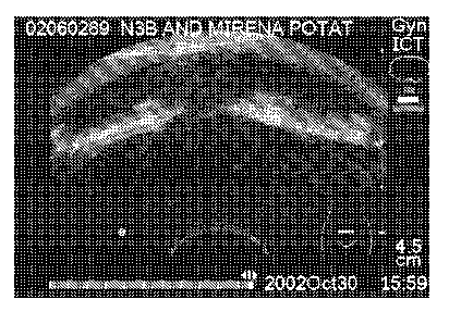
Figure 8. Acoustic shadowing behind the horizontal arms of MIRENA . Note tri-
ple shadowing from the thickest parts of horizontal arms. MIRENA is a
levonorgestrel-releasing intrauterine system (IUS), which consists of a
hormone-
elastomer capsule, mounted on a T-body and covered with an opaque tubing,
which
regulates the release of levonorgestrel.
Figure 9. Comparison of a glass microsphere modified 7-frame with a standard T-
frame in corn starch thickening by vaginal probe. The whole horizontal arm of
the 7-
frame is visible whereas only the three thickest parts of the T-frame and
their acous-
tic shadowing can be seen.
Figure 10. Spherical ends of Au-coated T-body (marked with arrows) were
located
from a sponge-water system.
Figure 11. A comparative picture about the brightness of Ag-rings on the
vertical
arm (transverse view, vaginal probe). A) Embedded single ring, B) Reference
(no
ring) C) Embedded double ring.
Figure 12. T-body design with positions for embedded metal rings at the ends
of a
vertical arm.
CA 02607079 2007-10-05
WO 2006/106180
PCT/F12006/050123
9
Figure 13. A schematic picture of different loop designs together with T-body
de-
sign for metals clips at the ends of a vertical arm.
Detailed description of the invention
Ultrasound visibility or echogenicity of an intrauterine device depends on the
density
difference of the adjacent materials, the propagation speed difference of
sound in the
adjacent materials, surface roughness, and the echogenicity of surrounding
materials.
The ultrasound visibility of different material modifications of IUS's can be
esti-
mated by evaluating the echogenicity of the material from the calculated
reflected
energies.
Sound travels through materials under the influence of sound pressure. Because
molecules or atoms are bound elastically to one another, the excess pressure
results
in a wave propagating through the solid. Acoustic impedance, Z (105 g/cm2s),
de-
termines the acoustic transmission and reflection at the boundary of adjacent
materi-
als:
Z = p = V
wherein p = density (g/cm) and V = propagation speed (mm/i.ts).
Reflected energy R, can be calculated from the acoustic impedances of adjacent
ma-
terials (Z1 and Z2):
Z
R _ ____________________________ ,
Z2 + Z1
CA 02607079 2007-10-05
WO 2006/106180
PCT/F12006/050123
For transmitted sound energy: T = 1 - R. With these formulas the ultrasound
visibil-
ity of different modifications of IUS can be estimated. The higher the
reflected en-
ergy, the better the echogenicity of the material.
5 In Table 1, the reflected and transmitted energies of various material
combinations
are compared.
Table 1. Comparison of different material combinations
Material 1 - Material 2 Reflected sound Transmitted
energy, R sound energy, T
Human tissue - Copper 0.860 0.140
Human tissue - MED 4735 tubing 0.032 0.996
Human tissue - PDMS 373 TW tubing 0.020 0.980
Human tissue - PE-LD 0.004 0.997
Human tissue - Glass (soda lime) 0.625 0.375
(PDMS = polydimethyl siloxane)
(PE-LD = low density polyethylene)
From Table 1 it can be seen that the copper wire of copper IUDs and glass
reflect
most of the sound energy back, thus providing good echogenicity and bright
picture.
Echogenicity of elastomers and the usual body raw material of an IUS (PE-LD
and
20-24% of BaSO4) is worse. Most of the sound energy is transmitted through the
material.
An intrauterine system according to the invention comprises at least one image
en-
hancing means for improving the ultrasound imaging of the system. The means
are
selected from the group consisting of
a) an inert metal coating on at least part of the body of the intrauterine
system,
b) at least one inert metal clip, pin, ring and/or sleeve fixedly positioned
on the body
of the intrauterine system, and
CA 02607079 2013-02-21
11
c) an inert metallic loop anchored to the vertical arm of the body of the
intrauterine system
in place of the usual loop.
The metal is advantageously selected so that the reflected energy at the
boundary of
adjacent materials is as high as possible. Preferably the metal is selected
from the group
consisting of inert metals, such as silver, gold, titanium, tungsten, bismuth,
platinum and
palladium. Preferred metals are silver, gold, titanium and platinum, which are
known to be
compatible (i.e. physically inert) with the human body. However, copper may
also be used.
In a preferred embodiment according to the invention, the metal coating or the
metal clips,
pins, rings or sleeves are located at the ends of the vertical arm(s) of the
IUS having the
shape of the letter T or 7. This enables a physician to reliably measure the
distance of IUS
from fundus. It is also possible to coat the "loop" at the end of the vertical
arm of the IUS,
or to fix a metal ring, pin or sleeve at the foot of the loop. In a further
preferred
embodiment, the metal coating or the metal clip, pin, ring or sleeve is
located only at the
"upper" end of the vertical arm of the IUS.
Sometimes it is also important to locate the position of horizontal arms of a
T-body. This
can be achieved by metal coating the whole T-body or by incorporating metal
clips, rings
or sleeves also to the end of horizontal arm(s) (before spherical ends) (Fig.
5).
Typically the thickness of the metal coating may vary from between about 0.1
nm and
about 500 nm, preferably between about 1 nm and about 50 nm. However, even
thicker
coatings of about 0.1 mm are possible.
The metal clips, pins, rings or sleeves may be unembedded or at least partly
embedded in
the body of an IUS. Partial embedding of the rings smooths the surface of the
IUS while
not yet impairing the visibility compared to unembedded counterpart. In case
of rings it is
advantageous to use double rings to enhance echogenicity. In case
CA 02607079 2007-10-05
WO 2006/106180
PCT/F12006/050123
12
of clips and sleeves, the broader the clip or sleeve, the better is the
visibility. The
width of the metal clip, pin, ring or sleeve may vary for example from 0.2 to
few
millimetres, being preferably about 1 mm, or in case of double rings about 0.5
mm A
further embodiment is to fix a metal pin of an appropriate size through the
loop, so
that the ends of the pin which are larger than the diameter of the loop are
visible.
The intrauterine system according to the invention may also have locking
means,
typically at least two locking parts, between which the medicated capsule is
mounted. The locking parts keep the capsule in the correct position during the
inser-
tion, use and removal of the IUS. Said locking parts may have different
shapes, e.g.
a shape of a truncated cone. They can be made of a polymeric material, which
can be
the same or different from the material of the body, but other materials can
also be
used, for example in this case an inert metal which improves visibility of the
IUS in
an ultrasound examination.
The intrauterine system according to the invention has been designed for a
relatively
long-term insertion into a uterine cavity. However, a long-term insertion may
vary
greatly, for example from a couple of weeks to several years, the maximum IUS
usage time being typically up to five years.
The invention is also directed to a method for improving the visualization of
an in-
trauterine system within the uterine cavity in an ultrasound examination,
comprising
at least one of the steps of
- applying an inert metal coating on at least part of the body of an IUS,
or
- providing the body of an IUS with at least one inert metal clip, pin, ring
and/or
sleeve, or
- anchoring a metallic loop to the vertical arm of the body of an IUS;
inserting the IUS into the uterine cavity and examining the position of the
IUS
within the uterine cavity in an ultrasound examination at an appropriate point
of
time.
CA 02607079 2007-10-05
WO 2006/106180
PCT/F12006/050123
13
Experimental
Experimental in vitro conditions:
= PE-container filled with water, corn starch thickening or potato starch
thicken-
ing
= Test specimen placed inside a sponge and the system immersed into water
Apparatus:
¨ Sonosite 180PLUS, with convex (2-4 MHz) and vaginal (4-7 MHz) probes
Or
¨ Aloka SSD 900, with convex (3.5 MHz) and vaginal (7.5 MHz) probes
Studied modifications:
= Group 1: Hollow glass microspheres have been incorporated in the raw
material
of the frames (bodies). Due to high density and entrapped air inside, the echo-
genicity should be improved.
= Group 2: Hollow glass microspheres have been incorporated in the hormone-
releasing core.
= Group 3: The whole T-body is Au-coated using Jeol Fine Coat ion sputter
JFC-
1100 equipment (1 kV voltage and 1 mA current for 20 minutes). The obtained
thickness of the Au-layer was few nanometers. See Figure 6.
= Group 4: Rings or double rings of 0.5 mm thick silver wire were
positioned ad-
jacent to the ends of the vertical arm of the T-body. Both embedded and unem-
bedded fixing was investigated with the currently available T-frames. A rough
embedding was made manually by scooping out a channel with depth of about
0.25 mm. See Figure 7.
Other in vitro conditions:
= Potato starch and corn starch thickenings behaved similarly in the
sonography.
CA 02607079 2007-10-05
WO 2006/106180
PCT/F12006/050123
14
= The scattering and attenuation of sound waves and the avoidable presence
of air
in the sponge system was so high that only NOVA T 380 (vertical arm) was lo-
cated. (NOVA T is a T-shaped plastic frame, which has a copper wire or a sil-
ver cored copper wire surrounding the vertical arm of the T.)
= Water as an in vitro medium was found worse than the other media due to
too
good echogenicity of studied specimens in water. No differences in
echogenicity
between the samples were detected. Sound wave proceeds easily through water
and no disturbing echoes are formed. Acoustic shadowing, the typical phenome-
non of IUD's and IUS's is very difficult to be detected in water as water is
seen
black in a sonograph. (In Figure 8 an example of the acoustic shadowing of
MIRENA in potato starch thickening is presented.)
Comparison of different modifications:
= Glass microspheres in T-frame improved echogenicity slightly. See Figure 9
where glass microsphere modified 7-frame and standard T-frame are compared
in corn starch thickening.
= Au-coating improved echogenicity of T-body. T-body is seen as a bright
image
under hormone releasing capsule. See Figure 3. Even in the sponge system
which was found to be very challenging in vitro medium, the spherical ends
were
located. See Figure 10.
= 0.5 mm thick Ag-wire placed on the upper and lower ends of the vertical
arm
enhanced the echogenicity. See Figure 4. Metal rings were seen as bright white
spots and their location during investigation was easy. Partial embedding of
the
rings did not impair the visibility compared to unembedded counterpart in any
projections. However, it was obvious that a double ring behaved better than a
single ring. The sonograph from double rings was larger and brighter. See a
comparative picture, Figure 11, where the ring, double-ring and no-ring have
been examined in optimal projection.