Note : Les descriptions sont présentées dans la langue officielle dans laquelle elles ont été soumises.
CA 02608352 2013-02-28
METHOD AND APPARATUS FOR SEQUENCED EXTRACTION FROM
ELECTROCARDIOGRAMIC WAVEFORMS
BACKGROUND OF ILLUSTRATIVE EMBODIMENTS OF THE INVENTION
[0002] The heart is a pump comprised of muscle tissue that responds to
electrical
stimulation. A heartbeat is a precisely controlled event that relies on
synchronization
between the atrial and ventricular chambers to maximize pumping efficiency.
The
sinoatrial node, which is located in the right atrium of the heart, generates
the
electrical stimulus. In a healthy person, the sinoatrial node normally
generates
electrical stimulus signals at a 60400 Hz rate, and the waves of myocardial
excitation
and contraction spread throughout the heart in well-defined manner. The
electrical
stimulus signals cause contractions in the heart's chambers, thereby pumping
blood
through the chambers. The left and right atria of the heart contract first and
for a brief
time, and then the left and right ventricles contract for a brief time. Normal
heart
rhythm is referred to as "sinus" rhythm, because it originates in the
sinoatrial node
(also referred to as the sinus node). The electrical stimulus signal output by
the
sinoatrial node is first sent to the left and right atria, then through the
atrioventricular
node and into the left and right ventricles.
[0003] An electrocardiogram (ECG) measures the heart's electrical activity.
Electrodes are placed at specific locations on the body to capture a tracing
of the
heart's electrical activity. The electrical activity resulting from heart
depolarization
1
CA 02608352 2007-11-13
WO 2006/124787
PCT/US2006/018755
and heart repolarization is recorded by each lead. The ECG is a summation of
the
information recorded from each lead. The captured ECG reflects the direction
of
electrical current flow, and the magnitude of the muscle that is depolarized.
Therefore, when the atria depolarize (and contract) the ECG tracing is smaller
as
compared to when the ventricles contract, since the atria are much smaller
than the
ventricles. Ventricle repolarization is in the same direction (positive) as
ventricle
depolarization. Although an ECG is positive during membrane depolarization and
negative during repolarization, the direction with respect to ventricles is
the same
since ventricles depolarize from the inside to the outside (endocardium to
epicardium), while repolarization occurs in the opposite direction.
[00041 Referring to FIG. 1, an ECG tracing is illustrated. The cardiac cycle
begins
with a P-wave, wherein the spontaneously firing cells in the sinoatrial node
reach a
threshold and generate action potentials. A wave of depolarization that
spreads to the
left and downward though left and right atria, which is labeled in FIG. 1 as
the "P
wave." The atria that were hyperpolarized suddenly become depolarized and the
ECG records a positive deflection. When the left and right atria become
depolarized,
the ECG returns to zero. The electrical current passes through the
atrioventricular
node, causing a delay of about one-tenth of a second, and due to the small
mass of the
atrioventricular node, the ECG tracing does not record any electrical
activity. When
the atrioventricular node is depolarized, it triggers depolarization of the
Purkinje
fibers. The Purkinje fibers spread the electrical current throughout the left
and right
ventricles, thereby causing depolarization across each ventricle
simultaneously. Since
the tissue mass of the Purkinje fibers is small, the ECG tracing does not
record any
electrical activity. The passing of the electrical current through the
atrioventricular
node and the Purkinje fibers is labeled in FIG. 1 as the "PR segment."
2
CA 02608352 2007-11-13
WO 2006/124787
PCT/US2006/018755
[0005] The depolarization of the left and right ventricles is referred to as
the "QRS
complex" and FIG. 1 is labeled as such. The QRS complex is quite large since
the
left and right ventricle tissue is large in comparison to the sinoatrial node.
The three
peaks are indicative of the manner in which the electrical current spreads
through the
left and right ventricles, i.e., from inside to outside, and are indicative of
the fact that
the tissue mass of the left ventricle is greater than the tissue mass of the
right
ventricle. The complete depolarization of the left and right ventricles
indicates that
the QRS complex has terminated.
[0006] Referring to FIG. 2, the points of the QRS complex are labeled. As
noted
above, the QRS complex is indicative of the depolarization of the left and
right
ventricles. The ventricular depolarization begins at a left side of the
intraventricular
septum and the peak of this depolarization is shown by the "Q" peak of the QRS
complex. The ventricular depolarization spreads from the endocardial surface
of the
left ventricle to the epicardial surface of the left ventricle, and is shown
by the "R"
peak of the QRS complex. The spread of the ventricular depolarization to the
right
ventricle is shown by the "S" peak of the QRS complex.
[0007] The segment labeled "T wave" indicates repolarization of the left and
right
ventricles. Although the left and right ventricles are repolarizing, the T
wave is
positive, since the heart repolarizes from outside to inside, the opposite
direction of
depolarization (inside to outside). The completion of the T wave signals the
end of
the cardiac cycle.
[0008] Referring to FIG. 3, the captured tracing of electrical activity is
printed out
on a paper tape or is presented on a display. Anomalies in an ECG are
indicative of
various heart-related conditions, such as ischemia, myocardial infarction,
conduction
disorder, electrolyte disturbance, pericarditis, valve disease or enlarged
heart. Certain
3
CA 02608352 2007-11-13
WO 2006/124787
PCT/US2006/018755
arrhythmias might occur only on an intermittent basis, or only if certain
psychological
or physical factors (i.e., stress, fatigue, etc.) are present. Since a typical
ECG tracing
is only a few minutes in length, arrhythmias of this type are difficult to
capture. A
more lengthy ECG tracing, referred to as a Holter monitor, will be used to
capture any
arrhythmias or other abnormal activity. The Holter monitor may record a
heart's
activity over a period of several days.
[0009] Referring to FIG. 1, one of the measured segments is referred to as the
QT
interval, and the QT interval indicates the duration of the electrical
activity that
controls contraction of the cells of the heart muscle. The QT interval
represents the
duration of ventricular depolarization and subsequent repolarization,
beginning at the
initiation of the Q wave of the QRS complex and ending where the T wave
returns to
isoelectric baseline. QT interval prolongation creates an electrophysiological
environment that favors the development of cardiac arrhythmias, most clearly
torsade
de pointes, but possibly other ventricular arrhythmias as well. Long QT
syndrome
identifies a condition wherein there exists an abnormally long QT interval on
the ECG
tracing. The term "congenital long QT" refers to a long QT interval that is
inherited.
The inherited form occurs due to irregularities in particular heart cell
proteins, and, of
course, these protein irregularities are caused by abnormalities in the genes
that
produce those proteins. The term "acquired long QT" refers to a long QT
interval that
is brought about by drugs or anomalous levels of the salts within blood, e.g.,
potassium and magnesium.
[0010] Although a person might have an unremarkable QT interval under normal
conditions, that person might develop a prolonged QT or suffer torsades de
pointes
(TdP) when taking certain medications. As shown in FIG. 4, TdP refers to the
characteristic appearance of the electrocardiogram indicative of a rhythm
abnormality,
4
CA 02608352 2007-11-13
WO 2006/124787
PCT/US2006/018755
and typically occurs in the setting of a prolonged QT interval on the
electrocardiogram. TdP is a polymorphic ventricular tachyarrhythmia that
manifests
on the ECG tracing as continuous twisting of the vector of the QRS complex
around
the isoelectric baseline. A feature of TdP is pronounced prolongation of the
QT
interval in the sinus beats preceding the arrhythmia. TdP can degenerate into
life-
threatening cardiac rhythms that can result in blackouts or sudden death.
Measurement of the QT interval on the ECG tracing is still the main method of
determining whether a person has long QT interval syndrome, whether inherited
or
acquired.
[0011] Non-antiarrhythmic drugs can have an undesirable side effect of causing
delayed cardiac repolarization. Due to its relationship to heart rate, the QT
interval is
normalized into a heart rate independent "corrected" value known as the QT e
interval,
which represents the QT interval at a standardized heart rate (essentially the
QT
interval at a heart rate of 60 bpm). Several drugs that have caused TdP
clearly
increase both the absolute QT and the QT.
SUMMARY OF ILLUSTRATIVE EMBODIMENTS OF THE INVENTION
[0012] Illustrative, non-limiting embodiments of the present invention
overcome
various disadvantages. In addition, the present invention is not required to
overcome
these disadvantages, and an illustrative, non-limiting embodiment of the
present
invention may not overcome any disadvantages.
[0013] Illustrative, non-limiting embodiments of the present invention provide
apparatuses and methods for automatically extracting a number of short
segments of
data from a single long ECG recording based on settings defined by a protocol
associated with a medical study, and transmitting the data to a central
location.
CA 02608352 2014-03-12
(0014] One aspect of an embodiment provides an extraction template that
specifies
times and amounts of data to extract relative to drug dosing time or time
points
specified in the study protocol. Different protocols may have different
specific
extraction templates which apply across all subjects and all days in the
particular
studies. As an example, an extraction template may be set to extract a few ten-
second
segments of an ECG, spaced apart by two to three minutes, every thirty to
sixty
minutes over the duration of the ECG. These values are merely examples, and
the
extraction template is completely configurable by the user.
[0015] Another aspect of an embodiment chooses ECG segments to extract in an
intelligent manner. The presence of signal artifacts and other specified
conditions in
the ECG segments are checked, and, if present, those ECG segments are avoided.
To
avoid extracting corrupted data, the extraction template may be automatically
adjusted
by a limited amount to extract data close to the desired extraction times.
Extracted
ECG segments may be exported to separate data files.
[0016] A further aspect of an embodiment provides a remote mode which extracts
sections of ECG data at the point of capture and transmits the extracted data
to a
central location at a higher priority. In the remote mode, artifact detection
and
avoidance may be suspended, and the extraction template may be widened to
include
segments of data on either side of the requested extraction times.
[0017] A further aspect of an embodiment provides a method of extracting ECG
data segments that includes specifying the parameters of the configurable
extraction
template, specifying data files from which to extract data, intelligently
extracting the
data, and writing the extracted data segments to separate data files. The
method may
also include transmitting the data files to a central location.
6
CA 02608352 2016-08-05
A further aspect of an embodiment provides a method for extracting segments
from an electrocardiogram tracing, the method comprising: acquiring an
electrocardiogram tracing; selecting the electrocardiogram tracing for segment
extraction;
associating a predetermined time with the electrocardiogram tracing to align
an extraction
template within the electrocardiogram tracing for segment extraction; scanning
the
electrocardiogram tracing for artifacts and annotating the electrocardiogram
tracing if any
artifacts are discovered; if artifacts are present in the segment designated
for extraction
by the extraction template, modifying the extraction template to avoid the
artifacts,
wherein the modification of the extraction template comprises shifting the
extraction
template to avoid extracting the segment containing the artifacts therein,
and, if unable to
modify the extraction template, annotating the electrocardiogram tracing as
unextractable; and if artifacts are not present in the segment designated for
extraction by
the extraction template or the extraction template was successfully modified,
extracting
the designated segment from the electrocardiogram tracing and writing the
extracted
segment to a storage medium.
A further aspect of an embodiment provides a computer program product
comprising: a tangible computer-readable medium; and computer executable code
embodied on the computer-readable medium that causes a computer to perform
predetermined operations, wherein the predetermined operations comprise:
selecting an
electrocardiogram tracing for segment extraction; associating a predetermined
time with
the electrocardiogram tracing to align an extraction template within the
electrocardiogram
tracing for segment extraction; scanning the electrocardiogram tracing for
artifacts and
annotating the electrocardiogram tracing if any artifacts are discovered; if
artifacts are
present in the segment designated for extraction by the extraction template,
modifying the
extraction template to avoid the artifacts, wherein the modification of the
extraction
template comprises shifting the extraction template to avoid extracting the
segment
containing the artifacts therein, and, if unable to modify the extraction
template,
annotating the electrocardiogram tracing as unextractable; and if artifacts
are not present
in the segment designated for extraction by the extraction template or the
extraction
template was successfully modified, extracting the designated segment from the
electrocardiogram tracing and writing the extracted segment to a storage
medium.
[0018] Additional aspects and advantages of the embodiments will be set forth
in
6a
CA 02608352 2016-08-05
part in the description that follows or may be learned by practice of the
embodiments. The
aspects and advantages of the embodiments may be realized and attained by
means of the
instrumentalities and combinations particularly pointed out in the appended
claims. Also, one of
ordinary skill in the art may learn other aspects by performing routine
experimentation after
reviewing the application.
BRIEF DESCRIPTION OF THE DRAWINGS
[0019] The above and other features and advantages will become more
apparent by describing
in detail exemplary embodiments. The accompanying drawings, which are
incorporated in and
constitute a part of this specification, illustrate the exemplary embodiments
and, together with
the description, serve to explain various aspects, advantages and principles.
In the drawings:
[0020] FIG. 1 is an illustration of an ECG tracing that identifies the
various segments of an
electrical profile of a normal heartbeat as known from prior art;
[0021] FIG. 2 is an illustration of an ECG tracing that identifies the
various peaks of an
electrical profile of a normal heartbeat as known from prior art;
[0022] FIG. 3 is an illustration of the output from a 12-lead Hotter
monitoring device as
known from prior art;
100231 FIG. 4 is an illustration of ECG tracing showing torsades de
pointes;
[0024] FIG. 5 is an illustration of an exemplary, non-limiting example of a
computer system
for extracting segments from an electrocardiogram tracing for analysis;
[0025] FIG. 6 is a exemplary flowchart illustrating a method for processing
electrocardiogram
tracings for segment extraction;
[0026] FIG. 7 is an exemplary flowchart illustrating the initialization
and/or reconfiguring of
control settings for a computer system implementing a method for
7
CA 02608352 2007-11-13
WO 2006/124787
PCT/US2006/018755
processing electrocardiogram tracings for segment extraction;
[0027] FIG. 8 is an exemplary flowchart illustrating a selection process for
segment
extraction from electrocardiogram tracings;
[0028] FIG. 9 is an exemplary flowchart illustrating pre-extraction processes
of
annotation data track creation and scanning of an electrocardiogram tracing
for
artifacts;
[0029] FIG. 10A is an exemplary flowchart illustrating the modification of
extraction templates to avoid artifact areas in an electrocardiogram tracing;
and
[0030] FIG. 10B is an exemplary flowchart illustrating the modification of
extraction templates while operating in remote mode.
DETAILED DESCRIPTION OF THE ILLUSTRATIVE, NON-LIMITING
EMBODIMENTS OF THE INVENTION
[0031] Illustrative, non-limiting embodiments of the invention will now be
described more fully with reference to the accompanying drawings. A general
example of a computer that can be used in accordance with the described
embodiment
will be described below.
[0032] The computer comprises one or more processors or processing units, a
system memory and a bus that couples various system components comprising the
system memory to the processors. The bus can be one or more of any of several
types
of bus structures, comprising a memory bus or memory controller, a peripheral
bus,
an accelerated graphics port and a processor or local bus using any of a
variety of bus
architectures. The system memory comprises read only memory (ROM) and random
access memory. A basic input/output system (BIOS) containing the routines that
help
to transfer information between elements within the computer, such as during
boot up,
is stored in the ROM or in a separate memory.
8
CA 02608352 2007-11-13
WO 2006/124787
PCT/US2006/018755
[0033] The computer further comprises a hard drive for reading from and
writing to
one or more hard disks (not shown). Some computers comprise a magnetic disk
drive
for reading from and writing to a removable magnetic disk and/or an optical
disk
drive for reading from or writing to a removable optical disk, such as a CD
ROM or
other optical media. The hard drive, the magnetic disk drive and the optical
disk drive
are connected to the bus by an appropriate interface. The drives and their
associated
computer-readable media provide nonvolatile storage of computer-readable
instructions, data structures, program modules and other data for the
computer.
Although the exemplary environment described herein employs a hard disk, a
removable magnetic disk and a removable optical disk, it should be appreciated
by
those skilled in the art that other types of computer-readable media which can
store
data that is accessible by a computer, for example, but not limited to,
magnetic
cassettes, flash memory cards, digital video disks, random access memories
(RAM),
read only memories (ROM), etc., may also be used in the exemplary operating
environment.
[0034] A number of program modules may be stored on the hard disk, magnetic
disk, optical disk, ROM or RAM, comprising an operating system, at least one
or
more application programs, other program modules and program data. In some
computers, a user might enter commands and information into the computer
through
input devices such as a keyboard and a pointing device. Other input devices
(not
shown) may comprise a microphone, a joystick, a game pad, a satellite dish
and/or a
scanner. In some instances, however, a computer might not have these types of
input
devices. These and other input devices are connected to the processing unit
through
an interface coupled to the bus. In some computers, a monitor or other type of
display
device might also connected to the bus via an interface, such as a video
adapter.
9
CA 02608352 2007-11-13
WO 2006/124787
PCT/US2006/018755
Some computers, however, do not have these types of display devices. In
addition to
the monitor, the computers might comprise other peripheral output devices (not
shown) such as speakers and printers.
[0035] The computer can, but need not, operate in a networked environment
using
logical connections to one or more remote terminals. The remote terminal may
be,
but is not limited to, another personal computer, a server, a router, a
network PC, a
peer device or other common network node, and typically comprises many or all
of
the elements described above relative to the computer. The logical connections
to the
computer may comprise a local area network (LAN) and a wide area network
(WAN).
Such networking environments are commonplace in offices, enterprise-wide
computer
networks, intranets, and the Internet.
[0036] When used in a LAN networking environment, the computer is connected to
the local network through a network interface or adapter. When used in a WAN
networking environment, the computer typically comprises a modem or other
means
for establishing communications over the wide area network, such as the
Internet.
The modem, which may be internal or external, is connected to the bus via a
serial
port interface. In a networked environment, program modules depicted relative
to the
computer, or portions thereof, may be stored in the remote memory storage
device. It
will be appreciated that the network connections shown are exemplary and other
means of establishing a communications link between the computers may be used.
[0037] Generally, the data processors of the computer are programmed by means
of
instructions stored at different times in the various computer-readable
storage media
of the computer. Programs and operating systems are typically distributed, for
example, on floppy disks or CD-ROMs. From there, they are installed or loaded
into
the secondary memory of the computer. At execution, they are loaded at least
CA 02608352 2007-11-13
WO 2006/124787
PCT/US2006/018755
partially into the computer's primary electronic memory. The embodiments
described
herein may comprise these and other various types of computer-readable storage
media when such media contain instructions or programs for implementing the
steps
described below in conjunction with a microprocessor or other data processor.
The
embodiments also may comprise the computer itself when programmed according to
the methods and techniques described below.
[0038] Referring to FIG. 5, an illustrative, non-limiting embodiment of the
invention includes a computer that comprises a processor 50, user interfaces
51, and
local storage 54. As described above, the processor 50 may comprise one or
more
processors, and the user interfaces 51 may comprise monitors, keyboards, mice,
touch-screens, etc. The processor 50 is connected to the local storage 54 via
a bus (or
busses) as described above, and the local storage 54 itself may comprise
various types
of disk memory, electronic memory (i.e., RAM, ROM), or various combinations
thereof. The processor 50 may also access the remote storage 53, which itself
may
comprise various types of data storage machines and/or server machines. The
Holter
recording file 52, also referred to as an electrocardiogram tracing, is stored
in either
the remote storage 53 or the local storage 54, and the processor 50 accesses
the Holter
recording file 52 therefrom.
[0039] In one implementation, the computer performs a method for assisting
cardiologists in evaluating electrocardiogram (ECG) tracings. The computer
acts in a
similar fashion to a relatively inexperienced cardiologist who is assisting an
expert
cardiologist in interpreting captured ECG tracings. Aspects of the embodiment
allow
the identification of artifacts in the ECG tracing and allow for tentative
interpretations
of the ECG tracing. Another aspect of the embodiment is the computer's ability
to
compare several ECG waveforms and group them accordingly, taking into account
all
11
CA 02608352 2007-11-13
WO 2006/124787
PCT/US2006/018755
presently known information about the waveforms. For instance, if a
cardiologist has
marked one waveform as normal and another as abnormal, the computer
understands
that both waveforms cannot be members of the same group, even if they do look
similar. In addition, if a cardiologist makes changes to a waveform's
interpretation,
the computer will regroup the remaining waveforms as needed.
[0040] As is known, ECG tracings are stored in a variety of different file
formats,
such as FDA XML, Mortara XML as exported from E-Scribe, and GE MUSE . In
one embodiment, the computer may contain conversion libraries to facilitate
the
conversion of an ECG tracing stored in one of these formats. The conversion
libraries
allow the computer to process an ECG tracing, without having to worry about
the
specific format, sample rate, length of recording or other details. Using the
conversion libraries, the exemplary embodiments of the present invention
operate
independently of the data file size, format, sample rate, bit depth and scale
factor.
Typically, each Holter recording file (i.e., an ECG tracing) will contain 24
or 48 hours
of 12-lead data at 1k samples per second. An exemplary embodiment of the
present
invention will be able to handle a Holter recording file of at least 48 hours
x 12 leads
x 1k samples per second. Also, embodiments do not have any intrinsic
limitations
that prevent the handling of longer recordings or recordings taken at higher
and/or
lower sampling rates.
[0041] Although there is no set time limit on the length of an ECG tracing,
the
typical length of an ECG tracing is about ten seconds. In one implementation,
the
computer in the present embodiment provides a configurable time limiting
function
that will truncate ECG tracings that are longer than the configured time
limit. Also,
the computer may provide a default time limit of ten seconds.
[0042] In one embodiment, the computer extracts a number of short segments
from
12
CA 02608352 2007-11-13
WO 2006/124787
PCT/US2006/018755
a single long ECG tracing, based on settings defined by a study's protocol.
The
computer may also use an extraction template, which specifies times and
amounts of
data to extract, relative to drug dosing time. Each protocol has a specific
template,
which applies across all subjects and all days in the study.
[0043] In an exemplary operation, the computer may extract, for example, about
six
to eighteen ten-second segments that equal approximately two to three minutes
out of
every thirty to sixty minutes of an ECG tracing. These are typical values, and
the
extraction template is completely configurable to accommodate drug study
protocols.
The computer may also choose the ECG tracing segments to extract by checking
for
the presence of signal artifacts and other defined conditions, and avoid
extracting the
affected ECG tracing segments. When such conditions in the ECG tracing are
detected, the extraction template is adjusted by a limited amount in order to
avoid
extracting corrupted data from the ECG tracing. For example, ECG traces are
usually
extracted from a twenty-four-hour Holter recording in triplicate in ten-second
time
slices. When an artifact or other defined condition is detected, the
extraction template
is moved to the right or to the left to find ECG traces at the same heart rate
within a
tolerance of one minute, thereby avoiding the artifact or other defined
condition that
would otherwise result in the extraction of corrupted data. The net result is
that the
computer of the present embodiment extracts clean ECG tracing segments that
are as
close as possible to the desired extraction times. Once the ECG tracing
segments are
chosen and extracted, the computer automatically exports each individual ECG
tracing segment to its own separate file.
[0044] The computer may also export the selected ECG tracing segments to a
single
"sparse" ECG file, i.e., a file that contains multiple non-continuous ECG
tracing
segments. The sparse file is easier to transmit and store than a group of
files,
13
CA 02608352 2007-11-13
WO 2006/124787
PCT/US2006/018755
administrative tasks are reduced, and metadata in the file remain unaltered
across all
of the extracted ECG tracing segments.
[0045] In an exemplary embodiment of the present invention, a user (such as a
cardiovascular technician) interfaces with a computer system through the
computer
interfaces described earlier.
[0046] Prior to extracting any ECG tracing segments from a Holter recording
file,
the user may first configure the control settings. In one implementation, the
computer
system will attempt to fetch and cache the latest control settings from one or
more
servers that serve as a central repository for control settings. If the server
(or servers)
is not reachable or accessible for some reason, the computer system may use
the last
cached control settings.
[0047] The control settings comprise a list of various parameters that are set
to
manage the extraction of the ECG tracing segments. For extraction templates,
the
control settings change the limits of the extraction template to avoid
artifacts in the
ECG tracing. For example, the control settings may change the number of
seconds to
shift the extraction window and the direction in which to shift the extraction
window.
For annotations to the ECG tracing, the control settings determine the type of
annotation channel or channels to create. For example, the control settings
can create
an artifact channel to be used when scanning the ECG tracing. Alternatively,
the
control settings can be entered, cached and saved locally for processing
Holter
recording files. Other types of annotations are possible as well, for example,
but not
limited to, significant variations in heart rate across ECUs when ECGs are
taken in
multiple replicates, poor lead attachment and impedance problems. Annotations
may
also be entered manually using selectable lists, for example, drop-down lists,
and free
text.
14
CA 02608352 2007-11-13
WO 2006/124787
PCT/US2006/018755
[0048] With respect to the extraction of ECG tracings, the control settings
can
determine what types of data are extracted from the annotations. For example,
the
control settings might command that only artifact data be extracted from the
annotation channel, and all other data in the annotation channel be ignored.
The
artifact detection settings are set via control settings as well. Artifact
detection
parameters such as jagged or wandering baseline and thickness of baseline are
indicators of artifacts that can be managed via the control settings. Pixel
movement
can be used to determine jaggedness of baseline. In addition, the control
settings can
be used to manage the detection of other types of conditions as well, for
example, data
file output format.
[0049] The control settings can also be used to manage the input of ECG
tracing
files for processing. For example, the control settings can control the
operational
mode of the file input, such as single file input, batch file input, automatic
scanning of
directories or integration/control with an automatic workflow control system.
The
control settings can be used to point to a particular file location,
repository, server
and/or remote storage system. The control settings can also be used to manage
the
types of input file formats that are acceptable for processing.
[0050] With respect to output of extracted ECG tracing segments, the control
settings can be used to set a location, repository, server and/or remote
storage system
for output files. The control settings can also be used to manage the type of
format
used for output file generation, and for output file integration/control with
an
automatic workflow control system.
[0051] In an exemplary embodiment of the present invention, updated control
settings can be fetched from a central source, which may be a database or
other such
data storage entity. If a central source for control storage cannot be
accessed and/or
CA 02608352 2007-11-13
WO 2006/124787
PCT/US2006/018755
located, the local copy of the most recent control settings may be used. The
active
control settings for each ECG analysis are archived for audit trail purposes.
[0052] In an exemplary embodiment of the invention, when allowed by a
particular
output file format, each extracted ECG tracing is tagged with metadata that
points
back to the control settings used to extract that ECG tracing.
[0053] After the control settings have been established, the user next
commands the
computer system to begin processing Holter recording files to extract the
desired ECG
tracing segments. The user has several options for commanding the computer
system
to process the Holter recording files, and these options can be configured via
control
settings. The simplest option is for the user to enter the filename of the
desired Holter
recording file on a command line. In one implementation, the computer system
processes only the Holter recording file, and no other filenames. If batch
processing
is commanded by the control settings, the user enters a batch file that lists
the
filenames of one or more Holter recording files, and the computer system
processes
these files in serial fashion. Another alternative is for the computer system
to receive
Holter recording files to process as commanded by an external workflow control
system. The computer system would process each Holter recording file as
commanded by the external workflow control system, and return the results
thereto.
Finally, if directory scan is commanded via the control settings, the computer
system
automatically and repeatedly scans a particular directory and/or directories
for new
Holter recording files. Once a new Holter recording file has appeared in a
targeted
directory, the computer system performs the ECG tracing segment extraction on
the
new Holter recording file.
[0054] In an exemplary embodiment of the invention, the dosing time associated
with a particular Holter recording file is recorded. Typically, a
cardiovascular
16
CA 02608352 2007-11-13
WO 2006/124787
PCT/US2006/018755
technician performs this step. The dosing time value is an actual hour/minute
time,
expressed in the local time. The extraction template is then positioned within
the time
window of the whole Holter recording file, based on the dosing time.
[0055] Once a Holter recording file or files have been selected for
processing, the
computer system scans the Holter recording file for artifacts and other
conditions as
specified in the control settings. One or more annotation streams that contain
metadata concerning the ECG tracing are added "in parallel" with the existing
data in
the Holter recording file. If the extraction of the ECG tracing segments must
rely on
annotations previously generated by the capture of the Holter recording file,
the
computer system verifies that the necessary annotations exist. If the required
annotations are non-existent in the Holter recording file, the computer system
rejects
the file and the user is notified.
[0056] Prior to actually extracting ECG tracing segments from the Holter
recording
file, the whole ECG tracing is analyzed, and at least one metadata/annotation
channel
is generated (if required by the control settings) that is specifically
intended to help
guide the extraction process. This metadata/annotation channel is similar to
other
types of annotations, such as those indicating patient activity or symptoms.
The
metadata/annotation channel contains information about the presence or absence
of
conditions that affect the extraction process. Like other running annotation
channels
or tracks, this channel describes the ECG tracing waveform on a moment-by-
moment
basis. Each annotation comprises a start time, an end time and other
information,
such as a Holter lead number. The annotation channel also comprises a
description of
the condition seen, in a machine-readable format and optionally comprises an
equivalent human readable description. A typical annotation message is as
follows:
"Artifact present at time point X, for Y seconds, on lead III." This
annotation
17
CA 02608352 2007-11-13
WO 2006/124787
PCT/US2006/018755
message could be encoded in any appropriate machine or human readable format
that
can be stored in a file, but the underlying message is independent of the
formatting
and/or representation.
[0057] The scanning and annotation of the ECG tracing is managed via control
settings that determine the subject matter to be scanned. The purpose of the
scanning
is to scan for conditions that have some impact on which ECG tracing segments
can
or cannot be extracted. Typically, the Holter recording file is scanned for
artifacts or
other conditions (based on control settings) that would prevent a given time
section of
the ECG tracing from being extracted. The control settings can also control
the
scanning to search for other user-defined conditions that might influence
extraction of
the ECG tracing segments. For example, an annotation track that recorded RR
intervals (FIG. 1) could be used to allow and/or disallow extraction based on
heart
rate. A patient symptoms track could be used to tailor the extraction to only
those
ECG tracing segments where certain symptoms were present. Multiple annotation
tracks could also be examined to decide which ECG tracing segments to extract.
Annotation tracks from different types of file formats also can be scanned, as
file
format converter tools become available.
[0058] After the annotation process, the computer system scans the Holter
recording
file and extracts the desired ECG tracing segments that are specified in the
extraction
template. The computer system uses the annotation track or tracks to avoid
corrupted
portions of the Holter recording file and to seek out the portions of the
Holter
recording file that are requested by the extraction template.
[0059] The extraction template specifies what ECG tracing segments should be
extracted relative to the timing of the drug dosing of the patient. In
addition,
information concerning the desired output format and the type and sensitivity
of
18
CA 02608352 2007-11-13
WO 2006/124787
PCT/US2006/018755
artifact detection may be specified in the template as well. In one
implementation, the
extraction template applies to all Holter recording files for a given drug
test protocol,
so that the all ECG tracings in that drug test protocol are subjected to the
same
extraction template. However, the extraction template may be overridden on a
per-
subject, per-cohort and/or per-day basis, as necessary. Enabling the
extraction
template to be overridden allows different settings to be used for each day,
each
cohort and/or each different subject.
[0060] Exemplary embodiments of the present invention may also support a
remote
mode that extracts important ECG segments from a full Holter recording file,
and
those extracted segments are transmitted to a cardiologist at a higher
priority than the
remainder of the Holter recording file. The remote mode does not attempt to
detect
and/or avoid artifacts in the Holter recording file, and the remote mode
expands the
extraction template to include the ECG tracing on either side of the specific
ECG
tracing requested by the extraction template. When the extracted data is
received by
the cardiologist, the extracted ECG tracing segments are reprocessed to remove
the
ECG tracing that was not requested by the extraction template, as well as
remove any
ECG tracing segments affected by artifacts.
[0061] When the remote mode is engaged, no artifact detection is performed on
the
ECG tracing, and the capture parameters of the extraction template are
expanded so
that ECG tracing on either side of any time segment specified in the
extraction
template is extracted as well. This expanded "window" is configurable, and
might be
from one to five minutes on either or both sides of the specified time
segment. The
widened extraction template compensates for the possibility that noise or
artifacts do
exist in the extracted ECG tracing segment.
[0062] Once the computer system has located one or more ECG tracing segments
to
19
CA 02608352 2007-11-13
WO 2006/124787
PCT/US2006/018755
extract from the Holter recording file, the ECG tracing segments are
extracted, and
one output file per extracted ECG tracing segment is generated. Each ECG
tracing
segment output file is tagged with relevant metadata from the Holter recording
file, as
well as an identifier that describes the control settings used for the
extraction. The
inclusion of the control settings is for audit trail purposes. An ECG tracing
within a
Holter recording file that has been previously marked as unextractable is
ignored
during the extraction process.
[0063] Exemplary embodiments of present invention also export the selected ECG
tracing segments to a single "sparse" ECG file, i.e., a file that contains
multiple non-
continuous ECG tracing segments. The sparse file is easier to transmit and
store than
a group of files, administrative tasks are reduced, and metadata in the file
remains
unaltered across all of the extracted ECG tracing segments.
[0064] With respect to the annotations, based on the control settings, the
computer
system may delete from the ECG tracing segment output file whatever
annotations it
created. If no other application needs the annotations, they just waste space
and
should be deleted from the ECG tracing segment output file.
[0065] As noted above, the computer system can be controlled by an external
workflow system. The user of the computer system can configure the control
settings
such that the computer system signals the external workflow system that a
Holter
recording file has been processed, and that the results are available. If
there were any
errors, the computer system sends a message to the external workflow system
instead.
In this manner, a plurality of Holter recording files can be processed on a
computer
system during periods of low system use.
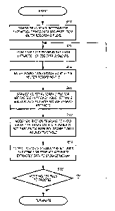
[0066] FIG. 6 shows a flowchart illustrating a non-limiting method for
processing
electrocardiogram tracings for segment extraction. At S100, prior to the
extraction of
CA 02608352 2007-11-13
WO 2006/124787
PCT/US2006/018755
ECG tracing segments, the user first configures the control settings. In an
exemplary
embodiment, the computer system attempts to fetch and cache the latest control
settings from one or more servers that serve as a central repository for
control
settings. If the server or servers are not reachable or accessible for some
reason, the
computer system uses its last cached control settings. Alternatively, the user
can
configure the control settings locally at the computer system.
[0067] At S200, after the control settings have been established, Holter
recording
files are selected in order to extract the desired ECG tracing segments. The
filename
of the desired Holter recording file can be entered on a command line, a batch
file that
lists the filenames of one or more Holter recording files for batch processing
can be
entered, Holter recording files for processing can be delivered by an external
workflow control system, or directory scan techniques can be used.
[0068] Subsequent to the selection of Holter recording files for processing,
at S300,
a dosing time associated with a particular Holter recording file or files is
entered.
Typically, a cardiovascular technician perfoims the entry of the dosing time
relative
to a Holter recording file or files.
[0069] At S400, once a Holter recording file or files have been selected for
processing, the Holter recording file is scanned for artifacts and other
conditions as
specified in the control settings. Prior to actually extracting ECG tracing
segments
from the Holter recording file, the whole ECG tracing is analyzed, and at
least one
metadata/annotation channel is generated (if required by the control settings)
that is
specifically intended to help guide the extraction process. This
metadata/annotation
channel is similar to other types of annotation, such as those indicating
patient activity
or symptoms. The metadata/annotation channel contains information about the
presence or absence of conditions that affect the extraction process. As
discussed
21
CA 02608352 2007-11-13
WO 2006/124787
PCT/US2006/018755
with reference to FIG. 9, if the remote mode of extracting ECG tracing
segments is
engaged, the scanning of a Holter recording file is not performed.
[0070] At S500, the extraction template is modified to avoid areas with
artifacts.
The extraction template is modified at little as possible, and each segment to
be
extracted within the extraction template is moved to avoid areas with
artifacts. If a
segment to be extracted is moved past a predetermined threshold, then the
region is
marked as unextractable. As discussed with reference to FIG. 10B, if the
remote
mode of extracting ECG tracing segments is engaged, the modification of the
extraction template is performed in a different manner.
[0071] At S600, one or more ECG tracing segments to be extracted from the
Holter
recording file are located, the ECG tracing segments are extracted, and one
output file
per extracted ECG tracing segment is generated. Each ECG tracing segment
output
file is tagged with relevant metadata from the Holter recording file input
file, as well
as an identifier that describes the control settings used for the extraction.
[0072] At S700, the process determines whether or not any more Holter files
need to
be processed. If so, the process returns to S200, and if not, the process
terminates.
[0073] FIG. 7 is a flowchart illustrating a detailed, non-limiting example of
the
process of S100 in FIG. 6 in which control settings are configured. Although
the
flowchart in FIG. 7 and other figures illustrate a particular sequence of
operations, the
sequence of at least some of the operations can be changed by design or
modified by a
user.
[0074] As noted above, the control settings may be retrieved from one or more
servers that serve as a central repository for control settings and cached
locally. If the
server or servers are not reachable or accessible for some reason, the last
cached
control settings can be used. Alternatively, the control settings can be
entered locally
22
CA 02608352 2007-11-13
WO 2006/124787
PCT/US2006/018755
for a particular Holter recording file if necessary or for the sake of
convenience.
[0075] At S110, the control settings for managing changes to the extraction
template
parameters to avoid identified artifacts in the ECG tracing are entered. For
example, a
baseline amplitude of less than or equal to seven millivolts (mV) is
considered a
normal baseline amplitude. The control settings may set a detection amplitude
of "n"
mV, where "n" is a number greater than seven, to identify a baseline amplitude
artifact. Further, a baseline movement tolerance, measured in pixels, may be
set.
[0076] At S120, the control settings for operating in a remote or local mode
are
entered. As discussed above, the local/remote setting has an effect on whether
a
Holter recording file is scanned for artifacts and pre-existing annotations,
as well as
the manner in which an extraction template may be modified in order to avoid
identified artifacts within the Holter recording file.
[0077] At S130, the control settings for determining the type of annotation
channel
or channels to create are entered. The control settings can create an artifact
channel to
be used when scanning the ECG tracing. Other types of annotations are possible
as
well, for example, variations in heart rate, poor lead attachment and
impedance
problems. With respect to the extraction of ECG tracing segments, the control
settings can determine what types of data is extracted from the annotations.
For
example, the control settings might require only artifact data to be extracted
from the
annotation channel, and all other data in the annotation channel, for example,
variations in heart rate and impedance problems, be ignored.
[0078] At S140, the control settings for artifact detection settings are
entered. For
example, artifact detection parameters such as jagged or wandering baseline,
and
thickness of baseline are entered. In addition, the artifact detection control
settings
can be used to manage the detection of other types of conditions as well, for
example,
23
CA 02608352 2007-11-13
WO 2006/124787
PCT/US2006/018755
significant variations in heart rate across ECGs when ECGs are taken in
multiple
replicates, poor lead attachment and impedance problems.
[0079] At S150, the control settings to manage the input of ECG tracing files
for
processing are entered. The control settings manage the operational mode of
the file
input, such as single file input, batch file input, automatic scanning of
directories or
integration/control with an automatic workflow control system. The control
settings
can point to a particular file location, repository, server and/or remote
storage system.
In addition, the control settings manage the types of input file formats that
are
acceptable for processing.
[0080] At S160, the control settings for outputting the extracted ECG tracing
segments are entered. The control settings set a location, a repository, a
server and/or
a remote storage system for the output files. The control settings manage the
type of
format used for output file generation, and for output file
integration/control with an
automatic workflow control system. When allowed by a particular output file
format
commanded by the control settings, each extracted ECG tracing is tagged with
metadata that points back to the control settings used to extract that ECG
tracing.
[0081] At S170, the control settings for the extraction template are entered,
for
example, times and amounts of data to extract relative to drug dosing time.
[0082] In an exemplary embodiment of the present invention, updated control
information can be fetched from a central source, which may be a database or
other
such data storage entity. If a central source for control storage cannot be
accessed
and/or located, the local copy of the most recent control setting is used. The
active
control settings for each ECG analysis are archived for audit trail purposes.
[0083] FIG. 8 is a flowchart illustrating a detailed, non-limiting example of
the
process of S200 of FIG. 6 in which Holter recording files are selected.
24
CA 02608352 2007-11-13
WO 2006/124787
PCT/US2006/018755
[0084] At S210, if the control setting for processing a single Holter
recording file is
active, then at S220, the filename of the desired Holter recording filenames
is entered,
and only that Holter recording filename is processed. Otherwise, at S230, if
the
control setting for processing a batch of Holter recording files is active,
then at S240,
a batch file that lists the filenames of one or more Holter recording files is
entered,
and the listed Holter recording files are processed in serial fashion.
Otherwise, at
S250, if the control setting for directory scan processing of Holter recording
files is
active, then at S260, a particular directory or directories are scanned for
new Holter
recording files. Once a new Holter recording file has appeared in a targeted
directory,
the ECG tracing segment extraction process is performed on the newly deposited
Holter recording file. Otherwise, at S270, if the control setting for
automated
processing under an external workflow control system is active, then at S280,
Holter
recording files are processed as commanded by the external workflow control
system,
and the results are returned thereto.
[0085] FIG. 9 is a flowchart illustrating a detailed, non-limiting example of
the
process of S400 of FIG. 6 in which the Holter recording files are scanned for
artifacts
and annotated.
[0086] If the control setting for the remote mode is active (S405: Yes), then
the
Holter recording file is not scanned for artifacts and is not
annotated.Otherwise (S405:
No), if the extraction of the ECG tracing segments need to rely on annotations
previously generated by the capture of the Holter recording file (S410: Yes),
verification of whether the necessary annotations exist takes place at S420.
During
capture, one or more annotation streams that contain metadata concerning the
ECG
tracing are added "in parallel" with the existing data in the Holter recording
file. If
the required annotations are non-existent in the Holter recording file (S420:
No), the
CA 02608352 2007-11-13
WO 2006/124787
PCT/US2006/018755
extraction of ECG tracing segments is not performed.
[0087] If the extraction of the ECG tracing segments does not rely on
previously
generated annotations (S410: No) or such annotations are present in the Holter
recording file (S420: Yes), at least one metadata/annotation channel is
generated (if
required by the control settings) (S430). This metadata/annotation channel is
specifically intended to help guide the extraction process and is similar to
other types
of annotation, such as those indicating patient activity or symptoms. The
metadata/annotation channel contains information about the presence or absence
of
conditions that affect the extraction process. Like other running annotation
channels
or tracks, this channel describes the ECG tracing waveform on a moment-by-
moment
basis. Each annotation comprises a start time, an end time and other
information,
such as a Holter lead number. The annotation channel also comprises a
description of
the condition seen, in a machine-readable format, and optionally comprising an
equivalent human readable description. A typical annotation message is as
follows:
"Artifact present at time point X, for Y seconds, on lead III." This
annotation
message could be encoded in any appropriate machine or human readable format
that
can be stored in a file, but the underlying message is independent of the
formatting
and/or representation.
[0088] At S440, the Holter recording file is scanned for artifacts or other
conditions
(based on control settings) that would prevent a given time section of the ECG
tracing
from being extracted. The control settings also control the scanning to search
for
other user-defined conditions that might influence extraction of the ECG
tracing
segments. For example, an annotation track that recorded RR intervals could be
used
to allow and/or disallow extraction based on heart rate. A patient symptoms
track
could be used to guide extraction to only those ECG tracing segments where
certain
26
CA 02608352 2007-11-13
WO 2006/124787
PCT/US2006/018755
symptoms were present. Multiple annotation tracks could also be examined to
decide
which ECG tracing segments to extract.
[0089] At S450, metadata concerning any uncovered artifacts in the Holter
recording file are inserted into the annotation channel created to assist the
extraction
of ECG tracing segments.
[0090] At S460, metadata concerning other annotations configured by the
control
settings are inserted into the added annotation track of the Holter recording
file.
[0091] FIGS. 10A and 10B show a flowchart illustrating a detailed, non-
limiting
example of the process of S500 of FIG. 6 in which the extraction template is
modified.
[0092] At S510, if the control setting for remote mode is active (S510: Yes),
then a
different modification of the extraction template is performed at S560.
Otherwise
(S510: No), at S520, the extraction template is modified such that ECG tracing
segments to be extracted are shifted time-wise to avoid the identified
artifact areas. If
an artifact is detected, the extraction template is shifted by "n" seconds to
the right or
to the left from a time point established by the protocol. At S530, a
determination is
made whether the time-wise shift of the extraction template has exceeded a
predetermined threshold. In one embodiment, the extraction window may be
shifted
by an amount not to exceed 1 minute to extract ECG tracing segments at the
same
heart rate. If the time-wise shift of the ECG tracing segments to be extracted
is too
great (S530: Yes), then at S540, those regions in the Holter recording file
are marked
as unextractable and are ignored during the ECG data segment extraction
process.
[0093] If remote mode is active (S510: Yes), then at S560, the capture
parameters of
the extraction template are expanded so that ECG tracing on either side of any
time
segment specified in the extraction template is extracted as well. This
expanded
27
CA 02608352 2013-02-28
"window" is configurable, and might be from one to five minutes on either side
of the
specified time segment. The widened extraction template compensates for the
possibility that noise or artifacts do exist in the extracted ECG tracing
segiiient. At
S570, a determination is made if any of the expanded extraction segments abut
one
another or overlap one another. If so (S570: Yes), then the
abutting/overlapping
expanded extraction segments are combined with each other with the extraction
template at S580.
[0094] The foregoing description of the exemplary embodiments of the invention
has been presented for purposes of illustration and description. It is not
intended to be
exhaustive or to limit the invention to the precise form disclosed, and
modifications
and variations are possible in light of the above teachings or may be acquired
from
practice of the invention. The exemplary embodiments were chosen and described
in
order to explain the principles of the invention and its practical application
to enable
one skilled in the art to utilize the invention in various exemplary
embodiments and
with various modifications as are suited to the particular use contemplated.
[0095] The scope of the claims should not be limited by the preferred
embodiments
set forth in the examples, but should be given the broadest interpretation
consistent
with the description as a whole.
28