Note : Les descriptions sont présentées dans la langue officielle dans laquelle elles ont été soumises.
CA 02615504 2008-01-15
WO 2007/014257 1 PCT/US2006/029001
MULTICELLULAR METABOLIC MODELS AND METHODS
BACKGROUND OF THE INVENTION
This invention relates generally to analysis of the activity of chemical
reaction networks and, more specifically, to computational methods for
simulating and
predicting the activity of multiple interacting reaction networks.
Therapeutic agents, including drugs and gene-based agents, are being
rapidly developed by the pharmaceutical industry with the goal of preveinting
or treating
human disease. Dietary supplements, including herbal products, vitamins and
amino
acids, are also being developed and marketed by the nutraceutical industry.
Because of
the complexity of the biochemical reaction networks in and between human
cells, even
relatively minor perturbations caused by a therapeutic agent or a dietary
component in the
abundance or activity of a particular target, such as a metabolite, gene or
protein, can
affect hundreds of biochemical reactions. These perturbations can lead to
desirable
therapeutic effects, such as cell stasis or cell death in the case of cancer
cells or other
pathologically hyperproliferative cells. However, these perturbations can also
lead to
undesirable side effects, such as production of toxic byproducts, if the
systemic effects of
the perturbations are not taken into account.
Current approaches to drug and nutraceutical development do not take into
account the effect of a perturbation in a molecular target on systemic
cellular behavior. In
order to design effective methods of repairing, engineering or disabling
cellular activities,
it is essential to understand human cellular behavior from an integrated
perspective.
Cellular metabolism, which is an example of a process involving a highly
integrated network of biochemical reactions, is fundamental to all normal
cellular or
physiological processes, including homeostatis, proliferation,
differentiation, programmed
cell death (apoptosis) and motility. Alterations in cellular metabolism
characterize a vast
number of human diseases. For example, tissue injury is often characterized by
increased
catabolism of glucose, fatty acids and amino acids, which, if persistent, can
lead to organ
dysfunction. Conditions of low oxygen supply (hypoxia) and nutrient supply,
such as
occur in solid tumors, result in a myriad of adaptive metabolic changes
including
activation of glycolysis and neovascularization. Metabolic dysfunctions also
contribute
CA 02615504 2008-01-15
WO 2007/014257 PCT/US2006/029001
2
to neurodegenerative diseases, cardiovascular disease, neuromuscular diseases,
obesity
and diabetes. Currently, despite the importance of cellular metabolism to
normal and
pathological processes, a detailed systemic understanding of cellular
metabolism in
human cells is currently lacking.
Thus, there exists a need for models that describe interacting reaction
networks within and between cells, including core metabolic reaction networks
and
metabolic reaction networlcs in specialized cell types, which can be used to
simulate
different aspects of multicellular behavior under physiological, pathological
and
therapeutic conditions. The present invention satisfies this need, and
provides related
advantages as well.
SUMMARY OF THE INVENTION
The invention provides a computer readable medium or media, having: (a)
a first data structure relating a plurality of reactants to a plurality of
reactions from a first
cell, each of said reactions comprising a reactant identified as a substrate
of the reaction, a
reactant identified as a product of the reaction and a stoichiometric
coefficient relating
said substrate and said product; (b) a second data structure relating a
plurality of reactants
to a plurality of reactions from a second cell, each of said reactions
coinprising a reactant
identified as a substrate of the reaction, a reactant identified as a product
of the reaction
and a stoichiometric coefficient relating said substrate and said product; (c)
a third data
structure relating a plurality of intra-system reactants to a plurality of
intra-system
reactions between said first and second cells, each of said intra-system
reactions
comprising a reactant identified as a substrate of the reaction, a reactant
identified as a
product of the reaction and a stoichiometric coefficient relating said
substrate and said
product; (d) a constraint set for said plurality of reactions for said first,
second and third
data structures, and (e) commands for determining at least one flux
distribution that
minimizes or maximizes an objective function when said constraint set is
applied to said
first and second data structures, wherein said at least one flux distribution
is predictive of
a physiological function of said first and second cells. The first, second and
third data
structures also can include a plurality of data structures. Additionally
provided is a
method for predicting a physiological function of a multicellular organism.
The method
includes: (a) providing a first data structure relating a plurality of
reactants to a plurality
CA 02615504 2008-01-15
WO 2007/014257 PCT/US2006/029001
3
of reactions from a first cell, each of said reactions comprising a reactant
identified as a
substrate of the reaction, a reactant identified as a product of the reaction
and a
stoichiometric coefficient relating said substrate and said product; (b)
providing a second
data structure relating a plurality of reactants to a plurality of reactions
from a second cell,
each of said reactions comprising a reactant identified as a substrate of the
reaction, a
reactant identified as a product of the reaction and a stoichiometric
coefficient relating
said substrate and said product; (c) providing a third data structure relating
a plurality of
intra-system reactants to a plurality of intra-system reactions between said
first and
second cells, each of said intra-system reactions comprising a reactant
identified as a
substrate of the reaction, a reactant identified as a product of the reaction
and a
stoichiometric coefficient relating said substrate and said product; (d)
providing a
constraint set for said plurality of reactions for said first, second and
third data structures;
(e) providing an objective function, and (f) deterinining at least one flux
distribution that
minimizes or maximizes an objective function when said constraint set is
applied to said
first and second data structures, wherein said at least one flux distribution
is predictive of
a physiological function of said first and second cells.
BRIEF DESCRIPTION OF THE DRAWINGS
Figure 1 shows a schematic representation of a hypothetical metabolic
network.
Figure 2 shows mass balance constraints and flux constraints (reversibility
constraints) that can be placed on the hypothetical metabolic network shown in
Figure 1.
Figure 3 shows the stoichiometric matrix (S) for the hypothetical
metabolic network shown in Figure 1.
Figure 4 shows, in Panel A, an exemplary biochemical reaction network
and in Panel B, an exemplary regulatory control structure for the reaction
network in
panel A.
Figure 5 shows a metabolic network of central human metabolism.
Figure 6 shows an example of a gene-protein-reaction association for trios-
phosphate isomerase.
CA 02615504 2008-01-15
WO 2007/014257 PCT/US2006/029001
4
Figure 7 shows a metabolic network of adipocyte metabolism.
Figure 8 shows muscle contraction in a myocyte metabolic model.
Figure 9 shows a metabolic network of myocyte metabolism.
Figure 10 shows a metabolic network of coupled adipoctye-myocyte
metabolism.
Figure 11 shows triacylglycerol degradation in an adipocyte model.
Figure 12 shows the impairment of muscle contraction as a result of lactate
accumulation during anaerobic exercise. Time is in arbitrary unit.
Concentration and
yield of lactate (YLac) production are in mol/mol glucose.
Figure 13 shows glycogen utilization versus (highlighted on the left)
glucose utilization (highlighted on the right) in myocyte.
DETAILED DESCRIPTION OF THE INVENTION
The present invention provides in silico models that describe the
interconnections between genes in the Homo sapiens genome and their associated
reactions and reactants. The invention also provides in silico models that
describe
interconnections between different biochemical networks within a cell as well
as between
cells. The interconnections among different biochemical networks between cells
can
describe interactions between, for example, groups of cells including cells
within
different locations, tissues, organs or between cells carrying out different
functions of a
multicellular organism. Therefore, the models can be used to simulate
different aspects
of the cellular behavior of a cell derived from a multicellular organism,
including a
human cell, as well as be used to simulate different aspects of cellular
behavioral
interactions of groups of cells. Such groups of cells include, for example,
eukaryotic
cells, such as those of the same tissue type or colonies of prokaryotic cells,
or different
types of eukaryotic cells derived from the same or different tissue types from
a
multicellular organism. The different aspects of cellular behavior, including
cellular
behavioral interactions, can be simulated under different normal, pathological
and
therapeutic conditions, thereby providing valuable information for
therapeutic, diagnostic
CA 02615504 2008-01-15
WO 2007/014257 PCT/US2006/029001
and research applications. One advantage of the models of the invention is
that they
provide a holistic approach to simulating and predicting the activity of
multicellular
organisms, cellular interactions and individual cells, including the activity
of Homo
sapiens cells. Therefore, the models and methods can be used to simulate the
activity of
5 multiple interacting cells, including organs, physiological systems and
whole body
metabolism for practical diagnostic and therapeutic purposes.
In one embodiment, the invention is exemplified by reference to a
metabolic model of a Homo sapien cell. This in silico model of an eukaryotic
cell
describes the cellular behavior resulting from two or more interacting
networks because it
can contain metabolic, regulatory and other network interactions, as described
below.
The models and methods of the invention applicable to the production and use
of a
cellular model containing two or more interacting networks also are applicable
to the
production and use of a multi-network model where the two or more networks are
separated between compartments such as cells or tissues of a multicellular
organism.
Therefore, a Homo sapien or other eulcaryotic cell model of the invention
exemplifies
application of the models and methods of the invention to models that describe
the
interaction of multiple biochemical networks between and among cells of a
tissue, organ,
physiological system or whole organism.
In another embodiment, the Homo sapiens metabolic models of the
invention can be used to determine the effects of changes from aerobic to
anaerobic
conditions, such as occurs in skeletal muscles during exercise or in tumors,
or to
determine the effect of various dietary changes. The Homo sapiens metabolic
models can
also be used to determine the consequences of genetic defects, such as
deficiencies in
metabolic enzymes such as phosphofructokinase, phosphoglycerate kinase,
phosphoglycerate mutase, lactate dehydrogenase and adenosine deaminase.
In a further embodiment, the invention provides a model of multicellular
interactions that includes the network reconstruction, characteristics and
simulation
performance of an integrated two cell model of human adipocyte and myocyte
cells. This
multicellular model also included an intra-system biochemical network for
extracellular
physiological systems. The model was generated by reconstructing each of the
component biochemical networks within the cells and combining them together
with the
CA 02615504 2008-01-15
WO 2007/014257 PCT/US2006/029001
6
addition of the intra-system biochemical network and achieved accurate
predictive
performance of the two cell types under different physiological conditions.
Such
multicellular metabolic models can be employed for the same determinations as
described
above for the Honio sapiens metabolic models. The determinations can be
performed at
the cellular, tissue, physiological system or organism level.
The multicellular and Hofno sapiens metabolic models also can be used to
choose appropriate targets for drug design. Such targets include genes,
proteins or
reactants, which when modulated positively or negatively in a simulation
produce a
desired therapeutic result. The models and methods of the invention can also
be used to
predict the effects of a therapeutic agent or dietary supplement on a cellular
function of
interest. Likewise, the models and methods can be used to predict both
desirable and
undesirable side effects of the therapeutic agent on an interrelated cellular
function in the
target cell, as well as the desirable and undesirable effects that may occur
in other cell
types. Thus, the models and methods of the invention can make the drug
development
process more rapid and cost effective than is currently possible.
The multicellular and Honio sapiens metabolic models also can be used to
predict or validate the assignment of particular biochemical reactions to the
enzyme-
encoding genes found in the genome, and to identify the presence of reactions
or
pathways not indicated by current genomic data. Thus, the models can be used
to guide
the research and discovery process, potentially leading to the identification
of new
enzymes, medicines or metabolites of clinical importance.
The models of the invention are based on a data structure relating a
plurality of reactants to a plurality of reactions, wherein each of the
reactions includes a
reactant identified as a substrate of the reaction, a reactant identified as a
product of the
reaction and a stoichiometric coefficient relating the substrate and the
product. The
reactions included in the data structure can be those that are common to all
or most cells
or to a particular type or species of cell, including Homo sapiens cells, such
as core
metabolic reactions, or reactions specific for one or more given cell type.
As used herein, the term "reaction" is intended to mean a conversion that
consumes a substrate or forms a product that occurs in or by a cell. The term
can include
CA 02615504 2008-01-15
WO 2007/014257 PCT/US2006/029001
7
a conversion that occurs due to the activity of one or more enzymes that are
genetically
encoded by a genome of the cell. The term can also include a conversion that
occurs
spontaneously in a cell. When used in reference to a Honao sapiens reaction,
the term is
intended to mean a conversion that consumes a substrate or forms a product
that occurs in
or by a Hoino sapiens cell. Conversions included in the term include, for
example,
changes in chemical composition such as those due to nucleophilic or
electrophilic
addition, nucleophilic or electrophilic substitution, elimination,
isomerization,
deamination, phosphorylation, methylation, reduction, oxidation or changes in
location
such as those that occur due to a transport reaction that moves a reactant
from one cellular
compartment to another. In the case of a transport reaction, the substrate and
product of
the reaction can be chemically the same and the substrate and product can be
differentiated according to location in a particular cellular compartment.
Thus, a reaction
that transports a chemically unchanged reactant from a first compartment to a
second
compartment has as its substrate the reactant in the first compartment and as
its product
the reactant in the second compartment. It will be understood that when used
in reference
to an in silico model or data structure, a reaction is intended to be a
representation of a
chemical conversion that consumes a substrate or produces a product.
As used herein, the term "reactant" is intended to mean a chemical that is a
substrate or a product of a reaction that occurs in or by a cell. The term can
include
substrates or products of reactions performed by one or more enzymes encoded
by a
genome, reactions occurring in cells or organisms that are performed by one or
more
non-genetically encoded macromolecule, protein or enzyme, or reactions that
occur
spontaneously in a cell. When used in reference to a Hon2o sapiens reactant,
the term is
intended to mean a chemical that is a substrate or product of a reaction that
occurs in or
by a Homo sapiens cell. Metabolites are understood to be reactants within the
meaning of
the term. It will be understood that when used in reference to an in silico
model or data
structure, a reactant is intended to be a representation of a chemical that is
a substrate or a
product of a reaction that occurs in or by a cell.
As used herein the term "substrate" is intended to mean a reactant that can
be converted to one or more products by a reaction. The term can include, for
example, a
reactant that is to be chemically changed due to nucleophilic or electrophilic
addition,
nucleophilic or electrophilic substitution, elimination, isomerization,
deamination,
CA 02615504 2008-01-15
WO 2007/014257 PCT/US2006/029001
8
phosphorylation, methylation, reduction, oxidation or that is to change
location such as by
being transported across a membrane or to a different compartment.
As used herein, the term "product" is intended to mean a reactant that
results from a reaction with one or more substrates. The term can include, for
example, a
reactant that has been chemically changed due to nucleophilic or electrophilic
addition,
nucleophilic or electrophilic substitution, elimination, isomerization,
deamination,
phosphorylation, methylation, reduction or oxidation or that has changed
location such as
by being transported across a membrane or to a different compartment.
As used herein, the term "stoichiometric coefficient" is intended to mean a
numerical constant correlating the number of one or more reactants and the
number of
one or more products in a chemical reaction. Typically, the numbers are
integers as they
denote the number of molecules of each reactant in an elementally balanced
chemical
equation that describes the corresponding conversion. However, in some cases
the
numbers can take on non-integer values, for example, when used in a lumped
reaction or
to reflect empirical data.
As used herein, the term "plurality," when used in reference to reactions or
reactants including Honio sapiens reactions or reactants, is intended to mean
at least 2
reactions or reactants. The term can include any number of reactions or
reactants in the
range from 2 to the number of naturally occurring reactants or reactions for a
particular of
cell or cells. Thus, the term can include, for example, at least 10, 20, 30,
50, 100, 150,
200, 300, 400, 500, 600 or more reactions or reactants. The number of
reactions or
reactants can be expressed as a portion of the total number of naturally
occurring
reactions for a particular cell or cells including a Honzo sapiens cell or
cells, such as at
least 20%, 30%, 50%, 60%, 75%, 90%, 95% or 98% of the total number of
naturally
occurring reactions that occur in a particular Homo sapiens cell.
Similarly, the term "plurality," when used in reference to data structures, is
intended to mean at least 2 data structures. The term can include any number
of data
structures in the range from 2 to the number of naturally occurring
biochemical networks
for a particular subsystem, system, intracellular system, cellular
compartment, organelle,
extra-cellular space, cytosol, mitochondrion, nucleus, endoplasmic reticulum,
group of
CA 02615504 2008-01-15
WO 2007/014257 PCT/US2006/029001
9
cells, tissue, organ or organism. Therefore, the term can include, for
example, at least
about 3, 4, 5, 6, 7, 8, 9, 10, 25, 20, 25, 50, 100 or more biochemical
networks. The term
also can be expressed as a portion of the total number of naturally occurring
networks for
any of the particular categories above occurring in prokaryotic or eukaryotic
cells
including Honzo sapiens.
As used herein, the term "data structure" is intended to mean a physical or
logical relationship among data elements, designed to support specific data
manipulation
functions. The term can include, for example, a list of data elements that can
be added
combined or otherwise manipulated such as a list of representations for
reactions from
which reactants can be related in a matrix or network. The term can also
include a matrix
that correlates data elements from two or more lists of information such as a
matrix that
correlates reactants to reactions. Information included in the term can
represent, for
example, a substrate or product of a chemical reaction, a chemical reaction
relating one or
more substrates to one or more products, a constraint placed on a reaction, or
a
stoichiometric coefficient.
As used herein, the term "constraint" is intended to mean an upper or
lower boundary for a reaction. A boundary can specify a minimum or maximum
flow of
mass, electrons or energy through a reaction. A boundary can further specify
directionality of a reaction. A boundary can be a constant value such as zero,
infinity, or
a numerical value such as an integer. Alternatively, a boundary can be a
variable
boundary value as set forth below.
As used herein, the term "variable," when used in reference to a constraint
is intended to mean capable of assuming any of a set of values in response to
being acted
upon by a constraint function. The term "function," when used in the context
of a
constraint, is intended to be consistent with the meaning of the term as it is
understood in
the computer and mathematical arts. A function can be binary such that changes
correspond to a reaction being off or on. Alternatively, continuous functions
can be used
such that changes in boundary values correspond to increases or decreases in
activity.
Such increases or decreases can also be binned or effectively digitized by a
function
capable of converting sets of values to discreet integer values. A function
included in the
term can correlate a boundary value with the presence, absence or amount of a
CA 02615504 2008-01-15
WO 2007/014257 PCT/US2006/029001
biochemical reaction network participant such as a reactant, reaction, enzyme
or gene. A
function included in the term can correlate a boundary value with an outcome
of at least
one reaction in a reaction network that includes the reaction that is
constrained by the
boundary limit. A function included in the term can also correlate a boundary
value with
5 an environmental condition such as time, pH, temperature or redox potential.
As used herein, the term "activity," when used in reference to a reaction, is
intended to mean the amount of product produced by the reaction, the amount of
substrate
consumed by the reaction or the rate at which a product is produced or a
substrate is
consumed. The amount of product produced by the reaction, the amount of
substrate
10 consumed by the reaction or the rate at which a product is produced or a
substrate is
consumed can also be referred to as the flux for the reaction.
As used herein, the term "activity," when used in reference to a Homo
sapiens cell or a multicellular interaction, is intended to mean the magnitude
or rate of a
change from an initial state to a final state. The term can include, for
example, the
amount of a chemical consumed or produced by a cell, the rate at which a
chemical is
consumed or produced by a cell, the amount or rate of growth of a cell or the
amount of
or rate at which energy, mass or electrons flow through a particular subset of
reactions.
The invention provides a computer readable medium, having a data
structure relating a plurality of Homo sapiens reactants to a plurality of
Horno sapiens
reactions, wherein each of the Honzo sapiens reactions includes a reactant
identified as a
substrate of the reaction, a reactant identified as a product of the reaction
and a
stoichiometric coefficient relating the substrate and the product.
Also provided is a computer readable medium or media, having: (a) a first
data structure relating a plurality of reactants to a plurality of reactions
from a first cell,
each of said reactions comprising a reactant identified as a substrate of the
reaction, a
reactant identified as a product of the reaction and a stoichiometric
coefficient relating
said substrate and said product; (b) a second data structure relating a
plurality of reactants
to a plurality of reactions from a second cell, each of said reactions
comprising a reactant
identified as a substrate of the reaction, a reactant identified as a product
of the reaction
and a stoichiometric coefficient relating said substrate and said product; (c)
a third data
CA 02615504 2008-01-15
WO 2007/014257 PCT/US2006/029001
11
structure relating a plurality of intra-system reactants to a plurality of
intra-system
reactions between said first and second cells, each of said intra-system
reactions
comprising a reactant identified as a substrate of the reaction, a reactant
identified as a
product of the reaction and a stoichiometric coefficient relating said
substrate and said
product; (c) a constraint set for said plurality of reactions for said first,
second and third
data structures, and (d) commands for determining at least one flux
distribution that
minimizes or maximizes an objective function when said constraint set is
applied to said
first and second data structures, wherein said at least one flux distribution
is predictive of
a physiological function of said first and second cells.
Depending on the application, the plurality of reactions for any of a
multicellular, multi-network or single cell model or method of the invention,
iiicluding a
Horno sapiens cell model or method, can include reactions selected from core
metabolic
reactions or peripheral metabolic reactions. As used herein, the term "core,"
when used
in reference to a metabolic pathway, is intended to mean a metabolic pathway
selected
from glycolysis/gluconeogenesis, the pentose phosphate pathway (PPP), the
tricarboxylic
acid (TCA) cycle, glycogen storage, electron transfer system (ETS), the
malate/aspartate
shuttle, the glycerol phosphate shuttle, and plasma and mitochondrial membrane
transporters. As used herein, the term "peripheral," when used in reference to
a metabolic
pathway, is intended to mean a metabolic pathway that includes one or more
reactions
that are not a part of a core metabolic pathway.
A plurality of reactants can be related to a plurality of reactions in any
data
structure that represents, for each reactant, the reactions by which it is
consumed or
produced. Thus, the data structure, which is referred to herein as a "reaction
network data
structure," serves as a representation of a biological reaction network or
system. An
example of a reaction network that can be represented in a reaction network
data structure
of the invention is the collection of reactions that constitute the core
metabolic reactions
of Horno sapiens, or the metabolic reactions of a skeletal muscle cell, as
shown in the
Examples. Further examples of reaction networks that can be represented in a
reaction
network data structure of the invention are the collection of reactions that
constitute the
core metabolic reactions and the triacylglycerol (TAG) biosynthetic pathways
of an
adipocyte cell; the core metabolic reactions and the energy and contractile
reactions of a
myocyte cell, and the intra-system reactions that supply buffering functions
of the kidney.
CA 02615504 2008-01-15
WO 2007/014257 PCT/US2006/029001
12
The choice of reactions to include in a particular reaction network data
structure, from among all the possible reactions that can occur in
multicellular organisms
or among multicellular interactions, including human cells, depends on the
cell type or
types and the physiological, pathological or therapeutic condition being
modeled, and can
be determined experimentally or from the literature, as described further
below.
The reactions to be included in a particular network data structure of a
multicellular interaction can be determined experimentally using, for example,
gene or
protein expression profiles, where the molecular characteristics of the cell
can be
correlated to the expression levels. The expression or lack of expression of
genes or
proteins in a cell type can be used in determining whether a reaction is
included in the
model by association to the expressed gene(s) and or protein(s). Thus, it is
possible to use
experimental technologies to determine which genes and/or proteins are
expressed in a
specific cell type, and to further use this information to determine which
reactions are
present in the cell type of interest. In this way a subset of reactions from
all of those
reactions that can occur in human cells are selected to comprise the set of
reactions that
represent a specific cell type. eDNA expression profiles have been
demonstrated to be
useful, for exainple, for classification of breast cancer cells (Sorlie et
al., Proc. Natl.
Acad. Sci. U.S.A. 98(19):10869-10874 (2001)).
The methods and models of the invention can be applied to any
multicellular interaction as well as to any Homo sapiens cell type at any
stage of
differentiation, including, for example, embryonic stem cells, hematopoietic
stem cells,
differentiated hematopoietic cells, skeletal muscle cells, cardiac muscle
cells, smooth
muscle cells, skin cells, nerve cells, kidney cells, pulmonary cells, liver
cells, adipocytes
and endocrine cells (e.g. beta islet cells of the pancreas, mammary gland
cells, adrenal
cells, and other specialized hormone secreting cells). Similarly, the methods
and models
of the invention can be applied to any interaction between any of these cell
types,
including two or more of the same cell type or two or more different cell
types.
Described below in Example IV is an example of the interactions that occur
between
myocyte cells and adipocyte cells during different physiological conditions.
The methods and models of the invention can be applied to normal cells,
pathological cells as well as to combinations of interactions between normal
cells,
CA 02615504 2008-01-15
WO 2007/014257 PCT/US2006/029001
13
interactions between pathological cells or interactions between normal and
pathological
cells. Normal cells that exhibit a variety of physiological activities of
interest, including
homeostasis, proliferation, differentiation, apoptosis, contraction and
motility, can be
modeled. Pathological cells can also be modeled, including cells that reflect
genetic or
developmental abnormalities, nutritional deficiencies, environmental assaults,
infection
(such as by bacteria, viral, protozoan or fungal agents), neoplasia, aging,
altered immune
or endocrine function, tissue damage, or any combination of these factors. The
pathological cells can be representative of any type of pathology, such as a
human
pathology, including, for example, various metabolic disorders of
carbohydrate, lipid or
protein metabolism, obesity, diabetes, cardiovascular disease, fibrosis,
various cancers,
kidney failure, immune pathologies, neurodegenerative diseases, and various
monogenetic metabolic diseases described in the Online Mendelian Inheritance
in Man
database (Center for Medical Genetics, Johns Hopkins University (Baltimore,
MD) and
National Center for Biotechnology Information, National Library of Medicine
(Bethesda,
MD)).
The methods and models of the invention can also be applied to cells or
organisms undergoing therapeutic perturbations, such as cells treated with
drugs that
target participants in a reaction network or cause an effect on an interactive
reaction
network, cells or tissues treated with gene-based therapeutics that increase
or decrease
expression of an encoded protein, and cells or tissues treated with radiation.
As used
herein, the term "drug" refers to a compound of any molecular nature with a
known or
proposed therapeutic function, including, for example, small molecule
compounds,
peptides and other macromolecules, peptidomimetics and antibodies, any of
which can
optionally be tagged with cytostatic, targeting or detectable moieties. The
term "gene-
based therapeutic" refers to nucleic acid therapeutics, including, for
example, expressible
genes with normal or altered protein activity, antisense compounds, ribozymes,
DNAzymes, RNA interference compounds (RNAi) and the like. The therapeutics can
target any reaction network participant, in any cellular location, including
participants in
extracellular, cell surface, cytoplasmic, mitochondrial and nuclear locations.
Experimental data that are gathered on the response of cells, tissues, or
interactions
thereof, to therapeutic treatment, such as alterations in gene or protein
expression profiles,
CA 02615504 2008-01-15
WO 2007/014257 PCT/US2006/029001
14
can be used to tailor a network or a combination of networks for a
pathological state of a
particular cell type.
The methods and niodels of the invention can be applied to cells, tissues
and physiological systems, including Honao sapiens cells, tissues and
physiological
systems, as they exist in any form, such as in primary cell isolates or in
established cell
lines, or in the whole body, in intact organs or in tissue explants.
Accordingly, the
methods and models can take iiito account intercellular colnmunications and/or
inter-
organ communications, the effect of adhesion to a substrate or neighboring
cells (such as
a stem cell interacting with mesenchymal cells or a cancer cell interacting
with its tissue
microenvironment, or beta-islet cells without normal stroma), and other
interactions
relevant to multicellular systems.
The reactants to be used in a reaction network data structure of the
invention can be obtained from or stored in a compound database. As used
herein, the
term "compound database" is intended to mean a computer readable medium or
media
containing a plurality of molecules that includes substrates and products of
biological
reactions. The plurality of molecules can include molecules found in multiple
organisms,
thereby constituting a universal compound database. Alternatively, the
plurality of
molecules can be limited to those that occur in a particular organism, thereby
constituting
an organism-specific compound database. Each reactant in a compound database
can be
identified according to the chemical species and the cellular compartment in
which it is
present. Thus, for example, a distinction can be made between glucose in the
extracellular compartment versus glucose in the cytosol. Additionally each of
the
reactants can be specified as a metabolite of a primary or secondary metabolic
pathway.
Although identification of a reactant as a metabolite of a primary or
secondary metabolic
pathway does not indicate any chemical distinction between the reactants in a
reaction,
such a designation can assist in visual representations of large networks of
reactions.
As used herein, the term "compartment" is intended to mean a subdivided
region containing at least one reactant, such that the reactant is separated
from at least one
other reactant in a second region. A subdivided region included in the term
can be
correlated with a subdivided region of a cell. Thus, a subdivided region
included in the
term can be, for example, the intracellular space of a cell; the extracellular
space around a
CA 02615504 2008-01-15
WO 2007/014257 PCT/US2006/029001
cell; the periplasmic space, the interior space of an organelle such as a
mitochondrium,
endoplasmic reticulum, Golgi apparatus, vacuole or nucleus; or any subcellular
space that
is separated from another by a membrane or other physical barrier. For
exainple, a
mitochondrial comparhnent is a subdivided region of the intracellular space of
a cell,
5 which in turn, is a subdivided region of a cell or tissue. A subdivided
region also can
include, for example, different regions or systems of a tissue, organ or
physiological
system of an organism. Subdivided regions can also be made in order to create
virtual
boundaries in a reaction networlc that are not correlated with physical
barriers. Virtual
boundaries can be made for the purpose of segmenting the reactions in a
network into
10 different compartments or substructures.
As used herein, the term "substructure" is intended to mean a portion of
the information in a data structure that is separated from other information
in the data
structure such that the portion of information can be separately manipulated
or analyzed.
The term can include portions subdivided according to a biological function
including, for
15 example, information relevant to a particular metabolic pathway such as an
internal flux
pathway, exchange flux pathway, central metabolic pathway, peripheral
metabolic
pathway, or secondary metabolic pathway. The term can include portions
subdivided
according to computational or mathematical principles that allow for a
particular type of
analysis or manipulation of the data structure.
The reactions included in a reaction network data structure can be obtained
from a metabolic reaction database that includes the substrates, products, and
stoichiometry of a plurality of metabolic reactions of Homo sapiens, other
multicellular
organisms or single cell organisms that exhibit biochemical or physiological
interactions.
The reactants in a reaction network data structure can be designated as either
substrates or
products of a particular reaction, each with a stoichiometric coefficient
assigned to it to
describe the chemical conversion taking place in the reaction. Each reaction
is also
described as occurring in either a reversible or irreversible direction.
Reversible reactions
can either be represented as one reaction that operates in both the forward
and reverse
direction or be decomposed into two irreversible reactions, one corresponding
to the
forward reaction and the other corresponding to the backward reaction.
CA 02615504 2008-01-15
WO 2007/014257 PCT/US2006/029001
16
Reactions included in a reaction network data structure can include
intra-system or exchange reactions. Intra-system reactions are the chemically
and
electrically balanced interconversions of chemical species and transport
processes, which
serve to replenish or drain the relative amounts of certain metabolites. These
intra-system
reactions can be classified as eitlier being transformations or
translocations. A
transformation is a reaction that contains distinct sets of compounds as
substrates and
products, while a translocation contains reactants located in different
compartinents.
Thus a reaction that simply transports a metabolite from the extracellular
environment to
the cytosol, without changing its chemical composition is solely classified as
a
translocation, while a reaction that takes an extracellular substrate and
converts it into a
cytosolic product is both a translocation and a transformation. Further, intra-
system
reactions can include reactions representing one or more biochemical or
physiological
functions of an independent cell, tissue, organ or physiological system. For
example, the
buffering function of the kidneys for the hematopoietic system and intra-
cellular
environments can be represented as intra-system reactions and be included in a
multicellular interaction model either as an independent reaction network or
merged with
one or more reaction networks of the constituent cells.
Exchange reactions are those which constitute sources and sinks, allowing
the passage of metabolites into and out of a compartment or across a
hypothetical system
boundary. These reactions are included in a model for simulation purposes and
represent
the metabolic demands placed on Homo sapiens. While they may be chemically
balanced
in certain cases, they are typically not balanced and can often have only a
single substrate
or product. As a matter of convention the exchange reactions are further
classified into
demand exchange and input/output exchange reactions.
The metabolic demands placed on a multicellular or Homo sapiens
metabolic reaction network can be readily determined from the dry weight
composition of
the cell, cells, tissue, organ or organism which is available in the published
literature or
which can be determined experimentally. The uptake rates and maintenance
requirements
for Homo sapiens cells can also be obtained from the published literature or
determined
experimentally.
CA 02615504 2008-01-15
WO 2007/014257 PCT/US2006/029001
17
Input/output exchange reactions are used to allow extracellular reactants to
enter or exit the reaction network represented by a model of the invention.
For each of
the extracellular metabolites a corresponding input/output exchange reaction
can be
created. These reactions are always reversible with the metabolite indicated
as a substrate
with a stoichiometric coefficient of one and no products produced by the
reaction. This
particular convention is adopted to allow the reaction to take on a positive
flux value
(activity level) when the metabolite is being produced or removed from the
reaction
network and a negative flux value when the metabolite is being consumed or
introduced
into the reaction network. These reactions will be further constrained during
the course
of a simulation to specify exactly which metabolites are available to the cell
and which
can be excreted by the cell.
A demand exchange reaction is always specified as an irreversible reaction
containing at least one substrate. These reactions are typically formulated to
represent the
production of an intracellular metabolite by the metabolic network or the
aggregate
production of many reactants in balanced ratios such as in the representation
of a reaction
that leads to biomass formation, also referred to as growth.
A demand exchange reactions can be introduced for any metabolite in a
model of the invention. Most commonly these reactions are introduced for
metabolites
that are required to be produced by the cell for the purposes of creating a
new cell such as
amino acids, nucleotides, phospholipids, and other biomass constituents, or
metabolites
that are to be produced for alternative purposes. Once these metabolites are
identified, a
demand exchange reaction that is irreversible and specifies the metabolite as
a substrate
with a stoichiometric coefficient of unity can be created. With these
specifications, if the
reaction is active it leads to the net production of the metabolite by the
system meeting
potential production demands. Examples of processes that can be represented as
a
demand exchange reaction in a reaction network data structure and analyzed by
the
methods of the invention include, for example, production or secretion of an
individual
protein; production or secretion of an individual metabolite such as an ainino
acid,
vitamin, nucleoside, antibiotic or surfactant; production of ATP for
extraneous energy
requiring processes such as locomotion or muscle contraction; or formation of
biomass
constituents.
CA 02615504 2008-01-15
WO 2007/014257 PCT/US2006/029001
18
In addition to these demand exchange reactions that are placed on
individual metabolites, demand exchange reactions that utilize multiple
metabolites in
defined stoichiometric ratios can be introduced. These reactions are referred
to as
aggregate demand exchange reactions. An example of an aggregate demand
reaction is a
reaction used to simulate the concurrent growth demands or production
requirements
associated with cell growth that are placed on a cell, for example, by
simulating the
formation of multiple biomass constituents simultaneously at a particular
cellular or
organismic growth rate.
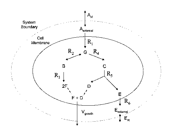
A specific reaction network is provided in Figure 1 to exemplify the
above-described reactions and their interactions. The reactions can be
represented in the
exemplary data structure shown in Figure 3 as set forth below. The reaction
network,
shown in Figure 1, includes intra-system reactions that occur entirely within
the
compartment indicated by the shaded oval such as reversible reaction R2 which
acts on
reactants B and G and reaction R3 which converts one equivalent of B to 2
equivalents of
F. The reaction network shown in Figure 1 also contains exchange reactions
such as
input/output exchange reactions A,,t and E,,t, and the demand exchange
reaction, Vgroõ,d,,
which represents growth in response to the one equivalent of D and one
equivalent of F.
Other intra-system reactions include RI which is a translocation and
transformation
reaction that translocates reactant A into the compartment and transforms it
to reactant G
and reaction R6 which is a transport reaction that translocates reactant E out
of the
compartment.
A reaction network can be represented as a set of linear algebraic
equations which can be presented as a stoichiometric matrix S, with S being an
m x n
matrix where m corresponds to the number of reactants or metabolites and n
corresponds
to the number of reactions taking place in the network. An example of a
stoichiometric
matrix representing the reaction network of Figure 1 is shown in Figure 3. As
shown in
Figure 3, each column in the matrix corresponds to a particular reaction n,
each row
corresponds to a particular reactant m, and each S,,,,, element corresponds to
the
stoichiometric coefficient of the reactant m in the reaction denoted n. The
stoichiometric
matrix includes intra-system reactions such as R2 and R3 which are related to
reactants
that participate in the respective reactions according to a stoichiometric
coefficient having
a sign indicative of whether the reactant is a substrate or product of the
reaction and a
CA 02615504 2008-01-15
WO 2007/014257 PCT/US2006/029001
19
value correlated with the number of equivalents of the reactant consumed or
produced by
the reaction. Exchange reactions such as -Et and -AXt are similarly correlated
with a
stoichiometric coefficient. As exemplified by reactant E, the same compound
can be
treated separately as an internal reactant (E) and an external reactant
(Eextemai) such that an
exchange reaction (R6) exporting the compound is correlated by stoichiometric
coefficients of -1 and 1, respectively. However, because the compound is
treated as a
separate reactant by virtue of its comparttnental location, a reaction, such
as R5, which
produces the internal reactant (E) but does not act on the external reactant
(Eexternal) is
correlated by stoichiometric coefficients of 1 and 0, respectively. Demand
reactions such
as Vg,o,,,tl, can also be included in the stoichiometric matrix being
correlated with
substrates by an appropriate stoichiometric coefficient.
As set forth in further detail below, a stoichiometric matrix provides a
convenient format for representing and analyzing a reaction network because it
can be
readily manipulated and used to compute network properties, for example, by
using linear
programming or general convex analysis. A reaction networlc data structure can
talce on a
variety of formats so long as it is capable of relating reactants and
reactions in the manner
exemplified above for a stoichiometric matrix and in a manner that can be
manipulated to
determine an activity of one or more reactions using methods such as those
exemplified
below. Other examples of reaction network data structures that are useful in
the invention
include a connected graph, list of chemical reactions or a table of reaction
equations.
A reaction network data structure can be constructed to include all
reactions that are involved in metabolism occurring during the interaction of
two or more
cells, Homo sapiens cell metabolism or any portion thereof. A portion of an
organisms
metabolic reactions that can be included in a reaction network data structure
of the
invention includes, for example, a central metabolic pathway such as
glycolysis, the TCA
cycle, the PPP or ETS; or a peripheral metabolic pathway such as amino acid
biosynthesis, amino acid degradation, purine biosynthesis, pyrimidine
biosynthesis, lipid
biosynthesis, fatty acid metabolism, vitamin or cofactor biosynthesis,
transport processes
and alternative carbon source catabolism. Examples of individual pathways
within the
peripheral pathways are set forth in Table 1. Other examples of portions of
metabolic
reactions that can be included in a reaction network data structure of the
invention
include, for example, TAG biosynthesis, muscle contraction requirements,
bicarbonate
CA 02615504 2008-01-15
WO 2007/014257 PCT/US2006/029001
buffer system and/or ammonia buffer system. Specific examples of these and
other
reactions are described further below and in the Examples.
Depending upon a particular application, a reaction network data structure
can include a plurality of Homo sapiens reactions including any or all of the
reactions
5 listed in Table 1. Similarly, a reaction network data structure also can
include the
reactions set forth in Examples I-IV and include, for example, single reaction
networks,
multiple reaction networks that interact within a cell as well as multiple
reaction networks
that interact between cells or physiological systems.
For some applications, it can be advantageous to use a reaction network
10 data structure that includes a minimal number of reactions to achieve a
particular Horno
sapiens activity or activity of a multicellular interaction under a particular
set of
environmental conditions. A reaction network data structure having a minimal
number of
reactions can be identified by performing the simulation methods described
below in an
iterative fashion where different reactions or sets of reactions are
systematically removed
15 and the effects observed. Accordingly, the invention provides a computer
readable
medium, containing a data structure relating a plurality of Homo sapiens
reactants to a
plurality of Homo sapiens reactions, wherein the plurality of Hoino sapiens
reactions
contains at least 65 reactions. For exainple, the core metabolic reaction
database shown
in Tables 2 and 3 contains 65 reactions, and is sufficient to simulate aerobic
and
20 anaerobic metabolism on a number of carbon sources, including glucose.
Similarly, the
invention provides a computer readable medium containing a data structure
relating a
plurality of reactants of multicellular interactions to a plurality of
reactions from
multicellular interactions, wherein the reactions contain at least 430 for a
two cell
interaction. Such reactions between multicellular interactions are exemplified
in Table
11, for example.
Depending upon the particular cell type or types, the physiological,
pathological or therapeutic conditions being tested, the desired activity and
the number of
cellular interactions of a model or method of the invention, a reaction
network data
structure can contain smaller numbers of reactions such as at least 200, 150,
100 or 50
reactions. A reaction network data structure having relatively few reactions
can provide
the advantage of reducing computation time and resources required to perform a
CA 02615504 2008-01-15
WO 2007/014257 PCT/US2006/029001
21
simulation. When desired, a reaction network data structure having a
particular subset of
reactions can be made or used in which reactions that are not relevant to the
particular
simulation are omitted. Alternatively, larger numbers of reactions can be
included in
order to increase the accuracy or molecular detail of the methods of the
invention or to
suit a particular application. Thus, a reaction network data structure can
contain at least
300, 350, 400, 450, 500, 550, 600 or more reactions up to the number of
reactions that
occur in or by multicellular interactions, including Horno sapiens, or that
are desired to
simulate the activity of the full set of reactions occurring in multicellular
interactions,
including Homo sapiens. A reaction network data structure that is
substantially complete
with respect to the metabolic reactions of a multicellular organism, including
Hoino
sapiens, provides an advantage of being relevant to a wide range of conditions
to be
simulated, whereas those with smaller numbers of metabolic reactions are
specific to a
particular subset of conditions to be simulated.
A Homo sapiens reaction network data structure can include one or more
reactions that occur in or by Homo sapiens and that do not occur, either
naturally or
following manipulation, in or by another organism, such as Saccharomyces
cerevisiae. It
is understood that a Homo sapiens reaction network data structure of a
particular cell type
can also include one or more reactions that occur in another cell type.
Addition of such
heterologous reactions to a reaction network data structure of the invention
can be used in
methods to predict the consequences of heterologous gene transfer and protein
expression, for example, when designing in vivo and ex vivo gene therapy
approaches.
Similarly, reaction networks for a multicellular interactions also can include
one or more
reactions that occur entirely within the species of origin of the cellular
interactions or can
contain one or more heterologous reactions from one or more different species.
The reactions included in a reaction network data structure of the invention
can be metabolic reactions. A reaction network data structure can also be
constructed to
include other types of reactions such as regulatory reactions, signal
transduction
reactions, cell cycle reactions, reactions controlling developmental
processes, reactions
involved in apoptosis, reactions involved in responses to hypoxia, reactions
involved in
responses to cell-cell or cell-substrate interactions, reactions involved in
protein synthesis
and regulation thereof, reactions involved in gene transcription and
translation, and
CA 02615504 2008-01-15
WO 2007/014257 PCT/US2006/029001
22
regulation thereof, and reactions involved in assembly of a cell and its
subcellular
components.
A reaction network data structure or index of reactions used in the data
structure such as that available in a metabolic reaction database, as
described above, can
be annotated to include information about a particular reaction. A reaction
can be
annotated to indicate, for example, assignment of the reaction to a protein,
macromolecule or enzyme that performs the reaction, assignment of a gene(s)
that codes
for the protein, macromolecule or enzyme, the Enzyme Commission (EC) number of
the
particular metabolic reaction, a subset of reactions to which the reaction
belongs, citations
to references from which information was obtained, or a level of confidence
with which a
reaction is believed to occur in Homo sapiens or other organism. A computer
readable
medium or media of the invention can include a gene database containing
annotated
reactions. Such information can be obtained during the course of building a
metabolic
reaction database or model of the invention as described below.
As used herein, the term "gene database" is intended to mean a computer
readable medium or media that contains at least one reaction that is annotated
to assign a
reaction to one or more macromolecules that perform the reaction or to assign
one or
more nucleic acid that encodes the one or more macromolecules that perform the
reaction.
A gene database can contain a plurality of reactions, some or all of which are
annotated.
An annotation can include, for exainple, a name for a macromolecule;
assignment of a
function to a macromolecule; assignment of an organism that contains the
macromolecule
or produces the macromolecule; assignment of a subcellular location for the
macromolecule; assignment of conditions under which a macromolecule is
regulated with
respect to performing a reaction, being expressed or being degraded;
assignment of a
cellular component that regulates a macromolecule; an amino acid or nucleotide
sequence
for the macromolecule; a mRNA isoform, enzyme isoform, or any other desirable
annotation or annotation found for a macromolecule in a genome database such
as those
that can be found in Genbank, a site maintained by the NCBI (ncbi.nlm.gov),
the Kyoto
Encyclopedia of Genes and Genomes (KEGG) (www.genome.ad.jp/kegg/), the protein
database SWISS-PROT (ca.expasy.org/sprot/), the LocusLink database maintained
by the
NCBI (www.ncbi.nlm.nih.gov/LocusLink/), the Enzyme Nomenclature database
CA 02615504 2008-01-15
WO 2007/014257 PCT/US2006/029001
23
maintained by G.P. Moss of Queen Mary and Westfield College in the United
Kingdom
(www.chem.qmw.ac.uk/iubmb/enzymeo.
A gene database of the invention can include a substantially complete
collection of genes or open reading frames in a multicellular organism,
including Homo
sapiens, or a substantially complete collection of the macromolecules encoded
by the
organism's genome. Alternatively, a gene database can include a portion of
genes or
open reading frames in an organism or a portion of the macromolecules encoded
by the
organism's genome, such as the portion that includes substantially all
metabolic genes or
macromolecules. The portion can be at least 10%, 15%, 20%, 25%, 50%, 75%, 90%
or
95% of the genes or open reading frames encoded by the organism's genoine, or
the
macromolecules encoded therein. A gene database can also include
macromolecules
encoded by at least a portion of the nucleotide sequence for the organism's
genome such
as at least 10%, 15%, 20%, 25%, 50%, 75%, 90% or 95% of the organism's genome.
Accordingly, a computer readable medium or media of the invention can include
at least
one reaction for each macromolecule encoded by a portion of an organism's
genome,
including a Hoino sapiens genome.
An in silico model of multicellular interactions, including a Homo sapiens
model, of the invention can be built by an iterative process which includes
gathering
information regarding particular reactions to be added to a model,
representing the
reactions in a reaction network data structure, and performing preliminary
simulations
wherein a set of constraints is placed on the reaction network and the output
evaluated to
identify errors in the network. Errors in the network such as gaps that lead
to non-natural
accumulation or consumption of a particular metabolite can be identified as
described
below and simulations repeated until a desired performance of the model is
attained. An
exemplary method for iterative model construction is provided in Exainple I.
For
multicellular interactions, an iterative process includes producing one or
more component
reaction networks followed by combining the components into a higher order
multi-
network system, as described in Example IV. For example, components can
include the
central metabolism reaction network and the cell specific reaction networks
such as TAG
biosynthesis for adipocytes or muscle contraction for myocytes. Combination of
the
central metabolism and the cell specific reaction networks into a single model
produces,
for example, a cell specific reaction network. Components also can include the
individual
CA 02615504 2008-01-15
WO 2007/014257 PCT/US2006/029001
24
cell types, tissues, physiological systems or intra-system reaction networks
that are
constituents of the larger multicellular system. Combining these components
into a larger
model produces, for example, a model describing the relationships and
interactions of the
multicellular system together with its various interactions.
Tlius, the invention provides a method for making a data structure relating
a plurality of reactants to a plurality of reactions in a computer readable
medium or
media. The method includes the steps of: (a) identifying a plurality of
reactions and a
plurality of reactants that are substrates and products of the reactions; (b)
relating the
plurality of reactants to the plurality of Honzo sapiens reactions in a data
structure,
wherein each of the reactions includes a reactant identified as a substrate of
the reaction, a
reactant identified as a product of the reaction and a stoichiometric
coefficient relating the
substrate and the product; (c) making a constraint set for the plurality of
reactions; (d)
providing an objective function; (e) determining at least one flux
distribution that
minimizes or maximizes the objective function when the constraint set is
applied to the
data structure, and (f) if the at least one flux distribution is not
predictive of physiology,
then adding a reaction to or deleting a reaction from the data structure and
repeating step
(e), if the at least one flux distribution is predictive of physiology, then
storing the data
structure in a computer readable medium or media. The method can be applied to
multicellular interactions within or among single or multicullar organisms,
including
Homo sapiens.
Information to be included in a data structure of the invention can be
gathered from a variety of sources including, for example, annotated genome
sequence
information and biochemical literature.
Sources of annotated human genome sequence information include, for
example, KEGG, SWISS-PROT, LocusLink, the Enzyme Nomenclature database, the
International Human Genome Sequencing Consortium and commercial databases.
KEGG
contains a broad range of information, including a substantial amount of
metabolic
reconstruction. The genomes of 304 organisms can be accessed here, with gene
products
grouped by coordinated functions, often represented by a map (e.g., the
enzymes involved
in glycolysis would be grouped together). The maps are biochemical pathway
templates
which show enzymes connecting metabolites for various parts of metabolism.
These
CA 02615504 2008-01-15
WO 2007/014257 PCT/US2006/029001
general pathway templates are customized for a given organism by highlighting
enzymes
on a given template which have been identified in the genome of the organism.
Enzymes
and metabolites are active and yield useful information about stoichiometry,
structure,
alternative names and the like, when accessed.
5 SWISS-PROT contains detailed information about protein function.
Accessible information includes alternate gene and gene product names,
function,
structure and sequence information, relevant literature references, and the
like.
LocusLink contains general information about the locus where the gene is
located and, of relevance, tissue specificity, cellular location, and
implication of the gene
10 product in various disease states.
The Enzyme Nomenclature database can be used to compare the gene
products of two organisms. Often the gene names for genes with similar
functions in two
or more organisms are unrelated. When this is the case, the E.C. (Enzyme
Commission)
numbers can be used as unambiguous indicators of gene product function. The
15 information in the Enzyme Nomenclature database is also published in Enzyme
Nomenclature (Academic Press, San Diego, California, 1992) with 5 supplements
to date,
all found in the European Journal of Biochemistry (Blackwell Science, Maiden,
MA).
Sources of biochemical information include, for example, general
resources relating to metabolism, resources relating specifically to human
metabolism,
20 and resources relating to the biochemistry, physiology and pathology of
specific human
cell types.
Sources of general information relating to metabolism, which were used to
generate the human reaction databases and models described herein, were J.G.
Salway,
Metabolism at a Glance, 2"d ed., Blackwell Science, Malden, MA (1999) and T.M.
25 Devlin, ed., Textbook of Biochemistry with Clinical Correlations, 4th ed.,
John Wiley and
Sons, New York, NY (1997). Human metabolism-specific resources included J.R.
Bronk,
Human Metabolism: Functional Diversity and Integration, Addison Wesley
Longman,
Essex, England (1999).
CA 02615504 2008-01-15
WO 2007/014257 PCT/US2006/029001
26
The literature used in conjunction with the slceletal muscle metabolic
models and simulations described herein included R. Maughan et al.,
Biochernistrv of
Exercise and Training, Oxford University Press, Oxford, England (1997), as
well as
references on muscle pathology such as S. Carpenter et al., Pathology of
Skeletal Muscle,
2"d ed., Oxford University Press, Oxford, England (2001), and more specific
articles on
muscle metabolism as may be found in the Journal of Physiology (Cambridge
University
Press, Cambridge, England).
In the course of developing an in silico model of metabolism during or for
multicellular interactions, the types of data that can be considered include,
for example,
biochemical information which is information related to the experimental
characterization
of a chemical reaction, often directly indicating a protein(s) associated with
a reaction and
the stoichiometry of the reaction or indirectly demonstrating the existence of
a reaction
occurring within a cellular extract; genetic information, which is information
related to
the experimental identification and genetic characterization of a gene(s)
shown to code
for a particular protein(s) implicated in carrying out a biochemical event;
genomic
information, which is information related to the identification of an open
reading frame
and functional assignment, through computational sequence analysis, that is
then linked
to a protein performing a biochemical event; physiological information, which
is
information related to overall cellular physiology, fitness characteristics,
substrate
utilization, and phenotyping results, which provide evidence of the
assimilation or
dissimilation of a compound used to infer the presence of specific biochemical
event (in
particular translocations); and modeling information, which is information
generated
through the course of simulating activity of cells, tissues or physiological
systems using
methods such as those described herein which lead to predictions regarding the
status of a
reaction such as whether or not the reaction is required to fulfill certain
demands placed
on a metabolic network. Additional information relevant to multicellular
organisms that
can be considered includes, for example, cell type-specific or condition-
specific gene
expression information, which can be determined experimentally, such as by
gene array
analysis or from expressed sequence tag (EST) analysis, or obtained from the
biochemical
and physiological literature.
The majority of the reactions occurring in a multicellular organism's
reaction networks are catalyzed by enzymes/proteins, which are created through
the
CA 02615504 2008-01-15
WO 2007/014257 PCT/US2006/029001
27
transcription and translation of the genes found within the chromosome in the
cell. The
remaining reactions occur either spontaneously or through non-enzymatic
processes.
Furthermore, a reaction network data structure can contain reactions that add
or delete
steps to or from a particular reaction pathway. For example, reactions can be
added to
optimize or improve performance of a model for multicellular interactions in
view of
empirically observed activity. Alternatively, reactions can be deleted to
remove
intermediate steps in a pathway when the intermediate steps are not necessary
to model
flux through the pathway. For example, if a pathway contains 3 nonbranched
steps, the
reactions can be combined or added together to give a net reaction, thereby
reducing
memory required to store the reaction network data structure and the
computational
resources required for manipulation of the data structure.
The reactions that occur due to the activity of gene-encoded enzymes can
be obtained from a genome database which lists genes identified from genome
sequencing and subsequent genome annotation. Genome annotation consists of the
locations of open reading frames and assignment of function from homology to
other
known genes or empirically determined activity. Such a genome database can be
acquired through public or private databases containing annotated nucleic acid
or protein
sequences, including Horno sapiens sequences. If desired, a model developer
can
perform a network reconstruction and establish the model content associations
between
the genes, proteins, and reactions as described, for example, in Covert et al.
Trends in
Biochemical Sciences 26:179-186 (2001) and Palsson, WO 00/46405.
As reactions are added to a reaction network data structure or metabolic
reaction database, those having known or putative associations to the
proteins/enzymes
which enable/catalyze the reaction and the associated genes that code for
these proteins
can be identified by annotation. Accordingly, the appropriate associations for
all of the
reactions to their related proteins or genes or both can be assigned. These
associations
can be used to capture the non-linear relationship between the genes and
proteins as well
as between proteins and reactions. In some cases one gene codes for one
protein which
then perform one reaction. However, often there are multiple genes which are
required to
create an active enzyme complex and often there are multiple reactions that
can be carried
out by one protein or multiple proteins that can carry out the same reaction.
These
associations capture the logic (i.e. AND or OR relationships) within the
associations.
CA 02615504 2008-01-15
WO 2007/014257 PCT/US2006/029001
28
Annotating a metabolic reaction database with these associations can allow the
methods
to be used to determine the effects of adding or eliminating a particular
reaction not only
at the reaction level, but at the genetic or protein level in the context of
running a
simulation or predicting a multicellular interaction activity, including Homo
sapiens
activity.
A reaction network data structure of the invention can be used to
determine the activity of one or more reactions in a plurality of reactions
occurring from
multicellular interactions, including a plurality of Horno sapiens reactions,
independent of
any knowledge or annotation of the identity of the protein that performs the
reaction or
the gene encoding the protein. A model that is annotated with gene or protein
identities
can include reactions for which a protein or encoding gene is not assigned.
While a large
portion of the reactions in a cellular metabolic network are associated with
genes in the
organism's genome, there are also a substantial number of reactions included
in a model
for which there are no known genetic associations. Such reactions can be added
to a
reaction database based upon other information that is not necessarily related
to genetics
such as biochemical or cell based measurements or theoretical considerations
based on
observed biochemical or cellular activity. For example, there are many
reactions that can
either occur spontaneously or are not protein-enabled reactions. Furthermore,
the
occurrence of a particular reaction in a cell for which no associated proteins
or genetics
have been currently identified can be indicated during the course of model
building by the
iterative model building methods of the invention.
The reactions in a reaction network data structure or reaction database can
be assigned to subsystems by annotation, if desired. The reactions can be
subdivided
according to biological criteria, such as according to traditionally
identified metabolic
pathways (glycolysis, amino acid metabolism and the like) or according to
mathematical
or computational criteria that facilitate manipulation of a model that
incorporates or
manipulates the reactions. Methods and criteria for subdviding a reaction
database are
described in further detail in Schilling et al., J. Theor. Biol. 203:249-283
(2000), and in
Schuster et al., Bioinformatics 18:351-361 (2002). The use of subsystems can
be
advantageous for a number of analysis methods, such as extreme pathway
analysis, and
can make the management of model content easier. Although assigning reactions
to
subsystems can be achieved without affecting the use of the entire model for
simulation,
CA 02615504 2008-01-15
WO 2007/014257 PCT/US2006/029001
29
assigning reactions to subsystems can allow a user to search for reactions in
a particular
subsystem which may be useful in performing various types of analyses.
Therefore, a
reaction network data structure can include any number of desired subsystems
including,
for example, 2 or more subsystems, 5 or more subsystems, 10 or more
subsystems, 25 or
more subsystems or 50 or more subsystems.
The reactions in a reaction networlc data structure or metabolic reaction
database can be annotated with a value indicating the confidence with which
the reaction
is believed to occur in one or more cells of a multicellular interaction or in
one or more
reaction networks within a cell such as a Homo sapiens cell. The level of
confidence can
be, for example, a function of the amount and form of supporting data that is
available.
This data can come in various forms including published literature, documented
experimental results, or results of computational analyses. Furthermore, the
data can
provide direct or indirect evidence for the existence of a chemical reaction
in a cell based
on genetic, biochemical, and/or physiological data.
The invention further provides a computer readable medium, containing
(a) a data structure relating a plurality of Horno sapiens reactants to a
plurality of Hoino
sapiens reactions, wherein each of the Homo sapiens reactions includes a
reactant
identified as a substrate of the reaction, a reactant identified as a product
of the reaction
and a stoichiometric coefficient relating the substrate and the product, and
(b) a constraint
set for the plurality of Homo sapiens reactions. Similarly, the computer
readable medium
or media can relate a plurality of reactions to a plurality of reactions
within first and
second cells and for an intra-system between first and second interacting
cells.
Constraints can be placed on the value of any of the fluxes in the metabolic
network using a constraint set. These constraints can be representative of a
minimum or
maximum allowable flux through a given reaction, possibly resulting from a
limited
amount of an enzyme present. Additionally, the constraints can determine the
direction
or reversibility of any of the reactions or transport fluxes in the reaction
network data
structure. Based on the in vivo environment where multiple cells interact,
such as in a
human organism, the metabolic resources available to the cell for biosynthesis
of essential
molecules for can be determined. Allowing the corresponding transport fluxes
to be
CA 02615504 2008-01-15
WO 2007/014257 PCT/US2006/029001
active provides the in silico interaction between cells with inputs and
outputs for
substrates and by-products produced by the metabolic network.
Returning to the hypothetical reaction network shown in Figure 1,
constraints can be placed on each reaction in the exemplary format shown in
Figure 2, as
5 follows. The constraints are provided in a format that can be used to
constrain the
reactions of the stoichiometric matrix shown in Figure 3. The format for the
constraints
used for a matrix or in linear programming can be conveniently represented as
a linear
inequality such as
bj < vj < aj :j = 1....n (Eq. l)
10 where vj is the metabolic flux vector, bj is the minimum flux value and aj
is the maximum
flux value. Thus, aj can take on a finite value representing a maximum
allowable flux
through a given reaction or bj can take on a finite value representing minimum
allowable
flux through a given reaction. Additionally, if one chooses to leave certain
reversible
reactions or transport fluxes to operate in a forward and reverse manner the
flux may
15 remain unconstrained by setting bj to negative infinity and aj to positive
infinity as shown
for reaction R2 in Figure 2. If reactions proceed only in the forward reaction
bj is set to
zero while aj is set to positive infinity as shown for reactions RI, R3, R4,
R5, and R6 in
Figure 2. As an example, to simulate the event of a genetic deletion or non-
expression of
a particular protein, the flux through all of the corresponding metabolic
reactions related
20 to the gene or protein in question are reduced to zero by setting aj and bj
to be zero.
Furthermore, if one wishes to simulate the absence of a particular growth
substrate one
can simply constrain the corresponding transport fluxes that allow the
metabolite to enter
the cell to be zero by setting aj and bj to be zero. On the other hand if a
substrate is only
allowed to enter or exit the cell via transport mechanisms, the corresponding
fluxes can
25 be properly constrained to reflect this scenario.
The ability of a reaction to be actively occurring is dependent on a large
number of additional factors beyond just the availability of substrates. These
factors,
which can be represented as variable constraints in the models and methods of
the
invention include, for example, the presence of cofactors necessary to
stabilize the
30 protein/enzyme, the presence or absence of enzymatic inhibition and
activation factors,
CA 02615504 2008-01-15
WO 2007/014257 PCT/US2006/029001
31
the active formation of the protein/enzyme through translation of the
corresponding
mRNA transcript, the transcription of the associated gene(s) or the presence
of chemical
signals and/or proteins that assist in controlling these processes that
ultimately determine
whether a chemical reaction is capable of being carried out within an
organism. Of
particular importance in the regulation of human cell types is the
implementation of
paracrine and endocrine signaling pathways to control cellular activities. In
these cases a
cell secretes signaling molecules that may be carried far afield to act on
distant targets
(endocrine signaling), or act as local mediators (paracrine signaling).
Examples of
endocrine signaling molecules include hormones such as insulin, while examples
of
paracrine signaling molecules include neurotransmitters such as acetylcholine.
These
molecules induce cellular responses through signaling cascades that affect the
activity of
biochemical reactions in the cell. Regulation can be represented in an in
silico Homo
sapiens model by providing a variable constraint as set forth below.
Thus, the invention provides a computer readable medium or media,
including (a) a data structure relating a plurality of Homo sapiens reactants
to a plurality
of Homo sapiens reactions, wherein each of the reactions includes a reactant
identified as
a substrate of the reaction, a reactant identified as a product of the
reaction and a
stoichiometric coefficient relating the substrate and the product, and wherein
at least one
of the reactions is a regulated reaction; and (b) a constraint set for the
plurality of
reactions, wherein the constraint set includes a variable constraint for the
regulated
reaction. Additionally, the invention provides a computer readable medium or
media
including data structures for two or more cells and for an intra-system and a
constraint set
for the plurality of reactions within the data structures that includes a
variable constraint
for a regulated reaction.
As used herein, the term "regulated," when used in reference to a reaction
in a data structure, is intended to mean a reaction that experiences an
altered flux due to a
change in the value of a constraint or a reaction that has a variable
constraint.
As used herein, the term "regulatory reaction" is intended to mean a
chemical conversion or interaction that alters the activity of a protein,
macromolecule or
enzyme. A chemical conversion or interaction can directly alter the activity
of a protein,
macromolecule or enzyme such as occurs when the protein, macroinolecule or
enzyme is
CA 02615504 2008-01-15
WO 2007/014257 PCT/US2006/029001
32
post-translationally modified or can indirectly alter the activity of a
protein,
macromolecule or enzyme such as occurs when a chemical conversion or binding
event
leads to altered expression of the protein, macromolecule or enzyme. Thus,
transcriptional or translational regulatory pathways can indirectly alter a
protein,
macromolecule or enzyme or an associated reaction. Similarly, indirect
regulatory
reactions can include reactions that occur due to downstream components or
participants
in a regulatory reaction network. When used in reference to a data structure
or in silico
Homo sapiens model, for example, the term is intended to mean a first reaction
that is
related to a second reaction by a function that alters the flux through the
second reaction
by changing the value of a constraint on the second reaction.
As used herein, the term "regulatory data structure" is intended to mean a
representation of an event, reaction or network of reactions that activate or
inhibit a
reaction, the representation being in a format that can be manipulated or
analyzed. An
event that activates a reaction can be an event that initiates the reaction or
an event that
increases the rate or level of activity for the reaction. An event that
inhibits a reaction can
be an event that stops the reaction or an event that decreases the rate or
level of activity
for the reaction. Reactions that can be represented in a regulatory data
structure include,
for example, reactions that control expression of a macromolecule that in
turn, performs a
reaction such as transcription and translation reactions, reactions that lead
to post
translational modification of a protein or enzyme such as phophorylation,
dephosphorylation, prenylation, methylation, oxidation or covalent
modification,
reactions that process a protein or enzyme such as removal of a pre- or pro-
sequence,
reactions that degrade a protein or enzyme or reactions that lead to assembly
of a protein
or enzyme.
As used herein, the term "regulatory event" is intended to mean a modifier
of the flux through a reaction that is independent of the amount of reactants
available to
the reaction. A modification included in the term can be a change in the
presence,
absence, or amount of an enzyme that performs a reaction. A modifier included
in the
term can be a regulatory reaction such as a signal transduction reaction or an
environmental condition such as a change in pH, temperature, redox potential
or time. It
will be understood that when used in reference to an in silico Homo sapiens
model or data
structure, or when used in reference to a model or data structure for a
multicellular
CA 02615504 2008-01-15
WO 2007/014257 PCT/US2006/029001
33
interaction, a regulatory event is intended to be a representation of a
modifier of the flux
through a Honao sapiens reaction or reaction occurring in one or more cells in
a
multicellular interaction that is independent of the amount of reactants
available to the
reaction.
The effects of regulation on one or more reactions that occur in Honao
sapiens can be predicted using an in silico Homo sapiens model or
multicellular model of
the invention. Regulation can be taken into consideration in the context of a
particular
condition being examined by providing a variable constraint for the reaction
in an in
silico Honzo sapiens model or multicellular model. Such constraints constitute
condition-dependent constraints. A data structure can represent regulatory
reactions as
Boolean logic statements (Reg-reaction). The variable takes on a value of 1
when the
reaction is available for use in the reaction network and will take on a value
of 0 if the
reaction is restrained due to some regulatory feature. A series of Boolean
statements can
then be introduced to mathematically represent the regulatory network as
described for
example in Covert et al. J. Theor. Biol. 213:73-88 (2001). For example, in the
case of a
transport reaction (A in) that imports metabolite A, where metabolite A
inhibits reaction
R2 as shown in Figure 4, a Boolean rule can state that:
Reg-R2 = IF NOT(A in). (Eq. 2)
This statement indicates that reaction R2 can occur if reaction A in is not
occurring (i.e.
if metabolite A is not present). Similarly, it is possible to assign the
regulation to a
variable A which would indicate an amount of A above or below a threshold that
leads to
the inhibition of reaction R2. Any function that provides values for variables
corresponding to each of the reactions in the biochemical reaction network can
be used to
represent a regulatory reaction or set of regulatory reactions in a regulatory
data structure.
Such functions can include, for example, fuzzy logic, heuristic rule-based
descriptions,
differential equations or kinetic equations detailing system dynamics.
A reaction constraint placed on a reaction can be incorporated into an in
silico Homo sapiens model or mulicellular model of interacting cells using the
following
general equation:
(Reg-Reaction) *bj< vj < aj*(Reg-Reaction), V
CA 02615504 2008-01-15
WO 2007/014257 PCT/US2006/029001
34
j = 1....n (Eq. 3)
For the example of reaction R2 this equation is written as follows:
(0)*Reg-R2 <_ R2 < (oo)*Reg-R2. (Eq. 4)
Thus, during the course of a simulation, depending upon the presence or
absence of metabolite A in the interior of the cell where reaction R2 occurs,
the value for
the upper boundary of flux for reaction R2 will change from 0 to infinity,
respectively.
With the effects of a regulatory event or network taken into consideration
by a constraint function and the condition-dependent constraints set to an
initial relevant
value, the behavior of the Homo sapiens reaction network or one or more
reaction
networks of a multicellular interaction can be simulated for the conditions
considered as
set forth below.
Although regulation has been exemplified above for the case where a
variable constraint is dependent upon the outcome of a reaction in the data
structure, a
plurality of variable constraints can be included in an in silico Honzo
sapiens model or
other model of nlulticellular interactions to represent regulation of a
plurality of reactions.
Furthermore, in the exemplary case set forth above, the regulatory structure
includes a
general control stating that a reaction is inhibited by a particular
environmental condition.
Using a general control of this type, it is possible to incorporate molecular
mechanisms
and additional detail into the regulatory structure that is responsible for
determining the
active nature of a particular chemical reaction within an organism.
Regulation can also be simulated by a model of the invention and used to
predict a Homo sapiens physiological function without knowledge of the precise
molecular mechanisms involved in the reaction network being modeled. Thus, the
model
can be used to predict, in silico, overall regulatory events or causal
relationships that are
not apparent from in vivo observation of any one reaction in a network or
whose in vivo
effects on a particular reaction are not known. Such overall regulatory
effects can include
those that result from overall environmental conditions such as changes in pH,
temperature, redox potential, or the passage of time.
CA 02615504 2008-01-15
WO 2007/014257 PCT/US2006/029001
As described previously and further below, the models and method of the
invention are applicable to a wide range of multicellular interactions. The
multicellular
interactions include, for example, interactions between prokaryotic cells such
as colony
growth and chemotaxis. The multicellular interactions include, for example,
interactions
5 between two or more eukaryotic cells such as the concerted action of two or
more cells of
the same or different cell type. A specific example of the concerted action of
the same
cell type includes the combined output of the contractile activity of
myocytes. A specific
example of the concerted action of different cell types includes the energy
production of
adipocyte cells and the contractile activity of myocyte cells based on the
consumption of
10 energy available from the adipocyte cells. Multicellular interactions also
can include, for
example, interactions between host cells and a pathogen, such as a bacteria,
virus or
worm, as well as symbiotic interactions between host cells and microbes, for
example. A
symbiotic microbe can include, for example, E. coli. Further examples of host
and
microbe interactions include bacterial communities that reside in the skin and
mouth and
15 the vagina flora, providing the host with a defense against infections.
Moreover, the
models and methods of the invention also can be used to reconstruction the
reaction
networks between a plurality of dynamic multicellular interactions including,
for
exainple, interactions between host cells or tissues, pathogen and symbiotic
microbe.
Multicellular interactions also include, for example, interactions between
20 cells of different tissues, different organs and/or physiological systems
as well as
interactions between some or all cells, tissues organs and/or physiological
systems within
a multicellular organism. Specific examples of such interactions include
organismic
homeostasis, signal transduction, the endocrine system, the exocrine system,
sensory
transduction, secretion, the hematopoietic system, the immune system, cell
migration, cell
25 adherence, cell invasion and neuronal and synaptic transduction. Numerous
other
multicellular interactions are well known in the art and can similarly be
reconstructed and
simulated to predict an activity thereof using the models and methods of the
invention.
Given the teachings and guidance provided herein with respect to the
construction and use of multiple reaction networks including, for example, the
regulated
30 and metabolic reaction networks of a Horno sapiens cell, those skilled in
the art will know
how to employ the models and methods of the invention for the construction and
use of
any multicellular interaction. Specific examples of such multicellular
interactions are
CA 02615504 2008-01-15
WO 2007/014257 PCT/US2006/029001
36
described above. Other examples of multicellular interactions include, for
example, all
interactions occurring between two or more cells such as those cells set forth
in Table 5
below. Such multicellular interactions can occur between cells within the same
or
different physiological category or functional characterization. Similarly,
such
multicellular interactions also can occur between cells within the same and
between
different physiological categories or functional characterizations. The number
and types
of different cellular interactions will be determined by the multicellular
model being
produced using the methods of the invention.
Models of multicellular interactions also can include, for example,
interactions between cells of one or more tissues and organs. The models and
methods of
the invention are applicable to predict the activity of interactions between
some or all cell
types of a tissue or organ. The models and methods of the invention also can
include
reaction networks that include interactions between some or all cell types of
two or more
tissues or organs. Specific examples of tissues or organs and their respective
cell types
and functions are shown below in Table 6. The models and methods of the
invention can
include, for example, some or all of these interactions to predict their
respective
activities.. Similarly, Table 7 exemplifies the cell types of a liver. Given
the teachings
and guidance provided herein, the models and methods of the invention can be
used to
construct an in silico reconstruction of the reaction networks for some or all
of these cell
types to predict some or all of the activities of the liver. Further, an in
silico
reconstruction of reaction networks for some or all multicellular interactions
exemplified
in Tables 5-7, including those within and between tissues and organs, can be
produced
that can be used to predict some or all activities of one or more tissues or
of an organism.
Therefore, the invention provides for the in silico reconstruction of whole
organisms,
including human organisms, tissues, cells and physical or physiological
functions
performed by such cellular systems.
The invention also provides for the in silico reconstruction of a plurality of
reaction networks that interact to perfonn the same or different activity. The
plurality can
be a small, medium or large plurality and can reside within the same cell,
different cells
or in different tissues or organisms. Specific examples of such pluralities
residing within
the same cell include the reaction networks exemplified below in Example IV
for a
myocyte or for an adipocyte. Specific examples of such pluralities residing in
different
CA 02615504 2008-01-15
WO 2007/014257 PCT/US2006/029001
37
cells or tissues include the reaction networks exemplified below in Example IV
for
coupled adipocyte-myocyte metabolism. Another example of interactions between
different reaction networks within different networks includes interactions
between
pathogen and host cells.
Briefly, and as described previously, a computer readable medium or
media can be produced that includes a plurality of data structures each
relating a plurality
of reactants to a plurality of reactions from each cell within the
multicellular interaction.
The reactions include a reactant identified as a substrate of the reaction, a
reactant
identified as a product of the reaction and a stoichiometric coefficient
relating the
substrate and said product In a two cell interaction, including populations of
two cell
types, the plurality of data structures can include a first data structure and
a second data
structure corresponding to the reactions within the two cells or populations
of two cell
types. The data structures will describe the reaction networks for each cell.
For optimization of the multicellular interaction containing two cells, a
third data structure is particularly useful for relating a plurality of intra-
system reactants
to a plurality of intra-system reactions between the first and second cells.
Each of the
intra-system reactions includes a reactant identified as a substrate of the
reaction, a
reactant identified as a product of the reaction and a stoichiometric
coefficient relating the
substrate and said product. The inta-system data structure can be included in
the
reconstruction as an independent data structure or as a component of one or
more data
structures for either or both cells within such a two cell interaction model.
A specific
example of intra-system reactions represented by a third data structure is
shown in Figure
10 for the bicarbonate and ammonia buffer systems employed in the two cell
model
describing adipocyte and myocyte interactions.
As with the models and methods of the invention described above and
below, a computer readable medium or media describing a multicellular
interaction also
will contain a constraint set for the plurality of reactions for each of the
first, second and
third data structures as well as commands for determining at least one flux
distribution
that minimizes or maximizes an objective function when said constraint set is
applied to
said first and second data structures. The objective function can be, for
example, those
objective functions exeinplified previously, those exemplified below or in the
Examples
CA 02615504 2008-01-15
WO 2007/014257 PCT/US2006/029001
38
as well as various other object functions well known to those skilled in the
art given the
teachings and guidance provided herein. Solving the optimization problem by
determining one or more flux distribution will predict a physiological
function of
occurring as a result of the interaction between the first and second cells of
the model.
Each of the first, second or third data structures can include one or more
reaction networks. For example, and with reference to Figures 5-10, a reaction
networlc
for each of the cells exemplified therein can be defined as the different
networks within
each cell such as central metabolism and the cell specific reactions. Applying
this view,
the adipocyte and myocyte cells each contain at least two reaction networks.
When
combined together with the intra-cellular reaction network and the exchange
reactions,
the interactions of the two cells exemplified in Figure 6 can be described by
at least five
different reaction networks. The interactions of this two cell model can
therefore be
described using at least five data structures. Alternatively, a reaction
network can be
defined as all the networks within each cell. When combined together with the
intra-
cellular reaction network and the exchange reactions, the interactions of the
exemplified
adipocyte and myocyte cells can be described by at least three different
reaction
networks. One reaction network for each cell and one reaction network for the
intra-
system reactions. Therefore, each of the first, second or third data
structures can consist
of a plurality of two or more reaction networks including, for example, 2, 3,
4, 5, 10, 20
or 25 or more as well as all integer numbers between and above these exemplary
numbers. Similarly, given the teachings and guidance provided herein, the
models and
methods of the invention can be generated and used to predict an activity
and/or
physiological function of the intercellular network interactions or the
intracellular
network interaction. The latter interactions, for example, also predict an
activity and/or a
physiological function of the interactions between two or more cells including
cells of
different tissues, organs of a multicellular organism or of a whole organism.
As with the number of reaction networks within a data structure, the
models and methods of the invention also can employ greater than three data
structures as
exemplified above. For example, the models and method of the invention can
comprise
one or more fourth data structures having one or more fourth constraint sets
where each
fourth data structure relates a plurality of reactants to a plurality of
reactions from a cell
already included in the model or from one or more third cells within the
multicellular
CA 02615504 2008-01-15
WO 2007/014257 PCT/US2006/029001
39
interaction. Use of one or more fourth data structures is particularly useful
when
reconstructing a interactions between three or more interacting cells
including a large
plurality of cells such as the cells within a tissue, organ, physiological
system or
organism. Each of the reactions within such fourth data structures include a
reactant
identified as a substrate of the reaction, a reactant identified as a product
of the reaction
and a stoichiometric coefficient relating the substrate and said product.
The number of fourth data structures can correspond to the number of cells
greater than the first and second cells of the multicellular interaction and
include, for
example, a plurality of data structures. As with the specific embodiment of a
two cell
interaction, the plurality of data structures for three or more interacting
cells can
correspond to different cells within the cellular interaction as well as
correspond to
different cell types within the cellular interaction. The number of cells can
include, for
example, at least 4 cells, 5 cells, 6 cells, 7 cells, 8 cells, 9 cells, 10
cells, 100 cells, 1000
cells, 5000 cells, 10,000 cells or more. Therefore, the number of cells within
a
multicellular interaction model or used in a method of predicting a behavior
of such
multicellular interactions can include some or all cells which constitute a
group of
interacting cells, a tissue, organ, physiological system or whole organism.
The
multicellular interaction models and methods of the invention also can include
some or all
cells which constitute a group of interacting cells of different types or from
different
tissues, organs, physiological systems or organisms. The organism can be
single cell
prokaryotic or eukaryotic organism or multicellular eukaryotic organisms.
Specific
examples of different cell types include a mammary gland cell, hepatocyte,
white fat cell,
brown fat cell, liver lipocyte, red skeletal muscle cell, white skeletal
muscle cell,
intermediate skeletal muscle cell, smooth muscle cell, red blood cell,
adipocyte,
monocyte, reticulocyte, fibroblast, neuronal cell epithelial cell or one or
more cells set
forth in Table 5. Specific examples of physiological functions resulting from
multicellular interactions that can be predicted include metabolite yield, ATP
yield,
biomass demand, growth, triacylglycerol storage, muscle contraction, milk
secretion and
oxygen transport capacity.
Intra-system reactions of a multicellular interaction model or method of
the invention has been exemplified above and below with reference to the
extracellular in
vivo environment and, in particular, with reference to buffering this
environment by
CA 02615504 2008-01-15
WO 2007/014257 PCT/US2006/029001
supplying functions of the renal system. Given the teachings and guidance
provided
herein, those skilled in the art will understand that any extracellular
reaction, plurality of
reactions, function of the extracellular space or function supplied into the
extracellular
space by another cell, tissue or physiological system can be employed as an
intra-system
5 reaction network. Such reactions or activities can represent normal or
pathological
conditions or both conditions occurring within this intra-system environment.
Specific
examples of intra-system reactions include one or more reactions performed in
the
hematopoietic system, urine, connective tissue, contractile tissue or cells,
lymphatic
system, respiratory system or renal system. Reactions or reactants included in
one or
10 more intra-system data structures can be, for example, bicarbonate buffer
system, an
ainmonia buffer system, a hormone, a signaling molecule, a vitamin, a mineral
or a
combination thereof.
The in silico models of multicellular or multi-network interactions,
including Flomo sapiens model and methods, described herein can be implemented
on
15 any conventional host computer system, such as those based on Intel®
microprocessors and running Microsoft Windows operating systenis. Other
systems, such
as those using the UNIX or LINUX operating system and based on IBM®,
DEC® or Motorola® microprocessors are also contemplated. The systems
and
methods described herein can also be implemented to run on client-server
systems and
20 wide-area networks, such as the Internet.
Software to implement a method or model of the invention can be written
in any well-known computer language, such as Java, C, C++, Visual Basic,
FORTRAN
or COBOL and compiled using any well-known compatible compiler. The software
of the
invention normally runs from instructions stored in a memory on a host
computer system.
25 A memory or computer readable medium can be a hard disk, floppy disc,
compact disc,
magneto-optical disc, Random Access Memory, Read Only Memory or Flash Memory.
The memory or computer readable medium used in the invention can be contained
within
a single computer or distributed in a network. A network can be any of a
number of
conventional network systems known in the art such as a local area network
(LAN) or a
30 wide area network (WAN). Client-server environments, database servers and
networks
that can be used in the invention are well lcnown in the art. For example, the
database
server can run on an operating system such as UNIX, running a relational
database
CA 02615504 2008-01-15
WO 2007/014257 PCT/US2006/029001
41
management system, a World Wide Web application and a World Wide Web server.
Other types of memories and computer readable media are also contemplated to
function
within the scope of the invention.
A database or data structure of the invention can be represented in a
markup language format including, for example, Standard Generalized Markup
Language
(SGML), Hypertext markup language (HTML) or Extensible Markup language (XML).
Markup languages can be used to tag the information stored in a database or
data
structure of the invention, thereby providing convenient annotation and
transfer of data
between databases and data structures. In particular, an XML format can be
useful for
structuring the data representation of reactions, reactants and their
annotations; for
exchanging database contents, for example, over a network or internet; for
updating
individual elements using the document object model; or for providing
differential access
to multiple users for different information content of a data base or data
structure of the
invention. XML programming methods and editors for writing XML code are known
in
the art as described, for example, in Ray, "Learning XML" O'Reilly and
Associates,
Sebastopol, CA (2001).
A set of constraints can be applied to a reaction network data structure to
simulate the flux of mass through the reaction network under a particular set
of
environmental conditions specified by a constraints set. Because the time
constants
characterizing metabolic transients and/or metabolic reactions are typically
very rapid, on
the order of milli-seconds to seconds, compared to the time constants of cell
growth on
the order of hours to days, the transient mass balances can be simplified to
only consider
the steady state behavior. Referring now to an example where the reaction
network data
structure is a stoichiometric matrix, the steady state mass balances can be
applied using
the following system of linear equations
S = v = 0 (Eq. 5)
where S is the stoichiometric matrix as defined above and v is the flux
vector. This
equation defines the mass, energy, and redox potential constraints placed on
the metabolic
network as a result of stoichiometry. Together Equations 1 and 5 representing
the
reaction constraints and mass balances, respectively, effectively define the
capabilities
CA 02615504 2008-01-15
WO 2007/014257 PCT/US2006/029001
42
and constraints of the metabolic genotype and the organism's metabolic
potential. All
vectors, v, that satisfy Equation 5 are said to occur in the mathematical
nullspace of S.
Thus, the null space defines steady-state metabolic flux distributions that do
not violate
the mass, energy, or redox balance constraints. Typically, the number of
fluxes is greater
than the number of mass balance constraints, thus a plurality of flux
distributions satisfy
the mass balance constraints and occupy the null space. The null space, which
defines the
feasible set of metabolic flux distributions, is further reduced in size by
applying the
reaction constraints set forth in Equation 1 leading to a defined solution
space. A point in
this space represents a flux distribution and hence a metabolic phenotype for
the network.
An optimal solution within the set of all solutions can be determined using
mathematical
optimization methods when provided with a stated objective and a constraint
set. The
calculation of any solution constitutes a simulation of the model.
Objectives for activity of a human cell can be chosen. While the overall
objective of a multi-cellular organism may be growth or reproduction,
individual human
cell types generally have much more complex objectives, even to the seemingly
extreme
objective of apoptosis (programmed cell death), which may benefit the organism
but
certainly not the individual cell. For example, certain cell types may have
the objective
of maximizing energy production, while others have the objective of maximizing
the
production of a particular hormone, extracellular matrix component, or a
mechanical
property such as contractile force. In cases where cell reproduction is slow,
such as
human skeletal muscle, growth and its effects need not be taken into account.
In other
cases, biomass composition and growth rate could be incorporated into a
"maintenance"
type of flux, where rather than optimizing for growth, production of
precursors is set at a
level consistent with experimental knowledge and a different objective is
optimized.
Certain cell types, including cancer cells, can be viewed as having an
objective of maximizing cell growth. Growth can be defined in terms of
biosynthetic
requirements based on literature values of biomass composition or
experimentally
determined values such as those obtained as described above. Thus, biomass
generation
can be defined as an exchange reaction that removes intermediate metabolites
in the
appropriate ratios and represented as an objective function. In addition to
draining
intermediate metabolites this reaction flux can be formed to utilize energy
molecules such
as ATP, NADH and NADPH so as to incorporate any maintenance requirement that
must
CA 02615504 2008-01-15
WO 2007/014257 PCT/US2006/029001
43
be met. This new reaction flux then becomes another constraint/balance
equation that the
system must satisfy as the objective function. Using the stoichiometric matrix
of Figure 3
as an example, adding such a constraint is analogous to adding the additional
column
Vgro,,,ch to the stoichiometric matrix to represent fluxes to describe the
production demands
placed on the metabolic system. Setting this new flux as the objective
function and
asking the system to maximize the value of this flux for a given set of
constraints on all
the other fluxes is then a method to simulate the growth of the organism.
Continuing with the example of the stoichiometric matrix applying a
constraint set to a reaction network data structure can be illustrated as
follows. The
solution to equation 5 can be formulated as an optimization problem, in which
the flux
distribution that minimizes a particular objective is found. Mathematically,
this
optimization problem can be stated as:
Minimize Z (Eq. 6)
where Z Yci v' (Eq. 7)
where Z is the objective which is represented as a linear combination of
metabolic fluxes
vi using the weights ci in this linear combination. The optimization problem
can also be
stated as the equivalent maximization problem; i.e. by changing the sign on Z.
Any
commands for solving the optimazation problem can be used including, for
example,
linear programming commands.
A computer system of the invention can further include a user interface
capable of receiving a representation of one or more reactions. A user
interface of the
invention can also be capable of sending at least one command for modifying
the data
structure, the constraint set or the commands for applying the constraint set
to the data
representation, or a combination thereof. The interface can be a graphic user
interface
having graphical means for making selections such as menus or dialog boxes.
The
interface can be arranged with layered screens accessible by making selections
from a
main screen. The user interface can provide access to other databases useful
in the
invention such as a metabolic reaction database or links to other databases
having
information relevant to the reactions or reactants in the reaction network
data structure or
CA 02615504 2008-01-15
WO 2007/014257 PCT/US2006/029001
44
to a multicellular organism's physiology, including Hoino sapiens physiology.
Also, the
user interface can display a graphical representation of a reaction network or
the results of
a simulation using a model of the invention.
Once an initial reaction network data structure and set of constraints has
been created, this model can be tested by preliminary simulation. During
preliminary
simulation, gaps in the network or "dead-ends" in which a metabolite can be
produced but
not consumed or where a metabolite can be consumed but not produced can be
identified.
Based on the results of preliminary simulations areas of the metabolic
reconstruction that
require an additional reaction can be identified. The determination of these
gaps can be
readily calculated through appropriate queries of the reaction network data
structure and
need not require the use of simulation strategies, however, simulation would
be an
alternative approach to locating such gaps.
In the preliminary simulation testing and model content refinement stage
the existing model is subjected to a series of functional tests to determine
if it can perform
basic requirements such as the ability to produce the required biomass
constituents and
generate predictions concerning the basic physiological characteristics of the
particular
cell type being modeled. The more preliminary testing that is conducted the
higher the
quality of the model that will be generated. Typically, the majority of the
simulations
used in this stage of development will be single optimizations. A single
optimization can
be used to calculate a single flux distribution demonstrating how metabolic
resources are
routed determined from the solution to one optimization problem. An
optimization
problem can be solved using linear programming as demonstrated in the Examples
below.
The result can be viewed as a display of a flux distribution on a reaction
map. Temporary
reactions can be added to the network to determine if they should be included
into the
model based on modeling/simulation requirements.
Once a model of the invention is sufficiently complete with respect to the
content of the reaction network data structure according to the criteria set
forth above, the
model can be used to simulate activity of one or more reactions in a reaction
network.
The results of a simulation can be displayed in a variety of formats
including, for
example, a table, graph, reaction network, flux distribution map or a
phenotypic phase
plane graph.
CA 02615504 2008-01-15
WO 2007/014257 PCT/US2006/029001
Thus, the invention provides a method for predicting a Homo sapiens
physiological function. The method includes the steps of (a) providing a data
structure
relating a plurality of Hoino sapiens reactants to a plurality of Horno
sapiens reactions,
wherein each of the Hoino sapiens reactions includes a reactant identified as
a substrate of
5 the reaction, a reactant identified as a product of the reaction and a
stoichiometric
coefficient relating said substrate and said product; (b) providing a
constraint set for the
plurality ofHorno sapiens reactions; (c) providing an objective function, and
(d)
determining at least one flux distribution that minimizes or maximizes the
objective
function when the constraint set is applied to the data structure, thereby
predicting a
10 Horno sapiens physiological function.
A method for predicting a Homo sapiens physiological function can
include the steps of (a) providing a data structure relating a plurality of
Horno sapiens
reactants to a plurality of Honao sapiens reactions, wherein each of the Horno
sapiens
reactions includes a reactant identified as a substrate of the reaction, a
reactant identified
15 as a product of the reaction and a stoichiometric coefficient relating the
substrate and the
product, and wherein at least one of the reactions is a regulated reaction;
(b) providing a
constraint set for the plurality of reactions, wherein the constraint set
includes a variable
constraint for the regulated reaction; (c) providing a condition-dependent
value to the
variable constraint; (d) providing an objective function, and (e) detertnining
at least one
20 flux distribution that minimizes or maximizes the objective function when
the constraint
set is applied to the data structure, thereby predicting a Homo sapiens
physiological
function.
Further, a method for predicting a physiological function of a multicellular
organism also is provided. The method includes: (a) providing a first data
structure
25 relating a plurality of reactants to a plurality of reactions from a first
cell, each of said
reactions comprising a reactant identified as a substrate of the reaction, a
reactant
identified as a product of the reaction and a stoichiometric coefficient
relating said
substrate and said product; (b) providing a second data structure relating a
plurality of
reactants to a plurality of reactions from a second cell, each of said
reactions comprising a
30 reactant identified as a substrate of the reaction, a reactant identified
as a product of the
reaction and a stoichiometric coefficient relating said substrate and said
product; (c)
providing a third data structure relating a plurality of intra-system
reactants to a plurality
CA 02615504 2008-01-15
WO 2007/014257 PCT/US2006/029001
46
of intra-system reactions between said first and second cells, each of said
intra-system
reactions comprising a reactant identified as a substrate of the reaction, a
reactant
identified as a product of the reaction and a stoichiometric coefficient
relating said
substrate and said product; (d) providing a constraint set for said plurality
of reactions for
said first, second and third data structures; (e) providing an objective
function, and (f)
determining at least one flux distribution that minimizes or maximizes an
objective
function when said constraint set is applied to said first and second data
structures,
wherein said at least one flux distribution is predictive of a physiological
function of said
first and second cells.
As used herein, the ternl "physiological function," when used in reference
to Honzo sapiens, is intended to mean an activity of an organism as a whole,
including a
multicellular organism and/or a Horno sapiens organism or cell as a whole. An
activity
included in the term can be the magnitude or rate of a change from an initial
state of, for
example, two or more interacting cells or a Homo sapiens cell to a final state
of the two or
more interacting cells or the Homo sapiens cell. An activity included in the
term can be,
for example, growth, energy production, redox equivalent production, biomass
production, development, or consumption of carbon nitrogen, sulfur, phosphate,
hydrogen
or oxygen. An activity can also be an output of a particular reaction that is
determined or
predicted in the context of substantially all of the reactions that affect the
particular
reaction in two or more interacting cells or a Homo sapiens cell, for example,
or
substantially all of the reactions that occur in a plurality of interacting
cells such as a
tissue, organ or organism, or substantially all of the reactions that occur in
a Homo
sapiens cell (e.g. muscle contraction). Exalnples of a particular reaction
included in the
term are production of biomass precursors, production of a protein, production
of an
amino acid, production of a purine, production of a pyrimidine, production of
a lipid,
production of a fatty acid, production of a cofactor or transport of a
metabolite. A
physiological function can include an emergent property which emerges from the
whole
but not from the sum of parts where the parts are observed in isolation (see
for example,
Palsson, Nat. Biotech 18:1147-1150 (2000)).
A physiological function of reactions within two or more interacting cells,
including Homo sapiens reactions, can be determined using phase plane analysis
of flux
distributions. Phase planes are representations of the feasible set which can
be presented
CA 02615504 2008-01-15
WO 2007/014257 PCT/US2006/029001
47
in two or tliree dimensions. As an example, two parameters that describe the
growth
conditions such as substrate and oxygen uptake rates can be defined as two
axes of a
two-dimensional space. The optimal flux distribution can be calculated from a
reaction
network data structure and a set of constraints as set forth above for all
points in this
plane by repeatedly solving the linear programming problem while adjusting the
exchange fluxes defining the two-dimensional space. A finite number of
qualitatively
different metabolic pathway utilization patterns can be identified in such a
plane, and
lines can be drawn to demarcate these regions. The demarcations defining the
regions
can be determined using shadow prices of linear optimization as described, for
example in
Chvatal, Linear Prograrrzrning New York, W.H. Freeman and Co. (1983). The
regions
are referred to as regions of constant shadow price structure. The shadow
prices define
the intrinsic value of each reactant toward the objective function as a number
that is either
negative, zero, or positive and are graphed according to the uptake rates
represented by
the x and y axes. When the shadow prices become zero as the value of the
uptalce rates
are changed there is a qualitative shift in the optimal reaction network.
One demarcation line in the phenotype phase plane is defined as the line of
optimality (LO). This line represents the optimal relation between respective
metabolic
fluxes. The LO can be identified by varying the x-axis flux and calculating
the optimal
y-axis flux with the objective function defined as the growth flux . From the
phenotype
phase plane analysis the conditions under which a desired activity is optimal
can be
determined. The maximal uptake rates lead to the definition of a finite area
of the plot
that is the predicted outcome of a reaction network within the environmental
conditions
represented by the constraint set. Similar analyses can be performed in
multiple
dimensions where each dimension on the plot corresponds to a different uptake
rate.
These and other methods for using phase plane aiialysis, such as those
described in
Edwards et al., Biotech Bioeng. 77:27-36(2002), can be used to analyze the
results of a
simulation using an in silico Honio sapiens model of the invention.
A physiological function of Homo sapiens can also be determined using a
reaction map to display a flux distribution. A reaction map of Homo sapiens
can be used
to view reaction networks at a variety of levels. In the case of a cellular
metabolic
reaction network a reaction map can contain the entire reaction complement
representing
a global perspective. Alternatively, a reaction map can focus on a particular
region of
CA 02615504 2008-01-15
WO 2007/014257 PCT/US2006/029001
48
metabolism such as a region corresponding to a reaction subsystem described
above or
even on an individual pathway or reaction.
Thus, the invention provides an apparatus that produces a representation of
a Homo sapiens physiological function, wherein the representation is produced
by a
process including the steps of: (a) providing a data structure relating a
plurality of Honzo
sapiens reactants to a plurality of Homo sapiens reactions, wherein each of
the Honao
sapiens reactions includes a reactant identified as a substrate of the
reaction, a reactant
identified as a product of the reaction and a stoichiometric coefficient
relating said
substrate and said product; (b) providing a constraint set for the plurality
of Homo sapiens
reactions; (c) providing an objective function; (d) determining at least one
flux
distribution that minimizes or maximizes the objective function when the
constraint set is
applied to the data structure, thereby predicting a Homo sapiens physiological
function,
and (e) producing a representation of the activity of the one or more Homo
sapiens
reactions. Similarly, the invention provides an apparatus that produces a
representation of
two or more interacting cells, including a tissue, organ, physiological system
or whole
organism wherein data structures are provided relating a plurality of
reactants to a
plurality of reactions for each type of interacting cell and for one or more
intra-system
functions. A constraint set is provided for the plurality of reactions for the
plurality of
data structures as well as an objective function that minimizes or maximizes
an objective
function when the constraint set is applied to predict a physiological
function of the two
or more interacting cells. The apparatus produces a representation of the
activity of one
more reactions of the two or more interacting cells.
The methods of the invention can be used to determine the activity of a
plurality of Homo sapiens reactions including, for example, biosynthesis of an
amino
acid, degradation of an amino acid, biosynthesis of a purine, biosynthesis of
a pyrimidine,
biosynthesis of a lipid, metabolism of a fatty acid, biosynthesis of a
cofactor, transport of
a metabolite and metabolism of an alternative carbon source. In addition, the
methods
can be used to determine the activity of one or more of the reactions
described above or
listed in Table 1.
The methods of the invention can be used to determine a phenotype of a
Homo sapiens mutant or aberrant cellular interaction between two or more
cells. The
CA 02615504 2008-01-15
WO 2007/014257 PCT/US2006/029001
49
activity of one or more reactions can be determined using the methods
described above,
wherein the reaction network data structure lacks one or more gene-associated
reactions
that occur in Horno sapiens or in a multicellular organism or multicellular
interaction.
Alternatively, the methods can be used to determine the activity of one or
more reactions
when a reaction that does not naturally occur in the model of multicellular
interactions or
in Hoino sapiens, for example, is added to the reaction network data
structure. Deletion
of a gene can also be represented in a model of the invention by constraining
the flux
through the reaction to zero, thereby allowing the reaction to remain within
the data
structure. Thus, simulations can be made to predict the effects of adding or
removing
genes to or from one or more cells within a multicellular interaction,
including Homo
sapiens and/or a Homo sapiens cell. The methods can be particularly useful for
determining the effects of adding or deleting a gene that encodes for a gene
product that
performs a reaction in a peripheral metabolic pathway.
A drug target or target for any other agent that affects a function of a
multicellular interaction, including a Homo sapiens function can be predicted
using the
methods of the invention. Such predictions can be made by removing a reaction
to
simulate total inhibition or prevention by a drug or agent. Alternatively,
partial inhibition
or reduction in the activity a particular reaction can be predicted by
performing the
methods with altered constraints. For example, reduced activity can be
introduced into a
model of the invention by altering the aj or bj values for the metabolic flux
vector of a
target reaction to reflect a finite maximum or minimum flux value
corresponding to the
level of inhibition. Similarly, the effects of activating a reaction, by
initiating or
increasing the activity of the reaction, can be predicted by performing the
methods with a
reaction network data structure lacking a particular reaction or by altering
the aj or bj
values for the metabolic flux vector of a target reaction to reflect a maximum
or minimum
flux value corresponding to the level of activation. The methods can be
particularly
useful for identifying a target in a peripheral metabolic pathway.
Once a reaction has been identified for which activation or inhibition
produces a desired effect on a function of a multicellular interaction,
including a Honzo
sapiens function, an enzyme or macromolecule that performs the reaction in the
multicellular system or a gene that expresses the enzyme or macromolecule can
be
identified as a target for a drug or other agent. A candidate compound for a
target
CA 02615504 2008-01-15
WO 2007/014257 PCT/US2006/029001
identified by the methods of the invention can be isolated or synthesized
using known
methods. Such methods for isolating or synthesizing compounds can include, for
example, rational design based on known properties of the target (see, for
example,
DeCamp et al., Protein Engineering Pf=inciples and Practice, Ed. Cleland and
Craik,
5 Wiley-Liss, New Yorlc, pp. 467-506 (1996)), screening the target against
combinatorial
libraries of compounds (see for example, Houghten et al., Nature, 354, 84-86
(1991);
Dooley et al., Science,. 266, 2019-2022 (1994), which describe an iterative
approach, or
R. Houghten et al. PCT/US91/08694 and U.S. Patent 5,556,762 which describe the
positional-scanning approach), or a combination of both to obtain focused
libraries.
10 Those skilled in the art will know or will be able to routinely determine
assay conditions
to be used in a screen based on properties of the target or activity assays
known in the art.
A candidate drug or agent, whether identified by the methods described
above or by other methods known in the art, can be validated using an in
silico model or
method of multicellular interactions, including a Hoino sapiens model or
method of the
15 invention. The effect of a candidate drug or agent on physiological
function can be
predicted based on the activity for a target in the presence of the candidate
drug or agent
measured in vitro or in vivo. This activity can be represented in an in silico
model of the
multicellular system by adding a reaction to the model, removing a reaction
from the
model or adjusting a constraint for a reaction in the model to reflect the
measured effect
20 of the candidate drug or agent on the activity of the reaction. By running
a simulation
under these conditions the holistic effect of the candidate drug or agent on
the
physiological function of the multicellular system, including Homo sapiens
physiological
function can be predicted.
The methods of the invention can be used to determine the effects of one
25 or more environmental components or conditions on an activity of, for
example, a
multicellular interaction, a tissue, organ, physiological function or a Homo
sapiens cell.
As set forth above an exchange reaction can be added to a reaction network
data structure
corresponding to uptake of an environmental component, release of a component
to the
environment, or other environmental demand. The effect of the environmental
30 component or condition can be further investigated by running simulations
with adjusted
aj or bj values for the metabolic flux vector of the exchange reaction target
reaction to
reflect a finite maximum or minimum flux value corresponding to the effect of
the
CA 02615504 2008-01-15
WO 2007/014257 PCT/US2006/029001
51
environmental component or condition. The environmental component can be, for
example an alternative carbon source or a metabolite that when added to the
environment
of a multicellular system, organism or Honao sapiens cell can be taken up and
metabolized. The environmental component can also be a combination of
components
present for example in a minimal medium composition. Thus, the methods can be
used to
determine an optimal or minimal medium composition that is capable of
supporting a
particular activity of a multicellular interaction or system, including a
particular activity
of Hotno sapiens.
The invention further provides a method for determining a set of environmental
components to achieve a desired activity for Hom.o sapiens. The method
includes the
steps of (a) providing a data structure relating a plurality of Homo sapiens
reactants to a
plurality of Homo sapiens reactions, wherein each of the Hozno sapiens
reactions includes
a reactant identified as a substrate of the reaction, a reactant identified as
a product of the
reaction and a stoichiometric coefficient relating the substrate and the
product;
(b)providing a constraint set for the plurality of Hofno sapiens reactions;
(c) applying the
constraint set to the data representation, tliereby determining the activity
of one or more
Horno sapiens reactions (d) deterinining the activity of one or more Homo
sapiens
reactions according to steps (a) through (c), wherein the constraint set
includes an upper
or lower bound on the amount of an environmental component and (e) repeating
steps (a)
through (c) with a changed constraint set, wherein the activity determined in
step (e) is
improved compared to the activity determined in step (d). Similarly, a method
for
determining a set of environmental components to achieve a desired activity
for a
multicellular interaction also is provided. The method includes providing a
plurality of
data structures relating a plurality of reactants to a plurality of reactions
for each type of
interacting cell and for one or more intra-system functions; providing a
constraint set for
the plurality of reactions for the plurality of data structures as well as
providing an
objective function that minimizes or maximizes an objective function when the
constraint
set is applied to predict a physiological function of the two or more
interacting cells;
determining the activity of one or more reactions within two or more
interacting cells
using a constraint set having an upper or lower bound on the amount of an
environmental
component and repeating these steps until the activity is improved.
CA 02615504 2008-01-15
WO 2007/014257 PCT/US2006/029001
52
It is understood that modifications which do not substantially affect the
activity of the various embodiments of this invention are also included within
the
definition of the invention provided herein. Accordingly, the following
examples are
intended to illustrate but not limit the present invention.
EXAMPLE I
This example shows the construction of a universal Honao sapiens
metabolic reaction database, a Hofrao sapiens core metabolic reaction database
and a
Homo sapiens muscle cell metabolic reaction database. This example also shows
the
iterative model building process used to generate a Homo sapiens core
metabolic model
and a Homo sapiens muscle cell metabolic model.
A universal Homo sapiens reaction database was prepared from the
genome databases and biochemical literature. The reaction database shown in
Table 1
contains the following information:
Locus ID - the locus number of the gene found in the LocusLink website.
Gene Ab. - various abbreviations which are used for the gene.
Reaction Stoichiometry - includes all metabolites and direction of the
reaction,
as well as reversibility.
E.C. - The Enzyme Commission number.
Additional information included in the universal reaction database,
although not shown in Table 1, included the chapter of Salway, su ra (1999),
where
relevant reactions were found; the cellular location, if the reaction
primarily occurs in a
given compartment; the SWISS PROT identifier, which can be used to locate the
gene
record in SWISS PROT; the full name of the gene at the given locus; the
chromosomal
location of the gene; the Mendelian Inheritance in Man (MIM) data associated
with the
gene; and the tissue type, if the gene is primarily expressed in a certain
tissue. Overall,
1130 metabolic enzyme- or transporter-encoding genes were included in the
universal
reaction database.
CA 02615504 2008-01-15
WO 2007/014257 PCT/US2006/029001
53
Fifty-nine reactions in the universal reaction database were identified and
included based on biological data as found in Salway supra (1999), currently
without
genome annotation. Ten additional reactions, not described in the biochemical
literature
or genome annotation, were subsequently included in the reaction database
following
preliminary simulation testing and model content refinement. These 69
reactions are
shown at the end of Table 1.
From the universal Honao sapiens reaction database shown in Table 1, a
core metabolic reaction database was established, which included core
metabolic
reactions as well as some amino acid and fatty acid metabolic reactions, as
described in
Chapters 1, 3, 4, 7, 9, 10, 13, 17, 18 and 44 of J.G. Salway, Metabolism at a
Glance, 2 a
ed., Blackwell Science, Maiden, MA (1999). The core metabolic reaction
database
included 211 unique reactions, accounting for 737 genes in the Homo sapiens
genome.
The core metabolic reaction database was used, although not in its entirety,
to create the
core metabolic model described in Example II.
To allow for the modeling of muscle cells, the core reaction database was
expanded to include 446 unique reactions, accounting for 889 genes in the Homo
sapiens
genome. This skeletal muscle metabolic reaction database was used to create
the skeletal
muscle metabolic model described in Example II.
Once the core and muscle cell metabolic reaction databases were
compiled, the reactions were represented as a metabolic network data
structure, or
"stoichiometric input file." For example, the core metabolic network data
structure
shown in Table 2 contains 33 reversible reactions, 31 non-reversible
reactions, 97 matrix
columns and 52 unique enzymes. Each reaction in Table 2 is represented so as
to indicate
the substrate or substrates (a negative number) and the product or products (a
positive
number); the stoichiometry; the name of each reaction (the term following the
zero); and
whether the reaction is reversible (an R following the reaction name). A
metabolite that
appears in the mitochondria is indicated by an "m," and a metabolite that
appears in the
extracellular space is indicated by an "ex."
To perform a preliminary simulation or to simulate a physiological
condition, a set of inputs and outputs has to be defined and the network
objective function
CA 02615504 2008-01-15
WO 2007/014257 PCT/US2006/029001
54
specified. To calculate the maximum ATP production of the Horno sapiens core
metabolic network using glucose as a carbon source, a non-zero uptake value
for glucose
was assigned and ATP production was maximized as the objective function, using
the
representation shown in Table 2. The network's performance was examined by
optimizing for the given objective function and the set of constraints defined
in the input
file, using flux balance analysis methods. The model was refined in an
iterative manner
by examining the results of the simulation and implementing the appropriate
changes.
Using this iterative procedure, two metabolic reaction networks were
generated,
representing human core metabolism and huinan skeletal muscle cell metabolism.
EXAMPLE II
This example shows how human metabolism can be accurately simulated
using a Horno sapiens core metabolic model.
The human core metabolic reaction database shown in Table 3 was used in
simulations of human core metabolism. This reaction database contains a total
of 65
reactions, covering the classic biochemical pathways of glycolysis, the
pentose phosphate
pathway, the tricitric acid cycle, oxidative phosphorylation, glycogen
storage, the
malate/aspartate shuttle, the glycerol phosphate shuttle, and plasma and
mitochondrial
membrane transporters. The reaction network was divided into three
compartments: the
cytosol, mitochondria, and the extracellular space. The total number of
metabolites in the
network is 50, of which 35 also appear in the mitochondria. This core
metabolic network
accounts for 250 human genes.
To perform simulations using the core metabolic network, network
properties such as the P/O ratio were specified using Salway, supra (1999) as
a reference.
Oxidation of NADH through the Electron Transport System (ETS) was set to
generate 2.5
ATP molecules (i.e. a P/O ratio of 2.5 for NADH), and that of FADH2 was set to
1.5 ATP
molecules (i.e. a P/O ratio of 1.5 for FADH2).
Using the core metabolic network, aerobic and anaerobic metabolisms
were simulated in silico. Secretion of metabolic by-products was in agreement
with the
known physiological parameters. Maximum yield of all 12 precursor-metabolites
CA 02615504 2008-01-15
WO 2007/014257 PCT/US2006/029001
(glucose-6-phosphate, fructose-6-phosphate, ribose-5-phosphate, erythrose-4-
phosphate,
triose phosphate, 3-phosphoglycerate, phosphoenolpyruvate, pyruvate, acetyl
CoA,
a-ketoglutarate, succinyl CoA, and oxaloacetate) was examined and none found
to exceed
the values of its theoretical yield.
5 Maximum ATP yield was also examined in the cytosol and mitochondria.
Salway, supra (1999) reports that in the absence of membrane proton-coupled
transport
systems, the energy yield is 38 ATP molecules per molecule of glucose and
otherwise 31
ATP molecules per molecule of glucose. The core metabolic model demonstrated
the
same values as described by Salway supra (1999). Energy yield in the
mitochondria was
10 determined to be 38 molecules of ATP per glucose molecule. This is
equivalent to
production of energy in the absence of proton-couple transporters across
mitochondrial
membrane since all the protons were utilized only in oxidative
phosphorylation. In the
cytosol, energy yield was calculated to be 30.5 molecules of ATP per glucose
molecule.
This value reflects the cost of metabolite exchange across the mitochondrial
membrane as
15 described by Salway, supra (1999).
EXAMPLE III
This example shows how human muscle cell metabolism can be accurately
simulated under various physiological and pathological conditions using a Homo
sapiens
muscle cell metabolic model.
20 As described in Example I, the core metabolic model was extended to also
include all the major reactions occurring in the skeletal muscle cell, adding
new functions
to the classical metabolic pathways found in the core model, such as fatty
acid synthesis
and }3-oxidation, triacylglycerol and phospholipid formation, and amino acid
metabolism.
Simulations were performed using the muscle cell reaction database shown in
Table 4.
25 The biochemical reactions were again compartmentalized into cytosolic and
mitochondrial compartments.
To simulate physiological behavior of human skeletal muscle cells, an
objective function had to be defined. Growth of muscle cells occurs in time
scales of
several hours to days. The time scale of interest in the simulation, however,
was in the
30 order of several to tens of minutes, reflecting the time period of
metabolic changes during
CA 02615504 2008-01-15
WO 2007/014257 PCT/US2006/029001
56
exercise. Thus, contraction (defined as, and related to energy production) was
chosen to
be the objective function, and no additional constraints were imposed to
represent growth
demands in the cell.
To study and test the behavior of the network, twelve physiological cases
(Table 8) and five disease cases (Table 9) were examined. The input and output
of
metabolites were specified as indicated in Table 8, and maximum energy
production and
metabolite secretions were calculated and taken into account.
Table 8
Metabolite Exchange 1 2 3 4 5 6 7 8 9 10 11 12
Glucose I I - - I I - - - - - -
02 I - I - I - I - I - I -
Palmitate I I - - - - - - I I - -
Glycogen I I I I - - - - - - - -
Phosphocreatine I I - - - - - - - - I I
Triacylglycerol I I - - - - I I - - - -
Isoleucine I I - - - - - - - - - -
Valine I I - - - - - - - - - -
Hydroxybutyrate - - - - - - - - - - - -
Pyruvate 0 0 0 0 0 0 0 0 0 0 0 0
Lactate 0 0 0 0 0 0 0 0 0 0 0 0
Albumin 0 0 0 0 0 0 0 0 0 0 0 0
CA 02615504 2008-01-15
WO 2007/014257 PCT/US2006/029001
57
Table 9
Disease Enzyme Deficiency Reaction Constrained
McArdle's disease phosphorylase GBE1
Tarui's disease phosphofructokianse PFKL
Phosphoglycerate kinase phosphoglycerate kinase PGK1R
deficiency
Phosphoglycerate mutase phosphoglycerate mutase PGAM3R
deficiency
Lactate dehydrogenase deficiency Lactate dehyrogenase LDHAR
The skeletal muscle model was tested for utilization of various carbon
sources available during various stages of exercise and food starvation (Table
8). The
by-product secretion of the network in an aerobic to anaerobic shift was
qualitatively
compared to physiological outcome of exercise and found to exhibit the same
general
features such as secretion of fermentative by-products and lowered energy
yield.
The network behavior was also examined for five disease cases (Table 9).
The test cases were chosen based on their physiological relevance to the
model's
predictive capabilities. In brief, McArdle's disease is marked by the
impairment of
glycogen breakdown. Tarui's disease is characterized by a deficiency in
phosphofructokinase. The remaining diseases examined are marked by a
deficiency of
metabolic enzymes phosphoglycerate kinase, phosphoglycerate mutase, and
lactate
dehydrogenase. In each case, the changes in flux and by-product secretion of
metabolites
were examined for an aerobic to anaerobic metabolic shift with glycogen and
phosphocreatine as the sole carbon sources to the network and pyruvate,
lactate, and
albumin as the only metabolic by-products allowed to leave the system. To
simulate the
disease cases, the corresponding deficient enzyme was constrained to zero. In
all cases, a
severe reduction in energy production was demonstrated during exercise,
representing the
state of the disease as seen in clinical cases.
CA 02615504 2008-01-15
WO 2007/014257 PCT/US2006/029001
58
EXAMPLE IV
This Example shows the construction and simulation of a multi-cellular
model demonstrating the interactions between human adipocytes and monocytes.
The specific examples described above demonstrate the use a constraint-
based approach in modeling metabolism in microbial organisms including
prokaryotes
such as E. coli and eukaryotes such as S. cerevisiae as well as for complex
multicellular
organisms requiring regulatory interactions such as humans. Described below is
the
modeling procedure, network content, and simulation results including network
characteristics and metabolic performance of an integrated two-cell model of
human
adipocyte (fatty cell) and myocyte (muscle cell) using the compositions and
methods of
the invention. Simulations were performed to exemplify the coupled function of
the two
cell types during distinct physiological conditions corresponding to the
coupled function
of adipocyes and myocytes during sprint and marathon physiological conditions.
A human metabolic network model was reconstructed using biochemical,
physiological, and genomic data as described previously. Briefly, the central
metabolic
network was used as a template for the construction of cell-specific models by
adding
biochemical reactions known to occur in specific cell-types of interest based
on genomic,
biochemical, and/or physiological information. Other methods for
reconstructing the cell-
specific models included reconstructing all the biochemical pathways and
biochemical
reactions that occur in the human metabolism regardless of their tissue
specificity and
location within the cell in a database and then reconstructing cell-, tissue-,
organ-specific
models by separating reactions that occur in specified cells, tissues, and/or
organs based
on genomic, physiological, biochemical, and/or high throughput data such as
gene
expression, proteomics, metabolomics, and other types of "omic" data. In this
latter
approach, in addition to the cell-, tissue-, and/or organ-specific reactions,
reactions can be
added to balance metabolites and represent the biochemistry, physiology, and
genetics of
the cells, tissues, organs, and/or whole human body. In the approach described
below, the
initial reconstruction of a central metabolic network followed by development
of cell-
specific models, the reconstruction of a generic central metabolic network is
not a
necessary step in reconstructing and modeling human metabolism. Rather, it is
performed to accelerate the reconstruction process.
CA 02615504 2008-01-15
WO 2007/014257 PCT/US2006/029001
59
Implementation of the multi-cellular adipocyte-myocyte model is
described below with reference to the reconstruction of the constituent
components. In
this regard, the reconstruction of a central human metabolic network is
described first
followed by the reconstruction procedures for fatty cell and muscle cell
specific networks.
The reconstruction procedure by which the two cell-specific models were
combined to
generate a multi-cellular model for human metabolism is then described.
Metabolic Network of Central Human Metabolism
The metabolic network of the central human metabolism was constructed
as a template and a starting point for reconstructing more specific cell
models. To
construct a central metabolic network for human metabolism, a compendium of
1557
annotated human genes obtained from Kyoto Encyclopedia of Genes and Genomes
KEGG, National Center for Biotechnology Information or NCBI, and the Universal
Protein Resource or UniProt databases was used. In addition to the genomic and
proteomic data, several primary textbooks and publications on the biochemistry
of human
metabolism also were used and includedthe Human Metabolism: Functional
Diversity
and Integration, Ed. by J.R. Bronk, Harlow, Addison, Wesley, Longman (1999);
Textbook of Biochemistry with Clinical Correlations, Ed. by Thomas M. Devlin,
New
York, Wiley-Liss (2002), and Metabolisin at a Glance, Ed. by J.G. Salway,
Oxford,
Malden, MA, Blackwell Science (1999). The network reconstruction of human
central
metabolism included metabolic pathways for glycolysis, gluconeogenesis,
citrate cycle
(TCA cycle), pentose phosphate pathway, galactose, malonyl-CoA, lactate, and
pyruvate
metabolism. The methods described previously were similarly used for this
reconstruction as well as those described below. Metabolic reactions were
compartmentalized into extra-cellular space, cytosol, mitochondrion, and
endoplasmic
reticulum. In addition to the biochemical pathways, exchange reactions were
included
based on biochemical literature and physiological evidence to provide the
transport of
metabolites across different organelles and cytosolic membrane.
The completed central metabolic network for human metabolism is shown
in Figure 5 where dashed lines indicate organelle, cell, or system boundary.
The large
dashed rectangle (black) represents the cytosolic membrane. The large dashed
circle
(red) represents the mitochondrial membrane and small dashed circle (green)
represents
CA 02615504 2008-01-15
WO 2007/014257 PCT/US2006/029001
the endoplasmic reticulum membrane. The human central metabolic network
contains 80
reactions of which 25 are transporters and 60 unique metabolites 5. A
representative
example of a gene-protein-reaction association is shown in Figure 6 where the
open
reading frame or ORF (7167) is associated to an mRNA transcript (TPI1). The
transcript
5 is then associated to a translated protein (Tpil) that catalyzes a
corresponding reaction
(TPI).
Adipocyte Metabolic Network
Adipocytes are specialized cells for synthesizing and storing
triacylglycerol. Triacylglycerols (TAG's) are synthesized from
dihydroxyacetone
10 phosphate and fatty acids in white adipose tissue. Triacylglycerol
synthesized in
adipocytes can be hydrolyzed (or degraded) into fatty acids and glycerol via
specialized
pathways in the fat cells. The fatty acids that are released from
triacylglycerol leave the
cell and are transported to other cell types such as myocytes for energy
production. The
fatty acid composition of triacylglycerol in human mammary adipose tissue has
been
15 experimentally measured (Raclot et al., 324:911-5 (1997)) and includes
essential, non-
essential, saturated, unsaturated, even-, and odd-chain fatty acids (Table
10).
CA 02615504 2008-01-15
WO 2007/014257 PCT/US2006/029001
61
Table 10. Fatty acid composition of fat cell TAG in human, NEFA released by
these cells
in vitro, and relative mobilization (% NEFA/% TAG) of fatty acids.
TAG NEFA ReltitiVe TAG NEFA Relrrtiue
Fatty tiClil (wglghlt %) (IAfe1gltt %) mobilization Ftltt'1+L' aCi{I (w@I(lhit
.+a) {'IAfei(lht %) OiObi1lZiitloll
012:0 0.50 0.07 0.45 0.06 0.88 0.02 C20:1,n-11 0.17 0.01 0.11 0.01 *** 0.66
0.03
C14:0 3.08 0.13 2.94 0.15 0.94 0.01 C20:1,n-9 0.84 0.02 0.53 0.02*** 0.62 0.01
01 4:1,n-7 0.03 0.00 0.03 0.00 1.07 0.14 C20:1,n-7 0.03 0.00 0.02 0.00* 0.67
0.03
014:1,n-5 0.20 0.01 0.19 0.02 0.96 0.03 C20:2,n-9 0.04 0.00 0.02 0.00** 0.63
0.06
C15:0 0.33 0.02 0.35 0.02 1.05 0.02 C20:2,n-6 0.31 0.02 0.26 0.01* 0.82 0.04
016:0 22.79 0.56 23.51 0.74 1.02 0.01 C20:3,n-6 0.26 0.03 0.24 0.03 0.90 0.05
016:1,n-9 0.54 0.01 0.42 0.02*** 0.77 0.01 C20:3,n-3 0.03 0.00 0.03 0.00 0.90
0.06
C16:1,n-7 2.77 0.21 3.69 0.34* 1.31 0.02 C20:4,n-6 0.35 0.03 0.57 0.04*** 1.60
0.04
017:1,n-8 0.29 0.02 0.36 0.02* 1.21 0.03 C20:4,n-3 0.03 0.01 0,04 0.01 1.13
0.16
018:0 6.67+_0.35 6.41 1.39 0.95 0.06 C20:5,n-3 0.04 0.01 0.10 0.01*** 2.25
0.08
018:1,n-9 40.79 0.52 39.77 0.57 0.96 0.01 C22:0 0.04 0.01 0.02 0.01* 0.42 0.05
018:1 ,n-7 1.90 0.05 2.12 0.10 1.10 0.03 C22:1,n-11 0.03 0.01 0.01 0.00* 0.37
0.02
018:1,n-5 0.27 0.01 0.31 0.03 1.12 0.04 C22:1,n-9 0.07 0.01 0.03 0.00*" 0.45
0.03
Gl 8;2,n-6 16.23 0.86 16.21 0.62 0.99 0.01 C22:4,n-6 0.17 0.02 0.10 0.01 0.58
0.03
018:3,n-6 0.04 0.00 0.05 0.01 1.27 0.07 C22:5,n-6 0.02 0.01 0.01 0.00 0.59
0.05
01 8:3,n-3 0.51 0.02 0.75 0.03*** 1.43 0.03 C22:5,n-3 0.20 0.03 0.11 t0.01 **
0.55 0.02
020:0 0.21 0.02 0.10 0.01*** 0.47 0.04 C22:6,n-3 0.21 0.04 0.14 0.02* 0.65
0.04
*P<0.05; **P<0.01; ***P<0.001
The adipocyte metabolic model was constructed by adding the non-
essential saturated, unsaturated, even- and odd-chain fatty acid biosynthetic
pathways to
the central metabolic network for 21 of the fatty acids listed in Table 10.
The remaining
13 essential fatty acids were supplied to the cell via the extra-cellular
space, representing
the nutritional intake from the environment. Pathway for biosynthesis of
triacylglycerol
(TAG) from all 34 fatty acids was included to account for the formation and
storage of
TAG in adipocytes. Reactions for hydrolysis of TAG into fatty acids were also
included
to represent TAG degradation. In addition to fatty acid synthesis and TAG
biosynthesis
and degradation, transport reactions were included to allow for the release of
fatty acids
from intra-cellular space to the environment.
The metabolic model of an adipocyte cell contains a total of 198 reactions
of which 63 are transporters. The adipocyte cell model is shown in Figure 7
where
dashed lines indicate organelle, cell, or system boundary. The large dashed
rectangle
CA 02615504 2008-01-15
WO 2007/014257 PCT/US2006/029001
62
(yellow) represents the adipocyte cytosolic membrane. The two large dashed
circles (red)
represent the mitochondrial membrane and the small dashed circle at the top
(green)
represents the endoplasmic reticulum membrane. As shown, metabolic reactions
were
compartmentalized into extra-cellular, cytosolic, mitochondrial, and
endoplasmic
reticulum. As described above, the extra-cellular space represents the
environment
outside the cell, which can include the space outside the body, connective
tissues, and
interstitial space between cells.
Myocyte Metabolic Network
The energy required for muscle contraction is generally supplied by
glucose, stored glycogen, phosphocreatine, and fatty acids. The myocyte model
was
constructed by adding phosphocreatine kinase reaction, myosin-actin activation
mechanism, and (3-oxidation pathway to the central metabolic network. Muscle
contraction was represented by a sequential conversion of myoactin to myosin-
ATP,
myosin-ATP to myosin-ADP-P, myosin-ADP-P to myosin-actin-ADP-P complex,
myosin-actin-ADP-P to myoactin, and subsequently the formation of muscle
contraction
as shown in Figure 8.
The conversion of myoactin to myosin-actin-ADP-P complex and muscle
contraction results in a net conversion of ATP and H20 to ADP, H+, and Pi.
The complete reconstructed metabolic model for myocyte cell metabolism
is shown in Figure 9 where dashed lines indicate organelle, cell, or system
boundary. The
large dashed rectangle (brown) represents the myocyte cytosolic membrane. The
two
large dashed circles (red) represent the mitochondrial membrane. The medium
sized
dashed circle (purple) represents the peroxisomal membrane and the small
dashed circle
(green) represents the endoplasmic reticulum membrane. The myocyte network
contains
a total of 205 reactions of which 46 are transport reactions. Reactions for
utilizing
phosphcreatine as well as selected pathways for (3-oxidation of saturated,
unsaturated,
even- and odd-chain fatty acids and their intermediates were also included in
the model
and are shown in Figure 9. As with the previous network models, metabolic
reactions
were compartmentalized into extra-cellular, cytosolic, mitochondrial,
peroxisomal, and
endoplasmic reticulum.
CA 02615504 2008-01-15
WO 2007/014257 PCT/US2006/029001
63
Multi-cellular Adipocyte-Myocyte Reconstruction
To generate a multi-cellular model for human metabolism, the metabolic
function of the two models of adipocyte and myocyte were integrated by
reconstructing a
model that includes all the metabolic reactions in the two individual cell
types. The
interaction of the two cell types were then represented within an "intra-
system" space,
which represents the connective tissues such as blood, urine, and interstitial
space, and an
outside environment or "extra-system" space. To represent the uptake of
metabolites and
essential fatty acids from the environment, appropriate transport reactions
were added to
exchange metabolites across the extra-system boundary. Additional reactions
also were
added to balance metabolites in the intra-system space by including the
bicarbonate and
ammonia buffer systems as they function in the kidneys. These reactions were
initially
omitted but were added to improve the model once the requirement for the
integrated
system to buffer extracellular protons in the interstitial space became
apparent once
simulation testing began. The combined adipocyte-myocyte model contains 430
reactions and 240 unique metabolites. The complete reconstruction is shown in
Figure 10
and a summary of the reactions is set forth in Table 11. A substantially
complete listing
of all the reactions set forth in Figure 10 is set forth below in Table 15.
Table 11. Network properties of central metabolic network, adipocyte, myocyte,
and
multi-cell adipocyte-myocyte models.
Model Reactions Transporters Compounds
Central Metabolism 80 25 60
Adipocyte 198 63 150
Myocyte 205 46 167
Adipocyte-Myocyte 430 135 240
In Figure 10, dashed lines again indicate organelle, cell, or system
boundaries. The outer most large dashed rectangle (black) separates the
environment
inside and outside the human body. The two interior dashed rectangles
represents the
adipocyte cytosolic membrane (top, yellow) and the myocyte cytosolic membrane
(bottom, brown). The pair of larger dashed circles within the adipocyte and
myocyte
cytosol (red) represent the mitochondrial membrane. The medium sized dashed
circle in
CA 02615504 2008-01-15
WO 2007/014257 PCT/US2006/029001
64
the myocyte cytosol (purple) represents the peroxisomal membrane and small
dashed
circle within the adipocyte and myocyte cytosol (green) represent the
endoplasmic
reticulum membrane.
METABOLIC SIMULATIONS
The computational and infrastructure requirements for producing the
integrated multi-cellular model were assessed by examining the network
properties of
first, the cell-specific models, and then the integrated multi-cellular
reconstruction.
Metabolic Model of Central Human Metabolism
The metabolic capabilities of the central human model was determined
through computation of maximum yield of the 12 precursor metabolites per
glucose. The
results are shown in Table 12. In all cases, the network's yield was less or
equal to the
maximum theoretical values except for succinyl-CoA. In the case of succinyl-
CoA, a
higher yield was possible by incorporating CO2 via pyruvate carboxylase
reaction, PCm.
In addition to precursor metabolite yields, the maximum ATP yield per mole of
glucose
was computed in the network. The maximum ATP yield for the central human
metabolism was computed to be 31.5 mol ATP/mol glucose, which is consistent
with
previously calculated values (Vo et al., J. Biol. Chem. 279:39532-40. (2004)).
Table 12. Maximuin theoretical and central human metabolic network yields for
the
precursor metabolites per glucose. Units are in mol/mol glucose.
CA 02615504 2008-01-15
WO 2007/014257 PCT/US2006/029001
Precursor Metabolites Theoretical Central Metabolism
Glucose 6-P 1 0.94
Fructose 6-P 1 0.94
Ribose 5-P 1.2 1.115
Erythrose 4-P 1.5 1.37
Glyceraldehyde 3-P 2 1.775
3-P Glycerate 2 2
Phosphoenolpyruvate 2 2
Pyruvate 2 2
Oxaloacetate, mitochondrial 2 1.969
Acetyl-CoA, mitochondrial 2 2
aKeto-glutarate, mitochondrial 1 1
Succinyl-CoA, mitochondrial 1 1.595
The biomass demand in living cells is a requirement for the production of
biosynthetic components such as amino acids, lipids and other molecules that
are needed
5 to provide cell integrity, maintenance, and growth. All the biosynthetic
components were
made from the 12 precursor metabolites in the central metabolism shown in
Table 12.
The rate of growth and biomass maintenance in mammalian cells however is
typically
much lower than the rate of metabolic activities. Thus to represent the cells'
biosynthetic
requirement, a small flux demand was imposed for the production of the 12
precursor
10 metabolites while maximizing for ATP. In the absence of experimental
measurements,
the capability of the network to meet the biosynthetic requirements was
examined by
constructing a reaction in which all the precursor metabolites were made
simultaneously
with stoichiometric coefficients of one as set forth in the reaction below:
Precursor Demand: 3pg[c] + accoa[m] + akg[m] + e4p[c] + f6p[c] +
15 g3p[c] + g6p[c] + oaa[m] + pep[c] + pyr[c] + r5p[c] + succoa[m] --> (2)
coa[m]
In the absence of quantitative measurement, the above reaction serves to
demonstrate the ability of the network to meet both biomass and energy
requirements in
the cell simultaneously. The maximum ATP yield for the central metabolism with
a
demand of 0.01 mmol/gDW of precursor metabolites was computed to be 29.0,
20 demonstrating that the energy and carbon requirements for precursor
metabolite
CA 02615504 2008-01-15
WO 2007/014257 PCT/US2006/029001
66
generation, as expected, reduce the maximum energy production in the cell and
this
amount can be quantified using the reconstructed model.
Triacylglycerol Storage and Utilization in Adipocyte Tissue
As described previously, a main function of adipocyte is to synthesize,
store, and hydrolyze triacylglycerols. The stored TAG can be used to generate
ATP
during starvation or under high-energy demand conditions. TAG hydrolysis
results in the
formation of fatty acids and glycerol in adipocyte. Fatty acids are
transported to other
tissues such as the muscle tissue where they can be utilized to generate
energy. Glycerol
is utilized furtlier by the liver and other tissues where it is converted into
glycerol
phosphate and enters glycolytic pathway.
To simulate the storage of triacylglycerol from glucose in adipocyte, TAG
synthesis was simulated by maximizing an internal demand for cytosolic
triacylglycerol.
The maximum yield of triacylglycerol per glucose was computed to be 0.06 mol
TAG/mol glucose, without any biomass demand. To demonstrate how the stored TAG
can be reutilized to produce fatty acids, the influx of all other carbon
sources including
glucose was constrained to zero and glycerol secretion, which is assumed to be
taken up
by the liver, was maximized. When 2 mol of cytosolic proton was allowed to
leave the
system, a glycerol yield of 1 mol glycerol/mol TAG or 100% was computed. The
excess
two protons were formed in TAG degradation pathway. As shown in Figure 11,
degradation of TAG was performed in the following three steps: (1) TRTGH
ac_HS_ub;
(2) 1 2DGRH ac HS ub, and (3) MGLYCH ac HS ub). Glycerol generated as an end
product of this pathway was transported out of the cell via a proton-coupled
symport
mechanism. TAG was hydrolyzed completely to fatty acids and glycerol in three
steps
and in each step one proton is released. Glycerol transport was coupled to one
proton.
Thus, a net amount of two protons were generated per mol TAG degraded.
To balance protons, an ATPase reaction across the cytosolic membrane
was used. However, since the (3-oxidative pathways were not included in this
adipocyte
model, this network is unable to use membrane bound ATPase to balance the
internal
protons. When 0-oxidative pathways are added to the adipocyte model, the model
can
completely balance protons.
CA 02615504 2008-01-15
WO 2007/014257 PCT/US2006/029001
67
In addition to triacylglycerol synthesis and hydrolysis, the maximum ATP
yield on glucose (YATP/glucose) was computed in the adipocyte model. As for
the
central human metabolic network, YATP/glucose was 31.5 mol ATP/mol glucose.
Muscle Contraction During Aerobic and Anaerobic Exercise
The required energy in muscle tissue is generally supplied by glucose,
stored glycogen, and phosphocreatine. During anaerobic exercise such as a
sprint, for
example, the blood vessels in the muscle tissue are compressed and the cells
are isolated
from the rest of the body (Devlin, supra). This compression restricts the
oxygen supply
to the tissue and enforces anaerobic energy metabolism in the cell. As a
result, lactate is
generated to balance the redox potential and must be secreted out of the cell.
In the liver,
lactate is converted into glucose. However, rapid muscle contraction and
decreased blood
flow to the muscle tissue cause lactate accumulation during anaerobic exercise
and
quickly impairs muscle contraction. During starvation or under high-energy
demands, the
glucose and glycogen storage of the muscle tissue quickly depletes and the
energy storage
in triacylglycerol molecules supplied by fatty cells is used to generate ATP.
To simulate the muscle physiology at steady state, phosphocreatine kinase
reaction, myosin-actin activation mechanism, and (3-oxidation pathway were
included in
the central metabolic network. The physiological function of muscle tissue was
simulated
by determining the maximum amount of contraction that is generated from the
energy
supplied by glucose, stored glycogen, phosphocreatine, and supplied fatty
acids.
The metabolic capabilities of the myocyte model were assessed by first
computing the maximum ATP yield on glucose. As for the central human metabolic
network, YATP/glucose was 31.5 mol ATP/mol glucose. The muscle contraction was
also examined with glucose as the sole carbon source. Maximum muscle
contraction with
glucose was computed to be 31.5 mol/mol glucose in aerobic and 2 mol/mol
glucose in
anaerobic condition. Lactate was secreted as a byproduct during anaerobic
contraction
(Yieldlactate/glucose = 2 mol/mol).
As lactate accumulates during anaerobic metabolism, its secretion rate
quickly fails to meet the demand to release lactate into the blood. To
simulation the
impairment of muscle contraction in anaerobic exercise, the maximum lactate
secretion
CA 02615504 2008-01-15
WO 2007/014257 PCT/US2006/029001
68
rate was constrained to 75%, 50%, 25%, and 0% of its maximum value under
anaerobic
condition. The results using these different constraints are shown in Figure
12 where the
time is shown as an arbitrary unit, rate of contraction and lactate secretion
are in mols per
cell mass per unit time, r corresponds to rate and lac corresponds to lactate.
The results
show that as more lactate accumulates in anaerobic metabolism, the maximum
allowable
lactate secretion decreases and maximum muscle contraction decreased
proportionally.
The muscle contraction was simulated also with stored glycogen and
phosphocreatine as the energy source. The maximum contraction for glycogen was
computed to be 32.5 mol/mol glycogen in aerobic and 3 mol/mol glycogen in
anaerobic
condition. The observed difference between the maximum contraction generated
by
glycogen in comparison to glucose arises from the absence of the
phosphorylation or
glucokinase step in the first step of glycolysis. The results of glycogen
versus glucose
utilization are illustrated in Figure 13 where the glycogen utilization
pathway is shown as
the thick bent arrow on the left (red) and the glucose utilization pathway is
shown as the
thick straight arrow on the right (blue). The dashed circle (green) represents
the
endoplasmic reticulum membrane. The maximum contraction from phosphocreatine
under both aerobic and anaerobic conditions was computed to be 1 mol/mol
phosphocreatine. The energy generated from phosphocreatine is independent of
the
energy produced through oxidative phosphorylation and thus was computed to be
the
same in both aerobic and anaerobic conditions.
In addition, (3-oxidative pathways in the myocyte tissue were examined by
supplying the network with eicosanoate (n-C20:0), octadecenoate (C18:1, n-9),
and
pentadecanoate (C15:0) as examples of fatty acid oxidation of odd- and even-
chain, and
saturated and unsaturated fatty acids. The results are shown in Table 13 and
demonstrate
that maximum contraction in the myocyte model was 134 mol/mol for eicosanoate,
118.5
mol/mol for octadecenoate, and 98.5 mol/mol for pentadecanoate. The results
also show
that on a carbon-mole basis, all the fatty acids yielded approximately the
same
contraction, which was equivalent to ATP yield. Contraction was observed to be
larger in
terms of carbon yield than that generated from glucose (i.e. -6.6 mol ATP/C-
mo1 fatty
acid in comparison to 5.3 mol ATP/C-mol glucose). The maximum ATP yield for
palmitate (C 16:0) was also computed to be 106 mol ATP/mol palmitate, which
was
CA 02615504 2008-01-15
WO 2007/014257 PCT/US2006/029001
69
consistent with the previously calculated values (Vo et al, supra). One mol of
cytosolic
protons per mol of fatty acid was supplied to the network for fatty acid
oxidation.
Table 13. Maximum contraction in the myocyte model given different fatty acids
Maximum Maximum
Fatty Acid Abbreviation* Contraction (mol/mol Contraction (mol/C-
fatty acid) mol)
Eicosanoate C20:0 134 6.7
Octadecenoate C18:1, n-9 118.5 6.6
Palmitate C16:0 106 6.6
Pentadecanoate C 15:0 98.5 6.6
*Abbreviation indicates: number of carbons in the fatty acid, number of double
bonds, carbon number where the 15S double bond appears if the fatty acid is
unsaturated.
A unit of proton per fatty acid is required in the network to balance fatty
acyl CoA formation in the cell as illustrated in the following reaction:
Fatty Acid CoA Ligase: Fatty Acid + ATP + CoA --> Fatty Acyl-CoA + AMP + PPi
Adenylate Kinase: AMP + ATP (-> (2) ADP
Inorganic Diphosphatase: PPi + H20 --> H++ (2) Pi
Net: Fatty Acid + CoA + (2) ATP + H20 -> Fatty Acyl-CoA + (2)0 ADP + (2) Pi +
H+
With respect to ATP balance (i.e. ATP + H20 -* ADP + Pi + H}), the net
reaction has one mol less H20 and W. Water can freely diffuse through the
membrane.
However, cell membrane is impermeable to free protons and thus protons were
balanced
in all compartments. The proton requirement in the cell can be fulfilled with
a proton-
coupled fatty acid transporter. It has been observed that the proton
electrochemical
gradient across the inner membrane plays a crucial role in energizing the long-
chain fatty
acid transport apparatus in E. coli and the proton electrochemical gradient
across the inner
membrane is required for optimal fatty acid transport (DiRusso et al., Mol.
Cell. Biochern.
192:41-52 (1999)). Fatty acid transporters in S. cerevisiae have also been
studied,
however, no evidence is currently available on the mechanism of transport.
When a
CA 02615504 2008-01-15
WO 2007/014257 PCT/US2006/029001
proton coupled fatty acid transporter was used in the model, the requirement
for
supplying a mol of proton to the system was eliminated.
Adipocyte-Myoctye Coupled Functions
Muscle cells largely rely on their stored glycogen and phosphocreatine
5 content. During aerobic exercise, however, glucose, glycogen, and
phosphcreatine storage
of muscle cells are depleted and energy generation in myocytes is achieved by
fatty acid
oxidation. Lipolysis or lipid degradation proceeds in muscle cells following
the transfer
of fatty acids from adipocytes to myocytes via blood.
Modeling of multi-cellular metabolism was performed using a constraint-
10 based approach as described herein where the metabolic networks of
adipocyte and
myocyte were combined into a multi-cellular metabolic model as shown in Figure
10.
The integrated model was assessed by computing the networlc energy
requirements during
anaerobic exercise such as that corresponding to a sprint and aerobic exercise
such as that
corresponding to a marathon. From a purely additive perspective, combining all
of the
15 reactions from the adipocyte model with those from the myocyte model was
initially
performed as a sufficient indicator for the combined networlc to compute
integrated
physiological results. However, with the two models strictly combined in this
manner
they were deficient at computing integrated functions such as those described
below and,
in particular, the results described in the "Muscle Contraction in a Marathon"
section
20 below. Addition of buffer systems for bicarbonate and ammonia allowed the
combined
model to function more efficiently and predictably. In retrospect, the
inclusion of intra-
system reactions is consistent with the role that, for example, the kidney
plays in
integrated metabolic physiology.
Simulation of an Integrated Model for Muscle Contraction During a
25 Sprint: The energy requirements of myocytes in a sprint are extremely high
and supplied
primarily from the fuel present in the muscle. In addition, oxygen cannot be
transported
to the cells fast enough to trigger an aerobic metabolism. It has been
estimated that only
5% of the energy in a sprint is supplied via oxidative phosphorylation and the
remaining
ATNs generated from anaerobic metabolism from stored glycogen and
phosphocreatine
CA 02615504 2008-01-15
WO 2007/014257 PCT/US2006/029001
71
(Bioclzernical and Physiological Aspects of Human Nutf=ition, Philadelphia,
Ed. by M.H.
Stipanuk, W.B. Saunders, (2000)).
To simulate the metabolic activity of the muscle in a sprint, the maximum
muscle contraction in an aerobic condition was computed by supplying the multi-
cellular
model with glucose, glycogen, and phosphocreatine as shown in Table 14. In
addition,
muscle contraction was simulated under anaerobic condition by constraining the
oxygen
supply to zero. Maximum contraction was computed to be the same as in the
isolated
myocyte model, as expected, demonstrating that the integrated model retains
the
functionalities observed in the single-cell model.
Table 14. Simulation results in the adipocyte-myocyte integrated model.l
Carbon Source Objective (Cell Aerobic Anaerobic
Type) mol/mol carbon source
Glucose Contraction (M) 31.5 2
Glycogen Contraction (M) 32.5 3
Phosphocreatine Contraction (M) 1 1
Glucose ATP synthesis (A) 32.5 -
Glucose TAG synthesis (A) 0.06 -
TAG Glycerol (I) 1* -
TAG supplying C12:0, C14:0, C15:0, Contraction (M) 253.9 -
C 16:0, C18:0, C18:1 n-9, and C20:0
* Two protons were allowed to leave the cytosol (see section "Triacylglycerol
Storage and
Utilization in Adipocyte Tissue")
- Not relevant
1M, inyocyte; A, adipocyte; I, intra-system; TAG, triacylglycerol; C12:0,
dodecanoate;
C14:0, tetradecanoate; C15:0, pentadecanoate; C16:0, palmitate, C18:0,
octadecanoate;
C18:1 n-9, octadecenoate; C20:0, eicosanoate
Sirnulation of an Integrated Model for Muscle Contraction During a
Marathon: The total energy expenditure in a marathon is about 12,000 kJ or
2868 kcal,
which is equivalent to burning about 750 g of carbohydrate or 330 g of fat
(Stipanuk,
supra). Since the total stored carbohydrate in the body is only about 400 to
900 g, the
mobilized fatty acids from adipose tissue provide an important part of the
supplied energy
to the muscle cells in an aerobic metabolism and especially in a marathon.
To simulate the aerobic oxidation of fatty acid in the muscle cells, the
integrated model was first demonstrated to be able to synthesize and store
triacylglycerol
in the adipocyte compartment when supplied by glucose. As for the single cell
model, the
CA 02615504 2008-01-15
WO 2007/014257 PCT/US2006/029001
72
integrated adipocyte-myocyte networlc was able to store TAG in adipocyte
compartment.
The results are shown in Table 14. In addition, TAG degradation and fatty acid
mobilization to the blood was simulated by maximizing glycerol secretion in
the intra-
system space generated from the stored TAG in adipocyte. As with the single
cell model,
TAG hydrolysis was simulated with the integrated adipocyte-myocyte model and
maximum glycerol secretion rate was shown to be the same.
To demonstrate the coupled function of the two cell types, muscle
contraction in an aerobic exercise was simulated by constraining all other
alternative
carbon sources including glucose, stored glycogen, and phosphocreatine to zero
and
supplying adipocyte with stored triacylglycerol as an energy source. Exchange
fluxes
were included to ensure the proper transfer of fatty acids between the two
models. The
maximum muscle contraction in the network that contains (3-oxidative pathways
for fatty
acids C 12:0, C 14:0, C 15:0, C 16:0, C 18:0, C 18:1 n-9, and C20:0 was
simulated and
computed to be 253.9 mol/mol TAG, The total contraction in this simulation is
the sum
of niaximum contraction that is generated if the model was supplied with each
fatty acid
individually. The results from using the integrated niodel demonstrated that
energy
generated in the muscle cell from triacylglycerol is produced in an additive
fashion and
metabolite balance in the two cell types does not reduce the energy production
in the cell.
These studies further demonstrate the the application of a constraint-based
approach to modeling multi-cellular integrated metabolic models. The results
also
indicate that modeling multi-cellular networks can be optimized by
incorporating intra-
system reactions such as the bicarbonate and ammonia buffer systems into the
integrated
adipocyte-myocyte model. The reconstructed models and simulation results also
demonstrated that metabolic functions of various cell types can be studied,
understood
and reproduced using the methods of the invention. Furthermore, coupling of
the
functions of multiple cell types in a system was demonstrated through the
transport of
various metabolites and the coupled function of different cell types were
studied by
imposing biologically appropriate objective function. Finally, the ability to
predict
further network modifications, such as the transport mechanism of fatty acids
into
myocyte, using the reconstructed models also was demonstrated. These results
also
indicate that multi-cellular modeling can be extended to the modeling of more
than two
cells and which correspond to various cell types including the same specie or
among
CA 02615504 2008-01-15
WO 2007/014257 PCT/US2006/029001
73
multiple different species, tissues, organs, and whole body by including
additional
genomic, biochemical, physiological, and high throughput datasets.
Throughout this application various publications have been referenced
within parentheses. The disclosures of these publications in their entireties
are hereby
incorporated by reference in this application in order to more fully describe
the state of
the art to which this invention pertains.
Although the invention has been described with reference to the disclosed
embodiments, those skilled in the art will readily appreciate that the
specific examples
and studies detailed above are only illustrative of the invention. It should
be understood
that various modifications can be made without departing from the spirit of
the invention.
Accordingly, the invention is limited only by the following claims.
CA 02615504 2008-01-15
WO 2007/014257 74 PCT/US2006/029001
Table 1
Locus ID Gene Ab. Reaction Stoichiometry E.C.
1. Carbohydrate Metabolism
1.1 Glycolysis / Gluconeogenesis [PATH:hsa00010]
3098 HK1 GLC + ATP -> G6P + ADP 2.7.1.1
3099 HK2 GLC + ATP -> G6P + ADP 2.7.1.1
3101 HK3 GLC + ATP -> G6P + ADP 2.7.1.1
2645 GCK, HK4, MODY2, NIDDM GLC + ATP -> G6P + ADP 2.7.12
2538 G6PC, G6PT G6P + H20 -> GLC + PI 3.1.3.9
2821 GP! G6P <-> F6P 5.3-1.9
5211 PFKL F6P + ATP -> FDP + ADP 2-7.1.11
5213 PFKM F6P+ATP->FDP+ADP 2.7.1.11
5214 PFKP, PFK-C F6P + ATP -> FDP + ADP 2-7.1.11
5215 PFKX F6P + ATP -> FDP + ADP 2.7-1-11
2203 FBP1, FBP FDP + H20 -> F6P + PI 3.1-3.11
8789 FBP2 FDP + H20 -> F6P + P! 3.1-3.11
226 ALDOA FDP <-> T3P2 + T3P1 4.1.2.13
229 ALDOB FDP <-> T3P2 + T3P1 4-1.2.13
230 ALDOC FDP <-> T3P2 + T3P1 4-1.2.13
7167 TPt1 T3P2 <-> T3P1 5-3.1.1
2597 GAPD, GAPDH T3P1 + PI + NAD <-> NADH + 13PDG 1.2.1.12
26330 GAPDS, GAPDH-2 T3P1 + PI + NAD <->. NADH + 13PDG 1.2.1.12
5230 PGK1, PGKA 13PDG + ADP <-> 3PG + ATP 2-7.2.3
5233 PGK2 13PDG + ADP <-> 3PG + ATP 2.7.2.3
5223 PGAM 1, PGAMA 13PDG -> 23PDG 5-4.2.4
23PDG + H20 -> 3PG + Pt 3.1-3-13
3PG <-> 2PG 5-4.2.1
5224 PGAM2, PGAMM 13PDG <-> 23PDG 5.4.2-4
23PDG + H20 -> 3PG + P! 3-1.3.13
3PG <-> 2PG 5-4.2.1
669 BPGM 13PDG <-> 23PDG 5.4.2A
23PDG + H20 <-> 3PG + PI 3.1-3.13
3PG <-> 2PG 5.4.2-1
2023 ENO1, PPH, ENO1 L7 2PG <-> PEP + H20 4.2.1-11
2026 ENO2 2PG <-> PEP + H20 4.2.1.11
2027 ENO3 2PG <-> PEP + H20 4.2.1.11
26237 ENO1B 2PG <-> PEP + H20 4.2.1-11
.5313 PKLR, PKI PEP + ADP -> PYR + ATP 2.7.1.40
5315 PKM2, PK3, THBP1, OtP3 PEP + ADP -> PYR + ATP 2-7.1.40
5160 PDHAi, PHEiA, PDHA PYRm + COAm + NADrn -> + NADHm + C02m + ACCOAm 1-2.4-1
5161 PDHA2, PDHAL PYRm + COAm + NADm -> + NADHm + C02m + ACCOAm 1.2:4-1
5162 PDHB PYRm + COAm + NADm -> + NADHm + C02m + ACCOAm 1-2.4-1
1737 DLAT, DLTA, PDC-E2 PYRm + COAm + NADm -> + NADHm + C02m + ACCOAm 2.3.1.12
8050 PDX1, E3BP PYRm + COAm + NADm -> + NADHm + C02m + ACCOAm ' 2.3-1-12
3939 LDHA, LDH1 NAD + LAC <-> PYR + NADH 1.1.1.27
3945 LDHB ' NAD + LAC <-> PYR + NADH 1-1.1.27
3948 LDHC, LDH3 NAD + LAC <-> PYR + NADH 1.1-1-27
5236 PGMI G1P <-> G6P 5-4.2.2
5237 PGM2 GIP <-> G6P 5.4-22
5238 PGM3 G1P <-> G6P 5-4.2.2
1738 DLD, LAD, PHE3, DLDH, E3 DLIPOm + FADm <-> LIPOm + FADH2m 1-8-1.4
124 ADH1 ETH + NAD <-> ACAL + NADH 1.1.1-1
125 ADH2 ETH + NAD <-> ACAL + NADH 1-1.1-1
126 ADH3 ETH + NAD <-> ACAL + NADH 1-1-1.1 .
127 ADH4' ETH t NAD <-> ACAL + NADH 1.1.1-1
128 ADH5 FALD + RGT + NAD <-> FGT + NADH 1-2-1.1
ETH + NAD <-> ACAL + NAD!-1 1-1-1.1
130 ADH6 ETH + NAD <-> ACAL + NADH 1.1.1-1
131 ADH7 ETH + NAD <-> ACAL + NADH 1-1.1-1
10327 AKRIAI, ALR, AlDR1 1-1.1-2
97 ACYP1 3-6-1.7
CA 02615504 2008-01-15
WO 2007/014257 75 PCT/US2006/029001
98 ACYP2 3.6.1.7
1.2 Citrate cycle (TCA cycle) PATH:hsa00020
1431 CS ACCOAm + OAm + H20m -> COAm + CITm 4.1.3.7
48 ACOI,1REt31, IRP1 CIT <-> ICIT 4_2_1.3
50 ACO2 CITm <-> ICITm 4.2.1.3
3417 IDH1 ICIT + NADP -> NADPH + C02 + AKG 1.1.1.42
3418 IDH2 ICITm + NADPm -> NADPHm + CO2m + AKGm 1.1.1.42
3419 IDH3A ICITm + NADm -> C02m + NADHm + AKGm 1.1.1.41
3420 1DH3B ICITm + NADm -> C02m + NADHm + AKGm 1_1.1.41
3421 1DH3G tCiTm + NADm -> C02m + NADHm + AKGm 1_1 _1.41
4067 OGDH AKGm + NADm + COAm - C02m + NADHm + SUCCOAm 1.2.4.2
1743 DLST, DLTS AKGm + NADm + COAm - C02m + NADHm + SUCCOAm 2.3_1.61
8802 SUCLG1, SUCLA1 GTPm + SUCCm + COAm <-> GDPm + Ptm+ SUCCOAm 6.2.1.4
8803 SUCLA2 ATPm + SUCCin + COAm <-> ADPm + PIm + SUCCOAm 6.2.1.4
2279 FH FUMm + H20m <-> MALm 4.2.1.2
4190 MDH1 MAL + NAD <-> NADH + OA 1.1.1.37
4191 MDH2 MALrn + NADm <-> NADHm + OAm 1.1.1.37
5091 PC; PCB PYRm + ATPm + C02m -> ADPm + OAm + Pfm 6.4.1.1
47 ACLY, ATPCL, CLATP ATP + CIT + COA + H20 -> ADP + PI + ACCOA + OA 4_1.3.8
3657
5105 PCKI OA + GTP -> PEP + GDP + C02 4.1.1.32
5106 PCK2, PEPCK OAm + GTPm -> PEPm + GDPm + CO2m 4.1-1.32
1.3 Pentose phosphate cycte PATH:hsa00030
2539 G6PD, G6PDI G6P + NADP <-> D6PGL + NADPH 1_1.1_49
9563 H6PD 1.1.1.47
D6PGL + H20 -> D6PGC 3_1_1.31
25796 PGLS, 6PGL D6PGL + H20 -> D6PGC 3.1.1.31
5226 PGD D6PGC + NADP -> NADPH + C02 + RL5P 1.1.1.44
6120 RPE RL5P <-> X5P 5_1.3.1
7086 TKT R5P + X5P <-> T3P1 + S7P 2_2_1_1
X5P + E4P <_> F6P + T3PI
8277 TKTLi, TKR, TKT2 R5P +X5P <-> T3P1 + S7P 2_2.1.1
X5P + E4P <-> F6P + T3P 1
6888 TALD01 T3P1 + S7P <_> E4P + F6P 2.2_1.2
5631 PRPSI, PRS 1, PRS, I R5P + ATP <-> PRPP + AMP 2_7.6_1
5634 PRPS2, PRS 11, PRS, II R5P + ATP <-> PRPP + AMP 2_7.6.1
2663 GDH ' 1_1.1.47
1.4 Pentose and glucuronate interconversions PATH:hsa00040
231 AKR1B1, AR, ALDRi, ADR 1-1_1.21
7359 UGP1 G 1 P + UTP -> UDPG + PPI 2.7.7.9
7360 UGP2, UGPP2 G1P + UTP -> UDPG + PPI 2_7_7.9
7358 UGDH, UDPGDH 1_1-122
10720 UGT2B11 2.4_ 1.17
54658 UGT1A1, UGTIA, GNT1, UGT1 2-4_1-17
7361 UGT1A, UGTi, UGT1A 2.4_1_17
7362 UGT2B, UGT2, UGT2B 2.4.1.17
7363 UGT2B4, UGT2B11 2.4.1.17
7364 UGT2B7, UGT2B9 2.4_1.17
7365 UGT2B10 2.4.1.17
7366 UGT21315, UGT268 2.4_1-17
7367 UGT2817 2.4.1.17
13 AADAC, DAC 3.1.1=
399i LIPE, LHS, HSL 3.1.1.-
1.5 Fructose and mannose metabolism PATH:hsa00051
4351 MPI, PMii MAN6P <-> F6P 5_3_1.8
5372 PMM1 MAN6P <-> MAN1P 5_4_2.8
5373 PMM2, CDGi, CDGS MAN6P <-> MANIP 5.4.2:.8
2762 GMDS 4.2_1.47
8790 FPGT, GFPP . 2_7-7.30
5207 PFKF81, PFRX ATP + F6P -> ADP + F26P 2_7.1-105
F26P -> F6P + PI 3.1.3.46
CA 02615504 2008-01-15
WO 2007/014257 76 PCT/US2006/029001
5208 PFKFB2 ATP + F6P -> ADP + F26P 2.7-1-105
F26P -> F6P + P1 3.1.3.46
5209 PFKFB3 ATP + F6P -> ADP + F26P 2.7.1.105
F26P -> F6P + PI 3.1.3.46
5210 PFKFB4 ATP + F6P -> ADP + F26P 2.7.1.105
F26P -> F6P + PI 3.1.3.46
3795 KHK 2.7.1.3
6652 SORD DSOT + NAD -> FRU + NADH 1.1.1.14
2526 FUT4, FCT3A, FUC-TIV 2.4.1.-
2529 FUT7 2.4.1.-
3036 HAS1, HAS 2=4-1:
3037 HAS2 2=4-1:
8473 OGT, O-GLCNAC 2=4=1:
51144 LOC51144 1=1=1:
1.6 Galactose metaboGsm PATH:hsa00052
2584 GALK1, GALK GLAC + ATP -> GAL1P + ADP 2.7.1.6
2585 GALK2, GK2 GLAC + ATP -> GAL1P + ADP 2.7=1.6
2592 GALT UTP + GAL1 P<-> PPI + UDPGAL 2-7.7.10
2582 GALE UDPGAL <-> UDPG 5-1.3.2
2720 GLB1 3.2.1.23
3038 LCT, LAC 3.2=1.62
3-2=1.108
2683 134GALTi, GGTB2, BETA4GAL-T1, 2-4.1.90
- GTi, GTB
2.4.1.38
2.4-1.22
3906 LALBA 2.4.1.22
2717 GLA, GALA MELI -> GLC + GLAC 3.2.1.22
2548 GAA MLT'-> 2 GLC 3.2.1.20
6DGLC -> GLAC + GLC
2594 GANAB MLT -> 2 GLC 3.2.1.20
6DGLC -> GLAC + GLC
2595 GANC MLT -> 2 GLC 3.2=1.20
6DGLC -> GLAC + GLC
8972 MGAM, MG, MGA MLT -> 2 GLC = 3.2.1.20
6DGLC -> GLAC + GLC
3-2.1-3
1.7 Ascorbate and a(darate metabolism PATH:hsa00053
216 ALDH1, PUMB1 ACAL + NAD -> NADH + AC 1.2-1-3
217 ALDH2 ACALm + NADm -> NADHm + ACm 1.2.1.3
219 ALDH5, ALDHX 1.2.1.3
223 ALDH9, E3 1.2.1.3
12.1.19
224 ALDH10, FALDH, SLS 1.2-1.3
8854 RALDH2 1.2.1.3
1591 CYP24 1-14_ .-
1592 CYP26A1, P450RAI 1.14.-:
1593 CYP27A1, CTX, CYP27 1.14.:
1594 CYP27B1, PDDR, VDD1, VDR, CYP1, 1 .14: :
VDDR,1, P450C1
1.8 Pyruvate metabolism PATH:hsa00620
54988 FLJ20581 ATP + AC + COA -> AMP -+ PPI + ACCOA 6.2-1.1
31 ACACA, ACAC, ACC ACCOA + ATP + C02 <-> MALCOA + ADP + PI + tt 6.4-1 =2
6.3.4.14
32 ACACB, ACCB, HACC275, ACC2 ACCOA + ATP + C02 <-> MALCOA + ADP + Pt + H
6.4.1.2
6.3.4.14
2739 GLO1, GLYf RGT + MTHGXL <-> LGT 4.4.1-5
3029 HAGH, GL02 LGT -> RGT + LAC 3.1.2.6
2223 FDH FALD + RGT + NAD <-> FGT + NADH 1.2.1.1
9380 GRHPR, GLXR 1=1.1.79
4200 ME2 MALm + NADm -> C02m + NADHm + PYRm 1.1.1-38
CA 02615504 2008-01-15
WO 2007/014257 PCT/US2006/029001
77
10873 ME3 MALm + NADPm -> C02m + NADPHm + PYRm 1.1.1.40
29897 HUMNDME MAL + NADP -> C02 + NADPH +. PYR 1.1.1.40
4199 ME1 MAL + NADP -> C02 + NADPH + PYR 1.1.1.40
38 ACAT1, ACAT, T2, THIL, MAT 2 ACCOAm <-> COAm + AACCOAm 2=3-1=9
39 ACAT2 2 ACCOAm <-> COAm + AACCOAm 2.3.1.9
1.9 Glyoxylate and dicarboxylate metabolism PATH:hsa00630
5240 PGP = 3.1.3.18
2758 GLYD 3HPm + NADHm -> NADm + GLYAm 1.1.1.29
10797 MTHFD2, NMDMC METHF <=> FTHF 3.5.4.9
METTHF + NAD -> METHF + NADH 1_5.1.15
4522 MTHFD1 METTHF + NADP <-> METHF + NADPH 1.5.1.15
METHF <-> FTHF 3.5.4.9
THF + FOR + ATP -> ADP + P{ + FTHF 6.3.4.3
1.10 Propanoate metabolism PATH:hsa00640
34 ACADM, MCAD MBCOAm + FADm -> MCCOAm + FADH2m 1_3.99.3
{BCOAm + FADm -> MACOAm + FADH2m
IVCOAm + FADm -> MCRCOAm + FADH2m
36 ACADSB. MBCOAm + FADm -> MCCOAm + FADH2m 1.3.99.3
IBCOAm + FADm -> MACOAm + FADH2m
IVCOAm + FADm -> MCRCOAm + FADH2m
1892 ECHS1, SCEH MACOAm + H20m -> HIBCOAm 4.2_1.17
MCCOAm + H2Om -> MHVCOAm
1962 EHHADH MHVCOAm + NADm -> MAACOAm+ NADHm 1.1_1_35
HIBm + NADm -> MMAm + NADHm
MACOAm + H20m -> HIBCOAm 4.2.1.17
MCCOAm + H20m -> MHVCOAm
3030 HADHA, MTPA, GBP MHVCOAm + NADm -> MAACOAm + NADHm 1.1 _1.35
HIBm + NADm -> MMAm + NADHm
MACOAm + H2Om -> HIBCOAm 4.21 _17
MCCOArn + H20m -> MHVCOAm
C160CARm + COAm + FADm + NADm -> FADH2m + NADHm + 1.1.1.35
C140COAm + ACCOAm 4.2.1.17
23417 MLYCD, MCD 4.1.1.9
18 ABAT, GABAT GABA + AKG -> SUCCSAL + t',Ll! 2.6.1_19
5095 PCCA PROPCOArn + C02m + ATPm -> ADPm + Pim + DMMCOAm 6.4.1 _3
5096'PCCB PROPCOAm + C02m + ATPm -> ADPm + Plm + DMMCOAm 6_4.1.3
4594 MUT, MCM LMMCOArn -> St1CCOAm 5.4.99.2
4329 MMSDH MMAm + COAm + NADm -> NADHm + C02m + PROPCOAm 1.2.1.27
8523 FACVL1, VLCS, VLACS 6-2-1 -
1_11 Butarroate metabolism PATH:hsa00650'
3028 HADH2, ERAB C140COAm + 7 COAm + 7 FADm + 7 NADm -> 7 FADH2m + 7 1 1_1 35
NADHm + 7 ACCOAm
3033 HADHSC, SCHAD 1.1_1.35
35 ACADS, SCAD, MBCOAm + FADm -> MCCOAm + FADH2m 1.3.99_2
IBCOAm + FADm -> MACOAm + FADH2m
7915 ALDH5A1, SSADH, SSDH 1.2-1.24
2571 GAD1, GAD, GAD67, GAD25 GLU -> GABA + C02 4.1-1.15
2572 GAD2 GLU -> GABA+ C02 4_1.1.15
2573 GAD3 GLU -> GABA+ C02 4-1.1:15
3157 HMGCSI, HMGCS H3MCOA + COA <-> ACCOA + AACCOA 4-1 _3.5
3158 HMGCS2 H3MCOA + COA <-> ACCOA + AACCOA 4.1.3_5
3155 HMGCL, HL H3MCOAm -> ACCOAm + ACTACm 4.1.3.4
5019 OXCT 2_8.3_5
622 SDH 3t-iBm + NADm -> NADHm + Hm + ACTACm 1.1.1.30
1629 DBT, BCATE2 OMVALm + COAm + NADm -> MBCOAm + NADHm + C02m 2.3.1:
OIVALm + COAm + NADm -> IBCOAm + NADHm + C02m
OICAPm + COAm + NADHm -> IVCOAm + NADHm + CO2m
1.13 Inosdol metabotism PATH:hsa00031
2_ Energy Metabolism
2_1 OzidaSve phosphorylation PATH:hsa00190 4535 MTND1 NADHm + Qm + 4 Hm ->
QH2m + NADm + 4 H 1.6.5.3
CA 02615504 2008-01-15
WO 2007/014257 PCT/US2006/029001
78
4536 MTND2 NADHm + Qm + 4 Hm -> QH2m + NADm + 4 H 1.6.5.3
4537 MTND3 NADHm + Qm + 4 Hm -> QH2m + NADm + 4 H 1_6.5.3
4538 MTND4 NADHm + Qm + 4 Hm -> QH2m + NADm + 4 H 1.6.5.3
4539 MTND4L NADHm + Qm + 4 Hm -> QH2m + NADm + 4 H 1.6.5.3
4540 MTND5 NADHm + Qm + 4 Hm -> QH2m + NADm + 4 H 1.6.5.3
4541 MTND6 NADHm + Qrn + 4 Hm -> QH2m + NADm + 4 H 1.6.5.3
4694 NDUFAI, MWFE NADHm + Qm + 4 Hm-> QH2m + NADm + 4 H 1.6.5.3
4695 NDUFA2, B8 NADHm + Qm + 4 Hm -> QH2m + NADm + 4 H 1.6.5.3
NADHm+Om+4Hrrm->QH2m+NADm+4H 1_6.99.3
4696 NDUFA3, B9 NADHm + Qm + 4 Hm -> QH2m + NADm + 4 H 1.6.5.3
NADHm + Qm + 4 Hm -> QH2m + NADm + 4 H 1:6.99.3
4697 NDUFA4, MLRQ NADHm + Qm + 4 Hm -> QH2m + NADm + 4 H 1_6.5_3
NADHm + Qm + 4 Hm -> QH2m + NADm + 4 H 1.6.99.3
4698 NDUFA5, UQOR13, B13 NADHm + Qrr- + 4 Hm -> QH2m + NADrn + 4 H 1.6.5.3
NADHm + Qm + 4 Hm -> QH2m + NADm + 4 H 1.6.99.3
4700 NDUFA6, 614 NADHm +Qm + 4 Hm -> QH2m + NADm + 4 H 1.6.5_3
NADHm+Qm+4Hm->QH2m+NADm+4H 1.6.99.3
4701 NDUFA7, B14.5a, B14.5A NADHm + Qm + 4 Hm -> QH2m + NADm + 4 H 1.6.5.3
NADHm + Qm + 4 Hm -> QH2m +'NADm + 4 H 1.6.99.3
4702 NDUFA8, PGIV NADHm.+ Qm + 4 Hm -> QH2m + NADm + 4 H 1.6.5.3
NADHm + Qm + 4 Hm -> QH2m + NADm + 4 H 1.6.99.3
4704 NDUFA9, NDUFS2L NADHm + Qm + 4 Hm -> QH2m + NADm + 4 H 1.6.5.3
NADHm + Qm + 4 Hm -> QH2m + NADm + 4 H 1.6.99.3
4705 NDUFAIO NADHm + Qm + 4 Hm -> QH2m + NADm + 4 H 1_6_5.3
NADHm + Qm + 4 Hm -> QH2m + NADm + 4 H 1.6_99.3
4706 NDUFABI, SDAP NADHm + Qm + 4 Hrn -> QH2m + NADm + 4 H 1.6-5.3
NADHm + Qm + 4 Hm -> QH2m + NADm + 4 H 1.6.99.3
4707 NDUFBI, MNLL, CI-SGDH NADHm + Qm + 4 Hm -> QH2m + NADm + 4 H 1.6.5.3
NADHm + Qm + 4 Hm -> QH2m + NADm + 4 H 1.6.99.3
4708 NDUFB2, AGGG NADHm + Qm + 4 Hm -> QH2m + NADm + 4 H 1.6_5.3
NADHm + Qm + 4 Hm -> QH2m + NADm +4 H 1.6.99.3
4709 NDUFB3, B12 NADHm + Qm + 4 Hm -> QH2m + NADm + 4 H 1_6_5_3
NADHm + Qm + 4 Hm -> QH2m + NADm + 4 H 1.6.99.3
4710 NDUFB4, B15 NADHm + Qm + 4 Hrn -> QH2m + NADm + 4 H 1.6_5_3
NADHm+Qm+4Hm->QH2m+NADm+4H 1.6.99.3
4711 NDUFB5, SGDH NADHm + Qm + 4 Hm -> QH2m + NADm + 4 H 1.6.5.3
NADHm + Qm + 4 Hm -> QH2m + NADm + 4 H 1.6_99.3
4712 NDUFB6, B17 NADHm + Qm + 4 Hm -> QH2m + NADm + 4 H 1_6_5.3
NADHm + Qm + 4 Hm -> QH2m + NADm + 4 H 1_6.99_3
4713 NDUFB7, B18 NADHm + Qm + 4 Hm -> QH2m + NADm + 4 H 1.6.5.3
NADHm + Qm + 4 Hm -> QH2m + NADrn + 4 H- 1.6.99.3
4714 NDUF88, ASHI NADHm + Qm + 4 Hm -> QH2m + NADm + 4 H 1.6.5.3
NADHm + Qm + 4 Hm -> QH2m + NADm + 4 H 1.6_99.3
4715 NDUFB9, UQOR22, B22 NADHm + Qm + 4 Hm -> QH2(n + NADm + 4 H 1.6.5_3
NADHm+Qm+4Hm->QH2m+NADm+4H 1.6.99.3
4716 NDUFBiO, PDSW NADHm + Qm + 4 Hm -> QH2m + NADm + 4 H 1.6_5.3
NADHrn + Qm + 4 Hm -> QH2m + NADm + 4 H 1.6_99.3
4717 NDUFCI, KFYI NADHm + Qm + 4 Hrrm -> QH2m + NADm + 4 H 1.6_5_3
NADHm + Qm + 4 Hm -> QH2m + NADm + 4 H 1_6_99.3
4718 NDUFC2, B14.5b, B14.5B NADHm + Qm + 4 Hm -> QH2m + NADm + 4 H 1.6_5.3
NADHm + Qm + 4 Hm -> QH2m + NADm + 4 H 1.6.99_3
4724 NDUFS4. AQDQ NADHm + Qm + 4 Hm -> QH2m + NADm + 4 H 1_6.5_3
NADHm + Qm + 4 Hm -> QH2m + NADm + 4 H 1_6.99.3
4725 NDUFS5 NADHm + Qm + 4 Hm -> QH2m + NADm + 4 H 1.6.5.3
NADHm + Qm + 4 Hm -> QH2r6 + NADm + 4 H 1.6:99.3
4726 NDUFS6 . NADHm + Qm + 4 Hm -> QH2m + NADm + 4 H 1.6_5.3
NADHm + Qm + 4 Hm -> QH2m + NADm + 4 H 1.6_99_3
4731 NDUFV3 NADHm + Qm + 4 Hm -> QH2m + NADm + 4 H 1.6.5_3
4727 NDUFS7, PSST NADHm + Qm + 4 Hm -> QH2m + NADm + 4 H 1.6.5_3
NADHm + Qrn + 4 Hm -> QH2m + NADrn + 4 H 1_6_99.3
4722 NDUFS3 NADHm + Qm + 4 Hm -> QH2m + NADm + 4 H 1_6.5.3
1
CA 02615504 2008-01-15
WO 2007/014257 79 PCT/US2006/029001
NADHm + Qm + 4 Hm -> QH2m + NADm + 4 H 1.6.99_3
4720 NDUFS2 NADHm + Qm + 4 Hm -> QH2m + NADm + 4 H 1.6.5.3
4729 NDUFV2 NADHm + Qm + 4 Hrn -> QH2m + NADm + 4 H 1.6.5.3
NADHm + Qm + 4 Hm -> QH2m + NADm + 4 H 1.6.99.3
4723 NDUFV1, UQOR1 NADHm + Qm + 4 Hm -> QH2m + NADm + 4 H 1.6.5.3
NADHrrm +. Om + 4 Hm -> QH2m + NAD,m + 4 H 1.6.99.3
4719 NDUFSI, PR01304 NADHm + Qm + 4 Hm -> QH2m + NADm + 4 H 1.6.99.3
NADHm + Qm + 4 Hm -> QH2m + NADm + 4 H 1.6.5.3
4728 NDUFS8 NADHm + Qm + 4 Hm -> QH2m + NADm + 4 H 1.6.5.3
NADHm + Qm + 4 Hm -> QH2m + NADm + 4 H 1.6_99.3
6391 SDHC SUCCm + FADm <-> FUMm + FADH2m 1.3.5.1
FADH2m + Qm <-> FADm + QH2m
6392 SDHD, CBT1, PGL, PGL1 SUCCm + FADm <-> FUMm + FADH2m 1.3.5.1
FADH2m + Qm 4> FADm + QH2m
6389 SDHA, SDH2, SDHF, FP SUCCm + FADm <-> FUMm + FADH2m 1.3.5.1
FADH2m + Qm <-> FADm + QH2m
6390 SDHB, SDH1, IP, SDH SUCCm + FADm <-> FUMm + FADH2m 1_3.5.1
FADH2m + Qm <-> FADm + QH2m
7386 UQCRFS1, RISI 02m + 4 FEROm + 4 Hm -> 4 FERIm + 2 H20m + 4 H 1.10.2.2
4519 MTCYB 02m + 4 FEROm + 4 Hm -> 4 FERim + 2 H20m + 4 H 1.10.2.2
1537CYC1 O2m+4,FEROm+4Hm->4FERIm+2H20m+4H 1.10.2.2
7384 UQCRC1, D3S3191 02m + 4 FEROm + 4 Hm -> 4 FEREm + 2 H20m + 4 H 1.10.22
7385 UQCRC2 02m + 4 FEROm + 4 Hm -> 4 FERIm + 2 H20m + 4 H 1.10.2.2
7388 tJQCRH 02m.+ 4 FEROm + 4 Hm '=> 4 FERIm + 2 H20m + 4 H 1.10_2.2
7381 UQCRB, QPC, UQBP, QP-C 02m + 4 FEROm + 4 Hm -> 4 FERim + 2 H20m + 4 H
1.10.2.2
27089 QP-C 02m + 4 FEROm + 4 Hm -> 4 FERim + 2 H2Om + 4 H 1.10.2.2
10975 UQCR 02m + 4 FEROrrm + 4 Hm-> 4 FERim + 2 H20m + 4 H 1.10.2.2
1333 COX5BL4 QH2m + 2 FERim + 4 Hm -> Om + 2 FEROrri + 4 H 1.9.3.1
4514 MTCO3 QH2m + 2 FERIm + 4 Hm -> Om + 2 FEROm + 4 H 1_9.3.1
4512 MTCO1 QH2rr- + 2 FERIm+ 4 tim -> Qm + 2 FEROm + 4 H 1_9.3.1
4513 MTCO2 QH2m + 2 FERtm + 4 Hm -> Qm + 2 FEROm + 4 H 1.9.3_1
1329 COXSB QH2m + 2 FERim + 4 Hm -> Qm + 2 FEROm + 4 H 1.9.3.1
1327 COX4 QH2m + 2 FERim + 4 Hm -> Qm + 2 FEROm + 4 H 1.9.3_1
1337 COX6A1, COX6A QH2m + 2 FERim + 4 Hm -> Qm + 2 FEROm + 4 H 1.9.3.1
1339 COX6A2 QH2m + 2 FERtm + 4 Hm -> Qm + 2 FEROm + 4 H 1_9.3.1
1340 COX6B QH2m + 2 FERIm + 4 Hm -> Qm + 2 FEROm + 4 H 1.9.3_1
1345 COX6C QH2m + 2 FERfm + 4 Hm -> Qm + 2 FEROm + 4 H 1_9.3.1
9377 COX5A, COX, VA, COX-VA QH2m + 2 FERIm + 4 Hm -> Qm + 2 FEROm + 4 H
1.9.3.1
1346 COX7A1, COX7AM, COX7A QH2m + 2 FERim + 4 Hm-> Qm + 2 FEROm + 4 H L9.3_1
1347 COX7A2, COX Viia-L QH2m + 2 FERIm + 4 Hm -> Qm + 2 FEROm + 4 H 1.9_3.1
1348 COX7A3 QH2m + 2 FERim + 4 Hm -> Qm + 2 FEROm + 4 H 1.9.3.1
1349 COX7B QH2m+2FERIrn+4Hm->Qm+2FEROm+4H 1_9.3.1
9167 COX7A2L, COX7RP, EBI QH2m + 2 FERIm + 4 Hm -> Qm + 2 FEROm + 4 H 1.9.3.1
1350 COX7C QH2m + 2 FERIm + 4 Nm -> Qm + 2 FEROm + 4~ H 1_9.3.1
1351 COX8, COX VIII Qt12m + 2 FERim + 4 Hm ,> Qm + 2 FEROm + 4 H 1.9_3_1
4508 MTATP6 ADPm + Pim + 3 H -> ATPm + 3 Hm + H20m 3.6:1.34
4509 MTATP8 ADPm + Pim + 3 H-> ATPm + 3 Hm + H20m 3_6.1.34
499 ATP5A2 ADPm + Pim + 3 H -> ATPm + 314m + H20m 3.6.1.34
507 ATP5BL7, ATPSBLI ADPm + Pim + 3 H -> ATPm + 3 Hm + H20m 3_6.1 _34
508 ATP5BL2, ATPSBL2 ADPm + Pim + 3 H -> ATPm + 3 Hm + H2Om 3.6.1.34
519 ATP5H ADPm + Pim + 3 H -> ATPm + 3 Hm + H20m 3.6.1.34
537 ATP6S1, ORF, VATPSI, XAP-3 ADPm + Pim + 3 H -> ATPm + 3 Hm + H20m 3.6-1.34
514 ATPSE ADPm + Pim + 3 H ->ATPm + 3 Hm + H20m 3.6_1:34
513 ATP5D ADPm + Pim + 3 H -> ATPm + 3 Hm +H20 :0 3.6.1.34
506 ATPSB, ATPSB ADPm + Pim + 3 H -> ATPm + 3 Hm + H20m 3.6.1.34
509 ATP5C1, ATP5C ADPm + Pim + 3 H -> ATPm + 3 Hm + H20m 3_6.1-34
498 ATP5A1, ATP5A, ATPM, OMR, HATP1 ADPm + Pim + 3 H -> ATPm + 3 Hm + H2Om
3.6_1.34
539 ATP5O, ATPO, OSCP ADPm + Pim + 3 H -> ATPm + 3 Hm + H20m 3.6.1.34
516 ATP5Gi, ATP5G ADPm + Pim + 3 H -> ATPm + 3 Hm + H20m 3.6_1-34~1,.
517ATP5G2 ADPmtPim+3H ->ATPm+3Hm+H20m 3.6.1.34
CA 02615504 2008-01-15
WO 2007/014257 PCT/US2006/029001
518 ATP5G3 ADPm + Pim + 3 H -> ATPm + 3 Hm + H20m 3.6.1.34
515 ATP5F1 ADPm + Pim + 3 H -> ATPm + 3 Hm + H20m 3.6.1.34
521 ATP51 ADPm + Pim + 3 H -> ATPm + 3 Hm + H20m 3.6.1.34
522 ATP5J, ATP5A, ATPM, ATP5 ADPm + Pim + 3 H -> ATPm + 3 Hm + H20m 3.6.1.34
9551 ATP5J2, ATP5JL, F1FO-ATPASE ADPm + Pim + 3 H -> ATPm + 3 Hm + H20m
3.6_1.34
10476 ATP5JD ADPm + Pim + 3 H -> ATPm + 3 Hm + H20m 3.6.1.34
10632 ATP5JG ADPm + Pim + 3 H -> ATPm + 3 Hm + H20m 3.6.1.34
9296 ATP6S14 ADPm + Pim + 3 H a ATPrm + 3 Hm + H20m 3.6.1.34
528 ATP6D ADPm + Pim + 3 H -> ATPm + 3 Hm + H20m 3.6.1_34
523 ATP6A1, VPP2 ADPm + Pim + 3 H -> ATPm + 3 Hrn + H20m 3.6.1_34
524 ATP6A2, VPP2 ADPm + Pim + 3 H -> ATPm + 3 Hm + H20m 3_6_1.34
525 ATP6B1, VPP3, VATB ADPm + Pim + 3 H -> ATPm + 3 Hm + H20m 3_6_1 _34
526 ATP6B2, VPP3 ADPm + Pim + 3 H -> ATPm + 3 Hm + H20m 3.6.1.34
529 ATP6E ADPm + Pim + 3 H -> ATPm + 3 Hm + H20m 3.6.1.34
527 ATP6C, ATPL ADPm + Pim + 3 H -> ATPm + 3 Hm + H20m 3.6-1.34
533 ATP6F ADPm + Pim + 3 H -> ATPm + 3 Hm + H2Om 3.6.1.34
10312 TCIRG1, TIRC7, OC-116, OC-116kDa, ADPm + Pim + 3 fi -> ATPm + 3 Hm +
H20m 3.6.1.34
OC-116KDA, ATP6N1C =
23545 TJ6 ADPm + Pim + 3 H -> ATPm + 3 Hm + H2Om 3.6.1.34
50617 ATP6N1 B ADPm + Pim + 3 H-> ATPm + 3 Hm + H20m 3.6.1 _34
535 ATP6N 1 ADPm + Pim + 3 H-> ATPm + 3 Hm + H2Orn 3.6.1.34
51382 VATD AOPm + Pim + 3 H-> AT.Pm + 3 Hm + H20m 3.6.1.34
8992 ATP6H ADPm + Pim + 3 H-> ATPm + 3 Hm + H20m 3.6.1.34
9550 ATP6J ADPm + Pim + 3 H-> ATPm + 3 Hm + H2Om 3.6.1.34
51606 LOC51606 ADPrn + Pim + 3 H-> ATPm + 3 Hm + H20m 3.6.1.34
495 ATP4A, ATP6A ATP + H+ Kxt + H20 <-> ADP + Pt + Hext + K 3.6.1.36
496 ATP4B, ATP6B ATP + H+ Kxt + H20 <-> ADP + Pi + Hext+ K 3_6_1.36
476 ATP1A1 ATP + 3 NA + 2 Kxt + H20 <-> ADP + 3 NAxt + 2 K + PI 3_6.1 _37
477 ATP1A2 ATP + 3 NA + 2 Kxt + H20 <-> ADP + 3 NAxt + 2 K+ Pt 3.6.1.37
478ATP1A3 ATP+3NA+2Kxt+H20<->ADP+3NAxt+2K+P1 3_6-1.37
479 ATPiAL1 ATP + 3 NA + 2 Kxt + H20 <-> ADP + 3 NAxt + 2 K+ Pl =3.6.1.37
23439ATP1B4 ATP+3NA+2Kxt+H20<->ADP+3NAxt+2K+Pt 3.6.1.37
481 ATPi Bt, ATP1 B ATP + 3 NA + 2 Kxt + H20 <> AD.P + 3 NAxt + 2 K+ PI 3.6.1
_37
482 ATP 1 B2, AMOG ATP + 3 NA + 2 Kxt + H20 <-> ADP + 3 NAxt + 2 K+ PI
3.6.1.37
483 ATP1 B3 ATP + 3 NA + 2 Kxt + H20 <-> ADP + 3 NAxt + 2 K + PI 3_6_ 1.37
27032 ATP2C 1, ATP2CIA, PMR1 ATP + 2 CA + H20 <-> ADP + PI + 2 CAxt 3.6.1.38
487 ATP2A1, SERCAI, ATP2A ATP + 2 CA + H20 <-> ADP + Pt + 2 CAxt 3_6.1-38
488 ATP2A2, ATP2B, SERCA2, DAR, DD ATP + 2 CA + H20 <-> ADP + Pt + 2 CAxt
3.6.1 _38
489 ATP2A3, SERCA3 ATP + 2 CA + H20 <_> ADP + Pl + 2 CAxt 3.6.1 _38
490 ATP2B1, PMCA1 ATP + 2 CA + H20 <-> ADP + PI + 2 CAxt 3.6.1.38-
491 ATP2B2, PMCA2 ATP + 2 CA + H20 <-> ADP + P1 + 2 CAxt 3.6_1.38
492 ATP2B3, PMCA3 ATP + 2 CA + H20 <-> ADP + Pi + 2 CAxt 3.6.1.38
493 ATP2B4, ATP2B2, PMCA4 ATP + 2 CA + H20 <-> ADP + PI + 2 CAxt 3.6.1.38
538 ATP7A, MK, MNK, OHS ATP + H20 + Cu2 -> ADP + Pt + Cu2xt 3_6.3.4
540 ATP7B, WND ATP + H20 + Cu2 -> ADP + PI + Cu2xt 3.6_3.4
5464 PP, SID6-8061 PPI -> 2 Pt 3.6_1.1
2_2 Photosynthesis PATH:hsa00195
2_3 Carbon fixation PATH:hsa00710
2805 GOTi OAm + GLUm <-> ASPm + AKGm 2.6_1.1
2806 GOT2 OA + GLU <-> ASP + AKG 2.6.1.1
2875 GPT PYR + GLU <-> AKG + ALA 2.6.1.2
2.4 Reductive carboxylate cycte (C02 fixation) PATH:hsa00720
2.5 Methane rnetabolisrn PATH:hsa00680
847 CAT 2 H202 -> 02 1,11.1.6
4025 LPO, SPO 1.11.1.7
4353 MPO 1-11.1.7
8288 EPX, EPX-PEN, EPO, EPP 1.11_1-7
9588 KtAA0106, AOP2 1.11.1.7
6470 SHMTI, CSHMT THF + SER <-> GLY + METTHF 2_1_2_1
6472 SI-tMT2, GLYA, SHMT THFm + SERm <-> GLYm + METTHFm 2_1.2.1
CA 02615504 2008-01-15
WO 2007/014257 81 PCT/US2006/029001
51004 LOC51004 2OPMPm + 02m -> 2OPMBm 1.14.13=
2OPMMBm + 02m -> 2OMHMBm
9420 CYP7BI 2OPMPm + 02m -> 2OPMBm 1.14.13:
2OPMMBm + 02m -> 2OMHMBm
2.6 Nitrogen metabolism PATH:hsa00910
11238 CA5B 4-2-1.1
23632 CA14 4-2=1-1
759 CA1 4-2-1-1
760 CA2 4-2-1-1
761 CA3, CAIII 4-2-1 =1
762 CA4, CAiV 4.2.1.1
763 CA5A, CA5, CAV, CAVA 42.1-1
765 CA6 4.2.1.1
766 CA7 4.2.1.1
767 CAB, CALS, CARP 4.2.1.1
768 CA9, MN 4-2.1.1
770 CA11, CARP2 4-2-1-1
771 CA12 4-2.1.1
1373 CPS1 GLUm + C02m + 2 ATPm -> 2 ADPm + 2 Plm + CAPm 6.3.4.16
GLYm + THFm + NADm <-> METTHFm + NADHrn + C02m +
275 AMT 2.1-2.10
NH3m
3034 HAL, HSTD,.HfS HIS -? NH3 + URO 4.3.1.3
2746 GLUD1, GLUD AKGm + NADHm + NH3m <-> NADm + H20m + GLUm 1.4.1.3
AKGm + NADPHm + NH3m <-> NADPm + H2Om + GLUm
8307 GLUD2 AKGm + NADHm + NH3m <-> NADm + H20m + GLUm 1.4.1-3
AKGm + NADPHm + NH3m <-> NADPm + H20m + GLUm
2752 GLUL, GLNS GLUm + NH3m + ATPm -> GLNm + ADPm + Pim 6.3.1-2
22842 KIAA0838 GLN -> GLU + NH3 3.5.1.2
27165 GA GLN -> GLU + NH3 3-5.1-2
2744 GLS GLNm -> GLUm + NH3m 3.5-1-2
440 ASNS ASPm + ATPm + GLNm -> GLUm + ASNm + AMPm + PPim 6-3.5-4
1491 CTH LLCT + H20 -> CYS + HSER 4.4-1.1
OBUT + NH3 <-> HSER 4.4.1.1
2.7 Suffur metabolism PATH:hsa00920
9060 PAPSS2, ATPSK2, SK2 APS + ATP -> ADP + PAPS 2.7-1.25
SLF + ATP -> PPI + APS 2-7.7.4
- 9061 PAPSSI, ATPSKI, SK1 APS + ATP -> ADP + PAPS 2-7.1.25
SLF + ATP -> PPI + APS 2.7.7-4
10380 BPNT1 PAP -> AMP + P- 3-1-3-7
6799 SULTIA2 2-8.2.1
6817 SULTIA1, STP1 2.8.2.1
6818 SULT1A3. STM 2.8-2.1
6822 SULT2A1, STD 2.8.2-2
6783 STE; EST 2.8.2.4
6821 SUOX 1.8.3.1
3. Lipid Metabolism
3.1 Fatty acid biosynthesis (path 1) PATH:hsa00061
2194 FASN 2-3.1.85 %
3-2 Fatty acid biosynthesis (path 2) PATHhsa00062
10449 ACAA2, DSAEC MAACOAm -> ACCOAm + PROPCOAm 2.3.1.16
30 ACAA1, ACAA MAACOA -> ACCOA + PROPCOA 2.3.1.16
3032 HADHB MAACOA -> ACCOA + PROPCOA 2.3.1.16
3-3 Fatty acid metabolism PATH:hsa00071
51 ACOX1, ACOX 9.3.3.6
33 ACADL, LCAD 1.3.99-13
2639 GCDH 1.3.99.7
2179 FACLI, LACS ATP + LCCA + COA <-> AMP + PPI + ACOA 6.2.1.3
2180 FACL2, FACL1, LACS2 ATP + LCCA + COA <-> AMP + PPI + ACOA 6-2.1.3
2182 FACL4, ACS4 ATP + LCCA + COA <-> AMP + PPI + ACOA 6-2.1.3
1374 CPT1A, CPTi, CPTi-L 2.3.1.21
1375 CPT1B, CPT1-M 2.3-1-21
CA 02615504 2008-01-15
WO 2007/014257 PCT/US2006/029001
82
1376 CPT2, CPT1, CPTASE 2.3.1.21
1632 DCI 5.3.3.8
11283 CYP4F8 1.14.14.1
1543 CYP1A1, CYP1 1.14.14.1
1544 CYP1A2 1.14.14.1
1545 CYP1B1, GLC3A 1.14.14.1
1548 CYP2A6, CYP2A3 1.14.14.1
1549 CYP2A7 1_14.14.1
1551 CYP3A7 1.14.14.1
1553 CYP2A13 1.14.14.1
1554 CYP2B 1.14.14.1
1555 CYP2B6 1-14.14.1
1557 CYP2C19, CYP2C, P45011C19 1.14.14.1
1558 CYP2C8 . 1.14.14.1
1559 CYP2C9, P45011C9, CYP2C10 1.14.14.1
1562 CYP2C18, P45011C17, CYP2C17 1.14.14.1
1565 CYP2D6 1.14.14.1
1571 CYP2E, CYP2E1, P450C2E 1.14.14-1
1572 CYP2F1, CYP2F 1.14.14.1
1573 CYP2J2 1_14.14_1
1575 CYP3A3 1_14.14.1
1576 CYP3A4 1.14-14.1
1577 CYP3A5, PCN3 1.14.14_1
1580 CYP4B1 1.14_14.1
1588 CYP19, ARO 1-14.14.1
1595 CYP51 1-14-14.1
194 AHHR, AHH 1_14_14.1
3.4 Synthesis and degradation of ketone bodies PATH:hsa00072
3-5 Sterol biosynthesis PATH:hsa00100
3156 HMGCR MVL + COA + 2 NADP <-> H3MCOA + 2 NADPH 1.1.1 _34-
4598 MVK, MVLK ATP + MVL -> ADP + PMVL 2-7.136
CTP + MVL -> CDP + PMVL
GTP + MVL -> GDP + PMVL
UTP + MVL -> UDP + PMVL
10654 PMVK, PMKASE, PMK, HUMPMKI ATP + PMVL -> ADP + PPMVL 2.7_4.2
4597 MVD, MPD ATP + PPMVL -> ADP + P1 + tPPP + C02 4.1.1.33
3422 ID11 IPPP -> DMPP 5-3.3.2
2224 FDPS GPP + IPPP -> FPP + PPI 2.5.1-10
DMPP + IPPP -> GPP + PPI 2.5.T-1
9453 GGPS1, GGPPS DMPP + IPPP -> GPP + PPI 2_5.1.1
GPP + IPPP -> FPP + PPI 2.5.1-10
2.5-1.29
2222 FDFT1, DGPT 2 FPP + NADPH -> NADP + SQL. 2.5.121
6713 SQLE SQL + 02 + NADP -> S23E+ NADPH 1_14_99.7
4047 LSS, OSC S23E -> LNST 5_4.99.7
1728 DIA4, NMOR1, NQO1, NMORI 1_6.99.2
4835 NMOR2, NQO2 1_6.99.2
37 ACADVL, VLCAD, LCACD 1.3.99.
3-6 Bile acid biosynthesis PATH:hsa00120
1056 CEL, BSSL, BAL =3.1.1.3
3_i-1.13
3988 UPA, LAL 3.1-1.13
6~6 SOATI. ACAT, STAT, SOAT, ACAT1, 2.3.1_26
ACACT
1581 CYP7A1, CYP7 1.14.13.17.
6715 SRD5A1 1.3.99_5
6716 SRD5A2 1-3.99.5
6718AKR1D1, SRD5B1, 3o5bred 1.3.99-6
570 BAAT, BAT 2.3.1.65
3.7 C21-Steroid hormone metabolism PATH:hsa00140
1583 CYP1 fA, P450SCC 1.14-15.6
CA 02615504 2008-01-15
WO 2007/014257 PCT/US2006/029001
83
3283 HSD3B1, HSD3B, HSDB3 IMZYMST -> IIMZYMST + C02 5.3.3_1
IMZYMST -> IIZYMST + C02
1.1-1.145
3284 HSD3B2 IMZYMST -> IIMZYMST + C02 5.3.3.1
IMZYMST -> IIZYMST + C02
1.1.1.145
1589 CYP21A2, CYP21, P450C21B, 1.14.99-10
- CA21 H. CYP21 B, P450c21 B
1586 CYP17, P450C17 1.14.99.9
1584 CYP2181, P450C11, CYP11B 114.15.4
1585 CYP11B2, CYP11B 1.14.15_4
3290 HSD11B1, FlSD11, HSD11L, HSD11B . 1.1-1-146
3291 HSD11132, HSD11K 1.1.1.146
3.8 Androgen and estrogen metaboGsm PATH_hsa00150
3292 HSD17B1, EDH1762, EDHB17, 1.1.1.62
- HSD17 1
3293 HSD17B3, EDH17B3 1.1-1.62
3294 HSD17B2, EDH17B2 1.1.1.62
3295 HSD17B4 1.1-1.62
3296 HSD17BP1, EDH17B1, EDHB17, 1.1.1.62.
HSD17 51478 HS01787, PRAP 1.1.1_62
412 STS, ARSC, ARSC1, SSDD 3.1.6.2
414 ARSD 3.1.6.1
415 ARSE, CDPX1; CDPXR, CDPX 3.1.6-1
11185 INMT 2-1-1:
24140 JM23 2.L1=
29104 N6AMTI, PRED28
29960 FJHI 2-1-1
3276 HRMT1L2, HCPI, PRMT1 2-1.1:
51628 LOC51628 2.1.1=
54743 HASJ4442 2.1.1:
27292 HSA9761 2-1-1--
4- Nucleotide Metabofism
4.1 Purine metabofism PATH:hsa00230
11164 NUDT5, HYSAH1, YSA1H 3-6.1.13
5471 PPAT, GPAT PRPP + GLN PPI + GLU + PRAM 2-4.2J4
2618 GART, PGFT, PRGS PRAM + ATP + GLY <-> ADP + P! + GAR 6.3.4_13
FGAM + ATP -> ADP + P1 + AIR 6_3-3_1
GAR + FTHF a THF + FGAR 2_1.2_2
5198 PFAS, FGARAT, K1AA0361, PURL FGAR + ATP + GLN -> GLU + ADP + Pl + FGAM
6.3.5.3
10606 ADE2H1 CAIR + ATP + ASP <-> ADP + P{ + SAICAR 6.12.6
CAIR <-> AIR + C02 4.1-1-21
5059 PAICS, AIRC, PAIS CAIR + ATP + ASP <-> ADP + Pl + SAICAR 6-3.2-6
158 ADSL ASUC <-> FUM + AMP 4-32.2
471 ATIC, PURH AICAR + FTHF <-> THF + PRFICA 2.1-2.3
PRFICA <-> IMP 35.4.10
3251 HPRT1, HPRT, HGPRT HYXAN + PRPP -> PPF+iMP 2.4.2.8
GN+PRPP-> PPI+GMP
3614 IMPDHI IMP + NAD -> NADH + XMP 1.1.1.205
3615 IMPDH2 IMP + NAD -> NADH + XMP 1_1.1205
'8833 GMPS 63.5-2
14923
2987 GUK1 GMP + ATP <-> GDP + ADP 2_7A-8
DGMP + ATP <a DGDP + ADP
GMP + DATP <-> GDP + DADP
2988 GUK2 GMP + ATP <-> GDP + ADP 2.7_4_8
DGMP + ATP <-> DGDP + ADP
GMP + DATP <-> GDP + DADP
10621 RPC39 2_7.7_6
CA 02615504 2008-01-15
WO 2007/014257 84 PCT/US2006/029001
10622 RPC32 2.7.7.6
10623 RPC62 2.7.7.6
11128 RPC155 2.7.7.6
25885 DKFZP586M0122 2.7.7.6
30834 ZNRD1 2.7.7.6
51082 LOC51082 2.7-7.6
51728 LOC51728 2.7.7.6
5430 POLR2A, RPOL2, POLR2, POLRA 2.7.7.6
5431 POLR2B, POL2RB 2_7.7.6
5432 POLR2C 2.7.7.6
5433 POLR2D, HSRBP4, HSRPB4 2.7.7_6
5434 POLR2E, RPB5, XAP4 2.7-7.6
5435 POLR2F, RPB6, HRBP14.4 2.7_7.6
5436 POLR2G, RPB7 2.7.7.6
5437 POLR2H, RPB8. RPB17 2.7.7.6
5438 POLR21 2.7.7.6
5439 POLR2J 2.7.7.6
5440 POLR2K, RPB7.0 2.7.7.6
5441 POLR2L, RPB7.6, RPBIO 2.7.7.6
5442 POLRMT, APOLMT 2.7.7.6
54479 FLJ10816, Rpol-2 2.7.7.6
55703 FLJ10388 2.7.7.6
661 BN51T 2.7.7.6
9533 RPA40, RPA39 2.7.7.6
10721 POLO 2_7.7-7
11232 POLG2, MTPOLB, HP55, POLB 2_7.7.7
23649 POLA2 2.7.7.7
5422 POLA 2.7.7.7
5423 POLB 2.7.7.7
5424 POLD1, POLD 2.7_7.7
5425 POLD2 . 2.7.7.7
5426 POLE 2.7.7.7
5427 POLE2 2.7.7.7
5428 POLG 2.7.7.7
5980 RE1l3L, POLZ, REV3 2_7.7.7
7498 XDH 1_1.3_22
1.1_1.204
9615 GDA, KIAA1258, GYPiN, NEDASIN 3.5.4.3
2766 GMPR 1.6.6.8
51292 10C51292 1.6_6.8
7377 UOX 1.7.3.3
6240 RRM1 ADP + RTHIO -> DADP + OTHIO 1.17.4.1
GDP + RTi-ttO -> DGDP + OTHtO
CDP + RTHIO -> DCDP + OTHiO
UDP + RTHIO -> DUDP'+ OTHIO
6241 RRM2 ADP + RTHIO -> DADP + OTHIO 1.17.4.1
GDP + RTHIO -> DGDP + OTH1O
CDP + RTHIO -> DCDP + OTHIO
UDP + RT141O > DUDP + OTHIO
4860NP,PNP AND+PI<->AD+R1P 2.4_2.1
GSN + P1 <-> GN + R1P
DA+Pt<->AD+R1P
DG+Pt<->GN+R1P
DtN+Pt<->HYXAN+RIP
tNS + Pt <-> HYXAN + R1P
XTSINE + Pt <-> XAN + RiP
1890 ECGF1, hPD-ECGF DU + PI <-> URA + DR1P 2-4.2.4
DT + Pl <-> THY + DR1 P
353 APRT AD + PRPP -> PPI + AMP 2.4.2-7
132 ADK ADN + ATP -> AMP + ADP 2_7_1.20
1633 DGK 2.7-1.74
CA 02615504 2008-01-15
WO 2007/014257 PCT/US2006/029001
1716 DGUOK 2.7.1.113
203 AK1 ATP + AMP <-> 2 ADP 2.7_4.3
GTP + AMP <-> ADP + GDP
ITP+AMP<->ADP+IDP
204 AK2 ATP + AMP <-> 2 ADP 2.7.4.3
GTP + AMP <-> ADP + GDP
(TP + AMP <-> ADP + IDP
205 AK3 ' ATP + AMP <-> 2 ADP 2.7.4.3
GTP + AMP <-> ADP + GDP
ITP + AMP <-> ADP + !DP
26289 AK5 ATP + AMP <-> 2 ADP 2.7.4.3
GTP + AMP <-> ADP + GDP
ITP + AMP <-> ADP + iDP
4830 NME1, NM23, NM23-H1 UDP+ATP <-> UTP+ADP 2.7_4.6 -
CDP + ATP <-> CTP + ADP
GDP + ATP <a GTP + ADP
lDP + ATP <-> ITP + IDP
DGDP + ATP <-> DGTP + ADP
DUDP + ATP <-> DUTP + ADP
DCDP + ATP <-> DCTP + ADP
DTDP + ATP <=> DTTP + ADP
DADP + ATP <-> DATP + ADP
4831 NME2, NM23-H2 UDP + ATP <-> UTP + ADP 2.7.4.6
CDP + ATP <-> CTP + ADP
GDP + ATP <-> GTP + ADP
lDP+ATP<->ITP+lDP
DGDP+ATP<->DGTP+ADP
DUDP + ATP <-> DUTP + ADP
DCDP + ATP <-> DCTP + ADP
DTDP +.ATP <-> DTTP + ADP
DADP + ATP <-> DATP + ADP
4832 NME3, DR-nm23, DR-NM23 UDP + ATP <-> UTP + ADP 2_7.4.6
CDP + ATP <-> CTP + ADP
GDP + ATP <-> GTP + ADP
!DP + ATP <-> 1TP + IDP
DGDP + ATP <-> DGTP + ADP
DUDP + ATP <-> DUTP + ADP
DCDP + ATP <-> DCTP + ADP
DTDP + ATP <-> DTTP + ADP
DADP + ATP <-> DATP + ADP
4833 NME4 UDPm + ATPm <-> UTPm + ADPm 2.7.4_6
CDPm + ATPm <-> CTPm + ADPm
GDPm "+ ATPrn <-> GTPm + ADPm
IDPm + ATPm <-> lTPnr + 1DPm
DGDPm + ATPm" <-> DGTPm + ADPm
DUDPm + ATPm <-> DUTPm + ADPm
DCDPm + ATPm <-> DCTPm + ADPm
DTDPm + ATPm <-> DTTPm + ADPm
DADPm + ATPm <-> DATPm + ADPm
22978 NT5B, PNT5, NT5B-PENDING AMP + H20 -> Pi + ADN 3.1 _3_5
GMP ->PI+GSN
CMP -> CYfD + Pi
UMP -> Pi + UR!
tMP->PI+,NS
DUMP -> DU + Pi
DTMP -> DT + Pi
DAMP -> DA + Pt
DGMP -> DG + PI
DCMP -> DC + Pi
XMP -> P1 + XTSINE
4877 NT3 AMP -> Pi + ADN 3.1 _3.5
CA 02615504 2008-01-15
WO 2007/014257 PCTIUS2006/029001
86
GMP -> PI + GSN
CMP -> CYTD + Pi
UMP -> Pi + URI
IMP -> P1 + INS
DUMP -> DU + PI
DTMP -> DT + Pi
DAMP -> DA + PI
DGMP-> DG+Pi
DCMP -=> DC + Pt
XMP -> P! + XTSINE
4907 NT5, CD73 AMP -> Pi + ADN 3.1.3.5
GMP -> Pt + GSN
CMP ->CYTD+PI
UMP -> Pi + URI
IMP -> 01 + INS
DUMP -> DU + Pi
DTMP -> DT + PI
DAMP => DA + Pi
DGMP-> DG + PI
DCMP -> DC + Pi
XMP -> Pi + XTSINE
7370 UMPHZ AMP -> Pi + ADN 3.1.3.5
GMP -> Pt + GSN
CMP -> CYTD + Pt
UMP -> Pt + URt
tMP-> Pt+ INS
DUMP->bU+P!
DTMP -> DT + Pi
DAMP -> DA + Pt
DGMP -> DG + Pt
DCMP -> DC + Pi
XMP -> Pi + XTSINE
10846 PDE10A cAMP -> AMP 3.1.4_17
cAMP -> AMP
cdAMP -> dAMP
cIMP -> IMP
cGMP -> GMP
cCMP -> CMP
27115 PI5E7B cAMP-> AMP 3.1.4_17
cAMP-> AMP
cdAMP -> dAMP
cIMP -> IMP
cGMP -> GMP
cCMP -> CMP
5136 PDE1A cAMP -> AMP 3_1.4.17
cAMP -> AMP
cdAMP -> dAMP
cIMP -> IMP
cGMP-> GMP
cCMP -> CMP
5137 PDE1C, HCAM3 cAMP -> AMP 3-1-4=17
cAMP -> AMP
cdAMP-> dAMP
ctMP -> IMP
cGMP -> GMP
cCMP -> CMP
5138 PDE2A cAMP a AMP 3.1_4.17
cAMP -> AMP
cdAMP -> dAMP
ciMP -> IMP
cGMP -> GMP
CA 02615504 2008-01-15
WO 2007/014257 87 PCT/US2006/029001
cCMP -> CMP
5139 PDE3A. CGI-PDE cAMP -> AMP 3.1.4.17
cAMP -> AMP
cdAMP -> dAMP
cIMP -> iMP
cGMP -> GMP
cCMP -> CMP
5140 PDE3B cAMP -> AMP 3.1.4.17
cAMP->AMP
cdAMP -> dAMP
clMP -> IMP
cGMP -> GMP
cCMP -> CMP
5141 PDE4A, DPDE2 cAMP -> AMP 3.1.4.17
5142 PDE4B, DPDE4, PDENB cAMP -> AMP 3.1.4.17
5143 PDE4C, DPDE1 cAMP -> AMP 3.1.4.17
5144 PDE4D, DPDE3 cAMP -> AMP 3.1.4-17
5145 PDE6A, PDEA, CGPR-A cGMP -> GMP 3.1.4.17
5146 PDE6C, PDEA2 cGMP => GMP 3.1.4.17
5147 PDE6D cGMP-> GMP 3.1-4,17
5148 PDE6G, PDEG cGMP -> GMP 3.1.4.17
5149 PDE6H cGMP -> GMP 3.1.4-17
5152 PDE9A cAMP -> AMP 3.1-4.17
cAMP -> AMP
cdAMP -> dAMP
cIMP -> IMP
cGMP -> GMP
cGMP -> CMP
5153 PDES 1 B cAMP -> AMP 3.1.4.17
cAMP -> AMP
cdAMP -> dAMP
ctMP -> IMP
cGMP -> GMP
cCMP -> CMP
5158 PDE6B, CSNB3, PDEB cGMP -> GMP 3.1.4.17
8654 PDE5A cGMP -> GMP 3_1.4.17
100 ADA ADN -> INS + NH3 3_5.4.4
DA -> DIN + NH3
270 AMPDI, MADA AMP -> IMP + NH3 3_5.4_6
271 AMPD2 AMP -> IMP + NH3 3_5.4_6
272 AMPD3 AMP -> IMP + NH3 3.5.4-6
953 ENTPDI, CD39 3.6.1.5
3704 ITPA 3.6.1-19
107 ADCY1 ATP -> cAMP + PPI 4.6.1.1
108 ADCY2, HBAC2 ATP -> cAMP + PPt 4.6.1 _ 1
109 ADCY3. AC3, K1AA0511 ATP -> cAMP + PPI 4.6.1.1
110 ADCY4 ATP a cAMP + pPi = 4.6-1.1
111 ADCY5 ATP a cAMP + PPI 4.6_1-1
112 ADGY6 ATP -> cAMP + PPi 4.6_1.1
113 ADCY7, KlAA0037 ATP -> cAMP + PPI 4-6.1_1
114 ADCY8, ADCY3, HBAC1 ATP -> cAMP + PPI 4_6.1-1 :*
115 ADCY9 ATP -> cAMP + PPI 4.6_1.1
2977 GUGYiA2, GUC1A2, GC-SA2 4_6.1.2
2982 SA3 GUCYIA3, GUCiA3, GUCSA3, GC- 4_6.1.2
2983 S63 GUCY1 GUC1B3, GUCSB3, GC- 4.6_12
2984 GUGY2C, GUC2G, STAR 4.6-1.2
2986 GUCY2F, GUC2F, GC-F,.GUC2DL, 4.6_1-2
RETGC-2
CA 02615504 2008-01-15
WO 2007/014257 gg PCT/US2006/029001
3000 GUCY2D, CORD6, GUC2D, LCA1, 4.6.1.2
GUC1A4, LCA, retGC
4881 NPR1, ANPRA. GUC2A, NPRA 4.6_1.2
4882 NPR2, ANPRB, GUC28, NPRB, 4.6.1.2
NPRBi
159 ADSS IMP + GTP + ASP -> GDP + PI + ASUC 6.3.4.4
318 NUDT2, APAH1 3.6.1.17
5167 ENPP1, M6S1, NPPS, PCAI, PC-1,
3.6.1.9
PDNP1
5168 ENPP2, ATX, PD-IALPHA, PDNP2 3.6.1.9
5169 ENPP3, PD-IBETA, PDNP3 3.6.1_9
3.1.4.1
2272 FHIT 3_6.1.29
4.2 Pyrimidine metabolism PATH:hsa00240
790CAD GLN+2ATP+C02->GLU+CAP+2ADP+Pt 6.3.5.5
CAP + ASP -> CAASP + Pt 2.1.3_2
CAASP <-> DOROA 3.5.2.3
1723 DHODH DOROA + 02 <-> H202 + OROA 1.3.3.1
7372 UMPS, OPRT OMP -> C02 + UMP 4.1.1.23
OROA + PRPP <-> PPI + OMP 2.42.10
51727 LOC51727 ATP + UMP <-> ADP + UDP 2.7.4.14
CMP + ATP <-> ADP + CDP
DCMP + ATP <-> ADP + DCDP
50808 AKL3L 2.7.4_10
1503 CTPS UTP + GLN + ATP -> GLU + CTP + ADP + P- 6.3.4.2
ATP+UTP+NH3->ADP+Pt+CTP
7371 UMPK, TSA903 URI + ATP -> ADP + UMP 2.7.1.48
URt + GTP -> UMP + GDP
CYTD + GTP -> GDP + CMP
7378 UP URI + PI <-> URA + R1P 2.4.2.3
1806 DPYD, DPD 1..3.1.2
1807 DPYS, DHPase, DHPASE, DHP 3_52.2
51733 L0C51733. 3.5.1.6
7296 TXNRD7, TXNR OTHIO + NADPH -> NADP + RTHIO 1.6.4_5
1854 DUT DUTP -> PPI + DUMP 3.6.1.23
7298 TYMS, TMS, TS DUMP + METTHF -> DHF + DTMP 2.1.1.45
978 CDA, CDD CYTD -> URI + NH3 3.5_4.5
DC -> NH3 + DU
1635 DCTD DCMP <-> DUMP + NH3 3.5.4.12
7083 TKt DU + ATP -> DUMP + ADP 2.7_1.21
DT + ATP -> ADP + DTMP
7084 TK2 DUm + ATPm -> DUMPm + ADPm 2.7.1.21
DTm + ATPm -> ADPm + DTMPm
1841 DTYMK, TYMK, CDC8 DTMP + ATP <-> ADP + DTDP 2.7.4.9
4.3 Nucteotide sugars metabolism PATH:hsa00520
23483 TDPGD 4.2-1.46
1486 CTBS, CTB 3.2.1.-
5_ Amino Acid Metabolism
5.1 Glutamate metabolism PATH:hsa00251
8659 ALDH4, P5CDf-t P5C + NAD + H20 -> NADH + GLU 1_5.1.12
2058 EPRS, QARS, QPRS GLU + ATP -> GTRNA + AMP + PPI 6.1.1.17
6.1_1-15
2673 GFPT1, GFA, GFAT, GFPT F6P + GLN -> GLU + GA6P 2_6.1.16
9945 GFPT2, GFAT2 FSP + GLN -> GLU + GA6P 2.6.1 _16
5859 OARS 6.1.1_18
2729 GLCLC, GCS, GLCL CYS + GLU + ATP -> GC + P! + ADP 6_3_22
2730 GLCLR CYS + GLU + ATP -> GC + Pt + ADP 6.3.2.2
2837 GSS, GSHS GLY +- GC + ATP -> RGT + Pt + ADP 6.3-2.3
2936 GSR NADPH + OGT -> NADP. + RGT 1.6_4.2
5188 PET112L, PET112 6.3.5:
5_2 Alanine and aspartate metabolism PATH:hsa00252
CA 02615504 2008-01-15
WO 2007/014257 89 PCT/US2006/029001
4677 NARS, ASNRS ATP + ASP + TRNA-> AMP + PPI + ASPTRNA 6.1.1.22
435 ASL ARGSUCC -> FUM + ARG 4.3.2.1
189 AGXT, SPAT SERm + PYRm <-> ALAm + 3HPm 2.6.1.51
ALA + GLX <-> PYR + GLY 2.6.1.44
16 AARS 6.1.1.7
1615 DARS 6.1.1.12
445 ASS, CTLN1, ASS1 CITR + ASP + ATP <-> AMP + PPI + ARGSUCC 6.3.4.5
443 ASPA, ASP, ACY2 3.5.i_15
1384 CRAT, CAT1 2.3.1.7
ACCOA + CAR -> COA + ACAR
8528 DDO 1.4.3.1
5.3 Glycine, serine and threonine metabolism PATH:hsa00260
5723 PSPH, PSP 3PSER + H20 -> PI + SER 3.1.3.3
29968 PSA PHP + GLU <-> AKG + 3PSER 2.6.1.52
OHB + GLU <-> PHT + AKG
26227 PHGDH, SERA, PGDH, PGD, PGAD 3PG + NAD <-> NADH + PHP 1.1.1.95
23464 GCAT, KBL 2_3.1.29
211 ALAS1, ALAS SUCCOA + GLY -> ALAV + COA + C02 2.3_1.37
212 ALAS2, ANH 1, ASB SUCCOA + GLY -> ALAV + COA + C02 2.3.1.37
4128 MAOA AMA + H20 + FAD -> NH3 + FADH2 + MTHGXL 1.4.3.4
4129 MAOB AMA + H20 + FAD a NH3 + FADH2 + MTHGXL 1.4.3.4
26 ABP1, AOCi, DAO 1.4.3.6
314 AOC2, DAO2, RAO = 1.4.3.6
8639 AOC3, VAP-1, VAP1, HPAO 1.4.3.6
2731 GLDC GLY + LIPO <-> SAP + C02 1.4.4_2
1610 DAO, DAMOX 1.4.3.3
2617 GARS 6_t_1.14
2628 GATM 2.1.4.1
2593 GAMT 2_1.1.2
PlSD, PSSC, DKFZP566G2246, PS ->. PE + C02 4.1.1_65
23761 DJ858B16
. 635 BHMT 2.1.1.5
29958 DMGDH 1.5.99.2
875 CBS SER + HCYS -> LLCT + H20 4_2.1 _22
6301 SARS, SERS 6_1.1.11
10993 SDS, SDH SER -> PYR + NIi3 + H20 4_2_1 _13
6897 TA12S 6.1.1_3
5.4 Methionine metaboCism PATH:hsa00271
4143 MAT1A, MATAI, SAMS1, MAT, SAMS MET + ATP + H20 a PPI + PI + SAM 2.5.1.6
4144 MAT2A, MATA2, SAMS2, MATIf MET + ATP + H2Q -> PPI + P! + SAM " 2-5.1.6
1786 DNMT1, MCMT, DNMT SAM + DNA -> SAH + DNA5MC 2.1.1.37
10768 AHCYLI, XPVKONA SAH + H20 -> HCYS + ADN 3_3.1.1
191 AHCY, SAHH SAH + H20 -> HCYS + ADN 3.3.1.1
4141 MARS. METRS, MTRNS 6.1.1.10
4548 MTR HCYS + MTHF -> THF + MET 2_1-1.13
5.5 Cysteine metaboG.sm PATH:hsa00272
833 CARS 6.1.1.16
1036 CDO1 CYS + 02 <-> CYSS 1-13.11.20
8509 NDST2, HSST2, NST2 2.8_2_
5_6 Valine, teucine and isoleucine degradation PATH:hsa00280
586 BCAT1, BCT1, ECA39, MECA39 AKG +!LE -> OMVAL + GLU 2.6.1.42
AKG + VAL -> OtVAL + GLU
AKG+LEU->OlCAP+GLU
587 BCAT2, BCT2 O1CAPm + GLUm <-> AKGm + LEUm 2.6.1.42
OMVALm + GLUm <-> AKGm + ILEm
5014 OVD1A 1.2.4.4
593 BCKDHA, MSUDI OMVALm + COAm + NADm -> MBCOAm + NADHm + C02m 12.4_4
OtVALm + COAm + NADm -> IBCOAm + NADHm + C02m
OfCAPm + COAm + NADm -> IVCOAm + NADHrr- + C02m
594 BCKDHB, E7 B OMVAtsn + COAm + NADm -> MBCOAm + NADHm + C02m 1.2_4.4
CA 02615504 2008-01-15
WO 2007/014257 PCT/US2006/029001
OtVALm + COAm + NADm -> IBCOAm + NADHm + C02m
=OICAPm + COAm + NADH -> IVCOAm + NADHm + C02m
3712 IVD tVCOAm + FADm -> MCRCOAm + FADH2m 1.3.99.10
316 AOX1, AO 1_2.3.1
4164 MCCC1 MCRCOAm + ATPm + C02m + H20m -> MGCOAm + ADPm + 6.4.1.4
Pim
4165 MCCC2 MCRCOAm + ATPm + C02m + H20m -> MGCOAm + ADPm + 6.4.1.4
Pim
5.7 Valine, Ieucine and is6leucine biosynthesis PATH:hsa00290
23395 KiAA0028, LARS2 6.4.1.4
3926 LARS 6.4_1.4
3376 IARS, ILRS 6.1.1.5
7406 VARS1, VARS 6.1.1.9
7407 VARS2, G7A 6.1.1.9
5.8 Lysine biosynthesis PATH:hsa00300
3735 KARS, KIAA0070 ATP + LYS+ LTRNA-> AMP + PPI + LLTRNA 6.1.1.6
5.9 Lysine degradation PATH:hsa00310
8424 BBOX, BBH, GAMMA-BBH, G-BBH 1-14.11.1
5351 PLOD, LLH 1.14.11.4
5352 PLOD2 1-14.11_4
8985 PLOD3, LH3 = 1-14.11.4
10157 LKRISDH, AASS LYS + NADPH + AKG -> NADP + H20 + SAC 1.5_1.9
SAC + H20 + NAD -> GLU + NADH + AASA
5.10 Arginine and proline metabolism PATtt:hsa00330
5009 OTC ORNm + CAPm-> CITRm + Pim + Hm 2.1.3.3
383 ARG1 ARG -> ORN + UREA 3_5_3.1
384 ARG2 ARG -> ORN + UREA 3.5.3_1
4842 NOS1, NOS 1.14.13.39
4843 NOS2A, NOS2 1.14.13.39
4846 NOS3, ECNOS 1_14.13_39
4942 OAT ORN + AKG <-> GLUGSAL + GLU 2_6.i.13
5831 PYCR1, P5C, PYCR P5C + NADPH -> PRO + NADP 1.5.1.2
P5C + NADH -> PRO + NAD
PHC + NADPH -> HPRO + NADP
PHC + NADH -> HPRO + NAD
5033 P4HA1, P4HA 1-14.11.2
5917 RARS ATP + ARG + ATRNA -> AMP + PPI + ALTRNA 6.1_1.19
1152 CKB, CKBB PCRE + ADP -> CRE + ATP 2.7.3.2
1156 CKBE 2_7.3.2
1158 CKM, CKMM , 2_7_3_2
1159 CKMT1, CKMT, UMTCK 2.7.3_2
1160 CKMT2, SMTCK 2.7.3.2
6723 SRM, SPSi, SRMLI PTRSC + SAM -> SPRMD + 5MTA 2.5.1.16
262 AMD1, ADOMETDC SAM <-> DSAM +C02 4.1.1.50
263 AMDP1, AMD, AMD2 SAM <-> DSAM + C02 4.1.1.50
1725 DHPS SPRMD + Qm'-> DAPRP + QH2m 1.5.99_6
6611 SMS QSAM + SPRMD -> 5MTA + SPRM 2_5.1.22
4953 ODC1 ORN -> PTRSC + C02 4_1.1.17-
6303 SAT, SSAT 2.3.1.57
5.11 Histidine metabofism PATH:hsa00340
10841 FTCD FIGLU + THF -> NFTHF + GLU 2_ 1.2.5
4.3.1.4
3067 HDC 4_1.1-22
1644 DDC. ~.,:,DC 4.1.1_28
3176 HNMT 2_1.1.8
218 ALDH3 AC.AL + NAD a NADH + AC 1_2.1.5
220 ALDH6 ACAL + NAD -> NADH + AC 1_2_1.5
221 ALDH7, ALDH4 ACAL + NAD -> NADH + AC 1.2.1.5
222 ALDHB ACAL + NAD -> NADH + AC 1.2_ L5
3035 HARS ATP + HIS + HTRNA-> AMP + PPI + HHTRNA 6_1-1_21
5_12 Tyrosine metabolism PATH:hsa00350
CA 02615504 2008-01-15
WO 2007/014257 91 PCT/US2006/029001
6898 TAT AKG + TYR -> HPHPYR + GLU 2.6.1.5
3242 HPD, PPD HPHPYR + 02 -> HGTS + C02 1.13.11.27
3081 HGD, AKU, HGO HGTS + 02 -> MACA 1_13.11.5
2954 GSTZ1, MAAI MACA-> FACA 5.2-1.2
2.5.1_18
2184 FAH FACA + H20 -> FUM + ACA 3.7.1.2
7299 TYR, OCAIA 1_14.18.1
7054 TH, TYH 1.14.16.2
1621 DBH 1.14.17.1
5409 PNMT, PENT 2.1.1.28
1112 COMT 2.1.1.6
7173 TPO, TPX 1.11,1.8
5.13 Phenytalanine meiabolism PATH:hsa00360
501 AT01 12.1 ~
5.14 Tryptophan metabolism PATH:hsa00380
6999 T002, TPH2, TRPO, TDO TRP + 02'-> FKYN 1.13,11_11
8564 KMO KYN + NADPH + 02 -> HKYN + NADP + H20 1.14_13.9
8942 KYNU KYN -> ALA + AN 3.7_1.3
HKYN + H20 -> HAN + ALA
23498 HAAO, HAO, 3-HAO HAN + 02 -> CMUSA 1_13.11-6
7666 TPH, TPRH , . 1.14.16.4
438 ASMT, HIOMT, ASMTY 2.1.1.4
15 AANAT, SNAT 2.3.1.87
3020 1NDO,IDO 1.13.11.42
10352 WARS2 ATPm + TRPm + TRNAm -> AMPm + PPtm + TRPTRNAm 6.1.1.2
7453 WARS, tFP53, tF153, GAMMA-2 ATP + TRP + TRNA-> AMP + PPI + TRPTRNA
6_1.1.2
4734 NEDD4, KIAA0093 6.3.2=
5.15 Phenytalanine, tyrosine and tryptaphan biosynthesis PATH:hsa00400
5053. PAH, PKU1 PHE + THBP + 02 a TYR + DHBP + H20 1_14.16.1
10667 FARS1 6_1_120
2193 FARSL, CML33 6.1.1.20
10056 PheHB 6.1.1.20
8165 YARS, TYRRS, YTS, YRS 6.1.1.1
5_16 Urea cycle and metabolism of amino groups PATH:hsaO0220
5832 PYCS 2.7.2.11
GLUP + NADH -> NAD + Pi + GLUGSAL 1.2_1.41
GLUP + NADPH -> NADP + Pt + GLUGSAL
95 ACYI 3.5.1.14
6_ Metabolism of Other Amino Acids
6.1 beta-Alanine metabolism PATH:hsa00410
6.2 Taurine and hypotaurine metabolism PATH:hsa00430
2678 GGT1, GTG, D22S672, D22S732, RGT + AtA -> CGLY + ALAGLY 2_3.2.2
2679 GGT2, 'GGT . RGT + ALA -> CGLY + ALAGLY 2_3.22
.2680 GGT3 RGT + ALA -> CGLY + ALAGLY 2.3.2.2
2687 GGTLA1, GGT-REL, DKFZP5660011 RGT + ALA -> CGLY + ALAGLY 2-3.2.2
6.3 Aminophosphonate metabolism PATH:hsaO0440
5130 PCYT1A, CTPCT, CT, PCYT1 PCHO + CTP - CDPCHO + PPI 2.7.7_15
9791 .PTDSSi, KtAA0024, PSSA CDPDG + SER <-> CMP + PS 2-7.8.
6_4 Selenoamino acid metabotism PATH:hsa00450
22928 SPS2 2.7.9.3
22929'SPS, SELD 2.7.9.3
6-5 Cyanoamino acid metabotism PATH:hsa00a60
6.6 D-Giutamine and D-glutamate metabolism PATH:hsa00471
6_7 D-Arginine and D-omiihine metabolism PATH-hsa00472
6.9 Glutathione metabolism PATH:hsa00480
5182 PEPB 3.4.11.4
2655 GCTG 2_32_4
2876 GPX1, GSHPXI 2 RGT + H202 <-> OGT 1.11 _1.9
2877 GPX2, GSHPX-Gi 2 RGT+ H202 <-> OGT 1.11.1.9
CA 02615504 2008-01-15
WO 2007/014257 PCT/US2006/029001
92
2878 GPX3 2 RGT + H2O2 <-> OGT 1-11.1.9
2879 GPX4 2 RGT + 1-i202 <-> OGT 1.11.1.9
2880 GPX5 2 RGT + H202 <-> OGT 1_11.1.9
2881 GPX6 2 RGT + H202 <-> OGT 1.11.1.9
2938 GSTA1 2.5-1.18
2939 GSTA2, GST2 2.5.1.18
2940 GSTA3 2.5.1.18
2941 GSTA4 2.5.1.18
2944 GSTM1, GST1, MU 2.5.1.18
2946 GSTM2, GST4 2.5.1.18
2947 GSTM3, GST5 2.5.1.18
2948 GSTM4 2.5.1.18
2949 GSTM5 2.5.1.18
2950 GSTPI. FAEES3, DFN7, GST3, P1 2.5.1.18
2952 GSTT1 2-5.1.18
2953 GSTT2 2.5.1.18
'4257 MGST1, GST12, MGST, MGST-1 2.5.1.18
4258 MGST2; GST2, MGST-11 2.5.1.18
4259 MGST3, GST-111 2.5.1.18
.7- Metabofism of Complex Carbohydrates
7.1 Starch and sucrose metabolism PATH:hsaOO500
6476 SI 3.2.1.10
3.2_1.48
11181 TREH, TRE, TREA TRE -> 2 GLC 3.2.1.28
2990 GUSB 3.2.1.31
2632 GBE1 GLYCOGEN + P[ -> G1 P 2.4.1.18
5834 PYGB GLYCOGEN + P[ -> G1P 2.4.1.1
5836 PYGL GLYCOGEN + Pf -> G1P 2.4.1.1
5837 PYGM GLYCOGEN + P( a G7 P 2.4.1.1
2997 GYS1, GYS UDPG -> UDP + GLYCOGEN 2.4.1.11
2998 GYS2 , UDPG -> UDP + GLYCOGEN 2.4.1.11
276 AMY1A, AMY1 3.2.1_1.
277 AMY9B, AMYI 3-2.1.1
278 AMY1C, AMY1 3.2.1.1
279 AMY2A, AMY2 3.2:1.1
280.AMY2B, AMY2 3.2.1-1
178 AGL, GDE 2.4.1.25
3.2.1.33
10000 AKT3, PKBG, RAC-GAMMA, PRKBG . = 2-7.1:
1017CDK2 2.7.1.-
1018 CDK3 2-7.1=
1019 CDK4, PSK-J3 2.7.1:
1020 CDK5, PSSALRE 2.7.1.-
1021 CDK6, PLSTIRE 2.7-1:
1022 CDK7, CAK1, STK1, CDKN7. 2-7.1:
1024 CDK8, K35 2.7.1-
1025 CDK9, PITALRE, CDC2L4 2.7.1:
10298.PAK4 2-7.1
10746 MAP3K2, MEKK2 2.7.1.-
1111 CHEKi, CHK1 2.7.1:
11200 RAD53, CHK2, CDSI, HUCDS1 2.7.1.-
1195 CLKt, CLK 2.7.1:
1326 MAP3K8, COT, EST, ESTF, TPL-2 2.7.1.-
1432 MAPK14, CSBP2, CSPB1, PRKM14, 2 7 1-
~ PRKM15, CSBP1, P38, MX12
1452 CSNKIAI 2,7-1.-
1453 CSNKID, HCKID 2.7.1:
1454 CSNK1E, HCKiE 2.7.1:
1455 CSNKIG2 2.7-1.-
1456 CSNKIG3 2.7.1_
CA 02615504 2008-01-15
WO 2007/014257 PCT/US2006/029001
93
1012 DAPK1, DAPK 2-7-1:
1760 DMPK, DM, DMK, DMt 2-7-1:
1859 DYRKIA, DYRK1, DYRK, MNB, MNBH 27.1:
208 AKT2, RAC-BETA, PRKBB, PKBBETA 2-7=1:
269 AMHR2, AMHR 2-7-1:
27330 RPS6KA6, RSK4 2.7.1.-
2868 GPRK2L. GPRK4 2.7.1:
2869 GPRK5, GRK5 2.7.1:
. 2670 GPRK6, GRK6 2.7.1.-
29904 HSU93850 2.7.1.-
30811 HUNK. 2.7.1:
3611 ILK, P59 2.7-1=
3654 IRAK1, IRAK 2-7.1.-
369 ARAF9, PKS2, RAFA1 2_7-1:
370 ARAF2P, PKS1, ARAF2 2-7.1:
3984 LIMK1, LIMK 2.7.1:
3985 LIMK2 ' 2.7.1:
4117 MAK . 2.7.1:
4140 MARK3, KP78 2.7.1.-
4215 MAP3K3, MAPKKK3, MEKK3 2.7-1_
4216 MAP3K4, MAPKKK4, MTKI, MEKK4, 2 7 1.
KfAA0213
4217 MAP3K5, ASKI, MAPKKK5, MEKK5 27.1.-
4293 MAP3K9, PRKEI, MLKi 2_7.1:
4294 MAP3K10, MLK2, MST 2-7-1=
4342 MOS 2.7.1=
4751 NEK2, tSLK1 2.7-t_
4752 NEK3 2.7-1_
5058 PAKt, PAKaipha 2.7.1:
5062 PAK2, PAK65, PAKgamma 2.7.1_
5063 PAK3, MRX30, PAK3bela 2.7.1.-
5127 PCTKt, PCTGAIRE 2.7.1--
5128 PCTK2 2-7.1:
5129 PGTK3, PCTAIRE 2.7.1: ,
5292 P1M1, P{M
5347 PLK, PLK1 = 2.7.1_
5562 PRKAAI 2-7-1=
5563 PRKAA2, AMPK, PRKAA 2-7.1_
5578 PRKCA, PKCA 2.7.1.-
5579 PRKCBI, PKCB, PRKCB, PRKCB2 2.7_1:
5580 PRKCD 2.7.11:
5581 PRKCE 2-7.1:
5582 PRKCG, PKCC, PKCG 27.1:
5583 PRKCH, PKC-L, PRKCL 2.7-1-
5584 PRKCI, DXS1179E, PKCI 2.7.1:
5585 PRKCLi, PAK1, PRKI, DBK, PKN 2.7.1:
5586 PRKCL2, PRK2 2_7.1:
5588 PRKCQ 2.7.11:
5590 PRKCZ 2.7.1.-
MAPKi, PRKMI, P4IMAPK,
5594 P42MAPK, ERK2, ERK, MAPK2, = 2.7.1=
PRKM2
5595 MAPK3, ERK1, PRKM3, P44ERKI, 2 7 1.
- P44MAPK
5597 MAPK6, PRKM6, P97MAPK, ERK3 2-7.1 :
5598 MAPK7, BMK1, ERKS, PRKM7 27.1--
5599 ~PK8, JNK, JNK1, SAPKI, PRKM8,
-- JNK1A2
CA 02615504 2008-01-15
WO 2007/014257 PCT/US2006/029001
94
5601 MAPK9, JNK2, PRKM9, P54ASAPK,
JUNKINASE 2'T1'
5602. MAPK10, JNK3, PRKMIO, P493F12, 2 7 1-
P54BSAPK"
5603 MAPK13, SAPK4, PRKM13, 2 7 1.
P38DELTA
5604 MAP2KI, MAPKKI, MEKi, MKK1,
PRKMK1 2'7'1 '
5605 MAP2K2, MEK2, PRKMK2 2,7,1=
5606 MAP2K3, MEK3, MKK3, PRKMK3 2.7_1_-
5607 MAP2K5, MEK5, PRKMK5 2.7.1:
MAP2K6, MEK6, MKK6, SAPKK3, 2 7 1.
5608 PRKMK6
MAP2K7, MAPKK7, MKK7, PRKMK7,
5609 2-7-1=
JNKK2
5610 PRKR, EIF2AKI, PKR 2.7.1.-
5613 PRKX, PKX1 27.1_
5894 RAF1 . 2.7_1 -
613 ~R, CML, PHL, BCR1, D22S11,
- 2.7_1:
D22S662
6195 RPS6KA1, HU-1, RSK, RSK1, 2 71_
MAPKAPKIA
6196 RPS6KA2, HU-2, MAPKAPKIC, RSK, 2 7 1.
RSK3
6197 RPS6KA3, RSK2, HU-2, HU-3, RSK, 2 7 1.
MAPKAPKIB, ISPK-1
6198 RPS6KB1, STK14A 2.7_1:
6199 RPS6KB2, P70-BETA, P70S6KB 2,7.1=
6300 MAPK12, ERK6, PRKM12, SAPK3, 2.7.1.
P38GAMMA, SAPK-3
6416 MAP2K4, JNKK1, MEK4, PRKMK4,
SERK1, MKK4 2=7-1=
6446 SGK 2_7.1:
658 BMPR 1 B. ALK-6, ALK6 2.7.1 _
659 BMPR2, BMPR-11, BMPR3, BRK-3 2_7-1=
673 BRAF
2.7.1.-
6792 STK9 2_7.1=
6794 STK11, LKB1, PJS 2_7-1=
6885 MAP3K7, TAK1 2.7.1 _
'699 BUBi 2.7.1:
701 BUBIB, BUBR1, MAD3L 2.7.1=
7016 TESK1 2.7.1=
7272 TTK, MPS1L1 2.7.1.=
7867 MAPKAPK3, 3PK, MAPKAP3 27.1 _
8408 ULK1
2.7.1=
8558 CDK70, PISSLRE 27.1=
8621 CDC2L5, CDC2L, CHED 2_7.1=
8737 RIPK1, RIP 2.7.1 -
8814 CDKL1, KKFALRE 2.7,1=
8899 PRP4, PR4H 2.7-1.
9064 MAP3K6, MAPKKK6 2.7_1.
9149 DYRKIB 2.7.1.-
92 ACVR2, ACTRlI 2.7.1_
9201 DCAMKLI, K1AA0369 2_7_1.-
93 ACVR2B 2-7_1=
983 CDC2
984 CDC2L1 2_7.1_
5205 FlC1, $RIC, PFlCl, PFIC, ATP8B1 3.6.1=
DHPP -> DHP + P[
GTP->GSN+3P[
DGTP -> DG + 3 P[
CA 02615504 2008-01-15
WO 2007/014257 95 PCT/US2006/029001
7.2 Gtycoprotein biosynthesis PATH:hsa00510
1798 DPAGTI, DPAGT, UGAT, UAGT, 2.7.8.15
D11S366, DGPT, DPAGT2, GPT
29880 ALGS 2.4.1.117
8813 DPMI GDPMAN + DOLP -> GDP + DOLMANP 2.4.1.83
1650 DDOST, OST, OST48, KIAA0115 2.4.1.119
6184 RPN1 2.4.1.119
6185 RPN2 2.4.1.119
10130 P5 5.3.4.1
10954 PDIR 5.3.4.1
11008 PDI 5.3_4.1
GRP58, ERp57, ERp60, ERp61,
2923 GRP57, P58, Pi-PLC, ERP57, ERP60, 5.3.4.1
ERP61
5034 P4HB, PROHB, P04DB, ERBA2L 5.3.4.1
7841 GCSI 3.2.1.106
4121 MAN1A1, MAN9, HUMM9 3.2.1.113
4245 MGAT1, GLl'T1, GLCNAC-Ti, GNT-f, 2 4 1 101
- MGAT
4122 MAN2A2, MANA2X 3.2-1.114
4124 MAN2A1, MANA2 3.2.1.114
4247 ~~22= CDGS2, GNT-11, GLCNACTII, 2.4_1.143 .
4248 MGAT3, GNT-111 " 2:4.1.144
6487 SIAT6, ST3GALII 2.4.99.6
6480 SlAT1 2.4.99.1
2339 FNTA, FPTA, PGGTIA
2342 FNTB, FPTB . 2.5.1.-
5229 PGGTIB, BGGI, GGTI 2.5J=
5875 RABGGTA 2.5.1:
5876 RABGGTB
1352 COXIO 7.3 Gtycoprotein degradation PATH:hsa00511
4758 NEU1, NEU 3.2.1.18
3073 H17fA, TSD " 3.2.1_52
3074 HEXB 3.2.1_52
4123 MAN2C1, MANA, MANA1, MAN6A8 3.2-1.24"
4125 MAN2BI, MANB, LAMAN 3.2.1.24
4126 MANBA, MANB1 32_1_25
2517 FUCAi 3.2.1.51
2519 FUCA2 3.2.1.51
175 AGA, AGU 3.5.1.26
7.4 Aniinosugars metabolism PATH:hsa00530
6675 UAP1, SPAG2, AGXI UTP + NAGA1 P<-> UDPNAG + PPI 2.7.7.23
10020 GNE, GLCNE 5>1.3.14
22951 CMAS 2.7.7.43
1727 DtA1 1.6.2_2
4669 NAGLU, NAG 32.1.50
7.5 Lipopolysaccharide biosynthesis PATH:hsa00540
6485 SlAT5, SAT3, STZ 2.4.99.-
7903 StATBD, PST, PST1, STBSlA-tV 2.4.99:
"8128 SIAT8B, STX, ST8SIA-11 2.4_99.-
7? Gtycasaminogtycan.degradation PATH:hsa00531
3423 IDS, MPS2, SIDS 3.1.6.13
3425 tDUA, IDA 3_2.1_76
411 ARSB 3-1.6.12
2799 GNS, G6S 3_1.6.14
2588 GALNS, MPS4A, GALNAC6S, GAS 3.1.6_4
8- Metabofism of Complex Lipids
8.1 Gfycerotipid metabolism PATH:hsa00561
CA 02615504 2008-01-15
WO 2007/014257 PCT/US2006/029001
96
AGL3P + 0.017 C100ACP + 0.062 C120ACP + 0.100 C140ACP +
10554 AGPAT1, LPAAT-ALPHA, G15 0.270 C160ACP + 0.169 C161ACP + 0.055 C180ACP +
0.235 23.1.51
C 181 ACP + 0.093 C 182ACP -> PA + ACP
AGL3P + 0_017 C100ACP + 0.062 C120ACP + 0.100 C140ACP +
10555 AGPAT2, LPAAT-BETA 0.270 C160ACP + 0.169 C161ACP + 0.055 C180ACP + 0.235
2_3.1.51
C181ACP + 0.093 C182ACP -> PA + ACP
1606 DGKA, DAGK, DAGK1 2.7.1.107
1608 DGKG. DAGK3 2.7.1.107
1609 DGKQ, DAGK4 2.7.1.107
8525 DGKZ, DAGKS, t-lDGKZETA 2-7.1.107
8526 DGKE, DAGK6; DGK 2.7.1.107
8527 DGKD, DGKDELTA, KlAA0145 2.7.1.107
1120 CHKL ATP + CHO -> ADP + PCHO 2.7.1.32
EKI1 ATP + ETHM - ADP + PETHM 2.7.1.82
1119 CHK, CKI ATP + CHO -> ADP + PCHO 2.7.1.32
43 ACHE, YT 3.1.1.7
1103 CHAT 2.3.1 _6
5337 PLD1 3.1.4.4
26279 PLA2G2D, SPLA2S 3_1.1.4
30814 PLA2G2E 3_1.1.4
5319 PLA2GI B, PLA2, PLA2A, PPLA2 3.1.1.4
5320 PLA2G2A, MOMI, PLA2B, PLA2L 3_1.1.4
5322 PLA2G5 3.1.1.4
8398 PLA2G6, IPLA2
3_1.1_4
8399 PiA2G 10, SPLA2 3.1.1.4
1040 CDS1 PA + CTP <-> CDPDG + PPI 2.7.7_41
10423 PIS CDPDG + MYOt -> CMP + PINS 2_7.8.11
2710 GK GL + ATP -> GL3P + ADP 2.7.1.30
2820 GPD2 GL3Pm + FADm -> T3P2m + FADH2m 1_1.99.5
2819 GPDI T3P2 + NADH <-> GL3P + NAD 1.1_1.8
248 ALPI AHTD -> DHP + 3 Pt 3.1.3.1
249 ALPL, HOPS, TNSALP AHTD -> DHP + 3 Pt 3_ 1_3.1
250 ALPP AHTD -> DHP + 3 PI 3.1.3.1
251 ALPPL2 AHTD -> DHP + 3 PI 3_1.3.1
439 ASNAI, ARSA-1 3_6-1.16
DAGLY + 0.017 CIOOACP + 0.062 C120ACP + 0.100 C140ACP
8694 DGAT, ARGP1 + 0.270 C160ACP + 0.169 C161ACP + 0.055 C180ACP + 0.235 2-
3.1_20
C 181ACP + 0.093 C182ACP -> TAGLY + ACP
3989 LIPB 3.1.1.3
3990 LIPC, HL 3.1.1.3
5406 PNLIP 3.1_1_3
5407 PNLIPRP1; PLRP1 3-1.1_3
5408 PNLIPRP2, PLRP2 3.1.1.3
8513 LlPF, HGL, HLAL 3.1.1.3
4023 E.PL, LlPD 3.1.1.34
8443 GNPAT, DHAPAT, DAP-AT 2_3.1.42
8540 AGPS, ADAP-S, ADAS; ADHAPS, 2.5.1.26
ADPS,ALDHPSY
4186 MDCR, MDS, LlS1 3.1.1.47
5048 PAFAHIB1, LISI, MDCR, PAFAH 3_1.1_47
5049 PAFAH1B2 3_1.1.47
5050 PAFAH1B3 3.1.1.47
5051 PAFA -!2, HSD-P> A2 3.1.1.47
7941 PLA2G7, PAFAH, LDL-PLA2 3_1.1.47
8.2 Inositot phosphate metabolism PATH:hsa00562
5290 PIK3CA ATP + PINS -> ADP + PINSP 2.7.1_137
5291 PIK3CB, PiK3C1 ATP + PlNS -> ADP + PINSP 2-7.1 _137
5293 P1K3CD ATP + P{NS -> ADP + PINSP 2.7.1.137
5294 PIK3CG ATP + PINS -> ADP + PINSP 2_7_1.137
5297 PIK4CA, P14K-ALPHA ATP + PINS -> ADP + PINS4P 2_7_1.67
CA 02615504 2008-01-15
WO 2007/014257 PCT/US2006/029001
97
5305 PIP5K2A PINS4P + ATP -> D45PI + ADP 2.7.1.68
5330 PLCB2 D45P! -> TPI + DAGLY 3.1.4.11
5331 PLCB3 D45P1-> "TP! + DAGLY 3.1.4.11
5333 PLCDI D45PI -> TPt + DAGLY 3.1.4.11
5335 PLCG1, PLC1 D45Pi -> TPI + DAGLY 3.1.4.11
5336 PLCG2 D45PI -> TPI + DAGLY 3.1.4.11
3612 1MPA1, fMPA MI1P -> MYOI + PI 3.1.3.25
3613 IMPA2 M11 P-> MYOI + PI 3.1 _3.25
3628 lNPP1 3.1.3.57
3632 INPP5A
3633 INPP5B 3.1.3.56
3636 INPPLI, SHIP2 3.1_3.56
4952 OCRL, LOCR, OCRL1, INPP5F 3.1.3.56
8867 SYNJ1, lNPP5G 3.1.3.56
3706 ITPKA 2.7.1_127
51477 ISYNAI G6P -> M11P 5_5.1.4
3631 INPP4A, INPP4 3_1_3.66
8821 INPP4B 3_1.3.66
8.3 Sphingophosphotipid biosynthesis PATH:hsa00570
6609 SMPDi, NPD 3.1.4.12
8.4 Phospfiolipid degradation PATH:hsa00580
1178 CLC 3.1.1.5
5321 PLA2G4A, CPLA2-ALPHA, PLA2G4 3.1.1.5
8_5 Sphingoglycolipid metabolism PATH:hsaOO600
10558 SPTLCI, LCB1., SPTI PALCOA+ SER -> COA+ DHSPH + C02 2.3.1.50
9517.SPTLC2, KIAA0526, LCB2 PALCOA + SER -> COA + DHSPH + C02 2.3.1.50
427 ASAH, AC, PHP32 3.5.1_23
7357 UGCG, GCS 2_4.1.80
2629 GBA, GLUC 3.2.1.45
2583 GALGT, GALNACT 2_4.1.92
6489 SIAT8A, StAT8, ST8SIA-1 2_4_99.8
6481 SIAT2 2.4_99.2
4668 NAGA, D22S674, GALB 3.2.1.49
9514 CST' 2_8_2.11
410 ARSA, MLD 3_1.6.8
8_6 Blood group glycol=rpid biosynthesis - lact series PATH:hsa00601
28 ABO 2.4_1.40
2.4.1.37
2525 FUT3, LE 2_4.1.65
-2527 FUT5, FUC-TV 2.4.1.65
2528 FUT6 2.4_1.65
2523 FUTi, H, HH = 2_4.1.69
2524 FUT2, SE 2.4.1.69
8.7 Blood group glycolipid biosynthesis - neotact series PATH:hsa00602
2651 GCNT2, IGNT, NACGTI, NAGCT1 2.4_1.150
8.8 Prostagtandin and leukotriene metabolism PATH:hsa00590
239 ALOX12, LOG12 1.13.11.31
246 ALOX15 1.13.11.33
240 ALOX5 1.13.11.34
4056 LTC4S 2.5.1.37
4048 =LTA4H 3.3.2.6
4051 CYP4F3, CYP4F, LTB4H 1.14.13.30 =
8529 CYP4F2 114.13_30
5742 PTGS1, PGHS-1 1.14_99.1
5743 PTGS2, COX-2, COX2 1_14.99.1
27306 PGDS 5.3.99.2
5730 PTGDS 5.3_99_2
5740 PTGIS, CYP8, PGIS 5_3.99_4
6916 TBXAS1. CYP5 5_3.99_5
873 CBR1, CBR 1.1.1.184
CA 02615504 2008-01-15
WO 2007/014257 98 PCT/US2006/029001
1.1.1.189
1.1.1.197
874 CBR3 1.1.1.184
9. Metabolism of Cofactors and Vitamins
92 Riboflavin metabolism PATH:hsa00740
52 ACPI 3.1.3.48
FMN -> RIBOFLAV + Pi 3.1.3.2
53 ACP2 FMN -> RIBOFLAV + PI 3.1.3.2
54 ACP5, TRAP FMN -> RtBOFLAV + PI 3.1.3.2
55 ACPP, PAP FMN -> RlBOFlAV + Pl 3.1.3.2
9.3 Vitamin B6 metabolism PATH:hsa00750
8566 PDXK, PKH, PNK PYRDX + ATP -> P5P + ADP 2.7.1.35
PDLA + ATP -> PDLA5P + ADP
PL+ATP-> PL5P+ADP
9-4 Nicotinate and nicotinamide metabolism PATH:hsa00760
23475 QPRT QA + PRPP -> NAMN + C02 + PPI 2.4.2_ 19
4837 NNMT ' 2.1.1.1
683 BST1, CD157 NAD -> NAM + ADPRtB 3.2.2.5
952 CD38 NAD -> NAM + ADPRIB 3_2.2.5
23530 NNT 1_6.1.2
9.5 Pantothenate and CoA biosynthesis PATH.hsa00770
9.6 Biotin metabolism PATH:hsa00780
3141 HLCS, HCS 6.3.4 -
fi.3.4_9
6.3.4.10
6.3.4.11
6.3.4.15
686 BTD 3.5.1.12
9.7 Fofate biosynthesis PATH:hsa00790
2643 GCHI, DYT5, GCH, GTPCHI GTP--> FOR + AHTD 3.5.4.16
1719 DHFR DHF + NADPH -> NADP + THF 1.5.1 _3
2356 FPGS THF + ATP + GLU <-> ADP + PI + THFG 6.3_2.17
8836 GGH, GH 3.4.19.9
5805 PTS 4.6_1.10
66.97 SPR 1_1.1_153
5860 QDPR, DHPR, PKU2 NADPH + DHBP -> NADP + THBP 1.6.99.7
9_8 One carbon pool by folate PATH:hsa00670
10840 FTHFD 1.5.1.6
10588 MTHFS ATP + FTHF -> ADP + Pt + MTHF 6_3.3_2
9.10 Porphyrin and chlorophyll metabolism PATH:hsa00860
210 A(AD 2 A[AV -> PBG 4-2_1.24
=3145 HMBS, PBGD, UPS 4 PBG -> HMB + 4 NH3 4.3.1.8
7390 UROS HMB-> UPRG 4_2.1.75,
7389 UROD UPRG a 4 C02 + CPP 4.1.1.37
1371 CPO,CPX 02 + CPP -> 2 C02 + PPHG 1.3.3.3
5498 PPOX, PPO 02 + PPHGm -> PPIXm 1_3_3.4
2235= FECH, FCE PPIXm -> PTHm 4.99.1.1
3162 HMOXi, HO-1 1.14_99.3
3163 HMOX2, HO-2 1.14.99.3
644 BLVRA, BLVR 1_3.1.24
645 BLVRB, FLR 1.3.1.24
1_6.99.1
2232 FDXR, ADXR 1_18.1.2
3052 HCCS, CCHL 4_4_1.17
1356 CP L16_3_t
9_11 Ubiquinone biosynthesis PATH:hsa00i 30
4938 OASI, iFl-4, OtAS 2_7_7.
4939 OAS2, P69 , 23.7.-
5557 PRIM1 2.7_7.
5558 PRIM2A, PRIM2 2.73 _
5559 PRiM2B, PRIM2 " 2.7_7.
CA 02615504 2008-01-15
WO 2007/014257 PCT/US2006/029001
99
7015 TERT, EST2, TCS1, TP2, TRT 2.7-7:
8638 OASL, TRIP14 2.7_7.-
10. Metabolism of Other Substances
10.1 Terpenoid biosynthesis PATH:hsa00900
10.2 Ftavonoids; stilbene and lignin biosynthesis PATH:hsa00940
10_3 Alkatoid biosynthesis I PATH:hsa00950
10.4 Alkaloid biosynthesis 11 PATH:hsa00960
10.6 Streptomycin biosynthesis PATH:hsa00521
10-7 Erythromycin biosynthesis PATH:hsa00522
10.8 Tetracycline biosynthesis PATH:hsa00253
10.14 gamma-Hexachlorocyclohexane degradation PATH:hsa00361
5444 PON1, ESA, PON 3.1.8.1
3.1.1.2
5445 PON2 - 3.1.1.2
3.1.8.1
10.18 1,2-Dichloroethane degradation PATH:hsa00631
10.20 Tetrachloroethene degradation PATH:hsa00625
2052 EPHXI, EPHX, MEH 3.3.2.3
2053 EPHX2 . 3.3.2.3
10.21 Styrene degradation PATH:hsa00643
11: Transcription (condensed)
11.1 RNA polymerase PATH:hsa03020
11-2 Transcription factors PATH:hsa03022
12. Translation (condensed)
12.1 Ribosome PATH:hsa03010
12.2 Translation factors PATH:hsa03012
EEF1A1, EF1A, ALPHA, EEF-1,
1915 EEF1A 3.6_1_48
1917 EEFIA2, EF1A 3_6.1.48
1938 EEF2, EF2, EEF-2 3_6.1.48
12.3 Aminoacyl-tRNA biosynthesis PATH:hsa00970
13. Sorting and Degradation (condensed)
13.1 Protein export PATH:hsa03060
23478 SPC18. 3.4.21.89
'13_4 Proteasome PATH:hsa03050
5687 PSMA6, IOTA, PROS27 3.4.99.46
5683 PSMA2, HC3, MU, PMSA2, PSC2 3.4_99.46
5085 PSMA4, HC9 3_4.99.46
5688 PSMA7, XAPC7 3.4.99.46
5686 PSMA5, ZETA, PSC5 3.4-99-46
5682 PSMA1, HC2, NU, PROS30 3.4.99_46
.5684 PSMA3, HC8 3.4_99_46
5698 PSMB9, LMP2, RING12 3.4.99.46
5695 PSM87, Z. 3.4.99.46
5691 PSMB3, HC10-11 3.4.99_46
5690 PSM82, HC7-f 3-4.99_46
5693 PSMB5, LMPX, MB1 3:4.99.46
5689 PSMBi, HC5, PMSB1 3.4-99-46
5652 PSMB4, HN3, PROS26 3.4_99_46
14_ Reptication and Repair
14.1 DNA polymerase PATH=hsa03030
14.2 Replication Complex PATHhsaO3032
23626 SPO11 5_99.1.3
7153 TOP2a TOP2 5.99_1.3
7155 TOP2B 5_99.1 _3
7156 TOP3A, TOP3 5_99.1 _2
8940 TOP3B 5_99_1_2
22. Enzyme Complex
22_ 1 Electron Transport System, Complex I PATH:hsa03100
222 Electron Transport System, Complex 11 PATH_hsa03150
22.3 Electron Transport System, Complex 111 PATH:hsa03140
CA 02615504 2008-01-15
WO 2007/014257 100 PCT/US2006/029001
22.4 Electron Transport System, Complex IV PATH:hsa03130
22.5 ATP Synthase PATH:hsa03110
22.8 ATPases PATH:hsa03230
23. Unassigned
23.1 Enzymes
5538 PPTi, CLN1, PPT, lNCL C160ACP + H20 -> C160 + ACP 3.1.2:22
23.2 Non-enzymes
2293412PIA, RPI RL5P <-> R5P 5.3.1.6
5250 SLC25A3, PHC Pt + H<-> Hm + Pim
6576 CIT +;JALm <-> C3Tm + MAL
51166 LOC51166 AADP + AKG -> GLU + KADP 2.6.1.39
5625 PRODH PRO + FAD -> P5C + FADH2 1_5.3.-
6517 SLC2A4, GLUT4 GLCxt -> GLC
6513 SLC2A1, GLUT1, GLUT GLCxt -> GLC
26275 HIBCH, HIBYL-COA-H HIBCOAm + H20m -> HIBm + COAm 3.1.2.4
23305 K1AA0837, ACS2, LACS5, LACS2 C160 + COA + ATP -> AMP + PPI + C 160COA
8611 PPAP2A, PAP-2A PA + H20 -> DAGLY + Pt
8612 PPAP2C, PAP-2C PA + H20 -> DAGLY + PI
'8613 PPAP2B, PAP-2B PA + H20 -> DAGLY + Pi
56994 LOC56994 CDPCHO + DAGLY -> PC + CMP
10400 PEMT, PEMT2 SAM + PE -> SAH + PMME
5833 PCYT2, ET PETHM + CTP -> CDPETN + PPI
10390 CEPT1 CDPETN + DAGLY <-> CMP + PE
8394 PIP5KIA PINS4P + ATP -> 1545Pt + ADP
8395 PIP5K1 B, STM7, MSS4 PINS4P + ATP -> D45P) + ADP
8396 PIP5K2B P1NS4P + ATP -> D45P1 + ADP
23396 PIP5KIC, KIAA0589, PIP5K-GAMMA PINS4P + ATP -> D45P1 + ADP
24. Our own reactions which need to be Found in KEGG
GL3P <-> GL3Pm
T3P2 <-> T3P2m
PYR <-> PYRm + Hm
ADP+ATPm+Pl+H->Hm+ADPm+ATP+Pim
AKG + MALm <-> AKGm + MAL
ASPm + GLU + H-> Hm + GLUm + ASP
GDP + GTPm + Pi + H -> Hm + GDPm + GTP + Ptm
C160Axt + FABP -> C160FP + ALBxt
C 160FP -> C 160 + FABP
C180Axt + FABP -> C180FP + ALBxt
C180FP a C180 + FABP
C16iAxt+ FABP-> C161FP+ALBxt
C161FP->C161+FABP
C18iAxt + FABP -> C181 FP + ALBxt
C181FP->C181+FABP
C182Axt + FABP -> C182FP + ALBxt
C182FP-> C182+FABP
C204Axt + FABP -> C204FP + ALBxt
C204FP -> C204 + FABP
O2xt-> 02
02 <-> 02m
ACTACm + SUCCOAm -> SUCCm + AACCOAm
3HB -> 3HBm
MGCOAm + H20m -> H3MCOAm 4_2_1.18
OMVAL a OhT/ALm
OfVAL -> OtVALm
OICAP -> OtCAPm
C160CAR <-> C160CARm
CAR <-> CARm
DMMCOAm -> LMMCOAm 5-1.991
amino acid metabolism
THR -> NH3 + H20 + OBUT 4_2_1-16
CA 02615504 2008-01-15
WO 2007/014257 PCT/US2006/029001
101
THR + NAD -> C02 + NADH + AMA 1_1.1 _103
THR + NAD + COA -> NADH + ACCOA+ GLY
AASA + NAD -> NADH + AADP 12.1.31
FKYN + H20 -> FOR + KYN 3_5.1.9
CMUSA-> C02 + AM6SA .4.1.1.45
AM6SA + NAD -> AMUCO + NADH 1.2.1.32
AMUCO + NADPH -> KADP + NADP + NH4 1.5.1:
CYSS + AKG <-> GLU + SPYR
URO + H20 -> 415P 4_2.1-49
415P + H20 -> FIGLU 3_5.2.7
GLU <-> GLUm + Hm
ORN + Hm -> ORNm
ORN + Hm + CITRm <-> CITR + ORNm
GLU + ATP + NADPH-> NADP + ADP=+ PI + GLUGSAL
GLYAm + ATPm -> ADPm + 2PGrh
AM6SA -> PIC
SPYR + H20 -> H2S03 + PYR
P5C <-> GLUGSAL
fatty acid synthesis MALCOA + ACP <-> MALACP + COA 2.3.1 _39
ACCOA + ACP <a ACACP + COA
ACACP + 4 MALACP + 8 NADPH -> 8 NADP + C100ACP + 4
G02+4ACP
ACACP + 5 MALACP + 10 NADPI-i -> 10 NADP + C 120ACP + 5
C02+5ACP
ACACP + 6 MALACP + 12 NADPH -> 12 NADP + C140ACP + 6
C02+6ACP
ACACP + 6 MALACP + 11 NADPH -> 11 NADP + C141ACP + 6
C02+6ACP
ACACP + 7 MALACP + 14 NADPH -> 14 NADP + C160ACP=+ 7
C02 + 7 ACP '
ACACP + 7 MALACP + 13 NADPH -> 13 NADP + C161ACP + 7
C02 + 7 ACP
ACACP + 8 MALACP + 16 NADPH -> 16 NADP + C180ACP + 8
C02+8ACP
ACACP + 8 MALACP + 15 NADPH -> 15 NADP + C181ACP + 8
CO2+8ACP
ACACP + 8 MALACP + 14 NADPH -> 14 NADP + C182ACP + 8
.C02 + 8 ACP
C160COA + CAR -> C160CAR + COA
C 160CARm + COAm -> C 160COAm + CARm
fatty acid'degredation .
GL3P + 0.017 C100ACP + 0.062 C120ACP + 0.1 C140ACP +
0.27 C160ACP + 0.169 C161ACP + 0.055 C180ACP + 0.235
C181ACP + 0.093 C182ACP -> AGL3P + ACP
TAGLYm + 3 H20m -> GLm + 3 C160m
Phospholipid metabolism -
SAM + PMME -> SAH + PDME
PDME + SAM -> PC + SAH
PE +' SER <-> PS + ETHM
Muscle contraction
MYOACT + ATP -> MYOATP + ACTIN
MYOATP + ACTIN -> MYOADPAC
MYOADPAC -> ADP + PI + MYOACT + CONTRACT
CA 02615504 2008-01-15
WO 2007/014257 102 PCT/US2006/029001
Table 2
Homo Sapiens Core Metabolic Network j/
Glycolysis //
-1 GLC -1 ATP +1 G6P +1 ADP 0 HK1
-1 G6P -1 H20 +1 GLC +1 PI 0G6PC
-1 G6P +1 F6P 0 GPIR
-1 FGP -1 ATP +1 FDP +1 ADP 0 PFKL
-1 FDP -1 H20 +1 FGP +1 PI 0 FBP1
-1 FDP +1 T3P2 +1 T3P1 0 ALDOAR
-1 T3P2 +1 T3P1 0 TPI1R
-1 T3P1 -1 PI -1 NAD +1 NADH +1 13PDG 0 GAPDR
-1 13PDG -1 ADP +1 3PG +1 ATP 0 PGK1k
-1 13PDG +1 23PDG 0 PGAM1
-1 23PDG -1 H20 +1 3PG +1 PI 0 PGAM2
-1 3PG +1 2PG 0 PGAM3R
-1 2PG +1 PEP +1 H20 0 ENO1R
-1 PEP -1 ADP +1 PYR +1 ATP 0 PKLR
-1 PYRm -1 COAm -1 NADm +1 NADHm +1 C02m +1 ACCOAm 0 PDHA1
-1 NAD -1 LAC +1 PYR +1 NADH 0 LDHAR
-1 G1P +1 G6P 0 PGM1R
// TC'A //
-1 ACCOAm -I OAm -1 H20m +1 COAm +1 CITm 0 CS
-1 CIT +1 ICIT 0 ACO1R
-1 CITm +i ICITm 0 ACO2R
-1 ICIT -1.NADP +1 NADPH +1 C02 +1 AKG 0 IDH1
-1 ICITm -1 NADPm +1 NADPHm +1 CO2m +1 AKGm 0 IDH2
-1 ICITm -1 NADm +1 C02m +1 NADHm +1 AKGfn 0 IDH3A
-1 AKGm -1 NADm -1 COAm +1 C02m +1 NADHm +1 SUCCOAm 0 OGDH
-1 GTPm -1 SUCCm -1 COAm +1 GDPm +1 PIm +1 SUCCOAm 0 SUCLGIR
-1 ATPm -1 SUCCm -1 COAm +1 ADPm +1 PIm +1 SUCCOAm 0 SUCLA2R
-1 FUMm -1 H20m +1 MALm 0 FHR
-1 MAL -1 NAD +1 NADH +1 OA 0 MDH1R
-1 MALm -1 NADm +1 NADHm-+I OAm 0 MDH2R
-1 PYRm -1 ATPm -1 C02m +1 ADPm +1 OAm +1 PIm 0 PC
-1 OA -1 GTP +1 PEP,+l GDP +1 C02 0 PCK1
-1 OAm -1 GTPm +1 PEPm +1 GDPm +1 C02m 0 PCK2
-1 ATP -1 CIT -1 COA -1 H20 +1 ADP +1 PI +1 ACCOA +1 OA 0 ACLY
CA 02615504 2008-01-15
WO 2007/014257 PCT/US2006/029001
103
J/ ppp -1 G6P -1 NADP +1 D6PGL +1 NADPH 0 G6PDR
-1 D6PGL -1 H20 +1 DGPGC 0 PGLS
-1 D6PGC -1 NADP +1 NADPH +1 C02 +1 RL5P 0 PGD
-1 RL5P +1 X5P 0 RPER
-1 R5P -1 XSP +1 T3P1 +1.S7P 0 TKT1R
-1 X5P -1 E4P +1 F6P +1 T3P1 0 TKT2R
-1 T3P1 -1 S7P +1 E4P +1 F6P 0 TALDO1R
-1 RLSP +1 R5P 0 RPIAR
// Glycogen //
-1 G1P -1 UTP +1 UDPG +1 PPI 0 UGPl
-1 UDPG +1 UDP +1 GLYCOGEN 0 GYS1
-1 GLYCOGEN -1 PI +1 G1P 0 GBE1
// ETS //
-1 MALm -1 NADPm +1 CO2m +1 NADPHm +1 PYRm 0 ME3
-l. MALm -1 NADm +l C02m +1 NADHm +1 PYRm 0 ME2
-I MAL -1 NADP +1 C02 +1 NADPH +1 PYR 0 MEl'
-1 NADHm -1 Qm -4 Hm +1 QH2m +1 NADm +4 T-I 0 MTNDI
-1 SUCCm -1 FADm +1 FUMm +l FADH2m 0 SDHCIR
-1 FADH2m -1 Qm +1 FADm +1 QH2m 0 SDHC2R
-1 02m -4 FEROm -4 Hm -i-4 FERIm +2 H20m +4 H 0 UQCRFSI
-1 QH2m -2 FERIm -4 Hm +1 Qm +2 FEROm +4 H 0 COX5BL4
-1 ADPm -1 PIm -3 H+1 ATPm'+3 Hm +1 H20m 0 MTAT
-I ADP -1 ATPm -1 PI -1 H+1 Htn +1 ADPm +1 ATP +1 PIm 0 ATPMC
-1 GDP -1 GTPm -1 PI -1 H+1 Hm +1 GDPm +1 GTP +1 PIm 0 GTPMC
-1 PPI +2 PI 0 PP
-1 ACCOA -1 ATP -1 C02 +1 MALCOA +1 ADP +1 PI 0 ACACAR
-1 GDP -1 ATP +1 GTP +1 ADP 0 GOT3R
/j Transporters jj
-i CIT -1 M.ALm +1 CITm +1 MAL' 0 CZTMCR
-1 PYR -].. H+1 PYRm +1 Hm 0 PYRMCR
Glycerol Phosphate Shuttle.//
-1 GL3Pm -1 FADm +1 T3P2m +1 FADH2m .0-GPD2
-1 T3P2 -1 NADH +l GL3P +1 NAD 0 GPD1
-1 GL3P +1 GL3Pm 0 GL3PMCR.
-1 T3P2 +l T3P2m 0 T3P2MCR
Malate/Aspartate Shuttle
-1 OAm -1 GLUm +1 ASPm +1 AKGm 0 GOT1R
- I AS P-1 AKG +1 OA -t-1 GLU 0 WT2R
-1 AKG -1 M.ALm +1 AKGm +1 MAL 0 MALMCR
-1 ASPm -1 GLU -1 H+1 Hm +1 GLUm -r-l. ASP 0 ASPMC
CA 02615504 2008-01-15
WO 2007/014257 PCT/US2006/029001
104
Exchange Fluxes //
+1 GLC 0 GLCexR
+1 PYR 0 PYRexR
+1 C02 0 CO2exR
+1 02 0 O2exR
+1 PI 0 PIexR
+1 H20 0 -H2OexR
+1 LAC 0 LACexR
+1 C02m 0 CO2min
-1 CO2m 0 C02mout
+1 02m 0 02min
-1 02m 0 O2mout
+1 H20m 0 H2Omi.n
-1 H20m 0 H20mout
+1 PIm 0 Plmin
-l PIm 0 PImout
Output J/
-1 ATP +1 ADP +1 PI 0 Output
0.0 end
end E 0
max
1 Output
0 end
0 GLCexR 1
-1000 PYRexR 0
-1000 LACexR 0
0 end 0
rev. rxn.33
nonrev. rxn 31-
total rxn 64
matrix columns 97
unique enzymes 52
CA 02615504 2008-01-15
WO 2007/014257 PCT/US2006/029001
105
TABLE 3
Abbrev. Reaction Rxn Name
Glycolysis
HK1 GLC + ATP -> G6P + ADP HKI
G6PC, G6PT G6P + H20 -> GLC + Pi G6PC
GPI G6P <-> F6P GPI
PFKL F6P + ATP -> FDP + ADP - PFKL
FBP1, FBP FDP + H20 -> F6P + PI FBP1
ALDOA FDP <-> T3P2 + T3P1 ALDOA
TPI1 T3P2 <->T3P1 TPI1
GAPD, GAPDH T3PI + Pi + NAD <-> NADH + 13PDG GAPD
PGK1, PGKA 13PDG + ADP <-> 3PG + ATP PGK1
PGAM1, PGAMA 13PDG <-> 23PDG PGAM1
23PDG + H20 -> 3PG + PI PGAM2
3PG <-> 2PG PGAM3
ENO1, PPH, ENOiL1 2PG <-> PEP + H20 ENO1
PKLR, PK1 PEP + ADP -> PYR + ATP PKLR
PDHA1, PHE1A, PDHA PYRm + COAm + NADm -> + NADHm + C02m + ACCOAm PDHA1
LDHA, LDH1 NAD + LAC <->,PYR + NADH LDHA
PGM1 G1P <a G6P PGM1
TCA
CS ACCOAm + OAm + H20m -> COAm + C[Tm CS
ACO1, IREB1, IRP1 CIT <-> ICIT ACO1
ACO2 CITm <-> ICITm = ACO2
iDH1 ICIT + NADP -> NADPH + C02 + AKG IDH1
IDH2 ICITm + NADPrn -> NADPHm + C02m + AKGm IDH2
IDH3A IClTm + NADm -> C02m + NADHm + AKGm IDH3A
OGDH AKGm + NADm + COAm -> C02m + NADHm + SUCCOAm OGDH
SUCLGI, SUCLA1 GTPm + SUCCm + COAm <-> GDPm + Pim + SUCCOAm SUCLGI
SUCLA2 ATPm + SUCCm + COAm <-> ADPm + Plm + SUCCOAm SUCLA2
FH FUMm + H2Om <-> MALm FH
MDH1 MAL + NAD <-> NADH + OA MDH1
MDH2 MALm + NADm <-> NADHm + OAm MDH2
PC, PCB PYRm +ATPm + C02m -> ADPm + OArn + Plm PC
ACLY, ATPCL, CLATP ATP + CIT + COA + H20 -> ADP + Pi + ACCOA + OA ACLY
PCK1 OA + GTP -> PEP + GDP + C02 PCK1
PPP
G6PD, G6PD1 - G6P + NADP <-> D6PGL + NADPIi G6PD
PGLS, 6PGL D6PGL + H20 -> D6PGC " PGLS
PGD - D6PGC + NADP -> NADPH + C02 + RL5P PGD
RPE RL5P <-> X5P RPE
TKT R5P + X5P <-> T3P9 + S7P TKT1
X5P + E4P <-> F6P + T3P1 TKT2
TALDOI T3P3 + S7P <->"E4P + F6P TALDOI
UGP1 G1P + tITP -> UDPG + PPI UGP1
ACACA, ACAC, ACC ACCOA + ATP + C02 <-> MALCOA + ADP + Pi + H ACACA
ETS
ME3 MALm + NADPm -> C02m + NADPHm + PYRm ME3
MTND1 NADHm + Qrn + 4 Hm -> QH2rn + NADm + 4 H MTND1
SDHC SUCCm + FADm <=>"FUMm + FADH2m SDHC1
FADH2m + E;Qm <-> FADm + QH2m SDHC2
UQCRFSi, RIS! 02m + 4 FEROm + 4 Hm -> 4 FERIm + 2 H20m .+ 4 H UQCRFSI
COX5BL4 QH2m + 2 FERim + 4 Hm a Qm + 2 FEROm + 4 H COX5BL4
MTATP6 ADPm + Ptm + 3 H-> ATPm + 3 Hm + H20m MTAT
PP. SID6-8061 PPI -> 2 Pi PP
Malate Aspartate shunttJe
GOT1 OAm + GLUm <-> ASPm + AKGm GOTI
GOT2 OA + GLU <-> ASP + AKG GOT2
GDP +ATP <-> GTP + ADP GOT3
CA 02615504 2008-01-15
WO 2007/014257 PCT/US2006/029001
106
Giycogen
GBE1 GLYCOGEN + PF-> G1P GBEI
GYS1. GYS UDPG -> UDP + GLYCOGEN GYS1
Glycerol Phosphate Shunttle
GPD2 GL3Prn + FADm -> T3P2m + FADH2rn GPD2
GPD1 T3P2 + NADH -> GL3P + NAD GPD1
RPIA, RPI RL5P <-> R5P RPIA
Mitochondria Transport
CIT + MALm <-> CITm + MAL CITMC
GL3P <-> GL3Pm GL3PMC
T3P2 <-> T3P2m T3P2MC
PYR <-> PYRm 4 Hm PYRMC
ADP + ATPm + P1 + H-> Hm + ADPm + ATP + P4rn ATPMC
AKG + MALm <-> AKGm + MAL MALMC
ASPm + GLU + H -> Hm +GLUm + ASP ASPMC
GDP + GTPm + P- + H -> Hm + GDPm + GTP + Plm GTPMC
CA 02615504 2008-01-15
WO 2007/014257 107 PCT/US2006/029001
TABLE 4
Metabolic Reaction for Muscle Cells
Reaction Rxi Name
GLC+ATP->G6P+ADP OHK1
G6P <-> F6P 0 GPI
F6P + ATP -> FDP + ADP 0 PFKL1
FDP+H20->F6P+PI OFBPI
FDP <-> T3P2 + T3PI 0 ALDOA
T3P2 <-> T3P1 0 TPIt
T3P1+PI+NAD<->NADH+I3POG oGAPD
13PDG + ADP <-> 3PG + ATP 0 PGK1
3PG <-> 2PG OPGAM3
2PG <-> PEP + H20 0 ENOt
PEP + ADP -> PYR + ATP 0 PKt
PYRm + COAm 4 NADm -> + NADHm + C02m + ACCOAm 0 POHAt
NAD + LAC <-> PYR + NADH 0 LDItA
G1P <-> G6P OPGM1
ACCOAm + OAm + 1i20m -> COAm + Cttm 0 CS
CIT <-> ICIT 0 ACO1
CITm <-> tCITm 0 ACO2
ICIT + NADP -> NADPH + C02 + AKG '0 IDH1
ICITm + NADPm -> NADPHm + C02m + AKGm 0 IDH2
tCtTm + NADm -> CO2m + NADHm + AKGrn 0 1DH3A
AKGm + NADm + COAm -> C02m + NADHm + SUCCOAm 0 OGDH
GTPm + SUCCm + COAm <-> GDPm + Pim + SUCCOAm 0 SUCLGi
ATPm + SUCCm + COAm <-> ADPm + Pim + SUCCOAm 0 SUCLA2
FUMm + H20m <-> MALm 0 FH
MAL+NAD<->NADH+OA OMDHi
MALm + NADm -> NADHm -t OAm 0 MDH2
PYRm + ATPm + CO2m -> ADPm + OAm + Plm 0 PC
ATP + CIT + COA + H20 -> ADP + PI + ACCOA + OA 0 ACLY
OA + GTP -> PEP + GDP + C02 0 PCK1
OAm + GTPm -> PEPm + GDPm + C02m 0 PCK2
G6P + NADP <-> D6PGL + NADPH 0 G6PD
D6PGL + 1i20 -> D6PGC - 0 H6PD
D6PGC + NADP -> NADPH + C02 + RL5P 0 PGD
RL5P <-> X5P 0 RPE
R5P + X5P <-> T3P1 + S7P 0 TKT7
X5P + E4P <-> F6P + T3P1 0 TKT2
T3Pi + S7P <-> E4P + F6P 0 TALDOI
RL5P <a R5P 0 RP{A
G1 P+ UTP -> UOPG + PPI 0 UGPt
GLYCOGEN + PI a G1P 0 GBEt
UDPG -> UDP + GLYCOGEN 0 GYS1
MALm + NADm -> C02m + NADHm + PYRm 0 ME2
MALm + NADPm -> C02m + NADPHm + PYRm 0 ME3
MAL + NADP -> C02 + NADPH + PYR 0 HUMNDME
NADHm+Qm+4Hm->QH2m+NADm+4H OMTNDf
SUCCm + FADm <-> FUMm + FADH2m 0 SDHC1
FADH2m + Qm <-> FADm + QH2m 0 SDHC2
02m + 4 FEROm + 4 Hm -> 4 FERim + 2 H20m + 4 H 0 UQCRFSI
QH2m + 2 FERIm + 4 Hm - Om + 2 FEROm + 4 H 0 COX5BL4
ADPm + Pim + 3 H -> ATPm + 3 Hm + H20m 0 MTAT1
ADP+ATPm+Pf+H->Hm+ADPm+ATP+PIm 0 ATPMC
GDP+GTPm+Pt+H->Hm+GDPm+GTP+Pim OGTPMC
PPl -> 2 Pt O PP
GDP +ATP <a GTP +ADP O NME1
ACCOA + ATP + C02 F> MALCOA + ADP + PI + H 0 ACACA
MALCOA + ACP <-> MALACP + COA 0 FASt_1
ACCOA + ACP <-> ACACP + COA 0 FAS.1 2
'ACACP + 4 MALACP + 8 NADPH -> 8 NADP + G100ACP + 4 C02 + 4 ACP 0 C100SY
ACACP + 5 MALACP + 10 NADPH -> 10 NADP + C120ACP + 5 C02 + 5
ACP 0 C120SY
ACACP + 6 MALACP + 12 NADPH -> 12 NADP + C140ACP + 6 C02 + 6
ACP 0 C140SY
ACACP + 6 MALACP + 11 NADPi-i -> 11 NADP + C141ACP + 6 C02 + 6
ACP 0 C141SY
ACACP + 7 MALACP + 14 NADPH -> 14 NADP + C160ACP + 7 C02 + 7
ACP 0 C1605Y
ACACP + 7MALACP + 13 NADPH -> 13 NADP + C16+ACP + 7 C02 + 7
ACP 0 C161SY
CA 02615504 2008-01-15
WO 2007/014257 108 PCT/US2006/029001
ACACP + 8 MALACP + 16 NADPH -> 16 NADP + C180ACP + 8 CO2 + 6
ACP O C180SY
ACACP + 8 MALACP + 15 NADPH-> 15 NADP + C181ACP + B C02 + 8
ACP ' 0 C1B1SY
ACACP + 8 MALACP + 14 NADPH -> 14 NADP + C182ACP + 8 C02 + 8
ACP 0 C182SY
C 160ACP + H20 ,> C160 + ACP 0 PPT1
C160 + COA + ATP -> AMP + PPI + C160COA 0 KlAA
C160COA + CAR -> C160CAR + COA 0 C160CA
C 160CARm + COAm -> C 160COAm + CARm 0 C 160C8
C 160CARm + COAm + FADm 4 NADm -> FADH2m + NADHm +
C140COAm + ACCOAm 0 HADHA
C 140COAm + 7 COAm + 7 FADm + 7 NADm -> 7 FADH2m + 7 NADHm + 7
ACCOAm 0 HADH2
TAGLYm + 3 H2Orn -> GLm 3 C 160m = 0 TAGRXN
GL3P + 0.017 CIOOACP + 0.062 C120ACP + 0.1 C140ACP + 0_27
C160ACP + 0.169 C161ACP + 0.055 C180ACP + 0.235 C181ACP + 0.093
C162ACP -> AGL3P +ACP 0 GAT1
AGL3P + 0_017 C100ACP + 0.062 C 120ACP + 0.100 C140ACP + 0.270
C160ACP + 0.169 C161ACP + 0.055 C180ACP + 0.235 C181ACP 0.083
C182ACP -> PA + ACP 0 AGPATI
ATP + CHO -> ADP + PCHO 0 CHKL1
PCHO + CTP -> COPCHO + PPi 0 PCYTIA
CDPCHO + DAGLY -> PC + CMP 0 LOC
SAM+PE- SAH+PMME OPEMT
SAM + PMME -> SAH + PDME 0 MFPS
PDME + SAM -> PC + SAH OPNMNM
G6P -> MI1P 01SYNA1
MIiP a MYOi + P- 0 IMPAi
PA + CTP <-> CDPDG + PPi 0 CDSi
CDPDG + MYOt-> CMP + PINS 0 P1S
ATP + PINS -> ADP + PINSP 0 PIK3CA
ATP + PINS-> ADP + PINS4P 0 PIK4CA
PiNS4P + ATP -> D45P! + ADP 0 PiP5K1
045PI-> TPi+ DAGLY 0 PLCB2
PA + H20 -> DAGLY + Pf 0 PPAP2A
DAGLY + 0-017 Ct00ACP + 0.062 C120ACP + 0.100 C140ACP + 0-270
C160ACP+ 0.169 C161ACP + 0.055 C180ACP + 0.235 C181ACP + 0.093
Ci82ACP -> TAGLY+ ACP 0 DGAT
CDPDG + SER <-> CMP + PS 0 PTDS
CDPETN + DAGLY <a CMP + PE 0 CEPTt
PE + SER <-> PS + ETHM 0 PESER
ATP + ETHM-> ADP + PETHM 0 EKI1
PETHM + CTP -> CDPETN + PPI 0 PCYT2
PS -> PE +' C02 0 PISD
3HSm + NADm-> NADHm + Hm + ACTACm 0 BDH
ACTACm+ SUCCOAm -> SUCCm + AACOAm 0 3OCT
THF + SER - GLY + METTHF 0 SHMT1
THFm + SERm <-> GLYm + METTHFm 0 SHMT2
SERm + PYRm <-> ALAm + 3HPm 0 AGXT
3PG + NAD <> NADH + PHP 0 PHGDH
PHP + GLU - AKG + 3PSER 0 PSA
3PSER + 1,120 -> Pi + SER 0 PSPH
3HPm + NADHm -> NADm + GLYAm 0 GLYD
SER -> PYR + NH3 + H20 . 0 SDS
GLYAm + ATPm -> ADPm + 2PGm 0 GLTK
PYR + GLU - AKG + ALA 0 GPT
GLUm + CO2rn + 2 ATPm -> 2 ADPm + 2 Pim + CAPm 0 CPSI
AKGrr- + NADHm + NH3m <-> NADm + H20m + GLUm 0 GLUDi
AKGm + NADPNm + NH3m u> NADPrrm + H20m + GLUm 0 GLUD2
GLUm + NH3m + ATPm-> GLUm + ADPm + Pim 0 GLUL
ASPm + ATPm + GLNm -> GLUm + ASNm + AMPm + PPIm 0 ASNS
ORN +AKG <-> GLUGSAL + GLU 0 OAT
GLU <> GLUm + Hm 0 GLUMT
GLU + ATP + NADPH -> NADP + ADP + Pi +. GLUGSAL 0 PSCS
GLUP + NADH -> NAD + PT + GLUGSAL 0 PYCS
PSC <-> GLUGSAL 0 SPTC
HIS -> NH3 + URO 0 HAL
URO + H20 -> 415P 0 UROH
415P + H20 -> FIGLU D IMPR
FIGLU + THF -> NFTHF + GLU 0 FTCD
MET+ATP+H20->PPl+Pt+SAM 0MAT1A
SAM + DNA-> SAN + DNA5MC 0 DNM7'1
SAH + H20 -> HCYS + ADN 0 AHCYLI
CA 02615504 2008-01-15
WO 2007/014257 109 PCT/US2006/029001
Table 5
Human Cell Types
Keratinizing epithelial cells
Epidermat keratinocyte (differentiating epidermal cell)
Epidermal basal cell (stem cell)
Keratinocyte of fingernails and toenails
Nail bed basal cell (stem cell)
Medullary hair shaft cell
Cortical hair shaft cell
Cuticular hair shaft cell
Cuticular hair root sheath cell
Hair root sheath cell of Huxley's layer
Hair root sheath cell of Henie's layer
External hair root sheath ceil
Hair matrix cell (stem cell)
Wet stratified barrier epithelial cells
Surface epithelial cell of stratified squamous epithelium of cornea, tongue,
oral cavity,
esophagus, anal canal, distal urethra and vagina
basal cell (stem cell) of epithelia of cornea, tongue, oral cavity, esophagus,
anal canal, distal
urethra and vagina
Urinary epithelium cell (lining u(nary bladder and urinary ducts)
Exocrine secretory epithelial cells
Salivary gland mucous cell (polysaccharide-rich secretion)
Salivary gland serous cell (glycoprotein enzyme-rich secretion)
Von Ebner's gland cell in tongue (washes taste buds)
Mammary gland cell (milk secretion)
Lacrimal gland cell (tear secretion)
Ceruminous gland cell in ear (wax secretion)
Eccrine sweat gland dark cell (glycoprotein secretion)
Eccrine sweat gland clear cell (small molecule secretion)
Apocrine sweat gland cell (odoriferous secretion, sex-hormone sensitive)
Gland of Moll cell in eyelid (specialized sweat gland)
Sebaceous gland cell (lipid-rich sebum secretion)
Bowman's gland cell in nose (washes olfactory epithelium)
Brunner's gland cell in duodenum (enzymes and alkaline mucus)
Seminal vesicle cell (secretes seminal fluid components, including fructose
for swimming
sperm)
Prostate gland cell (secretes seminal fluid components)
Bulbourethral gland cell (mucus,secretion)
Bartholin's gland cell (vaginal lubricant secretion)
Gland of Littre cell (muctis secretion)
Uterus endometrium cell (carbohydrate secretion)
Isolated goblet cell of respiratory and diges6ve tracts (mucus secretion)
Stomach lining mucous cell (mucus secretion)
Gastric gland zymogenic cell (pepsinogen secretion)
CA 02615504 2008-01-15
WO 2007/014257 PCT/US2006/029001
110
HCYS + MTHF -> THF + MET O MTR
SER + HCYS -> LLCT + H20 0 CBS
LLCT + H20 -> CYS + HSER 0 CTHI OBUT + NH3 <-> HSER 0 CTH2
CYS + 02 <-> CYSS 0 CDOi
CYSS + AKG <-> GLU + SPYR 0 CYSAT
SPYR + H20 -> H2S03 + PYR 0 SPTB
LYS + NADPH + AKG -> NADP + H20 + SAC 0 LKR 1
SAC + H20 + NAD -> GLU + NADH + AASA 0 LKR2
AASA + NAD -> NADH + AADP 0 2ASD
AADP + AKG a GLU + KADP 0 LOC5
TRP + 02 -> FKYN 0 TD02
FKYN + H2O -> FOR + KYN 0 KYNF
KYN + NADPH + 02 a HKYN + NADP + H20 0 KMO
HKYN + H20 -> HAN +.ALA 0 KYNO2
NAN + 02 -> CMUSA 0 HAAO
CMUSA -> C02 + AM6SA 0 ACSD
AM6SA-> PIC 0 SPTA
AM6SA + NAD -> AMUCO + NADH O AMSD
AMUCQ + NADPH -> KADP + NADP + NH4 0 2AMR
ARGaORN+UREA OARG2
ORN + Hm -> ORNm 0 ORNMT
ORN + Hm + CITRm <->. CITR + ORNm 0 ORNCITT
ORNm + CAPm -> CffRm + Pim + Hm 0 OTC
CITR + ASP + ATP <-> AMP + Ppl + ARGSUCC 0 ASS
ARGSUCC -> FUM + ARG 0 ASL
PRO + FAD -> P5C + FADH2 0 PRODH
P5C + NADPH -> PRO + NADP 0 PYCR1
THR->NH3+H20+OBUT =OWTDH
THR + NAD -> C02 + NADH + AMA O TDH
AMA + H20 + FAD a NH3 + FADH2 + MTHGXL 0 MAOA
GLYm + THFm + NADm <-> METTHFm + NADHm + CO2m NH3m 0 AMT -
PHE+THBP+O2=>TYR+DHBP+H20 OPAH
NADPH + DHBP -> NADP + THBP 0 ODPR
AKG + TYR -> HPHPYR + GLU 0 TAT
IiPHPYR + 02 -> HGTS + C02 0 HPD
HGTS + 02-> MACA 0 HGD,
MACA -> FACA 0 GSTZ7
FACA + H20 -> FUM +ACA O FAH
AKG + ILE -> OMVAL + GLU 0 BCATIA
OMVALm + COAm + NADm -> MBCOAm + NADHm + C02m 0 BCKDHAA
AABCOAm + FADm -> MCCOAm + FADH2m 0 ACADMA
MCCOAm + H2Om -> MHVCOAm 0 ECHSIB
MHVCOAm + NADm-> MAACOAm + NADHm 0 EHHADHA
MAACOAm -> ACCOAm + PROPCOAm 0 ACAA2
2 ACCOArn <-> COAm + AACCOAm 0 ACATmt
AKG + VAL-> OIVAL + GLU 0 BCATiB
OtVALm + COAm + NADm -> IBCOAm + NADHm + CO2m 0 BCKDHAB
lBCOAm + FADm -> MACOAm + FADH2m 0 ACADSB
MACOAm + H20rri -> HIBCOAm 0 EHHADHC
HIBCOAm + H20m -> HIBm + COAm 0 HIBCHA
- HIBm + NADm -> MMAm + NADHm 0 EHHADHB
MMAm + COAm + NADm a NADHm + C02m + PROPCOAm 0 MMSDH
PROPCOAm + CO2m + ATPm -> ADPm + P(m + DMMCOAm 0 PCCA
DMMCOAm a LMMCOAm. 0 HIBCHF
LMMCOAm-> SUCCOAm 0 MUT
AKG + LEU -> OICAP + GLU 0 BCATIC
OICAPrim + COAm + NADm -> NCOAm + NADHm + C02m 0 BCKDHAC
OICAPm + COAm + NADH -> IVCOAm + NADHm + CO2m 0 BCKDHBC
OICAPm + COAm + NADHm -> iVCOAm + NADHm + C02m 0 DBTC
iVCOAm + FADm -,> MCRCOAm + FADH2m 0 fVD
MCRCOAm + ATPm + C02m + H20m -> MGCOAm + ADPm + Pim 0 MCCC1
MGCOAm + H20m -> H3MCOAm 0 HIBCHB
-H3MCOAm -> ACCOAm + ACTACm 0 HMGCL
MYOACT + ATP -> MYOATP + ACTIN 0 MYOSA
MYOATP + ACTIN -> MYOADPAC 0 MYOSB
MYOADPAC a ADP + P1 + M1fOACT + CONTRACT 0 MYOSC
PCRE + ADP -> CRE +ATP 0 CREATA
AMP + H20 -> Pi + ADN 0 CREATB
ATP + AMP <-> 2 ADP 0 CREATC
02 <-> 02m 0 O2MT
3HB a 3HBm 0 HBMT
C!T + Mqlm <=> CITm + MAL 0 CITMC
PYR <-> PYRm + Hm 0 PYRMC
CA 02615504 2008-01-15
WO 2007/014257 111 PCT/US2006/029001
C160CAR + COAm -> C160COAm + CAR 0 C160CM
OMVAL -> OMVALm 0 HlBCHC
OlVAL-> ONALm 0 HIBCHD
OICAP -> OICAPm 0 HIBCHE
GL <-> GLm O GLMT
GL3Pm + FADm - T3P2m +. FADH2m 0 GPD2
T3P2 + NADH <-> GL3P + NAD 0 GPDI
GL3P <-> GL3Pm 0 GL3PMC
.T3P2 <-> T3P2m 0 T3P2MC
OAm + GLUm G> ASPm + AKGm 0 GOT1
OA + GLU <-> ASP + AKG O GOT2
AKG + tv1ALm <-> AKGm + iJiAL 0 MALMC
ASPm + GLU + H-> Hm + GLUm + ASP o ASPMC
GLCxt-> GLC 0 GLUT4
O2xt -> 02 0 02UP
C160Axt + FABP -> C 160FP + ALBxi 0 FAT1
C160FP a C160 + FABP 0 FAT2
C180Axt + FABP -> C 180FP + ALext 0 FAT3
C 980FP -> C180 + FABP 0 FAT4
'C161Axt+ FABP -> C161FP + ALBxt 0 FAT5
C161FPaC161+FA8P OFAT6
C181Axt + FABP -> C181FP + ALBxt O FAT7
C181FP->C181+FABP OFATB
C1824xt + FABP -> C 182FP + ALBxI 0 FAT9
C182FP -> C182 + FABP 0 FAT10
C204Axt + FABP -> C204FP + ALBxt 0 FAT41
C204FP -> C204 + FABP 0 FAT12
PYRxt + HEXT <->PYR + H 0 PYRUP
LACxI + HEXT <-> LAC + HEXT 0 LACUP
H <-> HEXT 0 HextUP
C02 <-> C02m 0 CO2MT
H20 <-> H20m 0 H2OMT
ATP + AC + COA -> AMP + PPI + ACCOA 0 FLJ2
C160CAR <-> C160CARm 0 C16OMT
CARm <-> CAR 0 CARMT
CO2xt <-> C02 0 CO2UP
1i20xt <-> H20 0 H2OUP
Pixt + HEXT <=> HEXT + P[ 0 P-UP
<-> GLCxt 0 GLCexR
'<-> . PYRxt 0 PYRexR
<-> C02xt 0 CO2exR
<-> 02xt 0 O2exR
<-> PIxt 0 PIexR
<-> H2Oxt 0 H2OexR
<-> LACxt 0 LACexR
<-> CI6OAxt 0 C160AexR
<-> C16IAxt 0 C161AexR
<-> C18oaxt' 0 C180AexR
<-> C281Axt 0 C181AexR
<-> C182nxt - 0 C182A6xR
<-> C204Axt 0 C204AexR
<-> ALSxt O ALBexR
<-> 3xB 0 HBexR
<-> GLYCOGEN 0 GLYex
<_> PCRE 0 PCREex
<> TAGLYm 0 TAGmex
<a !LE 0 ILEex
< > VAL 0 VALex
< > GRE 0 CREex
<a ADN 0 AONex
<ap( OPlex
CA 02615504 2008-01-15
WO 2007/014257 112 PCT/US2006/029001
Gastric gland oxyntic cell (hydrogen chloride secretion)
Pancreatic acinar cell (bicarbonate and digestive enzyme secretion)
Paneth cell of small intestine (lysozyme secretion)
Type tt pneumocyte of lung (surfactant secretion)
Clara cell of lung
Hormone secreting cells
Anterior pituitary cells
Somatotropes
Lactotropes
Thyrotropes
Gonadotropes
Corticotropes
Intermediate pituitary cell, secreting melanocyte-stimulatin.g hormone
Magnocellutar neurosecretory cells
secreting oxytocin
secreting vasopressin
Gut and respiratory tract cells secreting serotonin
secreting endorphin
secreting somatostatin
secreting gastrin
secreting secretin
secreting cholecystokinin
secreting insulin
secreting glucagon
secreting bombesin
Thyroid gland cells
thyroid epithelial cell
parafollicular cell
Parathyroid gland cells
Parathyroid chief cell
oxyphit cell
Adrenal gland cells
chromaffin cells
secretirig steroid hormones (mineralcorticoids and gluco corticoids)
Leydig cell of testes secreting testosterone
Theca interna cell of ovarian follicle secreting estrogen
Corpus luteum cell of ruptured ovarian follicle secreting progesterone
Kidney juxtagtomerutar apparatus ceU (renin secretion)
Macuta densa cell of kidney
Peripolar cell of kidney
Mesangial cell of kidney
Epithelial absorptive cells (Gut, Exocrine Glands and Urogenital Tract)
Intestinal brush border cell (with microvitii)
Exocrine gland striated duct ceti
Gall bladder epithelial cell
Kidney proximal tubule brush border cell
Kidney distal tubule cell
Ductulus efferens noncitiated cell
CA 02615504 2008-01-15
WO 2007/014257 PCT/US2006/029001
113
Epididymal principai cell
Epididymal basal cell
Metabolism and storage cells
Hepatocyte (liver cell)
White fat cell
Brown fat cell
Liver lipocyte
Barrier function cells (Lung, Gut, Exocrine Glands and Urogenital Tract}
Type t pneumocyte (lining air space of lung)
Pancreatic duct cell (centroacinaw= cell)
Nonstriated duct cetl (of sweat gland, salivary gland, mammary gtand, etc.)
Kidney glomerulus parietal cell
Kidney glomerulus podocyte
Loop of Henle thin segment cell (in kidney)
1Gdney collecting duct cell
Duct cell (of seminai vesicle, prostate gland, etc_)
Epithelial cells lining closed internal body cavities
Blood vessel and lymphatic vascular endothelial fenestrated cell
Blood vessel and lymphatic vascular endothelial continuous cell
Blood vessel and lymphatic vascular endothetial spenic cell
Synovial cell (lining joint cavities, hyaluronic acid secretion)
Serosal cell (lining peritoneat, pleural, and pericardial cavities)
Squamous cell-(lining perilymphatic space of ear)
Squamous cell (lining endotymphatic space of ear)
Cotumnar cell of endolymphatic sac with microvilti (lining endolymphatic space
of ear)
Columnar cell of endolymphatic sac without microvilli (lining endolymphatic
space of ear)
Dark cell (lining endolymphatic space of ear)
Vestibular membrane cell (lining endolymphatic space of ear)
Stria vascutaris basal cell (lining endolymphatic space of ear)
Stria vascularis marginal cell (lining endolymphatic space of ear)
Cell of Ciaudius (lining endolymphatic space of eat)
CeN of Boettcher (lining endolymphatic space of ear)
Choroid plexus cell (cerebrospinal fluid secretion)
Pia-arachnoid squamous cell
Pigmented ciliary epithelium cell of eye
Nonpigmented ciliary epithelium cell of eye
Corneat. endothelial cell
CA 02615504 2008-01-15
WO 2007/014257 PCT/US2006/029001
.114
Ciliated cells with propulsive function
Respiratory tract ciliated cell
Oviduct ciliated celt (in female)
tlteCine endometrial ciliated cell (in female)
Rete testis cilated cell (in male)
Ductulus efferens ciliated cell (in male)
Ciliated ependymal cell of central nervous system (lining brain cavities)
Extracellular matrix secretion cells
Ameloblast epithelial cell (tooth enamel secretion)
Planum semilunatum epithelial cell of vestibular apparatus of ear
(proteoglycan secretion)
Organ of Corti interdental epitheliat cell (secreting tectorial membrane
covering hair cells)
Loose connective tissue fibroblasts
Corneal fibroblasts
Tendon fibrobtasts
Bone marrow reticular tissue fibroblasts
Other nonepithelial fibroblasts
Blood capillary pericyte
Nucleus pulposus celt of intervertebral disc
Cementoblastlcementocyte (tooth root bonelike cementum secretion)
Odontoblast/odontocyte (tooth dentin secretion)
-Hyaline cartilage chondrocyte
t=ibrocartilage chondrocyte
Elastic cartilage chondrocyte
Osteoblast/osteocyte
Osteoprogenitor cell (stem cell of osteoblasts)'
Hyalocyte of vitreous body of eye
Stellate cell of perilymphatic space of ear
Contractile cells
Red skeletal muscle cell (slow)
White skeletal muscle cell (fast)
Intermediate skeletal muscle cell
nuctear bag cell of Muscle spindle
nuclear chain cell of Muscle-spindle
Satellite cell (stem cell)
Ordinary heart muscle cell
Nodal heari muscle cetl
Purkinje fiber cell
Smooth muscle cell (various types)
Myoepithelial cell of iris
Myoepitheliat cell of exocrine glands
CA 02615504 2008-01-15
WO 2007/014257 PCT/US2006/029001
115
Red Blood Cell
Blood and immune system cells
Erythrocyte (red blood cell)
Megakaryocyte (platelet precursor)
Monocyte
Connective tissue macrophage (various types)
Epidermal Langerhans cell
Osteociast (in bone)
Dendritic cell (in tymphoid tissues)
MicrogCal celtt (in central nervous system)
Neutrophil granulocyte
Eosinophil granulocyte
Basophil granutocyte
Mast cell
Helper T cell
Suppressor T cell
Cytotoxic T cell
B cells
Natural killer cell
Reticulocyte
Stem cells and committed.progenitors for the blood and immune system (various
types)
Sensory transducer cells
Photoreceptor rod cell of eye
Photoreceptor blue-sensitive cone cell of eye
Photoreceptor green-sensitive cone cell of eye
Photoreceptor red-sensitive cone cell of eye
Auditory inner hair cell of organ of Corti
Auditory outer hair cell of organ of Corti
Type I hair cell of vestibular apparatus of ear (acceleration and-gravity)
Type lI ,hair cell of vestibular apparatus of ear (acceleration and gravity)
Type I taste bud cell
Olfactory receptor neuron
Basal cell of olfactory epitheliurn (stem cell for olfactory neurons)
Type I carotid body cell (blood pti sensor)
Type 11 carotid body cell (blood pH sensor)
Merkel cell of epidermis (touch sensor)
Touch-sensitive primary sensory neurons (various types)
Cold-sensitive primary sensory neurons
Heat-sensitive primary sensory neurons
Pain-sensitive primary sensoryneurons (various types)
Proprioceptive primary sensory neuroris'(various types)
Autononiic neuron cells
Cholinergic neural cell (various types)
Adrenergic neural cell (various types)
Peptidergic neural cell (various types)
CA 02615504 2008-01-15
WO 2007/014257 PCT/US2006/029001
116
Sense organ and peripheral neuron supporting cells
Inner pillar cell of organ of Corti
Outer pillar cell of organ of Corti
Inner phatangeal cell of organ of Corti
Outer phafangeal cell of organ of Corti
Border cell of organ of Corti
Hensen cell of organ of Corti
iiestibulac apparatus supporting cell
Type. I taste bud supporting cell
Olfactory epithelium supporting cell
Schwann cell
Satellite cell (encapsulating peripherat nerve cell bodies)
Enteric glial cell
Central nervous system neurons and glial cells
Neuron cells (large variety of types, still poorly classified)
Astrocyte (various types)
Otigodendrocyte
.Lens cells
Anterior lens epitheGal cell
Crystallin-containing lens fiber ceii
Pigment cells
Metanocyte
Retinal pigrnented epithelial cell
Germ cells
Oogonium/Oocyte
Spermatid
Spermatocyte
Spermatogoniurn cell (stem cell for spermatocyte)
Spermatozoon
Nurse cells
Ovarian follicle cell
Sertoli cell (in testis)
Thymus epithelial.ceil
CA 02615504 2008-01-15
WO 2007/014257 PCT/US2006/029001
117
Table 6.
Human Tissues
Epitbelial Tissue Connective Tissues
Unilaminar (simple) epithelia Ffuid Connective Tissues
Squamous Lyrnph
Cuboidal Blood
Columnar Connective Tissues Proper
Sensory Loose Connective Tissues
Myoepitheliocyte Areolar
Multilaminar eipithefia Loose Connective Tissues and tntlammation
Replacing or sira6tied squamous epithelia Adipose
Stratfied cuboidal and columnar eipithelia Reticular
Urothetiurn (transitFonat epithelium) Dense Connective Tissues
Seminiferous eipthelium Regular(collagen)
Glands trregular(collagen)
Exocrine glands Regular(elastic)
Ducts and Tubules Supportive Connective Tissues
Endocrine glands Osseous Tissue
NenroUs Tissue Compact
Neurons Cancellous
Multipotar Neurons in CNS Cartilage
Nerves Hyaline
Nerves of 1he PNS Elastic
Receptors Fibrocartilage
' Miessner's and Pacinian Corpuscles Muscle Tissue
Non-striated
Smooth Muscle
Striated
Skeletal Muscle
Cardiac Muscle
CA 02615504 2008-01-15
WO 2007/014257 118 PCT/US2006/029001
Systems Major Structures
Skeletal Bones, cartilage, tendons, ligaments, and joints
Muscular Muscles (skeletal, cardiac, and smooth)
lntegumentary Skin, hair nails, breast
Circulatory Heart, blood vessels, blood
Respiratory Trachea, air passages, lungs
lmmune l.ymph nodes and vessels, white blood cells
Mouth, esophagus, stomach, Gver, pancreas, duodenum, jejunum, ileum,
Digestive caecum, rectum, gallbladder, pancreas, small and large intestines
Excretory and Urinary Kidneys, ureters, bladder, urethra
Nervous Brain, spinal cord, nerves, sense organs, receptors, dorsal root
ganglion
Endocrine glands, pineal gland, pituitary gland, adrenal gland, thyroid
Endocrine gland, and hormones
Lymphatic Lymph nodes, spleen, lymph vessels
Ovaries, uterus, fallopian tube, mammary gtands (in females), vas
Reproductive deferens, prostate, testes (in males), umbilical cord, placenta
CA 02615504 2008-01-15
WO 2007/014257 PCT/US2006/029001
119
Functions
provides structure; supports and protects internal organs
provides structure; supports and moves trunk and limbs; moves
substances through body
protects against pathogens; helps regulate body temperature
transports nutrients and wastes to and from all body tissues
carries air into and out of lungs, where gases (oxygen, and carbon
.dioxide) are exchanged
provides..protection against infection and disease
stores and digests food; absorbs nutrients; eliminates waste
eliminate waste; maintains water and chemical balance
controls and coordinates body movements and senses; controls
consciousness and creativity; helps monitor and maintain other body
systems
maintain homeostasis; regulates metabolism, water and mineral
balance, growth and sexual development, and reproduction
cleans and retums tissue fluid to the blood and destroys pathogens that
enter the body
produce gametes and offspring
CA 02615504 2008-01-15
WO 2007/014257 PCT/US2006/029001
120
Table 7
Cells of the Liver
Hepatocytes . .
.Perisinusoidal (Ito) cells
Endotheliocytes
Macrophages. (Kupffer cells)
Lymphocytes (pit cells)
Cells. oL the biliary tree
Cuboidal epitheliocytes
Columnar epitheliocytes
Conhective. tissue, cells
CA 02615504 2008-01-15
WO 2007/014257 PCT/US2006/029001
121
Tabte,15, Adipocyte-myocyte reactions
Reaction Reaction Name Equation Subsystem Protein
Abbreviation Classification =
G6PASEer ac g{ucose-6-phosphatase jf] : g6p + h2o -> glc-D + pi
Glycolysis/Gluconeoge EC-3.1.3:9
nesis
G6PASEer_mc gtucose-6-phosphatase [u] : g6p + h2o -> glc-D + pi
GlycolysislGluconeoge EC-3.1_3.9
nesis
PFK26_ac 6-phosphofructo-2-kinase {a] : atp + f6p -> adp + f26bp + h
Glycolysis/Gluconeoge EC-2,7.1.105
nesis
PGI_ac glucose-6-phosphate [a] : g6p <_=> f6p Giycotysis/Gluconeoge EC-5.3.1.9
isomerase nesis
PGK_ac phosphoglycerate kinase [a]: 13dpg + adp c==> 3pg + atp
GlycoiysislGluconeoge EG2.7-2-3
nesis
PGM_ac phosphoglycerate mutase [a] : 3pg <==> 2pg Glycoiysis/Gluconeoge EC-
5.4_2.1
nesis
PYK_ac pyruvate kinase [a]: adp + h+ pep -> atp + py'r GlycolysisJGluconeoge
EC-2.7_1.40
nesis
TPf ac triose-phosphate [a]: dhap <_> g3p GiycolysislGluconeoge EC-5.3.1.1
isomerase nesis
ACONTm_ac Aconitate hydratase [b] : cit <__> icit Central Metabolism EC-4.2.1-
3
ACONTrn_mc Aconitate hydratase [z] : cit <_=> icit Central Metabolism EC-4.2-
1.3
AKGDm_ac 2-oxoglutarate [b] : akg + coa + nad -> co2 + Central Metabolism
dehydrogenase, nadh + succoa
mitochondrial
AKGDm_mc 2-oxogiutarate [z] : akg + coa + nad -> co2 + Central Metabolism'
dehydrogenase, nadh + succoa
mitochondrial
CITL2 ac Citrate lyase (ATP- [a] : atp + cit + coa -> accoa + Central
Metabolism EC-4.1.3.8 .
requiring) adp + oaa + pi
CITL2_mc Citrate lyase (ATP- [y] : atp + cit + coa -> accoa + Central
Metabolism EC-4-1-3.8
requiring) adp + oaa + pi
CSm ac citrate synthase [b] : accoa + h2o + oaa -> cit + Central Metabolism EC-
4.1.3.7
coa+h
CSm_mc citrate synthase [z]: accoa + h2o + oaa -> ait + Central Metabolism EC-
4.1-3.7
coa+h
ENO_ac enolase [a]: 2pg <~=> h2o + pep Central Metabolism EC-4.2_1- t-1
ENOrnc enolase [y] : 2pg <__> h2o + pep Central Metabolism EG14.2.1.11
FBA ac fructuse-bisphosphate [a] : fdp <__> dhap + g3p Ceritral Metabolism
EG4:1213
aidolase
FBA_mc fructose-bisphosphate [y] : fdp <__> dhap + g3p Central Metabolism EC-4-
1.2.43
aldolase
FBP26 ac Fructose-2,6- [a] : f26bp + h2u -> f6p + pi Central Metabolism EC-
3_1_146
bisphosphate 2-
phosphatase
FBP26-mc Fructose-2,6- [y] : f26bp + h2o -> f6p + pi Central MetaboGsm
EG3.1.3.46
bisphosphate 2-
phosphatase
FBP ac fructose-bisphosphatase [aJ : fdp + h2o -> f6p + pi Central Metabolism
EC-3.1-3.11
FBP_mc fructose-bisphosphatase [y] : fdp + h2o -> f6p + pi Central Metabotism
EC-3.1.3.11
FUMrn ac fumarase, mitochondrial [b] : fum + h2o <_=> mal-L Central Metabolism
EC-4.2_1-2
CA 02615504 2008-01-15
WO 2007/014257 PCT/US2006/029001
122
FUMm_mc fumarase, mitochondrial [z] : fum + h2o <==> mal-L Central Metabolism
EC-4.21.2
G3PD1_ac glycerol-3-phosphate [a] : glyc3p + nad <==> dhap + h + Central
Metabolism EC-1.1.1.94
dehydrogenase (NAD), nadh
adipocyte
G3PD_mc Glycerol-3-phosphate [yj : dhap + h + nadh - glyc3p + Centraf
Metabolism EC-1.1.1_8
dehydrogenase (NAD) nad
G3PDm_ac glycerol-3-phosphate [b]: fad + glyc3p -> dhap + fadh2 Central
Metabolism EC-1.199-5
dehydrogenase
G3PDm_mc glycerol-3-phosphate [z] : fad + glyc3p -> dhap + fadh2 Central
Metabolism EC-1-1.99-5
dehydrogenase
G6PDH-ac glucose 6-phosphate [a]= g6p + nadp -> 6pgl + h + Central Metabolism
EC-1-1_1_49
dehydrogenase nadph
. G6PDH_mc glucose 6-phosphate [y] : g6p + nadp -> 6pgl + h + Central
Metabolism EC-1.1.1.49
dehydrogenase nadph.
GAPD_ac glyceraldehyde-3- [a} : g3p + nad + pi <__> 13dpg + Central
Metabolisrri EC-1-21.12
phosphate dehydrogenase h + nadh
(NAD)
GAPD mc glyceratdehyde-3- [yJ : g3p + nad + pi <=> 13dpg + Centrat Metabolism
EC-1.2.1.12
phosphate dehydrogenase= h + nadh
(NAD)
GL3Ptm_ac glycerol-3-phosphate glyc3p[aj <_-> glyc3p[b] Central Metabolism
transport, adipocyte
mitochondrial
GLC,P_ac glycogen phosphorylase [aJ _ glycogen + pi -> g1p = Central
Metabolism EC-24.1.1
HCO3Em_ac HCO3 equilibration. [b] : co2 + h2o <_=> h + hco3 Central Metabolism
EC-4_2_1.1
reaction, mitochondrial
HCO3Em_rnc HCO3 equilibration [z] : co2 + h2o.<==> h+ hco3 Central Metabolism
EC-4.2.1.1
reaction, mitochondriai
HEX1_ac hexokinase (D- [aJ : atp + gtc-D -> adp + 96p + h Central Metabolism
EC-2_7.1.2
glucose:ATP)
HEX1-mc hexokinase (D- [y] : atp + glc-D -> adp + g6p + h Central Metabolism
EC-27.1_2
glucose:ATP),
ICDHxm_ac tsocitrate dehydrogenase [b] : icit + nad -> akg + co2 + Central
Metabolism EC-1.1.1.41
(NAD+) nadh
ICDHxm_mc Isocitrate dehydrogenase [zj _ icit + nad --> akg + co2 + nadh
Central Metabolism EC-1.1.1.41
(NAD+)
iCDHym_ac Isocitrate dehydrogenase [b] : icit + nadp -> akg co2 + Central
Metabolism EC-1_1 _1.42
(NADP+) nadph
1CDHym_mc Isocitrate dehydrogenase [z] : icit + nadp -> akg + co2 + Central
Metabolism EC-1.1.1.42
(NADP+) nadph
LDH_L mc L-lactate dehydrogenase [y] : lac-L + nad <=> h + nadh + Central
Metabolism EC-4.1.1.27
pyr
MDI t ac malate dehydrogenase [a] : mal-L + nad <=> h + nadh + Central
Metabolism EC-1.1.1.37
oaa
MDH mc malate dehydrogenase [y]_ mal-L + nad <-> h + nadh + Central Metabolism
EC-1.1_1-37
oaa
MDHm_ac malate dehydrogenase, [b]: mal-L + nad <==> h + nadh + Central
Metabolism EC-1.1-1.37
mitochondrial oaa
MDHrm mc rnalate dehydrogenase, -[z] : mal-L + nad <_=> h+ nadh + Central
Metabolism EC-1.1.1.37
mitochondrial oaa
CA 02615504 2008-01-15
WO 2007/014257 PCT/US2006/029001
123
ME1m_ac malic enzyme (NAD), [b] : mal-L + nad -> co2 + nadh + Central
Metabolism EC-1.1.1.38
mitochondrial pyr
ME1m_mc malic enzyme (NAD), iz) : mal-L + nad -> co2 + nadh + Central
Metabolism EC-1.1.1.38
mitochondrial pyr
ME2_ac malic enzyme (NADP) (a] _ mal-L + nadp -> co2 + nadph Central
Metabolism EC-1.1.1.40
+ pyr
ME2 mc malic enzyme (NADP) [y] : mal-L + nadp -> co2 + nadph Central
Metabolism EC-1-1.1.40
+pyr
ME2m_ac matic enzyme (NADP), [b] : mal-L + nadp -> co2 + nadph Centrat
Metabolism EC-1.1.1.40
mitochondrial + pyr =
ME2m_mc malic enzyme (NADP), [z] : mal-L + nadp -> co2 + nadph Central
Metabolism EC-1.1.1.40
mitochondrial + pyr
PCm mc pyruvate carboxylase, [zJ : atp + hco3 + pyr -> adp + h + Central
Metabolism EC-6.4.1-1
mitochondrial oaa + pi
PDHmmc pyruvate dehydrogenase, [z]: coa + nad + pyr --> accoa + Central
Metabolism EC-1.2.1-51
mitochondrial co2 + nadh
PFK26_mc 6-phosphofructo-2-kinase [y] : atp + f6p -> adp + t26bp + h Central
Metabolism EC 2.7.1.105
PFK_ac phosphofructokinase [a]: atp + 16p -> adp + fdp + h Central Metabolism
EC-2.7-1.11
PFK_mc phosphofructokinase jy] : atp + f6p -> adp + fdp + h Central Metabolism
EC-2:7.1.11
PGDH mc . phosphogluconate [y] : 6pgc+ nadp -> co2=+ nadph Central Metabolism
EC-1.1.1.44
dehydrogenase + ru5p-D
PGl_mc glucose-6-phosphate [yJ : g6p <__> f6p Central Metabolism EC-5.3.1.9
isomerase
PGK_riic phosphogtycerate kinase jy] .: 13dpg + adp <==> 3pg + atp Central
Metabolism EC-2.7_2.3
.PGL mc 6- fy] : 6pgf + h2o -> 6pgc + h Central Metabolisin EC-I1_ 1.31
phosphogluconolactonase
PGM_mc phosphoglycerate mutase [y] : 3pg <=_> 2pg Central Metabolism EC-
5.4.2.1
PPA ac inorganic diphosphatase [aJ : h2a + ppi -> h+(2) pi Central Metabolism
EC-3.6.1.1
PPA mc inorganic diphosphatase [y]: h2o + ppi -> h+(2) pi Central Metabolism
EC-3.6.1.1
PPCKG ac phosphoenolpyruvate [a] : gtp + aaa -> co2 + gdp + pep Central
Metabolism EC-4.1.132
carboxykinase (GTP)
PPCKG mc phosphoenolpyruvate [y].: gtp + oaa -> co2 + gdp + pep Central
Metabolism EC-4_1.1_32
carbo)eykinase (GTP)
PYK mc pyruvate kinase [y] : adp + h+ pep -> atp + pyr Central Metabolism EC-2-
7.1. 40
RPE_mc ribulose 5-phosphate 3- [yJ : ru5p-D <==> xu5p-D" Central Metabolism EC-
5_ 1.3.1
epimerase
RPt_mc ribose-5-phosphate [y] : r5p <==> ru5p-D Central Metabolism EC-5.3.1.6
isomerase
SUCD1m_mc succinate dehydrogenase [z] : succ + ubq <_> furn + qh2 Central
Metabotism EC-13.5.1
SUCD3m_rnc succinate dehydrogenase [z] : fadh2 + ubq <==> fad + qh2 Central
Metabolism
cytochrome b
SUCOASAm_mc Succinate-CoA ligase jz] : atp + coa + succ <_=> adp + Central
Metabolism EC-6_2_1_4
(ADP-forming) pi + succoa
SUCC3ASGm_mc Succinate-CoA ligase [z] : coa + gtp + succ <==> gdp + Central
Metabolism EC-621.4
(GDP-forming) pi + succoa
TAL mc transafdolase [y] : g3p + s7p <= => e4p + f6p Central Metabolism EC-
2.2.1.2
CA 02615504 2008-01-15
WO 2007/014257 PCT/US2006/029001
124
TKT1_mc ' transketolase [y] : r5p + xu5p-D <__> g3p + s7p Central Metabolism
EC-2.2.1.1
TKT2 mc transketolase [y] : e4p + xu5p-D <__> f6p + g3p Central Metabolism EC-
2.21.1
TPt_mc triose-phosphate [y] : dhap <_=> 93p Central Metabolism EC-5.3.1.1
isomerase
SUCOASAm_ac Succinate-CoA ligase [b] _ atp + coa + succ <==> adp + Citrate
Cycle (TCA) EC-6.2.114
(ADP-torming) pi + succoa
SUCOASGm ae Succinate-CoA ligase [b]: coa + gtp + succ <==> gdp + Citrate
Cycle (TCA) EC-6.2.1_4
(GDP-forming) pi + succoa
PGDH_ac phosphogluconate [a] : 6pgc + nadp -> co2 + nadph Pentose Phosphate EC-
1.1.1.44
dehydrogenase + ru5p-D Cycle
PGL_ac 6- [a] : 6pgl + h2o -> 6pgc + h Pentose Phosphate EC-3.1.1.31
phosphog{uconotactonase cycle
RPE_ac ribulose 5-phosphate 3- [a] : ru5p-D <==> xu5p-D Pentose Phosphate EC-
5.1.3.1
epimerase Cycle
RPI -ac ribose-5-phosphate [a] : r5p <_=> ru5p-D Pentose Phosphate EC-5.3.1.6
isomerase Cycle
TAL ac transaldolase [a] : g3p + s7p <__> e4p + 16p . Pentose Phosphate EC-
2.2.1.2
Cycle
TKT1_ac transketolase [a] : r5p + xu5p-D <_=> g3p + s7p Peritose Phosphate EC-
2.2.1.1
Cycle
TKT2 ac transketolase [a] : e4p + xu5p-D <==> f6p + g3p Pentose Phosphate EC-
2.2.1.1
Cycle PCm_ac pyruvate carboxylase, [bJ : atp + hco3 i= pyr -> adp + ht
Pyruvate metabolism EC-6.4.1.1 .
mitochondrial oaa + pi
PDHm_ac pyruvate dehydrogenase, [b] : coa + nad + pyr -> accoa + Pyruvate
metabolism EC-1.2.1.51
mitochondrial co2 . nadh
ATPM_ac ATP maintenance [aJ : atp + h2o -> adp + h+ pi Energy Metabolism
requirment
ATPM_mc ATP maintenance [yj : atp + h2o -> adp + h + pi Energy Metabolism
requimient
ATPS4m_ac ATP synthase, adipocyte adp[b] + (4) h[aJ + pi[b] -> atp[b] + Energy
Metabolism EC-3.6.1.14,
mitochondrial (3) h[bJ + h2o[bj
ATPS4m_mc ATP synthase, myocyte adp[zJ + (4) h[yj + pi(zj-> atp[z] + Energy
Metabolism EC-3.6.1.14,
mitochondrial (3) h[z] + h2o[yj
ATPSis_ac ATPase, adipocyte atp[a] + h2o[a] -> adp[a] + h[i] + Energy
Metabolism EC-3_6.3_6,
cytosolic pi[a]
ATPSis mc ATPase, myocyte atp[y] + h2o[yj -> adp[y] + h[c] + Energy Metabolism
EC-3_6.3_6,
cytosolic pity]
CREATK mc creatine kinase, myocyte [y]: atp + creat <_=> adp + creatp Energy
Metabolism EC-2.7.3.2
cytosol
CREATPD_mc creatine phosphate [yj : creatp -> crtn + h+ pi Energy Metabolism
dephosphorylation,
spontaneous
CYO04m_ac cytochrome c oxidase (4) focyfc[b] +(8) h[b] + o2[b] -> Energy
Metabolism EC-1.9.3.1,
(adipocyte mit.ochondriat 4 (4) ficytc[b] + (4) h[a] +(2) h2o[b]
protons)
CYOO4m_mc cytochrome c oxidase (4) focytc[z] +(g) h[zJ + o2[z] -> Energy
Metabolism EC-1.9_3.1,
(myocyte mitochondral 4 (4) ficytc[z] + (4) h[y] +(2) h2o[z]
protons)
CA 02615504 2008-01-15
WO 2007/014257 PCT/US2006/029001
125
CYOR4m_ac ubiquinol cytochrome c (2) ficytc[b] +(2) h[b] + qh2[b] --> Energy
Metabolism EC=1 _10.2.2,
reductase, adipocyte (2) focytc[b] +(4) h[a] + ubq[b]
CYOR4m_mc ubiquinol cytochrome c (2) ficytc[z) + (2) h[z) + qh2[z] --> Energy
Metabolism EC-1.10.2.2,
reductase, myocyte (2) focyte[z] +(4) h[y] + ubq[z) I
NADH4m_mc NADH dehydrogenase, (5) h[zl + nadh[z] + ubqfz] -> (4) Energy
Metabofism EC-1.6.99.3,
mitochondrial h[y] + nad[z] + qh2[z]
NADH4m ac NADH dehydrogenase, (5) h[b] + nadh[b] + ubq[b] -> (4) Oxidative EC-
1.6.99.3,
adipocyte mitochondrial h[a) + nad[b) + qh2[b] phosplioryfation
SUCD1r--ac succinate dehydrogenase [bJ : succ + ubq <_=> fum + qh2 Oxidative
EC-1_3.5. i
phosphorylation
SUCD3m_ac succinate dehydrogenase [b] : fadh2 + ubq <=> fad + qh2 Oxidative
cytochrome b = phosphorylation
GALUi_ac UTP-glucose-l-phosphate [a] : g1 p+ h-+ utp --> ppi + udpg Galactose
metabolism EC-2.7_7.9
uridylyltransferase
(irreversibfe)
PGMT ac phosphoglucomutase [a] : g1p <__> g6p Galactose metabolism EC-5.4.2.2
GALUi_mc. UTP-gtucose-l-phosphate [y]': gip + h + utp ->.ppi + udpg
Carbohydrate EC-2.7.7.9
uridylyltransferase Metabolism
(irreversibte).
GLCP_mc glycogen phosphorylase [y) : gtycogen + pi -->'g1p Carbohydrate EC-
2.4.1.1
Metabolism
GLYGS .ac glycogen synthase [a] : udpg -> glycogen + h+ udp Carbohydrate EC-
2.4_1.11
(UDPGIc) Metabolism
GLYGS_mc glycogen synthase [y) : udpg -> glycogen + h udp Carbohydrate EC-
2.4_1211
(UDPGIc) Metabolism -
PGMT mc phosphogtucomutase [yJ : 9 1 p<==> g6p Carbohydrate EC-5.4_2.2
Metabolism
ACACT1Om-ac acetyl-CoA C- [b] : 2maacoa + coa --> accoa t Amino.Acid EC-
2.3.1.16
acyttransferase, adipocyte ppcoa Metabotism
mitochondrial
ACOAD3m_ac acyf-CoA dehydrogenase, [b] : 2mbeoa + fad <= > 2mb2coa Amino Acid
EC-1_3.99_3
adipocyte mitochondriaf + fadh2 Metabotism
ASPO_D_ac D-aspartate oxidasel [a] : asp-D + h2o + o2 -,> h + h2o2 Amino Acid
EC-1.4.3-16
+ nh3 + oaa Metabolism
ASPR_ac aspartase racerriase, [a] : asp-D <__> asp-L Amino Acid EC-5_1.1_13
adipocyte cytosolic Metabolism
ASPTAI_ac aspartate transaminase [a] : akg + asp-L <=> gfu-L + oaa Amino Acid
EC-26.1.1
Metabolism
ASPTAI_mc aspartate trarisaminase [y] : akg + asp-L <__> gtu-L+ oaa Amino Acid
EC-2.6.1-1
Metabolism
ASPTA1rn_ac aspartate transaminase, [b]: akg + asp-L <=> gtu-L + oaa Amino
Acid EC-2,6.1.1
mitochondriat Metabolism
ASPTA1m mc aspartate transaminase, [z] : akg + asp-L <__> glu-L + oaa Amino
Acid EC-2-6.1-1
mitochondrraf Metabolism
ECOAH3m_ac enoyl-CoA hydratase, [b] : 2mb2coa + h2o <_=> Amino Acid EC-4_2.1-
17
adipocyte mitochondriat 3h,mbcoa Metabolism
CA 02615504 2008-01-15
WO 2007/014257 PCT/US2006/029001
126
HACD8m_ac 3-hydroxyacyl-CoA [b] : 3hmbcoa + nad <==> Amino Acid EC-1.1.1.35
dehydrogenase (2- 2maacoa + h+ nadh Metabolism
Methytacetoacetyl-C aA),
adipocyte mitochondrial
1L.ETA ac isoleucine transaminase, [a) : akg :t ite-L <_=> 3mop + glu-L Amino
Acid EC-2.6.1.42
adipocyte cytosolic Metabolism
MOBD3m ac 3-Methyl-2-oxobutanoate [b] : 3mop + coa + nad -> 2mbcoa Amino Acid
dehydrogenase, adipocyte + co2 + nadh Metabolism
mitochondrial
CSNAT_mc carnitine 0- [y] : accoa + cm =-> acrn + coa Carnitine Shuttle EC-
2.3.1.7
acetyltransferase,
myocyte cytosol
CSNATPfm_mc carnitine 0- [z] : acm + coa -> accoa + crn Carnitine Shuttle EC-
2.3.1.7
aceyltransferase, forward
reaction, myocyte
mitochondrial
PPS_ac propionyt-CoA synthetase, [aj atp + coa +ppa <=> amp + Propanoate EC-
6.2.1.1
adipocyte cytosolic ppcoa + ppi Metabolism
PPSm ac , propionyt-CoA synthetase, [b]atp + coa + ppa <-_> amp + Propanoate
adipocyte mitochondriai ppcoa + ppi Metabolism
'ACACT10m_mc acetyl-CoA C- [z]: accoa + occoa <=> 3odcoa + Fatty Acid
Degradation EC-2.3.1.16
acyltransferase (octanoyl- coa
CoA)
ACACT11m_mc acetyl-CoAC- [z]: accoa. + -nncoa r_=> 3oedcoa . Fatty
Acid,Degradation EC-23.1.16
acyttr.ansferase (nonanoyl- + coa
CoA)
ACACT12m_mc acetyl-CoA C- [z] :, accoa + dccoa <==> 3oddcoa Fatty Acid
Degradation EC-2_3.1.16
acyttransferase (decanoyl- + cba
CoA)
ACACT13m_mc acetyl-CoA C- [z] : accoa + edcoa <==> 3otrdcoa Fatty Acid
Degradation EC-2_3.1.16
acyltransferase + coa
(endecanoyl-CoA)
ACACT145m me acefy!-CoA C- [zj accoa + cis.dd2coa <__> Fatty Acid Degradation
EC-2.3.1.16
acyltransferase 3otdecoa5 + coa
(dodecenoyi-CoA
C121CoA, n-3)
ACACT14nt mc acetyl-CoA C- [z] accoa + ddcoa <=_> 3otdcoa Fatty Acid
Degradation EC-2.3.1.16
acyftransferase + coa
(dodecanoyl-CoA)
ACACT15m mc acetyl-CoA G- [z] : accoa + trdcoa <==> 3opdcoa Fatty Acid
Degradation EC-2.3.1.16
acyltransferase + coa
(tridecanoyl-GoA)
ACACT167m_mc acetyl-CoA C- [z} : accoa + tdecoa5 <_-> Fatty Acid Degradation
EC-2_3_ 1_16
acyttransferase 3ohdecoa7 + coa
(tetradecenoyt-CoA
C14:1CoA, ri-5)
ACACT16m mc acetyl-CoA C- [z] : accoa + tdcoa <_> 3ohdcoa Fatty Aeid
degradation EC-2_3.1.16
acyltransferase + coa
(tetradecanoyl-CoA)
CA 02615504 2008-01-15
WO 2007/014257 PCT/US2006/029001
127
ACACT189m_mc acetyl-CoA C- [z] : accoa + hdcoa7 <=> Fatty Acid Degradation EC-
2.3.1.16
acyltransferase 3oodcecoa9 + coa
(hexadecenoyi-CoA
C16:1CoA, n-7)
ACACT18m rnc acetyl-CoA C- [z] : accoa + pmtcoa <==> Fatty Acid Degradation EC-
2.3.1.16
acyltransferase (palmitoyl- 3oodcoa + coa
CoA C16:OCoA)
ACACT24m-,mc acetyl-CaA C- [z] : accoa + strcoa <==> 3oescoa Fatty Acid
Degradation EC-2.3.1.16
acyltransferase + coa
(octadecanoyt-CoA
C18:OCoA)
ACACT22p_mc acetyl-CoA C- [w) : accoa + ecsacoa <==> Fatty Acid Degradation EC-
2.3.1.16
acyltransferase 3odscoa + coa
(eicosanoyl-CoA
C20:OCoA)
ACACT4m_mc acetyl-CoA C- iz] :(2) accoa <_=> aacoa + coa Fatty Acid
Degradation EC=2.3-1-16
acyltransferase (acetyl- -
CoA)
ACACT5m_mc acetyl-CoA C- [z] : accoa + ppcoa <==> 3optcoa Fatty Acid
Degradation EC-2-3.1.16
acyltransferase (propanoyl- + coa
CoA)
ACACT6m_mc ' acetyl-CoA C- [z] : accoa =t- btcoa <_> 3ohcoa + Fatty Acid
Degradation EC-2.3.1.16
acyltransferase (butanoyt- coa
CoA) = =
ACACT7m_mc acetyt-CoA C- [z] : accoa + ptcoa <==> 3ohpcoa Fatty Acid
Degradation EC-2.3.1-16
acyltransferase (pentanoyl-+ coa CoA). . = = ACACTBm_mc acetyl-CoA C- = [z) :
accoa + hxcoa <__> 3oocoa,+. Fatty Acid Degrada6on'EC-2-3.1_16
acyltransferase (hexanoyl- coa
CoA) ACACT9m_mc acetyl-CoA C- [z] : accoa + hpcoa <==> 3onncoa Fatty Acid
Degradatibn EC-2_3.1.16
acyltransferase (heptanoyl-+ coa
CoA)
ACOAD10m mc acyl-CoA dehydrogenase [z] : dccoa + fad <_=> dc2coa + Fatty Acid
Degradation EC-1.3.99-13
(decanoyl-CoA C10:OCoA) fadh2
ACOAD11m_rnc acyl-CoA dehydrogenase [z]: edcoa +fad <=> ed2coa + Fatty Aci:d
Degradation EC-1-3-99-13
(endecanoyi-CoA) fadh2 ACOAD12m_mc acyl-CoA dehydrogenase [z] : ddcoa + fad
<==>-fadh2 + Fatty Acid Degradation EC-1-3-99-13
(dodecanoyt-CaA trans-dd2coa
C12:OCoA) ACOAD13m_rnc acyl-CoA dehydrogenase [z] : fad + trdcoa <==> fadh2 +
Fatty Acid Degradation EC-1.3.99.13
(tridecanoyi-CoA) trd2coa
ACOAD745rn_m acyl-CoA dehydrogenase [z] : fad + tdecoa5 <_> fadh2 + Fatty Acid
Degradation EC-1:3-99.13
c (tetradecenoyl-CoA, tde2coa5
C14:1CoA, n-5)
ACOAD14m_mc acyl-CoA dehydrogenase [z] : fad + tdcoa <__> fadh2 + Fatty Acid
Degradatiori EC-1.3.99.13
(tetradecanoyi-CoA) td2coa
CA 02615504 2008-01-15
WO 2007/014257 PCT/US2006/029001
128
ACOAD15m_mc acyl-CoA dehydrogenase [z] : fad + pdcoa <=> fadh2 + Fatty Acid
Degradation EC-1.3.99.13
(pentadecanoyl-CoA) pd2coa
ACOAD167m-m acyl-CoA dehydrogenase [z] = fad + hdcoa7 <__> fadh2 + Fatty Acid
Degradation EC-1.3.99.13
c (hexadecenoyl=CoA, hde2coa7
C16:"1CoA, n-7)
ACOAD16m_mc acyl-CoA dehydrogenase [z] : fad + pmtcoa <__> fadh2 + Fatty Acid
Degradation EC-1.3.99.13
(hexadecanoyl-CaA hdd2coa
C16:OCoA)
ACOAD189m_m acyl-CoA dehydrogenase [z] : fad + odecoa9 <_=> fadh2 + Fatty Acid
Degradation EC-1.3.99.13
c (octadecenoyl-CoA, od82coa9
C18:1CoA, n-9)
ACOAD18m_mc acy!-CoA dehydrogenase [z] : fad + strcoa <__> fadh2 + Fatty Acid
Degradation EC-1.3_99_13
(Stearyl-CoA,C18:OCoA) od2coa
- ACOAD20irn-mc acyl-CoA dehydrogenase [zj : ecsacoa + fad r-=> es2c;oa +
Fatty Acid Degradation EC-1.3.99.13
(eicosanoyl-CoA, fadh2
C20:OCoA)
ACOAD22p_mc acyl-CoA =dehydrogenase [w] : dcsacoa + fad <_=> ds2coa + Fatty
Acid Degradation EC-1:3.99.13
(docosanoy{-CoA, fadh2
C22:OC6A)
ACOAD4m_mc acyl-CoA dehydrogenase [z] : btcoa + fad <=> b2coa + Fatty Acid
Degradation EC-1_3_99_13
(butanoyl-CoA C4:OCoA) fadh2
ACOAD5m_rnc acyl-CoA dehydrogenase [z] : fad + ptcoa <=;> fadh2 + Fatty Acid
Degradation EC-1,3.99.13
(pentanoy!-CoA) pt2coa
ACOAD6m_mc acyl-CoA dehydrogenase [z] _ fad + hxcoa <==> fadh2 + Fatty Acid
Degradation EC-1_3.99_13
(hexanoyi-CoA C8:OCoA) h)Qcoa
ACOAD7m_mc acyl-CoA dehydrogenase [z] : fad + hpcoa <==> fadh2 + Fatty Acid
Degradation EC-1-3.99-13
(heptanoyf-CoA) hp2coa
ACOAD8m_mc acyl-CoA dehydrogenase [z] : fad + occoa <==> fadh2 + Fatty Acid
Degradation EC-1.3.99_13
(octanoyt-CoA C8:OCoA) oc2coa
ACOAD9m_mc acyf-CoA dehydrogenase [z] : fad + nncoa <==> fadh2 + Fatty Acid
Degradation EC-1.3.99.13
(nonanoyl-CoA) nn2coa
CRNDST mc carnitine {y]: cm + dcsacoa -> coa + Fatty Acid Degradation EC-
2_3.1.21
docosanoyltransferase, dcsacrn
myocyte
CRNDSTp_mc carnitine coa[wj + dcsacrn[y] <==> crn[y] + Fatty Acid Degradation
docosanoyltransferase 11, dcsacoa[w]
myocyte
"CRNDT_mc carnitine [y] : crn + ddcoa <==> coa + ddcrn Fatty Acid Degradation
EC-2_3.1.21
dodecanoyltr,ansferas e,
myocyte
CA 02615504 2008-01-15
WO 2007/014257 PCT/US2006/029001
129
CRNDTm_mc carnitine coa[z] + ddcrn[yj <_=> crn[y] + Fatty Acid Degradation
dodecanoyltransferase il, ddcoa[z]
myocyte
CRNET_mc carnitine [y] : cm + ecsacoa <=_> coa Fatty Acid Degradation EC-
2.3_ 1.21
eicosanoyltransferase, ecsacrn
myocyte
CRNETm_mc carnitine coa[zj + ecsacrn[yJ <==> crn[y] + Fatty Acid Degradation
eicosanoy{transferase II, ecsacoa[z]
myocyte
CRNETp me carnitine coa[w] + ecsaern[y] <_=> crnjyj + Fatty Acid Degradation
eicosanoyltransferase 11, ecsacoa[w]
myocyte
CRNODET_mc carnitine 9-cis- [yJ : crn + odecoa9 <==> coa + Fatty Acid
Degrada6on EC-2.3_1.21
octadecenoyltransferase, odecrn9
myocyte
CRNOT mc carnitine [y]: crn + strcoa <==> coa + strcrn Fatty Acid Degradation
EC-2.3.1.21
octadecanoyftransferas e,
myocyte
CRNOTm_mc carnitine coa[z] + strcm[yJ <__> crnjyj + Fatty Acid Degradation
octadecanoyltransferase strcoa[z]
Ii, myocyte
CRNPTDT_mc carnitine = . [y]: crn + pdcoa <__> coa + pdcrn Fatty Acid
Degradation EC-2.3.121
pentadecanoyltra n sferase,
myocyte
CRNPT_mc carnitine 0- [y]: em + pmtcoa --> coa + pmtcrn Fatty Acid Degradation
EC-2.3_1_21
,palmi#oyitransferase,
myocyte
-CRNPTm_mc carnitine 0- coa[z] + pmtcrn[y] -> crn[y] + Fatty Acid
Degradation.;
.palmitoyltransferase II, pmtcoa[z]
myocyte
CRNTT_mc camitine [y]: cm + tdcoa <=> coa + fdcrn Fatty Aeid Degradation EC
2_3_1_21
tetradecanoyltransferase,
myocyte
CRNTTm. mc carnitine coa[z] + tdcrn[y] <_=> crn[yJ + Fatty Acid Degradation
tetradecanoyltransferase tdcoa[z]
11, myocyte
DDClm_mc dodecenoyt-CoA D- [z): cis-dd2coa <_=> trans-dd2coa Fatty Acid
DegradaGon EC-5_3_3-8
isomerase, myocyte
mitochondriai
ECOAH10m_mc 3-hydroxyacyi-CoA [z]: 3hdcoa <__> dc2coa + h2o Fatty Acid
Degradation EC-4-2.1.17
dehydratase (3-
hydroxydecanoyl-CoA)
ECOAH11m_mc 3-hydroxyacyl-CoA [zJ : 3hedcoa,<==> ed2coa + h2o Fatty Acid
Degradation EC-4_2.1.17
dehydratase(3-
hydroxyendecanoyl-CoA)
ECOAH12m_mc 3-hydroxyacyl-CoA [z]: 3hddcoa <_=> h2o + trans- Fatty Acid
Degradation EC-4.2.1-17
dehydratase (3- dd2coa
hydroxydodecanoyt-CoA)
ECOAH13m_mc 3-hydroxyacyf-CoA [z] : 3htrdcoa <==> h2o + trd2coa Fatty Acid
Degradation EC-4.2_1.17
dehydratase(3-
hydroxyttridecanoyl-CoA)
CA 02615504 2008-01-15
WO 2007/014257 PCT/US2006/029001
130
ECOAH145m_m 3-hydroxyacyl-CoA [z] : 3htdecoa5 <==> h2o + Fatty Acid
Degradation EC-4.2.1.17
c dehydratase (3- tde2coa5
hydroxytetra decen oyf-
CoA, C14:1CoA, n-5)
ECOAH14m_rnc 3-hydroxyacyl-CoA [z] : 3htdcoa <__> h2o + id2coa Fatty Acid
Degradation EC-4.2.1.17
dehydratase (3-
hydroxytetra decanoyl-
CoA)
ECOAH15m_mc 3-hydroxyacyl-CoA [z] : 3hpdcoa <__> h2o + pd2coa Fatty Acid
Degradation EC-4.2.1_17
dehydratase (3-
hydroxypentadecanoyf-
CoA)
ECOAH167m_rn 3-hydroxyacyl-CoA [z] : 3hhdecoa7 <=> h2o + Fatty Acid
Degradation EC-4.2.1.17-
c dehydratase (3- hde2coa7
hydroxyhexadecenoyl-
CoA, C16:1CoA, n-7)
ECOAH16m_mc 3-hydroxyacyl-CoA [z]: 3hhdcoa <_> h2o + hdd2coa FattyAcid
Degradation EC-4.2.1.17
dehydratase(3-
hydroxyhexadecanoyl-
.CaA)
ECOAH189m m 3-hydroxyacyi-CoA tz] : 3hodecoa9 <_> h2o + Fatty Acid Degradation
EC-42_1_17
c dehydratase (3- ode2coa9
hydroxyoctadecenoyl-
CoA, C18:1CoA, n-9)
ECOAH18ni_mc. 3-hydroxyacyl-CoA [z] _ 3hodcoa h2o + od2coa Fatty.Acid
Degradation 'EC-4.2:1-17
dehydratase(3-
hydroxyoctadecanoyl-
GoA, C18:OCoA) -
ECOAH2Om_mc 3-hydroxyacyt-CoA [z] : 3hescoa <_=> es2coa + h2o Fatty Acid
Degradation EC-4.2.1_17
dehydratase (3-'
hydroxyeicosanoy!-CoA,
C18:OCoA)
ECOAH22p_mc 3-hydroxyacyl-CoA [wJ : 3hdscoa <==> ds2coa + h2o Fatty Acid
Degradation EC-4.2-1.17
dehydratase(3-
hydroxydocosanoyl-CoA,
C18:OCoA)
ECOAH4m_rnc 3-hydroxyacyl-CoA [z]: 3hbycoa <__> b2coa h2o Fatty Acid
Degradation EC-4.2.1_17
dehydratase(3-
hydroxybutanoyl-CoA)
ECOAH5m_mc 3-hydroxyacyl-CoA [z] : 3hptcoa <=> h2o + pt2coa Fatty Acid
Degradation EC-4.2.1.17
dehydratase(3-
hydroxypentanoyl-CoA)
ECOAH6m_mc 3-hydroxyacyl-CoA [z] : 3hhcoa <__> h2o + hx2coa Fatty Acid
Degradation EC-4.2-1.17
dehydratase(3-
hydroxyhexanoyl-CoA)
ECOAH7m_mc 3-hydroxyacyl--CoA [z]: 3hhpcoa <_=> h2o + hp2coa Fatty Acid
Degradation EC-4.2.1.17
dehydratase (3-
hydroxyheptanoyl-CoA)
ECOAHBm_mc 3-hydroxyacyl-CoA [z]: 3hocoa <_=> h2o + oc2coa Fatty Acid
Degradation EC4.2.1.17
dehydratase (3-
hydroxyoctanoyl-CoA )
ECOAH9m_mc 3-hydroxyacyl-CoA [z] : 3hnncoa <_=> h2o + nn2coa Fatty Acid
Degradation FC-4.21.17
dehydratase (3-
hy d roxy n o n a n oyl-C oA )
CA 02615504 2008-01-15
WO 2007/014257 PCT/US2006/029001
131
HACD10m_mc 3-hydroxyacyl-CoA [z]_ 3odcoa + h + nadh <__> Fatty Acid
Degradation EC-1.1.1_35
dehydrogenase (3- 3hdcoa + nad
oxodecanoyl-CoA)
HACD11mmc 3-hydroxyacyl-CoA [z] - 3oedcoa + h+ nadh <==> Fatty Acid
Degradation EC-1_1.1.35
dehydrogenase (3- 3hedcoa+ nad
oxoendecanoyl-CoA)
HACD12m_mc 3-hydroxyacyl-CoA [z] : 3oddcoa + h + nadh <__> Fatty Acid
Degradation EC-1_1.1.35
dehydrogenase (3- 3hddcoa + nad
oxododecanoyl-CoA)
HACD13m_mc 3-hydroxyacyl-CoA [z] : 3otrdcoa + h+ nadh <_> Fatty Acid
Degradation EC-1.1.1_35
dehydrogenase (3- == 3htrdcoa + nad
oxotridecanoyi-CoA)
HACD145tn_mc 3-hydroxyacyl-CoA [z] : 3otdecoa5 + h+ nadh Fatty Acid
Degradation EC-1.1.1.35
dehydrogenase(3- 3htdecoa5 + nad
oxotetradecenoyt-CoA
C14:1CoA, h-5)
HACD14m_mc 3-hydroxyacyl-CoA [z] : 3otdcoa + h + nadh <==> Fatty Acid
Degradation EC-1.1.1.35
dehydrogenase (3- 3htdcoa + nad
oxotetradecanoyl-CoA)
HACD15m_mc 3-hydroxyacyl-CnA jz] : 3opdcoa + h+ nadh <__> Fatty Acid
Degradation EC-1-1.1.35
dehydrogenase(3- 3hpdcoa + nad
oxopentad ecanoyl-CoA)
HACD167m mc 3-hydroxyacyl-CoA - (z]: 3ohdecoa7 + h + nadh <==> Fatty Acid
Degradation EC-1.1.1.35
dehydrogenase (3- 3hhdecoa7 + nad
oxohexadecenoyi-CoA
C16:1CoA, n-7)
HACD16m_mc 3-hydroxyacyi-CoA [z] : 3ohdcoa h+ nadh <==> Fatty Acid
Degradation EC-1.1.1.35
dehydrogenase(3-3hhdcoa + nad
oxohexad eca poy.l-C oA)
HACD189m_mc 3-hydroxyacyl-CoA [z] : 3oodcecoa9 + h+ nadh <==> Fatty Acid
Degradation EC-1.1.1.35
dehydrogenase (3- 3hodecoa9 + nad
oxooctadecerioyl-CoA
C18:1CoA, n-9)
HACD18m_me 3-hydroxyacyl-CoA [z] : 3oodcoa + h + nadh <_=> Fatty Acid
Degradation EG1.1.1.35
-dehydrogenase(3- 3hodcoa + nad
o)(ooctadecanoyi-CoA - .
C18:OCoA)
HACD20m mc 3-hydroxyacyt-CoA [z] : 3oescoa + h+- nadh <__> Fatty Acid
Degradation EG1.1.1.35
dehydrogenase (3- 3hescoa + nad
oxoeicosanoyt-CoA -
C18_OCoA)
HACD22p_mc 3-hydroxyacyt-CoA- [w] : 3odscoa + h+ nadh <==> Fatty Acid
Degradation EC-1-1_1.35
dehydrogenase (3- 3hdscoa + nad
oxodocosanoyt-CoA
C18:OCoA)
HACD4m mc 3-hydroxyacyl-CoA [z] - aacoa + h'+ nadh <_=> Fatty Acid Degradation
EC-1 _1.1-35
dehydrdgenase(3- 3hbycoa + nad
oxobutanoyl-CoA) HACDSm_mc 3-hydroxyacyl-CoA [z] : 3optcoa + h+ nadh <==>
Fatty Acid Degradation EC-1.1.1.35
dehydrogenase(3- 3hptcoa + nad
oxopentanoyt-CoA)
HACD6m_me 3-hydroxyacyl-CoA [z] : 3ohcoa + h + nadh <==> Fatty Acid
Degradation EG1_ 1.1.35
deliydrogenase (3- 3hhcoa + nad
uxohexanoyl-CoA)
CA 02615504 2008-01-15
WO 2007/014257 PCT/US2006/029001
132
HACD7m_mc 3-hydroxyacyl-CoA [z] : 3ohpcoa + h + nadh <==> Fatty Acid
Degradation 'EC-1:1.1.35
dehydrogenase(3- 3hhpcoa + nad
oxoheptanoyi-CoA)
HACD8m_mc 3-hydroxyacyl-CoA [z] : 3oocoa + h + nadh <==> Fatty Acid
Degradation EC-1:1.1-35
dehydrogenase (3- 3hocoa + nad
oxooctanoyl-CoA)
HACD9m_mc 3-hydroxyacyi-CoA [z] : 3onncoa + h+ nadh <==> Fatty Acid
Degradation EC-1.1.1_35
dehydrogenase (3- 3hnncoa + nad
oxononanoyl-CoA)
MMEm_mc methylmalonyl-CoA [z] : mmcoa-S <_=> mmcoa-R Fatty Acid Degradation EC-
5.1.99.1
epimerase, myocyte
mitochondrial
MMMm-mc R-methyimalonyl-CoA [z] : mmcoa-R -> succoa Fatty Acid Degradation EC-
5.4-99-2
mutase, myocyte
mitochondrial
PPCOACm_mc Propionyt-CoA [z] : atp + hco3 + ppcoa -> adp + Fatty Acid
DegradaGon EC-6_4-1:3
carboxylase, myocyte h + rnmcoa-S + pi
mitochondrial
FACOAL120 mc fatty-acid--CoA ligase [y] : atp + coa + ddca <==> amp + Fatty
Acid Metabolism EC-62.1.3
~ (dodecanoate, C12.0), ddcoa + ppi
myocyte
FACOAL140_mc fatty-acid-CoA ligase [y] := atp + coa + ttdca <-=> amp + Fatty
Acid Metabolism EC-62.1-3
(tetradecanoate, C14:0), ppi + tdcoa
myocyte
FACOAL150_mc =fatty=acid-CoA ligase [y] = atp + coa + ptdca <_=> amp + Fatty
Acid Metabolism EC-62.1.3
(pentadecanoate, C15:0), pdcoa + ppi
myocyte ' =
FACOAL160_mc fatty-acid-CoA ligase [y] : atp + coa + hdca <__> amp-+ Fatty
Acid Metabolism EC-6:2.1-3
(hexadecanoat6; C16:0), . pmtcoa + ppi .
myocyte
FACOAL180_mc fatty-acid-CoA ligase [y] :. atp + coa + ocdca <==> amp Fatty
Acid Metabolism EC-6_2.1-3
(octadecanoate, C28:0), + ppi + strcoa
myocyte
FACOAL181_9 fatty-acid-CoA ligase [y] : atp + coa + ocdcea9 <__> Fatty Acid
Metabolism EC-6-2.1 _3
mc (octadecenoate, C18:1 n- amp + odecoa9 + ppi
9), myocyte
FACOAL200_mc fatty-acid-CoA ligase [y] : atp + coa + ecsa <__> amp + Fatty
Acid Metabolism EC-6-2-1.3
(eicosanoate, C20:0), ecsacoa + ppi
myocyte
ACCOAC ac acetyl-CoA carboxylase [a] : accoa + atp + hco3 -> adp + Fatty Acid
Synthesis EC-6.4.1.2
h+maicoa+pi
CA 02615504 2008-01-15
WO 2007/014257 PCT/US2006/029001
133
GAT ac_HS_u unbalanced 1-Acyl- [a]: 1ag3p_HS +(0.00032) Fatty AcidSynthesis
glycerol-3-phosphate dcsacoa + (0.00698) ddcoa +
acyltransferase, adipocyte (0.00024) dsecoa1l + (0.00056)
cytosol, Homo sapiens dsecoa9 +(0_00172) dshcoa3 +
specific (0.00163) dspcoa3 + (0.00016)
dspcoa6 + (0.00182) ecsdcoa +
(0.00272) esdcoa6 + (0.00035)
esdcoa9 + (0.00148) esecoall +
(0.00026) esecoa7 + (0.00732)
esecoa9 + (0.00036) espcoa3 +
(0.00027) estcoa3 + (0_0023)
estcoa6 + (0.00027) ettcoa3 +
(0.00311) ettcoa6 + (0.02985)
hdcoa7 + (0.00582) hdcoa9 +
(0.00295) hpdcoa8 + (0.15761)
ocdycacoa6 + (0-00499) odcoa3 +
(0.00039) odcoa6 + (0.0026)
odecoa5 + (0.01831) odecoa7 +
(0.39309) odecoa9 +.(0.00138)
osttcoa6 + (0.00375) pdcoa +
(0.24351) pmtcoa + (0.06379)
strcoa + (0.03728) tdcoa +
(0.00244) tdecoa5 + (0.00037)
tdecoa7 --> coa + pa_HS
3ESAT141_5~ac Myristicoyl-CoA [a]: h + nadph + o2 + tdcoa -> (2) Fatty Acid
Synthesis' EC-1.14_19_ 1
desaturase (n-C14:OCoA = h2o t nadp + tdecoa5
> C14_ 1 CoA; n-5),
adipocyte
DESAT141_7_ac Myristicoy!-CoA [a] : h+ nadph + o2 + tdcoa -> (2) Fatty Acid
Synthesis EC-1.14.19.1
desaturase (n-C14:OCoA - h2o + nadp + tdecoa7
> C14:1CoA, n-7),
adipocyte
DESAT161-7 ac Patmitoyl-CoA desaturase [a) : h + nadph + o2 + pmtcoa -> Fatty
Acid Synthesis EC-1.14.19.1
(n-C16:OCoA -> (2) h2o + hdcoa7 + nadp
C16:1CoA, n-7), adipocyte
DESAT161-9-ac Palmitoyi-CoA desaturase [a] : h + nadph + o2 + pmtcoa -> Fatty
Acid Synthesis EC-1.14.19.1
(n-C16:OCoA -> (2) h2o +- hdcoa9 + nadp
C16_1CoA, n-9), adipocyte
DESAT171-8 ac Paimitoyt-CoA desaturase [a] - h+ hpdcoa + nadph + o2 -> Fatty
Acid Synthesis EC-1.14.19.1
(n-C17:OCoA -> (2) h2o + hpdcoa8 + nadp
C17:1CoA, n-8), adipocyte
DESAT181_5_ac stearoyl-CoA desaturase [a] : h + nadph + o2 + strcoa -> . Fatty
Acid Synthesis EC-1.14.19.1
(n-C18:OCoA-> (2) h2o + r;adp + odecoa5
C18:1CoA, n-5), adipocyte
DESAT181-7_ac stearoyi-CoA desaturase [a] : h + nadph + o2 + strcoa -> Fatty
Acid Synthesis EC-1.14.19.1
(n-C18:OCoA-> (2) h2o + nadp + odecoa7
C18_1CoA, n-7), adipocyte
CA 02615504 2008-01-15
WO 2007/014257 PCT/US2006/029001
134
DESAT1.81_9_ac stearoyi-CoA desaturase [a] : h + nadph + o2 + strcoa -> Fatty
Acid Synthesis EC-1.14.19.1
(n-C18:OCoA -> (2) h2o + nadp + odecoa9
C18:1CoA, n-9), adipocyte
DESAT201_11_a stearoyi-CoA desaturase [a] : ecsacoa + h+ nadph + o2 --> Fatty
Acid Synthesis EC-1.14.19.1
c (n-C20:OCoA -> esecoa 11 + (2) h2o + nadp
C20:1CoA, n-11),
adipocyte
DESAT201_7_ac stearoyl-CoA desaturase [a] : ecsacoa + h+ nadph*+ o2-->
FattyAcid Synthesis EC-1_14_19_1
(n-C20:OCoA -> esecoa7 + (2) h2o + nadp
C20:1CoA, n-7), adipocyte
DESAT201_9_ac stearoyi-CoA desaturase [a]: ecsacoa + h+ nadph + o2 -> Fatty
Acid Synthesis EC-1_ 14.19_1
(n-C20:OCoA -> esecoa9 + (2) h2o + nadp
C20:1CoA, n-9), adipucyte
DESAT202 9_ac stearoyi-CoA desaturase [a] : ecsacoa +(2) h+(2) nadph + Fatty
Acid Synthesis EC-1.14.19.1
(lumped: n-C20:OCoA -> (2) o2 -> esdcoa9 + (4) h2o + (2)
C20:2CoA, n-9), adipocyte nadp
DESAT221_11 a stearoyt-CoA desaturase [a] : dcsacoa + h + nadph + o2 -> Fatty
Acid Synthesis EC-1.14.19.1
c (n-C22:OCoA-> dsecoa1l + (2) h2o+ nadp
C22:1 CoA, n-11),
adipocyte
DESAT221-9_ac stearoyi-CoA desaturase . [a] : dcsacoa + h+ nadph + o2 -->
Fatty Acid Synthesis EG-1.14.19:1
(n-C22:OCoA -> dsecoa9 + (2) h2o + nadp
C22:1CoA, n-9), adipocyte
FACOAL120_ac fatty-acid-CoA ligase [al : atp + coa.+ ddca <__> amp + Fatty
Acid Synthesis EC-6.2.1_3 =
(dodecanoate, C12:0), ddcoa + ppi
adipocyte
FACOAL140 ac fatty-acid-CoA ligase [a] : atp + coa + ttdca <__> amp + Fatty
Acid Synthesis EC-6.2.1_3
(tetradecanoate, C14:0), ppi + tdcoa
adipocyte
FACOAt,141_5 a fatty-acid-CoA ligasa [a] : atp + coa + ttdcea5 <_> amp Fatty
Acid Synthesis EC-6_2_ 1.3
c (tetradecenoate, C14:1 n- + ppi + tdecoa5
5), adipocyte
FACOAL141_7_a fatty-acid-CoA ligase [a] : atp + coa + ttdcea7 <_> amp Fatty
Acid Synthesis ; EC-621_3
(tetcadecenoate, C14:1 n- + ppi + tdecoa7
7), adipocyte
FACOAL150 ac fatty-acid-CoA ligase [a] : atp + coa + ptdca <==> amp + Fatty
Acid Synthesis EC-6.2.1.3
(heptadecanoate, C15:0), pdcoa + ppi
adipocyte =
FACOAL160_ac fatty-acid-CoA ligase [a] : atp + coa + hdca <==> amp + Fatty
Acid Synthesis EC-6.2_1_3
(hexadecanoate, C16:0), prntcoa + ppi
adipocyte
FACOAL161_7_a fat#y-acid=CoA ligase [a] : atp + coa + hdcea7 <_=> amp Fatty
Acid Synthesi~ EC-6.2_1_3
c (hexadecenoate, C16:1 n- + hdcoa7 + ppi
7), adipocyte
FACOAL161_9_a fatty-acid-CoA ligase [a] : atp + coa + hdcea9 <_=> amp Fatty
Acid- Synthesis EC.-6.2_1.3
c (hexadecenoate, C16:1 n- + hdcoa9 + ppi
9), adipocyte
FACOAL170_ac fatty-acid-CoA ligase [a]: atp + coa + hpdca <==> amp Fatty Acid
Synthesis EC-6.2-1_3
(heptadecanoate, C17:9), + hpdcoa + ppi
adipocyte
CA 02615504 2008-01-15
WO 2007/014257 PCT/US2006/029001
135
FACOAL171_8_a fatty-acid-CoA ligase [a] : atp + coa 4 hpdcea8 <__> Fatty Acid
Synthesis EC-6.2.1.3
c (heptadecenoate, C17:1 n- amp + hpdcoa8 + ppi
8), adipocyte
FACOAL180_ac fatty-acid-CoA ligase [a] : atp + coa + ocdca <_=> amp Fatty Acid
Synthesis EC-6.2.1.3
(octadecanoate, C18:0), + ppi + strcoa
adipocyte
FACOAL181_5_a fatty-acid--CoA ligase [a] : atp + coa + ocdcea5 <__> Fatty Acid
Synthesis EC-6.2.1.3
c (octadecenoate, C18:1 n- amp + odecoa5 + ppi
5), adipocyte
FACOAL181_7_a fatty-acid-CoA ligase [a] :atp + coa + ocdcea7 <==> Fatty Acid
Synthesis EC-62-1.3
c (octadecenoate, C18:1 n- amp + odecoa7 + ppi
7), adipocyte
FACOAL181_9_a fatt=y-acid-CoA ligase [a] : atp + coa + ocdcea9 <__> Fatty Acid
Synthesis EC-6.2.1.3
c (octadecenoate, C18:1 n- amp + odecoa9 + ppi
9), adipocyte
FACOAL182 6_a fatty-acid-CoA ligase [a] : atp + coa + ocddea6 <__> Fatty Acid
Synthesis EC-6.2.1.3
c (octadecadienoate, C18:2 amp + ocdycacoa6 + ppi
n-6), adipocyte
FACOAL183_3_a fatty-acid-CoA ligase [a].: atp + coa + ocdctra3 <__> Fatty Acid
Synthesis EC-6.2.1.3
c (octadecadienoate, C18:3 amp + odcoa3 + ppi
n-3), adipocyte
FACOALl 83 6_a fatty-acid-CoA ligase [a] : atp + coa + ocdctra6 <__> Fatty
Acid Synthesis EC-6.2.1.3
c (octadecadienoate, C18:3 amp + odcoa6 ppi
n-6), adipocyte
FACOAL200_ac fattyracid-CoA ligase [a] : atp +-coa + ecsa <==> amp + Fatty
Acid Synthesis EC-6.2.1.3 =
(eicosanoate, C20:0), ecsacoa + ppi
adipocyte.
FACOAL201_11_ fatty-acid-CoA ligase , [a] : atp + coa + ecsea1l= <=> Fatty
Acid Synthesis EC-6.2.1 _3
ac (eicosenoate, C20:1 n-11), amp +esecoall + ppi
adipocyte
FACOAL201_7 a fatty-acid-CoA tigase [a] : atp + coa + ecsea7 <==> amp Fatty
Acid Synthesis EC-6_2.1.3
c (eicosenoate, C20:9 n-7), + esecoa7 + ppi
adipocyte
FACOAL201-9_a fatty-acid-CoA ligase [a) : atp + coa + ecsea9 <_> amp Fatty
Acid Synthesis EC-6.2.1.3
c (eicosenoate, C20:1 ri-9), + esecoa9 + ppi
adipocyte
FACOAl202_6_a fatty-acid-CoA ligase [a] : atp + coa + ecsdea6 <=> Fatty Acid
Synthesis EC-6_21_3
~c (eicosadienoate. C20:2 n- amp + esdcoa6 + ppi
6), adipocyte
FACOAL202_9_a fatty-acid-CoA ligase [a]: atp + coa + ecsdea9 <__> Fatty Acid
Synthesis EC-621_3
c (eicosadienoate, C20:2 n- amp + esdcoa9 + ppi
9), adipocyte
FACOAL203 3_a fatty-acid-CoA ligase [a]: atp + coa + ecstea3 <-_> Fatty Acid
Synthesis EC-6.2_ 1_3
c ~ (eicosatrienoate, C20:3 n- amp + estcoa3 + ppi
6), adipocyte
FACOAL203_6_a fatty-acid-CoA ligase [a] : atp + coa + ecstea6 <__> Fatty Acid
Synthesis EC-6_2.1.3
c (eicosatrierioate, C20:3 n- amp + estcoa6 + ppi
6), adipocyte
FACOAL204_3_a fatty-acid-CoA ligase [a]: atp + coa + ecsttea3 <__> Fatty Acid
Synthesis EC-6_2_ 1.3
c (eicosatetraenoate, C20:4 amp + ettcoa3 + ppi
n-3), adipocyte
FACOAL204_6_a fatty-acid--CoA ligase [a] = atp + coa + ecsttea6 <==> Fatty
Acid Synthesis EC-6_2.1.3
c (eicosatetraenoate, C20:4 amp + ettcoa6 + ppi
n-6), adipocyte
CA 02615504 2008-01-15
WO 2007/014257 PCT/US2006/029001
136
FACOAL205_3_a fatty-acid--CoA ligase [a] : atp + coa + ecspea3 <==> Fatty Acid
Synthesis EC-6.21.3
c (eicosapentaenoate, amp + espcoa3 +,ppi
C20:5 n-3), adipocyte
FACOAL220_ac fatty-acid-CoA ligase [a] : atp + coa + dcsa <==> amp + Fatty
Acid Synthesis EC-621 _3
(docosanoate, C22:0), dcsacoa + ppi
adipocyte
FACOAL221_11_ fatty-acid-CoA ligase [a]: atp + coa + dcsea1l <=> Fatty Acid
Synthesis EC-6.2.1_3
ac (docosenoate, C22:1 n- amp + dsecoall + ppi
11), adipocyte
FACOAL221_9_a fatty-acid-CoA ligase [aj : atp + coa + dcsea9 <==> amp Fatty
Acid Synthesis EC-6.2.1.3
c (docosenoate, C22:1 n-9), + dsecoa9 + ppi
adipocyte
FACOAL224 6_a fatty-acid-CoA ligase [a] : atp + coa + ocsttea6 <_=> = Fatty
Acid Synthesis EC-6.2.1.3
c (ocosatetraenoate, C22:4 amp + osttcoa6 + ppi
n-6), adipocyte
FACOAL225_3_a fatty-acid-CoA ligase [a]: atp + coa + dcspea3 <__> Fatty Acid
Synthesis EC-6.2-1.3
.c (docosapentaenoate, amp + dspcoa3 + ppi
C22:5 n-3), adipocyte
FACOAL225 6_a fatty-acid-CoA ligase [a] : atp + coa + dcspea6 <==> Fatty Acid
Synthesis EC-6.2.1.3
c (docosapentaenoate, amp + dspcoa6 + ppi
C22:5 n-6), adipocyte
FACOAL226_6_a fatty-acid-CoA iigase [a] : atp + coa + dcshea3 <__> Fatty Acid
Synthesis EC-6.2.1.3
c (docosahexaenoate, amp + dshcoa3 + ppi
C226 n-6), adipocyte
FAS100_ae fatty acid synthase (n- [a]:(3) h+ matcoa + (2) nadph + Fatty Acid
Synthesis =EC-2.3_1_85=
C10:0), adipocyte octa --> co2 t coa -t dca + h2o +
(2) nadp
FAS120ac fatty acid=synthase (n-' [a] :.dca +'(3) h + malcoa +(2.) Fatty Acid
Synthesis = EC-2.3_1.85
C12:0), adipocyte nadph.->.co2 + coa + ddca + h2o =
+ (2) 'nadp
FAS140_ac fatty acid synthase (n- [a] : ddca +(3) h + malcoa + (2) Fatty Acid
Synthesis EC-23_185
C14:0), adipocyte nadph -> co2 + coa + h2o + (2) nadp + ttdca
FAS150_ac fatty acid synthase [a]:(17) h + (6) malcoa +(12,) Fatty Acid
Synthesis
(C15:0), adipocyte cytosol nadph + ppcoa -> (6) co2 + (7)
coa + (5) h2o + (12) nadp + ptdca
FAS160 ac fatty acid synthase (n- [a] :(3) h+ malcoa +(2) nadph + Fatty Acid
Synthesis EC-2.3_1-t35
C16:0), -adipocyte ttdca -> co2 + coa + h2o + hdca +
(2) nadp
FAS170_ac fatty acid synthase [a] :(3) h+ malcoa +(2) nadph + Fatty Acid
Synthesis
(C17:0), adipocyte cytosol ptdca -> co2 + coa -t h2o + hpdca
+ (2) nadp
FAS180_ac fatty acid synthase (n- [a] :(3) h+ hdca + malcoa + (2) Fatty Acid
Synthesis EC-2.3.1.85
C18:0), adipocyte nadph -> co2 + coa + h2o + (2)
nadp + ocdca
FAS200-ac fatty acid synthase (n- [a] :(3) h+ malcoa + (2) nadph + Fatty Acid
Syhthesis EC 2.3.1.85
C20:0), adipocyte ocdca -> co2 + coa + ecsa + h2o
+ (2) nadp
FAS220 ac fatty acid synthase (n- [a] : ecsa + (3) h+ malcoa + (2) Fatty Acid
Synthesis EC-2_3.1-85
C22:0), adipocyte nadph --> co2 + coa + dcsa + h2o
+ (2) nadp
FAS80-L ac fatty acid synthase (n- [a] : accoa +(8) h+(3) malcoa + Fatty Acid
Synthesis EC-2_3_ 1_85.
CB:n), lumped reaction, (6) nadph -> (3) co2 + (4) coa +
adipocyte (2) h2o + (6) nadp + octa
CA 02615504 2008-01-15
WO 2007/014257 PCT/US2006/029001
137
GAT1_ac_HS_ub unbalanced glycerol 3- (a) (0.00032) dcsacoa + Fatty Acid
Synthesis
phosphate acyltransferase (0.00698) ddcoa + (0.00024)
(glycerol 3-phosphate), dsecoa11 + (0.00056) dsecoa9 +
adipocyte cytosol, Horno (0.00172) dshcoa3 + (0.00163)
sapiens specific dspcoa3 + (0.00016) dspcoa6 +
(0.00182) ecsacoa + (0.00272)
esdcoa6 + (0.00035) esdcoa9 +
(0.00148)esecoal9 + (0.00026)
esecoa7 + (0.00732) esecoa9 +
(0.00036) espcoa3 + (0.00027)
estcoa3 + (0.0023) estcoa6 +
(0.00027) ettcoa3 + (0.00311)
ettcoa6 + gly63p + (0_02985)
hdcoa7 + (0.00582) hdcoa9 4
(0.00295) hpdcoa8 + (0.15761)
ocdycacoa6 + (0.00499) odcoa3 t
(0"00039) odcoa6 + (0.0026)
odecoa5 + (0.01831) odecoa7 +
(0.39309) odecoa9 + (0.00138)
osttcoa6 + (0.00375) pdcoa +
(0.24351) pmtcoa+ (0.06379)
strcoa + (0.03728) tdcoa +
(0_00244) tdecoa5.+ (0.00037)
tdecoa7 -> 1ag3p_HS + coa
12DGRH_ac HS, unbalanced diacylglycerol jaJ-: 12dgr_HS + h2o -> (0-
00032).Triglycerol Degradation EC-3.1"1.3
_ub hydrolase, adipocyte dcsa + (0.00024) dcseall +
cytosol, Horrio sapiens (0.00056) dcsea9+ (0.00172)"
specific dcshea3 + (0.00163) dcspea3 +
(0.00016) dcspea6 + (0.00696)
ddca + (0.00182) ecsa + (0.00272)
ecsdea6 + (0.00035) ecsdea9 +
(0_00148) ecseall + (0.00026)
ecsea7 + (0_00732) ecsea9 +
(0.00036) ecspea3 + (0.00027)
ecstea3 + (0_0023) ecstea6 +
(0.00027) ecsttea3+ (0.00311)
ecsttea6 + h + (0.24351) hdca +
(0_02985) hdcea7+ (0.00582)
hdcea9 + (0"00295) hpdceaB +
mgtyc HS + (0.06379) ocdca +
(0_0026) ocdcea5 + (0.01831)
ocdcea7 + (0.39309) ocdcea9 +
(0.00498) ocdctra3 + (0.00039)
ocdctra6 + (0.15761) ocddea6 +
(0_00138) ocsttea6 +, (0.00375)
ptdca + (0.03728) ttdca +
(0.00244) ttdcea5 + (0.00037)
ttdcea7
CA 02615504 2008-01-15
WO 2007/014257 PCT/US2006/029001
138
MGLYCH_ac_HS unbalanced monoglycerol [aJ : h2o + mglyc_HS -> (0.00032)
Triglycerol Degradation EC-3.1.1_3
-ub hydrolase, adipocyte dcsa + (0.00.024) dcseal1 +
cytosoi, Homo sapiens (0.00056) dcsea9 + (0.00172)
specific dcshea3 + (0.00163) dcspea3 +
(0.00016) dcspea6 + (0.00698)
ddca + (0.00182) ecsa + (0.00272)
ecsdea6 + (0_00035) ecsdea9 +
(0.00148) ecseal1 + (0.00026)
ecsea7 + (0.00732) ecsea9 +
(0.00036) ecspea3 + (0.00027)
ecstea3 + (0.0023) ecstea6 +
(0.00027) ecsttea3 + (0.00311)
ecsttea6 + glyc + h + (0.24351)
hdca + (0.02985) hdcea7 +
(0:00582) hdcea9 + (0.00295)
hpdcea8 -t (0.06379) ocdca +
(0.0026) ocdcea5 + (0.01831) .
ocdcea7 +(0.39309) ocdcea9 +
(0_00499) ocdctra3 + (0.00039)
ocdctra6 + (0.'i5761) ocddea6 +
(0.001.38) ocsttea6 + (0_ 00375)
ptdca + (0.03728) ttdca +
(0_00244) ttdcea5 + (0.00037)
ttdeea7
TRfGH ac_HS_u unbalanced triacyfglycerol [a]: h2o + trigtyc HS -> '
Triglycerol Degradation EC-3_1.1.3
b hydrofase, adipocyte . 12dgr_HS +(0.00032) dcsa +
cytosof; Homo sapiens (0.00024) dcseall + (0.00056)
specific dcsea9 + (0:00172) dcshea3 +
(0.00163) dcspea3 + (0.00016)
dcspea6 + (0.00698) dcica +
(0_00182) ecsa + (0_00272)
ecsdea6 + (0.00035) ecsdea9.+
(0_00148) ecseall + (0_00026)
ecsea7 + (0_00732) ecsea9 +
(0_00036) ecspea3 -+* (0.00027)
ecstea3 + (0.0023) ecstea6 +
(0_00027) ecsttea3 + (0.00311)
ecsttea6 + h + (0_24351) hdca +
(0.02985) hdcea7 + (0.00582)
hdcea9 + (0.00295) hpdcea8 +
(0_06379) ocdca + (0.0026)
ocdcea5 + (0.01831) ocdcea7 +
(0_39309) ocdcea9 + (0_00499)
ocdctra3 + (0.00039) ocdctra6 +
(1115761) ocddea6 + (0.00138)
ocsttea6 + (0.00375) ptdca +
(0:03728) ttdca + (0.00244)
ttdcea5 + (0_Q0037) ttdcea7
DAGPYP_ac_HS unbalanced diacytgfycerof [al : h2o + pa_HS - 12dgr HS +
Trig6ycerof Synthesis tF-C-3.1.3.4
_ub pyrophosphate pi
phosphatase, adipocyte
cytosot. Homo sapiens
specific
CA 02615504 2008-01-15
WO 2007/014257 PCT/US2006/029001
139
-RIGS ac HS u unbalanced triglycerol jai : 12dgr_H5 + (0.00032) Trigtycerol
Synthesis
synthesis, adipocyte dcsacoa + (0_00698) ddcoa +
cytosol, Homo sapiens (0.00024) dsecoa11 + (0.00056)
specific dsecoa9 + (0.00172) dshcoa3 +
(000163) dspcoa3 + (0.00016)
dspcoa6 + (0.00182) ecsacoa +
(0.00272)_esdcoa6 + (0.00035)
esdcoa9 + (0.00148) esecoall +
(0.00026) esecoa7 + (0.00732)
ese'coa9 + (0.00036) espcoa3 +
(0.00027) estcoa3 + (0.0023)
estcoa6 + (0.00027) ettcoa3 +
(0.00311) ettcoa6 + (0.02985)
hdcoa7 + (0.00582) hdcoa9 +
(000295) hpdcoa8 + (0.15761)
ocdycacoa6 + (0.00499) odcoa3 +
(0.00039) odcoa6 + (0_0026)
odecoa5 + (0.01831) ode6oa7 +
(0.39309) odecoa9 + (0-00138)
osttcoa6 + ~0_00375) pdcoa +
(0.24351) pmtcoa + (0_06379)
strcoa a- (0_03726) tdcoa + '
(0_00244)tdecoa5 +(0.00037)
tdecoa7 -> coa + triglyc HS
NDPK1_ac nucteoside-diphosphate [a] : atp +. gdp <__> adp +- gtp Nucleotide
Metabolism EC-2_7.4.6
kinase (ATP:GDP)
NDPK1_mc nucteoside-diphosphate [y] : atp + gdp <__> adp + gtp = Nucleotide
Metabolism EC-2.7.4_6
kinase (ATP:GDP)
ADK1_mc adenylate kinase, myocyte [y] : amp + atp <==> (2) adp Nuclectide
Salvage EC-2_7_4_3
cytosolic Pathways
NTPP6m_ac Nucleoside triphosphate [b] : atp + h2o -> amp + h+ ppi
Nucleotide.Sa{vage
pyrophosphoryiase (atp), ~ Pathways
adipocyte mitochondrial
ADK1_ac adenylate kinase, [a] _ amp + atp <__> (2) adp Nucleotide Savage EC-2-
7_4.3
adipocyte cytosolic Pathway
CAT ac catalase; adipocyte [a] :(2) h2o2 -> (2) h2o + o2 Other EC-1.11.1.6
cytosolic
HCO3E_ac carbonate dehydratase [aj : co2 + h2o <==> h + hco3 Other EC-4.2.1.1
(HCO3 equilibration
reaction), adipocyte
cytosolic
HCO3E_mc carbonate dehydratase [y] : co2 + h2o <__> h + hco3 Other EC 4.2_ 1.
i
(HCO3 equilibration
teaction), myocyte
cytosolic HCO3Ei carbonate dehydratase [i] : co2 + h2o <_=> h+ hco3 Other EC-
4.2_1.1
(HCO3 equilibration
reaction), intra-organism
CA 02615504 2008-01-15
WO 2007/014257 PCT/US2006/029001
140
NH4DIS_ac nh4 Dissociation [a]: nh4 <_=> h+ nh3 Other
CONTRACTION muscle contraction, [y) : myoactinADPPi -> adp + Contraction
mc myocyte cytosol m'yoactin + pi
MYOADPPIA_mc myosin-ADP-Pi [yJ : actin + myosinADPPi -> Contraction
attachment, myocyte myoactinADPPi
cytosol
MYOStNATPB_ mysosin ATP binding, [y]: atp + myoactin --> actin + Contraction
mc myocyte cytosol myosinATP
MYOSINATPt-{_ myosin-ATP hydrolysis, jy]: h2o + myosinATP -> h + Contraction
mc myocyte cytosol myosinADPPi
CREATt2is_mc Creatine Na+ symporter, creat[iJ + nal[c] <_=> creat(y) +
Transport
myocyte cytosol na1[y]
CRTNtis_mc creatinine transporf, crtn[iJ <__> crtn[y] Transport
myocyte cytosol
Clt xo chlorideion transport out cl[e] -> cl[i] Transport
via diffusion
DCSAtis_ac docosanoate (C22:0) dcsa[a] -> dcsaji] Transport
adipocyte transport
DCSEA11tis_ac docosenoate (C22:1, n- dcseall[aJ -> dcsea11ji] Transport
11) adipocyte transport
DCSEA9tis_ac docosenoate (C22:1, n-9) dcsea9[a] -> dcsea9[iJ Transport
adipocyte transport
DCSHEA3t docosahexaenoate 'dcshea3[e] <__> dcshea3[i] Transport
(C22:6, n-3) transport -
DCSHEA3tis ac docosahexaenoate dcshea3jiJ <_> dcshea3[al Transport
(C22:6, n-3) adipocyte - ' . -.
transport
DCSPEA3t Docosapentaenoate dcspea3[e) <__> dcspea3[iJ Transport -
(C22:5, n-3) transport
DCSPEA3tis_ac Docosapentaenoate dcspea3ji] <__> dcspea3[a] Transport
(C22:5, n-3) adipocyte
transport
DCSPEA6t Docosapentaenoate dcspea6[e] <__> dcspea6[i] Transport
(C22:5, n-6) transport
DCSPEA6tis_ac Docosapentaenoate dcspea6[i] <_> dcspea6[a] Transport
(022:5, n-6) adipocyte
transport
DDCAtis_ac dodecanoate (C12:0) ddca[a] -> ddcalij Transport
adipocyte transport
DDCAtis_mc . dodecanoate (C12:0) ddca[iJ.-> ddca[y] Transport
myocyte transport
ECSAtis_ac eicosanoate (C20:0) ecsa[aJ -> ecsa[iJ Transport
adipocyte transport
ECSDEA6t Eicosadienoate (C20:2, n- ecsdea6[eJ <==> ecsdea6[i] = Transport
6) transport
ECSDEA6tis_ac Eicosadienoate (C202, n- ecsdea6[iJ <=_> eesdea6[a] Transport
6) adipocyte transport
ECSDEA9tis_ac eicosadienoate (C202, n- ecsdea9[a] --> ecsdea9ji] Transport
9) adipocyte transport
ECSEA11tis_ac eicosenoate (C20:1, n-11) ecseall[a]-->.ecseal1[i] Transport
adipocyte transport
ECSEA7tis_ac eicosenoate (C20:1, n-7) ecsea7[a] -> ecsea7[iJ Transport
adipocyte transport
CA 02615504 2008-01-15
WO 2007/014257 PCT/US2006/029001
141
ECSEA9tisac eicosenoate (C20:1, n-9) ecsea9[aj --> ecsea9[i] Transport =
adipocyte transport
ECSFAtis_mc eicosanoate transport (n- ecsa[ij <_=> ecsa[y] Transport
C20:0)
ECSPEA3t Eicosapentaenoate ecspea3[e] <==>'ecspea3[i) Transport
(C20:5, n-3) transport
ECSPEA3tis_ac Eicosapentaenoate ecspea3[i] <==> ecspea3[a) Transport
(C20:5, n-3) adipocyte
transport
ECSTEA3t Eicosatrienoate (C20:3, n- ecstea3[e] <==> ecstea3[i] Transport
3) transport
ECSTEA3tis-ac Eicosatrienoate (C20:3, n- ecstea3[i] <=> ecstea3[a] Transport
3) adipocyte transport
ECSTEA6t Eicosatrienoate (C20:3, n- ecstea6[e] <=> ecstea6[ij Transport
6) transport
ECSTEA6tis_ac Eicosatrienoate (C20:3, n- ecstea6[i] <=> ecstea6[aJ Transport
6) adipocyte transport
ECSTTEA3t Eicosatetraenoate (C20:4, ecsttea3[ej <_=> ecsttea3[iJ Transport
n-3) transport
ECSTTEA3tis ac Eicosatetraenoate (C20:4, ecsttea3[ij <__> ecsttea3[a]
Transport
n-3) adipocyte transport
ECSTTEA6t Eicosatetraenoate (C20:4, ec sttea6[ej <__> ecsttea6[ij Transport n-
6) transport .
ECSTTEA6tis_ac Eicosatetraenoate (C20:4, ecsttea6[i] <__> ecsttea6[aJ
Transport =
n-6) adipocyte transport
'GLYCt6is_ae glycerol transport in/out glyc[aj + h[aj <==> glyc[i] +.h[i]
Transport
via symporter, adipocyte
HC03t2 HC03 transport out via hco3[ej <__> hco3[i] Transport
di#usion HDCAtis_ac hexadecanoate (C16:0) hdca[a] -> hdca[ij Transport
adipocyte transport
HDCAtis_mc hexadecanoate (C'i6:0) hdca[i] -> hdca[y] Transport
myocyte transport
HDCEA7tis_ac hexadecenoate (C16:1, n- hdcea7jaj-> hdcea7[i] Transport
7) adipocyte transport
HDCEA9tis ac hexadecenoate (C16:1, n- hdcea9[al -> hdcea9[ij Transport =
9).adipocyte transport HPDGEA8tis_ac heptadecenoate (C17:1, n- hpdcea8[aj ->
hpdcea8[i] Transport
8) adipocyte transport
ILEtis ac L-isoeucine transport h[iJ + ite-L[iJ <_> h[aj + ile-L[aj Transport
TC-2.A.26
in/out via protcln symport,
adipocyte
NAt sodium'transportin/out h[i]+na1[e]<==>h[e]+na1[ij Transport TC-2_A_36
via proton antiport (one
H+)
NAtis_mc sodium transport in/out na1[iJ - na1[y] Transport TC-1_A_95
via the non-selective
cation channel
NH4CLt_xo ammonium chloride cl[iJ + nh4[iJ <_=> c![e] + nh4[ej Transport
transport
NH4tis_ac ammonia transport via nh4[iJ <_=> nh4[a] Transport
diffusion, adipocyte
cytosolic
CA 02615504 2008-01-15
WO 2007/014257 PCT/US2006/029001
142
OCDCAtis_ac octadecanoate (C18:0) ocdca[aJ -> ocdca[iJ Transport
adipocyte transport
OCDCAtis_me octadecanoate (C18:0) ocdca[i] --> ocdca[y] Transport
myocyte transportOCDCEAStis_ac octadecenoate (C18:1, n- ocdcea5[a] -->
ocdcea5[iJ Transport
5) adipocyte transport
OCDCEA7tis_ac octadecenoate (C18:1, n- ocdcea7[a] -> ocdcea7[i] Transport
7) adipocyte transport
OCDCEA9tis_ac octadecenoate (C18:1; n- ocdcea9[a] ->> ocdcea9iJ Transport
9) adipocyte transport
'OCDCEA9tis-mc octadecenoate (Ci8:1, n- ocdcea9[i] -> ocdcea9[y] Transport
9) myocyte transport
OCDCTRA3t Octadecatrienoate (C18:3, ocdctra3[e] <__> ocdctra3[i] Transport
n-3) transport
OCDCTRA3tis_a Octadecatrienoate (C18:3, ocdctra3[i] <__> ocdctra3[a] Transport
c n-3) adipocyte transport
OCDCTRA6t Octadecatrienoate (C18:3, ocdetra6[e] <=>.ocdctra6[i] Transport
n-6) transport
OCDCTRA6tis_a Octadecatrienoate (C18:3, ocdctra6[i] <=> ocdctra6[a] Transport'
c n-6) adipocyte transport.
OCDDEA6t Octadecadienoate (C18:2, ocddea6[e] <_=> ocddea6[iJ Transport
n-6) transport
OCDDEA6tisac Octadecadienoate (C18:2, ocddea6[ij <_=> ocddea6[a] Transport
n-6) adipocyte transport
OCSTTEA6t Ocosatetraenoate (C22:4; ocsttea6[eJ <==> ocsttea6[i] Transport
n=6) transport,
OCSTTEA6tis- ac Ocosatetraenoate (C22:4, ocsttea6[i] <==> ocsttea6[a] -
Transport
n-6) adipocyte transport
PIt2 xo phosphate transport in via h[e] + pi[e] <_> h[iJ + pi[i] Transport
proton symport
PTDCAtis_ac pentadecanoate (C15:0) ptdca[a] -> ptdca[ij Transport
adipocyte transport
PTDCAtis_mc pentadecanoate (C15:0) ptdca[] -> ptdca[y] Transport
myocyte transport
TTDCAtis ac tetradecanoate (C14:0) ttdca[a] -> ttdca[i] Transport
adipocyte transport
TTDCAtis_mc tetradecanoate (C14:0) ttdca[i] -> ttdca[yj Transport
myocyte transport
TTDCEA5tis-ac tetradecenoate (C14:1, n- ttdcea5[a] -> ttdcea5[i] Transport
5) adipocyte transport
TTDCEA7tis_ac tetradecenoate (C14:1, n- ttdcea7[aj->ttdcea7[i] , Transport
7) adipocyte transport -
G6Pter_ac glucose 6-phosphate g6p[a] <_=> g6p[t] Transport,
adipoc-yte endoptasmic Endoplasmic Reticular
reticular transport via
diffusion
CA 02615504 2008-01-15
WO 2007/014257 PCT/US2006/029001
143
G6Pter mc glucose 6-phosphate g6p[yJ <==> g6p[u] Transport,
myocyte endoplasmic Endoplasmic Reticutar
reticular transport via
diffusion
GLCter_ac glucose transport, glc-D[a] <_=> glc-D[fl Transport,
endoplasmic reticulum Endoplasmic Reticular.
GLCter_mc glucose transport, gic-D[y] <_=> glc-D[u] Transport,
endop-lasmic reticulum Endoplasmic Reticular
C02t_xo C02 transport via co2[eJ <__> co2[iJ Transport, Ectracellular
difFusion
CO2tis_ac 002 adipocyte transport co2[i] <__> co2[a] Transport, Extraceitular
out yta diffusion
CO2tis_mc C02 myocyte transport co2[ij <=> co2[yJ Transport, Extraceliular
out via diffusion
CRTNt creatinine transport crtn[iJ <==> crtn[e] Transport, Extracelfular'
GLCt1_xo glucose trarisport (uniport_ glc-D[e] <=> gic-D[iJ Transport,
Extracel(ular
Facilitated diffusion), intra-
organism
GLCt1is_ac glucose transport into gic-D[ij <=> glc-D[aJ Transport,
Extracellutar
adipocyte (uniport:
facilitated diffusion).
GLCt1is_mc glucose transport.into gic-D[ij <==> gtc-D[yJ Transport,
Extracellular
myocyte (uniport_
facilitated diffusion). H20t5_xo H20 transport via, = h2o[e] <__> h2o[i]
Transport, Extracellular
diffusion
H2Ot5is_ac H20 transport into h2o[ij <__> ti2o[aj Transport,. Extracellular
adipocyte via diffusion
ti20t5is_mc H20 transport into h2o[i] <__> h2o[y] Transport, Extracellular
myocyte via diffusion
ILEt L-isoeucine transport h[eJ + ife-L[eJ <_=> h[ii + iFe-L[i] Transport,
Extracellular TC-2_A26
in/out via proton symport
L-LACt2_xo L-tactate transport via h[e] + Iao-L[e] <__> h[i] + tac-L[iJ
Transport, Extracellular
proton symport
L-LACt2is_mc L-lactate reversible - h[iJ+ lac-L(i] <=> h[yJ + lac-L[y]
Transport, Extracellular
transport into myocyte via
proton symport
02t_xo 02 transport via diffusion o2[e] <==> o2[i] Transport, Extraceltular
02tis_ac 02 transport into 02[i] <=> o2[a] Transport, Extracellular
adipocyte via diffusion
O2tis_mc 02 transport into myocyte o2[i] <_> o2[y] Transport, Extracellular
via diffusion
Pit2 xo [deleted phosphate transportin via h[e) + pi[e] -> h[i] + pi[i]
Transport, Extracellular
08/2612004 proton symport
0t.34:57 PM]
Plt6is_ac phosphate transport in/out h[iJ + pi[iJ <==> h[a] + pi[a] Transport,
Extracellular TC-2-A20
of adipocyte via proton -
symporter
CA 02615504 2008-01-15
WO 2007/014257 PCT/US2006/029001
144
Plt6is_mc phosphate transport in/out h[i) + pi[i] <==> h[y) + pi[y) Transport,
ExtraceUular TC-2.A.20
of myocyte via proton
symporter
3MOPtm_ac 3-Methyt-2-oxopentanoate 3mopja] <_=> 3mop[b] Transport,
transport, diffusion, Mitochondrial
adipocyte mitochondrial
ATP/ADPtm_ac ATP/ADP transport, adp[a) + atp[b] <==> adp[b] + Transport,
adipocy.te mitochondrial atp[al Mitochondriaf
ATP/ADPtm_mc ATP/ADP transport, adp[yJ + atp[zl <_=> adp[z] + atpjyJ
Transport,
myocyte mitochondriai .' Mitochondrial
ClTtam_ac citrate transport, adipocyte cit[a] + mal-L[b] <_=> cit[b] + mal-
Transport,
-mitochondrial L[a] Mitochondrial
CtTtam_me citrate transport, myocyte cit[yJ + mat-L[z] <==> cit[z] + mal-
Transport,
mitochondrial L[yJ Mitochondrial
CO2tm_ac C02 transport (diffusion), co2[aJ <==> co2[b] Transport,
adipocyte mitochondrial Mitochondrial
CO2tm_mc C02 transport (diffusion), co2[y] <==> co2[z] Transport,
'myocyte mitochondriat Mitochondriat
CRNCARtm_me carnithine-acetylcarnithine acrn[yJ + crnjz) -> acrn[zJ + crn[y]
Transport,
carrier, myocyte Mitochondrial
mitochondrial
-CRNODETm_mc carnitine 9-cis-coa[zJ + odecrn9[yJ <_=> crn[y) + Transport,
octadecenoyltransferase odecoa9[z) Mitochoridrial
tl, myocyte
CRNPTDTrn_mc carnitine coa[z] + pdcrn[y] <__> crn[y] + Transport,
pentadecanoyltransferase .. pdcoa[z] Mitochondrial
II, myocyte -
DHAP1tm ac dihydroxyacetone dhap[a] <=> dhap[b] Transport,
phosphate transport, Mitochondrial
adipocyte mitochondrial
DHAP1tm_mc dihydroxyacetone dhap[y] <=> dhap[z] Transport,
phosphate transport, Mitochondrial
myocyte mitochondriat
GACm_ac glutamate aspartate asp-L[bJ + glu-L[a] + h[a] -> asp- Transport,
carrier, adipocyte L[a] + glu-L[b] + h[b] Mitochondriaf
cytosolic/mi jochondriai
GACmmc gidtamate aspartate asp-LjzJ + gtu-L[yJ + h[y] -> asp- Transport,
carrier, myocyte L[yj + glu-L[z] + h[z] Mitochondria{
cytosotic/mitocho ndrial
GL3Ptrn mc glycerol-3-phosphate glyc3p[yJ <__> gtyc3p[z] Transport,
transport, myocyte Mitochondriat
mitochondrial
GTPt3m ac GTPIGDP transporter, gdp[b] + gtp[a) + h[a] -> gdpjaJ + Transport,
adipocyte mitoctiondriat gtp[b.] + h[bJ ~EVlitochondriat
GTPt3m_mc GTP/GDP transporter, gdp[z] + gtpjy] + h[y] -> gdp[y] + Transport,
myocyte mitochondriat gtp[zJ + h[z] Mitochondriat
H2Otm_ac H20 transport, adipocyte h2o[a) <__> h2o[b] Transport,
mitochondrial Mitochondriaf
H2Otrn mc H20 transport, myocyte h2o[y] <__> h2o[z] Transport,
mitochondrial Mitochondrial
CA 02615504 2008-01-15
WO 2007/014257 PCT/US2006/029001
145
MALAKGtm ac matate-alphaketogtutarate akg[b] + mal-L[aJ -> akg[aJ + mal-
Transport,
transporter, adipocyte L[b] Mitochondrial
mitochondria
MALAKGtm_mc malate-alphaketoglutarate akg[z] + mal-L[y] --> akg[y] + mal-
Transport,
transporter, myocyte L[z] Mitochondrial
mitochondria
O2trm_ac 02 transport into o2[a[ <__> o2[b] Transport,
adipocyte mitochondria Mitochondrial
(diffusion)
O2trm_mc 02 transport into myocyte o2[y] <__> o2[z] Transport,
mitochondria (diffusion) Mitochondrial
Pitin_ac phosphate transporter, h[aJ + pi[a] <__> h[b] + pi[b] Transport,
adipocyte mitochondrial Mitochondrial
Pitm_mc phosphate transporter, h[y] + pi[y] <__> h[z] + pi[zj Transport,
myocyte mitochondrial Mitochondrial
PPAtin_ac propion'ate transport in/out h(a] + ppa[al <==> h[b] + ppa[b)
Transport, TC-2A.20
via proton symport, Mitochondrial
adipocyte
PYRtm_ac pyruvate transport, h[aJ + pyr[a] <__> h[b] + pyr[b] Transport,
adipocyte mitochondrial Mitochondrial
PYRtm_mc pyruvate transport, h[yj + pyr[y] <__> h[z] + pyr[z] Transport,
myoc.yte mitochondrial Mitochondrial
CRNCARtp mc carnithine-acetylcarnithine acrn[y] + crn[in+].<==> acrn[w]+ .
Transport, Peroxisomat
carrier, myocyte crn[y]
peroxixome =