Note : Les descriptions sont présentées dans la langue officielle dans laquelle elles ont été soumises.
CA 02615771 2008-01-17
WO 2007/016132 PCT/US2006/028936
OPHTHALMIC DEVICE LATERAL POSITIONING SYSTEM AND
ASSOCIATED METHODS
CROSS-REFERENCE TO RELATED APPLICATIONS
This application claims priority under 35 U.S.C. 119 to U.S. Provisional
Patent Application No. 60/703,669, filed July 29, 2005, the entire contents of
which
are incorporated herein by reference.
TECHNICAL FIELD OF THE INVENTION
The present invention relates to systems and methods for performing comeal
wavefront measurements and laser-assisted corneal surgery, and, more
particularly, to
such systems and methods for optimizing a lateral positioning of the eye
undergoing
such surgery.
BACKGROUND OF THE INVENTION
It is known in the art to perform corneal ablation by means of wavefront-
guided refractive laser surgery. Typically a wavefront sensor measures the
aberrations in an eye to produce an aberration map and determines its position
relative
to anatomical landmarks, which can be intrinsic or externally applied
features.
Aberration data, sometimes along with geometric registration information, can
be
transferred directly to a treatment excimer laser, which is typically used to
perform
the ablation.
In ophthalmic devices the positioning of a measuring or ablation device in a
3o known position laterally relative to an eye such that the device can be
therapeutically
effective is of great importance. In some systems the eye must be centered and
in
clear focus for interaction of the image with an operator. It can also be
important for
a laser beam to come to focus at a predetermined plane with respect to the
eye, for
example, in an excimer laser system, or to have the eye positioned for an
effective
subsequent measurement of the eye, for example, a wavefront measurement.
Among the known techniques for assisting in positioning are the breaking of a
plurality of light beams, such as infrared light beams, by the comeal apex,
and the
-1-
CA 02615771 2008-01-17
WO 2007/016132 PCT/US2006/028936
projection onto the cornea of a plurality of light beams, which can
subsequently be
analyzed either automatically or by an operator to assess accuracy of eye
positioning.
If the eye is deemed not to be in a therapeutically effective position, then
the device
and/or head/eye can be moved so as to reposition the eye optimally or to
within
defined acceptable tolerances.
Known current approaches to solving the positioning problem are typically
subject to error and require intervention by an operator and/or additional
hardware.
Therefore, it would be advantageous to provide a system and method for
improving
accuracy and automation in eye alignment, without the need for human operator
input
or for additional hardware.
-2-
CA 02615771 2008-01-17
WO 2007/016132 PCT/US2006/028936
BRIEF SUMMARY OF THE INVENTION
The present invention is directed to a system and method for determining a
lateral position of an eye relative to an ophthalmic device. An optimal
lateral position
can be any position that places the eye such that the ophthalmic device can be
therapeutically effective in its designed for purpose. Optimal lateral
positioning can
include positioning the eye such that the ophthalmic device can perform to the
limits
of its design tolerances, as well as anywhere in the ophthalmic devices
designed for
therapeutically effective range. An embodiment of the method of the present
invention comprises the step of receiving data comprising an image of a
surface of an
eye. An edge feature in the image is located, wherein the edge feature is in a
known
relationship to a pupil of the eye. The image is mapped from the edge feature
to
laterally define the pupil, and a center of the pupil is determined using the
pupil
definition. The pupil center comprises a location from which to achieve an
optimal
lateral eye position relative to an ophthalmic device.
An =embodiment of the system of the present invention can comprise a
processor and a software package executable by the processor. The software
package
is adapted to carry out the above method steps.
Embodiments of the system and method of the present invention have an
advantage that no additional hardware is required if the ophthalmic device
already
comprises means for imaging the surface of the eye and for capturing that
image. An
additional element can comprise a software package for computing optimal
centering
and focal position, and for either driving the ophthalmic device position or
for
indicating a required ophthalmic device movement, depending upon the presence
of
an automatic positioning capability.
The features that characterize the invention, both as to organization and
method of operation, together with further objects and advantages thereof,
will be
better understood from the following description used in conjunction with the
accompanying drawing. It is to be expressly understood that the drawing is for
the
purpose of illustration and description and is not intended as a definition of
the limits
of the invention. These and other objects attained, and advantages offered, by
the
present invention will become more fully apparent as the description that now
follows
is read in conjunction with the accompanying drawing.
-3-
CA 02615771 2008-01-17
WO 2007/016132 PCT/US2006/028936
BRIEF DESCRIPTION OF THE SEVERAL VIEWS OF THE DRAWINGS
A more complete understanding of the present invention and the advantages
thereof may be acquired by referring to the following description, taken in
conjunction with the accompanying drawings in which like reference numbers
indicate like features and wherein:
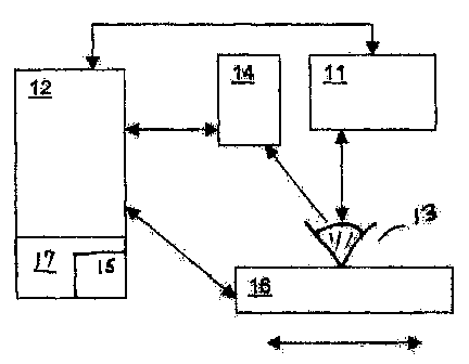
FIGURE 1 is a simplified block diagram illustrating one embodiment of the
eye lateral positioning system of the present invention;
FIGURE 2 is a flowchart of an exemplary embodiment of the method of the
present invention;
FIGURE 3 is an image of an eye with the pupil de-centered; and
FIGURE 4 is an image of the eye with the pupil centered in accordance with
the teachings of this invention.
-4-
CA 02615771 2008-01-17
WO 2007/016132 PCT/US2006/028936
DETAILED DESCRIPTION OF THE PREFERRED EMBODIMENTS
A description of the preferred embodiments of the present invention will now
be presented with reference to FIGUREs 1-4. An exemplary eye positioning
system
10 is depicted schematically in FIGURE 1, and an exemplary method 100, in
FIGUREs 2a and 2b.
An embodiment 100 of the method for determining an optimal position of an
eye 13 relative to an ophthalmic device 11 comprises the step of receiving
data into a
processor 12 (block 102). The data comprise an image of a surface of an eye 13
that
has been collected (block 101) with, for example, a video camera, digital
camera, still
camera or frame grabber 14, in communication with the processor 12. The image
is
collected with the eye at a first position relative to the ophthalmic device
11 (block
101), and typically coinprises a plurality of pixels, with each pixel having
an intensity
value associated therewith. Ophthalmic device 11 can be, for example, and
without
limitation, a femtosecond laser microkeratome, a treatment laser, such as an
excimer
laser, an aberrometer, or any other ophthalmic device, as will be known to
those
having skill in the art, for which accurate lateral positioning of an eye may
be
required.
A software package 15, which can be resident in a memory 17 (here shown as
part of processor 12), includes a code segment for locating an edge feature in
the
image (block 103). Memory 17 can be a separate memory operably coupled to
processor 12, or can be an integral part of processor 12. The edge feature may
include, but is not intended to be limited to, a pupil feature or a feature of
the iris.
Processor 12 (control circuit) may be a single processing device or a
plurality
of processing devices. Such a processing device may be a microprocessor, micro-
controller, digital signal processor, microcomputer, central processing unit,
field
programmable gate array, programmable logic device, state machine, logic
circuitry,
analog circuitry, digital circuitry, and/or any device that manipulates
signals (analog
and/or digital) based on operational instructions. The memory 17 coupled to
the
processor 12 or control circuit may be a single memory device or a plurality
of
memory devices. Such a memory device may be a read-only memory, random access
memory, volatile memory, non-volatile memory, static memory, dynamic memory,
flash memory, cache memory, and/or any device that stores digital information.
Note
that when the microprocessor or control circuit implements one or more of its
fiinctions via a state machine, analog circuitry, digital circuitry, and/or
logic circuitry,
-5-
CA 02615771 2008-01-17
WO 2007/016132 PCT/US2006/028936
the memory storing the corresponding operational instructions may be embedded
within, or external to, the circuitry comprising the state machine, analog
circuitry,
digital circuitry, and/or logic circuitry. The memory stores, and the
microprocessor or
control circuit executes, operational instructions (e.g., software package 15)
corresponding to at least some of the steps and/or functions illustrated and
described
in association with FIGUREs 2A and 2B.
The image is mapped from the edge feature to laterally define the pupil, for
example, by scanning from the edge feature to locate a darkest region in the
image.
This may be accomplished in an exemplary method by setting a rectangular area,
or
"window," that has a predefined size significantly smaller than a size of the
image,
but sufficiently large to contain a plurality of pixels (block 104). This
rectangular area
is "slid" across the image, scanning every row until substantially the entire
image has
been scanned (block 105). For each of the rectangular areas, the intensity
values of
each pixel within that area are summed (block 106), yielding an intensity
value for
each of a plurality of regions within the image. A region having a smallest
intensity
value comprises a darkest region and is assigned to contain at least a portion
of the
pupil (block 107).
Next, the image is scanned radially outward from a central pixel of the
darkest
region (block 108). The intensity value of each subsequent pixel is compared
with the
intensity value of the central pixel (block 109). If the intensity value of
the currently
examined pixel is equal to or less than the central pixel's intensity value,
the program
continues to the next radially outward pixel (block 108). If the intensity
value of the
currently examined pixel is greater than the central pixel's intensity value,
the current
pixel is considered to define a point on the pupil boundary (block 110).
This procedure is repeated a predetermined number of times (block 111) along
different radii (block 112), with the pupil boundary points collectively
defining the
pupil boundary (block 113). A center of the pupil can then be determined from
the
boundary points (block 114), as illustrated in FIGURE 3. The pupil center
comprises
a location from which to achieve an optimal lateral eye position relative to
the
ophthalmic device 11. An optimal lateral position can be any position that
places the
eye such that the ophthalmic device 11 can be therapeutically effective in its
designed
for purpose. Optimal lateral positioning can include positioning the eye such
that the
ophthalmic device 11 can perform to the limits of its design tolerances, as
well as
anywhere in the ophthalmic devices designed for therapeutically effective
range. An
-6-
CA 02615771 2008-01-17
WO 2007/016132 PCT/US2006/028936
optimal lateral position can be a preferred lateral position of an eye
relative to an
ophthalmic device.
If the eye is in a position other than an optimal lateral position (block
115), as
determined from the determined pupil center and the intended ophthalmic device
11
operating parameters, the eye and the ophthalmic device 11 are relatively
repositioned
(block 116) to place the eye in the optimal lateral position (block 117), as
illustrated
in FIGURE 4. Such repositioning may be effected manually or automatically
under
control of the software 15 and processor 12, by means which will be familiar
to those
having skill in the art and which are intended to be within the scope of the
present
invention, such as by using a positioning device 16. For example, and without
limitation, the patient can be manually repositioned, the ophthalmic device 11
can be
manually repositioned, and/or the ophthalmic device 11 or table/chair (e.g.,
positioning device 16) on which the patient is being supported can be
automatically
repositioned by mechanical and electrical control systems, or any combination
of
these methods. Once the eye is in the desired position, a desired procedure
can be
performed on the eye 13 using the ophthalmic device 11. The embodiments of
this
invention thus provide a pupil center reference point from which an optimal
positioning of an eye and a treating ophthalmic device 11 can be determined.
In the foregoing description, certain terms have been used for brevity,
clarity,
and understanding, but no unnecessary limitations are to be implied therefrom
beyond
the requireinents of the prior art, because such words are used for
description
purposes herein and are intended to be broadly construed. Moreover, the
embodiments of the apparatus illustrated and described herein are by way of
example,
and the scope of the invention is not limited to the exact details of
construction.
-7-
