Note : Les descriptions sont présentées dans la langue officielle dans laquelle elles ont été soumises.
CA 02621154 2008-02-29
WO 2007/028098 PCT/US2006/034367
PRC7STPIETIC INTERVERTEBRAL DISCS
Background of the Invention
The intervertebral disc is an anatomically and functionally complex joint. The
intervertebral disc is composed of three component structures: (1) the nucleus
pulposus; (2)
the annulus fibrosus; and (3) the vertebral endplates. The biomedical
composition and
anatomical arrangements within these component structures are related to the
biomechanical
function of the disc.
The spinal disc may be displaced or damaged due to trauma or a disease
process. If
displacement or damage occurs, the nucleus pulposus may herniate and protrude
into the
vertebral canal or intervertebral foramen. Such deformation is known as
herniated or slipped
disc. A herniated or slipped disc may press upon the spinal nerve that exits
the vertebral canal
through the partially obstructed foramen, causing pain or paralysis in the
area of its
distribution.
To alleviate this condition, it may be necessary to surgically remove the
involved disc
and fuse the two adjacent vertebrae. In this procedure, a spacer is inserted
in the place
originally occupied by the disc and additional fixation devices, such as
plates and rods, may be
added to provide increased stability. Despite the excellent short-term results
of such a "spinal
fusion" for traumatic and degenerative spinal disorders, long-term studies
have shown that
alteration of the biomechanical environment leads to degenerative changes at
adjacent mobile
levels. The adjacent discs have increased motion and stress due to the
increased stiffness of the
fused segment. In the long term, this change in the mechanics of the motion of
the spine
causes these adjacent discs to degenerate.
To circumvent this problem, an artificial intervertebral disc replacement has
been
proposed as an alternative approach to spinal fusion. Although various types
of artificial
intervertebral discs have been developed to restore the normal kinematics and
load-sharing
properties of the natural intervertebral disc, they can be grouped into two
categories: ball and
socket joint type discs and elastomer type discs.
Artificial discs of ball and socket type are usually composed of metal plates,
one to be
attached to the upper vertebra and the other to be attached to the lower
vertebra, and a
polyethylene or metal bearing surface working as a ball. The metal plates may
have concave
areas to house the bearing surface. The ball and socket type allows free
rotation or movement
between the vertebrae between which the disc is installed and thus has no load
sharing
capability against bending and translation. (Some ball and socket type
artificial discs have
rotation limiting features, which still do not address appropriate torque for
a natural disc.)
1
CA 02621154 2008-02-29
WO 2007/028098 PCT/US2006/034367
"'t~rfitiil M'c~ {RITH-lig tjN~ale''S very high stiffness in the vertical
direction; they cannot
replicate the normal compressive stiffness of the natural disc. Also, the lack
of load bearing
capability in these types of discs causes adjacent discs to bear the extra
load, resulting in the
eventual degeneration of the adjacent discs and facets. These types of discs
also cannot
replicate a natural disc's instantaneous access of rotation (IAR) as a direct
result of lacking
natural compressibility.
In elastomer type artificial discs, an elastomeric polymer is between metal
plates and
these metal plates are fixed to the upper and the lower vertebrae. The
elastomeric polymer
may be bonded to the metal plates by having the interface surface of the metal
plates be rough
and porous. This type of disc can absorb a shock in the vertical direction and
has a load
bearing capability. However, this structure has a problem in the interface
between the
elastomeric polymer and the metal plates. Even though the interface surfaces
of the metal
plates may be treated for better bonding, polymeric debris may nonetheless be
generated after
long term usage. Furthermore, the bond of the elastomer to the metal substrate
tends to fail
after a long usage because of its insufficient shear-fatigue strength.
Because of the above described disadvantages associated with either the ball
and socket
or elastomer type discs, there has existed a continued need for the
development of new
prosthetic devices. Several such new prosthetic devices are described in
United States Patent
Application Serial No. 10/632,538, filed August 1, 2003, and United States
Patent Application
Serial No. 10/903,276, filed July 30, 2004, each of which applications is
hereby incorporated
by reference herein. The foregoing applications describe, inter alia,
prosthetic intervertebral
discs that include an upper endplate, a lower endplate, and a compressible
core member
disposed between the two endplates. Several prosthetic disc embodiments are
described,
including single-piece, two-piece, three-piece, and four-piece structures.
While such prosthetic intervertebral discs and methods for their use show
great
promise, there remains a need for improved prosthetic discs and methods for
their use.
Relevant Literature
United States PatentNos. 3,867,728; 4,911,718; 5,039,549; 5,171,281;
5,221,431;
5,221,432; 5,370,697; 5,545,229; 5,674,296; 6,162,252; 6,264,695; 6,533,818;
6,582,466;
6,582,468; 6,626,943; 6,645,248. Also of interest are published United States
Patent
Application Nos. 2002/0107575, 2003/ 0040800, 2003/0045939, and 2003/0045940.
See also
Masahikio Takahata, Uasuo Shikinami, Akio Minami, "Bone Ingrowth Fixation of
Artificial
Intervertebral Disc Consisting of Bioceramic-Coated Three-dimensional Fabric,"
SPINE, Vol.
28, No. 7, pp. 637-44 (2003).
2
CA 02621154 2008-02-29
WO 2007/028098 PCT/US2006/034367
Prosthetic intervertebral discs and methods for using such discs are provided.
The
subject prosthetic discs typically include an upper endplate, a lower
endplate, and a
compressible core member disposed between the two endplates.
In several embodiments, the subject prosthetic discs are characterized by
including top
and bottom endplates separated by a compressible element. The two plates are
held together by
at least one fiber wound around at least one region of the top endplate and at
least one region of
the bottom endplate. The fibers are generally high tensile strength fibers
with a high modulus
of elasticity and high wear resistance. The elastic properties of the fibers,
as well as factors
such as the number of fibers used, the thickness of the fibers, the number of
layers of fiber
windings, the tension applied to each layer, and the crossing patCern of the
fiber windings
enable the prosthetic disc structure to mimic the functional characteristics
and bioniechanics of
a normal-functioning, natural disc. Alternatively, the two plates are held
together by an
engagement mechanism connecting each plate to the compressible element. The
subject discs
may be employed with separate vertebral body fixation elements, or they may
include
integrated vertebral body fixation elements.
Several optional core materials and structures may be incorporated in each of
the
prosthetic disc embodiments described herein. For example, the core member may
be formed
of an appropriately stiff material, such as polyurethane or silicone, and is
typically fabricated
by injection or compression molding. In other examples, the core member may be
formed by
layers of fabric woven from fibers. In still further examples, the core member
may comprise a
combina.tion of these materials, such as a fiber-reinforced polyurethane or
silicone. As an
additional option, one or more spring members may be placed between the upper
and lower
endplates in combination with the core member, such as in a coaxial
relationship in which the
core member has a generally cylindrical or toroidal shape and a spring is
located at its center.
In other embodiments, the core structure comprises two or more core members
having
different load bearing properties and having the ability to vary the center of
rotation of the core
structure. The varying properties of the core members may be provided by
selection of
materials, construction, or other features. In still further embodiments, the
core structure
comprises one or more core members that are formed of materials or are
otherwise constructed
to provide varying stiffness or other material properties to accommodate
different loads or
loading configurations. Examples of these core structures include cores having
discrete
portions formed of different materials, cores having grooves or other features
formed on
3
CA 02621154 2008-02-29
WO 2007/028098 PCT/US2006/034367
-pdi'ti&is o'f''tlie 'co'r"e'mem.be'rTo'r"btner purposes (such as
sterilization), and cores having coils or
couplers attached to or formed integrally with the core member.
In still further embodiments, the core structure is provided with one or more
mechanisms adapted to adjust the size, shape, orientation, or other feature or
combination of
features of the core member. For example, the core member may include threads,
slots and
tabs, or other mechanisms that provide the ability to adjust the height of the
core, or to adjust
other properties of the core.
Several particularly preferred core structures include a llollow member that
is adapted
to be inflated after iunplantation of the prosthetic disc. In this way, the
prosthetic disc is
provided with a contracted condition (core uninflated) for delivery and
implantation of the disc,
and an expanded condition (core inflated) that is adapted for use by the
patient after
implantation. These core structures may be provided with a fluid port that is
adapted to
facilitate inflation of the core. Alternatively, a fluid communication lumen
may be provided
that extends from the hollow core member and provides a lumen through which
inflation media
may be injected into the core. The hollow core may be provided with two or
more
compartments, each of which may be independent, or which may be in fluid
communication
with one another.
Several optional endplates and related mechanisms may be incorporated in each
of the
prosthetic disc embodiments described herein. For example, the endplates may
be curved or
kidney bean shaped to facilitate rotation of the disc within the
intervertebral void space.
Alternatively, the endplates may be of a partially cylindrical shape adapted
to engage and
retain a substantially cylindrical core member.
Other and additional devices, apparatus, structures, and methods are described
by
reference to the drawings and detailed descriptions below.
Brief Descriptions of the Fig;ures
The Figures contained herein are not necessarily drawn to scale, with some
components
and features being exaggerated for clarity.
Figures 1A and 1B provide a three dimensional view of two different prosthetic
discs
according to the subject invention.
Figure 2 provides a three-dimensional view of a fibrous compressible element
that
includes a polymeric nucleus and a fibrous annulus according to one embodiment
of the subject
invention.
Figure 3 provides a three-dimensional cross-sectional view of a prosthetic
disc.
Figures 4A-B provide three-dimensional views of two embodiments of a core
member.
4
CA 02621154 2008-02-29
WO 2007/028098 PCT/US2006/034367
'Fi~uf"e"4C p"r6vid"es'aerid'view of a core member located between a pair of
endplates.
Figures 5A-B provide side views of prosthetic discs having cores formed of a
plurality
of core members.
Figures 6A-N and 6P-T provide illustrations of several embodiments of core
members
suitable for use in prosthetic discs described herein.
Figures 7-10 provide illustrations of several embodiments of adjustable core
structures.
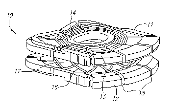
Figure 11 provides a top view of an endplate.
Figures 12A-B provide illustrations of implantation methods for prosthetic
discs having
endplates such as that shown in FIG. 11.
Figure 13 provides a perspective view of a prosthetic disc having a generally
elongated
tubular core member. '
Figures 14A-D provide illustrations of a selectably expandable prosthetic disc
and its
components.
Figures 15A-B provide illustrations of a prosthetic disc having an elongated
tubular
core member.
Figures 16A-C, 17A-B, 18A-C, and 19A-C provide illustrations of prosthetic
discs that
are constructed to mimic the physiology of the natural functional spinal unit.
Figures 20 and 2lA-B provide illustrations of two-piece endplates including
inner
endplates and outer endplates.
Figures 22A-D provide illustrations of a prosthetic disc having a plurality of
fixed
anchoring fins on its outer surface.
Figures 23A-B provide illustrations of a partially cylindrical endplate and a
removable
keel.
Figures 24A-B and 25A-C provide illustrations of selectively deployable
fixation
screws and associated mechanisms.
Figures 26A-C provide illustrations of another prosthetic disc fixation
mechanism.
Figures 27A-C provide illustrations of an insertable keel structure.
Figure 28 provides an illustration of a fiber winding construction for
attaching upper
and lower endplates of a prosthetic disc.
Figures 29A-B provide illustrations of a system for maintaining a prosthetic
disc in a
low profile condition during an implantation procedure.
Figure 30 provides an illustration of a core structure adapted for use in a
prosthetic disc.
5
CA 02621154 2008-02-29
WO 2007/028098 PCT/US2006/034367
"Figui'e'"s 3'1A=D provid"e illustrations of spinal motion preservation
systems.
Figures 32A-B provide illustrations of disc interlocking mechanisms.
Figures 33A-C provide illustrations of prosthetic discs adapted to be deployed
in an
approximately X-shaped configuration.
Figures 34A-B provide illustrations showing a surgical method for implanting a
prosthetic disc.
Figures 35A-D provide illustrations showing another surgical method for
implanting a
prosthetic disc.
Figures 36A-I provide illustrations of ineclianisms for attaching a pair of
adjacent
prosthetic discs.
Figures 37A-F provide illustrations showing another surgical method for
implanting a
prosthetic disc.
Figures 38A-F provide illustrations of several embodiments of generally "J"-
shaped
prosthetic discs.
Figure 39 provides an illustration of an encapsulated spring disc replacement
system.
Descriptions of the Preferred Embodiments
Before the present invention is described, it is to be understood that this
invention is not
limited to particular embodiments described, as such may, of course, vary. It
is also to be
understood that the terminology used herein is for the purpose of describing
particular
embodiments only, and is not intended to be limiting, since the scope of the
present invention
will be limited only by the appended claims.
Where a range of values is provided, it is understood that each intervening
value, to at
least the tenth of the unit of the lower limit unless the context clearly
dictates otherwise,
between the upper and lower limit of that range and any otlier stated or
intervening value in
that stated range is encompassed within the invention. The upper and lower
limits of these
smaller ranges may independently be included in the smaller ranges is also
encompassed
within the invention, subject to any specifically excluded limit in the stated
range. Where the
stated range includes one or both of the limits, ranges excluding either or
both of those
included limits are also included in the invention.
Unless defined otherwise, all technical and scientific terms used herein have
the same
meaning as commonly understood by one of ordinary skill in the art to which
this invention
belongs. Although any methods and materials similar or equivalent to those
described herein
can also be used in the practice or testing of the present invention, the
preferred metliods and
6
CA 02621154 2008-02-29
WO 2007/028098 PCT/US2006/034367
' miltei'ials 'a'r''e"no"'wescn"Cied.'-A7T'publications mentioned herein are
incorporated herein by
reference to disclose and describe the methods and/or materials in connection
with which the
publications are cited.
It must be noted that as used herein and in the appended claims, the singular
forms "a",
"an," and "the" include plural referents unless the context clearly dictates
otherwise.
The publications discussed herein are provided solely for their disclosure
prior to the
filing date of the present application. Nothing herein is to be construed as
an admission that
the present invention is not entitled to antedate such publication by virtue
of prior invention.
Further, the dates of publication provided may be different from the actual
publication dates
which may need to be independently confirmed.
As will be apparent to those of skill in the art upon reading this disclosure,
each of the
individual embodiments described and illustrated herein has discrete
components and features
which may be readily separated from or combined with the features of any of
the other several
embodiments without departing from the scope or spirit of the present
inventions.
I. OVERVIEW OF THE DESCRIBED PROSTHETIC INTERVERTEBRAL DISCS
Prosthetic intervertebral discs, methods of using such discs, apparatus for
implanting
such discs, and methods for implanting such discs are described herein. It is
to be understood
that the prosthetic intervertebral discs, implantation apparatus, and methods
are not limited to
the particular embodiments described, as these may, of course, vary. It is
also to be understood
that the terminology used herein is for the purpose of describing particular
embodiments only,
and is not intended to be limiting, since the scope of the present inventions
will be limited only
by the appended claims.
The prosthetic intervertebral discs are preferably artificial or manmade
devices that are
configured or shaped so that they can be employed as replacements for an
intervertebral disc in
the spine of a vertebrate organism, e.g., a mammal, such as a human. The
subject prosthetic
intervertebral discs have dimensions that permit them to substantially occupy
the space
between two adjacent vertebral bodies that is present when the naturally
occurring disc
between the two adjacent bodies is removed, i.e., a disc void space. By
substantially occupy is
meant that the prosthetic disc occupies a sufficient volume in the space
between two adjacent
vertebral bodies that the disc is able to perform some or all of the functions
performed by the
natural disc for which it serves as a replacement. In certain embodiments,
subject prosthetic
discs may have a roughly bean shaped structure analogous to naturally
occurring intervertebral
body discs. In many embodiments, the length of the prosthetic discs range from
about 5 mm to
about 40 mm, preferably from about 10 mm to about 25 mm, the width of the
prosthetic discs
7
CA 02621154 2008-02-29
WO 2007/028098 PCT/US2006/034367
"rafxge'fr''oriY''Woti$ mm, preferably from about 10 mm to about 35 mm, and
the height of the prosthetic discs range from about 2 mm to about 15 mm,
preferably from
about 5 mm to about 12 mm.
The subject discs are characterized in that they typically include both an
upper (or top)
and lower (or bottom) endplate or bone interfacing structure (e.g., contiguous
plates,
interrupted plates, spikes, keels, porous surfaces, and the like), where the
upper and lower
endplates are separated from each other by a compressible element, where the
combination
structure of the endplates and compressible element provides a prosthetic disc
that functionally
closely mimics real disc. A feature of some of the subject prostlietic discs
is that the top and
bottom endplates are held together by at least one fiber, e.g., of the fibrous
compressible
element, wound around at least one portion of each of the top and bottom
endplates. As such,
in these embodiments, the two endplates (or substrates) are held to each other
by one or more
fibers that are wrapped around at least one domain/portion/area of the upper
endplate and lower
endplate such that the plates are joined to each other.
Also provided are methods of using the subject prosthetic intervertebral
discs. The
subject prosthetic intervertebral discs find use in the replacement of damaged
or dysfunctional
intervertebral discs in vertebrate organisms. Generally the vertebrate
organisms are "mammals"
or "mammalian," where these terms are used broadly to describe organisms which
are within
the class mammalia, including the orders carnivore (e.g., dogs and cats),
rodentia (e.g., mice,
guinea pigs, and rats), lagomorpha (e.g., rabbits) and primates (e.g., humans,
chimpanzees, and
monkeys). In many embodiments, the subjects will be humans.
In general, the devices are employed by first removing part or all of the
native disc to
be replaced from the subject or patient according to typical surgical
technique to produce a disc
void space. Next, the subject prosthetic disc is implanted or positioned in
the disc void space,
resulting in replacement of the removed disc with the prosthetic disc. This
implantation step
may include a vertebral body fixation element implantation substep, a post
implantation
vertebral body securing step, or other variations, depending on the particular
configuration of
the prosthetic device being employed. In addition, the implantation step
described above may
include use of one or more implantation devices (or disc delivery devices) for
implanting the
system components to the site of implantation.
Two different representative intervertebral discs are shown in Figures 1A and
1B.
These discs, and others, are also described more fully in United States Patent
Application
Serial No. 10/632,538, filed August 1, 2003, ("the '538 application"), and
United States Patent
Application Serial No. 10/903,276, filed July 30, 2004, ("the '276
application"), each of which
8
CA 02621154 2008-02-29
WO 2007/028098 PCT/US2006/034367
applicatioris is iric6rporafed by 'r'eference herein. A substantial portion of
this overview
description, including FIGS. lA-B, 2, and 3, is adapted from portions of the
'276 application.
As can be seen in Figures lA and 1B, prosthetic discs 10 each include a top
endplate 11
and a lower endplate 12. Top and bottom endplates 11 and 12 are substantially
planar
substrates, where these plates typically have a length from about 5mm to about
40mm, such as
from about 10nun to about 25mm, a width of from about 2mm to about 50mm, such
as from
about 10mm to about 35mm and a thickness of from about 0.25mm to about 6mm,
such as
from about lmm to about 4mm. The top and bottom endplates or equivalent are
fabricated
from a biocompatible material that also provides for the requisite mechanical
properties, where
representative materials from whicli the endplates may be fabricated are known
to those of skill
in the art and include, but are not limited to: titanium, titaniunl alloys,
stainless steel,
cobalt/chromium/molybdenum alloys, multiphase alloys such as MP-35N, etc.;
plastics such as
polyethylene with ultra high molar mass (molecular weight) (UHMWPE), polyether
ether
ketone (PEEK), etc.; ceramics; graphite; etc. As shown in Figures 1A and 1B,
separating the
top and bottom endplates is a compressible element 17. The thickness of the
compressible
element may vary, but ranges in many embodiments from about 1mm to about 15mm,
including from about 2mm to about 10mm.
The disc is further characterized in that it includes an annular region 13
(i.e., annulus),
which is the region, domain or area that extends around the periphery of the
disc, and a nuclear
region (i.e., nucleus) 14, which is the region, domain or area in the center
of the disc and
surrounded by the annulus.
As shown in Figures 1 A and 1 B, the plates include a single region around
which a fiber
is wound in order to hold the plates together, although in many embodiments
the plates have a
plurality of such regions. As shown in Figures 1A and 1B, endplates 11 and 12
include a
plurality of slots 15 through which fibers, e.g., of the fibrous compressible
element, may be
passed through or wound, as shown. In many embodiments, the number of
different slots
present in the periphery of the device ranges from about 4 to about 36, such
as from about 5 to
about 25. As shown in Figures lA and 1B, at least one fiber 16 forming part of
the
compressible eleinent is wrapped around a region of the top and bottom plates,
e.g., by being
passed through slots in the top and bottom plates, in order to hold the plates
together.
The compressible elements, 17, are typically made up of one or more fibers,
where the
fibers are generally high tenacity fibers with a high modulus of elasticity
and high wear
resistance. By high tenacity fibers is meant fibers that can withstand a
longitudinal stress
without tearing asunder of at least about 50 MPa, such as at least about
250MPa. As the fibers
9
CA 02621154 2008-02-29
WO 2007/028098 PCT/US2006/034367
"hd-* & hig1[''ftlodu1u's"ot"Ma9ticity; their modulus of elasticity is
typically at least about
100MPa, usually at least about 500MPa. The fibers are generally elongate
fibers having a
diameter that ranges from about 0.1mm to about 5mm, such as about 0.2mm to
about 2mm,
where the length of each individual fiber making up the fibrous component may
range from
about 0.1m to about 20m, such as from about 0.3m to about 3m.
The fibers making up the fibrous compressible elements may be fabricated from
any
suitable material, where representative materials of interest include, but are
not limited to:
metals, including alloys, polymers, including polyester (e.g., Dacron),
polyethylene,
polyaramid, polytetrafluoroethylene, carbon or glass fibers, polyethylene
terephthalate, arciylic
polymers, methacrylic polymers, polyurethane, polyurea, polyolefin,
halogenated polyolefin,
polysaccharide, vinylic polymer, polyphosphazene, polysiloxane, nylon, and the
like.
The fibrous compressible elements made up of one or more fibers wound around
one or
more regions of the top or bottom plates may make up a variety of different
configurations. For
example, the fibers may be wound in a pattern that has an oblique orientation
to simulate the
annulus of intact disc, where a representative oblique fiber configuration or
orientation is
shown in Figure lA. The number of layers of fiber winding may be varied to
achieve similar
mechanical properties to an intact disk. Where desired, conzpliancy of the
structure may be
reduced by including a horizontal winding configuration, as shown in Figure
1B.
In certain embodiments, the fibrous compressible element 20 has a fibrous
component
21 limited to the annular region of the disc 22, e.g., to the region along the
periphery of the
disc. Figure 2 provides a representation of this embodiment, where the fibrous
component is
limited solely to the annular region of the disc and includes both oblique and
horizontal
windings. Also shown is a separate polymeric component 23 present in the
nucleus. The fiber
windings of the various layers of fiber may be at varying angles from each
other where the
particular angle for each layer may be selected to provide a configuration
that best mimics the
natural disc. Additionally, the tension placed on the fibers of each layer may
be the same or
varied.
In yet other embodiments the fibrous component of the fibrous compressible
element
may extend beyond the annular region of the disc into at least about a
portion, if not all, of the
nucleus.
In certain embodiments, the fibrous compressible element further includes one
or more
polymeric components. The polymeric component(s), when present, may be
fabricated from a
variety of different physiologically acceptable materials. Representative
materials of interest
include, but are not limited to: elastomeric materials, such as
polydimethylsiloxane,
CA 02621154 2008-02-29
WO 2007/028098 PCT/US2006/034367
pdi'ycatbonafe=polyureth'ane, aromatic and aliphatic polyurethanes,
poly(ethylene propylene)
copolymer, polyvinylchloride, poly(tetrafluoro ethylene) and copolymers,
hydrogels, and the
like.
The polymeric component may be limited to particular domains, e.g., the
annular and/or
nucleus domains, or extend throughout the entire region of the fibrous
compressible elements
positioned between the two endplates. As such, in certain embodinlents the
polymeric
component is one that is limited to the nuclear region of the disc, as shown
in Figure 2. In
Figure 2, fibrous compressible element 20 includes a distinct fibrous
component 21 that is
located in the annular region of the disc 22, while polymeric component 23 is
located in the
nuclear region of the disc. In other embodiments, the polymeric component is
located in both
the annulax and nuclear regions. In yet other embodiments, the polymeric
component may be
located solely in the annular region.
Depending on the desired configuration and mechanical properties, the
polymeric
component may be integrated with the fibrous component, such that at least a
portion of the
fibers of the fibrous component is embedded in, e.g., complexed with, at least
a portion of the
polymeric component. In other words, at least a portion of the fibrous
component is
impregnated with at least a portion of the polymeric component. For example,
stacked two-
dimensional layers of the fibrous component may be present inside the
polymeric component,
such that the fibrous component is impregnated with the polymeric component.
In those configurations where the fibrous and polymeric components are present
in a
combined format, the fibers of the fibrous component may be treated to provide
for improved
bonding with the polymeric component. Representative fiber treatments of
interest include, but
are not limited to: corona discharge, 02 plasma treatment, oxidation by strong
acid (HNO3,
H2S04). In addition, surface coupling agents may be employed, and/or a monomer
mixture of
the polymer may be polymerized in presence of the surface-modified fiber to
produce the
composite fiber/polymeric structure. Additionally, the fiber may be of a
composite
construction with an outer layer composed of a material optimized for surface
coupling. The
composite structure can also be composed of an outer jacket that provides
bonding to the
polymeric component but allows the relative motion of the fibrous component
within the
jacket.
As indicated above, the devices may include one or more different polymeric
components. In those embodiments where two or more different polymeric
components are
present, any two given polymeric components are considered different if they
differ from each
other in terms of at least one aspect, e.g., composition, cross-linking
density, and the like. As
11
CA 02621154 2008-02-29
WO 2007/028098 PCT/US2006/034367
sdCh,"the two"o'r"'riiore diffe"reri't'polymeric components may 76e fabricated
from the same
polymeric molecules, but differ from each other in terms of one or more of:
cross-linking
density; fillers; etc. For example, the same polymeric material may be present
in both the
annulus and nucleus of the disc, but the crosslink density of the annulus
polymeric component
may be higher than that of the nuclear region. In yet other embodiments,
polymeric materials
that differ from each other with respect to the polymeric molecules from which
they are made
may be employed.
By selecting particular fibrous component and polymeric component materials
and
configurations, e.g., froni the different representative fomiats described
above, a disc with
desired functional characteristics, e.g., that mimics the functional
characteristics of the
naturally occurring disc, may be produced.
Representative particular combinations of interest include, but are not
limited to, the
following:
1. Biocompatible polyurethane, such as Ethicon Biomer, reinforced with Dacron
poly(ethylene terephthalate) fiber, or Spectra polyethylene fiber, or Kevlar
polyaramide fiber,
or carbon fiber.
2. Biocompatible polysiloxane modified styrene-ethylene butylene block
copolymer sold
under C-Flex tradename reinforced with Dacron poly(ethylene terephthalate)
fiber, or Spectra
polyethylene fiber, or Kevlar polyaramide fiber, or carbon fiber.
3. Biocompatible Silastic silicone rubber, reinforced with Dacron
poly(ethylene
terephthalate) fiber, or Spectra polyethylene fiber, or Kevlar polyaramide
fiber, or carbon fiber.
In using the subject discs, the prosthetic disc is fixed to the vertebral
bodies between
which it is placed. More specifically, the upper and lower plates of the
subject discs are fixed
to the vertebral body to which they are adjacent. As such, the subject discs
are employed with
vertebral body fixation elements during use. In certain embodiments, the
vertebral body
fixation elements are integral to the disc structure, while in other
embodiments the vertebral
body fixation elements are separate from the disc structure.
Another representative prosthetic intervertebral disc 100 is shown in Figure
3, and is
also described more fully in the '276 application. The prosthetic disc 100 has
an integrated
structure that includes an upper endplate 110, a lower endplate 120, and a
core member 130
retained between the upper endplate 110 and the lower endplate 120. One or
more fibers 140
are wound around the upper and lower endplates to attach the endplates to one
another. The
wind of the fibers 140 allows a degree of axial rotation, bending, flexion,
and extension by and
between the endplates. The core member 130 may be provided in an uncompressed
or a pre-
12
CA 02621154 2008-02-29
WO 2007/028098 PCT/US2006/034367
compre'ssed slate: An arinu'la'r" capsule 150 is optionally provided in the
space between the
upper and lower endplates, surrounding the core meniber 130 and the fibers
140. The upper
endplate 110 and lower endplate 120 are generally flat, planar members, and
are fabricated
from a biocompatible material that provides substantial rigidity. Examples of
materials
suitable for use in fabricating the upper endplate 110 and lower endplate 120
include titanium,
titanium alloys, stainless steel, cobalt/chromium/molybdenum, etc., which are
manufactured by
machining, forging, casting or metal injection molding; plastics such as
polyethylene with ultra
high molar mass (molecular weight) (UHMWPE), polyether ether ketone (PEEK),
etc., which
are manufactured by injection molding or compression molding; ceramics;
graphite; and
others. Optionally, the endplates may be coated with hydroxyapatite, titanium
plasma spray, or
other coatings to enhance bony ingrowth.
As noted above, the upper and lower endplates typically have a length of from
about 5
mm to about 40 mm, preferably from about 10 mm to about 25 mm, a width of from
about 2
mm to about 50 mm, preferably from about 10 mm to about 35 mm, and a thickness
of from
about 0.25 mm to about 6 mm, preferably from about 1 mm to about 4 mm. The
sizes of the
upper and lower endplates are selected primarily based upon the size of the
void between
adjacent vertebral bodies to be occupied by the prosthetic disc. Accordingly,
while endplate
lengths and widths outside of the ranges listed above are possible, they are
not typical. The
upper surface of the upper endplate 110 and the lower surface of the lower
endplate 120 are
preferably each provided with a mechanism for securing the endplate to the
respective opposed
surfaces of the upper and lower vertebral bodies between which the prosthetic
disc is to be
installed. For example, in Figure 3, the upper endplate 110 includes a
plurality of anchoring
fins 111 a-b. The anchoring fins 111 a-b are intended to engage mating grooves
that are formed
on the surfaces of the upper and lower vertebral bodies to thereby secure the
endplate to its
respective vertebral body. The anchoring fins 111 a-b extend generally
perpendicularly from
the generally planar external surface of the upper endplate I 10, i.e., upward
from the upper
side of the endplate as shown in Figure 3. In the Figure 3 embodiment, the
upper endplate 110
includes three anchoring fins l l la-c, although only two are shown in the
cross-sectional view.
A first of the anchoring fins, 111 a, is disposed near an external edge of the
external surface of
the upper endplate and has a length that approximates the width of the upper
endplate 110. A
second of the anchoring fiiis, 111b, is disposed at the center of external
surface of the upper
endplate and has a relatively shorter length, substantially less than the
width of the upper
endplate 110. Each of the anchoring fins 111 a-b has a plurality of serrations
1121ocated on the
top edge of the anchoring fin. The serrations 112 are intended to enhance the
ability of the
13
CA 02621154 2008-02-29
WO 2007/028098 PCT/US2006/034367
aridhoriing"tin to brigage the vertebral body and to thereby secure the upper
endplate 110 to the
spine.
Similarly, the lower surface of the lower endplate 120 includes a plurality of
anchoring
fins 121a-b. The anchoring fins 121a-b on the lower surface of the lower
endplate 120 are
identical in structure and function to the anchoring fins l l la-b on the
upper surface of the
upper endplate I 10, with the exception of their location on the prosthetic
disc. The upper and
lower anchoring fins are not necessarily identical or similar; they could be
different from each
other in terms of geometry, size, or location. Such differences are used to
accommodate
anatomical differences between the superior and inferior vertebral bodies. The
anchoring fins
121 a-b on the lower endplate 120 are intended to engage mating grooves formed
on the lower
vertebral body, whereas the anclloring fins 111 a-b on the upper endplate I 10
are intended to
engage mating grooves on the upper vertebral body. Thus, the prosthetic disc
100 is held in
place between the adjacent vertebral bodies.
The anchoring fins 111, 121 may optionally be provided with one or more holes
or slots
115, 125. The holes or slots help to promote bony ingrowth that assist in
anchoring the
prosthetic disc 100 to the vertebral bodies.
The upper endplate 110 contains a plurality of slots 114 through which the
fibers 140
may be passed through or wound, as shown. The actual number of slots 114
contained on the
endplate is variable. Increasing the number of slots will result in an
increase in the
circumferential density of the fibers holding the endplates together. In
addition, the shape of
the slots may be selected so as to provide a variable width along the length
of the slot. For
example, the width of the slots may taper from a wider inner end to a narrow
outer end, or visa
versa. Additionally, the fibers may be wound multiple times within the same
slot, thereby
increasing the radial density of the fibers. In each case, this improves the
wear resistance and
increases the torsional and flexural stiffness of the prosthetic disc, thereby
further
approximating natural disc stiffness. In addition, the fibers 140 may be
passed through or
wound on each slot, or only on selected slots, as needed.
As described above, the purpose of the fibers 140 is to hold the upper
endplate 110 and
lower endplate 120 together and to limit the range-of-motion to mimic the
range-of-motion and
torsional and flexural resistance of a natural disc. Accordingly, the fibers
preferably comprise
high tenacity fibers with a high modulus of elasticity, for example, at least
about 100 MPa, and
preferably at least about 500 MPa. By high tenacity fibers is meant fibers
that can withstand a
longitudinal stress of at least 50 MPa, and preferably at least 250 MPa,
without tearing. The
fibers 140 are generally elongate fibers having a diameter that ranges from
about 100 m to
14
CA 02621154 2008-02-29
WO 2007/028098 PCT/US2006/034367
,==- . , r . ,.
alidu~ 1'000' m, anpreferalily'ab6ut 200 m to about 500 m. Optionally, the
fibers may be
processed (e.g., injection molded or extruded) with an elastomer to
encapsulate the fibers,
thereby providing protection from tissue ingrowth and improving torsional and
flexural
stiffness, or the fibers may be coated with one or more other materials to
improve fiber
stiffness and wear. Additionally, the core may be injected with a wetting
agent such as saline
to wet the fibers and facilitate the mimicking of the viscoelastic properties
of a natural disc.
The fibers 140 may be fabricated from any suitable material. Examples of
suitable
materials include polyester (e.g., Dacron ), polyethylene, polyaramid, poly-
paraphenylene
terephthalamide (e.g., Kevlar(D), carbon or glass fibers, polyethylene
terephthalate, acrylic
polymers, methacrylic polymers, polyurethane, polyurea, polyolefin,
halogenated polyolefin,
polysaccharide, vinylic polymer, polyphosphazene, polysiloxane, and the like.
The fibers 140 may be terminated on an endplate by tying a knot in the fiber
on the
superior surface of an endplate. Alternatively, the fibers 140 may be
terminated on an endplate
by slipping the terminal end of the fiber into a slot on an edge of an
endplate, similar to the
manner in wliich thread is retained on a thread spool. The slot may hold the
fiber with a crimp
of the slot structure itself, or by an additional retainer such as a ferrule
crimp. As a further
alternative, tab-like crimps may be machined into or welded onto the endplate
structure to
secure the terminal end of the fiber. The fiber may then be closed within the
crimp to secure it.
As a still further alternative, a polymer may be used to secure the fiber to
the endplate by
welding. The polymer would preferably be of the same material as the fiber
(e.g., PE, PET, or
the other materials listed above). Still further, the fiber may be retained on
the endplates by
crimping a cross-member to the fiber creating a T-joint, or by crimping a ball
to the fiber to
create a ball joint.
The core member 130 is intended to provide support to and to maintain the
relative
spacing between the upper endplate 110 and lower endplate 120. The core member
130 is
made of a relatively compliant material, for example, polyurethane or
silicone, and is typically
fabricated by injection molding. A preferred construction for the core member
includes a
nucleus formed of a hydrogel and an elastomer reinforced fiber annulus. For
example, the
nucleus, the central portion of the core member 130, may comprise a hydrogel
matei-ial such as
a water absorbing polyurethane, polyvinyl alcohol (PVA), polyethylene oxide
(PEO),
polyvinylpyrrolidone (PVP), polyacrylamide, silicone, or PEO based
polyurethane. The
annulus may comprise an elastomer, such as silicone, polyurethane or polyester
(e.g., Hytrel ),
reinforced with a fiber, such as polyethylene (e.g., ultra high molecular
weight polyethylene,
UHMWPE), polyethylene terephthalate, or poly-paraphenylene terephthalamide
(e.g.,
Kevlar ).
CA 02621154 2008-02-29
WO 2007/028098 PCT/US2006/034367
0 f'the'co're"memiber 130 is typically generally cylindrical or bean-shaped,
although the shape (as well as the materials making up the core member and the
core member
size) may be varied to obtain desired physical or performance properties. For
example, the
core member 130 shape, size, and materials will directly affect the degree of
flexion, extension,
lateral bending, and axial rotation of the prosthetic disc.
The annular capsule 150 is preferably made of polyurethane or silicone and may
be
fabricated by injection molding, two-part component mixing, or dipping the
endplate-core-fiiber
assembly into a polymer solution. A function of the annular capsule is to act
as a barrier that
keeps the disc materials (e.g., fiber strands) within the body of the disc,
and that keeps natural
in-growth outside the disc.
II. CORE STRUCTURES
Several alternative core structures are described hereinbelow. These core
structures are
preferably incorporated in one or more of the prosthetic intervertebral discs
constructed
according to the descriptions above, or they may be used or adapted for use
with other known
prosthetic discs.
Turning to Figures 4A-4C, a first alternative core structure is shown. The
core structure
includes a substantially cylindrical core member 150 that is configured to be
located between a
pair of endplates 110, 120 in a prosthetic intervertebral disc. The endplates
110, 120, as shown
in FIG. 4C, have a size, shape, and are made of materials such as any of those
described
elsewhere herein. The core member 150 is a solid, cylindrical structure having
a length and
width adapted to substantially occupy the internal volume of the prosthetic
disc between the
upper and lower endplates. The core 150 may comprise any one or more of the
materials
described above, including hydrogels, polyurethanes, polyvinyl alcohol (PVA),
polyethylene
oxide (PEO), polyvinylpyrrolidone (PVP), polyacrylamide, silicone, PEO based
polyurethane,
elastomers such as silicone, polyurethane, or polyester (e.g., Hytrel ),
reinforced with a fiber,
such as polyethylene (e.g., ultra high molecular weight polyethylene, UHMWPE),
polyethylene terephthalate, or poly-paraphenylene terephthalamide (e.g.,
Kevlar ).
In some preferred embodiments, the core member 150 includes an inner core
member
152 and an outer core member 154 as shown, for example, in FIG. 4B. The inner
152 and
outer 154 core members may be constructed of a single material, or they may be
constructed of
different materials, or they may be constructed of the same material having
different material
properties. When different materials or different material properties are
used, the performance
of the core 150 may be varied to obtain desired results. For example, a
relatively harder
material (i.e., higher durometer measurement) may be used to construct the
inner core member
16
CA 02621154 2008-02-29
WO 2007/028098 PCT/US2006/034367
157 wlule"a felatively soiter matenal (i.e., lower durometer measurement) is
used to construct
the outer core member 154. In this manner, the inner core member 152 is
adapted to provide a
primary source of support for the core member 150 and the outer core member
154 provides
compliance for the composite core structure.
Due to the substantially cylindrical shape of the core menlber 150, the
endplates 110,
120 each engage the core member 150 over a limited contact area along the
upper and lower
surface of the core member. The compressive loading that is applied to each of
the endplates is
applied perpendicular to the longitudinal axis of the cylindrical core member.
Additionally, as
the load on the upper I 10 and lower 120 endplates increases, the load bearing
contact areas
will enlarge due to the flattening out of the generally cylindrical core
member 150. This
flattening out of the core member contributes to maintaining the integrity of
the core and its
performance under higher compressive loads, and provides a progressively
greater resistive
force against the compression force of the two endplates.
The cylindrical shape of the core member 150 also allows for a relatively
larger amount
of rotation of the upper and lower endplates around the longitudinal axis of
the core member -
as shown, for example, by the arrows "R" in FIG. 4C - than is allowed by an
otherwise similar
core having a more conventional shape. This rotation of the endplates 110, 120
around the
longitudinal axis of the core member 150 is intended to mimic the rotation
provided by the
natural disc, or to produce other desired effects. The prosthetic disc 100 is
preferably oriented
within the space between the upper and lower vertebral bodies such that the
rotation about the
longitudinal axis of the core member is available for the desired effect.
The upper and lower endplates 110, 120 are each connected directly to the core
member
150, or the endplates are connected to each other by fibers woven through or
connected to the
endplates, as described elsewhere herein. Additional mechanisms for connecting
the disc
components may be utilized as well, as will be appreciated by those of skill
in the art. In
addition, an optional annular capsule may be attached to the prosthetic disc
in the manner
described above.
Turning to FIGS. 5A and 5B, another alternative core structure is shown. The
core
structure includes a plurality of core members 160 having different
performance properties that
provide varying load bearing properties and the ability to vary the center of
rotation of the core
structure. For example, FIG. 5A shows a core structure having two core members
160a, 160b.
An anterior core member 160a is formed of one or more materials or is
otherwise constructed
in a manner that provides a core member having a relatively low stiffness. A
posterior core
meniber 160b is formed of one or more materials or is otherwise constructed in
a marmer that
17
CA 02621154 2008-02-29
WO 2007/028098 PCT/US2006/034367
pfisvicles d"'co"re'riiembe"rhaviri'g"a 'r'elatively high stiffness. In this
way, the relatively stiffer
posterior core member 160b will support a greater amount of the load than the
relatively soft,
flexible anterior core member 160a, and the anterior core member 160a will
have relatively
greater movement because it is located away from the axis of rotation. In
addition, by varying
the stiffnesses of each of the anterior core member 160a and the posterior
core member 160b,
the axis of rotation of the core structure is able to be moved to thereby
provide for different
ranges. of motion of each of the anterior and posterior core members.
Another example is shown in FIG. 5B. A relatively stiff central core member
160a is
located between a first relatively softer peripheral core member 160b and a
second relatively
softer peripheral core member 160a. This configuration provides relatively
softer, more mobile
core members to be located on the periphery of the core structure to provide
an increased range
of motion for the core structure, while a relatively stiffer core member is
located near the
center of the core structure to provide the primary axial load bearing portion
of the core
structure.
Other variations of the structures shown in FIGS. 5A and 5B are also possible.
For
example, additional core members may be provided, such as four, five, or six
or more discrete
core members. Each of the core members may have a cylindrical cross-sectional
shape, such
as the core members shown in FIGS. 5A-B, or they may be of different cross-
sectional shapes,
such as oval, kidney-shaped, rectangular, or other geometric or irregular
shape. Each of the
core members may be formed of materials or otherwise be configured such that
it is relatively
stiff, relatively soft and flexible, or some other desired physical property.
The individual core
members may then be placed at desired locations between the two endplates of
the core
structure to obtain desired physical effects, such as by varying the range of
motion or the
degree of load borne by each discrete core member.
In addition, where the core structure is formed using fiber windings as
described above,
the location of the fiber windings are adapted to cooperate with the locations
of the discrete
core members located between the upper endplate and lower endplate. For
example, in one
embodiment, the fiber windings are located only around the periphery of the
endplates
themselves. In alternative embodiments, the windings are located around the
periphery of each
of the individual core members. In still other embodiments, the fiber windings
are formed in a
continuous serpentine pattern, or one or more figure-8 patterns, each
surrounding each of the
core members. Other variations of the winding pattern may be implemented to
obtain desired
physical properties of the core structure.
18
CA 02621154 2008-02-29
WO 2007/028098 PCT/US2006/034367
''Tt{rtiiiTg"to'VIGS':" 6A "6T; 'several additional alternative core members
are shown. The
exemplary core members are formed of materials or are otherwise constructed to
provide
varying stiffnesses or other material properties to accommodate different
loads or loading
configurations. As a first example, as shown in FIG. 6A, a generally
cylindrical core member
170 includes a posterior aspect 172 and an anterior aspect 174. In a preferred
embodiment, the
anterior aspect 174 is less stiff than the posterior aspect 172. The
difference in stiffness may be
gradual, such as by forming a stiffness gradient through the core member 170
from the anterior
aspect 174 to the posterior aspect 172. Alternatively, the difference in
stiffness may be stark,
such as by forming the portion of the core member 170 containing the anterior
aspect 174 of a
different material, or in an otherwise different manner, from the portion of
the core member
containing the posterior aspect 172. Other variations and methods are also
contemplated to
obtain the difference in stiffness or other material properties between the
anterior aspect 174
and posterior aspect 172 of the core member 170.
Other example core members are shown in FIGS. 6B-D. Each of these exemplary
core
members is generally cylindrical. Turning to the core member shown in FIG. 6B,
the core
member 180 includes an upper portion 182 and a lower portion 184 located on
either side of a
middle portion 186. The upper portion 182 and lower portion 184 are preferably
formed of a
relatively stiff polymeric material, or other material having a relatively
high degree of stiffness.
The middle portion 186 is preferably formed of a relatively softer material
having a relatively
lower degree of stiffness. This construction provides a core structure 180
having a relatively
larger degree of torsional motion relative to a comparable core not having a
softer middle
portion. Similarly, the core member 188 shown in FIG. 6C is an integrated
structure formed of
a polymeric or other material, and has a plurality of grooves 190 formed
around the periphery
of the core member. Each of the grooves has a depth and width that is selected
to obtain
desired performance characteristics, such as increased or decreased torsional
resistance and
load bearing capacity. Finally, the core member 192 shown in FIG. 6D is a
composite
structure including a plurality of sections 194a-n. Each section is formed of
a material or is
otherwise constructed to have desired physical properties, and the composite
structure is
formed such that the overall core member 192 possesses a desired combination
of such
physical properties to obtain a desired performance. For example, the core
member 192 may
be formed by alternating stiff sections with flexible, soft sections. Although
the Figure shows
four such sections 194a-d, the core member may be provided with more or fewer
sections to
obtain desired results.
Another example of a core member is shown in FIGS. 6E-G. The core member 196
includes a generally cylindrical central portion 197 that is typically formed
of a polymeric
19
CA 02621154 2008-02-29
WO 2007/028098 PCT/US2006/034367
"niaterial or"othd'r' suifable'cor"e'ineniber material. A coiled member 198 is
positioned around
the periphery of the central portion 197. The coiled member 198 may be in the
form of a
compression spring or other suitable member. In the embodiment shown, the
coiled member
198 provides a restraint substantially preventing radial expansion of the
central portion as it is
brought under load. For example, FIG. 6F shows the core member 196 in an
unloaded,
uncompressed state in which the coiled member 198 is not compressed and
extends around the
periphery of the central portion. As a load "L" is applied, as shown in FIG.
6G, the central
portion 197 and the coiled member 198 are compressed. The coiled member 196
substantially
prevents radial expansion, or bulging, of the central portion 197 of the core
member. In an
alternative embodiment, not shown, the coiled member may be replaced with a
thin outer layer
that is corrugated or otherwise shaped to provide for loading and unloading of
the central
portion while substantially preventing radial expansion of the central portion
of the core
member.
Another example of a core member is shown in FIG. 6H. The generally
cylindrical
core member 200 includes an upper portion 202 and a lower portion 204, with a
coupler
portion 206 located between the upper portion 202 and lower portion 204. Each
of the upper
portion 202 and lower portion 204 is preferably formed of a polymeric material
or other
suitable material having a relatively high stiffness. The coupler portion 206
is preferably
formed of a material that is sufficiently soft and flexible to allow for axial
compression and for
a relatively high degree of rotational freedom.
Additional examples of core members are illustrated in FIGS. 61-K. These
exemplary
core members include mechanisms adapted to increase the height of the core
member. In
several preferred embodiments, the height of the core member is able to be
adjusted in situ,
o.g., after deployment of the core member between two vertebral bodies.
Turning first to FIG.
61, the core member 208 includes a top portion 210 and a separate bottom
portion 216. The top
portion includes an upper end 212 and a generally cyliiidrical upper side wall
214. The bottom
portion 216 includes a bottom end 218 and a generally cylindrical bottom side
wall 220. The
inner portion of the upper side wall 222 and the outer portion of the bottom
side wall 224 each
includes a mating member, such as mating threads, notches and tabs, or other
similar
mechanism. The mating members of the top portion 210 and bottom portion 216
are adapted
to selectively connect the top portion to the bottom portion, and to allow for
adjustment of the
connection position such that the height of the core member 208 is able to be
adjusted. For
example, in the case of mating screw threads, the height of the core member
208 may be
adjusted by rotating the top portion 210 relative to the bottom portion 216 to
screw down the
top portion or to raise the top portion relative to the bottom portion. In the
case of mating
CA 02621154 2008-02-29
WO 2007/028098 PCT/US2006/034367
'rnd'tcli'egar"tti 1g1i's; t1i'e'top portiiori"2'10 may be raised or lowered
relative to the bottom portion
216 to place the core member at a desired overall height.
An example of a core member 208 having a top portion 210 and bottom portion
216
connected by a mating member is shown in FIG. 6J. The mating member comprises
a pair of
tabs 230 formed on the outer periphery of the bottom sidewall, and a notch 232
formed on the
inner periphery of the upper sidewall. In this configuration, the top portion
210 may be placed
in a first position relative to the bottom portion 216, wherein the top
portion notch 232 engages
the lower tab 230 of the bottom portion. The first position corresponds to a
relatively lower
overall height of the core member 208. Alternatively, the top portion 210 may
be placed in a
second position relative to the bottom portion 216, wherein the top portion
notch 232 engages
the upper tab 230 of the bottom portion. The second position corresponds to a
relatively higher
overall heiglit of the core member.
Another example of a core member 208 having a top portion and bottom portion
connected by a mating member is shown in FIG. 6K. The mating member comprises
mating
threads 236 formed on the outer periphery of the bottom sidewal1220 and the
inner periphery
of the upper sidewall 214. In this configuration, the top portion 210 is
rotated relative to the
bottom portion 216 (or the bottom portion is rotated relative to the top
portion) to cause the top
portion to either raise or lower relative to the bottom portion, thereby
adjusting the overall
height of the core member 208.
FIGS. 6L-N illustrate a method of forming a composite core member 208. In a
first
step, shown in FIG. 6L, a center portion 240 of the core member 208 is formed
of a relatively
stiff material, such as a polymeric material or other suitable material. The
center portion may
be extruded, molded, or formed in any other suitable manner known to those of
skill in the art.
A braid 242 is then applied to or placed on the center portion 240, as shown,
for example, in
FIG. 6M. The braid 242 is preferably formed of a material having properties
that provide a
desired amount of torsional resistance to the core member 208 to obtain a
desired performance
characteristic for the core structure. A preferred material for use as a braid
is a polymer, such
as polyester, polyethylene, or Kevlar. Other materials that may be used
include metals such as
stainless steel, or suitable metal alloys. Once the braid is applied, an outer
layer 244 is applied
over the braid 242 and the center portion 240 to finish the core member 208.
The outer layer
244 preferably comprises a relatively soft, flexible material to enhance the
bending, flexion,
and extension of the core member.
FIGS. 6P-T illustrate several core constructions and methods adapted to
facilitate
sterilization of the core. Turning first to FIGS. 6P, 6Q, and 6R, a core
member 208 is shown
21
CA 02621154 2008-02-29
WO 2007/028098 PCT/US2006/034367
havirig'a pl'u'r'ality"of furrows"2'~'0"f~ormed on its upper (superior)
surface and lower (inferior)
surface. The core member 208, as illustrated in the Figures, is generally
cylindrical, although
other core member shapes and sizes are also contemplated. For example, the
core member 208
may be provided having a construction or formed of materials in a manner
according to any of
the other embodiments described herein. The furrows 250 formed on the upper
and lower
surfaces include a first plurality of raised, semi-circular portions forming a
generally radial
pattern 252 with each of the first plurality of raised, semi-circular portions
extending from a
location near the center of the surface radially to the outer edge. The
furrows 250 include a
second plurality of raised, semi-circular portions forming a generally
circular pattern 254 with
each of the second plurality of raised, semi-circular portions extending in a
generally circular
pattern near the edge of the surface of the core member. The generally radial
pattern 252
formed by the first plurality of raised, semi-circular members thereby
intersects the generally
circular pattern 254 formed by the second plurality of raised, semi-circular
portions.
The purpose of the furrows 250 formed on the upper surface and lower surface
of the
core member is to separate the main portion of the core member 208 from each
of the upper
endplate and lower endplate. This provides a relatively small volume of
unoccupied space
between the core member 208 and the upper endplate and lower endplate. The
unoccupied
space facilitates passage of a sterilization medium between the core member
and the respective
endplates, thereby enhancing the effectiveness of the sterilization procedure.
As noted, the furrows 250 illustrated in the embodiments shown in FIGS. 6P-R
are
generally in the shape of raised, semi-circular portions extending outward
from the upper
surface and lower surface of the core member. Each of the raised, semi-
circular portions is
generally elongated and extends in either the generally radial pattern or the
generally circular
pattern. Other patterns and other shapes of the furrows are also contemplated.
For example,
the furrows may be formed by a plurality of generally aligned raised portions,
by a plurality of
concentric circular raised portions, or by any other geometric or non-
geometric pattern.
Another core member embodiment is shown in FIG. 6S. There, a core member 208
includes a plurality of raised bumps 260 formed on its upper surface and lower
surface (the
lower surface is not shown in FIG. 6S). The raised bumps 260 also function by
separating the
main portion of the core member 208 from the upper endplate and the lower
endplate, thereby
providing a relatively small unoccupied volume of space between the core
member and each of
the endplates. As described above, this unoccupied volume of space facilitates
sterilization by
enhancing the ability of the sterilization medium to pass between the core
member and each of
the endplates.
22
CA 02621154 2008-02-29
WO 2007/028098 PCT/US2006/034367
St1'1'T M18'ther coremem 'be"r embodiment is shown in FIG. 6T. In this
embodiment, the
core member 208 includes an integrated mesh 270 formed of polyethylene
terephthalate (PET).
The integrated mesh 270 includes a plurality of non-geometric raised portions
that function to
create an unoccupied space between the main portion of the core member and
each of the upper
and lower endplates. As noted above, this unoccupied space facilitates
sterilization of the
resulting prosthetic disc by enhancing the ability of the sterilization media
to pass between the
core member 208 and each of the endplates.
Turning to FIGS. 7 through 10, several embodiments of adjustable core
structures are
shown. In these preferred embodiments, the core structures are configured such
that they may
be adjusted in situ, e.., after deployment between a pair of vertebral bodies.
In FIG. 7, a
prosthetic disc 280 is implanted between a pair of adjacent vertebral bodies
296, 298. The
prosthetic disc 280 includes an upper endplate 282, a lower endplate 284, and
a core member
286 located between the upper and lower endplates. The upper endplate 282 and
lower
endplate 284 preferably are secured to the respective vertebral bodies in a
manner described
above in relation to the other exemplary disc structures described herein. The
core member
286 comprises a hollow member that is adapted to receive an inflation media
via an inflation
port 288 to thereby adjust the effective volume of the core member 286. The
amount of
inflation media contained within the hollow member will determine the physical
properties of
the core member 286. For example, when the hollow portion of the core member
286 is full of
inflation media, the core member 286 will be relatively firm and will have a
volume that is at
or near its maximum. As the amount of inflation media in the hollow portion of
the core
member is decreased, the core member 286 will gradually soften and become more
flexible,
and its volume will decrease. Thus, the user is able to adjust the physical
properties and size of
the core member by adjusting the amount of inflation media contained in the
hollow portion of
the core.
The core member 286 may be provided in any size or shape needed to achieve
desired
clinical results. For example, the core member may occupy the entire space
between the upper
endplate 282 and lower endplate 284, or it may occupy only a portion of the
space with one or
more other core member portions of different constructions making up the
remainder. The
core member 286 may be generally cylindrical, kidney-shaped, or any other
geometric or
irregular shape suitable for a particular application.
FIG. 7 illustrates a method for adjusting the volume of the core member 286. A
needle
290 is inserted into the spinal region to provide access to the hollow portion
of the core
niember. The needle 290 is inserted through the inflation port 288 into the
hollow portion of
the core member. Inflation media is then added to or taken from the hollow
portion by way of
23
CA 02621154 2008-02-29
WO 2007/028098 PCT/US2006/034367
e11290P'referalily, a radiopaque marker 292 or other similar indicator is
fixed to the core member 286 at the location of the inflation port 288 to
facilitate locating the
inflation port via fluoroscopy.
FIG. 8 illustrates an alternative structure for the core member that includes
a fluid
communication lumen 294. The fluid communication lunzen 294 comprises an
extended
tubular member defining an internal lumen that connects the interior of the
hollow portion of
the core member to a port 296 located at the proximal end of the fluid
communication lumen.
The fluid communication lumen 294 extends outward from the posterior portion
of the core
member 286. Preferably, when the prosthetic disc 280 is implanted, the fluid
communication
lumen 294 is oriented such that access may be obtained to the port 296 at the
proximal end of
the channel without having the need to obtain access to the interior of the
spinal column. For
example, the proximal end of the fluid communication lumen 294 may be located
just beneath
the skin surface of the patient in a location that provides ready access for
adjustment of the
core member 286. Thus, the port 296 may be accessed by an inflation needle or
other member
just beneath the surface of the skin, and the inflation media injected or
removed from the
hollow portion of the core member through the fluid communication lumen 294.
In either of the embodiments shown in FIGS. 7 and 8, the prosthetic disc 280
may be
implanted while the core member 286 is in its uninflated condition,
corresponding with its
lowest profile. This will provide the ability to implant the prosthetic disc
280 through a
relatively smaller implantation window than would be needed if the prosthetic
disc were to be
deployed in its fully inflated condition. Alternatively, if the prosthetic
disc 280 is to be
deployed in a disassembled condition, the core member 286 still is able to be
implanted in its
lowest profile state, and then inflated after deployment, in situ. In either
case, the ability to
deliver the core member 286 in its uninflated state allows the surgeon to
implant the device
through a relatively smaller implantation window.
Preferably, the inflation media comprises saline or another incompressible
inert fluid.
Other materials may be used for desired effect. The inflation niedia may be
added to or
removed from the core member 286 at any time post operatively to adjust the
performance of
the prosthetic disc 280. It is also contemplated that the hollow portion of
the core member may
comprise a plurality of independent or interdependent chambers, each of which
may be
adjustable to alter the height, size, or physical properties of one or more
portions of the core
member. For example, a system of four chambers would provide the ability to
adjust the
orientation of the core member to adjust for scoliosis, kyphosis, and
lordosis.
24
CA 02621154 2008-02-29
WO 2007/028098 PCT/US2006/034367
'Art~kdmpl"e"of amulti'cha'm.ber core member is shown in FIGS. 9A-B. The core
member 302 is located between an upper endplate 304 and a lower endplate 306,
and includes
a first fluid chamber 308, a second fluid chamber 310, and a fluid
communication channe1312
interconnecting the first and second fluid chambers. The core member 302 also
optionally
includes an inflation port and (also optionally) a fluid inflation lumen to
provide a mechanism
for inflating or deflating the core member in situ, as described above in
relation to FIGS. 7 and
8. The two fluid chambers 308, 310 provide the compression stiffness required
for the core
member of the prosthetic disc 300. The two fluid chambers 308, 310 are formed
in any desired
shape suitable for providing the desired physical performance, such as a
generally cylindrical
shape, kidney-shaped cross-section, or other geometric or irregular shape.
FIG. 9A illustrates the core member 302 in a condition in which each of the
first fluid
chamber 308 and second fluid chamber 310 is of a generally equivalent size and
shape. An
inflation fluid, such as saline, occupies the interior space of each of the
fluid chambers, and is
able to flow from the first fluid chamber 308 to the second fluid chamber 310
by way of the
fluid communication channel 312. FIG. 9B illustrates the core member in a
flexed condition.
Fluid has passed from the first fluid chamber 308 to the second fluid chamber
310due to the
flexion loading of the upper and lower endplates 304, 306. The core member
allows this range
of motion while providing a physiological stiffness in compression by
providing the ability for
one or more of the fluid chambers to expand to accommodate the fluid flow
required to provide
the range of motion.
Although two fluid chambers are shown in the embodiments illustrated in FIGS.
9A-B,
otlier einbodiments containing more than two fluid chambers are also
contemplated. For
example, a single core member having three or more separate fluid chambers may
be provided.
In such a case, fluid communication channels may be provided between each of
the fluid
chambers, or only for selected chambers. In addition, separate core members
may be provided
and fluid flow between the separate core members may be provided by a fluid
communication
member connecting the two or more separate core members.
FIG. 10 shows a prosthetic disc 320 having a fluid communication channel 322
connecting to an interspinous stabilization device 324. The prosthetic disc
320 is preferably of
a construction identical or similar to one of those described above in
relation to FIGS. 7, 8, and
9A-B, having an upper endplate attached to an upper vertebral body, a lower
endplate attached
to a lower vertebral body, and a core member located between the two
endplates. The core
member includes at least one fluid chamber. The interspinous stabilization
device 324 includes
an upper fixation member 326 attached to an upper transverse process 332, a
lower fixation
member 328 attached to a lower transverse process 334, and a fluid chamber 330
extending
CA 02621154 2008-02-29
WO 2007/028098 PCT/US2006/034367
"betWdefi a!hd''dtfaclied to""each'ofthe upper fixation member 326 and the
lower fixation member
328. The fluid chamber 330 functions by providing resistance to compression,
flexion, and
rotation of the vertebral bodies to which the interspinous stabilization
device 324 is attached.
The fluid communication channe1322 provides fluid flow between the core member
of
the prosthetic disc 320 and the fluid chamber 330 of the interspinous
stabilization device 324.
Thus, as the spine flexes or extends, fluid will flow between the prosthetic
disc 320 and the
interspinous stabilization device 324, thereby increasing the volume of one of
the components
and decreasing the volume of the other. Depending on the relative sizes of the
fluid chambers
of the interspinous stabilization device and the fluid chambers of the core
member of the
prosthetic disc, the motion and range of motion of the spine is controlled.
It will be understood that the core member of the prosthetic disc 320 may
optionally
include any one or more of the features described above in relation to the
cores shown in FIGS.
7, 8, and 9A-B. For example, the core member size (e.g., height, volume) may
be adjusted by
provision of an inflation port and a fluid communication lumen providing fluid
communication
between the user and the core member. Other combinations of features are also
contemplated,
as will be understood by those skilled in the art.
III. ENDPLATES AND RELATED MECHANISMS
Several alternative endplate structures and fixation mechanisms are described
hereinbelow. These endplate structures and fixation mechanisms are preferably
incorporated in
one or more of the prosthetic intervertebral discs constructed according to
the descriptions
above, or they may be used or adapted for use with other known prosthetic
discs.
FIG. 11 illustrates an alternative configuration of an endplate 110 for use in
a prosthetic
disc and configured for implantation through a minimally invasive, posterior
implantation
approach such as described, for example, in the '276 application. The endplate
110 has a
curved or kidney bean shape with two parallel keels 111 a, 11 lb having a
similar curvature to
the endplate 110. A chisel (not shown) having a similar keel configuration
could also be used.
The curved shape of the prosthetic disc 336 (and chisel) facilitates rotation
of the disc within
the intervertebral void space as it more closely matches the cross-sectional
anatomy of the
vertebral bodies thereby helping to avoid nerves, vessels and other bony
structures along or
adjacent to the implantation paths 340 used during a posterior implantation
approach, as
illustrated in FIG. 12A. While two parallel implantation paths 340 are shown,
one for each
disc 336, a single implantation path 340 may be used to implant both discs.
The minimally
invasive discs of the present invention may also be implanted from a posterior-
lateral or a
lateral approach as illustrated in FIG. 12B. Here, a single disc 336 has been
implanted by way
26
CA 02621154 2008-02-29
WO 2007/028098 PCT/US2006/034367
"of"an iriipIaiitgtio"ri"pa'th'3'42' p""'sitioned anteriorly of the transverse
process. The curvature of
the illustrated disc also facilitates implantation by this approach as well.
FIG. 13 shows a prosthetic disc 350 including an upper endplate 352, lower
endplate
354, and a generally cylindrical core member 356. The cylindrical core member
356 is
preferably constructed of a hollow material such that the core member may be
selectively
deflated to provide a lower profile for deployment, then inflated in situ to
its operative size and
volume. Alternatively, the core member may be of a construction identical or
similar to those
described, for example, in relation to FIGS. 4A-C.
The upper endplate352 and lower endplate 354 are preferably each of a
partially
cylindrical shape such that the inward-facing surfaces of each of the
endplates are generally
concave, and the outward-facing surfaces of each of the endplates are
generally convex. The
inward-facing surfaces are thereby adapted to engage and retain the generally
cylindrical core
member 356. In a particularly preferred enlbodiment, the upper endplate 352
and lower
endplate 354 each have a keel 358 formed on or attached to its upper surface
and lower surface,
respectively. The keels 358 are adapted to engage the respective vertebral
bodies to secure the
endplates against movement upon implantation.
The prosthetic disc so described may be implanted in separate parts, or as a
complete
unit. In either case, the cylindrical core member 356 is preferably deflated
or compressed prior
to implantation, then inflated or expanded after implantation. The degree of
inflation will
determine the physical properties of the core member 356, such as the height,
stiffness, and
load bearing capabilities of the core member. The selectable inflation of the
core member
provides a prosthetic disc 350 having a minimized deployment profile while
still having the
necessary height, volume, and size after inflation upon deployment.
FIGS. 14A-D illustrate another alternative prosthetic disc 360 having a first,
low profile
position for use when deploying the device, and a second, fully expanded
condition for use
after deployment. The low profile position is preferable for the deployment
process because it
requires less boney structure to be removed during a posterior, minimally
invasive implantation
procedure. Reinoval of excess boney structure from the vertebrae may result in
spinal
instability, which is to be avoided where possible. On the other hand, after
deployment, it is
preferable to have a prosthetic disc having a relatively large cross sectional
area. For example,
if the artificial disc is provided with upper and lower surfaces having
relatively smaller surface
area, the disc has a tendency to subside, or sink, into the bone of the upper
and lower vertebral
bodies.
27
CA 02621154 2008-02-29
WO 2007/028098 PCT/US2006/034367
'Ihe'p"rost'Iiefic disc 360'9llown in FIGS. 14A-D provides the ability to
increase the
surface area of the endplates interfacing with the vertebral bodies after
implantation by raising
adjacent superior and inferior surfaces. Referring to FIG. 14A, the upper
endplate 362 includes
a central portion 364 having a pair of anchoring fins 366 extending over at
least a portion of the
5- upper surface. A drop-leaf 368 is pivotably attached to each side of the
central portion 364
along its length. Each drop-leaf 368 may be attached to the central portion by
a hinge, such as
a standard piano hinge, a living hinge 370, or other suitable mechanism. Each
drop-leaf 368
has a first, delivery position (shown in FIG. 14A) in which the drop-leaf 368
extends
downward from the edge of the central portion 364. Each drop-leaf also has a
second,
deployment position (shown in FIG. 14B) in which the drop-leaf 368 is raised
to be coplanar
with the central portion 364, thereby forming a portion of the upper endplate.
Drop-leaves 378
are also formed on the lower endplate 372, which also has a central portion
374. The lower
endplate drop-leaves 378 also have a first, delivery position (shown in FIG.
14A) and a second,
deployment position (FIG. 14B).
FIGS. 14C-D illustrate a mechanism for supporting and stabilizing the drop-
leaves after
the drop-leaves are placed in the deployment position. FIG. 14D shows an upper
drop-leaf 368
positioned above a lower drop-leaf 378, each in its deployment position. For
clarity, the rest of
the prosthetic disc 360 is not shown in FIG. 14D. Each of the upper drop-leaf
368 and lower
drop-leaf 378 includes a spring slot 380 extending over its facing surface. A
separation spring
382 is shown in FIG. 14C. The separation spring 382 includes a first flat end
384a, a curved
spring portion 386, and a second flat end 384b. The spring 382 is adapted to
be placed into the
spring slots 380 of an opposed pair of upper and lower drop-leaves after the
drop-leaves have
been moved to the deployment position after implantation of the prosthetic
disc. Once in
place, the separation spring 382 maintains the spacing of the upper and lower
drop-leaves to
thereby provide a relatively larger surface area for each of the upper and
lower endplates.
FIGS. 15A-B illustrate a prosthetic disc 390 having an elongated tubular core
member
392 and a partially cylindrical upper endplate394 and partially cylindrical
lower endplate 396.
Each of the endplates includes a relief portion 398 on each of the anterior
and posterior eiids of
the endplates. The relief portions comprise a partial cutaway that extends
from the leading
anterior or posterior edge of the respective endplate, thereby forming a
generally curved relief
portion. The relief portions together cooperate to provide enhanced flexion
and extension of
the prosthetic disc so constructed, relative to a similarly constructed
prosthetic disc not having
such relief portions.
In reference to FIG. 15B, a fiber 400 is woven through a series of slots 402
formed on
the edges of each of the upper endplate 394 and lower endplate 396 to secure
the endplates
28
CA 02621154 2008-02-29
WO 2007/028098 PCT/US2006/034367
"toketY4e'r'. The sYots'4'02"are spacedevenly along the edges of the
endplates, though unevenly
spaced slots may be provided as an alternative option. One or more fiber
layers 400 may be
used, and each fiber layer may be formed of a different material and/or have
different range of
material properties, such as stiffness. In some embodiments, the fibers 400
are wound over and
under the core member, and between the core and the endplates.
In reference to the prosthetic discs shown in FIGS. 16A-C, 17A-B, 18A-C, and
19A-C,
the subject prosthetic discs are constructed in a manner that allows the
replacement disc to
closely mimic the physiology of the natural functional spinal unit. The spine
is composed of
motion segments, each of which is composed of three joints that together
comprise a functional
spinal unit. The intervertebral disc and the two facet joints create spinal
stability and motion.
A prosthetic disc will serve to replace the natural intervertebral disc. But,
in many cases, prior
prosthetic discs do not adequately compensate for the natural disc because
they do not
cooperate with the facet joints in the same manner as the natural disc. In
addition, in cases in
which a prosthetic disc is delivered to the spine by the posterior approach,
the approach may
require partial or total removal of the facet joints to gain access to the
intervertebral space.
This creates a concern about the stability of the spine and a potential
biomechanical disruption
that the prosthetic disc itself may not fully correct. The prosthetic discs
shown in FIGS. 16A-
C, 17A-B, 18A-C, and 19A-C are designed and constructed to provide the
replacement
functionality of the natural intervertebral disc, but also to provide the
replacement functionality
of the facet. In this way, the subject prosthetic discs provide the
appropriate stiffness and
mobility closely comparable to the entire functional spinal unit.
A pair of prosthetic discs 410 are shown in a top view in FIG. 16A. Each of
the
prosthetic discs 410 includes a generally bullet shaped upper endplate 412 and
a generally
bullet shaped lower endplate 414, each having an anterior end A and a
posterior end P. Each
prosthetic disc 410 also includes an anterior core member 416 and a posterior
core member 418
located between and supporting the upper and lower endplates. The anterior
core members 416
and posterior core members are preferably generally cylindrical, and may be
formed of any of
the materials and have any of the constructions of the core members described
in the preceding
section or in the '276 application, provided, however, that the anterior core
members 416 are
relatively larger and axially stiffer than the posterior core members 418. The
larger size and
stiffness of the anterior core members may be provided by selection of
materials, by the form
of construction, by the provision of a fiber wrapping, or by other mechanisms
described
elsewhere herein or otherwise known to those skilled in the art. In this way,
the anterior core
members 416 provide many of the physiological functions of the natural disc,
and the posterior
core members 418 provide many of the physiological functions of the facets.
For example, as
29
CA 02621154 2008-02-29
WO 2007/028098 PCT/US2006/034367
r"w'rapping 420a applied to the anterior core member 416 includes
fibers wound in a relatively more vertical pattern in comparison with the
pattern of the fibers
420b wound around the posterior core member 418. In this manner, the anterior
core member
portion of the disc is more rotationally compliant than the posterior core
member section, in the
manner of the natural disc. The posterior core member section, on the other
hand, is relatively
rotationally stiff, in the manner of the natural facets.
In another alternative construction, a pair of prosthetic discs 410 are shown
in FIG.
16C. Each of the prosthetic discs includes upper and lower endplates 412, 414
that each have a
generally curved shape such that the posterior ends P of each of the
prosthetic discs 410 are in
a generally parallel alignment but the anterior ends A of each of the discs
are in an abutting
arrangement facing one another. In this orientation, the anterior core members
416 are located
closer to the saggital midline in comparison with the posterior core members
418. The anterior
portions of the combination of the two prosthetic discs are thus more
rotatationally compliant
and more compliant in lateral bending, comparable to the natural disc. The
posterior portions
of the combination of the two prosthetic discs, on the other hand, are stiffer
and less
rotationally compliant, comparable to the natural facets.
Turning to FIGS. 17A-B, another embodiment of a prosthetic disc 410 is shown.
The
prosthetic disc includes an upper endplate 412, a lower endplate 414, and a
pair of core
members 416, 418 located between and supporting the pair of endplates. The
upper endplate
412 is provided with an anchoring fin 422, and the lower endplate is also
provided with an
anchoring fin 424. A first fiber winding 420a is located around the anterior
core member 416,
and a second fiber winding 420b is located around the posterior core member
418.
The upper endplate includes a downward extending face 426 at its posterior
end.
Similarly, the lower endplate includes an upward extending face 428 at its
posterior end.
Together the upper face 426 and lower face 428 form a pair of matching faces
that mimic the
translational limiting functions of the natural facet. For example, in the
embodiment shown in
FIG. 17A, the upper face 426 has a posteriorly angled surface 427, and the
lower face 428 has
an anteriorly angled surface 429. In the embodiment shown in FIG. 17B, the
upper face 426
and lower face 428 have vertical surfaces. Due to their spatial relationsliip,
the two facing
surfaces prevent the upper endplate 412 from translating anteriorly, and
prevent the lower
endplate 414 from translating posteriorly.
A gap 430 is preferably maintained between the bottom portion of the upper
face and
the lower endplate, and between the top portion of the lower face and the
upper endplate. The
gap 430 will determine the clearance available for the prosthetic disc to flex
and extend due to
CA 02621154 2008-02-29
WO 2007/028098 PCT/US2006/034367
'iirYrafted ioYdc'sTn"the "angled"coii"struction shown in FIG. 17A, the
matching faces will engage
with greater force as the upper endplate and lower endplate are more heavily
loaded and the
prosthetic disc encounters compressive displacement. Accordingly, the matching
faces are
preferably constructed of a highly wear-resistant material.
An optional gasket 432 is shown in the embodiment illustrated in FIG. 17B. The
gasket
432 functions by preventing tissue ingrowth into the interior of the
prosthetic disc 410, and to
otherwise seal the interior space of the prosthetic disc.
Turning to FIGS. 18A-C, other embodiments of the prosthetic discs 410 are
shown.
The prosthetic discs each include an upper endplate 412, a lower endplate 414,
and a pair of
core members (not shown in any of FIGS. 18A-C) located between and supporting
the upper
and lower endplates. The upper endplate 412 is provided with a downwardly
extending
matching face 426, and the lower endplate 414 is also provided with an
upwardly extending
matching face 428. In these embodiments, the matching faces are provided in an
offset manner
on the external posterior corner of each of the two prosthetic discs. These
matching faces are
otherwise identical to those described above in relation to FIGS. 17A-B. The
offset location of
the matching faces provides a mechanism to mimic the torsional resistance
provided by the
natural facets.
For example, as shown in the posterior view shown in FIG. 18B, the interface
of the
matching faces 426, 428 of the prosthetic disc 410 located on the right side
of the figure resists
movement of the upper endplate 412 to the left and anteriorly relative to the
lower endplate
414. Similarly, the interface of the matching faces 426, 428 of the prosthetic
disc located on
the left side of the figure resists movement of the upper endplate 412 to the
right and anteriorly
relative to the lower endplate 414. An opposite lateral resistance orientation
is obtained by
reversing the relative orientation of the matching faces, as shown in FIG.
18C.
The physiological functions of anterior-posterior resistance and torsional
(lateral)
resistance otherwise performed by the natural facets may be mimicked by the
materials,
construction, and orientation of the core structure of the prosthetic disc.
Turning to FIGS.
19A-C, several alternative structures are described for performing these
functions. For
example, FIG. 19A shows a pair of prosthetic discs 410 in a parallel
relationship relative to one
anotlier. Each prosthetic disc includes an upper endplate 412, a lower
endplate 414, an anterior
core member 416, and a posterior core member 418. As shown in FIG. 19A, the
posterior core
members 418 of the two prosthetic discs are each centered at the posterior end
of the respective
disc, aiid each is relatively larger than its respective anterior core member
416. The anterior
core members 416 are each located near the inner edge of the endplates,
thereby placing the
31
CA 02621154 2008-02-29
WO 2007/028098 PCT/US2006/034367
"cd'n'tefs''of't1ie a'nte'r'ior co'r'e'm"e'mbe'rs 416 relatively closer to one
another than are the centers of
the posterior core members 418. In this orientation, the relatively smaller
anterior core
members 4161ocated relatively closer to one another provide for a relatively
greater amount of
torsion than is allowed by the relatively larger posterior core members 418
that are spaced
further apart relative to the anterior core members. In this way, the
illustrated orientation
produces translation and torsional resistance intended to mimic the natural
physiological forces
imparted by the functional spinal unit.
Similarly, in the prosthetic discs 410 illustrated in FIG. 19B, fibers 420a,
420b are
wound in patterns surrounding each of the anterior core members 416 and
posterior core
members 418 of the pair of prosthetic discs. In the disc on the left in the
figure, the posterior
core member 418 is wound with relatively more fibers 420b than the fiber
windings 420a
surrounding the anterior core member 416. This will provide a relatively
greater limit to
torsional and translational movement at the posterior end P of the disc than
is allowed at the
anterior end A of the disc. In the disc on the right of the figure, fiber
windings 420a, 420b are
concentrated on the anterior and posterior margins of the prosthetic disc,
thereby resisting
anterior-posterior translation relative to lateral translation.
Finally, in the prosthetic disc 410 illustrated in FIG. 19C, the anterior core
member 416
is relatively taller than the posterior core member 418. Each core member is
provided with a
fiber winding layer 420a, 420b around its periphery. The relatively taller
anterior core member
416 allows a relatively greater amount of translational and rotational freedom
than is allowed
by the shorter posterior core member 418.
Advantageously, the several features described above in relation to the
prosthetic discs
shown in FIGS. 16A-C, 17A-B, 18A-C, and 19A-C may be combined in other
combinations to
obtain a desired biomechanical reproduction of the functional spinal unit.
Turning now to FIGS. 20-21, there is illustrated an endplate 430 comprising an
outer
endplate 432 and an inner endplate 434. The general design and construction of
the endplate is
described in the '276 application. The inner endplate 434 includes a post 436
that extends
through a mating hole 438 in the outer endplate 432, and the peripheral edge
440 of the inner
endplate rests in a recess 442 formed on the inner surface of the outer
endplate 432. The inner
endplate 434 is then welded to the outer endplate 432 at the post 436 and at
the peripheral
engagement surface 440. In prior designs, a great deal of stress is created in
the weld joints
holding the inner and outer endplates together. In the design illustrated in
FIGS. 20-21, the
inner endplate is provided with four peripheral wings 444 that extend radially
outward at
equispaced positions around the periphery of the inner endplate 434.
Similarly, the outer
32
CA 02621154 2008-02-29
WO 2007/028098 PCT/US2006/034367
eridpl'a.te 43L'"i''s'p"rovide'd'-withour'mating recesses 446 that are adapted
to receive and retain
the wings 444 formed on the inner endplate. The inner endplate 434 is then
welded to the outer
endplate 432 at the locations of the interfaces between the extensions 444 and
the recesses 446,
thereby distributing the stresses over a larger area.
An alternative construction for attaching upper and lower endplates is shown
in FIG.
28. A prosthetic disc 410 includes an upper endplate 412, a lower endplate
414, and a core
member 416. This general construction may comprise any of the specific
embodiments
described above, those described in the '276 application, or others known in
the art. A
plurality of fibers 420 extend between and are connected to each of the upper
endplate 412 and
the lower endplate 414 around the periphery of the core member 416. The fibers
420 provide
structural integrity to the prosthetic disc 410 and retain the endplates
together on opposed sides
of the core member 416.
To better mimic the physiological function of the natural disc, the prosthetic
disc 410
shown in FIG. 28 includes five layers of fibers 420 extending from the outer
periphery inward
toward the core member 416. The outermost fiber layer 421 is preferably formed
using fibers
that are relatively stiff and inelastic. The innermost fiber layer 423, on the
other hand, is
preferably formed using fibers that are more flexible and compliant. The
intermediate layers of
fibers are preferably formed of fibers having an intermediate range of
stiffness and elasticity.
It is contemplated that more or fewer fiber layers 420 may be included in the
structure
while obtaining the same or similar performance by providing stiffer fibers on
the outer
periphery and ranging to relatively flexible fibers on the interior of the
prosthetic disc.
Alternatively, the stiffness range may be reversed, such that the stiffer
fibers are provided on
the interior of the disc near the core member, and the fibers are provided
that have gradually
less stiffness toward the outer periphery of the disc. Other variations are
also contemplated.
IV. ENDPLATE FIXATION MECHANISMS
A number of mechanisms suitable for fixation of endplates to vertebral bodies
will now
be described. These fixation mechanisms are typically adapted for use with
endplates
incorporated in the prosthetic discs described herein and elsewhere. Other
uses for these
fixation mechanisms will also be apparent from consideration of the
descriptions below.
Turning first to FIGS. 22A-D, an endplate 450 for use in a prosthetic disc
includes a
plurality of fixed anchoring fins 452 on its outer surface. The fixed
anchoring fins 452 are
adapted to engage grooves that are cut in the inward facing surface of the
vertebral body, as
described, for example, in the '276 application. Although these anchoring fins
452 are
intended to fixedly engage the endplate to the vertebral body, it commonly
happens that the
33
CA 02621154 2008-02-29
WO 2007/028098 PCT/US2006/034367
within the groove. In the course of doing so, the prosthetic
disc will be moved from its preferred location.
To remedy this situation, retractable or moveable spikes 454 or fins 456 are
placed on
the endplate 450 in a manner that allows their selective engagement. The
retractable or
moveable fins 456 provide additional fixation to the vertebral body.
Advantageously, the
retractable or moveable fins 456 are oriented at an angle, preferably a right
angle, relative to
the fixed anchoring fins 4521ocated on the outer surface of the endplate. In
this way, once
they are engaged, the retractable or moveable fins 456 prevent unwanted
migration of the
endplate 450 and, hence, the prosthetic disc. FIG. 22A, for example,
illustrates a top view
showing the fixed anchoring fins 452 and a plurality of retractable fins 456,
each in its
extended state. FIG. 22B is a cross-sectional view illustrating the fixed
anchoring fins 452 and
the retractable fins 456, also in the extended state.
The retractable fins 456 may be moved from an undeployed to a deployed state
by one
of many suitable mechanisms. For example, an expansion balloon 458 may be
deployed
between the upper and lower endplates 450, 460 after deployment. See, e.g.,
FIG. 22D. The
expansion balloon may be expanded to cause the retractable fins 456 to move
from an
undeployed state to the deployed state, extending outward from the outer
facing surfaces of the
endplates 450, 460. Other mechanical spacer or screw-type devices 462 could
alternatively be
used to perform the deployment function. See, e.g., FIG. 22C.
Turning next to FIGS. 23A-B, a partially cylindrical endplate 470 and
removable keel
472 are shown. The partially cylindrical endplate 470 is generally similar to
that described
above in relation to FIGS. 13 and 15A-B. The removable keel 472 is an
elongated member
having a generally triangular cross section. The base 474 of the triangular
cross-section of the
keel 472 is adapted to engage an elongated trapezoidal slot 476 formed on the
upper surface of
the endplate. Accordingly, the endplate 470 may be deployed initially not
having the
removable keel 472 in order to minimize the profile of the endplate for
implantation. Once
implanted, the keel 472 may be attached to the endplate 470 by sliding the
base portion 474 of
the keel lengthwise into the trapezoidal slot 476. The keel 472 is then in
position to engage the
surface of the vertebral body to fix the endplate in place relative to the
vertebral body upon
deployment of the prosthetic disc.
A selectively deployable fixation screw and its associated mechanism are shown
in
FIGS. 24A-B and 25A-C. The fixation screw 480 is adapted for use in a
prosthetic disc having
an endplate 482 formed of an inner endplate portion 484 and an outer endplate
portion 486, in
which the inner endplate portion 484 is capable of rotation relative to the
outer endplate portion
34
CA 02621154 2008-02-29
WO 2007/028098 PCT/US2006/034367
ttie 11i!rRdd&"Uat'i~in ~screw ~F80 is located in a slot 488 formed in the
inner endplate 484
of the prosthetic disc. The fixation screw 480 is retained in the slot 488
such that the screw is
able to travel axially within the slot but cannot rotate relative to the inner
endplate 484. The
outer endplate 486 includes a threaded hole 490 through which the fixation
screw 480 extends.
Thus, rotation of the iimer endplate 484 relative to the outer endplate 486
causes the fixation
screw 480 to advance through the slot 488 in the inner endplate and out of the
hole 490 in the
outer endplate.
FIG. 24B illustrates a mechanism 492 adapted to effect rotation of the inner
endplate
484 relative to the outer endplate 486 as described above. The mechanism 492
includes an
elongated actuator 494 having a plurality of teeth 496 formed along an edge
thereof. The inner
endplate 484, is also provided with teeth 485 that are adapted to mate with
the actuator teeth
496. When the teeth are engaged, advancement of the actuator 494 causes
rotation of the inner
endplate 484 relative to the outer endplate 486, thereby causing the
retractable fixation screw
480 to extend outward and engage the vertebral body. Withdrawal of the
actuator 494 (while
the teeth are engaged) would cause retraction of the fixation screws.
FIGS. 25A-C illustrate a prosthetic disc 500 having a similar retractable
fixation
mechanism structure. The prosthetic disc 500 includes an upper endplate 502, a
lower endplate
504, and three core members 5061ocated between the upper and lower endplates.
Each core
member 506 includes a compressible inner member 508 which may be optionally
spring-
loaded, and an upper fixation member 510 and a lower fixation member 512. The
fixation
assembly is constructed such that rotation of an inner endplate member (not
shown) associated
with each of the three core members 506, (see FIG. 25B), causes the respective
fixation
member 510, 512to extend outward through holes 514 in the outer surface of the
respective
outer endplates (see FIG. 25C).
The retractable fixation screw structures so described provide an ability to
deliver a
prosthetic disc in a relatively lower profile condition during, for example, a
minimally invasive
implantation procedure. As shown in FIG. 25A, the prosthetic disc 500 has a
relatively shorter
height prior to extension of the fixation screws 510, 512. When the prosthetic
disc is delivered
in this condition, less of the spinal bony mass must be removed to provide
access to the
intervertebral disc space. In addition, there is a reduced likelihood of an
occurrence of damage
to the adjacent tissue, e.g., nerves, during insertion. After insertion, the
retractable fixation
screws 510, 512 are extended, as shown, for example, in FIGS. 25A-B, to secure
the prosthetic
disc to the adjacent vertebral bodies.
CA 02621154 2008-02-29
WO 2007/028098 PCT/US2006/034367
Anothe'r alt"e'mative fixation mechanism is shown in FIGS. 26A-C. This
mechanism is
also intended to provide a lower profile structure during the implantation
procedure. The lower
profile will reduce the likelihood of tissue or nerve damage caused by the
fixation mechanism,
and, in the case of a posterior implantation, will reduce the size of the
laminotomy and
facetectomy required to accommodate the implantation.
Turning to the Figures, FIG. 26A illustrates a pair of prosthetic discs 520
after a
posterior minimally invasive implantation. Each of the prosthetic discs 520 is
generally
lozenge-shaped, and the pair is provided in a parallel orientation within the
intervertebral disc
space. A plurality of anchoring spikes 522 extend radially outward from the
sides of each of
the prosthetic discs. The spikes extend into and engage the remnant portions
of the natural disc
that remains within the intervertebral disc space after implantation of the
prosthetic discs.
Preferably, the spikes 522 are provided with a spring mechanism (not shown)
that causes each
spike to flex outward from the retracted delivery position (FIG. 26B) to the
extended
deployment position (FIG. 26C) after the prosthetic discs have been implanted.
Other
actuation mechanisms are contemplated as well. For example, an alternative
actuation
mechanism includes a screw mechanism that is accessible by the user at the
posterior end of
each prosthetic disc. Rotation of the screw mechanism is translated by a
linkage to cause each
spike to extend to the deployment position.
The lateral orientation of the anchoring spikes 522 shown in FIGS. 26A-C may
provide
sufficient retention force to perform the function of anchoring the prosthetic
discs in place. If
additional anchoring force is required, more lateral spikes may be added to
the structure.
Alternatively, or additionally, anchoring fins may also be included on the
outer surfaces of the
upper endplate and lower endplate to engage the inner surfaces of the
vertebral bodies.
Additional fixation may be provided by suturing or surgically stapling the
disc to the remnant
natural disc.
Another embodiment of a selectively removable fixation member is shown in
FIGS.
27A-C. The fixation member comprises an insertable keel structure 530 adapted
to selectively
attach to the outer surface of a prosthetic disc endplate, such as those
endplates described
herein, in the '276 application, and elsewhere. The kee1530 includes a base
portion 532 and an
anchoring fin 534 extending upward from the upper surface of the base portion.
An attachment
member 536 is formed on the bottom surface of the base portion 532. In the
embodiment
shown, the attachment member 536 is a generally trapezoidal extension that is
adapted to slide
into a mating trapezoidal slot formed on the outer surface of the endplate,
thereby attaching the
kee1530 to the endplate. The anchoring fin includes three peaks 53 8a-c,
although more or
36
CA 02621154 2008-02-29
WO 2007/028098 PCT/US2006/034367
"fewerpea&m'ayVe'provided:'fUe'anchoring fin 534 is adapted to physically
engage the inner
face of the vertebral body to thereby retain the prosthetic disc in place.
Advantageously, the base portion 532 of the removable keel is in the form of a
generally wedge-shaped member having an upper surface that is located in a
plane at an acute
angle (3 relative to the plane of the lower surface of the base portion. The
purpose for the
wedge shape of the removable keel is to provide a lordosis angle to
accommodate the angle
between the vertebral bodies, particularly in the case of lumbar prosthetic
disc implants. In this
manner, the endplates of the prosthetic disc may be provided such that they
are in a parallel
relationship relative to one another, and the removable keel provides the
preferred lordosis
angle for the prosthetic disc structure.
V. PROSTHETIC DISC SYSTEMS
A number of systems and optional features that may be incorporated in or with
a
prosthetic disc will now be described.
Turning first to FIGS. 29A-B, a system is shown for maintaining a prosthetic
disc in a
low profile condition during an implantation procedure. The system includes a
prosthetic disc
550 having an upper endplate 552, a lower endplate 554, and a core member
5561ocated
between and attached to the upper endplate and lower endplate. A retention
mechanism 558
extends between the upper endplate 552, the core member 556, and the lower
endplate 554.
Preferably, the retention mechanism 558 extends through a hole formed in each
of the upper
and lower endplates 552, 554 for the purpose, and a channel through the core
member 556.
The retention mechanism 558 acts to selectively maintain the prosthetic disc
550 in a
compressed, low profile condition. In particular, the retention mechanism 558
includes a shaft
560 that extends through the prosthetic disc, an upper attachment mechanism
562 that attaches
the shaft 560 to the upper endplate 552, and a lower attachment mechanism 564
that attaches
the shaft 560 to the lower endplate 554. One example of an attachment
mechanism is shown in
FIG. 29B, in which the end of the shaft 560 is provided with a notch 561 that
engages a
keyhole 566 formed in a locking plate 568. A locking plate 568 is slidably
attached to one or
both of the upper endplate 552 and the lower endplate 554, and the notch 561
at the end of the
shaft 560 engages the keyhole 566, locking the shaft 560 in place relative to
the locking plate
568. When the locking plate 568 slides, the shaft 560 is allowed to pass
through the keyhole
566, releasing the retention mechanism. Other attaclunent mechanisms are also
contemplated.
In practice, prior to implantation, the prosthetic disc 550 is conipressed to
a height that
is reduced relative to its operational height. The retention mechanism 562,
564 is then
engaged, effectively restraining the compressed disc from expanding to its
operational height.
37
CA 02621154 2008-02-29
WO 2007/028098 õ õ PCT/US2006/034367
TH6 cbi'iipres'sed disc is 't'fieri iriiplainted, preferably by a minimally
invasive surgical procedure.
Once the disc has been placed into the intervertebral space, the retention
mechanism 562, 564
is disengaged by, for example, sliding the locking plate 568 to release the
shaft 560 end
through the keyhole 566. The unrestrained prosthetic disc is thus returned to
its operational
height, and is in operational condition.
Turning now to FIG. 30, a core structure for use in a spinal implant device is
shown.
The core structure is adapted to provide a method for adjusting the torsional
stiffness of the
spinal implant device. For example, the core structure includes a generally
cylindrical core
member 570 formed of materials and in a manner such as those described above.
The core
member includes a plurality of generally cylindrical recesses572 extending
down from the
upper surface and up from the bottom surface of the core member. The core
member 570 is
thus configured to engage an upper endplate having a mating plurality of
generally cylindrical
pins extending downward from the inner surface of the upper endplate. The core
member 570
is also configured to engage a lower endplate having a mating plurality of
generally cylindrical
pins extending upward from the inner surface of the lower endplate. The
interaction of the
recesses formed in the core member and the pins formed on the inner surfaces
of the upper
endplate and lower endplate causes the core member to be rotationally fixed to
both of the
endplates.
Advantageously, the numbers, sizes, shapes, materials, and material properties
of the
core member recesses 572 and the mating endplate pins are subject to design
choice in order to
obtain a desired performance. For example, the recesses 572 may be provided
relatively
shallow and the pins provided relatively short to obtain a relatively lower
degree of torsional
stiffness between the core member and the endplates. Lengthening the recesses
572 and
endplate pins will tend to increase the degree of torsional stiffness. Other
variations are also
contemplated, including location of the pins (and recesses) with respect to
the central axis of
the endplates. Also, the recesses 572 may be formed on the endplates and
mating pins formed
on the upper and lower surfaces of the core member to achieve other desired
results.
Turning now to FIGS. 31A-D, a preferred system of spinal motion preservation
devices
is shown. Spinal motion preservation devices are used to treat disorders or
diseases of the
spine. Two types of such preservation devices are total artificial discs and
dynamic
stabilization devices. These devices are used to treat, for example,
degenerative disc disease
and spondylolisthesis. Although such devices have been used independently,
they have not
been used in conjunction with one another in the manner described herein.
38
CA 02621154 2008-02-29
WO 2007/0280981~ 11 ~,,,1i ; ;,,l, ,,,,l õ ~~ ~..,~ ~,. PCT/US2006/034367
Fo'r''16k~'nipi6, FIG. 3~lA s'hows a dynamic stabilization device 580 attached
to the
transverse processes 582, 584 of a pair of adjacent vertebral bodies 586, 588.
The dynamic
stabilization device 580 includes an upper attachment member 590 (such as a
pedicle screw)
that provides an attachment to the upper vertebral body 586, a lower
attachment member 592
(such as a pedicle screw) that provides and attachment to the lower vertebral
body 588, and a
stabilizer 594 extending between and connected to each of the upper attachment
member 590
and the lower attachment member 592. The construction and functional details
of the dynamic
stabilization device 580 are beyond the scope of the present description. Most
are generally
known to those skilled in the art, and are generally available in the industry
literature.
A prosthetic disc 600 is located in the intervertebral space between the two
vertebral
bodies 586, 588. Natural discs 610 are located in the intervertebral spaces
above and below the
prosthetic disc 600. The prosthetic disc 600 includes an upper endplate 602, a
lower endplate
604, and a core member 606 extending between and attached to each of the upper
endplate 602
and lower endplate 604. The prosthetic disc 600 may be constructed according
to any of the
embodiments described herein, in the '276 application, or elsewhere.
One or more motion preservation devices (including prosthetic discs, dynamic
stabilization devices, interspinous spacers, and others) may also be combined
with replacement
devices, such as facet or vertebral body replacements.
A "lozenge" shaped prosthetic disc 620 is shown in FIG. 31B. The disc 620 is
similar
to those described above and in the '276 application, including an upper
endplate 622 having a
plurality of anchoring fins 623, a lower endplate 624 having a plurality of
anchoring fins 625,
and a pair of core members 626a, 626b extending between and attached to each
of the upper
endplate and the lower endplate. The "lozenge" shaped prosthetic disc 620 is
particularly
adapted to be implanted by a minimally invasive surgical procedure using a
posterior access.
The prosthetic disc 620 so described is suitable for use in combination with
one or more
dynamic stabilization devices 580 in the manner described above.
Alternatively, as shown in FIGS. 31C-D, the prosthetic disc 620 and dynamic
stabilization device 580 may be merged into an integrated structure. FIG. 31C
illustrates a first
embodiment of such a device, including a prosthetic disc 620 having an upper
endplate 622, a
lower endplate 624, and a pair of core members 626a-b extending between and
attached to each
of the upper endplate and lower endplate. The prosthetic disc is in the
"lozenge" shape similar
to that described above in relation to FIG. 31B. As shown in FIG. 31D, in an
alternative
embodiment the prosthetic disc 630 includes an upper endplate 632 and lower
endplate 634
that are angled to facilitate insertion of the device in a minimally invasive
surgical procedure.
39
CA 02621154 2008-02-29
WO 2007/028098 PCT/US2006/034367
I.,.,. ;t 11 .r . q,..l1 1:, ,1 u ..~~ tir: ,...h i
A dynamic sta ilization evice ' d is attached to the posterior side of the
prosthetic disc 630.
The dynamic stabilization device 580 is able to restrain movement of the
vertebral bodies to
which it is attached in either or both of the axial and lateral directions, to
accommodate varying
anatomical structures.
Turning now to FIGS. 32A and 32B, where two or more prosthetic disc implants
are
employed within one disc space, the discs may be positioned spaced apart or
engaged with
each other. In the latter event, the engagement may be end to end, side to
side, or end to side.
To facilitate positioning and alignment of two or more discs relative to each
other, one or more
components or portions of components may be configured to interlock with each
other. For
example, the peripheral edge of one or more endplates or the sides of the
gaskets may be keyed
to maintain locked engagement between the discs. Figure 32A show interlocking
disc
endplates 110 having annular tongue-and-groove configurations 640. Similarly,
the disc
gaskets 132 of Figure 32B are configured with interlocking bellows 650. Each
of these
interlocking mechanisms facilitates positioning and alignment of the adjacent
prosthetic discs
relative to one another.
FIGS. 33A-C illustrate prosthetic disc mechanisms adapted to be deployed in an
approximately X-shaped configuration. The approximately X-shaped configuration
is believed
to provide better alignment of the natural center of rotation and to provide
support for lateral
bending, flexion, and extension. As shown in FIG. 33A, the approximately X-
shaped
configuration may be obtained by providing a pair of curved prosthetic discs
660 oriented such
that the apices 662 of the curves of each disc are pointed toward one another
and located near
the center 664 of the intervertebral disc space.
Alternatively, as shown in FIG. 33B-C, each of the prosthetic discs may be
provided
with an upper endplate and lower endplate each having a central (or off-
center) linkage 670.
The core members are located between and attached to each of the upper
endplate and the
lower endplate, preferably on either side of the linkage. As shown in FIG.
33B, the prosthetic
disc may be implanted while in its straight orientation, thereby minimizing
its implantation
profile. Then, after implantation, the prosthetic discs may be curved by
pivoting the ends of
the disc through the linkage 670, thereby forming curved prosthetic discs such
as those shown
in FIG. 33A.
FIGS. 34A-B illustrate a surgical method for implanting a prosthetic disc
using a single
implant, single sided posterior approach. As shown, for example, in FIG. 34A,
posterior access
680 is created to the intervertebral disc space 682, and a cannula 684 is
inserted to maintain the
access. A generally lozenge shaped prosthetic disc 686 is inserted through the
cannula 684 to
CA 02621154 2008-02-29
WO 2007/028098 PCT/US2006/034367
ttio"in'tetverte6raldisc space 692;"with the longitudinal axis extending in
the same axis as the
cannula. An insertion tool 688 having a gripping end 690 facilitates insertion
of the prosthetic
disc 686. As shown in FIG. 34A, upon initial insertion, the prosthetic disc
686 is misaligned
by 90 from the desired alignment within the disc space 682.
Turning to FIG. 34B, the misalignment of the prosthetic disc is corrected by
gripping
the disc using the insertion tool 688 and rotating the disc 686 through a 90
rotation until the
longitudinal axis of the prosthetic disc is located perpendicular to the
insertion path. After
rotation, the prosthetic disc 686 is in proper alignment within the disc space
682.
FIGS. 35A-D illustrate an alternative minimally invasive surgical procedure
for
implantation of one or more prosthetic discs. The illustrated procedure
employs a lateral
approach that avoids several of the disadvantages inherent in either of the
posterior approach or
anterior approach. As shown in FIG. 35A, a cannula 700 is inserted laterally
through the
patient's side to provide access to the intervertebral disc space 702. A pair
of generally
lozenge shaped prosthetic discs704a-b is aligned for insertion through the
cannula into the disc
space.
Turning to FIG. 35B, when the leading prosthetic disc 704a exits the cannula
700 into
the disc space 702, the distal edge of the following prosthetic disc 704b
contacts the proximal
edge of the leading prosthetic disc 704a at an angle. The angular contact
causes the leading
prosthetic disc 704a to shift posteriorly within the disc space 702, while the
following
prosthetic disc 704b shifts anteriorly. (See FIG. 35 C). After further
advancement of the
following prosthetic disc704b, the leading disc 704a and following disc 704b
arrive at a final
parallel orientation, as shown in FIG. 35D.
FIGS. 36A-J illustrate several embodiments of an interlocking mechanism
suitable for
interlocking a pair of adjacent prosthetic discs 710, such as those described
above in relation to
FIG. 35D. Turning first to FIGS. 36A-B, these figures illustrate side and top
views,
respectively, of a first prosthetic disc 710 having a first attachment
mechanism 712. The first
attachment mechanism 712 is in the form of a plurality of pins projecting from
the sides of the
upper and lower endplates 714, 716 of the prosthesis. As shown in the
illustrated example, two
pins project from the side of each of the upper endplate 714 and the lower
endplate 716. More
or fewer pins may be suitable.
Turning next to FIGS. 36C-D, these figures illustrate side and top views,
respectively,
of a second prosthetic disc 720 having a second attachment mechanism 722, with
the second
attachment mechanism being complementary to the first attachment mechanism 712
shown in
FIGS. 36A-B. The second attachment mechanism 722 is in the form of a matching
plurality of
41
CA 02621154 2008-02-29
WO 2007/028098 PCT/US2006/034367
arYgleti "rariip's' p'r'dj"e'ctin 'g""from flie' sides of each of the upper
and lower endplates724, 726 of the
prosthesis.
Next, turning to FIGS. 36E-F, these figures illustrate side and top views,
respectively,
of a third prosthetic disc 730 having a third attachment mechanism 732, which
third attachment
mechanism 732 is also complementary to the first attachment mechanism 712
shown in FIGS.
36A-B. The third attachment mechanism 732 is in the form a matching plurality
of C-shaped
clamps projecting from the sides of each of the upper and lower endplates 734,
736 of the
prosthesis.
FIG. 36G shows the first and second prosthetic discs 710, 720 depicted in
FIGS.36A-B
and FIGS.36C-D, respectively, partially assembled with the first attachment
mechanism 712
pins as shown in FIGS. 36A-B moving laterally to the right and slightly down
to partially
engage the second attaclunent mechanism 722 ramps as shown in FIGS. 36C-D
which are
moving to the left and slightly up. The two-component disc prosthesis is shown
fully
assembled in FIG. 36H with the pins dropped behind the back of the ramps,
thereby preventing
movement of pin half of the prosthesis to the left and movement of the ramp
half of the
prosthesis to the right. FIG. 361 shows the alternative third attachment
mechanism 732
depicted in FIGS. 36E-F fully assembled with the first prosthetic disc 710
depicted in FIGS.
36A-B by use of the projecting pins of the first attachment mechanism 712
depicted in FIGS.
36A-B fully snapped into the projecting clamps of the third attachment
mechanism 732
depicted in FIGS. 36E-F. It will be appreciated by those skilled in the art
that the attachment
mechanism shown engaged in FIG 361 prevents movement of the two halves of the
prosthesis
relative to each other both laterally in both directions and vertically in
both directions.
Turning next to FIGS. 37A-F, another embodiment of a minimally invasive
surgical
procedure for delivering a pair of prosthetic disc implants is illustrated.
The procedure is
intended to provide a repeatable orientation of the implanted discs. It is
preferable to provide a
method that produces relatively consistent implantation results, because
variations in the final
positioning of the implanted prosthetic discs relative to one another and
relative to the vertebral
bodies will create variations in the biomechanical performance of the
implanted discs.
The procedure is adapted for use with a prosthetic disc 740 such as the
embodiment
shown in FIG. 37A. The disc 740 includes an upper endplate 742 having an
optional anchoring
fin 743, a lower endplate 744 also having an optional anchoring fin 745, and a
core member
746 extending between and attached to each of the upper endplate and lower
endplate. A guide
channel 748 is formed in each of the upper endplate and lower endplate. The
guide channel
748 preferably extends through the length of each of the endplates. A
guidewire 750 is shown
42
CA 02621154 2008-02-29
WO 2007/028098 PCT/US2006/034367
'in"FIUuJ 7V'Tfi'e guidechanne4'8 formed in each of the upper endplate 742 and
lower
endplate 744 of the prosthetic discs 740 is of a size sufficient to allow
passage of the guidewire
750. The guidewire 750 is preferably formed of a braided, coiled, monofilament
material.
To begin, the surgical procedure entails creation of an access to both sides
of the
posterior disc space 760. A pair of cannulas 762, 764 is inserted into the
incisions to provide
the access. The nucleus and the lateral and anterior annulus of the natural
disc are removed.
(See FIG. 37C). The guidewire 750 is then passed into one of the access
channels, through the
cleared nuclear cavity, and back out of the opposite access channel. Once the
guidewire 750 is
in place, a prosthetic disc 740a, 740b is threaded over each of the ends of
the guidewire 750,
and the pair of prosthetic discs 740a-b is advanced over the guidewire 750
into the cleared
nuclear cavity 760, as shown in FIG. 37D. In the preferred embodiment, the
guidewire 750 is
provided with a pair of fixed stops 752a-b that prevent further advancement of
the prosthetic
disc 740 along the guidewire 750. Accordingly, provided that the guidewire 750
has been
properly positioned within the disc space, each of the prosthetic discs is
advanced to a
predetermined position within the disc space and relative to one another.
In a particularly preferred embodiment of the foregoing methods, each of the
prosthetic
discs 740a-b is formed in a "J" shape and each includes a pair of core members
746a-b. See
FIG. 37E. In this embodiment, the distal ends of each of the discs butt up
against one another
(see at 749)within the disc space upon final implantation, as shown in FIG.
37F.
FIGS. 3 8A-F illustrate several embodiments of the pairs of "J" shaped
prosthetic discs
740a-b described above. The pairs of discs shown in these figures include
attachment
mechanisms that provide the ability to attach the pair of discs 740a-b to one
another after
deployment. For example, in FIGS. 3 8A-B, a first prosthetic disc 740a
includes an enlarged
extension 770 that is sized to provide a snap-fit engagement with a recess 772
formed in the
second prosthetic disc 740b. When the distal ends of the discs are forced
together, the
extension 770 is inserted into the recess 772 and snaps in place, thereby
attaching the first disc
740a to the second disc 740b. Similarly, in FIGS. 38C-D, the first prosthetic
disc 740a is
provided with a hook extension 780 at its distal end that is adapted to engage
and attach to a
mating slot 782 formed on the distal end of the second disc 740b. Finally, in
FIGS. 38E-F, a
suture 790 is inserted through the guide channels 748 formed in each of the
first prosthetic disc
740a and the second prosthetic disc 740b. After the distal ends of the discs
are forced together,
a knot 792 is tied in each end of the suture 790 to maintain the relative
positions of the pair of
prosthetic discs. Alternatively, a clip may be applied to each end of the
suture. The ends of the
suture may then be trinuned to remove any excess material. Additionally the
guide wire can
have crimps attached at both ends and the unnecessary portion cut away.
43
CA 02621154 2008-02-29
WO 2007/028098 PCT/US2006/034367
'Tuhiirig"t'o'VYO.'3'~; ari''ehcapsulated spring disc replacement system 800
is shown. The
system includes an internal spring element 802 that is contained in an
elastomeric capsule 804.
The spring element 802 may comprise a flat spring (e.g., elliptic or leaf
springs), a spiral
spring, a helical spring (e.g., coil springs), or the like. The spring element
802 may be formed
of a metallic material (e.g., stainless steel, metal alloys), an elastomeric
material, or any other
suitable material. The spring element 802 is preferably attached to an upper
(superior) fixation
member 806 and a lower (inferior) fixation member 808. The upper and lower
fixation
members 806, 808 may comprise spikes, as shown, or fins, anchors, or any other
members
suitable for engaging the superior and inferior vertebral bodies to
substantially fix the disc
replacement system in place. Examples of suitable fixation members are
described above. The
elastomeric capsule 804 may be generally spherical, cubic, kidney-shaped, or
any other size or
shape suitable its intended use.
Each encapsulated spring system 800 may be implanted via cannula delivery by
compressing the spring element 802 to decrease the profile of the system 800.
The
encapsulated spring system 800 is then allowed to expand to its normal
condition after
delivery. In this way, the encapsulated spring system is suitable for
deployment and
implantation between a pair of adjacent vertebral bodies. A single
encapsulated spring system
or a small plurality of such systems may be iniplanted as a partial disc
replacement, or to
provide disc assistance or disc repair. Alternatively, a relatively larger
plurality of
encapsulated spring systems may be implanted to provide a total disc
replacement.
VI. INFORMATION CONCERNING THE DESCRIPTIONS CONTAINED HEREIN
It is to be understood that the inventions that are the subject of this patent
application
are not limited to the particular embodiments described, as such may, of
course, vary. It is also
to be understood that the terminology used herein is for the purpose of
describing particular
embodiments only, and is not intended to be limiting, since the scope of the
present invention
will be limited only by the appended claims.
Unless defined otherwise, all technical and scientific terms used herein have
the same
meaning as commonly understood by one of ordinary skill in the art to which
these inventions
belong. Although any methods and materials similar or equivalent to those
described herein
can also be used in the practice or testing of the present inventions, the
preferred methods and
materials are herein described.
All patents, patent applications, and other publications mentioned herein are
hereby
incorporated herein by reference in their entireties. The patents,
applications, and publications
discussed herein are provided solely for their disclosure prior to the filing
date of the present
44
CA 02621154 2008-02-29
WO 2007/028098 PCT/US2006/034367
Weiii is t'o''b'e construed as an admission that the present invention is not
entitled to antedate such publication by virtue of prior invention. Further,
the dates of
publication provided may be different from the actual publication dates which
may need to be
independently confirmed.
As will be apparent to those of skill in the art upon reading this disclosure,
each of the
individual embodiments described and illustrated herein has discrete
components and features
which may be readily separated from or combined with the features of any of
the other several
embodiments without departing from the scope or spirit of the present
inventions.
The preceding merely illustrates the principles of the invention. It will be
appreciated
that those skilled in the art will be able to devise various arrangements
which, although not
explicitly described or shown herein, embody the principles of the invention
and are included
within its spirit and scope. Furthermore, all examples and conditional
language recited herein
are principally intended to aid the reader in understanding the principles of
the invention and
the concepts contributed by the inventors to furthering the art, and are to be
construed as being
without limitation to such specifically recited examples and conditions.
Moreover, all
statements herein reciting principles, aspects, and embodiments of the
invention as well as
specific examples thereof, are intended to encompass both structural and
functional equivalents
thereof. Additionally, it is intended that such equivalents include both
currently known
equivalents and equivalents developed in the future, i.e., any elements
developed that perform
the same function, regardless of structure. The scope of the present
invention, therefore, is not
intended to be limited to the exemplary embodiments shown and described
herein. Rather, the
scope and spirit of present invention is embodied by the appended claims.