Note : Les descriptions sont présentées dans la langue officielle dans laquelle elles ont été soumises.
CA 02621508 2008-03-05
WO 2007/033963 PCT/EP2006/066509
APPARATUS AND METHODS FOR PROTECTED ANGIOPLASTY
AND STENTING AT A CAROTID BIFURCATION
FIELD OF THE INVENTION
The present invention relates generally to catheter based treatments for
vascular
disease. More particularly, it relates to an improved apparatus and methods
for performing
angioplasty and stenting utilizing embolic protection to capture any potential
embolic
debris. The apparatus and methods are particularly applicable for treatment of
vascular
disease at a carotid bifurcation.
BACKGROUND OF THE INVENTION
Catheter based treatments, including angioplasty and stenting, represent a
tremendous advancement in the treatment of obstructive vascular disease.
Percutaneous
transluminal angioplasty (PTA) of stenotic lesions in peripheral arteries
using a balloon
dilatation catheter was first reported by Gruentzig et al in 1974
(Percutaneous
recanalization after chronic arterial occlusion with a new dilator-catheter
modification of
the Dotter technique; Dtsch Med Wochenschr 1974 Dec 6;99(49):2502-10, 2511).
The first
cases of percutaneous transluminal angioplasty of coronary arteries (PTCA) in
humans
were reported by Gruentzig et al in 1978 (Percutaneous transluminal dilatation
of chronic
coronary stenosis; First experiences, Schweiz Med Wochenschr 1978 Nov
4;108(44):1721-
3). (See also Gruentzig et al, U.S. Patent 4,195,637, Catheter arrangement,
method of
catheterization, and method of manufacturing a dilatation element.) The use of
a self-
expanding vascular stent or endovascular prosthesis to prevent acute reclosure
after
coronary angioplasty in humans was reported by Sigwart et al. in 1987
(Intravascular stents
to prevent occlusion and restenosis after transluminal angioplasty; N Engl J
Med 1987 Mar
19;316(12):701-6). The first angioplasty of the carotid artery in humans was
reported by
Kerber et al in 1980 (Catheter dilatation of proximal carotid stenosis during
distal
bifurcation endarterectomy; Am J Neuroradiol 1980;1:348-9). Multiple centers
reported
results for stent-supported angioplasty of the carotid artery beginning in
1996 (Yadav et al,
Angioplasty and stenting for restenosis after carotid endarterectomy. Initial
experience.
Stroke 1996;27:2075-2079; Wholey et al, Percutaneous transluminal angioplasty
and stents
in the treatment of extracranial circulation. J Invasive Cardiol 1996;9:225-
31; Dorros,
Carotid arterial obliterative disease: Should endovascular revascularization
(stent supported
angioplasty) today supplant carotid endarterectomy. J Intervent Cardiol
1996;9:193-196;
Bergeron et al, Recurrent carotid disease: will stents be an alternative to
surgery? J
CA 02621508 2008-03-05
WO 2007/033963 PCT/EP2006/066509
2
Endovasc Surg 1996;3:76-9; 21; Amor et al, Endovascular treatment of
atherosclerotic
internal carotid artery stenosis. J Endovasc Surg 1997;4(Suppl l):1-14.)
Despite this tremendous progress, problems and difficulties remain in the
treatment
of carotid artery disease by angioplasty and stenting. In particular, the
manipulation of
catheters in the carotid arteries can dislodge embolic materials, such as
thrombotic material
and atherosclerotic plaque, which have the potential of being carried distally
by the
bloodstream into the cerebral vasculature and causing ischemic damage in the
brain.
(Naylor et al, Randomized study of carotid angioplasty and stenting versus
carotid
endarterectomy: a stopped trial. J Vasc Surg 1998;28:326-34; DeMonte et al,
Carotid
transluminal angioplasty with evidence of distal embolisation. J Neurosurg
1989;70:138-
41.)
Methods and devices for embolic protection have been devised to reduce the
potential risks of embolization and ischemic damage during carotid angioplasty
(Theron et
al, New triple coaxial catheter system for carotid angioplasty with cerebral
protection.
AJNR 1990; 11:869-874) and during carotid stenting (Theron et al, Carotid
artery stenosis:
treatment with protected balloon angioplasty and stent placement. Radiology.
1996
Dec;201(3):627-36). (See also Theron, U.S. Patent 5, 423,742, Method for the
widening of
strictures in vessels carrying body fluid, and Theron, U.S. Patent 6,156,005
Ballon catheter
for stent implantation.)
Other recent advances in stent delivery technology are described in U.S.
Patent
Application, serial number 10/950,179, filed on September 24, 2004, Method for
protected
angioplasty and stenting at a carotid bifurcation, U.S. Patent Application,
serial number
10/950,180, filed on September 24, 2004, Catheter system for protected
angioplasty and
stenting at a carotid bifurcation, and U.S. Patent Application, serial number
10/833,494,
filed on April 27, 2004, Catheter system for stenting bifurcated vessels.
Where allowable,
the disclosures of these and all patents and patent applications referred to
herein are
incorporated by reference.
Distal embolic protection devices currently available for use in performing
protected angioplasty and stenting of carotid arteries include filter devices
to capture
potential emboli and occlusion balloon catheters combined with aspiration to
remove
potential emboli. The commercially available systems tend to be costly and
somewhat
cumbersome to use. Another disadvantage of using distal embolic protection
devices is that
placement of the device distal to the treatment site tends to cause a spasm of
the distal
cervical internal carotid artery, which can sometimes lead to serious
complications. Other
approaches, such as retrograde blood flow or proximal occlusion of the carotid
artery, have
not yet been shown to be effective at reducing embolic complications.
CA 02621508 2008-03-05
WO 2007/033963 PCT/EP2006/066509
3
What is desired therefore is improved apparatus and methods for performing
protected angioplasty and stenting of carotid arteries, which is simple to
operate, that
effectively reduces embolic complications and which is free from complications
due to
spasm of the distal cervical internal carotid artery.
SUMMARY OF THE INVENTION
In keeping with the foregoing discussion, the present invention provides
improved
apparatus and methods for performing angioplasty and stenting that utilize an
embolic
protection device combined with aspiration to capture and remove any potential
embolic
debris. The apparatus and methods are particularly applicable to the treatment
of vascular
disease at a carotid bifurcation.
The apparatus of the invention takes the form of an integrated catheter system
for
angioplasty and stenting with distal embolic protection and aspiration. The
catheter system
can be configured in a rapid-exchange version or in an over-the-wire version.
The rapid-
exchange version of the catheter system includes a self-expanding stent, a
stent delivery
sheath, a combination angioplasty balloon catheter and stent pusher catheter,
an embolic
protection device and an auto-releasing sheath. The over-the-wire version of
the catheter
system includes a self-expanding stent, a stent delivery sheath, a combination
angioplasty
balloon catheter and stent pusher catheter, and an embolic protection device.
The embolic
protection device can be configured as an embolic protection balloon catheter
or an embolic
protection filter catheter.
According to a first aspect, the present invention concerns a catheter system
for
stenting and angioplasty, comprising:
a stent delivery sheath having a proximal end and a distal end and an internal
lumen;
a self-expanding stent having an unexpanded condition and an expanded
condition;
and a combination angioplasty and stent pusher catheter having a catheter
shaft with an
expandable member mounted near a distal end of said catheter shaft;
wherein said catheter system has an undeployed configuration in which said
self-expanding
stent is in said unexpanded condition and is positioned within a distal
portion of said
internal lumen of said stent delivery sheath, and said combination angioplasty
and stent
pusher catheter is positioned within said internal lumen of said stent
delivery sheath
proximal to said self-expanding stent, whereby said combination angioplasty
and stent
pusher catheter can be used to deploy said self-expanding stent by retracting
said stent
delivery sheath while maintaining said combination angioplasty and stent
pusher catheter to
release said self-expanding stent out of said distal end of said stent
delivery sheath thereby
allowing said self-expanding stent to expand to said expanded condition, and
subsequently
CA 02621508 2008-03-05
WO 2007/033963 PCT/EP2006/066509
4
advancing said combination angioplasty and stent pusher catheter distally
until said
expandable member is located within said self-expanding stent and expanding
said
expandable member to further expand said self-expanding stent.
A method according to the invention includes steps of: inserting a guiding
catheter
into a target vessel in a patient's vascular system, for example at the site
of a carotid
bifurcation; inserting the catheter system into the guiding catheter and
advancing the distal
end of the catheter system to the distal end of the guiding catheter (when
using the rapid
exchange version of the catheter system, the auto-release sheath will
automatically release
itself from the catheter system during this step); advancing the embolic
protection device
beyond the lesion in order to support stent delivery; positioning the stent
and balloon
segment of the catheter system at the lesion; releasing the self-expanding
stent by pulling
the stent delivery sheath while maintaining the position of the combination
angioplasty
balloon catheter and stent pusher catheter; pulling the stent delivery sheath
back into the
guiding catheter; positioning and deploying the embolic protection device,
preferably
within the lumen of the deployed stent; advancing the combination angioplasty
balloon
catheter and stent pusher catheter and inflating the angioplasty balloon
within the lesion;
deflating the angioplasty balloon and withdrawing the combination angioplasty
balloon
catheter and stent pusher catheter and stent delivery sheath together;
aspirating through the
guiding catheter; then undeploying and withdrawing the embolic protection
device to
complete the procedure.
Among the three standard technical steps in the technique of carotid
angioplasty and
stenting, (A) prestenting angioplasty, (B) deployment of the stent, and (C)
poststenting
angioplasty, the most dangerous, by far, is the poststenting angioplasty step
in terms of the
embolic risk from detachment of cholesterol particles in the cerebral
circulation. Theron et
al have reported results from a series of patients confirming this and now
routinely use
cerebral protection only at the poststenting angioplasty step without any
complication. The
technical evolution in stent devices has made this possibility even more
favorable because
the lower profile and flexibility of most new stents allows them to be
positioned without
performing a prestenting angioplasty in most cases.
With the catheter system of the present invention, the embolic protection
device is
preferably deployed only after initial stent placement, and preferably with
the occlusion
balloon inflated within the lumen of the deployed stent, rather than
downstream or distally
from the stent. This technique has significant advantages over prior methods
in that (a)
inflation of the occlusion balloon inside the stent provides a full and
reliable occlusion of
the carotid artery; (b) inflation within the stent provides a more positive
fixation of the
balloon without migration of the balloon or movement of the balloon during
catheter
CA 02621508 2008-03-05
WO 2007/033963 PCT/EP2006/066509
exchanges; (c) the volume to purge is significantly less than with occlusion
balloons
positioned more distally, which will increase the efficacy of the aspiration
of potential
embolic particles after angioplasty; and (d) spasm of the distal carotid
artery is effectively
eliminated. The configuration of the catheter system, however, allows some
flexibility in
5 this step of the method. In situations where it is preferred, the occlusion
balloon can be
positioned and inflated prior to deployment of the stent and/or at a position
distal to the
treatment site.
Preferably, the guiding catheter is introduced into the lumen of the stent
after
deployment of the stent. This step provides additional advantages by: (e)
simplifying
catheter manipulations in the subsequent steps by providing a positive pathway
for
advancing the catheters into the lumen of the stent; and (f) further reducing
the volume that
must be purged of potential emboli.
These and other advantages will be apparent upon reading the following
detailed
description of the invention.
BRIEF DESCRIPTION OF THE DRAWINGS
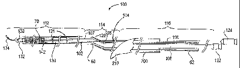
FIG 1 is a side view of a rapid exchange version of a catheter system for
protected
stenting and angioplasty of a patient's carotid artery.
FIG 2 is a detail drawing of the slotted portion of the stent delivery sheath.
FIG 3 shows a cross section of a proximal section of the catheter system of
FIG 1.
FIG 4 is a detail drawing showing an optional configuration of the embolic
protection balloon catheter.
FIG 5 is a detail drawing showing an optional feature of the catheter system
wherein
the embolic protection balloon is configured to act as a catheter tip for the
stent delivery
sheath.
FIG 6 is a detail drawing showing an optional feature of the catheter system
wherein
the stent delivery sheath has a floating catheter tip.
FIG 7 is a detail drawing showing an optional configuration of the proximal
end of
the catheter system.
FIG 8 is a side view of a coaxial over-the-wire version of a catheter system
for
protected stenting and angioplasty of a patient's carotid artery.
FIG 9 illustrates a patient's carotid arteries with an atherosclerotic plaque
at the
carotid bifurcation.
FIG 10 shows a guiding catheter positioned in the patient's common carotid
artery
and a guidewire advanced across the stenosis.
CA 02621508 2008-03-05
WO 2007/033963 PCT/EP2006/066509
6
FIG I 1 illustrates the optional step of dilating the stenosis prior to
stenting with a
small diameter angioplasty balloon.
FIG 12 shows the embolic protection balloon catheter advanced across the
stenosis.
FIG 13 shows the stent delivery sheath advanced across the stenosis and
deploying
a self-expanding stent within the lesion.
FIG 14 illustrates the self-expanding stent deployed within the lesion.
FIG 15 shows the distal end of the guiding catheter advanced into the lumen of
the
deployed self-expanding stent and the occlusion balloon inflated within the
lumen of the
self-expanding stent.
FIG 16 shows the combination angioplasty balloon catheter and stent pusher
catheter positioned with the angioplasty balloon across the lesion.
FIG 17 shows an angiography study performed to confirm occlusion of the
internal
carotid artery prior to dilatation of the lesion.
FIG 18 shows the angioplasty balloon inflated to dilate the stenosis and
complete
the deployment of the self-expanding stent.
FIG 19 illustrates potential embolic material being aspirated through the
lumen of
the guiding catheter.
FIG 20 illustrates the patient's carotid bifurcation after completion of the
protected
angioplasty and stenting procedure.
DETAILED DESCRIPTION OF THE INVENTION
FIG 1 is a side view of a rapid exchange version of a catheter system 100 for
protected stenting and angioplasty of a body passage, such as a patient's
carotid artery. This
version of the catheter system 100 has five major components: a self-expanding
stent 70, a
stent delivery sheath 60, a combination angioplasty balloon catheter and stent
pusher
catheter 102, an embolic protection device 104 and an auto-releasing sheath
200. The
catheter system 100 has a distal section 112, a transition section 114 and a
proximal section
116. The embolic protection device 104 can be configured as an embolic
protection balloon
catheter or an embolic protection filter catheter.
In the distal section 112, the catheter system 100 has a coaxial arrangement
with the
embolic protection device 104 on the inside, the combination angioplasty
balloon catheter
and stent pusher catheter 102 in between, and the stent delivery sheath 60 on
the outside.
The self-expanding stent 70 is positioned inside the stent delivery sheath 60,
distal to the
combination angioplasty balloon catheter and stent pusher catheter 102.
CA 02621508 2008-03-05
WO 2007/033963 PCT/EP2006/066509
7
In the transition section 114 of the catheter system 100, the embolic
protection
device 104 passes through a first slit or hole 118 on the side of the
combination angioplasty
balloon catheter and stent pusher catheter 102 and a second slit or hole 120
in the side of
the stent delivery sheath 60. The first slit or hole 118 in the side of the
combination
angioplasty balloon catheter and stent pusher catheter 102 and the second slit
or hole 120 in
the side of the stent delivery sheath 60 allow the embolic protection device
104, the
combination angioplasty balloon catheter and stent pusher catheter 102, and
the stent
delivery sheath 60 to be moved independently of one another after insertion
into the
patient's vascular system.
In the proximal section 116 of the catheter system 100, the embolic protection
device 104 is arranged side-by-side with the stent delivery sheath 60, and the
combination
angioplasty balloon catheter and stent pusher catheter 102 and the stent
delivery sheath 60
are arranged coaxially with one another. Prior to insertion into the patient's
vascular
system, the auto-releasing sheath 200 is arranged on the outside of the
embolic protection
device 104 and the stent delivery sheath 60. The auto-releasing sheath 200
extends a
substantial portion of the proximal section 116 of the catheter system 100 to
hold the
embolic protection device 104 and the stent delivery sheath 60 together in
longitudinal
alignment so that the catheter system 100 can be conveniently handled and
inserted into the
patient as a single unit. FIG 3 shows a cross section of a proximal section
116 of the
catheter system of FIG 1.
In one preferred embodiment, the embolie protection device 104 is configured
as an
embolic protection balloon catheter constructed with a tubular catheter shaft
108 with an
embolic protection member 132 in the form of an inflatable embolic protection
balloon
mounted at its distal end and a proximal connector 124, such as a luer
fitting, attached at its
proximal end. Preferably, a radiopaque marker 133 is located near the distal
end of the
tubular catheter shaft 108 to show the position of the embolic protection
balloon 132 on
fluoroscopy. The inflatable embolic protection balloon is preferably molded
from of a
highly elastic polymer, such as latex, silicone or polyurethane. An inflation
lumen extends
through the tubular catheter shaft from the proximal connector to an inflation
port that
communicates with the interior of the inflatable embolic protection balloon.
The embolic
protection balloon will preferably have an inflated diameter in the range of
approximately 6
to 9 mm and a deflated diameter as close as practically possible to the
outside diameter of
the tubular catheter shaft. In a preferred embodiment of the embolic
protection device 104,
the tubular catheter shaft 108 is constructed of a flexible metal tube with a
diameter of
0.014 to 0.018 inches and a length that is preferably approximately 120-170 cm
or longer.
The tubular catheter shaft 108 can be made, for example, from stainless steel
such as 302 or
CA 02621508 2008-03-05
WO 2007/033963 PCT/EP2006/066509
8
304 stainless, a cobalt alloy such as MP35 or Elgiloy, or a highly flexible or
superelastic
Titanium or NiTi alloy. In a preferred embodiment, a short length of coiled-
wire guidewire
134 with a tapered core wire is attached to the metal tube, for example by
welding,
soldering, crimping, and/or adhesive. Alternatively, the flexible guidewire
tip may be
constructed of a resilient polymer or polymer composite with similar
characteristics to a
coiled-wire guidewire. This construction provides the embolic protection
device 104 with
highly desirable handling characteristics similar to a floppy tip steerable
guidewire.
Alternatively, the tubular catheter shaft can be constructed from a polymer
tube or a
reinforced polymer composite tube.
Optionally, the embolic protection device 104 may be constructed with a
through-
lumen instead of having a guidewire tip and the catheter system 100 may also
include a
separate steerable guidewire that extends through the lumen of the embolic
protection
device 104.
FIG 4 is a detail drawing showing an optional configuration for the distal end
of the
embolic protection device 104. A short length of coiled-wire guidewire 134 is
attached to
the distal end of a tapered core wire 136, for example by welding, soldering,
crimping,
and/or adhesive. Preferably, the tapered distal end of the core wire extends
to the distal end
of the guidewire where it is attached by welding, soldering, crimping, and/or
adhesive.
Optionally, the tapered core wire may have a flattened distal section to
create a very
flexible floppy tip. Alternatively, there may be a flexible safety wire with a
flat cross
section that extends to the distai end of the guidewire. The core wire 136
extends
proximally of the guidewire 134 and has a proximal end with an increased
diameter portion
138. The increased diameter portion 138 can be created by flattening the
proximal end of
the core wire. Alternatively, the increased diameter portion 138 can be
created by welding
to form a weld bead, or by adding material to the core wire 136 by welding,
soldering or
adhesive.
The tubular catheter shaft 108 is constructed of a flexible metal tube with an
inflation lumen 109 extending though the tube. The increased diameter portion
138 on the
proximal end of the core wire 136 is inserted into the inflation lumen 109 at
the distal end
of the tube. Then, the distal end of the metal tube is swaged, for example by
rotary
swaging, to decrease the diameter of the inflation lumen 109, effectively
trapping the
increased diameter portion 138 on the proximal end of the core wire 136 in the
inflation
lumen 109. This creates a sliding attachment between the guidewire core wire
and the
flexible metal tube. Swaging also creates an external shoulder 140 on the
distal end of the
tubular catheter shaft that provides a recessed area for attachment of the
proximal sleeve of
the embolic protection balloon 132 with adhesive. The distal sleeve of the
embolic
CA 02621508 2008-03-05
WO 2007/033963 PCT/EP2006/066509
9
protection balloon is attached at the proximal end of the guidewire with
adhesive. The
sliding attachment of the guidewire core wire within the inflation lumen of
the tubular
catheter shaft allows the guidewire to move distally during balloon inflation
to
accommodate the expansion of the balloon. This feature reduces the stress on
the
attachments of the proximal and distal balloon sleeves during balloon
inflation.
In an alternate embodiment, the embolic protection device 104 can be
configured as
an embolic protection filter catheter. In this embodiment, the embolic
protection device 104
is constructed with an elongated catheter shaft 108 with an embolic protection
member 132
in the form of an expandable embolic protection filter mounted near the distal
end. The
embolic protection filter may be self-actuating or it may be selectively
actuated with an
actuating mechanism operable from the proximal end of the catheter shaft 108.
Other
examples of embolic protection filter catheters that can be adapted for use
with the present
invention are described in the following patents: US5941896, US6355051,
US6991641,
US6755846, US6391044, US 6142987, US6887256, US6645224, US6432122,
US6336934, US 6027520, US7048752, US7033375, US6989019, US6949103,
US6712835, US6605102, US6506204, US6168622, US6620182, US6616679, US6589263,
US6544279, US6530939, US6371970, US6348062, US6214026, US6203561, US6179861,
US6129739, US6969396, US6726702, US6663651, US6361546.
The combination angioplasty balloon catheter and stent pusher catheter 102 has
a
construction similar to a rapid exchange angioplasty balloon catheter with a
single-lumen
proximal catheter shaft 106 connected to a two-lumen distal catheter shaft
107. An
approximately cylindrical inflatable angioplasty balloon 130 is mounted near
the distal end
of the two-lumen distal catheter shaft 107. Preferably, two radiopaque markers
131 are
positioned on the distal catheter shaft 107 to show the position of the
angioplasty balloon
130 on fluoroscopy. A balloon inflation lumen extends from a proximal
connector 122,
such as a luer fitting, on the proximal end of the catheter through the single-
lumen proximal
catheter shaft and through most of the two-lumen distal catheter shaft where
it makes a
fluid connection with the interior of the inflatable angioplasty balloon. A
guidewire lumen
extends from the distal tip of the catheter 106 through the two-lumen distal
catheter shaft
and terminates at the first slit or hole 118 on the side of the catheter. The
guidewire lumen
is sized to have a sliding fit with the tubular catheter shaft 108 of the
embolic protection
device 104. The balloon inflation lumen and the guidewire lumen may be
arranged
coaxially or side-by-side in the two-lumen distal catheter shaft. A shoulder
142 is formed
near the distal end of the combination angioplasty balloon catheter and stent
pusher catheter
102. The shoulder 142 is sized and configured to have a sliding fit with the
inside of the
CA 02621508 2008-03-05
WO 2007/033963 PCT/EP2006/066509
stent delivery sheath 60 and to act as a pusher for pushing the self-expanding
stent 70 out
the distal end of the stent delivery sheath 60.
In one preferred embodiment of the combination angioplasty balloon catheter
and
stent pusher catheter 102, the single-lumen proximal catheter shaft 106 is
constructed of a
5 flexible metal tube made, for example, from stainless steel such as 302 or
304 stainless, a
cobalt alloy such as MP35 or Elgiloy, or a highly flexible or superelastic
Titanium or NiTi
alloy. Alternatively, the single-lumen proximal catheter shaft may be made
from an
extruded polymer tube or a reinforced polymer composite tube. The two-lumen
distal
catheter shaft is preferably made from an inner extruded polymer tube that
forms the
10 guidewire lumen and a coaxial outer extruded polymer tube that forms the
distal portion of
the balloon inflation lumen. The first slit or hole 118 is located in the
transition section 114
on the side of the catheter just distal to the junction between the single-
lumen proximal
catheter shaft and the two-lumen distal catheter shaft. The inflatable
angioplasty balloon is
typically formed by blow molding of an extruded polymer tube made, for
example, from
polyethylene, polyolefin, polyethylene terephthalate, polyvinyl chloride,
polyamide, or
alloys or copolymers thereof. The angioplasty balloon is bonded to the distal
catheter shaft
by welding and/or adhesive.
The stent delivery sheath 60 is constructed as a thin-walled single-lumen tube
with
an inner lumen sized to fit the self-expanding stent 70 in a compressed state.
Preferably, the
stent delivery sheath 60 has an external diameter as small as practically
possible in order to
fit through the guiding catheter and to pass through a stenosis without
predilatation.
Preferably, the external diameter of the stent delivery sheath 60 will be less
than 6 French
(approximately 2 mm diameter), more preferably, less than 5 French
(approximately 1.7
mm diameter). The stent delivery sheath 60 is preferably formed of an extruded
polymer
tube made, for example, from polyimide, polyethylene, polypropylene,
polyolefin,
polyurethane, polyethylene terephthalate, polyvinyl chloride, polyamide, or
alloys,
copolymers or reinforced composites thereof. At the level of the transition
section of the
catheter system there is a second slit or hole 120 in the side of the stent
delivery sheath 60.
Preferably, a gripping portion 62 is formed at the proximal end of the stent
delivery sheath
60 for convenience in handling. In one particularly preferred embodiment of
the stent
delivery sheath 60 shown in FIG 7, the proximal end of the stent delivery
sheath 60 has a
side port 144 with a connector, such as a luer fitting, for purging the
catheter system 100 of
air prior to use and, optionally, for aspirating or irrigating through the
lumen of the stent
delivery sheath 60 and a hemostasis valve 146 forming a sliding seal with the
proximal
catheter shaft 106 of the angioplasty balloon catheter and stent pusher
catheter 102.
CA 02621508 2008-03-05
WO 2007/033963 PCT/EP2006/066509
11
The self-expanding stent 70 is typically constructed as a braided tube of
resilient
wire made, for example, from stainless steel such as 302 or 304 stainless, a
cobalt alloy
such as MP35 or Elgiloy, or a highly flexible or superelastic Titanium or NiTi
alloy. The
self-expanding stent 70 will preferably have an expanded diameter of
approximately 7 to 9
mm and a length that is preferably 5 cm or more to accommodate a majority of
patients'
carotid arteries. The self-expanding stent 70 will have a compressed diameter
as small as
practically possible in order to fit inside of the stent delivery sheath 60.
The length of the
self-expanding stent 70 in the compressed state will depend on the
foreshortening of the
stent when it expands. Other constructions are possible for the self-expanding
stent 70. For
example, a self-expanding stent 70 having a nested ring pattern or the like
can be laser cut
from a tube of highly resilient material, such as a highly flexible or
superelastic Titanium or
NiTi alloy. This construction of self-expanding stent can be configured to
have little or no
foreshortening of the stent when it expands.
The auto-releasing sheath 200 is preferably constructed as a single lumen
extruded
polymer tube with a longitudinal slit 202 along the length of the sheath. The
lumen of the
sheath is sized and configured to have a snug fit around the tubular shaft 108
of the embolic
protection device 104 side-by-side with the stent delivery sheath 60 (with the
combination
angioplasty balloon catheter and stent pusher catheter 102 located coaxially
inside the
sheath 60). The auto-releasing sheath 200 extends a substantial portion of the
proximal
section 116 of the catheter system 100 to hold the embolic protection device
104 and the
stent delivery sheath 60 together in longitudinal alignment so that the
catheter system 100
can be conveniently handled and inserted into the patient as a single unit.
The distal end of
the auto-releasing sheath 200 is cut at a diagonal with a tab 210 located on
the opposite side
from the slit 202 to facilitate the auto-releasing of the sheath 200 during
insertion of the
catheter system 100 into the patient. Other possible configurations of the
auto-releasing
sheath 200 and other catheter linking devices usable with the catheter system
100 are
described in copending patent application 10/833,494.
In one particularly preferred embodiment shown in cross section in FIG 3, the
auto-
releasing sheath 200 is manufactured as an extruded profile with an
approximately circular
outer profile and an approximately oval inner lumen 204. The longitudinal
split 202
connects the inner lumen 204 with the exterior of the auto-releasing sheath
200 at a thin
part of the wall that coincides with the major axis of the oval inner lumen
204. The
longitudinal split 202 is preferably formed during the extrusion of the auto-
releasing sheath
200. Alternatively, the tube 200 can be extruded without the longitudinal
split 202 and then
slitted along the length to form the longitudinal split 202 in a secondary
operation. The
longitudinal split 202 allows the auto-releasing sheath 200 to be placed over
the proximal
CA 02621508 2008-03-05
WO 2007/033963 PCT/EP2006/066509
12
sections 106, 108 of the catheters 102, 104 during assembly of the catheter
system 100 and
to be removed from the catheters 102, 104 at the appropriate time during the
protected
angioplasty and stenting procedure. Suitable materials for the auto-releasing
sheath 200
include polyamide copolymers (e.g. PEBAX 6333 or PA 8020 from ATOFINA),
polypropylene, and any extrudable medical grade polymer with a suitable
combination of
strength, flexibility and friction characteristics.
The auto-releasing sheath 200 has the advantage that, once it is started, the
auto-
releasing sheath 200 will demount itself as the catheter system 100 is
advanced so that the
physician does not need to unpeel, remove or displace a linking member that
would
otherwise require a "third hand". The catheter system 100 is prepared for use
by aligning
the combination angioplasty balloon catheter and stent pusher catheter 102 and
the embolic
protection device 104 in the desired longitudinal alignment and then pressing
the
longitudinal split 202 of the auto-releasing sheath 200 against the proximal
sections 106,
108 of the catheters until they are enclosed within the inner lumen 204 of the
auto-releasing
sheath 200, as shown in FIG 3. This preparation is preferably carried out at
the
manufacturing facility or, alternatively, it may be performed at the point of
use by a
medical practitioner. The distal ends of the combination angioplasty balloon
catheter and
stent pusher catheter 102 and the embolic protection device 104 are inserted
into the patient
in the usual manner through a guiding catheter with a Y-fitting or other
hemostasis adapter
on the proximal end of the guiding catheter. The distal pull-tab 210 is pulled
toward the
side to start demounting the auto-releasing sheath 200 from the combination
angioplasty
balloon catheter and stent pusher catheter 102 and the embolic protection
device 104, and
then the catheter system 100 is advanced as a unit. When the auto-releasing
sheath 200
encounters the Y-fitting, the auto-releasing sheath 200 will peel away or
demount itself
from the proximal sections 106, 108 of the combination angioplasty balloon
catheter and
stent pusher catheter 102 and the embolic protection device 104. Once the
combination
angioplasty balloon catheter and stent pusher catheter 102 and the embolic
protection
device 104 have been advanced into the distal part of the guiding catheter,
the auto-
releasing sheath 200 can be set aside and discarded.
FIG 5 is a detail drawing showing an optional feature of the catheter system
100
wherein the embolic protection balloon 132 is configured to act as a catheter
tip for the
stent delivery sheath 60. During insertion of the catheter system 100, the
embolic protection
balloon device 104 is withdrawn into the stent delivery sheath 60 so that the
embolic
protection balloon nestles into the distal end of the stent delivery sheath 60
and creates a
smooth tapered distal end on the sheath 60 for passing though a stenosis in an
artery
without disturbing the plaque on the arterial walls.
CA 02621508 2008-03-05
WO 2007/033963 PCT/EP2006/066509
13
FIG 6 is a detail drawing showing an optional feature of the catheter system
100
wherein the stent delivery sheath 60 has a floating catheter tip 64. The
floating catheter tip
64 has a conical distal shape 65 and has a proximal shoulder 66 configured to
nest into the
distal tip of the stent delivery sheath 60. The tubular catheter shaft 108 of
the embolic
protection device 104 passes through a central hole 67 in the floating
catheter tip 64.
During insertion of the catheter system 100, the floating catheter tip 64
provides a smooth
tapered distal end on the sheath 60 for passing though a stenosis in an artery
without
disturbing the plaque on the arterial walls. When the self-expanding stent 70
is deployed,
the floating catheter tip 64 is pushed out of the lumen of the stent delivery
sheath 60 to
allow deployment of the stent 70. The floating catheter tip 64 is effectively
trapped on the
tubular catheter shaft 108 of the embolic protection device 104.
FIG 8 is a side view of a coaxial over-the-wire version of a catheter system
100 for
protected stenting and angioplasty of a body passage, such as a patient's
carotid artery. This
version of the catheter system 100 has four major components: a self-expanding
stent 70, a
stent delivery sheath 60, a combination angioplasty balloon catheter and stent
pusher
catheter 102 and an embolic protection device 104. The embolic protection
device 104 can
be configured as an embolic protection balloon catheter or an embolic
protection filter
catheter. The catheter system 100 has a coaxial arrangement throughout the
entire length,
with the embolic protection device 104 on the inside, the combination
angioplasty balloon
catheter and stent pusher catheter 102 in between, and the stent delivery
sheath 60 on the
outside. The self-expanding stent 70 is positioned inside the stent delivery
sheath 60, distal
to the combination angioplasty balloon catheter and stent pusher catheter 102.
In most
respects, the components in this version of the catheter system are similar in
construction to
those described above. However, in this version the combination angioplasty
balloon
catheter and stent pusher catheter 102 is configured similar to an over-the-
wire angioplasty
balloon catheter with a guidewire lumen and a balloon inflation lumen that
extend the full
length of the catheter and which connect, respectively, to a guidewire
insertion hub 150 and
a balloon inflation hub 152 on the proximal end of the catheter 102. Since the
components
are coaxial throughout the entire length of the catheter system, there is no
need for the
transition section or the first and second slits or holes in the stent
delivery sheath 60 and the
combination angioplasty balloon catheter and stent pusher catheter 102, as in
the rapid-
exchange version of the catheter system described above. Also there is no need
for the auto-
releasing sheath 200. In this version, the tubular catheter shaft 108 of the
embolic
protection device 104 has a length of approximately twice the length of the
stent delivery
sheath 60 and the combination angioplasty balloon catheter and stent pusher
catheter 102 to
allow these components to be withdrawn completely out of the guiding catheter
while the
CA 02621508 2008-03-05
WO 2007/033963 PCT/EP2006/066509
14
embolic protection member 132 remains deployed and in place during the
aspiration step of
the method.
Alternative configurations that allow the stent delivery sheath 60 and the
combination angioplasty balloon catheter and stent pusher catheter 102 to be
withdrawn
completely out of the guiding catheter while the embolic protection balloon
remains
inflated and in place include: a removable luer hub combined with a valve
within the
tubular shaft of the embolic protection catheter, as described in U.S. patent
6,156,005; and
a low profile catheter shaft with means for inflating the embolic protection
balloon, as
described in U.S. patent 6,641,573.
FIGS 9-20 illustrate a method for protected stenting and angioplasty according
to
the invention. The method includes steps of: inserting a guiding catheter into
a target vessel
in a patient's vascular system, for example at the site of a carotid
bifurcation; inserting the
catheter system into the guiding catheter and advancing the distal end of the
catheter system
to the distal end of the guiding catheter (when using the rapid exchange
version of the
catheter system, the auto-release sheath will automatically release itself
from the catheter
system during this step); advancing the embolic protection balloon catheter
(or embolic
protection filter catheter) beyond the lesion in order to support stent
delivery; positioning
the stent and balloon segment of the catheter system at the lesion; releasing
the self-
expanding stent by pulling the stent delivery sheath while maintaining the
position of the
combination angioplasty balloon catheter and stent pusher catheter; pulling
the stent
delivery sheath back into the guiding catheter; positioning and inflating the
embolic
protection balloon, preferably within the lumen of the deployed stent;
advancing the
combination angioplasty balloon catheter and stent pusher catheter and
inflating the
angioplasty balloon within the lesion; deflating the angioplasty balloon and
withdrawing
the combination angioplasty balloon catheter and stent pusher catheter and
stent delivery
sheath together; aspirating through the guiding catheter; then deflating and
withdrawing the
embolic protection balloon catheter to complete the procedure.
FIG 9 illustrates a patient's carotid arteries with an atherosclerotic plaque
50 at the
carotid bifurcation. The carotid bifurcation is a unique anatomical spot of
the human body
because of the carotid sinus. This dilatation at the origin of the internal
carotid artery and
the externai carotid artery creates an area of turbulent flow that represents
a kind of filter
for the cerebral vasculature: the particles of cholesterol that circulate in
the artery deposit
on the arterial wall, mainly the posterior wall. There is usually no deposit
of cholesterol
above the site of the bifurcation. One of the goals of the present invention
is to concentrate
the whole procedure on the actual pathological area, which is limited in
length and volume.
CA 02621508 2008-03-05
WO 2007/033963 PCT/EP2006/066509
The procedure begins by establishing arterial access, typically with a needle
puncture of the femoral artery or radial artery. A 7 or 8 French introducer
sheath is
positioned in the artery at the puncture site using a standard Seldinger
technique or other
known insertion technique. The common carotid artery is catheterized with a 5
French
5 diagnostic catheter and an exchange guidewire is advanced through the
diagnostic catheter
into the common carotid artery.
The diagnostic catheter is withdrawn and a 7 or 8 French guiding catheter 52,
with a
vertebral curve or other suitable distal curve, is advanced over the exchange
guidewire into
the common carotid artery. The exchange guidewire is withdrawn and angiography
is
10 performed by injecting radiopaque dye through the lumen of the guiding
catheter 52.
Next, a guidewire 54 is advanced through the guiding catheter 52 and across
the
stenosis 50 in the carotid artery. FIG 10 shows a guiding catheter 52
positioned in the
patient's common carotid artery and a guidewire 54 advanced across the
stenosis.
Preferably, a coronary style steerable guidewire with a diameter of 0.0 14 to
0.0 18 inches is
15 used. Alternatively, the catheter system can be modified to use other
diameters of
guidewire such as 0.035 to 0.038 inches.
When necessary (in less than 5% of the cases), a prestenting angioplasty
(typically
using a rapid exchange style angioplasty catheter 56 with a 2 mm diameter
dilatation
balloon 58) is performed without embolic protection to facilitate stent
crossing. FIG 11
illustrates this optional step of dilating the stenosis prior to stenting.
After the stenosis has
been dilated, the balloon 58 is deflated and the angioplasty catheter 56 and
the guidewire 54
are withdrawn.
FIG 12 shows the embolic protection device 104 advanced across the stenosis
50.
The tubular shaft 108 of the embolic protection device 104 serves the function
of a
guidewire for establishing catheter access across the stenosis 50.
FIG 13 shows a stent delivery sheath 60 advanced across the stenosis 50 and
deploying a self-expanding stent 70 within the lesion. The self-expanding
stent 70 is
deployed by pulling the stent delivery sheath 60 by the gripping portion 62 on
its proximal
end while maintaining the position of the combination angioplasty balloon
catheter and
stent pusher catheter 102. The stent 70 is typically deployed without embolic
protection as
this step presents very low risk for release of embolic material. Optionally
however, the
occlusion balloon 132 on the embolic protection device 104 can be inflated to
provide
embolic protection during this step of the procedure if desired. The occlusion
balloon 132
can be inflated distally to the treatment site prior to release of the self-
expanding stent 70
or, alternatively, the occlusion balloon 132 can be inflated at a distal
location within the
CA 02621508 2008-03-05
WO 2007/033963 PCT/EP2006/066509
16
lumen of the self-expanding stent 70 after it has been partially deployed as
indicated by the
phantom lines 132 in FIG 13.
Most practitioners presently consider it very important to cover the whole
atherosclerotic plaque with the stent from a normal arterial wall to a normal
arterial wall.
This implies the use of long stents. Because of the strong flow in the carotid
artery there is
no evidence, contrary to the experience in other arteries, that a long stent
produces more
restenosis than short stents at the carotid bifurcation. Alternatively, the
catheter system 100
can be used for delivering shorter length stents, for example 3 cm or even
shorter, where it
is clinically indicated.
The recommended characteristics of the stent 70 for use in carotid
bifurcations are:
(a) the stent should be self-expanding, (b) preferably a minimum of 5 cm
length should be
used, (c) an expanded diameter of 7 to 9 mm is typically necessary to fit with
the common
carotid artery, (d) a good radial expansion force is mandatory to rule out
secondary
complications due to aggregation on poorly deployed stents, (e) continuous,
not segmented,
framework of the stent is recommended to get a straightening of the carotid
artery that
facilitates the stenting technique, (f) longer and conic stents might be
considered in the
future. These characteristics may be varied for adapting the stenting
technique to other parts
of the vasculature.
FIG 14 illustrates the self-expanding stent 70 deployed within the lesion. The
stent
delivery sheath 60 has been pulled back into the guiding catheter 52. A
residual stenosis 50
may remain at the site of the original stenosis, but the entire length of the
lesion is
effectively covered by the expanded stent 70.
FIG 15 shows the distal end of the guiding catheter 52 advanced into the lumen
of
the deployed self-expanding stent 70 and the occlusion balloon 132 of the
embolic
protection device 104 inflated within the lumen of the self-expanding stent.
The occlusion
balloon 132 is inflated in the distal part of the stent 70 to occlude the
carotid artery and to
prevent any embolic debris from traveling downstream from the treatment site.
The guiding
catheter 52 is firmly positioned into the lumen of the deployed self-expanding
stent 70
leaving an open road for the following steps of the technique.
FIG 16 shows the combination angioplasty balloon catheter and stent pusher
catheter 102 positioned with the angioplasty balloon 130 across the lesion 50.
FIG 17 shows an angiography study performed to confirm occlusion of the
internal
carotid artery prior to dilatation of the lesion 50. The patient is clinically
tested. An
angiography series is performed to confirm the effective temporary occlusion
of the internal
carotid. The contrast 90 should remain close to the bifurcation site and
usually does not
reach the occlusion balloon 132.
CA 02621508 2008-03-05
WO 2007/033963 PCT/EP2006/066509
17
FIG 18 shows the angioplasty balloon 130 inflated to dilate the stenosis 50
and to
complete the deployment or expansion of the self-expanding stent 70. It is
recommended
that atropine be injected at least 5 minutes previously to rule out
bradycardia induced by the
compression of the carotid glomus.
After completion of the poststenting angioplasty, the angioplasty balloon 130
is
deflated and the combination angioplasty balloon catheter and stent pusher
catheter 102 and
the stent delivery sheath 60 are withdrawn from the guiding catheter 52. The
tubular shaft
108 of the embolic protection device 104 has sufficient length that the distal
section 112 of
the combination angioplasty balloon catheter and stent pusher catheter 102 and
the stent
delivery sheath 60 can be "parked" on the shaft 108 near the proximal end of
the embolic
protection device 104 so that it will not interfere with the aspiration step,
which is to
follow. (In the case of the over-the-wire version of the catheter system 100
of FIG 8, the
tubular shaft 108 of the embolic protection device 104 has additional length
so that the full
length of the combination angioplasty balloon catheter and stent pusher
catheter 102 and
the stent delivery sheath 60 can be "parked" on the shaft 108 near the
proximal end of the
embolic protection device 104.) Alternatively, if the embolic protection
device 104 is made
with a removable proximal fitting, the fitting may be removed at this point so
that the
angioplasty catheter 102 can be removed completely. The internal sealing
member
described above will maintain the occlusion balloon 132 in the inflated state.
With the occlusion balloon 132 still inflated, blood is aspirated back through
the
lumen of the guiding catheter 52. FIG 19 illustrates potential embolic
material 92 being
aspirated through the lumen of the guiding catheter 52.
Alternatively, the stent delivery sheath 60 can be left in place and the lumen
of the
sheath 60 can be used for aspiration and/or irrigation of potential embolic
materia192.
The occlusion balloon 132 is then deflated and the angioplasty catheter 102
and
embolic protection device 104 are withdrawn. An angiography series is
performed through
the guiding catheter 52 to verify patency of the lumen and full deployment of
the self-
expanding stent 70. Then, the guiding catheter 52 and introducer are withdrawn
and the
puncture site is closed.
FIG 20 illustrates the patient's carotid bifurcation with the fully deployed
stent 70
after completion of the protected angioplasty and stenting procedure.
Although it has been described in relation to treatment of obstructive carotid
artery
disease, the method of the present invention can be adapted for performing
protected
angioplasty and stenting in other parts of the vasculature, for example in the
coronary
arteries or renal arteries.
CA 02621508 2008-03-05
WO 2007/033963 PCT/EP2006/066509
18
While the present invention has been described herein with respect to the
exemplary
embodiments and the best mode for practicing the invention, it will be
apparent to one of
ordinary skill in the art that many modifications, improvements and
subcombinations of the
various embodiments, adaptations and variations can be made to the invention
without
departing from the spirit and scope thereof.