Note : Les descriptions sont présentées dans la langue officielle dans laquelle elles ont été soumises.
CA 02624953 2008-04-04
WO 2007/044140
PCT/US2006/033138
HIGH-SPEED MOLECULAR ANALYZER
SYSTEM AND METHOD
BACKGROUND OF THE INVENTION
1. Field of the Invention
The present invention relates in general to an improved method, apparatus and
system for analyzing molecules. More specifically, the invention relates to an
improved method, apparatus and system for determining characteristics or
properties
of molecules isolated, for example, according to their mass.
2. Description of the Background
Advances in the understanding of molecular biology and genetics and the
future promise of biotechnology have created a need for improved tools to
further the
research that will revolutionize the world. New information provided by
projects such
as the Human Genome Project has created even more demand for faster, higher
throughput methods for sequencing DNA. The tremendous efforts put into
sequencing .
in the last decade have helped other researchers begin to understand
fundamental cell
function. These efforts have accelerated the pace of research and discoveries
and have
created a growing need for improved tools for analyzing a large variety of
molecules
in addition to DNA. The benefits to mankind in medicine, agriculture and for
the
environment, as well as the economic potential that these fields promise, are
driving
researchers to decipher the function of individual genes, molecules and the
cells that
contain them. By sequencing an organisms' DNA and analyzing the molecules that
make up its cells, researchers are able to develop an understanding of the
systems and
structure that make it function.
DNA sequencing has become an extremely important tool in molecular
biology. DNA sequencing is the process of determining the nucleotide order of
a
given DNA fragment, called the DNA sequence. The amount of DNA sequence that
organisms have varies from species to species but in all but the simplest
organisms,
the amount that must be determined is enormous. The Human Genome for example,
consists of more than 3 billion nucleotide or "bases." The real benefit from
genomics
will not be derived from just the sequence data; it will be from an
understanding of
CA 02624953 2008-04-04
WO 2007/044140
PCT/US2006/033138
the function of the genes and the proteins that they encode. In order to
determine the
function and significance of different genes it is particularly helpful to
compare the
DNA sequence of entirely different species as well as the DNA sequence of like
species. The DNA sequence varies even for organisms of the same species and it
is
these differences that determine the different characteristics of different
individuals.
By obtaining the sequence data from many different organisms and individuals
and
correlating the different characteristics with differences in the genes, great
insight can
be gained about genetic function. However, this requires very large amounts of
sequencing capacity. There have been many methods and machines developed to
improve the speed and throughput of DNA sequencing, however it has taken
thousands of people, hundreds of machines and several years just to sequence
the
human genome using the current technology. This is entirely too slow and too
costly
to be practical to meet the future needs of genomics.
A variety of different sequencing approaches have been developed however
currently, almost all DNA sequencing is performed using a version of the chain
termination method, developed by Frederick Sanger. This technique uses
sequence-
specific termination of an in vitro DNA polymerase catalyzed synthesis
reaction using
modified nucleotide substrates. The synthesized copies of the original DNA are
then
separated by electrophoresis and analyzed to determine the sequence of the
original
DNA.
The 'tremendous amount of DNA sequence that is now available in databases
such as GenBank serves as a valuable resource and strong enticement to
generate
more sequence. Disciplines such as functional genomics and proteomics have
arisen
and use this data along with other research techniques to go beyond simple
genes to
begin to decipher the secrets of life. The growth of research in these fields
has
created a need for improved methods for analyzing other molecules such as
proteins,
carbohydrates, RNA, lipids and other bio-molecules in addition to the need for
higher
throughput, less expensive DNA sequencing technology.
Researchers make use of numerous analytical techniques to characterize and
decipher the functions of molecules from living systems. Analytical techniques
such
as high performance liquid chromatography, mass spectrography, nuclear
magnetic
resonance, electron microscopy, x-ray fluorescence analysis, x-ray
crystallography,
2
CA 02624953 2008-04-04
WO 2007/044140
PCT/US2006/033138
spectrographic absorption and fluorescence analysis and many others each yield
different bits of information very helpful to researchers. As the amount of
research
increases so has the number of samples to be analyzed. Many techniques in use
today
are still suited mainly for low throughput analysis.
Much of the sequence generated for the Human Genome Project was made
possible in part by processes using electrophoretic analysis.
Electrophoretic sorting of copies of DNA to sequence a segment having 1000
bases even in some of the fastest equipment can take up to an hour or more.
Typically
after each run, the gel or medium for electrophoresis must be discarded or
otherwise
replaced or replenished which can add even more time to the process.
Electrophoresis
is slow, complex, and expensive and the equipment requires regular
maintenance.
This method is also subject to resolution problems due to the different
mobility's
imparted by different fluorescent dyes. Since each different dye affects the
mobility
differently, the movement of the tagged molecules through the gel is not
purely
dependant on the size of the original DNA and will be affected by which dye
has been
incorporated. The equipment must be reconditioned between runs which costs
time
and requires additional consumables. In order to sequence a single organism in
a
reasonable time frame it is necessary to perform a very high volume of reads
in a
short period. Since electrophoresis is slow, many electrophoresis machines
must be
purchased making the sequencing process very expensive (if not impractical for
some
projects) in both capital costs as well as maintenance costs. Electrophoresis
is not
suited to satisfy the needs for significantly higher throughput.
Another approach to sequencing DNA involves the use of mass spectrometers.
This method uses the mass spectrometer to determine the sequence from mass
measurements made on copies of the original sequence or on probe molecules.
Mass
spectrometry is also used to analyze atomic composition and in the
identification and
quantification of various molecular species in a sample. Mass spectrometry is
growing in importance in molecular biology and is particularly important for
use in
protein analysis.
Mass spectrometry is also a tool of choice for analyzing bio-molecules. Many
different approaches have been taken to improve detectors to help increase
their
utility. A common limitation that time of flight mass spectrometers have is
the
3
CA 02624953 2008-04-04
WO 2007/044140
PCT/US2006/033138
resolution that they are able to achieve when trying to simultaneously measure
a
broad range of molecules with large differences in mass. For example, when
sequencing DNA using mass spectrometry, it is difficult to resolve the mass
differences necessary to accurately identify the base for a given position
when trying
to sequence a molecule with more than about 50 bases.
To achieve good resolution in mass spectrometry, it is desirable that
molecules
of like size be tightly clumped with minimal overlap to provide discrete
arrival times
at the detector. Poor separation of different molecule species results in
less
resolution. Since the velocity of the molecule is proportional to its mass,
small
relative differences in mass result in small differences in velocity. One
source of
error is due to initial velocities that the molecules have before
acceleration. These
differences in velocity provide error that is difficult to distinguish from
velocity
differences caused by differences in mass. This means that measurements on
large
molecules such as oligonucleotides from a sequencing reaction that differ by
only the
slight difference in molecular mass between A, C, G or T become more difficult
to
resolve as the size of the entire molecule increases. This method has
typically been
limited to sequencing shorter lengths of nucleic acid due to the accuracy and
resolution required for larger molecules. Additionally to improve resolution
four
separate reactions have been run for each of the A, C, G and T and then
sequenced
separately and re-assembled.
The detectors in time of flight mass spectrometers are typically less
sensitive
to larger molecules with low energies. If a mixture of nucleic acid sequence
fragments is analyzed that contains a large number of fragments of different
lengths,
the small molecules will be detected, but the larger molecules must be
accelerated at
the end of the drift region in order to provide enough impact to provide a
signal on the
detector. This introduces additional complexity and source for error. This is
another
aspect that contributes to the difficulty that mass spectrometers have in
providing
good resolution when analyzing a group of molecules with a large range of mass
values. Since many molecules of interest in molecular biology are large this
is a
limitation that would be helpful to overcome.
The detectors also have a limited life that depends on the number of molecules
that strike them. This means that regular maintenance and replacement is
usually
4
CA 02624953 2014-04-03
required to keep them accurate, this increases cost and down time. This is
problematic
for a machine that is to be used for high-volume sequencing since by the very
nature
of the process, very large quantities of molecules must be processed.
Background noise is also a problem with many devices. Collisions of stray
molecules with the detector cause noise that reduces sensitivity. For example,
molecules that are either from the desorption matrix (in the case of MALDI-
TOF) or
become fragmented during acceleration and or drift can produce a signal that
is not
discernable from the actual molecules being measured.
Several examples of patents and publications which disclose various DNA
sequencing methods and devices or attempts to solve some of the above problems
are
set forth as follows.
U.S. Patent No. 5,171,534 to Smith et al., entitled "Automated DNA
sequencing technique," sets forth a system for the electrophoretic analysis of
DNA
fragments produced in DNA sequencing operations comprising: a source of
chromophore or fluorescent tagged DNA fragments; a zone for contacting an
electrophoresis gel; means for introducing said tagged DNA fragments to said
zone;
and photometric means for monitoring said tagged DNA fragments as they move
through said gel.
U.S. Patent No. 6,847,035B2 to Sharma, entitled "Devices and methods for the
detection of particles," discloses devices and methods for determiningthe
masses of
particles by measuring the time between a first event such as a sample being
ionized,
(or a beam of electromagnetic radiation being scattered by a particle and
electromagnetic radiation scattered by said particle being detected by a
detection
means,) and a second event in which a beam of electromagnetic radiation is
scattered
by a particle from said ionized sample and electromagnetic radiation from said
beam
scattered by said particle is detected by a detection means.
U.S. Patent No. 6,995,841B2 to Scott et al., entitled "Pulsed-multiline
excitation for color-blind fluorescence detection," discloses a technology
called Pulse-
Mainline Excitation or PME. This technology provides a novel approach to
fluorescence detection with application for high-throughput identification of
informative SNPs, which could lead to more accurate diagnosis of inherited
disease,
5
CA 02624953 2008-04-04
WO 2007/044140
PCT/US2006/033138
better prognosis of risk susceptibilities, or identification of sporadic
mutations. The
PME technology has two main advantages that significantly increase
fluorescence
sensitivity: (1) optimal excitation of all fluorophores in the genomic assay
and (2)
"color-blind" detection, which collects considerably more light than standard
wavelength resolved detection. Successful implementation of the PME technology
will have broad application for routine usage in clinical diagnostics,
forensics, and
general sequencing methodologies and will have the capability, flexibility,
and
portability of targeted sequence variation assays for a large majority of the
population.
U.S. Publication No. 2004/0057050 to Beck et al., entitled "Analysis systems
detecting particle size and fluorescence," sets forth particle analyzing
systems with
fluorescence detection, primarily in connection with particle sizing based on
scattered
light intensity or time-of-flight measurement. In one system, emission of
fluorescence
is used as a threshold for selecting particles for further analysis, e.g. mass
spectrometry. In another embodiment, laser beams arranged sequentially along
an
aerosol path are selectively switched on and off, to increase the useful life
of
components, and diminish the potential for interference among several signals.
Other
embodiments advantageously employ color discrimination in aerodynamic particle
sizing, single detectors positioned to sense both scattered and emitted
fluorescent
radiation, and laser beam amplitude or gain control to enhance the range of
fluorescence detection.
U.S. Patent No. 6,806,464 to Stowers et al., entitled "Method and device for
detecting and identifying bio-aerosol particles in the air," discloses a
method for
detecting and identifying bioaerosol particles in the air, the bioaerosol
particles in a
particle stream are selected in an ATOFMS (aerosol time-of-flight mass
spectrometer)
by means of fluorescence techniques, and only the selected bioaerosol
particles are
ionized, for instance on the basis of MALDI (matrix-assisted laser
desorption/ionization), after which the resulting ions are detected and the
bioaerosol
particles are identified. The selection of bioaerosol particles takes place by
means of
laser radiation, generated by a first laser device, of a wavelength which in
specific
substances in bioaerosol particles effects a fluorescence, after which by
means of a
fluorescence detector the bioaerosol particles are selected and a second laser
device is
6
CA 02624953 2008-04-04
WO 2007/044140
PCT/US2006/033138
triggered to emit light of a wavelength which effects the ionization of the
bioaerosol
particles selected only by the fluorescence detector.
U.S. Patent No. 5,003,059 to Brennan, entitled "Determining DNA sequences
by mass spectrometry," relates to the methods, apparatus, reagents and
mixtures of
reagents for sequencing natural or recombinant DNA and other polynucleotides.
In
particular, this invention relates to a method for sequencing polynucleotides
based on
mass spectrometry to determine which of the four bases (adenine, guanine,
cytosine or
thymine) is a component of the terminal nucleotide. In particular, the present
invention relates to identifying the individual nucleotides by the mass of
stable
nuclide markers contained within either the dideoxynucleotides, the DNA
primer, or
the deoxynucleotide added to the primer. This invention is particularly useful
in
identifying specific DNA sequences in very small quantities in biological
products
produced by fermentation or other genetic engineering techniques. The
invention is
therefore useful in evaluating safety and other health concerns related to the
presence
of DNA in products resulting from genetic engineering techniques.
U.S. Patent No. 5,643,798 to Beavis, et al., entitled "Instrument and method
for the sequencing of genome," is directed to improved techniques for DNA
sequencing, and particularly for sequencing of the entire human genome.
Different
base-specific reactions are utilized to use different sets of DNA fragments
from a
piece of DNA of unknown sequence. Each of the different sets of DNA fragments
has
a common origin and terminates at a particular base along the unknown
sequence. The
molecular weight of the DNA fragments in each of the different sets is
detected by a
matrix assisted laser absorption mass spectrometer to determine the sequence
of the
different bases in the DNA. The methods and apparatus of the present invention
provide a relatively simple and low cost technique which may be automated to
sequence thousands of gene bases per hour, and eliminates the tedious and time
consuming gel electrophoresis separation technique conventionally used to
determine
the masses of DNA fragments.
U.S. Patent No. 5,691,141 to Koster, entitled "DNA sequencing by mass
spectrometry," sets forth a new method to sequence DNA. The improvements over
the
existing DNA sequencing technologies are high speed, high throughput, no
electrophoresis and gel reading artifacts due to the complete absence of an
7
CA 02624953 2008-04-04
WO 2007/044140
PCT/US2006/033138
electrophoretic step, and no costly reagents involving various substitutions
with stable
isotopes. The invention utilizes the Sanger sequencing strategy and assembles
the
sequence information by analysis of the nested fragments obtained by base-
specific
chain termination via their different molecular masses using mass
spectrometry, as for
, example, MALDI or ES mass spectrometry. A further increase in throughput can
be
obtained by introducing mass-modifications in the oligonucleotide primer,
chain-
terminating nucleoside triphosphates and/or in the chain-elongating nucleoside
triphosphates, as well as using integrated tag sequences which allow
multiplexing by
hybridization of tag specific probes with mass differentiated molecular
weights.
U.S. Patent Nos. 6,541,765B1 and 6,281,493B1 to Vestal, both entitled
"Time-of-flight mass spectrometry analysis of biomolecules," are directed to a
time-
of-flight mass spectrometer for measuring the mass-to-charge ratio of a sample
molecule. The spectrometer provides independent control of the electric field
experienced by the sample before and during ion extraction. Methods of mass
spectrometry utilizing the principles of this invention reduce matrix
background,
induce fast fragmentation, and control the transfer of energy prior to ion
extraction.
U.S. Patent No. 5,998,215 to Prather et al., entitled "Portable analyzer for
determining size and chemical composition of an aerosol," discloses a portable
analyzer for determining the size and chemical composition of particles
suspended in
an aerosol. The aerosol is accelerated through a nozzle and skimmers, to
produce a
well-defined beam of particles, the speed of which is inversely related to the
particle
size. A dual-beam laser system positioned along the beam path detects light
scattered
from each particle, to determine the particle's velocity and thus its
aerodynamic size.
The laser system also triggers a laser to produce a beam that irradiates the
particle, to
desorb it into its constituent molecules. The particle is desorbed in a source
region of
a bipolar, time-of-flight mass spectrometer, which provides a mass-to-charge
spectrum of the desorbed molecule, thereby chemically characterizing the
material of
the particle. Several structural features provide sufficient ruggedness to
allow the
analyzer to be easily used in the field with minimum calibration and
maintenance.
U.S. Patent No. 5,681,752 to Prather et al., entitled "Method and apparatus
for
determining the size and chemical composition of aerosol particles," sets
forth an
improved mass spectrometer apparatus, and related method, that characterizes
aerosol
8
CA 02624953 2008-04-04
WO 2007/044140
PCT/US2006/033138
particles, in real time, according not only to their chemical composition, but
also to
their size. This added information can be of critical importance when
evaluating risks
associated with aerosol particles of particular chemical composition. The
apparatus
achieves this beneficial result in a reliable fashion by first detecting the
presence and
size of individual aerosol particles moving along a predetermined particle
path and by
then directing a pulse of high-intensity light at the particle, to desorb and
ionize the
particle, for analysis of its chemical composition.
U.S. Patent No. 5,654,545 to Holle et al., entitled "Mass resolution in time-
of-
flight mass spectrometers with reflectors," discloses a method for the high
resolution
analysis of analyte ions in a time-of-flight mass spectrometer. The method
consists of
the generation of an intermediate time-focus plane for ions of a certain mass
at a
location between an ion source and an ion reflector, and then using the ion
reflector to
temporally focus the ions of equal mass and differing velocities which pass
this plane
at the same time onto a detector. For time-of-flight mass spectrometers with
an ion
selector, the ion selector is particularly favorable location for this
intermediate plane
with time focus; and with a collision cell for the collision fragmentation of
the ions,
the collision cell is a particularly favorable location.
Various articles and publications include the following:
Mark T. Roskey, Peter Juhasz, Igor P. Smimov, Edward J. Takach, Stephen A.
Martin and Lawrence A. Haff (1996). DNA sequencing by delayed extraction-
matrix-assisted laser desorption/ionization time of flight mass spectrometry.
Proc.
Natl. Acad. Sci. USA. Vol. 93, pp. 4724-4729, May 1996. Biochemistry.
PerSeptive
Biosystems, 500 Old Connecticut Path, Framingham, MA 01701. Communicated by
Klaus Biemann, Massachusetts Institute of Technology, Cambridge, MA, January
11,
1996 (received for review November 10, 1995).
http://www.pubmedcentral.nih.gov/picrender.fcgi?artid-----393468zblobtype=pdf
Finn Kirpekar*, Eckhard Nordhoff, Leif K. Larsen, Karsten Kristiansen, Peter
Roepstorff, Franz Hillenkamp (1998). DNA sequence analysis by MALDI mass
spectrometry. Depai __ intent of Molecular Biology, Odense University,
Campusvej 55,
DK-5230 Odense M, Denmark and 'Institute for Medical Physics and Biophysics,
University of Munster, Robert-Koch-Strasse 31, D-48149 Munster, Germany.
Received March 10, 1998; Revised and Accepted April 16, 1998.
9
CA 02624953 2008-04-04
WO 2007/044140
PCT/US2006/033138
http://nar.oxfordjournals. org/cgi/content/abstract/26/11/2554
N. I. Taranenko, S. L. Allman, V. V. Golovlev, N. V. Taranenko 1, N. R. Isola
and C. H. Chen* (1998). Sequencing DNA using mass spectrometry for ladder
detection. 2488-2490 Nucleic Acids Research, 1998, Vol. 26, No. 10. Life
Science
Division, Oak Ridge National Laboratory, Oak Ridge, TN 37831-6378, USA and 1
Yale University Medical Center, New Haven, CT, USA. Received December 3,
1997; Revised and Accepted March 23, 1998.
http://www.pubmedcentral.nih.gov/picrender.fcgi?artid--.147544&blobtype=pdf
Eckhard Nordhoffa Christine Luebbert, Gabriela Thiele, Volker Heiser, and
Hans Lehrach (2000). Rapid determination of short DNA sequences by the use of
MALDI-MS. Nucleic Acids Res. 2000 October 15; 28(20): e86. Max Planck
Institute for Molecular Genetics, Ihnestrasse 73, 14195 Berlin, Germany. To
whom
correspondence should be addressed. Tel: +49 30 8413 1542; Fax: +49 30 8413
1139;
Email: nordhoff@molgen.mpg.de. Received August 21, 2000; Accepted August 22,
2000.
http ://wvvw.pubmedcentral.nih. goviarticlerender. fcgi?artid=110802
Annette Kaetzke and Klaus Eschrich (2002). Simultaneous determination of
different DNA sequences by mass spectrometric evaluation of Sanger sequencing
reactions. Nucleic Acids Research, 2002, Vol. 30, No. 21 e117. Institute of
Biochemistry, Medical Faculty, University of Leipzig, Liebigstrasse 16, D-
04103
Leipzig, Germany. *To whom correspondence should be addressed. Tel: +49 341
9722105; Fax: +49 341 9739371; Email: eschrich@uni-leipzig.de
http://nar. oxfordj tuna's org/cgi/content/abstract/30/21/e117
While the mass spectrometer can provide fast analysis of molecules, numerous
practical limitations prevent it from being the high throughput tool that is
needed. A
sequencing method that can provide the speed of mass spectrometry and the
convenience of one sequencing reaction for all bases as well as good read
lengths is
clearly needed. Therefore, there is a need to determine the sequence of
nucleic acids
and analyze other molecules and collections of molecules in a much faster and
more
economical way. Additionally, a high throughput instrument for analyzing
molecules
such as bio-molecules is needed and would be particularly helpful in analyzing
biological systems. Whatever the precise merits features and advantages of the
above
CA 02624953 2008-04-04
WO 2007/044140
PCT/US2006/033138
cited references, none of them achieves or fulfills the purpose of the present
invention
as set forth below.
Those of skill in the art will appreciate the present invention which
addresses
the above needs and other significant needs the solution to which are
discussed
hereinafter.
11
CA 02624953 2008-04-04
WO 2007/044140
PCT/US2006/033138
SUMMARY OF THE INVENTION
The present invention contemplates a method and apparatus for analyzing at
least one molecule. An aspect of the invention is Isolating at least one
molecule
wherein the isolating depends substantially on the mass of the at least one
molecule;
subsequently Interacting the at least one molecule with a radiant signal
inducer; and
detecting a signal resulting from the interacting of the at least one molecule
and the
radiant signal inducer.
Analyzing includes but is not limited to determining: the atomic composition
of one or more molecules; the mass of one or more molecules; at least one
subunit of
at least one molecule comprising two or more subunits; and the concentration
of one
or more molecules in a sample. Analyzing also may include but is not limited
to
nucleic acid sequencing; DNA sequencing; single nucleotide polymorphism (SNP)
analysis; and protein sequencing.
The at least one molecule includes but is not limited to organic molecules as
well as inorganic molecules. Organic molecules include but are not limited to
bio-
molecules. Inorganic molecules include but are not limited to inorganic
monomers
and inorganic polymers. Bio-molecules include but are not limited to: small
molecules; organic monomers; organic polymers and macromolecules. Small
molecules include but are not limited to: lipids, phospholipids, glycolipids,
sterols;
vitamins; hormones, neurotransmitters, carbohydrates, sugars and
disaccharides.
Monomers include but are not limited to amino acids, nucleotides, phosphates
and
monosaccharides. Organic polymers include but are not limited to nucleic
acids,
peptides, oligosaccharides and polysaccharides. Macromolecules include but are
not
limited to prions. Nucleic acids include btit are not limited to DNA, RNA and
oligonucleotides. Peptides include but are not limited to oligopeptides,
polypeptides,
proteins and antibodies.
Other embodiments of the invention are configured to analyze molecules such
as: at least one fragment molecule wherein the fragment has been prepared by
using
the at least one molecule as a template. The at least one fragment molecule
may also
have a known subunit in a known position on the fragment. Additionally,
molecules
to be analyzed may comprise a tag that is capable of producing a signal when
interacted with a signal inducer such as but not limited to a fluorophore. One
12
CA 02624953 2008-04-04
WO 2007/044140
PCT/US2006/033138
embodiment of the invention analyzes a molecule comprising a subunit
comprising a
fluorescent tag in a known position wherein the tag is characteristic of the
subunit
comprises it.
The isolating depending substantially on the mass of the at least one molecule
may have numerous embodiments including but not limited to the following: one
embodiment comprises ionizing and accelerating the at least one molecule to be
analyzed and allowing it to drift a sufficient distance to allow isolation
dependent
upon its mass; one embodiment of the invention comprises a time of flight
(TOF)
mass analyzer; one embodiment of the invention comprises a quadrupole mass
analyzer; another embodiment comprises a magnetic-sector mass analyzer;
another
embodiment comprises a quadrupole ion trap mass analyzer and a further
embodiment
comprises a Fourier transform ion cyclotron resonance mass analyzer.
In one embodiment of the invention the radiant signal inducer comprises
electromagnetic radiation. Another embodiment of the invention comprises
particle
radiation. Examples of electromagnetic radiation suitable for various
embodiments of
the invention include: radio frequency radiation, microwave radiation,
infrared
radiation, visible light, ultraviolet light, x-ray radiation and gamma ray
radiation.
Examples of particle radiation suitable for various embodiments of the
invention
include: protons neutrons, electrons, positrons, alpha particles and
molecules. These
types of radiation can be used separately or in combination
One example embodiment comprises a laser as a source of a radiant signal
inducer. The laser may independently comprise a diode laser, a semiconductor
laser,
a gas laser, such as an argon ion, krypton, or helium-neon laser, a diode
laser, a solid-
state laser such as a Neodymium laser which will include an ion-gain medium,
such
as YAG and yttrium vanadate (YV04), or a diode pumped solid state laser. Other
devices, which produce light at one or more discrete excitation wavelengths,
may also
be used as a source of radiant signal inducer such as a flash lamp. One
example
embodiment the source of radiant signal inducer comprises a xenon flash lamp.
Another example embodiment the source of a radiant signal inducer comprises an
x-
ray tube.
13
CA 02624953 2015-04-30
According to one aspect of the present invention, there is provided an
apparatus
for analyzing at least one molecule, the apparatus comprising:
a mass dependent molecule isolator adapted to isolate at least one molecule
wherein the isolation depends substantially on the mass of the at least one
molecule; and
a molecule detector in communication with the isolator the molecule detector
comprising at least one source of a radiant signal inducer, and a signal
detector.
According to another aspect of the present invention, there is provided an
apparatus for analyzing at least one molecule, the apparatus comprising:
a mass dependent molecule isolator adapted to isolate at least one molecule
wherein the isolation depends substantially on the mass of the at least one
molecule; and
a molecule detector in communication with the isolator the molecule detector
comprising
at least one source of a radiant signal inducer wherein the radiant
signal inducer is emitted continuously from the at least one source, and
a signal detector comprising at least one wavelength dependent photon
detector.
According to another aspect of the present invention, there is provided an
apparatus for analyzing a property of at least one molecule, the apparatus
comprising:
a mass dependent molecule isolator adapted to isolate at least one molecule
wherein the isolation depends substantially on the mass of the at least one
molecule; and
a molecule detector in communication with the isolator, the molecule detector
comprising
at least one source of a radiant signal inducer,
a signal detector, and
an analyzer in communication with the signal detector configured to supply an
output signal that is a function of an input signal and one or more reference
values.
According to another aspect of the present invention, there is provided a
method
for analyzing at least one molecule, the method comprising:
13a
CA 02624953 2015-04-30
isolating at least one molecule, wherein said isolating depends substantially -
on
the mass of the at least one molecule;
subsequently interacting the at least one molecule with a radiant signal
inducer;
and
detecting a signal resulting from the interacting of the at least one molecule
and the radiant signal inducer.
According to another aspect of the present invention, there is provided a
method
for analyzing at least one molecule, the method comprising:
isolating at least one molecule, wherein said isolating depends substantially
on the
mass of the at least one molecule;
subsequently interacting the at least one molecule with a radiant signal
inducer,
wherein the radiant signal inducer is emitted continuously from at least one
source;
causing the at least one molecule to emit at least one photon; and
detecting the at least one photon.
According to another aspect of the present invention, there is provided a
method
for analyzing a property of at least one molecule, the method comprising:
isolating at least one molecule wherein said isolating depends substantially
on the mass of the at least one molecule;
subsequently interacting the at least one molecule with a radiant signal
inducer;
detecting absorption of at least a part of the radiant signal inducer
resulting from
the interacting of the at least one molecule and the radiant signal inducer;
and
determining at least one property of the at least one molecule based on the
detecting.
According to another aspect of the present invention, there is provided a
method
for analyzing a property of at least one molecule, the method comprising:
isolating at least one molecule wherein said isolating depends substantially
on the mass of the at least one molecule;
subsequently interacting the at least one molecule with a particle beam; and
13b
detecting a signal resulting from the interacting of the at least one molecule
and the particle beam.
According to another aspect of the present invention, there is provided a
method
for determininL, at least one subunit of at least one sample molecule
comprising two or
more subunits, the method comprising:
isolating at least one fragment molecule having a known subunit in a known
position of the fragment molecule. wherein the fragment molecule has been
prepared
using the at least one sample molecule;
wherein said isolating depends substantially on the mass of the at least one
fragment molecule:
subsequently interacting the at least one fragment molecule with a radiant
signal
inducer;
detecting a portion of the radiant signal inducer scattered as a result of the
interacting of
the at least one fragment molecule and the radiant signal inducer; and
determining at least a part of a sequence of subunits based on the
detecting.
13c
CA 2624953 2017-06-28
CA 02624953 2008-04-04
WO 2007/044140
PCT/US2006/033138
In one example embodiment the source of a radiant signal inducer comprises a
source of particles. Examples of sources of particles include an electron gun
and
radioisotopes.
An aspect of the present invention includes interacting at least one molecule
with a radiant signal inducer after being isolated depending substantially
upon the
mass of the at least one molecule. One example embodiment comprises a time of
flight mass analyzer and a laser beam directed to intersect the flight path of
the at least
one molecule through the TOF mass analyzer such that when the at least one
molecule
passes through the TOP mass analyzer it interacts with the laser beam. Another
example embodiment comprises a quadrupole ion cyclotron mass analyzer and an x-
ray tube disposed generally at the exit of the quadrupole ion cyclotron mass
analyzer
such that when the at least one molecule exits the mass analyzer it passes
through the
x-ray radiation emitted from the x-ray tube and thereby interacts with the x-
ray
radiation and produces a signal characteristic of a property of the at least
one
molecule.
In one example embodiment the radiant signal inducer radiates in substantially
parallel paths from at least one source. In another example embodiment the
radiant
signal inducer radiates in substantially non-parallel paths from at least one
source.
The signal inducer may be emitted continuously from at least one source or be
emitted in one or more pulses from at least one source. The pulsed emission
may
comprise control circuitry to control the emission of the one or more pulses.
The signal produced as a result of the interaction of the radiant signal
inducer
may comprise any form of electromagnetic radiation or particle radiation or
combinations of the same. The signal produced can be the result of
luminescence
such as fluorescence resulting from the interaction. Interaction of the
radiant signal
inducer and the at least one molecule can be detected by: detecting absorption
of the
radiant signal inducer by the at least one molecule or by detecting emission
of a
particle or electromagnetic radiation or by detecting scattering of the
radiant signal
inducer or by detecting reflection of the radiant signal inducer or by
detecting a
combination of two or more of these phenomena.
The detector may comprise a charged couple device, a photomultiplier tube, a
silicon avalanche photodiode, a silicon PIN detector, a wavelength dispersive
14
CA 02624953 2008-04-04
WO 2007/044140
PCT/US2006/033138
spectrometer or an energy dispersive spectrometer. It may also comprise
filters to
selectively pass or block electromagnetic radiation or particles depending
upon the
wavelength of the electromagnetic radiation or the energy of the signal to be
detected
or the type of particle.
The present invention may further comprise data processing apparatus for
processing of the signals detected.
In one example embodiment, a method for analyzing at least one molecule is
provided. The method includes at least: providing at least one molecule;
isolating the
at least one molecule; causing the at least one molecule to emit a signal; and
detecting
the signal.
Another example embodiment of an apparatus includes a novel device for the
analysis of nucleic acid fragments including at least: a source of chromophore
or
fluorophore tagged nucleic acid fragments, the chromophore of fluorophore
being
distinguishable by the spectral characteristics; means for vaporization and
acceleration of said nucleic acid fragments; means for introducing the tagged
nucleic
acid fragments to the vaporization and acceleration means; a drift region;
said
vaporization and acceleration means being located at one end of said drift
region and
directed so as to propel said nucleic acid fragments through said drift
region;
detecting means located at the end of said drift region generally opposite
said
accelerating and vaporization means; said detecting means comprises means for
inducing emission from the tagged nucleic acid fragments and means for
detecting
emissions from said tagged nucleic acid fragments and distinguishing said
tagged
nucleic acid fragments.
In another example embodiment of the apparatus, the apparatus includes at
least vaporization and ionization means comprising electro-spray ionization.
In another example embodiment of the apparatus, the apparatus includes at
least a vaporization and ionization means comprising matrix assisted laser
desorption
ionization.
In another example embodiment of the apparatus, the apparatus includes at
=
least a source of illumination comprising a laser.
CA 02624953 2008-04-04
WO 2007/044140
PCT/US2006/033138
In another example embodiment of the apparatus, the apparatus includes at
least means for detecting emissions comprising a prism and one or more photo
detectors located at positions corresponding to unique spectral positions.
Another example embodiment of a method includes a method of determining
the sequence of nucleic acids comprising the following steps: introduction of
chromophore of fluorophore tagged nucleic acid fragments, said chromophore of
fluorophore being distinguishable by its spectral characteristics;
vaporization of said
nucleic acid fragments; acceleration of said nucleic acid fragments;
stimulation of
said nucleic acid fragments by external means so as to induce emissions from
said tag;
and detection of said emissions.
Another example embodiment of an apparatus includes a device for the
determination of the sequence of a nucleic acid sample comprising: a generally
tubular chamber; said chamber being evacuated sufficiently to prevent
degradation of
said sample during analysis; means for electrospray ionization of said sample;
an
accelerating grid adjacent the injector; an un-obstructed section of
sufficient length to
allow separation of said sample after acceleration by said accelerating grid;
a laser
directed to intersect the path of flight of said sample, positioned at the end
of said un-
obstructed section, opposite said accelerating grid; a photo-detector located
sufficiently close to said intersection of said illumination source and said
path of
flight of said sample.
Another example embodiment comprises a photo-detector located sufficiently
close to said intersection of said illumination source and said path of flight
of said
sample.
Another example embodiment comprises an un-obstructed section of sufficient
length to allow separation of said sample after acceleration by said
accelerating grid.
Another example embodiment comprises a source of illumination directed to
intersect said path of flight of said nucleic acid fragments, positioned at
the end of
said tubular chamber, opposite said vaporization and acceleration means.
Another example embodiment comprises a chamber being evacuated
sufficiently to prevent degradation of said nucleic acid fragments during
analysis.
16
CA 02624953 2008-04-04
WO 2007/044140
PCT/US2006/033138
Another example embodiment comprises at one end of said chamber, means
for vaporization and acceleration of said nucleic acid fragments along a path
of flight
generally in the direction of the axis of said tubular chamber.
Another example embodiment provides a method for analyzing at least one
molecule Comprising: Providing item to be analyzed; isolating the item to be
analyzed; causing the item to be analyzed to emit a signal.
Another example embodiment provides a method for analyzing at least one
molecule comprising: providing at least one molecule; isolating the at least
one
molecule; causing the at least one molecule to emit a signal; and detecting
the signal.
Another example embodiment provides a method for analyzing at least one
molecule comprising: providing at least one molecule; causing the at least one
molecule to have a non-neutral charge; separating the at least one molecule
based on
its mass to charge ratio; causing the at least one molecule to emit a
detectable signal;
detecting said signal; recording said signal.
Another example embodiment provides a method for determining the identity
of at least one base of at least one polynucleotide comprising:; providing a
population
of fluorescently labeled fractions; each fraction having a unique fluorescent
label
characteristic of the base at its end position; accelerating the population of
fractions in
a manner so as to impart generally the same amount of energy to each molecule;
allowing the population of fractions to travel a distance sufficient to
separate like
fractions into differentiable groups; causing at least one of the fluorescent
labels on at
least one of the fractions to fluoresce; and detecting the signal emitted from
the label.
Another example ,embodiment provides a method for analyzing at least one
molecule comprising: providing at least one molecule; accelerating the at
least one
molecule; allowing the at; least one molecule to travel a distance; causing
the at least
one molecule to emit a detectable signal; detecting said signal; recording
said signal.
Another example embodiment of the method includes at least sequencing a
group of molecules, wherein each molecule comprises multiple sub-units of
differing
sub-unit types, wherein each of the molecules includes at least one tag
specific to the
sub-unit type, the method comprising: accelerating said molecules, separating
said
molecules dependant upon at least said accelerating, and radiant detecting of
each of
the at least one tags by the tag type of each of the at least one tags.
17
CA 02624953 2008-04-04
WO 2007/044140
PCT/US2006/033138
In another example embodiment of the method, the method includes at least
radiant detecting comprises electromagnetic radiant detecting.
In another example embodiment of the method, the method includes at least
radiant detecting comprising phosphorescent radiant detecting.
In another example embodiment of the method, the method includes at least
radiant detecting comprising fluorescent radiant detecting.
In another example embodiment of the method, the method includes at least
radiant detecting comprising thermal radiant detecting.
In another example embodiment of the method, the method includes at least
radiant detecting comprising radioactive radiant detecting.
In another example embodiment of the method, the method includes at least
radiant detecting comprising particle radiant detecting.
In another example embodiment of the method, the method includes at least
radiant detecting comprising chemical-reactive radiant detecting.
In another example embodiment of the method, a further method includes at
least radiant detecting comprising detecting the radiation of the tag with a
detector.
In another example embodiment of the further method, the method includes at
least radiant detecting comprising electromagnetic radiant detecting.
In another example embodiment of the further method, the method includes at
least radiant detecting comprising phosphorescent radiant detecting.
In another example embodiment of the further method, the method includes at
least radiant detecting comprising fluorescent radiant detecting.
In another example embodiment of the further method, the method includes at
least radiant detecting comprising thermal radiant detecting.
In another example embodiment of the further method, the method includes at
least radiant detecting comprising radioactive radiant detecting.
In another example embodiment of the further method, the method includes at
least radiant detecting comprising particle radiant detecting.
In another example embodiment of the further method, the method includes at
least radiant detecting comprising chemical-reactive radiant detecting.
18
CA 02624953 2008-04-04
WO 2007/044140
PCT/US2006/033138
In another example embodiment of the method, a further method includes at
least radiant detecting comprising detecting the radiation of a detection
substance
uPon contact with the tag.
In another example embodiment of the further method, the method includes at
least radiant detecting comprising electromagnetic radiant detecting.
In another example embodiment of the further method, the method includes at
least radiant detecting comprising phosphorescent radiant detecting.
In another example embodiment of the further method, the method includes at
least radiant detecting comprising fluorescent radiant detecting.
In another example embodiment of the further method, the method includes at
least radiant detecting comprising thermal radiant detecting.
hi another example embodiment of the further method, the method includes at
least radiant detecting comprising radioactive radiant detecting.
In another example embodiment of the further method, the method includes at
least radiant detecting comprising particle radiant detecting.
In another example embodiment of the further method, the method includes at
least radiant detecting comprising chemical-reactive radiant detecting.
In molecular biology and materials science there is a growing need for the
identification and characterization molecules. The device of the current
invention
would allow the determination of various characteristics such as mass,
absorbance and
fluorescence signatures and possibly molecular structure.
An embodiment of the invention is an apparatus for determining the sequence
of DNA molecules, however the invention can be applied to many analytical
purposes
in characterizing molecules.
A method for analyzing at least one molecule comprising: accelerating the at
least one molecule; allowing the molecule to travel a distance; remotely
detecting a
signal from the molecule after traveling said distance; recording said signal
from said
detecting.
The apparatus for determining the sequence of DNA is similar to a time of
flight mass spectrometer and has four basic components:
19
CA 02624953 2008-04-04
WO 2007/044140
PCT/US2006/033138
1. A molecule accelerator that ionizes and accelerates the molecule of
interest. This can be an apparatus such as an electro-spray device or a
matrix assisted laser desorption ionization device.
2. A flight tube that is connected to the accelerator and provides a path
for the molecules to travel after they are accelerated. This flight tube
would be held at a vacuum to minimize collisions during the flight of
the molecule being analyzed.
3. A detection device that comprises: a laser directed generally normal to
the flight path of the molecules and located at the end of the flight tube
opposite from the accelerator; 4 photon detectors such as photo-
multiplier tubes located in the same plane as the laser and oriented
generally normal to the laser beam; a refractor for dispersing light into
its component colors and directing the light at one of each of the 4
photon detectors.
4. A data recording
device that records the signals from each of the
detectors.
The operation of the apparatus is as follows: The DNA to be analyzed is
prepared in a manner typical for analysis in a 4 color capillary sequencing
device.
This process produces a population of molecules that range in length from a
few
molecules to the original length of the DNA molecule to be analyzed. During
the
sequencing reaction a fluorescent dye is incorporated at the end of each of
these
molecules. The tags fluoresce when excited by a laser and emit one of 4 colors
representing the base for that end position.
The DNA prepared as described above is introduced into the accelerator
component of the apparatus of the current embodiment of the invention. A group
of
these molecules are ionized and accelerated by the accelerator and directed to
travel
down the flight tube.
As a result of traveling the distance of the flight tube the molecules are
fractionated by length. Since all molecules are imparted the same amount of
energy
by the accelerator, each molecule of a given length travels at a different
velocity. The
smallest molecules travel the fastest and the next smallest next fastest, etc.
until the
largest molecules which travel the slowest. This velocity difference causes
the
CA 02624953 2008-04-04
WO 2007/044140
PCT/US2006/033138
molecules to pass the detector at different times and thus accomplishes the
fractionation.
As each molecule group passes the detector they are illuminated by the laser.
This illumination causes the fluorescent dyes to emit light which passes
through the
refractor and is directed to the appropriate photo detector.
The data recording device records the detector signal strength and the time
detected.
After all of the molecules have passed the detector, the data recorded then
can
be analyzed and the exact sequence of the original DNA molecule determined by
correlating the wavelength detected and the order in which it was detected.
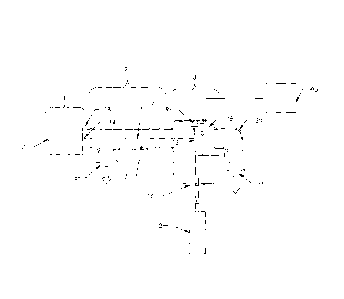
The present invention is shown as a block diagram in FIG. 1. The present
invention comprises a sample accelerator 1, a drift tube 2 and a detector 3.
The
chamber in the drift tube 8 and the area inside the detector are maintained at
high
vacuum by vacuum pumps connected at ports 5 and 6. The sample accelerator
vaporizes, ionizes and accelerates the sample molecules down the drift tube
along the
path 7 and through the detector chamber 15. While passing through the detector
3,
the sample ions are illuminated by the laser beam 11 causing the fluorescent
dye
terminator molecules incorporated into the sample molecules to emit light. The
photo
detector 9 then detects this light. The particular dye terminator incorporated
at the
end of the molecule corresponds to the original nucleotide of the molecule
being
sequenced. Once past the detector, the sample molecules are then cleared from
the
chamber mainly by the vacuum pump connected to port 6.
Referring to the block diagram in FIG. 1, the sample molecules to be analyzed
are vaporized and ionized by ionizing means 1. The ionizing means 1 can be any
device that provides a source of ionized molecules of sample without causing
excessive degradation of the sample molecules. Devices that are commonly used
to
do this use techniques such as Matrix Assisted Laser Desorption Ionization
(MALDI)
and Electrospray Ionization (ES). These techniques are commonly used to
provide
sample ion sources for Time of Flight Mass Spectrometers and are well known.
Each
device has particular advantages and disadvantages but serves as means to
convert the
sample to be analyzed to a gaseous ionized collection of molecules. The
ionizing
means accelerates the sample molecules to a velocity that is proportional to
their mass
21
CA 02624953 2008-04-04
WO 2007/044140
PCT/US2006/033138
to charge ratio. Thus, the smaller molecules will have higher velocities than
the larger
molecules. The molecules exit the ionizing meansl through exit port 14 with a
velocity directed down the drift tube 2. The dashed line 7 represents the
flight path of
the molecules, which travel down the drift tube past the detection point13. As
the
molecules travel the distance down the drift tube, the smaller (faster moving)
molecules travel the distance faster than the larger molecules. This results
in a
separation of the sample such that the molecules pass the detection point in
order of
increasing size with smallest arriving first and largest arriving last. The
chamber
areas in the drift tube 7 and detector 15 are maintained at a high vacuum. The
vacuum should be sufficient so as to prevent collisions between the sample and
stray
molecules causing excessive fragmentation and disruption of the sorting
process.
The sample to be sequenced is injected at 1. Very quickly after injection the
sample breaks into very small droplets that evaporate and leave the individual
molecules in a charged state.
After the sample is fully vaporized the accelerating grid 2 is turned on
accelerating the molecules from the sample through the grid. After passing
through
the grid they travel down a drift section that is an -un-obstructed section of
the
chamber. This section is of sufficient length to allow separation of said
sample after
acceleration by the accelerating grid. The molecules are accelerated to a
velocity that
is proportional to their mass to charge ratio. Therefore molecules of like
mass (size)
will be accelerated to very near the same velocity. As the molecules travel
down the
drift section, the fastest (smallest) molecules are the first to reach the
detector section.
The next smallest molecules arrive next and so on until all of the molecules
from the
sample have passed the detector section.
An object of the invention is to make large-scale sequencing of nucleic acids
faster, simpler and lower in cost. Several other objects and advantages of the
present
invention are to provide a method and an; apparatus to sequence polymeric or
chain
type molecules such as nucleic acids:
a) in larger volumes in a shorter amount of time;
b) having larger molecular size with greater accuracy;
c) as a continuous process without requiring reconditioning
between
each run;
22
CA 02624953 2008-04-04
WO 2007/044140
PCT/US2006/033138
d) with lower maintenance requirements;
e) with a lower sequencing cost per base.
An example embodiment of the invention is a method and apparatus for
determining the sequence polymeric or chain type molecules such as nucleic
acids.
This example embodiment comprises a source of chromophore or fluorophore
tagged
molecule fragments each being distinguishable by its spectral characteristics;
a means
for vaporization and acceleration of the molecule fragments; means for
introducing
the tagged molecule fragments to the vaporization and acceleration means; a
drift
region having the vaporization and acceleration means located at one end of
the drift
region and directed so that it propels the molecule fragments through the
drift region;
detecting means located at the end of the drift region generally opposite the
accelerating and vaporization means. The detecting means comprises means for
inducing emission from the tagged molecule fragments; means for detecting
emissions from the tagged molecule fragments and distinguishing the tagged
molecule
fragments.
Sequencing of polymeric or chain type molecules such as DNA is
accomplished by producing duplicate copies of varying lengths of the original
sequence that are terminated with a base specific chromophore or fiuorophore.
Four
different chromophores or fluorophores are used (one for each possible
nucleotide)
and each terminating molecule emits a unique emission spectrum when excited.
The
prepared DNA or nucleic acid is then loaded into the present invention for
analysis.
The nucleic acid fragments are then vaporized, ionized and accelerated by an
electric
field and directed down the drift region. The nucleic acid fragments are all
subjected
to approximately the same force in the accelerating field; however, since each
fragment of a different length has a different mass, each is accelerated to a
different
final velocity. As the nucleic acid fragments travel through the drift region,
their
differences in velocity cause them to be sorted from smallest to largest, the
smallest
arriving first and largest last. The detector illuminates the molecules as
they pass and
a sensor receives the resulting emission. The detector is designed to sense
characteristic emission spectrum of each tagged nucleotide allowing
determination of
the individual bases. The output from each sensor is then an accurate, ordered
sequential representation of the bases in the original molecule under
analysis.
23
CA 02624953 2014-04-03
This design achieves very high throughputs in contrast with electrophoresis.
Electrophoresis can typically take at least an hour for the sample to pass
completely
by the detector compared to fractions of a second for the present invention.
The
present invention requires virtually no reconditioning. All that is necessary
to
prepare the machine to sequence another sample is for the vacuum pump to clear
the
molecules from the previous sample out of the vacuum chamber, which happens
very quickly.
The present invention has advantages over mass spectrometry since the
detection method depends on detection of the wavelength of the emission from
the
florescent tags not precise measurements of time between discrete collisions.
The apparatus required is relatively simple with very few parts to fail;
therefore, the maintenance requirements are lower than the prior art. The
machine
can be made to operate automatically and there is next to no reconditioning
required
between runs so the labor cost per sample is lower than the prior art.
Other and further objects, advantages and features of the present invention
will become apparent from a consideration of the following discussions and
drawings describing various embodiments of the invention.
There has thus been outlined, rather broadly, the more important features of
the invention in order that the detailed description thereof may be better
understood,
and in order that the present contribution to the art may be better
appreciated. There
are additional features of the invention that will be described hereinafter.
In this respect, before explaining at least one example embodiment of the
invention in detail, it is to be understood that the invention is not limited
in its
application to the details of construction and to the arrangements of the
components
set forth in the following description or illustrated in the drawings. The
invention is
capable of other embodiments and of being practiced and carried out in various
ways. Also, it is to be understood that the phraseology and terminology
employed
herein are for the purpose of the description and should not be regarded as
limiting.
24
CA 02624953 2008-04-04
WO 2007/044140
PCT/US2006/033138
To the accomplishment of the above and related objects, this invention may be
embodied in the form illustrated in the accompanying drawings, attention being
called
to the fact, however, that the drawings are illustrative only, and that
changes may be
made in the specific construction illustrated.
25
CA 02624953 2008-04-04
WO 2007/044140
PCT/US2006/033138
BRIEF DESCRIPTION OF THE DRAWINGS
The drawings as noted below form part of the present specification and are
included to further demonstrate certain aspects of the present invention.
Various other
objects, features and attendant advantages of the present invention will
become fully
appreciated as the same becomes better understood when considered in
conjunction
with the accompanying drawings, in which like reference characters designate
the
same or similar parts throughout the several views, and wherein:
FIG. 1 shows a schematic diagram of a example nucleotide-sequencing device
in accordance with the current invention.
FIG. 2 shows a symbolic representation of a hypothetical nucleic acid
sequence paired with a complimentary nucleic acid copy terminated with a base
specific label molecule.
FIG. 3 shows a symbolic representation of a vaporized group of hypothetical
nucleic acid copies of different lengths; illustrating a random special
orientation of the
different length molecules.
FIG. 4 shows a symbolic representation of the same molecules in FIG. 3
shortly after being accelerated; illustrating separation of the sizes.
FIG. 5 shows a symbolic representation of the same molecules in FIG. 3 after
being accelerated and traveling for sufficient time to effect significant
separation by
size.
FIG. 6 shows a schematic representation of the detector optics of an example
embodiment.
FIG. 7 shows a symbolic representation of a group of molecules under
analysis and the corresponding outputs from the detectors sensing them.
FIG. 8 shows a cross section of an example detector having a single photo
detector.
FIG. 9 shows a schematic diagram of an example nucleic acid sequencing
device in accordance with the present invention.
FIG. 10 shows a schematic diagram of an example molecular analysis device
in accordance with the present invention.
FIG. 11 shows a schematic diagram of an example a wavelength dependent
photon detector in accordance with the present invention.
26
CA 02624953 2008-04-04
WO 2007/044140
PCT/US2006/033138
FIG. 12 shows a schematic diagram of an example molecular detector in
accordance with the present invention.
FIG. 13 shows a schematic diagram of an enlarged view of the region where a
molecule is in a position to interact with the signal inducer pulses in
accordance with
the present invention.
FIG. 14 shows a schematic diagram of an enlarged view of the region where a
molecule is in a position to interact with the signal inducer pulses in
accordance with
the present invention.
FIG. 15 shows a schematic diagram of an enlarged view of the region where a
molecule is in a position to interact with the signal inducer pulses in
accordance with
the present invention.
FIG. 16 shows a schematic diagram of an enlarged view of the region where a
molecule is in a position to interact with the signal inducer pulses in
accordance with
the present invention.
FIG. 17 shows a schematic diagram of an enlarged view of the region where a
molecule is in a position to interact with the signal inducer pulses in
accordance with
the present invention.
FIG. 18 shows a schematic diagram of an enlarged view of the region where a
molecule is in a position to interact with the signal inducer pulses in
accordance with
the present invention.
FIG. 19 shows a schematic diagram of the molecule in a position to interact
with the radiant signal inducer and illustrates how the pulse time can be
calculated in
accordance with the present invention.
FIG. 20 shows a schematic diagram of an example molecular analysis device
in accordance with the present invention.
FIG. 21 shows a schematic diagram of an example molecular analysis device
in accordance with the present invention.
FIG. 22 shows a schematic diagram of an example molecular analysis device
in accordance with the present invention.
FIG. 23 shows a schematic diagram of an example molecular analysis device =
in accordance with the present invention.
27
CA 02624953 2014-04-03
FIG. 24 shows a schematic diagram of an example molecular sequencing
device in accordance with the present invention.
FIG. 25 shows a schematic diagram of a molecule with two or more subunits
in accordance with the present invention.
FIG. 26 shows a schematic diagram of a molecule fragments in accordance
with the present invention.
FIG. 27 shows a schematic diagram of a molecule fragments in accordance
with the present invention.
FIG. 28 shows a schematic diagram of a molecule fragments in accordance
with the present invention.
FIG. 29 shows a schematic diagram of time measurements recorded for
fragment groups accordance with the present invention.
FIG. 30 shows a schematic diagram of an example molecular analysis device
in accordance with the present invention.
While the present invention will be described in connection with presently
preferred embodiments, it will be understood that it is not intended to limit
the invention
to those embodiments. On the contrary, it is intended to cover all
alternatives,
modifications, and equivalents defuted in the appended claims.
25
28
CA 02624953 2008-04-04
WO 2007/044140
PCT/US2006/033138
DETAILED DESCRIPTION OF EXAMPLE EMBODIMENTS OF THE
INVENTION
The present invention is a novel device and method for the high speed analysis
of molecules for determining characteristics such as atomic composition; mass;
sequence of subunits and the concentration of one or more molecules in a
sample.
The invention may also be used for nucleic acid sequencing; DNA sequencing;
single
nucleotide polymorphism (SNP) analysis; and protein sequencing.
In one example embodiment of the invention an apparatus is provided for
determining the sequence of bases or nucleotides in a nucleic acid such as DNA
or
RNA.
The basic steps involved in the process include:
a) Making copies ranging in length from 1 nucleotide to the same length
as the molecule under analysis;
b) Incorporating a base specific molecule at the end of the copy that
corresponds to the base of the original molecule at that position and
has a dye molecule that emits a uniquely identifiable spectrum when
induced by external means;
c) Vaporizing the molecules;
d) Accelerating the molecules in a way so as to impart substantially the
same energy to each molecule;
e) Allowing the molecules to travel for a sufficient time after
acceleration
so that the molecules are able to be separated as a consequence of their
differences in velocity;
Inducing an emission from the molecules in a localized area of the path
of travel after time for separation has elapsed;
g) Detecting the emissions from the molecules.
A detailed description of each of the steps listed above will now be given
generally in the order that they are presented.
In an example embodiment, nucleic acid that is to be analyzed is prepared by
producing copies ranging in length from a few nucleotides up the same length
as the
original sample molecule. When these copies are produced care is taken so as
to
produce generally equivalent numbers of molecules of each given length. At the
end
29
CA 02624953 2008-04-04
WO 2007/044140
PCT/US2006/033138
of each molecule, a fluorescent dye is incorporated in place of the original
nucleotide.
Four different dyes are used in the preparation of the copies, one for each of
the four
possible nucleotides. Each of these dyes has unique emission spectra when
induced
by external means such as illumination by a light source such as a laser.
There are various techniques for preparing the samples to achieve the desired
results mentioned above. The most common method involves the use of the
enzymatic chain termination reaction. This method is widely used and well
known.
This technique involves the Polymerase Chain Reaction (PCR) to make copies of
the
original sequence. During the copying, a dideoxymicleotide with a fluorescent
dye
molecule attached is incorporated randomly during PCR this halts the copying
of the
chain at the point where it is incorporated. Sufficient PCR cycles are run so
that large
enough populations of base specific terminated fragments of different lengths
exist to
allow detection by the detector as described later in this disclosure. This
process is
generally referred to as a sequencing reaction. This method of preparation is
commonly used in preparing molecules for sequencing using electrophoresis.
Several
variations of this technique exist, are well known and are mostly based on
methods
proposed by Sanger, F., Nicken, S. and Coulson, A. R. Proc. Natl. Acad. Sci.
USA 74,
5463 (1977) and the methods proposed by Maxam, A. M. and Gilbert, W. Methods
in
Enzymology 65, 499-599 (1980).
Referring to the example embodiment in FIG. 2, a schematic view is shown of
a short strand of DNA prepared using a sequencing reaction. 21 represents the
original sequence of nucleotides that is to be analyzed. The ellipses 22, 23,
24 and 28
indicate the positions of an arbitrary number of intervening bases that are
not shown
due to space limitations in the drawing. The bases shown in this view are A
representing adenine, C representing cytosine, G representing guanine and T
representing thymine. The particular sequence shown has no particular
significance
and was chosen randomly for the purposes of illustration only. The invention
does not
depend upon any specific bases or number of bases in the molecule under
analysis.
20 represents the primer region. The strand shown generally at 25 above and
complementary to the original sequence represents the copy of the original
sequence
generated by PCR. The molecule is shown in the state after the polymerase has
completed the copying of the original sequence 21 and the polymerization has
been,
CA 02624953 2008-04-04
WO 2007/044140
PCT/US2006/033138
in an example embodiment, terminated by molecule 27. The terminating molecule
27
has label 26 attached to it. In the case shown, the terminating molecule is
shown as a
T and is complimentary to the corresponding molecule A on the original
sequence.
In the example embodiment, the terminating molecule 27 that is incorporated
is a dideoxynucleotide with a fluorophore molecule 26 attached to it. The
terminating
molecule 27 is shown as a T in this case since T is complimentary to A; this
example
embodiment was chosen for illustration. The tag molecule 26 in this case is a
fluorophore. It emits light when stimulated by an external source such as a
laser. The
emission spectrum of this molecule is chosen to be unique for the particular
terminating molecule that it is attached to. For example the terminating
molecule that
is complementary to A will have a unique fluorophore that will have a unique
emission spectra to the fluorophore that is attached to the terminating
molecule
complimentary to T and likewise unique for C and G. This allows each
terminating
molecule to be uniquely identified when stimulated so that they can be
differentiated
from the other bases. The tag molecule 26 could alternatively be a chromophore
or
any molecule that will emit a detectable emission when stimulated by an
external
source and that can be uniquely distinguished from the emissions of the other
tag
molecules in the sample. The present discussion refers to the analysis of DNA
and
the bases present therein, however, RNA could be analyzed in a similar
fashion. In
the case of RNA, it would be necessary to use a terminating molecule that
would be
complimentary to Uracil and use a polymerase appropriate for the reaction. The
present invention is not intended to be limited only to the sequencing of DNA.
During the sequencing reaction, a sufficient number of copies of the original
sequence are generated to provide sufficient signal for the detector when
stimulated.
As the molecules are synthesized by the polymerase, the terminating molecules
are
randomly incorporated which halts extension. The reaction is prepared to
produce a
generally uniform quantity of copies ranging from the first base to the entire
length of
the original molecule.
The example sequencing reaction for the present invention makes uses of the
polymerase chain termination reaction however; any method that yields copies
of the
original sequence that can be distinguished from the other terminating
molecules
representing a different base is acceptable. What is important for the process
is to
31
CA 02624953 2008-04-04
WO 2007/044140
PCT/US2006/033138
have one or more copies of the original sequence for each base in the original
sequence and that each copy has a length representative of the position that
each base
occupies. For example if a molecule having 5 bases were to be analyzed there
should
be at least 5 molecules with lengths of 1, 2, 3, 4 and 5 nucleotides. Each of
the 5
molecules will have a terminating molecule that is complimentary to the
original base
at the terminating position in the original molecule. The terminating position
refers to
the position of the base at the location where copying was terminated.
In chain terminator sequencing (Sanger sequencing), extension is initiated at
a
specific site on the template DNA by using a short oligonucleotide 'primer'
complementary to the template at that region. The oligonucleotide primer is
extended
using a DNA polymerase, an enzyme that replicates DNA. Included with the
primer
and DNA polymerase are the four deoxynucleotide bases (DNA building blocks),
along with a low concentration of a chain terminating nucleotide (most
commonly a
di-deoxynucleotide). Limited incorporation of the chain terminating nucleotide
by the
DNA polymerase results in a series of related DNA fragments that are
terminated
only at positions where that particular nucleotide is used. The fragments are
then size-
separated by electrophoresis in a slab polyacrylamide gel, or more commonly
now, in
a narrow glass tube (capillary) filled with a viscous polymer.
The classical chain termination method or Sanger method first involves
preparing the DNA to be sequenced as a single strand. The DNA sample is
divided
into four separate samples. Each of the four samples have a primer, the four
normal
deoxynucleotides (dATP, dGTP, dCTP and dTTP), DNA polymerase, and only one of
the four dideoxynucleotides (ddATP, ddGTP, ddCTP and ddTTP) added to it. The
dideoxynucleotides are added in limited quantities. In an example embodiment,
the
primer or the dideoxynucleotides are either radiolabeled or have a fluorescent
tag.
As the DNA strand is elongated the DNA polymerase catalyses the joining of
deoxynucleotides to the corresponding bases. However, if a dideoxynucleotide
is
joined to a base, then that fragment of DNA can no longer be elongated since a
dideoxynucleotide lacks a crucial 3'-OH group. Fragments of all sizes should
be
obtained due to the randomness of when a dideoxynucleotide is added. However,
to
make sure that all different lengths will occur, only short stretches of DNA
can be
sequenced in one test.
32
CA 02624953 2008-04-04
WO 2007/044140
PCT/US2006/033138
The DNA is then denatured and the resulting fragments are separated (with a
resolution of just one nucleotide) by gel electrophoresis, from longest to
shortest. The
shorter fragments have greater mobility in the gel than the longer fragments
and
therefore arrive at the detector first with successively longer fragments
following.
Each of the four DNA samples is run on one of four individual lanes (lanes A,
T, G,
C) depending on which dideoxynucleotide was added. Depending on the whether
the
primers or dideox3mucleotides were radiolabeled or fluorescently labeled, the
DNA
bands can be detected by exposure to X-rays or UV-light and the DNA sequence
can
be directly read off the gel. Bands in the gel indicate the positions of the
DNA
molecules of different lengths. A band in a lane indicates a chain termination
for that
particular DNA subunit and the DNA sequence can be read off accordingly.
There can be various problems with sequencing through the Sanger Method.
The primer used can also be annealed to a second site. This would cause two
sequences to be interpreted at the same time. This can be solved by higher
annealing
temperatures and higher G and C content in the primer. Another problem can
occur
when RNA contaminates the reaction, which can act like a primer and leads to
bands
in all lanes at all positions due to non specific priming. Other contaminants
can be
I- from other plasmids, inhibitors of DNA polymerase, and low
concentrations of
template in general. Secondary structure of DNA being read by DNA polymerase
can
lead to reading problems and will be visualized on the readout by bands in all
lanes of
only a few positions.
There are two sub-types of chain-termination sequencing. In the original
method, the nucleotide order of a particular DNA template can be inferred by
performing four parallel extension reactions using one of the four chain-
terminating
bases in each reaction. The DNA fragments are detected by labeling the primer
with
radioactive phosphorous prior to performing the sequencing reaction. The four
reactions would then be rim out in four adjacent lanes on a slab
polyacrylamide gel.
A development of this method used four different fluorescent dye-labeled
primers. This has the advantage of avoiding the need for radioactivity;
increasing
safety and speed, and also that the four reactions can be combined and run in
a single
gel lane, if they can be distinguished. This approach is known as 'dye primer
sequencing'.
33
CA 02624953 2008-04-04
WO 2007/044140
PCT/US2006/033138
An alternative to the labeling the primer is to label the terminators instead,
commonly called 'dye terminator sequencing'. The major advantage of this
approach
is the complete sequencing set can be performed in a single reaction, rather
than the
four needed with the labeled-primer approach. This is accomplished by labeling
each
of the dideoxynucleotide chain-terminators with a separate fluorescent dye,
which
fluoresces at a different wavelength. This method is easier and quicker than
the dye
primer approach, but may produce more uneven data peaks (different heights),
due to
a template dependent difference in the incorporation of the large dye chain-
terminators. This problem has been significantly reduced with the introduction
of new
enzymes and dyes that minimize incorporation variability.
This method is now used for the vast majority of sequencing reactions as it is
both simpler and cheaper. The major reason for this is that the primers do not
have to
be separately labeled (which can be a significant expense for a single-use
custom
primer), although this is less of a concern with frequently used 'universal'
primers.
To produce detectable labeled products from the template DNA, 'cycle
sequencing' is most commonly performed. This approach uses repeated (25 - 40)
rounds of primer annealing, DNA polymerase extension and disassociation
(melting)
of the template DNA strands. The major advantages of cycle sequencing is the
more
efficient use of the expensive sequencing reagent (BigDye) and the ability to
sequence
templates with strong secondary structures such as hairpins or GC-rich
regions. The
different stages of cycle sequencing are performed by altering the temperature
of the
reaction using a PCR thermal cycler. This relies on the fact that
complementary DNA
will anneal at a lower temperatures and disassociate at higher temperatures.
An
important part of making this possible is the use of DNA polymerase from a
thermophillic organism, which is not rapidly denatured at the high (>95C)
temperatures involved. In the past, new DNA polymerase had to be added
individually every cycle of PCR.
Various large-scale sequencing strategies include several current methods
which can directly sequence only short lengths of DNA at a time. For example,
modern sequencing machines using the Sanger method can achieve a maximum of
around 1000 base pairs This limitation is due to the geometrically decreasing
34
CA 02624953 2008-04-04
WO 2007/044140
PCT/US2006/033138
probability of chain termination at increasing lengths, as well as physical
limitations
on gel size and resolution.
It is often necessary to obtain the sequence of much larger regions. For
example, even simple bacterial genomes contain millions of base pairs, and the
human
genome has more than 3 billion. Several strategies have been devised for large-
scale
DNA sequencing, including primer walking (see also chromosome walking) and
shotgun sequencing. These involve taking many small reads of the DNA through
the
Sanger method and subsequently assembling them into a contiguous sequence. The
different strategies have different tradeoffs in speed and accuracy; for
example, the
shotgun method is the most practical for sequencing large genomes, but its
assembly
process is complex and potentially error-prone.
It is easier to obtain high quality sequence data when the desired DNA is
purified and amplified from any contaminants that may be in the original
sample. This
can be achieved through PCR if it is practical to design primers that cover
the entire
desired region. Alternatively, the sample can be cloned using a bacterial
vector,
harnessing bacteria to "grow" copies of the desired DNA a few thousand base
pairs at
a time. Most large-scale sequencing efforts involve the preparation of a large
library
of such clones.
Certain areas of molecular biology research are very dependent upon
identifying and sequencing RNA. RNA is less stable in the cell, and also more
prone
to nuclease attack experimentally. As RNA is generated by transcription from
DNA,
the information is already present in the cell's DNA. However, it is sometimes
desirable to sequence RNA molecules. In particular, in Eukaryotes RNA
molecules
are not necessarily co-linear with their DNA template, as introns are excised.
To
sequence RNA, the usual method is first to reverse transcribe the sample to
generate
DNA fragments. This can then be sequenced as described above.
= Referring to one example embodiment shown in FIG. 1, once a sample has
been prepared, it is ready for use. An example embodiment of the present
invention
comprises a source of nucleic acid fragments each being distinguishable by its
spectral characteristics as described above. The example embodiment further
comprises a mass dependent molecule isolator which in the current embodiment
comprises means for vaporization and acceleration of the nucleic acid
fragments
CA 02624953 2008-04-04
WO 2007/044140 PCT/US2006/033138
shown generally at 1; means 17 for introducing the nucleic acid fragments to
the
vaporization and acceleration means; a drift region 2 having two ends 18 and
19 and
having the vaporization and acceleration means 1 located at one end 18 of the
drift
region and directed so that it propels the nucleic acid fragments through the
drift
region along the path generally represented by the dashed line 7; detecting
means
shown generally at 3 located at the end 19 of the drift region 2 generally
opposite the
accelerating and vaporization means 1. The detecting means 3 comprises means
12
for inducing emission from the nucleic acid fragments represented by the
dashed line
7; and means 9 for detecting emissions from the tagged nucleic acid fragments,
represented schematically by the wavy arrow 10 and distinguishing the tagged
nucleic
acid fragments. Referring again to FIG. 1, the vaporizing and accelerating
means 1 in
the example embodiment is an electrospray device. The purpose of this device
is to
=
vaporize the molecules of the sample and accelerate them to a velocity that is
proportional to their masses. Typically with electrospray the molecules of the
sample
are vaporized, ionized and accelerated by an ion accelerator. The velocity
that the
molecules are accelerated to is proportional to their mass to charge ratio.
Electrospray
is a common technique used in mass spectrography for vaporizing and
accelerating a
sample to be analyzed and is well understood. U. S. Patent 5,015,845 Allen et
al.,
shows such a device. This patent is cited for reference; there are many
different
designs for this technique that will work well for the purposes of the present
invention. Electrospray is used in the example embodiment because it
accelerates
large molecules without causing significant degradation of the molecules and
because
it lends itself to a continuous process. With electrospray, the sample can be
introduced continuously to the device while maintaining the vacuum in the
drift
region. This means that the drift region 2 and detector 3 do not have to
undergo
periodic pump downs just to introduce more samples. This is highly desirable
in
achieving high throughput since it eliminates the down time that would be
incurred if
these chambers had to be pumped down periodically.
Vaporization and acceleration of the sample may be accomplished by many
other methods. Other methods used for mass spectrography may be used providing
)different advantages as can be appreciated by those skilled in the art. Some
of these
methods are Matrix Assisted Laser Desorption Ionization, Fast-atom
bombardment,
36
CA 02624953 2008-04-04
WO 2007/044140
PCT/US2006/033138
Electron impact, Field ionization, Plasma-desorption ionization or Laser
ionization.
The particular technique is not important as long as the sample is vaporized
so that the
molecules are generally separated from each other and that the molecules all
receive
generally the same amount of energy during acceleration. Another important
characteristic of the vaporization and acceleration means 1 is that
vaporization and
acceleration be accomplished without significant degradation of the sample
molecules. Significant degradation of the sample for example, would be a
situation in
which the sample molecules were broken apart to a degree that prevented an
accurate
signal to be detected by the detection means 3. In this situation, the
molecules would
not be of the correct size to represent the position of the base nucleotide
indicated by
the attached tag. The molecule would then be accelerated to a velocity
inappropriate
for the base. Upon reaching the detector, they would contribute noise that
would
inhibit accurate determination of the base for that position. If the noise
signal from
the degraded molecules is greater than the proper signal, it would cause
inaccurate
detection.
Referring again to FIG.1, each molecule in the sample is accelerated and
allowed to travel down drift region 2 generally along the path indicated by
dashed line
7. The drift region 2 has an chamber area 8 which is generally free of
obstruction that
would inhibit free travel of the molecules. The chamber 8 is maintained at
sufficient
vacuum so as not to cause collisions with stray molecules that might cause
degradation of the sample molecules or significantly disturb the flight of the
sample
molecules. A vacuum port is shown generally at 5 and is connected to a vacuum
pump capable of maintaining sufficient vacuum as described above. The location
of
this port is shown generally close to the exit port 14 of the vaporizing and
accelerating
means. This is to more efficiently remove stray molecules entering the chamber
8
through exit port 14. The sample molecules will be essentially unaffected.
Alternatively, one or more vacuum pumps may be used and positioned anywhere
along the drift region as long as they are capable of maintaining sufficient
vacuum as
described earlier.
As the sample molecules travel down the drift region 2, the smaller (faster
moving nucleic acid fragments) move ahead of the larger ones and are thereby
sorted
sequentially by size. FIG. 3 shows a hypothetical mixture of sample fragments
37
CA 02624953 2008-04-04
WO 2007/044140
PCT/US2006/033138
generally at 40. The mixture is depicted symbolically to represent a mixture
of
randomly positioned fragments of different lengths. This is representative of
the
molecules after vaporization and immediately before acceleration. FIG. 4 shows
the
same molecules as depicted in FIG. 3 shortly after acceleration generally at
50, 51, 52
and 53. FIG. 4 illustrates symbolically the process of separation that occurs
due to
differing velocities of each different fiagment length. The arrow 54 shows the
general
direction of travel of all of the molecules in the sample. The smallest
molecules
shown generally at 50 have begun to move ahead of the larger molecules shown
generally at 51, 52 and 53. The same is true of the next smallest molecules
51, which
are shown moving ahead of larger molecules at 52 and 53. Likewise, the
molecules at
52 have begun to move ahead of the larger molecules at 53. FIG. 5 illustrates
symbolically the same molecules depicted in FIGS. 3 and 4 but at a point in
time
sufficiently later to allow more complete separation of the molecules. The
arrow 64
represents the general direction of travel of the molecules and each different
size
molecule is represented generally at 60, 61, 62 and 63 where the smallest
molecules
are depicted at 60, next largest at 61, next largest at 62 and largest at 63.
At this point
in time the differences in velocity of each different size molecule has caused
a
separation and sorting by size to occur. In reality the number of different
sized
molecules in the sample will usually be more than four as shown in FIGS. 3, 4
and 5;
however it can be appreciated that for the purposes of illustration, this
small number
was chosen to more simply illustrate the separation process in a symbolic
manner.
The length of the drift region 2 as shown in FIG. 1, is chosen to allow
sufficient distance and time for the molecules to separate suffiCiently to
allow
individual detection of each size molecule. The length of the drift region in
the
example embodiment is typically 1 to 2 meters but can be longer or shorter
depending
upon the velocity of the molecules and upon the type of molecule being
analyzed.
What is important is that the length be sufficient to allow sufficient
separation of the
molecules for accurate detection by the detector 3.
Referring again to FIG. 1, once the molecules reach the end of the drift
region
19, they enter the detector 3. The detector of the example embodiment includes
a
vacuum chamber 15 that is generally contiguous with the chamber 8 of the drift
region and a vacuum pump connected to port 6. The vacuum port 6 has a
generally
38
CA 02624953 2008-04-04
WO 2007/044140
PCT/US2006/033138
curved section 20 where the sample molecules strike after leaving the
detector. The
curvature of the port at 20 helps slow down the molecules and deflect them to
the
vacuum pump connected at 6.
The detector 3 also includes means for inducing emission from the sample
nucleic acid fragments, which for the example embodiment is a laser 12. The
laser 12
is directed through a transparent window 16 in the wall of the chamber and is
aimed
to intersect the flight path of the molecules 7 as shown generally at 13. The
wavy
arrow 10 is a symbolic representation of the emissions from the molecules as
they are
illuminated by the laser beam 11. In the case of the example embodiment, these
emissions are photons. The laser has associated optics that focus and
condition the
emission inducing photons so that they illuminate the sample molecules in a
sufficiently narrow region. The size of the region in the direction of travel
of the
molecules should be narrow enough to prevent significant illumination of
neighboring
molecules of different sizes and thus avoid stray signals that could give an
erroneous
reading. The width of the beam in the plane perpendicular to the flight path
of the
molecules should be sufficient to illuminate enough of the molecules to
generate a
detectable signal and maximize the signal to noise ratio. The wavelength of
the laser
is chosen to best coincide with the excitation maxima for all the fluorescent
dye
molecules in the sample and thus provide a reasonable compromise for optimal
emission from all of the fluorophores.
FIG. 6 shows a block diagram of the optics for a detector in accordance with
the present invention. This view is shown looking parallel to a plane that is
perpendicular to the flight path of the sample molecules 7 as shown in FIG. 1.
Referring to FIG. 6, the laser 12 emits a beam of photons that are that
focused and
conditioned by optics 76 and is directed to illuminate the sample molecules
77. Some
of the photons emitted from the sample are focused and separated into spectral
bands
by detector optics shown generally at 78. The detector optics shown in FIG. 6
includes a lens 71 and a prism 70. The lens focuses the beam and the prism
separates
the beam into spectral bands that then strike photomultiplier tubes 72,73,74
and 75.
FIG. 7 shows a hypothetical stream of molecules symbolically represented by
the ovals generally at 80. Each molecule has a fill pattern that represents
the
particular tag present in that group of molecules. Group 81 is tagged with the
39
CA 02624953 2008-04-04
WO 2007/044140
PCT/US2006/033138
molecule indicating A, group 82 is tagged with the molecule indicating C,
group 83 is
tagged with the molecule indicating G and group 84 is tagged with the molecule
indicating T. Like fill indicates like tags. The lines below the stream
labeled Tag 1
(A), Tag 2 (C), Tag 3 (G) and Tag 4 (T) are hypothetical outputs from each of
the four
detectors 72,73,74 and 75 that correspond to the tags on the molecules shown
generally at 80 above. These outputs illustrate amplitude of the output signal
vs. time
for each detector. As each group of molecules pass through the laser, they are
illuminated causing them to fluoresce. The light emitted passes through lens
71 is
refracted by prism 70 and directed to one of the four photomultiplier tubes 72
through
75 depending upon the wavelength of light emitted.
The outputs from the photomultiplier tubes are fed into a computer ihaving a
high-speed interface to capture the data. As the data comes in from each
input, the
computer makes the conversion from input source to corresponding base and
combines the data sequentially to yield the sequence of the original molecule
under
analysis. Since the molecules pass the detector in order of increasing size,
the order
of the out put signals is the same as the order of the original sequence being
analyzed.
While for the purposes of disclosure and illustration, the example embodiment
has been discussed in detail there are numerous other possible components that
can be
used in combination to achieve the same purposes and still fall within the
scope of the
invention. Some of these have been listed above and additional possibilities
are listed
below for illustration purposes.
An example embodiment of the invention has been explained for sequencing
of nucleic acids such as DNA and RNA. Other example embodiments of the
invention will be obvious to those skilled in the art and can be used for
sequencing
proteins or any polymer or chain type molecule. Common elements in the
analysis
are:
a) the molecules analyzed in the apparatus be duplicates of the original
molecule;
b) the duplicates have some distinguishing characteristic representative of
the original component molecule occupying the end position;
CA 02624953 2008-04-04
WO 2007/044140
PCT/US2006/033138
c) and the
distinguishing characteristic be induced to emit some
detectable signal that is differentiable from other distinguishing
characteristics of the other component molecules being analyzed.
An example detection means for the invention comprises a laser to induce
fluorescent emission from the molecules and a photomultiplier to detect these
emissions. Other embodiments could use a light from a source such as an
electric
lamp, directed at the molecules and optical detectors to measure the
absorption of
light by the molecules. Still another embodiment might sense the emission from
molecules tagged with different chromophores. Other embodiments could sense
radio
frequency emission from molecular tags that emit a distinguishable RF signal
when
stimulated. Still other embodiments of the detector could sense higher energy
emissions such as X-rays when stimulated.
Some alternate methods of stimulation include electron beam, ion beam, and
other electro magnetic radiation such as radio frequency, x-ray, ultra violet
and
gamma ray. High energy collisions with a surface could be used wherein the tag
emits radiation of a differentiable spectrum when impact occurs. An example of
this
is a metal atom incorporated as a tag, and stimulation by a high-energy
collision with
a surface.
Some other example embodiments of methods of isolating the molecule to be
analyzed include various techniques employed by mass spectrometry.
Mass spectrometry is an analytical technique used to measure the mass of
molecules based on mass-to-charge ratio (m/q) of ions generated from the
molecules.
It is most generally used to find the composition of a physical sample by
generating a
mass spectrum representing the masses of sample components. Mass spectrometers
do this by separating one or more molecules according to their mass and by
detecting
the molecules after the separation. Based on the detection and separation the
mass
can be determined. The technique has several applications, including:
a) identifying
unknown compounds by the mass of the compound and/or
fragments thereof.
b) determining the
isotopic composition of one or more elements in a
compound.
41
CA 02624953 2008-04-04
WO 2007/044140
PCT/US2006/033138
c) determining the structure of compounds by observing the
fragmentation of the compound.
d) quantitating the amount of a compound in a sample using carefully
designed methods (mass spectrometry is not inherently quantitative).
e) studying the
fundamentals of gas phase ion chemistry (the chemistry of
ions and neutrals in vacuum).
f) determining
other physical, chemical or even biological properties of
compounds with a variety of other approaches.
A mass spectrometer is a device used for mass spectrometry, and produces a
mass spectrum of a sample to fmd its composition. This is normally achieved by
ionizing the sample and separating ions of differing masses and recording
their
relative abundance by measuring intensities of ion flux. A typical mass
spectrometer
comprises three parts: an ion source, a mass analyzer, and a detector.
The ion source is the part of the mass spectrometer that ionizes the material
under analysis (the analyte). The ions are then transported by magnetic or
electrical
fields to the mass analyzer.
Techniques for ionization have been key to determining what types of samples
can be analyzed by mass spectrometry. Electron ionization and chemical
ionization
are used for gases and vapors. In chemical ionization sources, the analyte is
ionized
by chemical ion-molecule reactions during collisions in the source. Two
techniques
often used with liquid and solid biological samples include electrospray
ionization
(due to John Fenn) and matrix-assisted laser desorption/ionization (MALDI, due
to
M. Karas and F. Hillenkamp). Inductively coupled plasma sources are used
primarily
for metal analysis on a wide array of samples types. Others include fast atom
bombardment (FAB), thermospray, atmospheric pressure chemical ionization
(APCI),
secondary ion mass spectrometry (SIIVIS) and thermal ionization.
Mass analyzers separate the ions according to their mass-to-charge ratio
(m/q).
All mass spectrometers are based on dynamics of charged particles in electric
and
magnetic fields in vacuum where the following to laws apply:
F = q(E B), (Lorentz force law)
42
CA 02624953 2008-04-04
WO 2007/044140
PCT/US2006/033138
F=--- ma (Newton's second law of motion)
where F is the force applied to the ion, m is the mass of the ion, a is the
acceleration, q is the ionic charge, E is the electric field, and v x B is the
vector cross
product of the ion velocity and the magnetic field.
Using Newton's third law of motion yields:
(n/q)a = E v x B
This differential equation is the classic equation of motion of charged
particles. Together with the particles initial conditions it completely
determines the
particles motion in space and time and therefore is the basis of every mass
spectrometer. It immediately reveals that two particles with the same physical
quantity m/q behave exactly the same. This is why all mass spectrometers
actually
measure m/q and strictly speaking should be called mass-to-charge
spectrometers. In
mass spectrometry it is very common to use the dimensionless m/z, where z is
the
number of elementary charges (e)on the ion (z=q/e) instead of the mass-to-
charge
ratio m/q.
There are many types of mass analyzers, some using static fields, some using
dynamic fields, some using magnetic fields, some using electric fields, but
all operate
according this same law. Several examples are provided as follows:
a) Section MS:
It uses an electric and/or magnetic field to affect the path
and/or velocity of the charged particles in some way. As shown above, sector
instruments change the direction of ions that are flying through the mass
analyzer.
The ions enter a magnetic or electric field which bends the ion paths
depending on
their mass-to-charge ratios (m/q), deflecting the more charged and faster-
moving,
lighter ions more. The ions eventually reach the detector and their relative
abundances
are measured. The analyzer can be used to select a narrow range of in/q's or
to scan
through a range of in/q's to catalog the ions present.
Besides the original magnetic-sector analyzers, several other types of
analyzer
are now more common, including time-of-flight, quadrupole ion trap, quadrupole
and
Fourier transform ion cyclotron resonance mass analyzers.
43
CA 02624953 2008-04-04
WO 2007/044140
PCT/US2006/033138
b) TOFMS: Perhaps the easiest to understand is the Time-of-flight (TOF)
analyzer. It boosts ions to the same kinetic energy by passage through an
electric field
and then measures the times they take to reach the detector. While the nominal
kinetic
energy of all the ions is the same, the resultant velocity is different,
thereby causing
lighter ions (and also more highly charged ions) to reach the detector first.
c) QMS: Quadrupole mass analyzers use oscillating electrical fields to
selectively stabilize or destabilize ions passing through a RF quadrupole
field.
d) QIT: The quadrupole ion trap works on the same physical principles as
the QMS, but the ions are trapped and sequentially ejected. Ions are created
and
trapped in a mainly quadrupole RF potential and separated by m/q, non-
destructively
or destructively. There are many mass/charge separation and isolation methods
but
most commonly used is the mass instability mode in which the RF potential is
ramped
so that the orbit of ions with a mass a> b are stable while ions with mass b
become
unstable and are ejected on the z-axis onto a detector. Ions may also be
ejected by the
resonance excitation method, whereby a supplemental oscillatory excitation
voltage is
applied to the endcap electrodes, and the trapping voltage amplitude and/or
excitation
voltage frequency is varied to bring ions into a resonance condition in order
of their
mass/charge ratio. The cylindrical ion trap mass spectrometer is a derivative
of the
quadrupole ion trap mass spectrometer.
e) Linear QIT: In the linear quadrupole ion trap the ions are trapped in a
2D quadrupole field instead of the 3D quadrupole field of the QIT.
FTICR: Fourier transform mass spectrometry or more precisely Fourier
transform ion cyclotron resonance mass spectrometry measures mass by detecting
the
image cm-rent produced by ions cyclotroning in the presence of a magnetic
field.
Instead of measuring the deflection of ions with a detector such as a electron
multiplier, the ions are injected into a Penning trap (a static
electric/magnetic ion trap)
where they effectively form part of a circuit. Detectors at fixed positions in
space
measure the electrical signal of ions which pass near them over time producing
cyclical signal. Since the frequency of the ions' cycling is determined by its
mass to
charge ratio, this can be deconvoluted by performing a Fourier transform on
the
signal. FTMS has the advantage of improved sensitivity (since each ion is
'counted'
more than once) as well as much higher resolution and thus precision.
44
CA 02624953 2008-04-04
WO 2007/044140
PCT/US2006/033138
ICR: Ion cyclotron resonance is an older mass analysis technique that
is similar to FTMS above except ions are detected with a traditional detector.
Ions
trapped in a Penning trap are excited by an RF electric field until they
impact the wall
of the trap where the detector is located with ions of different mass being
resolved in
time.
h) Orbitrap:
Orbitraps are the most recently introduced mass analyzers
(commercially available since 2005). Ions are electrostatically trapped in an
orbit
around a central, spindle-shaped electrode. They perform two kinds of
movements in
parallel: First, they cycle in an orbit around the central electrode. Second,
they also
move back and forth along the axis of the central electrode. Thus, the ion
movement
resembles a ring that oscillates along the axis of the spindle. This
oscillation generates
an image current in detector plates which is recorded. The frequencies of
these image
currents depend on the mass to charge ratios of the ions in the Orbitrap. Mass
spectra
are obtained by Fourier transformation of the recorded image currents. Similar
to
Fourier transform ion cyclotron resonance mass spectrometers, Orbitraps have a
high
mass accuracy, high sensitivity and an increased dynamic range.
Each of the above analyzer types has its strengths and weaknesses. In
addition,
there are many more less-common mass analyzers. Many mass spectrometers use
two
or more mass analyzers for tandem mass spectrometry (MS/MS).
The final element of the mass spectrometer is the detector. The detector
records the charge induced or current produced when an ion passes by or hits a
surface. In a scanning instrument the signal produced in the detector during
the course
of the scan versus where the instrument is in the scan (at what ni/q) will
produce a
mass spectrum, a record of how many ions of each rn/q are present.
Typically, some types of electron multiplier is used, though other detectors
(such as Faraday cups) have been used. Because the number of ions leaving the
mass
analyzer at a particular instant is typically quite small, significant
amplification is
often necessary to get a signal. Microchannel Plate Detectors are commonly
used in
modern commercial instruments. In FTMS, the detector consists of a pair of
metal
plates within the mass analyzer region which the ions only pass near. No DC
current
is produced, only a weak AC image current is produced in a circuit between the
plates.
CA 02624953 2008-04-04
WO 2007/044140
PCT/US2006/033138
Another example embodiment runs 4 differently tagged molecule groups
simultaneously. The different emissions from the different tags distinguish
between
A, C, G and T. Alternately, a single tagged molecule group could be run and
the
output data could then be combined afterwards to achieve the same results as
running
4 simultaneously. Likewise, any combination of tagged molecule groups could be
run
together to obtain data for the molecules represented by the tags.
The invention is well suited to fulfill the objects of the invention. Since
the
= molecules to be analyzed are accelerated to a high velocity to effect
separation, the
travel time through the apparatus is very short, on the order of 10'6 seconds.
Therefore, the time to analyze a single sample is very small. The samples can
be
loaded into the vaporizer and accelerator in a way such that the vacuum can be
maintained and the next sample can be introduced as soon as the previous
sample has
fully passed the detector. Once the sample is detected, it enters a scrubbing
area
where it is deflected and immediately removed by the vacuum pump. This allows
almost a continuous flow of samples to be run through the apparatus, which
allows for
very high throughput.
The present invention may or may not rely upon impact type detectors like a
micro channel device. In one example embodiment, the present invention does
not
rely upon impact type detectors like a micro channel device. This means that
the
detector life does not degrade as a function of sample molecules being run.
This
provides for significantly longer detector life, higher throughput and the
reduction of
down tine.
In addition, the sequence determination may or may not be dependant upon
very precise measurements of differences in arrival times of the molecules to
distinguish between terminating molecules. In one ' example embodiment, the
sequence determination is not dependant upon very precise measurements of
differences in arrival times of the molecules to distinguish between
terminating
molecules. As molecule size increases the difference in mass between different
terminating molecules becomes a very small difference compared to the total
mass of
the molecule. This makes differentiation much more difficult for larger
molecules.
Differentiation of the terminating molecule in the present invention is not
dependant
upon precise measurements in arrival time and therefore is not subject to the
problems
46
CA 02624953 2008-04-04
WO 2007/044140
PCT/US2006/033138
encountered by mass spectrometry. The present invention is therefore, well
suited to
determine the sequence of larger molecules with greater accuracy than before.
Figure 10 shows an example embodiment apparatus 100 for analyzing at least
one molecule 102 the apparatus comprises: A mass dependent molecule isolator
101
adapted to isolate at least one molecule wherein the isolation depends
substantially on
the mass of the at least one molecule; a molecule detector 103 in
communication with
the isolator the molecule detector comprises: at least one source of a radiant
signal
inducer 104 wherein the radiant signal inducer 105 is emitted continuously
from the at
least one source and; a signal detector 106 comprising at least one wavelength
dependent photon detector.
The mass dependent molecule isolator 101 in the present embodiment is a time
of flight mass analyzer. It uses an electric field to accelerate ionized
molecules
through the same potential, and then allows them to drift. If the particles
all have the
same charge, then their kinetic energies will be identical, and their
velocities will
depend only on their masses. Lighter ions will reach the detector first.
Molecules will
therefore be isolated depending substantially upon their mass.
The at least one molecule 102 is accelerated in the molecule isolator 101 and
allowed to drift along flight path 107 until it reaches the molecule detector
103 where
the radiant signal inducer 105, a laser beam, intersects the flight path and
interacts
with the molecule 102. At least one photon 108 is emitted from the molecule
102 as a
result of the interaction of the molecule and the signal inducer. The laser
beam is
emitted from the source of a radiant signal inducer 104 which is a continuous
wave
laser in the current embodiment and comprises a control system 113. Continuous
emission of the radiant signal inducer from the source ensures interaction
with the
molecule being analyzed and obviates the need for additional control circuitry
to
ensure proper interaction.
The photon 108 is detected by the signal detector 106. Data from the signal
detector is collected by data collector 112. The signal
detector comprises a
wavelength dependent photon detector which comprises a photomultiplier tube
109
and a filter 110 placed in front of the photon receiving portion 111 of the
photomultiplier tube. The filter 110 provides the ability to selectively
detect photons
depending upon the wavelength of the photon.
47
CA 02624953 2008-04-04
WO 2007/044140
PCT/US2006/033138
Other embodiments of a wavelength dependent photon detector may be used
in the current in invention to accomplish the same result. Another example
wavelength dependent photon detector is shown in figure 11 generally at 114
and
comprises a diffraction grating 115, a charge coupled device (CCD) 116,
collimating
optics 117 and data collection system 119. The one or more photons enter the
collimating optics 117 and are collimated into a beam 118 which strikes
diffraction
grating 115. The diffraction grating directs photons of different wavelengths
118a
and 118b to different locations on the CCD where they generate a signal that
is
collected by the data collection system 119. The data from the data collection
system
then can be selectively used depending upon the wavelength detected as needed.
Another example wavelength dependent photon detector is shown in figure 6
comprising detector optics shown generally at 78 including a lens 71 and prism
70.
The lens collimates the signal beam 10 and the prism 70 refracts the signal
beam
depending upon the wavelength of the photons comprising it. Photomultiplier
tubes
72, 73, 74 and 75 are placed appropriately to receive photons of the desired
wavelength.
While the current embodiment mass dependent molecule isolator comprises
time of flight mass analyzer other mass analyzers may be substituted to
accomplish
the same result. Other example mass dependent isolators include a quadrupole
mass
analyzer and a magnetic-sector mass analyzer.
The current embodiment radiant signal inducer comprises light from a
continuous wave laser however further embodiments may comprise other signal
inducers as will occur to those skilled in the art for analysis of a
particular molecule of
interest. Some examples of other signal inducers include particles and other
electromagnetic radiation.
The example embodiment shown in figure 10 comprises a method for
analyzing at least one molecule. this method comprises Isolating at least one
molecule wherein said isolating depends substantially on the mass of the at
least one
molecule; subsequently Interacting the at least one molecule with a radiant
signal
inducer 105 wherein the radiant signal inducer is emitted continuously from at
least
one source 104; causing the at least one molecule to emit at least one photon
108 and
detecting the at least one photon. The isolating step is performed by mass
dependent
=
48
CA 02624953 2008-04-04
WO 2007/044140
PCT/US2006/033138
molecule isolator 101. The Interacting step occurs when molecule 102 travels
along
flight path 107 and is struck by radiating signal inducer 105 . The step of
causing the
at least one molecule to emit a photon happens as a result of the interaction
of the
radiating signal inducer and the molecule interaction. The detecting step
happens as a
result of the emitted photon 108 striking the signal detector 106.
Another example embodiment comprises a mass dependent molecule isolator
adapted to isolate at least one molecule wherein the isolation depends
substantially on
the mass of the at least one molecule and a molecule detector in communication
with
the isolator. The molecule detector of the current embodiment is shown in
figure 12
comprises: at least one source of a radiant signal inducer 120 , wherein the
radiant
signal inducer is emitted from the at least one source and comprises at least
two on-
pulses 121a and 121b separated by an off-pulse 121c; a duration control system
126 to
control the duration of the off-pulse 121c to be less than the time that the
at least one
molecule122 is in a position to interact with the signal inducer as it travels
along
flight path 123; a photon discriminator 125 comprising at least one wavelength
dependent photon detector.
The present invention comprises a pulsed radiant signal inducer. A pulsed
radiant signal inducer is generated by a pulsed source. A pulsed source in
some cases
includes advantages such as improved signal to noise ratio and a reduction in
component cost. Lower cost components can be used in some instances since a
higher power output can be achieved operating in pulsed mode rather than in
continuous mode for a given size source.
When a pulsed radiant signal inducer is used there is a chance of the molecule
to be analyzed passing through the detector without interacting with the
radiant signal
inducer as illustrated in figures 13 and 14 and 15. Figures 13, 14 and 15 show
an
enlarged view of the region where the molecule 122 is in a position to
interact with
the signal inducer pulses 128a and 128b. The series of figures show a sequence
time
slices where molecule 122 passes through the region where interaction with the
signal
inducer is possible but does not occur because the molecule passes through
this region
at the same time the off-pulse passes through the region. Figure 13 shows
molecule
122 traveling along flight path 123 and beginning to enter the interaction
region 127.
Signal inducer pulse 128b has just left the interaction region and signal
inducer pulse
49
CA 02624953 2008-04-04
WO 2007/044140
PCT/US2006/033138
128a has not yet arrived. Figure 14 shows molecule 122 midway through the
interaction region 127 while signal inducer pulse 128a is still approaching
the
interaction region but has still not arrived. Figure 15 shows molecule 122
exiting the
interaction region 127 just before signal inducer pulse 128a enters the
interaction
region. These series of figures illustrate how a molecule can miss interaction
with the
radiant signal inducer if the duration of the off-pulse is greater than the
time that the
molecule 122 is in a position 127 to interact with the signal inducer as the
molecule
travels along its flight path 123. Figures 16, 18 and 19 show similar views to
those of
figures 13, 14 and 15 however they illustrate the effect of an off-pulse time
less than
the time that the molecule is in a position to interact with the signal
inducer as it
travels along its flight path. Figure 16 shows molecule 122 traveling along
flight path
123 and beginning to enter the interaction region 130. Signal inducer pulse
129b is
still in the interaction region and signal inducer pulse 129a is just
arriving. Figure 14
shows molecule 122 midway through the interaction region 130 while signal
inducer
pulse 129a is midway through the interaction region while pulse 129b is just
leaving
the interaction region. Figure 15 shows molecule 122 exiting the interaction
region
130 and still interacting with signal inducer pulse 129a. Thus by controlling
the
duration of the off-pulse so that it is less than the time that the molecule
is in a
position to interact with the radiant signal inducer detection of the molecule
can be
ensured.
For one example embodiment the time that the molecule is in a position to
interact with the radiant signal inducer can be calculated as shown in figure
19. W is
. the width of the interaction region which in the present embodiment is the
width of
the signal inducing beam; d is the diameter of the molecule and v is the
velocity of the
velocity along the flight path 123. The time T can be calculated as shown in
the
formula in figure 19.
An alternate example embodiment molecule detector for use with a pulsed
radiant signal inducer is shown in figure 20 generally at 139 and comprises:
at least
one source of a radiant signal inducer131wherein the radiant signal inducer is
emitted
from the at least one source and comprises at least two off-pulses 133a and
133b
separated by an on-pulse 132b; a timing control system 136 to time the
emission of
the at least one on-pulse 132b of the signal inducer to allow interaction with
the at lest
CA 02624953 2008-04-04
WO 2007/044140
PCT/US2006/033138
one molecule 138; and a photon discriminator 135 comprising at least one
wavelength
dependent photon detector. In the present embodiment the molecule is isolated
by the
mass dependent molecule isolator 210 and travels along flight path 137. The
timing
control system receives a signal from the mass dependent molecule isolator 210
which
is then used to time the emission of the at least one on-pulse 132b of the
signal
inducer to allow interaction with the at lest one molecule 138. Without the
timing
control circuit, the on-pulse might not properly interact with the molecule
and proper
detection of the signal from the molecule might not occur.
Figure 20 shows an example embodiment apparatus comprising a method for
analyzing a property of at least one molecule. The current example embodiment
method comprises: Isolating at least one molecule 138 wherein said isolating
depends
substantially on the mass of the at least one molecule; subsequently
Interacting the at
least one molecule 138 with a radiant signal inducer 132b, wherein the signal
inducer
is emitted from at least one source 131 and comprises at least two off-pulses
133a and
133b separated by an on-pulse 132b, wherein the interacting further comprises:
determining when the at least one molecule will be in a position to interact
with the
signal inducer and timing the emission of the at least one on-pulse 132b of
the signal
inducer to allow interaction with the at lest one molecule 138 based on said
determining; detecting a signal 134 emitted from the at least one molecule
resulting
from the interacting 4 the at least one molecule and the radiant signal
inducer 132b.
The step of isolating is performed by the mass dependent molecule isolator
210. The
step of interacting is performed by the travel of the molecule 138 along
flight path
137 and intersecting the path of the radiant signal inducer 132b and the
determining
and emission timing steps of the interacting step are performed by the timing
control
system 136. The detecting step is performed by the photon discriminator 135.
Figure 12 illustrates a detector for a further example embodiment comprising a
method for analyzing a property of at least one molecule the method comprises:
Isolating at least one molecule wherein said isolating depends substantially
on the
mass of the at least one molecule; subsequently Interacting the at least one
molecule
with a radiant signal inducer 121b, wherein the signal inducer is emitted from
at least
one source 120 and comprises at least two on-pulses 121a and 121b separated by
an
off-pulse 121c, wherein the interacting comprises: determining the amount of
time
51
CA 02624953 2008-04-04
WO 2007/044140
PCT/US2006/033138
that the at least one molecule will be in a position to interact with the
signal inducer
and controlling the off-pulse duration to be less than the time that the at
least one
molecule is in a position to interact with the signal inducer; detecting a
signal emitted
from the at least one molecule resulting from the interacting of the at least
one
molecule and the radiant signal inducer. The Isolating step is performed by a
mass
dependent molecule isolator. The interacting step is performed by the motion
of the
molecule along flight path 123 so as to intersect the path of the radiating
signal
inducer 121b. The determining step can be performed by the manufacturer, the
user
of the apparatus or automatically by the apparatus of the current embodiment.
The
step of controlling the off-pulse duration is performed by the duration
control system
126. The step of detecting is performed by the photon discriminator 125.
Figure 21 shows another alternate example embodiment molecule detector for
use with a pulsed radiant signal inducer generally at 150. This embodiment
comprises: at least one source of a radiant signal inducer 142 wherein the
radiant
signal inducer is emitted from the at least one source and comprises at least
two off-
pulses 148a and 148b separated by an on-pulse 147b; a timing control system
144 to
time the emission of the at least one on-pulse 147b of the signal inducer to
allow
interaction with the at least one molecule 140; and a photon discriminator 145
comprising at least one wavelength dependent photon detector. The present
embodiment also comprises a second source of a radiant signal inducer 143 that
emits
a continuous beam of a radiant signal inducer 151 and is directed to intersect
the flight
path 149 of the molecules at detection point 154. A second molecule 141 is
shown at
detection point 154 and emits a signal 152 when interacted with radiant signal
inducer
151. Signal 152 is detected by detector 153. The signal from detector 153 is
communicated to the timing control system 144 and is used to time the emission
of
the at least one on-pulse to interact properly with the molecule 141 detected
at 154
when it arrives at 155
Figure 22 shows an example embodiment molecule detector generally at 165
in communication with the mass dependent molecule isolator 164 comprising; at
least
one source of a radiant signal inducer 156; a signal detector 160; and an
analyzer 161
in communication with the signal detector 160 configured to supply an output
signal
that is a function of an input signal 159 and one or more reference values. In
the
52
CA 02624953 2008-04-04
WO 2007/044140
PCT/US2006/033138
current embodiment, molecule 158 interacts with radiant signal inducer 157 and
produces a signal 159. In the current embodiment, detector 160 detects the
absorption
of radiant signal inducer 157 by molecule 158 and signal 159 is normally lower
when
molecule 158 is present than if no molecule is present. One quantitative
measure of
absorption of a signal can be deterniined by calculating optical density.
Optical density is the absorbance of an optical element for a given wavelength
X per unit distance:
AA 1 1
op, = log10 T = - 100-10 (-)
I 1 I
Where:
1 the distance that light travels through the sample (i.e., the sample
thickness), measured in cm
= the absorbance at wavelength X
= the per-unit transmittance
JO ¨ the intensity of the incident light beam
I = the intensity of the transmitted light beam
Many suitable methods exist (including optical density) for quantitating
absorption of a signal and analyzer 161 may be configured to supply an output
signal
that is a function of an input signal by making use of such methods of
calculation.
Another method that may be used involves measuring a reference signal that is
detected by detector 160 with no molecule present and then subtracting the
value of
the signal 159 when the molecule is present. The difference will represent a
signal
value characteristic of the absorption of the molecule.
The example embodiment of figure 22 comprises a method for analyzing a
property of at least one molecule. The method of the current example
embodiment
comprises: Isolating at least one molecule wherein said isolating depends
substantially
on the mass of the at least one molecule 158; subsequently Interacting the at
least one
molecule with a radiant signal inducer 157; detecting absorption of at least a
part of
the radiant signal inducer resulting from the interacting of the at least one
molecule
and the radiant signal inducer determining at least one property of the at
least one
molecule based on the detecting. The step of isolating is performed by the
mass
dependent molecule isolator 164. The step of interacting is performed when the
53
CA 02624953 2008-04-04
WO 2007/044140
PCT/US2006/033138
molecule travels along its flight path 163 and intersects the path of the
radiating signal
inducer 157. The step of detecting absorption is performed by the signal
detector 160.
The step of determining at least one property is performed by the analyzer
161.
Figure 23 shows an example embodiment molecule detector generally at 173
in communication with the mass dependent molecule isolator 172 the molecule
detector comprises: a radiating signal inducer 167 which in the current
embodiment is
a particle beam; and a signal detector 171. The molecule 168 is isolated by
the mass
dependent molecule isolator 172 and travels along flight path 169 to the
detection
region generally at 174 where it interacts with the particle beam 167 and
produces a
signal 170 which is detected by detector 171. The signal 170 in the current
embodiment comprises electromagnetic radiation. In alternate embodiments, the
signals detected by detector 171 may comprise electromagnetic radiation or
particle
radiation and will depend upon the particle type of the radiating signal
inducer and the
molecule type being analyzed.
The example embodiment of figure 23 comprise s a method for analyzing a
property of at least one molecule. The method of the current example
embodiment
comprises: Isolating at least one molecule wherein said isolating depends
substantially
on the mass of the at least one molecule; subsequently Interacting the at
least one
molecule with a particle beam 167; detecting a signal 170 resulting from the
interacting of the at least one molecule and the particle beam. The step of
isolating is
performed by the mass dependent molecule isolator 172. The step of interacting
is
performed when the molecule travels along its flight path 169 and intersects
the path
of the particle beam 167. The step of detecting is performed by the signal
detector
171.
Figure 24 shows an example embodiment apparatus generally at 175 for
determining the sequence of subunits of at least one sample molecule
comprising two
or more subunits by analyzing two or more fragment groups having two or more
fragment molecules 180a through 180d; each of the two or more fragment
molecules
having a known subunit in a known position; and each fragment group being
prepared
using the at least one sample molecule.
Figure 25 shows an example molecule generally at 189 comprising two or
more subunits (in this example 7) 188a through 188g, in this case the types of
54
CA 02624953 2008-04-04
WO 2007/044140
PCT/US2006/033138
subunits that the molecule comprises are A, B and C. Figures 26, 27 and 28
each
show different fragment groups. Figure 26 shows a fragment group for the A
subunit
type and has three fragment molecules in its group. Figure 27 shows a fragment
group for the C subunit type and has two fragment molecules in its group.
Figure 28
shows a fragment group for the B subunit type and has two fragment molecules
in its
group. Each of the two or more fragment molecules have a known subunit in a
known position. For example the fragment group shown in figure 26 has a known
subunit, A at a known position - the right hand end position, these positions
are 190a,
190b and 190c on each of the fragment molecules. Likewise, the fragment group
shown in figure 27 has a known subunit, C at the right hand end position 191a
and
191b on each of the fragment molecules. Similarly, the fragment group shown in
figure 28 has a known subunit, C at the right hand end position 192a and 192b
on
each of the fragment molecules. Each of the fragment groups in figures 26, 27
and 28
have been prepared using the molecule shown in figure 25. Many methods for
preparing fragment groups for different molecule types exist and are well
known.
Some examples include enzymatic methods that use the sample molecule as a
template such as the chain terminator sequencing (Sanger sequencing) reaction
and
the dye terminator sequencing reaction which may be used to prepare fragment
groups
for DNA. When DNA is sequenced in accordance with the present embodiment, the
subunits are adenine, guanine, cytosine and thyrnine and the known position of
the
subunit in both dye terminator sequencing and Sanger sequencing is the end
position
of the fragment. Other methods for generating fragments for other molecules
such as
proteins RNA and polysaccharides make use of digestion or degradation.
Referring again to figure 24 the example embodiment shown generally at 175
comprises: A mass dependent molecule isolator 193; a molecule detector 194; a
time
measuring device 185 and an analyzer 195.
The mass dependent molecule isolator 193 is adapted to isolate at least one
molecule wherein the isolation depends substantially on the mass of the at
least one
molecule and comprises: a molecular ionizer 182; and a molecular accelerator
183;
In the present example embodiment the molecular ioni7er 182 is a matrix
assisted laser desorption ionizer. The shape at 196 schematically represents a
molecule sample to be analyzed. Other ionizers such as electro-spray ionizers
may be
CA 02624953 2008-04-04
WO 2007/044140
PCT/US2006/033138
substituted as will be evident to those skilled in the art. The molecular
accelerator
includes accelerating grid 196 and accelerating control unit 197 and
accelerates the
molecules after they are ionized by the molecular ionizer.
The molecule detector 194 is in communication with the mass dependent
molecule isolator 193 and allows the isolated molecules to travel to the
detector. The
detector comprises at least one source of a radiant signal inducer 176 and a
signal
detector 178. The source of a radiant signal inducer in the present invention
comprises a laser. The signal detector 178 comprises a photomultiplier tube.
The time measuring device 185 measures the time between acceleration of a
fragment group by the molecular accelerator 183 and the reception of at least
one
signal 179 by the signal detector 178.
The analyzer 195 comprises a time measurement recorder 186and a data
processor 187. The time measurement recorder 186 is configured to record the
time
measurements made by the time measuring device 185 and to associate the
measurements with a corresponding fragment group.
The data processor 187 configured to combine time measurements recorded
for the two or more fragment groups an place the measurements in time order to
thereby indicate at least a part of the sequence of subunits in the at least
one sample
molecule.
When a signal is detected by the molecule detector 194 the time measuring
device communicates a time measurement to the time measurement recorder 186
where the data is stored and associated with the corresponding fragment group.
In the
present example embodiment different fragment groups are um separately and
recorded by the time measurement recorder 186. Figure 29 shows a schematic
representation of the data recorded by the time measurement recorder. The
lanes
indicated by 198a, 198b and 198c schematically represent data recorded for
each of
the fragment groups. Lane 198a represents time recordings made for the
fragment
group representing the A subunits. Lane 198b represents time recordings made
for
the fragment group representing the B subunits. Lane 198c represents time
recordings
made for the fragment group representing the C subunits. The bars such as 199a
an
199b indicate an individual time recording. The arrow labeled "Time"
represents the
time scale and bars farther right indicate time measurements made later in the
run.
=
56
CA 02624953 2008-04-04
WO 2007/044140
PCT/US2006/033138
The bar labeled 199a, for example, represents a time measurement for a
fragment
having an A subunit. The bar labeled 199b, for example, represents a time
measurement for a fragment having an B subunit and was detected before bar
199a in
time.
Once all fragment groups have been run, the data processor 187 combines the
time measurements recorded for the fragment groups and places the measurements
in
time order. The process is illustrated schematically in figure 29. The data
processor
takes each of the time measurements from each lane and combines them into a
single
lane 200 in time order to thereby indicate at least a part of the sequence of
subunits in
the sample molecule. Figure 29 represents this process schematically and the
concept
of lanes has been used to illustrate the data combination process in a visual
fashion.
For purposes of illustration the sample molecule depicted in figure 29 is
shown in
figure 25 and each of the fragment groups are shown in figures 26, 27 and 28.
The
subunit designation has no significance other that to illustrate the
principle. In
example embodiments the subunits may be from, for example, DNA, RNA or
proteins
and may have more or less fragment groups.
The example embodiment of figure 24 comprises a method for determining at
least one subunit of at least one sample molecule comprising two or more
subunits.
The method of the current example embodiment comprises: Isolating at least one
fragment molecule180c having a known subunit in a known position of the
fragment
molecule wherein the fragment molecule has been prepared using the at least
one
sample molecule, wherein said isolating depends substantially on the mass of
the at
least one fragment molecule; subsequently Interacting the at least one
fragment
moleculel 80c with a radiant signal inducer 177; detecting a portion of the
radiant
signal inducer scattered179 as a result of the interacting of the at least one
fragment
molecule and the radiant signal inducer; determining at least a part of the
sequence of
subunits based on the detecting. The Step of isolating is performed by the
mass
dependent molecule isolator 193. The step of interacting is performed when the
molecule travels along its flight path 181 and intersects the path of the
radiating signal
inducer 177. The step of detecting is performed by the signal detector 178.
The step
of determining at least a part of the sequence of subunits is performed by the
analyzer
195.
57
CA 02624953 2008-04-04
WO 2007/044140
PCT/US2006/033138
Figure 30 shows an example embodiment generally at 208 comprising a mass
dependent molecule isolator 202 adapted to isolate at least one molecule
wherein the
isolation depends substantially on the mass of the at least one molecule and a
molecule detector 209 in communication with the isolator.
The mass dependent molecule isolator 202 in the current embodiment
comprises a Fourier transform ion cyclotron resonance molecule isolator and
further
comprises a Fourier transform ion cyclotron resonance mass analyzer similar to
that
used in a Fourier transform ion cyclotron resonance mass spectrometer. In this
type of
molecule isolator the molecules are accelerated and allowed to circulate in a
circular
path as shown by 203a 203b and 203c. The circulation of the ions on this path
depends upon the mass-to-charge ratio (m/z) of the ions based on the cyclotron
frequency of the ions in the fixed magnetic field of the molecule isolator.
Isolation of
molecules in this detector occurs spatially as illustrated by the concentric
paths 203a,
203b and 203c.
The detector 209 comprises a source of radiant signal inducer 201, a
continuous wave laser in the present embodiment and a signal detector 205. The
laser
emits a radiant signal inducer 207, the laser beam that intersects the ion
paths 203a,
203b and 203c. The interaction of the molecule ions with the radiant signal
inducer
207 generates signals 204a, 204b and 204c that are detected by the signal
detector
205. An analyzer unit 206 process the signals received by the detector 205 and
performs a furrier transform on the data to deconvolute the data and associate
it with
its appropriate molecule.
Figure lA shows an example embodiment detector in cross-section view. This
example embodiment detector is configured to detect a signal emitted from the
moleculel3a who's flight path is perpendicular to the plane of the drawing.
The
detector comprises a source of a radiant signal inducer 12a; and
photomultiplier tube
9a. The radiant signal inducer interacts with the molecule 13a and emits a
signal 10a
that is detected by photomultiplier tube 9a. The configuration shown in Figure
lA is
illustrative of the configuration comprised in the example embodiment of
figure 1.
Figure 1B shows an example embodiment detector in cross-section view. This
example embodiment detector is configured to detect a signal absorbed by the
58
CA 02624953 2014-04-03
molecule 13b who's flight path is perpendicular to the plane of the drawing.
The
detector comprises a source of a radiant signal inducer 12b; and
photomultiplier tube
9b. The radiant signal inducer interacts with the molecule 13b and absorbs
part of the
radiant signal inducer to generate a signal 10b that is detected by
photomultiplier
tube 9b.
Figure 1C shows an example embodiment detector in cross-section view. This
example embodiment detector is configured to detect a signal scattered from
the
moleculel3c who's flight path is perpendicular to the plane of the drawing.
The detector
comprises a source of a radiant signal inducer 12c; and photomultiplier tube
9c. The
radiant signal inducer interacts with the molecule 13c and scatters a signal
95a that is
detected by photomultiplier tube 9c.
The present invention is capable of very high throughput, requires less
maintenance and can be easily automated. This means that sequencing and
molecular
analysis can be performed at a significantly higher rate with fewer machines
at
substantially lower cost. This makes the invention well suited for large-scale
sequencing and molecular analysis.
The present invention is well adapted to carry out the objects and attain the
ends
and advantages mentioned, as well as others inherent therein. While, for the
purposes
of disclosure there have been shown and described what are considered at
present to be
the example embodiments of the present invention, it will be appreciated by
those
skilled in the art that other uses may be resorted to and changes may be made
to the
details of construction, combination of shapes, size or arrangement of the
parts, or "other
characteristics without departing from the scope of the invention. It is
therefore desired
that the invention not be limited to these embodiments, and it is intended
that the
appended claims cover all such modifications as fall within the scope of the
invention.
REFERENCES
U.S. Patent No. 5,171,534
59
CA 02624953 2008-04-04
WO 2007/044140
PCT/US2006/033138
U.S. Patent No. 6,847,035B2
U.S. Patent No. 6,995,841B2
U.S. Publication No. 2004/0057050
U.S. Patent No. 6,806,464
U.S. Patent No. 5,643,798
U.S. Patent No. 5,691,141
U.S. Patent No. 6,541,765B1
U.S. Patent Nos. 6,281,493B1
U.S. Patent No. 5,998,215
U.S. Patent No. 5,681,752
Various articles or publications include the following:
Michael C. Fitzgerald and Lloyd M. Smith (1995). Mass Spectrometry of
Nucleic Acids: The Promise of Matrix-Assisted Laser Desorption-Ionization
(MALDI) Mass Spectrometry. Annu. Rev. Biophys. Struct. Vol. 24, 117-140.
Zhengdong Fei and Lloyd M. Smith ( ). Analysis of Single Nucleotide
Polymorphisms in Human DNA by Single-Base Extension & MALDI-TOF Mass
Spectrometry. Department of Chemistry, University of Wisconsin, Madison, WI
53706
John E. Coligan, Ben M. Dunn, Hiddle L. Ploegh, David W. Speicher, Paul T.
Wingfield (1997). Chemical Analysis; Mass Spectrometry; and other chapter
titles.
Current Protocols in Protein Science. Vols. 1 and 2 (e.g., Vol. 2, sec. Chap.
11, and
Vol. 2, pp. 11Ø1-11.9.29; 11.8.1-11.8.4). John Wiley & Sons, Inc.
C. G. Edmonds, L. M. Smith and R. D. Smith. (1990). Proceeding of the
Relevance of Mass Spectrometry to DNA Sequence Determination: Research Needs
for the Human Genome Program. Pacifi Northwest Laboratory, Seattle,
Washington.
Gary R. Parr, Michael C. Fitagerald and Lloyd M. Smith. (1992). Matrix-
assisted Laser Desorption/lonization Mass Spectrometry of Synthetic
Oligodeoxyribonucleotides. Rapid Communications in Mass Spectrometry, Vol. 6,
369-372.
Lloyd M.Smith. (1993). The Future of DNA Sequencing. Science, Vol. 262,
530-532.
CA 02624953 2008-04-04
WO 2007/044140
PCT/US2006/033138
Michael C. Fitzgerald, Lin Zhu and Lloyd M. Smith. (1993). The Analysis of
Mock DNA Sequencing Reactions Using Matris-assisted Laser
Desorption/Ionization
Mass Spectrometry. Rapid Communications in Mass Spectrometry, Vol. 7, 895-897.
Christine M. Nelson, Lin Zhu, Wei Tang, Lloyd M. Smith, Kevin Crellin,
Jamal Berry and Jesse L. Beauchamp. ( ). Fragmentation Mechanisms of
Oligonucleotides in MALDI Mass Spectrometry.
Chad C. Nelson, Lesa M. Nelson and Kenneth Ward. ( ). Large-Scale
Populationh Screening of Genetic Variants using ESUMS Analysis of Multiplex
PCR
Products. Dept. of Human Genetics, University of Utah, Salt Lake City, Utah.
Huaiqin Wu and Hoda Aboleneen. ( ). Ladder Sequencing of
Oligonucleotides with Both Termini Blocked Using Exonulease Digestion and
Ionspray Mass Spectrometry. Abbott Laboratories, Diagnostics Division, Abbott
Park, IL 60064.
Stephane Mouradian, Jeffrey W. Skogen, Frank D. Dorman, Fahimeh Zarrin,
Stanley L. Kaufman and Lloyd M. Smith. (1997). DNA Analysis Using an
Electrospray Scenning Mobility Particle Sizer. Analytical Chemistry, Vol. 69,
919-
925.
Timothy, J. Griffin, Wei Tang and Lloyd M. Smith. (1997). Genetic Analysis
by Peptide Nucleic Acid Affinity MALDI-TOF Mass Spectrometry. Nature
Biotechnology, Vol. 15, 1368-1372.
Zhengdong Fei, Tetsuyoshi Ono and Lloyd M. Smith. (1998). MALDI-TOF
Mass Spectrometic Typing of single Nucleotide Polymoiphisms with Mass-tagged
ddNTPs. Nculeic Acids Research, Vol. 26, 2827-2828.
Stephane Mouradian, David R. Rank and Lloyd M.Smith. (1996). Analyzing
Sequencing Reactions from Bacteriophage M13 by Matrix-assisted Laser
Desorption/Ionization Mass Spectrometry. Rapid
Communications in Mass
Spectrometry, Vol. 10, 1475-1478.
Xian Chen, Zhengdong Fei, Lloyd M. Smith, E. Morton Bradbury and Vahid
Majidi. (1999) Stable-Isotope-Assisted MALDI-TOF Mass Spectrometry for
Accurate
Determination of Nucleotide Compositions of PCR Products. Anal. Chem. Vol. 71,
3118-3125.
61
CA 02624953 2008-04-04
WO 2007/044140
PCT/US2006/033138
Timothy J. Griffin and Lloyd M. Smith. (2000). Genetic Identification by
Mass Spectrometric Analysis of Single-Nucleotide Polymorphisms: Ternary
Encoding of Genotypes. Anal. Chem. Vol. 72, 3298-3302.
Zhengdon Fei and Lloyd M. Smith. (2000). Analysis of Single Nucleotide
Polymorphisms by primer Extension and Matric-assisted Laser
Desorption/ionization
Time-of-Flight Mass Spectrometry. Rapid Communications in Mass Spectrometry,
Vol. 14, 950-959.
Lloyd M. Smith. (1997). Diagnostic Gene Detection and Quantification
Technologies. In: Diagnostic Gene Detectikon & Quantification Technologies for
Infectious Agents & Homan Genetic Diseases. Editor: Shelley A. Minden.
Managing
Editor: Lynn M. Savage. International Business Communications, Inc.
Southborough,
MA 01772.
Lin Zhu, Gary R. Parr, Michael C. Fitzgerald, Christine M. Nelson and Lloyd
M. Smith. (1995). Oligodeoxynucleotide Fragmenation in MALDI/TOF Mass
Spectrometry Using 355-nm Radiation. J. American Chemical Society, Vol. 117,
6048-6056.
Wei Tang, Joern Krause, Lin Zhu and Lloyd M. Smith. (1997). Factor
Influencing Oligonucleotide Stability in Matris-assisted Laser
Desorption/Ionization
(MALDI) Mass Spectroscopy. International J. of Mass Spectromery and Ion
Processes. Vol. 169/170, 301-311.
Joern Krause, Mark Scalf and Lloyd M. Smith. (1999). High Resolution
Characterization of DNA Fragment Ions Produced by Ultraviolet Matrix-Assisted
Laser Desorption/Ionization Using Linear and Reflecting Time-Flight Mass
Spectrometry. J. Am. Soc. Mass Spectrom. Vol. 10, 423-429.
Mark Seat; Michael S. Westphill, Joan Krause, Stanley L. Kaufman and
Lloyd M. Smith. (1999). Controlling Charge States of Large Ions. Science, Vol.
283,
194-197.
Timothy J. Griffin, Jeff G. Hall, James R. Piudent and Lloyd M. Smith.
(1999). Direct Genetic Analysis by Matrix-assisted Laser
Desorpition/Ionization
Mass Spectrometry. Proc. Natl. Acad. Sci. USA, Vol. 96, 6301-6306.
62
CA 02624953 2008-04-04
WO 2007/044140
PCT/US2006/033138
Mark Scalf, Michael S. Westphill and Lloyd M. Smith. (2000). Charge
Reduction Electroscopy Mass Spectrometry. Anal. Chem. Vol. 72, 52-60.
Daniel D. Ebeling, Michael S. Westphill, Mark ScaIf and Lloyd M. Smith. (
). Corona Discharge in Charge Reduction Electrospray Mass Spectrometry.
Lloyd M. Smith. (1993). Automated DNA Sequencing: A Look into the
Future. Caner Detection and Prevention, Vol. 17, 283-288.
David Rank. (1996). Automated DNA Sequencers: The Next Generation.
Optics & Photonics News, March, 30-33.
Michael S. Westphall, Robert L. Brumley Jr., Eric C. Buxton and Lloyd M.
Smith. ( ). High-Speed Automated DNA Sequencing Utilizing from-the-side Laser
Excitation. SPIE, Vol 2386, 45-54.
Joseph Sambrook, and David W. Russell. (2001) Molecular Cloning, A
Laboratory Manual. 3rd edition. Cold Spring Harbor Laboratory Press, Cold
Spring
Harbor, NY.
T. A. Brown. (1994). DNA Sequencing, The Basic. IRL Press at Oxford
University Press, New York, NY.
J. Throck Watson. (1997). Introduction to Mass Spectrometry. 3rd Edition.
Lippincott-Raven Publishers, New York, NY.
Hobart H. Willard, Lynne L. Merritt, Jr. John A. Dean and Frank A. Settle, Jr.
(1981). Instrumental Methods of Analysis. 6th Edition. Wadsworth Publishing
Company, Belmont, CA.
Mark T. Roskey, Peter Juhasz, Igor P. Smimov, Edward J. Takach, Stephen A.
Martin and Lawrence A. Haff (1996). DNA sequencing by delayed extraction-
matrix-assisted laser desorption/ionization time of flight mass spectrometry.
Proc.
Natl. Acad. Sci. USA. Vol. 93, pp. 4724-4729, May 1996. Biochemistry.
PerSeptive
Bio systems, 500 Old Connecticut Path, Framingham, MA 01701. Communicated by
Klaus Biemarm, Massachusetts Institute of Technology, Cambridge, MA, January
11,
1996 (received for review November 10, 1995).
http://www.pubmedcentral.nih.gov/picrender.fcgi?artid=39346&blobtype=pdf
Finn Kirpekar*, Eckhard Nordhoff, Leif K. Larsen, Karsten Kristiansen, Peter
Roepstorff, Fran7 Hillenkamp (1998). DNA sequence analysis by MALDI mass
spectrometry. Department of Molecular Biology, Odense University, Campusvej
55,
63
CA 02624953 2008-04-04
WO 2007/044140
PCT/US2006/033138
DK-5230 Odense M, Denmark and 'Institute for Medical Physics and Biophysics,
University of Miinster, Robert-Koch-Strasse 31, D-48149 Minster, Germany.
Received March 10, 1998; Revised and Accepted April 16, 1998.
http://nar.oxfordjoumals.org/cgi/content/abstract/26/11/2554
N. I. Taranenko, S. L. Allman, V. V. Golovlev, N. V. Taranenkol, N. R. Isola
and C. H. Chen* (1998). Sequencing DNA using mass spectrometry for ladder
detection. 2488-2490 Nucleic Acids Research, 1998, Vol. 26, No. 10. Life
Science
Division, Oak Ridge National Laboratory, Oak Ridge, TN 37831-6378, USA and 1
Yale University Medical Center, New Haven, CT, USA. Received December 3,
1997; Revised and Accepted March 23, 1998.
http://www.pubmedcentral.nillgov/picrender.fegi?artid=1475448thlobtype=pdf
Eckhard Nordhoff, Christine Luebbert, Gabriela Thiele, Volker Heiser, and
Hans Lehrach (2000). Rapid determination of short DNA sequences by the use of
MALDI-MS. Nucleic Acids Res. 2000 October 15; 28(20): e86. Max Planck
Institute for Molecular Genetics, Ihnestrasse 73, 14195 Berlin, Germany. To
whom
correspondence should be addressed. Tel: +49 30 8413 1542; Fax: +49 30 8413
1139;
Email: nordhoff@molgen.mpg.de. Received August 21, 2000; Accepted August 22,
2000.
http ://www.pubmedeentralmih.gov/articlerender.fcgi?artid=110802
Annette Kaetzke and Klaus Eschrich (2002). Simultaneous determination of
different DNA sequences by mass spectrometric evaluation of Sanger sequencing
reactions. Nucleic Acids
Research, 2002, Vol. 30, No. 21 el17. Institute of
Biochemistry, Medical Faculty, University of Leipzig, Liebigstrasse 16, D-
04103
Leipzig, Germany. *To whom correspondence should be addressed. Tel: +49 341
9722105; Fax: +49 341 9739371; Email: eschrich@uni-leipzig.de
http://nar.oxfordjournals. org/cgi/content/abstract/30/21/e 1 17
John R. Edwards, Yasuhiro Itagaki and Jingyue Ju *(2001). DNA
sequencing using biotinylated dideoxynucleotides and mass spectrometry.
Nucleic
Acids Research, 2001, Vol. 29, No. 21 e104.
http://nar.oxfordjournals .org/cgi/content/ful1/29/21/e104
Kai Tang*, Dong-Jing Flit, Dominique Julien, Andreas Braun, Charles R.
Cantor, and Hubert Koster (1999). Biochemistry Chip-based genotyping by mass
spectrometry
64
CA 02624953 2008-04-04
WO 2007/044140
PCT/US2006/033138
(DNA chip / single nucleotide polymorphism) Vol. 96, Issue 18, 10016-10020,
August 31,
1999. Sequenom Inc., 11555 Sorrento Valley Road, San Diego, CA 92121.
Contributed by Charles R. Cantor, July 12, 1999.
http://www.pnas.org/cgi/content/ful1/96/18/10016
John M. Butler and Christopher H. Becker (2001). Improved Analysis of
DNA Short Tandem Repeats with Time-of-Flight Mass Spectrometry - Science and
Technology Research Report. U.S. Department of Justice, Office of Justice
Programs, National Institute of Justice. October 2001. NCJ 188292.
http://www.ncjrs.gov/pdffiles 1 /nij/188292 .pdf
Beatrice Spoftke, Julia Gross, Hans-Joachim Galla, and Franz Hillenkamp
(2004). Reverse Sanger sequencing of RNA by MALDI-TOF mass spectrometry
after solid phase purification. Nucleic Acids Res. 2004; 32(12): e97.
Published
online 2004 July 7. doi: 10.1093/narignh089. Institute for Medical Physics and
Biophysics and 'Institute for Biochemistry, University of Milnster, Munster,
Germany
and 2Department of Chemistry, Washington University, St Louis, MO 63130, USA.
To whom correspondence should be addressed. Tel: +49 251 83 55103; Fax: +49
251
83 55121; Email: hillenk@uni-muenster.de. Received April 27, 2004; Revised
June 4,
2004; Accepted June 4, 2004.
http://wwvv.pubmedcentral.nih.gov/articlerender.fegi?artid=484192
Tae Seok Seo , Xiaopeng Bai, Dae Hyun Kim, Qinglin Meng, Shundi Shi,
Hameer Ruparel , Zengmin Li, Nicholas J. Turro , and Jingyue Ju. (2005).
Chemistry/Biophysics. Four-color DNA sequencing by synthesis on a chip using
photocleavable fluorescent nucleotides. Published online before print April
13, 2005,
10.1073/pnas.0501965102. PNAS I April 26, 2005 vol. 102 I no. 17 5926-5931.
Columbia Genome Center, Columbia University College of Physicians and
Surgeons,
New York, NY 10032; and Departments of Chemical Engineering, Chemistry, and
Biomedical Engineering, Columbia University, New York, NY 10027. Contributed
by Nicholas J. Truro, March 9, 2005.
http://www.pnas. org/cgi/content/ful1/102/17/5926
Hahner S, Olejnik J, Ludemann HC, Krzymanska-Olejnik E, Hillenkamp F,
Rothschild KJ. (1999). Matrix-assisted laser desorption/ionintion mass
spectrometry
CA 02624953 2008-04-04
WO 2007/044140
PCT/US2006/033138
of DNA using photocleavable biotin. Biomol Eng 1999 Dec 31;16(1-4):127-33.
AmberGen, Inc., Boston, MA 02215, USA.
http://www.ncbi.nlm.nih.gov/entrez/query. fegi?cmd=Retrieve&db--
PubMed&list_uid
s=10796995&dopt=Abstract
Ineke Kleefsman', Michael Stowers', Jan. C.M. Marijnissenl, Arjan L. van
Wuijekhuijse2 and Charles E. Kientz' Aerosol Maldi ¨ Mass Spectrometry for
Bioaerosol Analysis. (1)Particle Technology Group, Faculty of Applied Physics,
University of Technology - Delft, Julianalaan 136, Delft, Netherlands,
(2)Prins
Maurits Laboratory, TNO, Lange Kleiweg 137, Rijswijk, Netherlands
http ://aiche.confex.com/aiche/s06/preliminaryprogram/abstract 44635. htm
Leo Kohyama, Hamamatsu Corporation. Photonic Detectors in Bioweapon
Detection Systems.
http://www.usa.hamamatsu.com/assets/applications/Combined/Bioweapon-
detection.pdf
IUPAC Compendium of Chemical Terminology 2nd Edition (1997). Tandem
mass spectrometer. 1991, 63, 1546.
http://www.iupac.org/go1dboo1c/T06250.pdf
The University of Bristol, School of Chemistry. MASS SPECTROMETRY
RESOURCE Tandem Mass Spectrometry (MS/MS).
http://www.ehm.bris.ac.uk/ms/theory/tandem-ms.html
Hybrid Mass Spectrometer Reduces Time Required To Identify Metabolites.
Bio science Technology.
http://www.biosciencetechnology.com/ShowPR Print¨PUBCODE-090¨ACCT-900
0000100¨IS SUE-0310¨RELTYPE¨PR¨ORIGRELTYPE¨PAA¨PROD CODE-0000
0000¨PRODLETT¨B¨CALLFROM¨RELPGM.htm1
66