Note : Les descriptions sont présentées dans la langue officielle dans laquelle elles ont été soumises.
CA 02627741 2008-04-21
WO 2007/047974 PCT/US2006/041125
Title
[0001] ROTATING OPTICAL CATHETER TIP FOR OPTICAL COHERENCE
TOMOGRAPHY
Background of the Invention
[0002] The present invention relates to catheter probes based on the use of a
fiber that
does not rotate. More specifically, the present invention relates to optical
coherence
tomography based on the use of an optical fiber that does not rotate, which is
enclosed in a
catheter portion.
[0003] Myocardial infarction or heart attack remains the leading cause of
death in our
society. Unfortunately, most of us can identify a family member or close
friend that has
suffered from a myocardial infarction. Until recently many investigators
believed that
coronary arteries critically blocked with atherosclerotic plaque that
subsequently progressed
to total occlusion was the primary mechanism for myocardial infarction. Recent
evidence
from many investigational studies, however, clearly indicates that most
infarctions are due to
sudden rupture of non-critically stenosed coronary arteries due to sudden
plaque iupture. For
exainple, Little et al. (Little, WC, Downes, TR, Applegate, RJ. The underlying
coronary
lesion in myocardial infarction: implications for coronary angiography. Clin
Cardiol 1991,
14: 868-874, incoiporated by reference herein) obseived that approximately 70%
of patients
suffering from an acute plaque rupture were initiated on plaques that were
less than 50%
occluded as revealed by previous coronaiy aiigiography. This and similar
observations have
been confinned by other investigators (Nissen, S. Coronary angiography and
intravascular
ultrasound. A777 J Cardiol 2001, 87 (suppl): 15A -20 A, incorporated by
reference herein).
[0004] The development of technologies to identify these unstable plaques
holds the
potential to decrease substantially the incidence of acute coronary syndromes
that often lead
to premature death. Unfortunately, no methods are currently available to the
cardiologist that
may be applied to specify which coronary plaques are vulnerable and thus prone
to rupture.
Although treadmill testing has been used for decades to identify patients at
greater
cardiovascular risk, this approach does not have the specificity to
differentiate between stable
and vulnerable plaques that are prone to rupture and frequently result in
myocardial
infarction. Inasmuch as a great deal of information exists regarding the
pathology of unstable
plaques (deteiniined at autopsy) technologies based upon identifying the well
described
1
CA 02627741 2008-04-21
WO 2007/047974 PCT/US2006/041125
pathologic appearance of the vulnerable plaque offers a proniising long term
strategy to solve
this problem.
[0005] The unstable plaque was first identified aiid cliaracterized by
pathologists in
the early 1950's. Davis aild coworlcers noted that with the reconstruction of
serial histological
sections in patients with acute myocardial infarctions associated with death,
a rupture or
fissuring of atheimanous plaque was evident (Davis MJ, Thomas AC. Plaque
fissuring: the
cause of acute myocardial infarction, sudden death, and crescendo angina. Br
Heart J 1985;
53: 3 63-37 3, iticorporated by reference herein). Ulcerated plaques were
further
characterized as having a thin fibrous cap, increased macrophages with
decreased smooth
muscle cells and an increased lipid core when compared to non-ulcerated
atllerosclerotic
plaques in hunzan aortas (Davis MJ, Richardson ED, Woolf N. Katz OR, Maun J.
Risk of
tlirombosis in human atherosclerotic plaques: role of extracellular lipid,
macrophage, and
smooth muscle cell content, incorporated by reference herein). Furthermore, no
correlation in
size of lipid pool and percent stenosis was observed when imaging by coronaiy
angiography.
In fact, most cardiologists agree that unstable plaques progress to more
stenotic yet stable
plaques tluough progression via rupture with the fot7nation of a mural
thrombus and plaque
remodeling, but without complete luininal occlusion (Topol EJ, Rabbaic R.
Strategies to
achieve coronary arterial plaque stabilization. Cardiovasc Res 1999; 41: 402-
417,
incorporated by reference herein). Neovascularization with intra-plaque
hemorrhage may also
play a role in this progression from small lesions, i.e., those less than
about 50% occluded, to
larger significant plaques. Yet, if the unique features of unstable plaque
could be recognized
by the cardiologist and then stabilized, a dramatic decrease may be realized
in both acute
myocardial infarction and uzistable angina syndromes, and in the sudden
progression of
coronary artery disease.
[0006] The present invention uses depth-resolved light reflection or Optical
Coherence Tomography (OCT) to identify the pathological features that have
been identified
in the vulnerable plaque. In OCT, light from a broad band light source or
ttmable laser source
is split by an optical fiber splitter with one fiber directing light to the
vessel wall and the other
fiber directing light to a moving reference mirror. The distal end of the
optical fiber is
interfaced with a catheter for interrogation of the coronary artery during a
heart
catheterization procedure. The reflected light from the plaque is recombined
with the signal
2
CA 02627741 2008-04-21
WO 2007/047974 PCT/US2006/041125
from the reference miiTor forming interference fringes (measured by an
photovoltaic
detector) allowing precise depth-resolved imaging of the plaque on a micron
scale.
[0007] OCT uses a superluminescent diode source or tunable laser source
emitting a
1300 iun Nvave length, with a 50-250 iun band width (distribution of wave
length) to make in
situ tomographic images with axial resolution of 2 - 20 m and tissue
penetration of 2- 3
rrun. OCT has the potential to image tissues at the level of a single cell. In
fact, the inventors
have recently utilized broader band width optical sources so that axial
resolution is improved
to 4 um or less. With such resolution, OCT can be applied to visualize intimal
caps, their
thickness, and details of structure including fissures, the size and extent of
the underlying
lipid pool and the presence of inflaminatory cells. Moreover, near infrared
light sources used
in OCT instrumentation can penetrate into heavily calcified tissue regions
characteristic of
advanced coronary artery disease. With cellular resolution, application of OCT
may be used
to identify other details of the vulnerable plaque such as infiltration of
monocytes and
macrophages. In short, application of OCT can provide detailed images of a
pathologic
specimen without cutting or disturbing the tissue.
[0008] One concern regarding application of this technology to image
atherosclerotic
plaques within the arterial lumen is the strong scattering of light due to the
presence of red
blood cells. Once a catheter system is positioned in a coronaiy artery, the
blood flow between
the OCT optical fiber and artery can obscure light penetration into the vessel
wall. One
proposed solution is the use of saline flushes. Salitie use is limited in
duration, however, since
myocardial ischemia eventually occurs in the distal inyocardium. The inventors
have
proposed the use of artificial blood substitutes in the place of saline.
Artificial hemoglobin or
artificial blood including hemoglobin is non-pai-ticulate and therefore does
not scatter light.
Moreover, artificial hemoglobin is about to be approved by the LTnited States
Food and Drug
Administration as a blood substitute and can carry oxygen necessary to prevent
nlyocardial
ischeinia. Recently, the inventors demonstrated the viability of using
artificial hemoglobin to
reduce light scattering by blood in mouse myocardium coronary arteries
(Villard JW,
Feldman MD, Kim Jeehyun, Milner TO, and Freeman GL. Use of a blood substitute
to
determine instantaneous murine right ventricular thickening with optical
coherence
tomography. Circulation 2002, Volume 105: Pages 1843-1849, incorporated by
reference
herein).
3
CA 02627741 2008-04-21
WO 2007/047974 PCT/US2006/041125
[0009] An OCT catheter to image coronary plaques has been built and is
currently
being tested by investigators. (Jang IK, Bouma BE, Hang OH, et al.
Visualization of coronary
atherosclerotic plaques in patients using optical coherence tomography:
comparison with
intravascular ultrasound. JACC 2002; 3 9: 604-609, incorporated by reference
herein). The
prototype catheter consists of a single light source and is able to image over
a 360 degree arc
of a coronary arterial lwnen by rotating a shaft that spins the optical fiber.
Because the
rotating shaft is housed outside of the body, the spinning rod in the catheter
must rotate witli
unifoim angular velocity so that the light can be focused for equal intervals
of time on each
angular segment of the coronary artery. Mechanical drag in the rotating shaft
can produce
significant distortion and artifacts in recorded OCT images of the coronary
artery.
Unfortunately, because the catheter will always be forced to make several
bends between the
entry point in the femoral artery to the coronary artery (e.g., the 180 degree
turn around the
aortic arch), uneven mechanical drag will result in OCT image artifacts As the
application of
OCT is shifted from imaging gross anatoinical stiuctures of the coronary
artery to its
capability to image at the level of a single cell, non-uniform rotation of the
single fiber OCT
prototype will become an increasingly problematic source of distortion and
image artifact.
[0010] Essentially, current endoscope type single channel OCT systems suffer
by
non-constant rotating speed that foi7ns irregular images of a vessel target.
See U.S. Patent
6,134,003, incoiporated by reference herein. The approach of a rotary shaft to
spin a single
mode fiber is prone to produce artifacts. The catheter will always be forced
to make several
bends from its entry in the femoral artery, to the 180 degree tum around the
aortic arch, to its
final destination in the coronary artery. All these bends Ivill cause uneven
friction on the
rotary shaft, and uneven time distribution of the light on the entire 360
degree arch of the
coronary artery. As the application of OCT is shifted from gross anatomical
structures of the
coronary artery to its capability to image at higher resolutions (i.e., the
level of a single cell),
then non-uniform rotation of the single fiber OCT will become a greater source
of artifact.
[0011] The present invention overcomes this disadvantage of current single
mode
endoscope OCT by putting a rotating part at the end of the fiber probe. The
rotating part is
driven by biocompatible gas or liquid pumped externally. The rotating part is
based on a
miniature turbine, screw or water wheel, or nanotechnology. The single mode
fiber itself
remains stationary, but only a prism reflecting incident light to the target
vessel wall will
rotate at constant speed.
4
CA 02627741 2008-04-21
WO 2007/047974 PCT/US2006/041125
Summary of the Invention
[0012] The present invention pertains to a catheter imaging probe for a
patient. The
probe comprises a conduit through wliich energy is transmitted. The probe
comprises a first
portion through which the conduit extends. The probe conzprises a second
portion which
rotates relative to the conduit to redirect the energy from the conduit.
[0013] The present invention also pertains to a rotating tip assembly suitable
for use
with the inventive catheter imaging probe. The rotating tip assembly comprises
generally an
axle having a plurality of turbine-like members projecting generally radially
outward from a
central longitudinal axis of the axle, the axle further having a central
longitudinal bore
extending along, the entire longitudinal axis of the axle. A distal end of the
axle is beveled at
an angle suitable to permit the reflection or refi=action of optical energy at
a predeteimined
angle away from the central iongitudinal axis of the axle, then to gather
light reflected back
from the enviroiunent surrounding the catheter tip and transmit the same to
the optical fiber.
An outer housing having optically transparent properties is provided and is
mounted on a
distal end of a catheter body. A catheter end cap having a central
longitudinal bore and a
plurality of fluid flow ports passing through the catheter end cap and
oriented co-axial with
the longitudinal axis of the catheter end cap and the catheter body is
provided. The catheter
end cap is affixed within a distal end of the central longitudinal bore in the
catheter body, and
axle having the plurality of turbine-like members is concentrically and co-
axially engaged
within the central longitudinal bore of the catheter end cap and is rotatable
therein. A second
cap is provided which comprises generally concentrically aligned annular
members, a first
inner annular member defining a central longithidinal bore of the second cap
and being in
concentric spaced-apart relationship with a second outer cylindrical ineinber
so as to defme
an annular opening there between. The annular opening is maintained by spacer
or rib
members. The second outer cylindrical member has a plurality of fluid flow
ports passing
through a distal end surface thereof.
Brief Description of the Drawings
[0014] FIG. 1 is a perspective view of the rotating tip assembly of the
present invention
depicting fluid flows there through and optical inputs.
[0015] FIG. 2 is a perspective view of a first embodiment of a turbine member
in accordance
with the present invention.
5
CA 02627741 2008-04-21
WO 2007/047974 PCT/US2006/041125
[0016] FIG. 3 is a perspective cut-away view of the rotating tip assembly of
the present
invention.
[0017] FIG. 4A is an end elevational view of a housing cap for the rotating
tip assembly of
the present invention.
[0018] FIG. 4B is a perspective end view of the housing cap for the rotating
tip assembly of
the present invention.
[0019] FIG. 5a is a side end elevational view of the cap member for the
rotating tip assembly
of the present invention.
[0020] FIG. 5b is a perspective view of the cap member for the rotating tip
assembly of the
present invention
[0021] FIG. 6 is an end elevational view of an alternative embodiment of the
housing cap in
accordance with the present invention.
[0022] FIG. 7 is a perspective view of an alternative embodiment of the
turbine member in
accordance with the present invention.
[0023] FIG. 8 is a perspective view of an alternative embodiment of the second
cap member
in accordance with an embodiment of the present invention.
[0024] FIG. 9 is a perspective view of an alternative embodiment of the
rotating tip assembly
in accordance with the present invention.
Detailed Description of the Preferred Embodiments
[0025] In the accompanying figures, like elements are identified by like
reference numerals
among the several preferred embodiments of the present invention. A rotating
catheter tip
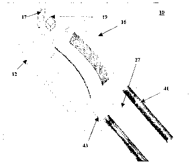
assembly 10 coniprises a housing 12 and a turbine 16, as shown in FIG. 1. The
housing 12
includes a conduit 27 that extends through the housing 12 and turbine 16,
whereby the turbine
16 rotates relative to the conduit 27 to redirect energy from the conduit 27.
Preferably,
conduit 27 is a radiation waveguide, and more preferably the radiation
waveguide is an
optical fiber. The rotating catheter tip assembly 10 rotates a reflecting
material 17, which
then reflects energy emanating from the conduit 27. The reflecting material 17
is coupled
with a focusing element 19 to focus the energy from conduit 27 to a target.
For purposes of
this detailed description, it will be understood that light is redirected
fi=om an optical fiber and
reflected light from a given in vivo target is then gathered and redirected
back to the optical
fiber through the focusing element 19. The focusing element 19 may be any type
of lens,
GRIN lens, and the like suitable to focus optical energy. The focusing element
19 can be
attached to the conduit, as to not rotate and alternatively, there is a space
in between the
6
CA 02627741 2008-04-21
WO 2007/047974 PCT/US2006/041125
focusing element 19 and the conduit 27, whereby the focusing element 19 is
attaclled to
turbine 16 as to rotate thereby.
[0026] The turbine 16 includes a center axle 22 and a plurality of vane
members 18, as
shown in FIG. 2. The center axle 22 includes a central longitudinal bore 26,
through which
the conduit 27 extends. The center axle 22 includes a window opening 24 at the
distal end,
through which reflecting material 17 reflects energy emanating from the
conduit 27. The
vane members 18 project radially outward from center axle 22 and provide a
rotating torque
to the center axle 22 when a flowing fluid (gas or liquid) flows against the
vane meinbers 16,
thereby causing the center axle 22 to rotate about the conduit 27. Preferably,
the vane
members 16 can have a pre-determined curvature along the longitudinal axis of
the turbine
16. The vane members 16 can be spiral shaped, or in any other configuration
which pernzits
rotation of the turbine 16. Preferably, the turbine 16 is made from staiidess
steel, plastic
tygon or Tetlon. Alternatively, the turbine 16 includes knobs to support the
axle 22 and
allows the axle 22 to rotate without wobbling.
[0027] The housing 12 includes a cylinder 32, a housing cap 14, and a cap
member 20, as
shown in FIG. 3. The cylinder 32 includes a central chamber 33, a distal
opening 29, and
outlet channels 30. The central chamber 33 houses the turbine member 16 and
includes an
inflow and an outflow, which define a fluid flow pathway 48. The inflow runs
along the
turbine member 16, while the outflow runs along the outlet channels 30. The
housing cap 14
includes a plurality of fluid inlet ports 42, a plurality of fluid outlet
ports 44, and a central
opening 40, as shown in FIGS. 4a aiid 4b. The fluid inlet ports 42 attach to
fluid inlet ti.ibes
41, as shown in FIG. 1. The fluid inlet tubes 41 are connected to a fluid
source (not shown).
The fluid inlet ports 42 pass through a generally central portion of the
housing cap 14, to
transmit fluid to central chanlber 33. The fluid inlet ports 42 generally
align with turbine
member 16. The fluid outlet ports 44 pass through a relatively peripheral
portion of the
housing 14 and align with the outlet channels 30 and outlet tubes 43, as shown
in FIG. 1.
The central opening 40 includes a concentric recessed seat 39, as shown in
FIG. 4, in which
the axle 22 sits and substantially rotates thereabout. Concentric recessed
seat 39 is for-med to
permit the axle 22 to rotate without wobbling. The central opening 40 co-
axially aligns with
longitudinal bore 26 and permits conduit 27 to be passed there through,
whereby the turbine
meinber 16 is freely rotatable without rotate conduit 27. The axle 22 is co-
axially aligned to
an opening 29 at a distal end of the housing 12 and opening 29 permits axle to
rotate about an
7
CA 02627741 2008-04-21
WO 2007/047974 PCT/US2006/041125
axis. Preferably the housing 12 is made from Teflon. Alternatively, the
housing 12 includes a
cover transparent to the energy and wliich encapsulates the turbine 16, so
that no fluid can
escape from the housing except tluough the charulels 30. Preferably, the
transparent cover is
made from any biocompatible transparent plastic. Such plastic can include
Polymethyl
methacrylate (PMMA) or the like.
[0028] The cap member 20 includes an inner annular member 28, an outer annular
member
27, a plurality of spacer rib members 34, and a plurality of spaces 35, as
shown in FIG. 5a
aiid 5b. The cap member 20 is concentrically mounted onto the distal end of
the axle 22
through inner annular member 28, as shown in FIG. 5b. The inner annular member
28
pennits axle 22 to freely rotate thereabout, without wobbling. The inner
annular meniber 28
and outer annular member 27 are comiected by spacer rib members 34 and are
concentrically
spaced apart. The spaces 35 between adjacent pairs of spacer rib members 34
provide
outflow pathways for the fluid flow 48 to pass from the central chamber 33 to
the distal end
of housing 12 and then to outlet channels 30. A plurality of fluid flow ports
(not shown)
may be provided in a distal surface of the cap member 20 and define a distal
end of spaces 35
to channel fluid flow out of spaces 35.
[0029] At the distal end of the axle 22, a reflecting material 17 (not shown)
is attached to the
center axle 22 at window 24, as showii in FIG. 1. The reflecting material
redirects energy
from the conduit 27. The reflecting material preferably includes a prism or a
mirror, wliich
reflects energy from the conduit, the prism rotating with the center axle 22.
In one
embodiment the energy is radiant energy. Preferably, a lens focuses energy
onto the patient.
The lens can be a microlens, GRIN lens, or optical fiber lines. The probe
preferably includes
a fluid source connected to the inlet tube.
[0030] The fluid is provided to the inlet tubes 41, as shown ui FIG. 1. The
fluid is provided
by a fluid source (not sho m). Preferably, the fluid source is a punip. The
pump can be any
standard fluid pump, as known and recognized by those skilled in the art.
Preferably, the
fluid is chosen from a group consisting of oxygen, carbon dioxide, nitrogen,
helium, saline,
water, d5W or ailificial blood such as Oxyglobin. Alternatively, any gas that
can be
dissolved into blood or tissue relatively easily can be used. Accordingly, a
gas pump would
used to provide fluid to the inlet tubes 41.
8
CA 02627741 2008-04-21
WO 2007/047974 PCT/US2006/041125
[0031] The preferred dimensions of the outer diameter of the housing 12 is
2mm, the outer
diameter of the turbine 16 is 1.4mm, the outer diameter of the iiilet tube 42
is 0.2nnn, the
outer diameter of the outlet tube 44 is 0.2mm. The speed can be 30 rotations
per second.
The turbine pitch can be 4 pitch/mm, while the speed of the gas flow can be
120nun/sec and
target flow rate is 3nun3/sec. The above are all examples. The invention is
not limited to
these values. For instance, to obtain a finer image, the flow rate is lower
and the time it talces
to obtain an image is then longer.
[0032] Alternatively, the turbine 16 includes wart to reflect energy coniing
through a
radiation energy guide back to the radiation energy guide. The reflective wart
can be any
reflective material on the axle 22. Preferably, the wart is block shape with a
flat wall shape.
The wart rotates with the turbine and the energy reflected by the wart
indicates cw.Tent
angular position of the prism. The wart identifies one angular position of the
rotating portion
when the light hits and gets back form the wart. The wart may be a flat wall
facing the
radiation energy guide to reflect back. The wart can be molded into the axle,
and flat wall
can have a reflective material, such as a mirror placed on it to increase the
reflection. The
width of the wart is sniall compared to the circumference of axle 22, so as to
identify a given
point, and is high enough to block the energy emitted from optical fiber, so
it is reflected by
wart.
[0033] In operation, the assembly may be connected to a sample ai-in of a
single mode fiber
OCT. In the center of an OCT probe, the turbine 16 is connected to a prism.
Gas or liquid
flows through the inlet port 42 into the turbine chainber 32. The turbine 16
is supported by
positioning between the housing cap 14 and cap member 20 to maintain constant
position
during rotation. At the center of the turbine 16, the central longitudinal
bore 26 includes an
optical fiber. During rotation of the turbine 16, the optical fiber remains
stationary. In
spectral domain phase sensitive OCT, the reference reflecting surface is
within the catlieter.
[0034] A probing light will be launched from the single nlode optical fiber
through a lens
having a curvature to focus the light onto target tissue area. A rotating
prism connected to the
turbine reflects incoming light toward target tissue area on the vessel wall,
enabling the
imaging system to scan 360 degrees around an inner vessel wall at a constant
speed. The
reflected light from the target tissue returns to the fiber through the prism.
A standard
analysis of the light is then performed to obtain the image, as in U.S. Patent
6,134,003,
9
CA 02627741 2008-04-21
WO 2007/047974 PCT/US2006/041125
incoiporated by reference herein. Gas or liquid gone through the turbine 16
exits the probe
through an outlet tube 44. The rotation direction and speed of the turbine are
controlled by
the pressure difference between inlet ports 42 and outlet ports 44. Applying a
gas or liquid
through an iiilet tube pressure is induced to the turbine which rotates;
therefore, a prism put
on the end of the turbine rotates as well. Finally, an imaging system can scan
360 degrees
around the iiiner vessel wall at a constant speed.
[0035] FIG. 6 depicts an alternative embodiment of a housing cap 14,
synonymously termed
a catheter cap 14, which is mountable on a distal open end of a catheter body
(not shown)
such that central flange 41 seats against the distal end of the catheter body
(not shown). The
fluid inlet openings 42 and fluid outlet openings 44 consist of channels which
permit fluid
flow to pass through the catheter cap 14 in the manner discussed above.
Central opening 40
again accommodates passage of the optical fiber 27 therethrough and is co-
axially aligned
with the central bore of 26 of the turbine member 16 as depicted in FIG. 7.
The proximal
and distal ends of the catheter cap 14 projects from the central flange 41 and
are preferably
mirror images of one another about the central flange 41.
[0036] An alteniative embodiment of the turbine member 16 is illustrated in
FIG. 7. The
principal difference between the first embodiment of the turbine member
illustrated in FIGS.
1-5 is that there is a space in between the focusing element 19 and the
conduit 27. The space
may be an air space or an optical gap providing for the optical energy
permission to expand
before being focused by the focusing element. In this embodiment, the focusing
element 19
and the reflecting material 17 both rotate about the axis by the axle 22, by
being substantially
contiected to the axle by optical glue, or the like. Also, the cuived or
helical pitch of the
turbine vanes 18 is greater than that depicted in FIGS. 1-5, such that they
subtend
approximately a 90 degree arc about the circumference of the axle 22.
[0037] A second embodiment of a cap member 20 is depicted in FIG. 8, and is
synonymously termed second cap member 60. The second cap member 60 includes a
central
opening 64, a collection channel 65 and a plurality of outflow ports 66. The
central opening
64 is concentrically mounted onto the distal end of the axle 22 to peimit axle
22 to rotate
freely thereabout. The collection channel 65 is connected to the outflow ports
66, to permit
the outflow of fluid. The outflow ports are substantially aligned with the
outflow ports 66of
the catheter cap 14, to allow the outflow to return to the fluid source (not
shown). Second
CA 02627741 2008-04-21
WO 2007/047974 PCT/US2006/041125
cap member 60 is similar to second cap member 60, in that it has an irnier
annular member 64
through which the axle 22 of turbine meniber, and an outer annular member 62
which is in
concentrically spaced apart relationship therewitli 16 passes except that
after fluid flows
through the spaces 35 it enters a retui7i path by passing through outlet flow
ports 66 which are
provided about a peripheral portion of a distal surface of the second cap
member 60 and enter
the fluid outlet chaivnels 30 in the housing 12.
[0038] FIG. 9 demonstrates the complete assembly 100 of the catheter cap 14,
second cap
member 60, with turbine member 16 therebetween.
[0039] The present invention also pertains to a method for imaging a patient.
The method
comprises the steps of inserting a catheter into a patient, rotating a turbine
16 of the catheter
relative to a conduit 27, extending through the turbine 16 of the catheter,
redirecting energy
transmitted through the conduit 27 to the patient and receiving the energy
reflected or
backscattered to the turbine, and redirecting reflected energy to the conduit
27.
[0040] Preferably, the rotating step includes flowing fluid through an inlet
tube 41 to the
turbine 16 to turn an axle 22 of the turbine 16.
[0041] Preferably, the flowing step includes flowing the fluid against a
plurality of vane
members 18 which extend from a rotating center axle 22 of the turbine 16 to
create a rotating
torque on the center axle 22 to rotate about the conduit 27 that extends
through the center
axle 22. The axle 22 preferably has reflecting material 17 attached to the
distal end of the
axle 22, which redirects the energy fiom the conduit 27. Preferably, the
conduit 27 is an
optical fiber.
[0042] The reflecting material 17 preferably includes a prism or inirror which
reflects light
from the conduit, atid includes rotating the prism with the axle as the axle
is rotated by the
flowing fluid. Preferably, the rotating step includes the step of rotating the
center axle 22 that
is supported by knobs of the cylinder of the turbine in which the center axle
22 is disposed.
Preferably, flowing the fluid from the inlet tube 41 through a chamber 33 and
removing the
fluid flowing from the housing 12 through at least one outlet tube 43.
11
CA 02627741 2008-04-21
WO 2007/047974 PCT/US2006/041125
[0043] hi the foregoing described embodiment of the invention, those of
ordinaiy skill in the
art will understand and appreciate that an assembly is described wliich
provides a fluid drive
mechanism for rotating a mirror about the central longitudinal axis of the
assembly while
transmitting optical energy from a co-axial optical fiber which is maintained
stationary witlun
the central axis of the assembly, such that light energy niay be reflected or
refracted
perpendicular to the central longitudinal axis of the catheter and traverse a
360 degree arc.
12