Note : Les descriptions sont présentées dans la langue officielle dans laquelle elles ont été soumises.
CA 02628824 2008-05-06
WO 2007/056734 PCT/US2006/060628
APPARATUS AND METHOD FOR MOUNTING A THERAPEUTIC DEVICE
Cross-Reference to Related Applications
[0001] This application claims the benefit of U.S. Provisional Application No.
60l734,245, filed November 7, 2005. The disclosure of this prior application
is incorporated
by reference in its entirety.
Statement Regarding Federally Sponsored Research or Development.
[0002] Not Applicable.
Appendix.
[0003] Not Applicable.
Background of the Invention
1. Field of the Invention
[0004] This uzvention relates generally to therapeutic ultrasound devices and,
more
par-ticularly, to an apparatus and method for mounting a therapeutic device to
an oi-thopaedic
cast or other rnedical wrapping.
2. RelatedArt
[0005] The use of ultrasound to therapeutically treat and evaluate bone
injuries is
known. Impinging ultrasonic pulses having appropriate parameters, e.g.,
frequency, pulse
repetition, and amplitude, for suitable periods of time and at a proper
external location
adjacent to a bone injury has been determined to accelerate the natural
healing of, for
example, bone breaks and fractures. For patients with reduced healing
capacity, such as
elderly persons with osteoporosis, ultrasonic therapy may promote healing of
bone injuries
that would otherwise require prosthetic replacement or leave the patient
permanently
disabled.
[0006] The ultrasound therapy is often used in conjunction with medical wraps,
such
as an orthopaedic cast. A rigid or semi-rigid plastic transducer port is
mounted onto a
1
CA 02628824 2008-05-06
WO 2007/056734 PCT/US2006/060628
fracture cast allowing its use for all large and small bones. Providing
reliable bonding of the
port into the cast with minimal increase in elevation and radius is highly
desirable.
Currently, reliance is placed on the adhesive properties of the cast resin in
shear to bond the
port to the cast. This is a disadvantage as a shear bond is not as strong as
other types of
bonding.
[0007] Another problem associated with the prior art transducer mounting
apparatus
becomes apparent to physicians duiing the installation of the apparatus.
Typically, a cast
will be mounted on the patient prior to the time that the decision is made to
administer
ultrasound therapy. Therefore, the physician is required to cut a hole in the
existing cast to
accommodate placement of an ultrasound transducer head module adjacent a body
portion
of a patient requiring treatment. Because a substantial number of transducer
head modules
are circular, a corresponding circular hole is required in the cast. However,
physicians are
commonly equipped with a tool having a blade that may be adjusted to limit
penetration to
the depth of the cast to cut a square or rectangular void in the cast.
Moreover, it is
inefficient to require the physician to be concerned with the precision with
which the void is
made in the cast. Therefore, a need exists for an apparatus that can be placed
within a void
in a cast and convert the square or rectangular void to a circular hole for
receiving an
ultrasound transducer head module and also an apparatus that is adaptable and
.versatile to
minimize a precisioia associated with the dimensions of the void.
[0008] Typically, an ultrasonic therapy device may be applied to a cast in one
of
three ways. First, a medical practitioner may cut a hole in the cast and strap
an ultrasonic
transducer directly over the hole. Second, the medical practitioner may cut a
hole in the cast
and force fit a plastic transducer port into the cast, and then place the
ultrasonic transducer
in the port. Third, the medical practitioner may build a cast around a plastic
transducer port
and thereafter mount the ultrasonic transducer in the port.
2
CA 02628824 2008-05-06
WO 2007/056734 PCT/US2006/060628
[0009] Alternatively, the physician may know, at the time the injury occurs,
that
ultrasound therapy is likely a preferred future treatment. However, the
installation of a
spacer which creates a void in the cast has heretofor been delayed until a
period of time has
elapsed such that the danger of swelling around the affected injury site has
transpired,
because it has been determined that the skin within the void is prone to
window edema
(especially during the swelling period). Therefore, a need exists for an
apparatus that allows
the surgeon to install an insert or support fixture in the cast at the time of
injury which will
insertably receive an ultrasound transducer treatment head module and also
prevent window
edema when the module is not in place.
[0010] There remains a need in the art for an improved assembly for accurately
mounting and positioning a therapeutic treatment device onto a cast that
overcomes the above-
noted disadvantages, is easy to use, and provides better results in healing
musculoskeletal and
bone injuries.
Summary of the Inventiorz
[0011] It is in view of the above problems that the present invention was
developed.
According to an aspect of the invention there is provided an apparatus ' for
ultrasonically
treating an injury in conjunction with an orthopaedic cast. The apparatus
includes: a
portable self-contained main operating unit; a support fixture configured and
adapted for
attachment to the orthopaedic cast adjacent an external site corresponding to
an internal
injury remote from the main operating unit, the support fixture having a body
and at least
one mesh projection extending from the body; an ultrasonic transducer
treatment head
module operatively connected to the main operating unit and detachably engaged
with the
body of the support fixture; and casting material for attaching the support
fixture to the
orthopaedic cast, wherein at least a portion of the casting material
impregnates the at least
one mesh projection.
3
CA 02628824 2008-05-06
WO 2007/056734 PCT/US2006/060628
[0012] In one embodiment of the invention, the at least one mesh projection
comprises a planar mesh base.
[0013] In another embodiment of the invention, the at least one mesh
projection
comprises a plurality of mesh tabs.
[0014] In another embodiment of the invention, the at least one mesh
projection
further comprises at least one living hinge.
[0015] In yet another embodiment of the invention, the at least one mesh
projection
conforms to a small radius of curvature.
[0016] In one particular embodiment of the invention, the main operating unit
has an
internal power source and is dimensioned to be carried by a patient during
treatment.
[0017] In another embodiment of the invention, the treatment head module has
an
ultrasonic signal generator and signal generator circuitry operatively
associated therewith.
[0018] In yet another embodiment of the invention, the treatment head module
comprises an ultrasound transducer.
[0019] In still another embodiment of the invention, the ultrasound transducer
has
piezoelectric properties and is made from a material selected from the group
consisting of a
ceramic material, a single-crystal relaxor ferroelectric, lead zirconate
titanate, lead
metaniobate, barium titanate, and piezoelectric co-polymers of polyvinylidene
fluoride
(PVDF).
[0020] In another embodiment of the invention, the treatment head module is
connected to the main operating unit through a wireless connection.
[0021] In one particular embodiment of the invention, the main operating unit
has an
ultrasonic signal generator and signal generator circuitry, wherein the signal
generator
circuitry includes a processor, a pulsed signal generator, and a switch
coupled to the
processor for regulating the pulsed signal.
4
CA 02628824 2008-05-06
WO 2007/056734 PCT/US2006/060628
[0022] In another embodiment of the invention, the main operating unit has a
display
panel coupled to the signal generator circuitry to display treatment sequence
data and a
keypad coupled to the signal generator circuitry to permit user control of the
signal
generator.
[0023] In another embodiment of the invention, there is provided an optical
transmitter connected to the switch, the optical transmitter being configured
to convert the
pulsed signal to an optical signal.
[0024] In yet another embodiment of the invention, there is provided a
communication interface connected between a communication port and the
processor to
provide a communication link between the ultrasonic signal generator and an
external
computer/modem.
[0025] In still another embodiment of the invention, there is provided an
alarm
connected to the processor to indicate accurate compliance with a treatment
protocol.
[0026] liz one particular embodiment of the invention, the body of the support
fixture has an aperture configured to receive a portion of the ultrasonic
transducer treatinent
head module.
[0027] In another embodiment of the invention, the body has at least two
bayonet
lugs extending into the aperture which are electrically connected to form a
conductive path
therebetween.
[0028] In yet another embodiment of the invention, the ultrasonic transducer
treatment head module includes at least two slotted lugs having at least a
portion thereof
extending from an outer surface of the module and configured to engage the at
least two
bayonet lugs, the at least two slotted lugs being fabricated from conductive
plastic such that
when the slotted lugs engage the bayonet lugs a conductive path is formed
between the
slotted lugs.
5
CA 02628824 2008-05-06
WO 2007/056734 PCT/US2006/060628
[0029] In another embodiment of the invention, the support fixture has an
outer
surface and an inner surface defining an axial bore therethrough with a
proximal inlet and a
distal outlet.
[0030] In yet another embodiment of the invention, there is provided a spacer
configured to fit within a void in the orthopaedic cast, the spacer having an
opening therein,
the opening having a shape corresponding to an outer periphery of the support
fixture, and
the support fixture being at least partially positioned within the opening. In
one particular
embodiment of the invention, the spacer is formed of felt.
[0031] In another embodiment of the invention, there is provided an ultrasound
transmission-enhancing medium positioned within the body. In one particular
embodiment
of the invention, the ultrasound transrnission-enhancing medium is a gel pad.
[0032] In another embodiment of the invention, there is provided a locking
structure
on an outside periphery of the body.
[0033] In still another embodiinent of the invention, the body extends
upwardly in a
transverse direction relative to the at least one inesh projection.
[0034] In yet another embodiment of the invention, there is provided a cap
attached
to the body and a biasing element connected to the cap, wherein the biasing
element engages
the ultrasonic transducer treatment head module.
[0035] In another embodiment of the invention, the body is a cylindrical
hollow
tube.
[0036] In yet another embodiment of the invention, the body further comprises
a lip.
[0037] In still another embodiment of the invention, the body has a proximal
end
portion and a distal end portion, and the at least one mesh projection is
located at the distal
end portion.
6
CA 02628824 2008-05-06
WO 2007/056734 PCT/US2006/060628
[0038] In another embodiment of the invention, the casting material comprises
at
least one cast material strip.
[0039] In yet another embodiment of the invention, the support structure
further
comprises a hemispherical notch.
[0040] In another embodiment of the invention, the mesh projection is made
from a
material selected from the group consisting of a polymer or a composite. In
one particular
embodiment of the invention, the polymer is selected from the group consisting
of
thermoplastic polymers, thermosetting polymers, and elastomers. In another
embodiment of
the invention, the polymer is made of a fourteen count polyester core yarn
having a vinyl
coating.
[0041] In one particular embodiment of the invention, the mesh projection is
made
from a material selected from the group consisting of polyvinyl chloride,
polyethylene,
acrylonitrile butadiene styrene, or silicone.
[0042] hi another einbodiment of the invention, the mesh projection has a
thickness
A, and the thickness A is in the range from about one-half inilliineter to
about seven
millimeters. In one particular embodiment of the invention, the thickness A is
about one
millimeter.
[0043] In another embodiment of the invention, the mesh projection has a first
dimension D1 and a second dimension D2, and the first dimension Dl is in the
range from
about twenty-five millimeters to about one hundred fifty millimeters, and the
second
dimension D2 is in the range from about twenty-five millimeters to about one
hundred fifty
millimeters. In one particular embodiment of the invention,D1 and D2 are each
about 57
millimeters.
[0044] In another embodiment of the invention, the body is rigid or semi-
rigid.
7
CA 02628824 2008-05-06
WO 2007/056734 PCT/US2006/060628
[0045] In another embodiment of the invention, a portion of the mesh
projection is
constructed of a material that may be seen with a chosen medical visualizing
system.
[0046] In another embodiment of the invention, a portion of the mesh
projection is
at least partially opaque to X-radiation.
[0047] In another embodiment of the invention, a portion of the mesh
projection is
at least partially opaque to infra-red radiation.
[0048] In another embodiment of the invention, a portion of the mesh
projection is
at least partially paramagnetic.
[0049] In another embodiment of the invention, a portion of the mesh
projection
includes an adhesive coating.
[0050] In another embodiment of the invention, the mesh projection further
comprises one or more peripheral markers. In one particular embodiment of the
invention,
there are four peripheral markers. In another embodiinent of the invention,
the peripheral
markers are selected fioin the group consisting of a radio-opaque thread woven
into the
mesh, radio-opaque ink, cut pieces of lead tape.
[0051] In another embodiment of the invention, there is provided a central
marker.
[0052] In another embodiment of the invention, the mesh projection has a
varying
weave spacing.
[0053] In another embodiment of the invention, the support fixture has a
concave
lower end.
[0054] In another embodiment of the invention, the support fixture includes at
least
one circurnferential groove in an upper portion thereof.
[0055] In another embodiment of the invention, the support fixture includes at
least
one circumferential flange.
8
CA 02628824 2008-05-06
WO 2007/056734 PCT/US2006/060628
[0056] In another embodiment of the invention, the support fixture features
locking
structure on the outside periphery of the body.
[0057] In another embodiment of the invention, the locking structure includes
a
circumferential ridge on the outside periphery of the support fixture which is
configured to
engage at least one locking member extending downward from an outer periphery
of a
cover. In one particular embodiment of the invention, there are three locking
members.
[0058] In another embodiment of the invention, the locking member is formed of
a
resilient material such that it will flex outward as the ridge thereon is
forced over ridge, and
it will snap back into position after it moves beyond ridge.
[0059] In another embodiment of the invention, there is provided a support
fixture
configured and adapted for attachment to a medical wrap adjacent an external
site
corresponding to an internal injury, the support fixture comprising: a body,
the body having
an inner wall and an outer wall, the inner wall forming an opening adapted to
receive, in
use, an ultrasonic therapy device; and at least one mesh projection, the at
least one mesh
projection extending from the body and having a plurality of openings, and
wherein at least
a portion of the plurality of openings is impregnated with a portion of the
inedical wrap.
[0060] The invention has several advantages over prior devices and techniques.
First, the apparatus has increased strength in comparison to existing ports
due to the
impregnation of the mesh projection by the casting material, such as resin.
Second, the
mesh, projection provides the ability to use the fixture on smaller radius
limbs. Third, the
mesh projection allows for a reduction in size of the cast opening, thereby
reducing the
possibility of cast failure.
[0061] Thus, in furtherance of the above goals and advantages, the present
invention
is, briefly, a rigid or semi-rigid transducer body with a flexible mesh or
weave projection
which enables conformation to a small radius of curvature, for example, around
a limb or
9
CA 02628824 2008-05-06
WO 2007/056734 PCT/US2006/060628
finger. The weave is sufficiently open to allow resin impregnation through the
weave for
incorporation of the transducer body to the cast. The resin impregnated weave
does not rely
on shear properties (which are weaker) for attachment to the cast but instead
relies on the
strength of the resin in tension and compression as the resin passes through
the weave and
attaches to the cast.
[0062] Further features, aspects, and advantages of the present invention, as
well as
the structure and operation of various embodirnents of the present invention,
are described in
detail below with reference to the accompanying drawings.
Brief Description of the Drawings
[0063] The accompanying drawings, which are incorporated in and form a part of
the
specification, illustrate embodiments of the present invention and together
with the
description, serve to explain the principles of the invention. In the
drawings:
[0064] Figure 1 is a perspective view of a portable ultrasonic treatment
apparatus
according to the present invention, illustrating a main operating unit or
controller and an
ultrasonic transducer treatment head module;
[0065] Figure 2 is an exploded view of the main operating unit;
[0066] Figure 3 is a block diagram of the circuitry for the main operating
unit;
[0067] Figure 4 is a block diagram of one embodiment of the circuitry for the
ultrasonic transducer assembly;
[0068] Figure 5 is a block diagram of an alternative embodiment of the
circuitry for
the ultrasonic transducer assembly;
[0069] Figure 6 is a schematic diagram illustrating an alternative embodiment
of the
portable ultrasonic treatment apparatus;
[0070] Figure 7 is a sectional side view of an ultrasonic transducer assembly
to deliver
ultrasound waves into a medium B;
CA 02628824 2008-05-06
WO 2007/056734 PCT/US2006/060628
[0071] Figure 8 is a top view of a fixture with a cap mounted thereon;
[0072] Figure 9 is a top view of the fixture alone;
[0073] Figure 10 is a perspective view of a locating ring and strap for
locating bone
injuries;
[0074] Figure 11 is a perspective view of the locating ring of Figure 10
affixed to a
patient wearing a cast and illustrating a mark to define the location of a
bone injury;
[0075] Figure 12 is a top view of a mesh base in a second embodiment;
[0076] Figure 13 is a perspective view of a template centrally located over
the mark;
[0077] Figure 14 is a perspective view with parts separated of the patient's
cast with
a reinoved section and a fixture for retaining and aligning the ultrasonic
transducer assembly
of Figure 1;
[0078] Figure 15 is a perspective top view of the fixture located on a cast;
[0079] Figure 16 is a perspective view of the fixture being secured to the
cast at the
removed section;
[0080] Figure 17 is a perspective view with parts separated of the cast, the
fixture and
a cap for the fixture;
[0081] Figure 18 is a top view of a tenlplate in a second embodiment;
[0082] Figure 19 is a side perspective view of the mesh base being used as a
template;
[0083] Figure 20 is a perspective view with parts separated, illustrating the
ultrasonic
transducer assembly aligned for releasable attachment to the fixture;
[0084] Figure 21 is a top view of the fixture within a void in a cast;
[0085] Figure 22 is a side cross-sectional view of a fixture partially secured
within a
void in a cast;
[0086] Figure 23 is a top view of the fixture having a plurality of tabs
extending
radially therefrom;
11
CA 02628824 2008-05-06
WO 2007/056734 PCT/US2006/060628
[0087] Figure 24 is an exploded side view of an apparatus for mounting an
ultrasound
transducer;
[0088] Figure 25 is an enlarged side view of an assembled apparatus for
mounting an
ultrasound transducer;
[0089] Figure 26 is a perspective view of the fixture secured in a cast ready
to receive
an ultrasound transducer head;
[0090] Figure 27 is a perspective view of a fully assembled apparatus for
mounting an
ultrasound transducer in a cast;
[0091] Figure 28 is a partial enlarged side view of another embodiment of an
apparatus for mounting an ultrasound transducer;
[0092] Figures 29-32 are various views of a cover illustrating alternative
locking
structure;
[0093] Figure 33 is an exploded side view of an apparatus for mounting an
ultrasound
transducer having a cover with external locking structure;
[0094] Figure 34 is a perspective view of another embodiment of a cover with
alternative locking structure;
[0095] Figure 35 is a side view in cross-section of an apparatus for the
installation of
the fixture adjacent a treatment location prior to installing a cast thereon;
[0096] Figure 36 is a perspective view of a piece of casting tape and a sealed
package
therefor;
[0097] Figures 37-39 are perspective views illustrating a system for mounting
an
ultrasound transducer receiving apparatus adjacent a treatment location; and
[0098] Figures 40-42 are perspective views illustrating a system for mounting
an
ultrasound transducer receiving apparatus adjacent a treatment location in a
cast.
12
CA 02628824 2008-05-06
WO 2007/056734 PCT/US2006/060628
Detailed Description of f the Embodiments
[00991, Referring to the accompanying drawings in which like reference numbers
indicate like elements, Figure 1 illustrates a portable ultrasonic treatment
apparatus 10. The
ultrasonic treatment apparatus 10 includes a portable main operating unit
(MOU) 12 and an
ultrasonic transducer treatment head module 14 coupled to the MOU 12 by a
first cable 16.
The MOU 12 provides control signals for the ultrasonic transducer treatment
head module
14. The first cable 16 may be a multi-conductor cable capable of transmitting
relatively low
frequency or optical signals, as well as digital signals. The first cable- 16
may include
coaxial cable or other type of suitable shielded cable. Altematively, the
first cable 16 may
include fiber optic cable for transmitting optical signals. In operation, the
transducer
treatment head module is positioned adjacent the injured area and excited for
a
predetermined period of time. To ensure that the transducer treatment head
module is
properly positioned, a safety interlock may be provided to prevent inadvertent
excitation of
the transducer assembly and to insure patient compliance. Although shown
herein with a
single transducer treatment head module, the present invention also envisions
a plurality of
head modules for use with a single MOU.
[00100] Referring to Figure 2, MOU 12 includes a housing 20 which is typically
constructed in two half-sections joined together by screws, ultrasonic welds
or adhesives. A
piinted cucuit board 22 is positioned within the housing 20 and coupled to
display assembly
24 via a second cable 26. Display assembly 24 includes mounting board 28,
display 30 and
a keypad 31, shown in Figure 1. Display 30 may be, for example, a liquid
ciystal type
display or an LED type display suitable for displaying text and numerals.
Battery holder 32
is connected to printed circuit board 22 for portable operation of the real
time clock and the
ultrasonic treatment head module of the present invention. In addition, a
suitable battery,
such as a bank of three (3) lithium batteries, is positioned in the battery
compartment.
13
CA 02628824 2008-05-06
WO 2007/056734 PCT/US2006/060628
[00101] Communication port 34 is affixed to printed circuit board 22 and
accessible
through channel 36 in housing 20. Communication port 34 is coupled to signal
generator
circuitry 38 on printed circuit board 22 and provides a communication link,
e.g., for serial
communications, between the MOU 12 and an external computer. In this
configuration, a
physician can download information, such as the number, date, time of day,
and/or duration
of actual treatments initiated by the patient, stored within signal generator
circuitry 38.
[00102] Figure 3 illustrates a block diagram of the signal generator circuitry
38 within
the MOU 12 which generates and controls the pulses transferred to the
ultrasonic transducer
assembly 14. Signal generator circuitry 38 includes a processor 44 having
memory 43 (e.g.,
RAM and ROM) and stored programs (e.g., system and application) for
controlling the
operation of the processor, as well as the transducer treatment head module
14. Processor
44 is coupled to display 30 and keypad 31 and is configured to receive data
from the keypad
31 and to transfer data to the display 30. Processor 44 may include a
microprocessor or
processor 44 may be a microcontroller having internal memory. Communication
interface
35 is connected between communication port 34 and processor 44 and is provided
to
communicate with, for example, an external computer. Communication interface
35 may be
a serial interface, such as an RS-232 interface, a parallel interface, a
universal serial bus
interface, or a modein.
[00103] Processor 44 is also utilized to control the treatment sequence, i.e.,
the star.=t
time and the stop time of the ultrasonic treatment. The processor may be
preprogrammed
for treatment times and the user (e.g. the physician or patient) selects one
of the treatment
times via keypad 31, or the processor may be programmed by the user via keypad
31 to set
the start and stop sequence. Treatment times may range between about one and
about sixty
minutes, although treatments in the order of 10-20 minutes are typical. When
the treatment
time is activated, processor 44 closes switch 60 which permits the modulated
signal to pass
14
CA 02628824 2008-05-06
WO 2007/056734 PCT/US2006/060628
to cable 16. When the treatment time expires, switch 60 is opened and the
modulated signal
is inhibited from passing to cable 16. In addition, when the treatment time
expires,
processor 44 may send an alarm signal to alarm 62 which activates.
[00104] Referring to Figure 4, a block diagram of one embodiment of the
transducer
treatment head module circuitry is shown. The transducer treatment head module
circuitry
includes a receiver 66 which receives the signals transferred iby MOU 12 via
cable 16.
Receiver 66 is connected to transducer driver 67 which excites transducer 68.
[00105] An alternative embodiment of the transducer treatment head module
circuitry
is shown in Figure 5. In this embodiment, the transducer treatment head module
14 includes
an internal battery 69 which supplies power to the internal components of the
transducer
treatment head module. For example, battery 69 supplies power to signal
monitoring circuit
70 and signal driver 71. The signal monitoring circuit 70 provides a digital
output signal 72
which represents the waveform characteristics of the output of transducer
driver 67. Such
characteristics may include, for example, the frequency, pulse repetition
frequency, the pulse
width and the average output power of the signal driving transducer 68. The
output signal
72 of signal monitorirng circuit 70 is transferred to the MOU 12 via driver 71
and the first
cable 16. Optional fixture interlock 73, which may include switches on the
outer surface of
the transducer treatment head module, provides power to the internal
components of the
transducer treatinent head module 14 so as to ensure that the transducer
treatinent head
module 14 is properly positioned before the transducer is excited.
[00106] Figure 6 illustrates an alternative embodiment of the ultrasonic
treatment
apparatus, generally indicated by reference numeral 10'. The ultrasonic
treatment apparatus
10' includes a logic voltage convertor 76, a power source 77, battery voltage
sense circuitry
78, a drive signal voltage converter 79, a drive signal voltage adjustor 80, a
real time clock
81, an EEPROM 82, gel sense circuitry 83, a transducer drive circuitry 84, an
ultrasound
CA 02628824 2008-05-06
WO 2007/056734 PCT/US2006/060628
transducer 85, a first user interface 86, a second user interface 87, and a
microprocessor 88.
The configuration of the ultrasonic treatment apparatus 10' allows for most of
the
components to be placed in the MOU 12 and only the ultrasound transducer 85
placed in the
head module 14. In some embodiments, the head module 14 may consist solely of
the
ultrasound transducer 85.
[00107] The logic voltage converter 76 steps down the voltage for the
microprocessor
and other electronics. The power source 77 supplies power to the ultrasonic
treatment
apparatus 10' and may be a battery or battery pack. The battery voltage sense
circuitry 78
senses the power of the power source 77 and prevents operation if there is
insufficient power
to drive the transducer and the other electronic components. The drive signal
voltage
converter 79 steps down the voltage for the transducer drive circuitry 84. The
drive signal
voltage adjustor 80 is used to calibrate the transducer drive circuitry 84 to
obtain a
predetermined acoustic wave from the transducer 85. The real time clock 81 is
the clock for
the microprocessor 88. The EEPROM 82 is used to record data after a treatment.
Such data
may include date, time, length of treatment, and other variables.
[00108] The gel sense circuitry 83 senses an impedance change through the
transducer to ascertain whether the transducer is emitting acoustic waves into
a fluid or gel
medium. Every transducer has an iinpedance when it is acting on a medium, such
as air or
water. The iinpedance is different for each inediuin. By using this
difference, the gel sense
circuitly 83 can determine whether or not the transducer is acting upon a gel
medium.
[00109] The transducer drive circuitry 84 creates the sinusoidal wave that
drives the
transducer 85. The ultrasound transducer 85 creates the ultrasonic wave for
treating the
injury. The first user interface 86 is a display. The first user interface 86
may indicate items
such as time duration, and errors. The second user interface 87 is one or more
switches that
are used to control the ultrasonic treatment apparatus 10'. Such switches may
include a start
16
CA 02628824 2008-05-06
WO 2007/056734 PCT/US2006/060628
button, a stop button, and a reset button. The microprocessor 88 executes
stored programs
to apply the treatment in a predefined manner.
[00110] Further, some embodiments of the ultrasonic treatment apparatus 10'
eliminate the wired connection between the transducer drive circuitry 84 and
the ultrasound
transducer 85 and instead implement a wireless connection, such as ZIGBEETm,
BLUETOOTHTm, IEEE 802.11, or other Radio Frequency (RF) technology. ZigBee is
a
published specification set of high level communication protocols designed for
wireless
personal area networks (WPANs). The ZIGBEE trademark is owned by ZigBee
Alliance
Corp., 2400 Camino Ramon, Suite 375, San Ramon, California, U.S.A. 94583.
Bluetooth is
a technical industry standard that facilitates short range communication
between wireless
devices. The BLUETOOTH trademark is owned by Bluetooth Sig, Inc., 500 108th
Avenue
NE, Suite 250, Bellevue Washington, U.S.A. 98004. IEEE 802.11 denotes a set of
Wireless
LAN/WLAN standards developed by working group 11 of the IEEE LAN/MAN Standards
Committee (IEEE 802). RF is a wireless communication technology using
electromagnetic
waves to transmit and receive data using a signal above approximately 0.1 MHz
in
frequency.
[00111] Figures 7-9 illustrate an ultrasonic transducer assembly 100 to
deliver
ultrasound waves into a medium B, which may be coinposed of soft tissue or
bone in order
to promote healing of a wound in the soft issue and/or healing of a fracture
in a bone. The
ultrasonic transducer assembly 100 includes a support fixture 110. The support
fixture may
also be termed a transducer port or simply a port. The support fixture 110
includes a body
114 and a mesh projection 112 that extends from the body 114. In the depicted
embodiments, the mesh projection 112 is a planar mesh base, and the body 114
that extends
upwardly from the mesh base 112 in a transverse direction. The ultrasonic
transducer
assembly 100 also includes a cap 116, a spring 118, and a transducer 120. In
the depicted
17
CA 02628824 2008-05-06
WO 2007/056734 PCT/US2006/060628
embodiments, the base 112 is shown assuming that the interface with the medium
B is flat.
However, those skilled in the art would understand that the base 112 can be
shaped to fit the
interface with medium B.
[00112] The base 112 is made of a mesh or woven material. The mesh or weave
allows the base 112 to conform to a small radius of curvature. For example,
the mesh or
weave permits the base to conform to a small limb or finger. Further, the mesh
or weave
aids in the incorporation of the support fixture 110 to a cast 288 (best seen
in Figure 13). As
those skilled in the art understand, an orthopaedic cast is a shell that
encases a limb (or, in
some cases, large portions of the body) to hold a broken bone (or bones) in
place until the
broken bone has healed. Typically, the cast is formed of plaster, cotton
bandages that have
been impregnated with plaster of paris, or knitted fibreglass bandages
impregnated with
polyurethane. However, other materials may be used. As used herein, the term
"resin"
means a material that hardens to form a portion of the cast shell. While the
depicted
embodiments illustrate an orthopaedic cast, those having ordinary skill in the
art would
understand that other types of medical wrappings may be used.'
[00113] As explained in greater detail below, the mesh or weave captures a
portion of
the cast resin, which strengthens the attachment of the support structure to
the cast in
comparison to prior devices. Prior devices utilized shear properties to retain
the support
structure to the cast. hnpregnating the mesh or weave with resin pennits the
present
invention to utilize shear, tension, and compression characteristics to adhere
the support
stiucture to the cast. This is a significant improvement over the state of the
art.
[00114] The base 112 may be made from many different types of materials, such
as a
polymer or a composite. Polymers may include thermoplastic polymers,
thermosetting
polymers, and elastomers. Examples of polymers may include, among others,
polyvinyl
chloride, polyethylene, acrylonitrile butadiene styrene, or silicone. In the
embodiment
18
CA 02628824 2008-05-06
WO 2007/056734 PCT/US2006/060628
depicted in Figures 7-9, the base 112 is made of a fourteen count polyester
core yarn having
a vinyl coating. In the depicted embodiments, the base 112 has a square or
rectangular
shape, but those of ordinary skill in the art would understand that other
shapes may be used.
For example, the base 112 may be in the shape of a circle or triangle. The
base 112 has a
thickness A, which is in the range from about one-half millimeter to about
seven
millimeters. In the embodiment depicted in Figure 7, A is about one
millimeter. The
embodiment depicted in Figure 8 illustrates a first dimension D1 and a second
dimension
D2. The first dimension D1 is in the range from about twenty-five millimeters
to about one
hundred fifty millimeters. The second dimension D2 is in the range from about
twenty-five
millimeters to about one hundred fifty millimeters. In the embodiment depicted
in Figures
6-8, D 1 and D2 are each about 57 millimeters. The mesh base 112 may be
oversized such
that it can be cut-to-fit for a custom application of the support structure
110.
[00115] The body 114 is rigid or semi-rigid. The body 114 is shaped to receive
the
transducer 120. The transducer 120 is constructed of materials and designs
that are
commonly used in ultrasound applications. The transducer 120 may have
piezoelectric
properties and may be made from, as examples, a ceramic material, a single-
crystal relaxor
ferroelectric, lead zirconate titanate, lead metaniobate, barium titanate, and
piezoelectric co-
polymers of polyvinylidene fluoride (PVDF). Alternatively, the transducer 120
may have
magiietostrictive properties.
[00116] In the embodiment depicted in Figures 7-9, the body 114 is a
cylindrical,
hollow tube as the transducer 120 has a circular cross-section but other
shapes may be used.
The body 114 has an inner portion 126 and an outer portion 128. In the
embodiments
depicted in Figures 7-9, the body 114 has an inner wall 130 and an outer wall
132 which
define the inner portion 126 and the outer portion 128. The inner wall 130
forms an
aperture 134 that receives the transducer 120. The inner wall 130 of the body
114 is
19
CA 02628824 2008-05-06
WO 2007/056734 PCT/US2006/060628
dimensioned such that the transducer 120 fits tightly within the body 114. The
inner wall
130 may have a diameter the same as or slightly larger than the diameter of
the transducer
120. For example, the diameter of the inner wall 130 may be about zero to
about two mm
larger than the diameter of the transducer 120. The body 114 also has a
proximal end
portion 135 and a distal end portion 136. In the depicted embodiment, the mesh
base 112 is
connected to the body 114 at the proximal end portion 135. However, the mesh
base 112
may be at the very end of the proximal end portion 135 or slightly above it
such that the
mesh base 112 attaches a few millimeters above the proximal end portion 135.
[00117] A cap 116 is connected to the body 114 at the distal end portion 136.
For
example, the body 114 may have a lip 140 (best seen in Figure 7) such that the
cap 116
snaps onto the body 114. Further, the spring 118 is mounted between the cap
116 and the
transducer 120 in order to exert pressure on the transducer 120. In some
embodiments, the
spring 118 may be mounted to the cap 116. The spring 118 biases the transducer
120
toward the proximal end portion 135 of the body 114. In some embodiments, the
ultrasonic
transducer assembly 100 includes gel or a gel bladder 121 to aid in the
transference of
acoustic waves from the transducer 120 to the medium B.
[00118] A system controller 122 spatially and temporally controls the acoustic
waves
that emanate from the transducer 120. The design and fabrication of the system
controller
122 are well known to those who practice the art. The systein controller 122
is electl7cally
connected to a signal generator 124, and the signal generator 124 is
electrically connected to
the transducer 120. The system controller 122 triggers the prograrnmable
signal generator
124 to produce ultrasonic excitation signals that are sent to the transducer
120. The
transducer 120 receives the excitation signal and emits an acoustic
longitudinal wave that
propagates on to medium B. The system controller 122 and the signal generator
124 may
take the form of the MOU 12 described above.
CA 02628824 2008-05-06
WO 2007/056734 PCT/US2006/060628
[00119] The transducer 120 produces specific sequential or simultaneous
transmissions of acoustic waves, which is controlled by the system controller
122, in order
to non-invasively irradiate or interrogate the medium B ultrasonically. The
system
controller 122 may be a programmable microprocessor, but may also include,
though is not
limited to, integrated circuits, analog devices, programmable logic devices,
personal
computers or servers. The timing sequences may be established by the user at
any time or
established during the manufacturing process.
[00120] The ultrasonic transducer assembly 100 may be used to administer
therapeutic treatment composed of an ultrasound dosage administered once or
twice a day,
and repeated daily for several months to effectively stimulate the healing
process. In some
embodiments, one dosage of acoustic waves ranges between one and sixty minutes
in length
for the transducer 120. The ultrasonic transducer assembly 110 may be used to
facilitate and
enhance application of therapeutic ultrasound dosages to shallow or deep
anatomical
structures, or both, in an effort to expedite tissue wound healing, including
both the
endosteal and periosteal healing phases in the bone fracture healing process.
[00121] Referring now to Figure 10, a locating ring 280 for detennining the
location
of injured bone is shown. The locating ring 280 includes a strap 282 for
releasably securing
the ring to a patient. The strap 282 preferably has two sections 284 and 286
which permit
quick fastening and unfastening of the ring 280 on and off the patient. The
ring 280 is
constructed of material that may be seen with a chosen medical visualizing
system. Thus, if
X-rays are used, the ring 280 is at least partially opaque to X-radiation. If
infra-red radiation
is used, the ring 280 is at least partially opaque to infra-red radiation,
and, if magnetic
resonance imaging is used, the ring 280 is at least partially paramagnetic.
However, the
other materials may be used for the ring which permits detection by medical
visualizing or
imaging systems.
21
CA 02628824 2008-05-06
WO 2007/056734 PCT/US2006/060628
[00122] The dimensions of the ring 280 are typically a function of the size of
the
patient, the estimated size and location of the injury, and the type of
visualizing system used.
For example, to determine the location of a bone fracture in an average human
limb, e.g.,
the ulna or radius, and using an X-ray imaging system, the diameter of the
ring may
nominally be about thirty-eight millimeters. In this example, the ring may be
a rigid torus of
metallic material of cross-sectional diameter nominally five millimeters. As
another
example, if the visualization system utilized is an ultrasonic imaging system,
the ring 280
may be substantially flexible and planar, so that it may contour to a surface
it is placed
adjacent to, thereby allowing the scanning or imaging transducer to be moved
across the
surface and the ring.
[00123] As noted, the strap 282 has two sections 284 and 286, each section has
one
end fastened to the ring 280. The two sections 284 and 286 may have hook and
loop type
fastening assembly, such as VELCROTm, so that they may be fastened together
and quickly
unfastened. Other quick release fastening techniques are also contemplated.
[00124] Alternatively, a portion of the mesh base 112 may be used to identify
the
location of injured bone. Thus, a portion of the mesh base 112 may be
constructed of a
material that may be seen with a chosen medical visualizing system. For
example, if X-rays
are used, a poi-tion of the inesh base 112 is at least partially opaque to X-
radiation. If infra-
red radiation is used, a portion of the mesh base 112 is at least partially
opaque to infra-red
2 0 radiation, and, if magnetic resonance imaging is used, a portion of the
mesh base 112 is at
least partially paramagnetic. However, the other materials may be used for the
mesh base
112 which permits detection by medical visualizing or imaging systems.
Additionally, the
mesh base 112 may have an adhesive coating on one side for temporarily
locating the mesh
base 112 while the medical visualizing system is activated.
22
CA 02628824 2008-05-06
WO 2007/056734 PCT/US2006/060628
[00125] Figures 11 through 16 illustrate locating an injured bone, affixing
the support
fixture 110 configured to maintain the transducer treatment head module 14
adjacent the
area of the injured bone, and connecting the ultrasonic transducer assembly 10
to the fixture
110 for treating the injured bone. Initially, the locator ring 280 is
positioned on the cast 288
on, for example, a patient's arrn, at a location corresponding to the
estimated or
approximated location of the injury. This initial position is a preliminary
approximation of
the external location of the bone injury, and may be based on previously taken
X-rays, a
physician's diagnosis or the patient's recall of the point of injury.
[00126] An external image, e.g., an X-ray, of the fractured region is taken to
include
the locating ring 280. Although the initial position of the locating ring 280
with respect to
the bone injury is a preliminary approximation, in many instances the initial
placement will
be sufficiently accurate so that the X-ray will depict the bone injury framed
by the ring 280.
The resulting X-ray image indicates the position of the bone injury relative
to the locating
ring 280. The X-rays are used as a guide to locate and mark 290 the
corresponding point on
the cast relative to the actual locating ring 280. The mark 290 gives an
approximate
external location on the cast of the bone injury. If greater accuracy is
required, the ring 280
may be centered about the mark 290, another X-ray is taken, and a new mark
(not shown) is
made on the cast based on the location of the bone fracture relative to the
ring on the X-ray.
Successive iterations of repositioning the locating i7ng 280 and X-raying the
site will yield
even greater accuracy.
[00127] As noted above, in some embodiments, the mesh base 112 may be used
instead of the ring 280 in order to locate the mark 290. Figure 12 illustrates
one example of
the mesh base 112 with one or more peripheral markers 170 that may be used in
conjunction
with a medical visualizing system, such as an x-ray machine. In the embodiment
depicted in
Figure 12, the mesh configuration of the mesh base 112 has been omitted for
clarity.
23
CA 02628824 2008-05-06
WO 2007/056734 PCT/US2006/060628
Additionally, in the embodiment depicted in Figure 12, there are four
peripheral markers
170 but a greater or lesser number may be used. The peripheral markers 170 may
be a
radio-opaque thread woven into the mesh. Alternatively, the peripheral markers
170 may be
radio-opaque ink. Finally, the peripheral markers 170 may be cut pieces of
lead tape. Some
embodiments may further include a central marker 172 to indicate the center of
the mesh
base 112. The peripheral markers 170 indicate the outline of the mesh base 112
while the
central marker 172 may be used to locate the mark 290.
[00128] As shown in Figures 13 and 14, a first embodiment of a marking
template
292 is pressed against the cast 288 and centered on the mark 290 of the
external location on
the cast 288 of the bone fracture. The outline of the inner edges of the
rectangular template
opening is traced on the cast 288, and the traced portion of the cast is
removed so that the
opening 294 in the cast 288 exposes the skin, as shown in Figure 14. The
opening 294 in
the cast 288 receives a felt pad 296 having a thickness approximately the same
as the
thickness of the cast. The felt pad 296 also has a cylindrical bore that
receives a cylindrical
felt pad 298. Felt pad 296 is provided to support the fixture 110 and to
maintain pressure
against the skin which helps prevent window edema (swelling) and is
substantially
equivalent to the pressure exerted by the cast 288 against the skin and is
described in more
detail below.
[00129] The teinplate 292, and consequently the opening 294 in the cast 288,
is
smaller than the mesh base 112 of the fixture 110 for retaining and aligning
the ultrasonic
transducer assembly 14, so that the mesh base 112 engages the cast sui-face
surrounding the
opening 294 when the fixture 110 is placed over the opening 294. The fixture
110 includes
the circular aperture 134 and may include bayonet locking lugs 306. Aperture
134 has
substantially the same diameter as the cylindrical felt pad 298.
24
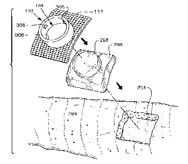
CA 02628824 2008-05-06
WO 2007/056734 PCT/US2006/060628
[00130] Figure 15 illustrates the fixture 110 placed over the felt 296 in the
opening
294.
[00131] Figure 16 shows the fixture 110 positioned over the opening 294 in the
cast
288 and the felt pad 296 so that the apei-ture 134 and the cylindrical felt
pad 298 are
coaxially aligned. The fixture 110 partially compresses the felt pad 296,
shown in Figure
14, against the skin as mesh base 112 of fixture 110, shown in Figure 15,
engages the cast
288, thereby approximating the pressure of the removed portion of the cast
where the felt
pad engages the skin.
[00132] In some embodiments, a portion of the mesh base 112 includes an
adhesive.
The adhesive may be used to temporarily affix the mesh base 112 to the cast
until resin
impregnates the mesh and affixes the mesh base 112 in a more permanent manner.
[00133] Referring to Figurel7, a cap 308 for the fixture 110 is shown. The cap
308 is
provided to maintain pressure on the body tissue exposed in fixture ] 10 when
the ultrasonic
treatment is completed. The cap 308 has a cylindrical portion 310 that extends
into the
aperture 134 of the fixture 110. The cap 308 has slotted lugs 312 on the
cylindrical portion
310 that engage the bayonet lugs 306 in the fixture 110. The cylindT.-ical
felt pad 298 is
positioned in the aperture 134 and the cylindrical poi-tion 310 is inserted
into the aperture
134 with the slotted lugs 312 offset from the bayonet lugs 306. The cap 308 is
pressed
against the cylindrical felt pad 298 until the pressure exerted by the cap 308
and cylindrical
felt pad 298 against the skin approximates the pressure exerted by the cast
288 against the
skin. The cylindrical felt pad 298 may also be comprised of substantially
planar circular
layers that may be removed one layer at a time in order to adjust the
thickness of the felt pad
and the resulting pressure against the skin. This pressure helps to inhibit
window edemas.
The cap 308 is then rotated so that its slotted lugs 312 engage the bayonet
lugs 306. While
the depicted embodiment includes the bayonet lugs 306 and slotted lugs 312,
those skilled in
CA 02628824 2008-05-06
WO 2007/056734 PCT/US2006/060628
the art would understand that other locking mechanisms may be used to connect
the cap 308
to the support fixture 110.
[00134] Figure 18 illustrates a second embodiment of the template for marking
the
opening 294, which is generally indicated by reference numeral 400. The
template 400 is
pressed against the cast 288 and centered on the mark 290 of the external
location on the
cast 288 of the bone fracture. The template 400 includes one or more slots 402
and a central
cutout 404. In the depicted embodiment, the template 400 has four peripherally
located
slots 402. The cutout 404 is centered upon the mark 290, and the outline of
the inner edges
of the template opening is traced on the cast 288 via the slots 402. The
traced portion of the
cast is removed so that the opening 294 in the cast 288 exposes the skin, as
shown in Figure
14.
[00135] In some embodiments, the mesh base 112 may be used as a template to
cut
the opening 294. Figure 19 illustrates the mesh base 112 resting on the cast
288. The mesh
base 112 is located relative to the mark 290. In Figure 19, the body 114 has
been omitted
for clarity and in ordei- to reveal the mark 290. Optionally, adhesive tape
420 can be placed
over the cast 288 and the mesh base 112 to temporarily hold the mesh base 112
while
marking. Alternatively, an adhesive may be applied to a bottom surface of the
mesh base
112 to temporarily affix it to the cast 288. After the mesh base 112 is
located relative to the
inark 290, a inarking instruinent 424, such as a pen or marker, is used to
trace the outline of
the mesh base 112. In some embodiments, the mesh base 112 has a vatying weave
spacing
to allow the marking instru.ment 424 to be traced through the weave to allow
reliable
marking of the cast for cutting.
[00136] In, yet other embodiments, the opening 294 may be cut free-hand. In
other
words, the opening 294 may be cut without a template. Once the opening 294 is
cut, the
26
CA 02628824 2008-05-06
WO 2007/056734 PCT/US2006/060628
mesh base 112 is trimmed with scissors or some other cutting device to match
the shape of
the mesh base 112 with the opening 294.
[00137] Figure 20 is a perspective view illustrating the alignment of the
ultrasonic
transducer treatment head module 14 with the fixture 110 for ultrasonic
treatment of the
injured bone. With the cap 308 and cylindrical felt pad 298, shown in Figure
17, removed,
the projection 314 fits into the aperture 134 of the fixture 110 and the bore
of the felt pad
296. The operative surface 318 of the transducer treatment head module 14 is
pressed
adjacent the skin. In some embodiments, the transducer treatment head module
projection
314 has slotted lugs 316 that engage the bayonet lugs 306 in the fixture 110,
and the
transducer treatment head module 14 is rotated so that its slotted lugs 316
engage the
bayonet lugs 306. The ultrasonic treatment then commences.
[00138] Referring again to Figures 3 and 4, to prevent inadvertent excitation
of
transducer treatment head module 14 and to insure compliance with treatment
protocol,
some embodiments include the fixture interlock 73, which includes switches on
the outer
surface of the transducer treatment head module. In this embodiment, slotted
Iugs 316 are
fabricated from a conductive plastic and the bayonet lugs 306 in fixture 1 10
are electrically
connected, such that when the slotted lugs 316 engage the bayonet lugs 306 an
electrical
path is coinpleted between at least two of the slotted lugs 316. Suitable
conductive plastics
which may be utilized include conductive ABS plastics with either carbon,
stainless steel,
nickel or aluminum fibers.
[00139] The operative surface 318 of transducer treatment head module 14
includes a
gel sensing element for confirming the presence of ultrasonic conductive
material on the
operative surface 318. This surface 318 is pre-coated with a coupling gel
before it is
inserted in the fixture 110 and engages the skin. Alternatively, the gel may
be contained
27
CA 02628824 2008-05-06
WO 2007/056734 PCT/US2006/060628
adjacent the operative surface 318 of transducer treatment head module 14
using a gel sack,
gel bladder or like container.
[00140] Figure 21 illustrates a further embodiment of the support fixture 520,
positioned within a void 522 formed in a portion of a cast 524. Void 522 has a
substantially
square shape and is delineated by the dashed lines. Fixture 520 is shown
having a
substantially circular periphery and a plurality of mesh tabs 526 extending
radially therefrom,
as an alternative to the planar mesh base. Four mesh tabs 526 are visible in
Figure 21.
Additional mesh tabs 526 are hidden by cast material in a plane beneath the
visible tabs as will
become apparent in Figure 23. Fixture 520 preferably includes an axial bore
within the
substantially circular periphery to mount an ultrasound transducer to initiate
a treatment, as
will be discussed in further detail below. Fixture 520 is forrned of a
polymer, such as
polypropylene or polyvinyl chloride.
[00141] Figure 22 illustrates a side cross-sectional view of fixture 520
within cast 524.
Prior to placing fixture 520 into void 522, a spacer 530 is placed within void
522. -Spacer 530
is configured to have a shape on its periphery which corresponds with the
shape of void 522,
and a hole in its center which corresponds to the shape of fixture 520. Spacer
530 is preferably
formed of a medical grade felt or a similar material which will exhibit
comfortable
characteristics agauist a body portion of a patient, and inay be fabricated in
a plurality of layers
so that the thickness can be adjusted depending on the thickness of cast 524.
[00142] Spacer 530 maintains fixture 520 at a predetermined distance from the
body
portion 534 of a patient, to prevent window edema or a similar injury to the
patient due to
uneven pressure at a casted site. As shown, fixture 520 is partially inserted
into the hole
within spacer 530 and is supported thereon by at least one of the radially
extending mesh tabs
526. Mesh tabs 526 contain living hinges formed by a reduction in cross-
section of the mesh
tabs 526 at a proximal end adjacent the bore of fixture 520 which weakens mesh
tabs 526 at
28
CA 02628824 2008-05-06
WO 2007/056734 PCT/US2006/060628
the hinge point, thus allowing them to bend freely. The living hinges provide
for lateral
flexure of mesh tabs 526 to enhance the ability to conform to varying angles
which are a
function of the anatomy of the patient. Moreover, the living hinges allow
fixture 520 to be
articulated to correct for other angular misalignments.
[00143] Fixture 520 is secured within void 522 in cast 524 by weaving strips
532 of
cast material between mesh tabs 526. A plurality of layers of cast material
strips 532 are
placed around fixture 520 until a desired thickness is achieved. Further, due
to its mesh
nature, the cast material impregnates the openings of the mesh tabs 526,
thereby increasing the
structural attachment of the fixture 520 to the cast 524. The configuration of
fixture 520
having mesh tabs 526 allows the fixture to be installed before or after the
cast is installed.
Advantageously, when the layers of cast material strips cure, fixture 520 will
be an integral
part of the cast. Thus, any impact on the skin of the patient, which would
otherwise be
transferred through fixture 520, will be minimized as it is absorbed by the
cast.
[00144] Fixture 520 may optionally include a hemispherical notch 536 in an
upper end
thereof to accommodate a cord extending from an ultrasound treatment head
module while the
module is positioned within the fixture. A lower end 538 of fxture 520 is
preferably concave
to correspond to fit a convex body portion 534 of a patient, without impacting
the skin which
may cause edema or a siunilar injury.
[00145] An ultrasound transmission-enhancing medium 528 is preferably
positioned
2 0 within spacer 530 adjacent a treatment location to minimize or eliminate
an air gap between an
ultrasound transducer head module and a treatment location. The ultrasound
t.ransmission-
enhancing medium 528 is preferably a conductive gel bladder but may be simply
gel.
[00146] The apparatus of the present invention is configured to adapt to and
fit within a
substantially rectangular or square-shaped void in a cast as shown in a top
view thereof in
29
CA 02628824 2008-05-06
WO 2007/056734 PCT/US2006/060628
Figure 23. Advantageously, the fixture 520 converts the void into a circular
receptacle for
receiving a corresponding circular-shaped ultrasound transducer head module.
[00147] Turning now to the exploded side view in Figure 24, and proceeding
from the
bottom, spacer 530 is shown in cross-section, having a hole therein configured
to insertably
receive fixture 520. Fixture 520 includes a plurality of mesh tabs 526 and a
concave lower end
538. Fixture 520 preferably includes at least one circumferential groove 540
in an upper
portion thereof. The purpose of the circumferential groove 540 is to enable
the removal of at
least one layer of fixture 520 to adjust the height of fixture 520 to
correspond to a thickness of
a cast. In the depicted embodiment of the ultrasound transmission-enhancing
medium 528, a
means for facilitating removal of the medium from fixture 520 is provided. In
this
embodiment, the means for facilitating removal is a lead 542 shown extending
from medium
528.
[00148] An ultrasound transducer head module 544 is positioned adjacent
ultrasound
transmission-enhancing medium 528 within fixture 520. Cord 550 connects module
544 with
electronic driving circuitry. Housing 546 is then inserted in the upper
portion of fixture 520 to
enclose the components within fixture 520. A bias element 548 extends from a
bottom portion
of housing 546. Bias element 548 may be a spring and, more particularly, may
be a conical
helical spring. The conical helical spring is advantageously configured to
fully collapse withiui
itself and will therefore require less space within fixture 520. A conical
helical spring will also
maintain a uniform force on ultrasound transducer head module 544 and will
allow module
544 to pivot to conform to the shape of transmission-enhancing medium 528.
[00149] Figure 25 illustrates an enlarged side view of an assembled apparatus
for
mounting an ultrasound transducer in accordance with the present invention.
This enlarged
view illustrates the living hinge 552 on mesh tab 526 which allows for free
lateral movement.
CA 02628824 2008-05-06
WO 2007/056734 PCT/US2006/060628
Also shown, spring 548 in its compressed state urgingly biases transducer
module 544 toward
ultrasound transmission-enhancing medium 528.
[00150] Referring to Figures 26 and 27, fixture 590 is shown secured within a
cast 592
of a patient requiring ultrasound treatment. Lead 594 which is attached at its
lower end to a
transmission-enhancing medium is shown extending from fixture 590. Following
the
placement of ultrasound transducer head module 596 into fixture 590, cover 598
is placed over
the top of fixture 590 and strap 600 is adjusted to secure the entire
apparatus in place.
[00151] Figure 28 illustrates an embodiment of an apparatus for mounting an
ultrasound transducer which features locking structure on the outside
periphery of fixture 110
for retaining a transducer head module within the fixture. As illustrated, the
locking structure
includes a circumferential ridge 612 on the outside periphery of fixture 610
which is
configured to engage at least one latch 614 extending downward from an outer
periphery of
cover 616. Although only one latch 614 is visible in Figure 28, it is
preferable to have three
latches extending from cover 616 and spaced about one hundred twenty degrees
apart. Latch
614 is formed of a resilient material such that it will flex outward as the
ridge thereon is forced
over ridge 612, and it will snap back into position after it moves beyond
ridge 612. The
locking structure advantageously eliminates the need for a strap to secure the
cover in place, as
described above with other embodiments of the presently disclosed apparatus.
[00152] Conical helical spring 618 is held in contact with a lower suiface of
cover 616
by resilient housing 620. Resilient housing 620 is designed to maintain spring
618 in its
position under cover 616 while exhibiting resiliency corresponding to the
compressive
property of spring 618. Housing 620 is secured to cover 616 by lock ring 622
which may be
affixed to cover 616 by epoxy or any other means known to one having ordinary
skill in the
art. Housing 620 is preferably fonned of polyurethane having a thickness of
about 0.01 inches
to about 0.10 inches.
31
CA 02628824 2008-05-06
WO 2007/056734 PCT/US2006/060628
[00153] Also illustrated in Figure 28 are flanges 624. It is contemplated that
flanges
624 may be a plurality of separate continuous circumferential flanges, a
single circumferential
flange having a spiral configuration around the periphery of fixture 610 or at
least one
interrupted flange. The flanges 624 fonn a groove therebetween which may be
used to receive
casting material, casting tape, or a strap.
[00154] Figures 29-32 illustrate an alternative locking structure associated
with cover
630 to removably engage cover 630 with fixture 632. In the cross-sectional
view shown in
Figure 29, cover 630 is illustrated locked within fixture 632 by means of a
hinged locking tab
634 on a first side of cover 630 and a protrusion 636 on a second side of
cover 630. To
remove cover 630 from fixture 632, the portion of locking tab 634 which
extends outwardly
from cover 630 is depressed to release protrusion 638 from a groove formed on
the inner
surface of fixture 632. Cover 630 may then be pivoted upward to disengage
protrusion 636
from a corresponding groove in fixture 632, and remove the cover. The
disclosed locking
structure advantageously eliminates the need for a strap to secure the cover
in place, as
described above with other embodiments of the presently disclosed apparatus.
Furthermore,
the configuration of locking tab 634 provides a structure for easily removing
the cover by a
single hand of the user. Alternatively, a cover may be provided with locking
structure having
two locking tabs. The cover may be removed by depressing one locking tab,
similar to the
einbodiunent desciibed above, or by depressing both locking tabs
siinultaneously.
[00155] Similar to cover 616 illustrated in Figure 29, cover 630 includes a
conical
helical spring 640 which is held in contact with a lower surface of cover 630
by a resilient
housing 642. Resilient housing 642 is designed to maintain spring 640 in its
position under
cover 630 while exhibiting resiliency corresponding to the compressive
property of spring 640.
Housing 642 is secured to cover 630 by a lock ring which may be affixed to
cover 630 by
epoxy or any other structure known to one having ordinary skill in the art.
32
CA 02628824 2008-05-06
WO 2007/056734 PCT/US2006/060628
[00156] Additionally, as an alternative to the internal locking structure
illustrated in
Figures 29-32, Figure 33 illustrates an embodiment of the presently disclosed
mounting
apparatus which employs external locking structure. As shown in this exploded
view, cover
820 includes external latches 822 integrally formed therewith. As cover 820 is
moved in the
direction of fixture 824, as indicated by Arrow C, the lower portions of
latches 822 contact the
circumferential lip 826 formed on the upper portion of fixture 824. As cover
820 continues in
this direction, latches 822 are forced outwardly until the lower portions
clear lip 826 and
resiliently snap back to their original position, thereby locking cover 820 on
fixtuxe 824.
Cover 820 may be removed from fixture 824 by depressing the upper portions of
latches 822
in a direction toward the center of cover 820, and simultaneously lifting
cover 820 off fixture
824.
[00157] Figure 34 illustrates a perspective view of cover 650 having locking
structure
similar to that which was described above with reference to Figures 29-32.
Cover 650 differs
from cover 630 in that cover 650 has two locking tabs 654 for locking the
cover within a
fixture. Protrusion 658 is similarly formed on locking tab 654 to engage a
groove on the inner
surface of a fixture. Also shown in Figure 34 is an ultrasound treatment
module with
treatment head 660. Furthermore, conical helical spring 662 is connected to a
lower surface of
cover 650 to bias treatment head 660 in a direction toward a treatment site.
[00158] Figure 35 illustrates an apparatus 670 for the installation of an
fixture adjacent
a treatment location prior to installing a cast thereon for insertably
receiving an ultrasound
transducer treatment head. Apparatus 670 comprises a fixture 672 having radial
flanges 674
on an outer periphery thereof, a spacer 676 to maintain fixture 672 a
predetermined distance
away from the skin of the patient, and padding portion 678 which wraps around
the intended
treatment location.
33
CA 02628824 2008-05-06
WO 2007/056734 PCT/US2006/060628
[00159] The pre-cast installation of apparatus 670 will now be described with
reference
to Figures 37-39. Referring initially to Figure 37, a stocking 680 is
typically placed over the
portion of the patient's body over which a cast will be installed. A hole 682
is then cut in
stocking 680 at the precise location for receiving ultrasound treatment.
Apparatus 670 is then
positioned over stocking 680 such that fixture 672 is adjacent hole 682.
Turning now to
Figure 38, padding 678 is then draped around the intended treatment location
and apparatus
670 is- secured in place by a piece of casting tape 684. As illustrated in
Figure 36, casting tape
684 is preferably supplied having a pre-cut hole therein and is stored in a
sealed package 686
to maintain sterile conditions. Advantageously, the resin or adhesive of the
casting tape 684 at
least partially impregnates the mesh tabs of the fixture 672, thereby
improving the mounting of
the apparatus 670 to a cast 688. Casting tape 684 advantageously provides
structural strength
to apparatus 670 and simplifies the main cast wrapping. Referring now to
Figure 38, apparatus
670 is shown secured within the main cast 688, ready for a cover 690 or an
ultrasound
transducer head module as discussed above.
[00160] Turning now to Figure 40-42, a system for installing an apparatus for
receiving
an ultrasound treatment head module in a cast which has already been installed
about a
treatment location is illustrated. As shown in Figure 40, a felt pad 700 is
provided to be placed
within a void 704 cut in a cast 702. Felt pad 700, having a centrally located
circular hole 705,
is dimensioned cor7esponding to the thickness of cast 702. Advantageously,
felt pad 700 may
initially be used as a template for cutting void 704 in cast 702. Felt pad 700
is then installed
within void 704. Referring to Figure 41, felt pad 700 is shown within void 704
adjacent a
treatment location on a patient and apparatus 706 is positioned such that
fixture 708 fits within
the hole 705 in felt pad 700. Padding 710 is then draped around cast 702.
Turning now to
Figure 42, apparatus 706 is then secured in place with a precut piece of
casting tape 712 which
is configured and dimensioned to fit over fixture 708. Apparatus 706 and tape
712 may then
34
CA 02628824 2008-05-06
WO 2007/056734 PCT/US2006/060628
be further secured in place by strips of about twenty-five millimeter wide
casting tape 714.
Advantageously, the resin or adhesive of the casting tape 712 at least
partially impregnates the
mesh tabs of the fixture 708, thereby improving the mounting of the fixture
708 to the cast
702. A cover 716 or ultrasound transducer may then be placed in fixture 708.
[00161] The invention also includes a method of mounting a therapeutic device
to an
orthopaedic cast. The method includes the steps of: providing a support
fixture, the support
fixture comprising a body and a mesh projection extending from the body;
placing a mark on
the orthopaedic cast; making an opening in the orthopaedic cast relative to
the mark; placing
the support fixture over the opening; and placing cast material over the
support fixture and the
cast such that a portion of the cast material at least partially impregnates
the mesh projection of
the support fixture. Optionally, the support fixture further comprises an
adhesive coating
applied to the mesh base, and the adhesive coating is used to temporarily
affix the mesh base
to the cast until a portion of the cast material at least partially
impregnates the mesh base. The
method may further include the steps of locating a bone fracture and the step
of placing a
locating ring and strap on a cast. Alternatively, the method may include the
step of placing a
support fixture having peripheral markers on a cast. The method also may
include the step of
engaging a medical visualizing system. The method may further include the step
of marking
the opening relative to the mark using a template. Alternatively, the method
may further
include the step of marking the opening relative to the mark using the support
fixture. The
step of marking the opening relative to the inark using a teinplate inay
include the step of
outlining peripheral slots of the template. Alternatively, the step of
inarking the opening
relative to the mark using the support fixture may include the step of placing
adhesive tape
over the support fixture and the cast.
[00162] In some embodiments, the invention may comprise a kit for
ultrasonically
treating injuries while maintaining patient mobility. The kit includes a
support fixture
CA 02628824 2008-05-06
WO 2007/056734 PCT/US2006/060628
configured and adapted to be worn by a patient adjacent an external site of an
internal injury,
the support fixture having a mesh base and a body extending from the mesh
base; an
ultrasonic transducer treatment head module including an ultrasonic signal
generator and
detachably engaged with the body of the support fixture; and a portable self-
contained main
operating unit operatively connected to the treatment head module. The body of
the support
fixture may include an aperture configured to receive a portion of the
ultrasonic transducer
treatment head module. Further, the body may include one or more bayonet lugs
extending
into the aperture, which are electrically connected to form a conductive path
therebetween.
The ultrasonic transducer treatment head module may include one or more
slotted lugs
having at least a portion thereof extending from an outer surface of the
transducer treatment
head module and configured to engage the at least two bayonet lugs, the at
least two slotted
lugs being fabricated from conductive plastic such that when the slotted lugs
engage the
bayonet lugs a conductive path is fonned between the slotted lugs. The
ultrasonic signal
generator may include signal generator circuitry and an internal power source
connected to
the signal generator circuitry, a display coupled to the signal generator
circuitry to display
treatment sequence data, a keypad coupled to the signal generator circuitry to
permit user
control and/or entry of data, the signal generator circuitry including a
processor, a pulse RF
signal generator, and a switch coupled to the processor for regulating the
pulsed RF signal.
Some embodiments of the kit may include a communication interface connected
between a
coin.inunication port and the processor to provide a coinxnunication link
between the
ultrasonic signal generator and an external computer. Additionally, soine
embodiinents may
include a cap engagable with the support fixture to replace the ultrasonic
transducer
treatment head module when the module is not being used for treatment. The may
include a
pad configured and adapted to be positioned between the cap and a skin suiface
of the
patient for applying a predetermined pressure to the skin surface.
36
CA 02628824 2008-05-06
WO 2007/056734 PCT/US2006/060628
[00163] In view of the foregoing, it will be seen that the several advantages
of the
invention are achieved and attained.
[00164] The embodiments were chosen and described in order to best explain the
principles of the invention and its practical application to thereby enable
others skilled in the
art to best utilize the invention in various embodiments and with various
modifications as are
suited to the particular use contemplated.
[00165] As various modifications could be made in the constructions and
methods
herein described and illustrated without departing from the scope of the
invention, it is
intended that all matter contained in the foregoing description or shown in
the accompanying
drawings shall be interpreted as illustrative rather than limiting. For
example, while many
illustrations depict the mesh base as having a rectangular shape, those of
ordinary skill in the
art would understand that the mesh base may have any number of shapes. Thus,
the breadth
and scope of the present invention should not be limited by any of the above-
described
exemplary embodiments, but should be defined only in accordance with the
following claims
appended hereto and their equivalents.
37