Note : Les descriptions sont présentées dans la langue officielle dans laquelle elles ont été soumises.
CA 02630662 2008-05-21
WO 2007/062050
PCT/US2006/045080
OPTICAL IMAGING PROBE CONNECTOR
TECHNICAL FIELD
This patent document pertains generally to imaging, and more particularly,
but not by way of limitation, to an optical imaging probe connector.
BACKGROUND
Bates et al. United States Published Patent Application US 2004/0067000
discusses a minimally-invasive optical-acoustic device for vascular and non-
vascular imaging. It discloses an elongated optical imaging guidewire,
catheter, or
like probe with one or more ultrasound transducers at its distal end to
provide
ultrasound energy to nearby tissue or the like. Light energy produced at the
external
instrumentation is transmitted to the distal end of the implanted instrument,
where it
is converted to sound energy that is directed at nearby tissue or the like.
Sound
energy returned by such tissue modulates light energy at the distal end of the
implanted section of the instrument. Such modulated light is then communicated
to
back to the proximal end of the instrument, and then to externally located
diagnostic
instrumentation.
SUMMARY
The present Applicant has recognized that the imaging system can use
different sections of optical fiber, e.g., one section for inserting into a
patient, and
the other section for connecting to the external instrumentation. Efficient
communication of information between external instrumentation and the
ultrasound
transmitting or receiving element relies on efficient light coupling between
optical
fibers included in the catheter.
However, optical fibers are difficult to reliably align accurately and quickly
because, for the present application, the typical single-mode optical fiber
transmission core is less than 10 micrometers in diameter (e.g., 3 ¨ 4
micrometers in
core diameter; 15 ¨ 30 micrometers in outer diameter). A small misalignment
1
CA 02630662 2008-05-21
WO 2007/062050 PCT/US2006/045080
between fiber cores may produce significant coupling losses¨particularly
because
optical fiber also tends to have a small numerical aperture. Moreover,
efficient
coupling of light between ends of multiple (e.g., 32) pairs of parallel
optical fibers
along the instrument may be difficult using fiber cut from different cable
regions or
different cable. The relative spatial variations of the optical fibers running
along the
cable length make it unlikely that all fiber ends can be mechanically aligned
if later
joined.
In the context of a medical imaging instrument, ease of alignment in
coupling a minimally-invasive instrument to an external instrumentation system
is
an important consideration. In a medical procedure, such instrumentation
coupling
time may affect the length of time a patient is exposed to risk, such as from
bacteria
or anesthesia. Moreover, product costs are influenced by the complexity of a
design
and how easily it can be manufactured. Reducing the number of components
needed for manufacturing and assembling an optical fiber coupler will likely
yield a
less expensive final product, which will help reduce health care costs. For
these and
other reasons, the present applicant has recognized that there is an unmet
need in the
art for improved connectors for optical imaging catheters.
In one embodiment, this document discloses an optical coupler. The optical
coupler includes a housing and at least one first optical fiber having a
beveled end
located at the housing. The coupler is configured to accept an elongated
"probe"
member, its distal end configured for imaging within an organism. The
elongated
probe member includes at least one second optical fiber having a beveled end
that
butts against and mates in self-alignment to the beveled end of the first
optical fiber
to couple light between the beveled end of the first optical fiber and the
beveled end
of the second optical fiber.
Moreover, in certain examples, an external instrumentation lead portion
(e.g., attached to the coupler) and the probe portion are manufactured from
the same
optical cable assembly, such as by cutting the same optical cable assembly
into the
separate external instrumentation lead portion and the probe portion. The
benefit of
dividing the optical cable assembly into probe and external instrumentation
lead
2
CA 02630662 2008-05-21
WO 2007/062050 PCT/US2006/045080
portions after the optical cable assembly is manufactured from a center body
and
peripheral optical fibers, is that the optical fibers will be substantially
perfectly
aligned at the division location. Therefore, each connector will uniquely fit
each
imaging probe optimally, which is okay because both are typically discarded
after a
single patient use.
In Example 1, an apparatus comprises an optical coupler. The coupler
comprising a housing and at least one first optical fiber having a beveled end
located
at the housing. The coupler is configured to accept an elongated member
configured for imaging within an object and including at least one second
optical
fiber having a beveled end that butts against and mates in self-alignment to
the
beveled end of the first optical fiber to couple light between the beveled end
of the
first optical fiber and the beveled end of the second optical fiber.
In Example 2, the apparatus of Example 1 optionally is configured such that
the first and second optical fibers are cut, at their respective beveled ends,
from a
single optical fiber.
In Example 3, the apparatus of Examples 1 or 2 optionally comprises the
elongated member configured for imaging within the object.
In Example 4, the apparatus of Examples 1 ¨3 is optionally configured such
that the elongated member includes proximal and distal ends and comprises at
least
one acousto-optical transducer at or near the distal end.
In Example 5, the apparatus of Examples 1 ¨4 is optionally configured such
that the coupler housing includes a view port positioned to permit receiving
light
from the beveled end of at least one of the first and second optical fibers.
In Example 6, the apparatus of Example 5 optionally comprises a detector
coupled to receive light from the view port.
In Example 7, the apparatus of Examples 5 or 6, optionally comprises a lens
that is positioned to receive light from the view port.
Example 8 comprises a method of making or using an optical coupler. The
method comprises angularly cutting at least one optical fiber into first and
second
portions to obtain beveled ends of the first and second portions. The method
also
3
CA 02630662 2008-05-21
WO 2007/062050 PCT/US2006/045080
comprises attaching a first portion of the optical fiber to a coupler housing
such that
the beveled end of the first portion is located at the coupler housing,
wherein the
coupler housing includes a receptacle opening sized and shaped to receive an
elongated member configured for imaging within an object, wherein the
elongated
member includes the second portion of the at least one optical fiber, such
that the
beveled end of the second portion is permitted to butt against the beveled end
of the
first portion in self-alignment to couple light between the first and second
portions.
In Example 9, the method of Example 8 is optionally performed such that
the cutting at least one optical fiber into first and second portions includes
cutting
from the same optical fiber to obtain the first and second portions, such that
the first
and second portions have mating beveled ends resulting from the cutting.
In Example 10, the method of Examples 8 or 9 optionally further comprises
butting the beveled ends of the first and second portions against each other.
In Example 11, the method of Examples 8 ¨ 10 is optionally performed such
that the angularly cutting at least one optical fiber includes angularly
cutting an
optical fiber assembly that includes a center body and a plurality of optical
fibers
disposed about the center body.
In Example 12, the method of Examples 8-11 is optionally performed such
that the angularly cutting includes sawing, and comprising polishing the
beveled
ends of the first and second portions.
Example 13 comprises a method. The method comprises receiving a
beveled proximal end of a first elongated imaging member into an optical
coupler
that is connected to an external instrumentation lead, the coupler including a
beveled end of the external instrumentation lead against which the beveled
proximal
end of the first elongated imaging member self-aligns. The method also
comprises
coupling light between the external instrumentation lead and the elongated
imaging
member.
In Example 14, the method of Example 13 optionally comprises inserting
into an object a distal portion of a first elongated imaging member, and
imaging a
4
CA 02630662 2008-05-21
WO 2007/062050 PCT/US2006/045080
region near a distal portion of the first elongated imaging member by
modulating
the coupled light with acoustic energy.
In Example 15, the method of Example 14 is optionally performed such that
the imaging the region includes delivering acoustic energy to the region.
In Example 16, the method of Examples 13 ¨ 15 optionally comprises
receiving light through a view port of the connector, and using a
characteristic of the
received light to determine whether the beveled proximal end of the first
elongated
imaging member is properly aligned to the beveled end of the external
instrumentation lead.
In Example 17, the method of Example 16 is optionally performed such that
the receiving light through a view port of the connector includes receiving
light
through a lens associated with the view port of the connector.
In Example 18, the method of Examples 16 or 17 is optionally performed
such that the receiving light through a view port of the connector includes
receiving
light at a photodetector that is operatively associated with the view port.
Example 19 discloses an apparatus. The apparatus comprises an optical
imaging coupler for an elongated member configured for imaging within an
object.
The coupler comprises at least one first optical fiber. The coupler also
comprises at
least one lens, positioned to couple light with the at least one first optical
fiber. The
coupler also comprises a stop, configured to accept the elongated member at a
predetermined location relative to the at least one lens such that at least
one second
optical fiber of the elongated member is positioned to couple light with the
at least
one lens.
In Example 20, the apparatus of Example 19 optionally further comprises the
elongated member.
In Example 21, apparatus of Examples 19-20 is optionally configured such
that the elongated member includes a distal end and a proximal end, and in
which
the elongated member comprises at least one acousto-optical transducer at or
near
the distal end.
CA 02630662 2008-05-21
WO 2007/062050 PCT/US2006/045080
In Example 22, the apparatus of Examples 19-21 is optionally configured
such that the at least one first optical fiber includes a first blazed Bragg
grating
positioned to couple light with the at least one lens.
In Example 23, the apparatus of Examples 19 ¨ 22 is optionally configured
such that the stop will accept the elongated member at a predetermined
location
relative to the at least one lens such that a second blazed Bragg grating of
the at
least one second optical fiber of the elongated member is positioned to couple
light
with the at least one lens.
In Example 24, the apparatus of Examples 19-23 is optionally configured
such that the stop is configured to position the at least one lens between the
ends of
the at least one first and the at least one second optical fibers.
In Example 25, the apparatus of Examples 19-24 is optionally configured
such that the at least one lens comprises at least one graded index (GRIN)
refractive
lens.
In Example 26, the apparatus of Examples 19-24 is optionally configured
such that the at least one lens comprises at least one of: a ball lens; a half
ball lens;
a holographic lens; and a Fresnel lens.
Example 27 describes an apparatus. The apparatus comprises an optical
imaging coupler for receiving an elongated member configured for imaging at a
location within an object. The coupler comprises a coupler housing. The
coupler
also comprises at least one first optical fiber. The coupler also comprises a
stop,
configured to accept the elongated member at a predetermined location. The
coupler also comprises at least one guide, configured to position the at least
one first
optical fiber and at least one second optical fiber of the elongated member so
as to
couple the light between ends of the at least one first and the at least one
second
optical fibers.
In Example 28, the apparatus of Example 28 optionally further comprises the
elongate member.
6
CA 02630662 2008-05-21
WO 2007/062050
PCT/US2006/045080
In Example 29, the apparatus of Examples 27 or 28 is optionally configured
such that the elongated member comprises at least one acousto-optical
transducer at
or near the distal end.
In Example 30, the apparatus of Examples 27-29 is optionally configured
such that the coupler comprises an external instrumentation lead portion
including a
center body and a plurality of optical fibers disposed about a circumferential
surface
of the center body. This example is also optionally configured such that the
elongated member includes a center body and a plurality of optical fibers
disposed
about a circumferential surface of the center body. This example is also
optionally
configured such that the guide is formed at an interior portion of the coupler
housing, and the guide includes a recess for each of the optical fibers
disposed about
the circumferential surface of the center body of the external instrumentation
lead
portion, wherein the guide includes a recess to receive each of the optical
fibers
disposed about the circumferential surface of the center body of the elongated
member, such that the optical fibers of the elongated member are aligned to
the
optical fibers of the external instrumentation lead.
Example 31 describes a method. In this example, the method comprises
positioning a proximal portion of an elongated member configured for imaging
within an object with respect to an external coupler to permit transmission or
reception of light between the elongated member and the coupler. In this
example,
the method also comprises coupling light between the elongated member and the
coupler, the coupling including refracting the light between an optical fiber
of the
elongated member and an optical fiber of the external coupler.
In Example 32, the method of Example 31 optionally further comprises
imaging a region near a distal portion of the elongated member by modulating
the
refracted light with acoustic energy.
In Example 33, the method of Examples 31 or 32 is optionally performed
such that the coupling the light between the elongated member and the coupler
includes using the at least one lens to refract the light, and using a blazed
Bragg
grating associated with each of the coupler and the elongated member.
7
CA 02630662 2014-03-06
In Example 34, the method of Examples 31 ¨ 33 is optionally performed such
that
the positioning includes positioning a first optical fiber associated with the
external
coupler end-to-end with a second optical fiber associated with the elongated
member, and
in which the refracting the light includes transmitting the light through at
least one lens
between nearby ends of first and second optical fibers.
In Example 35, the method of Examples 31-34 optionally comprises mechanically
guiding the coupler into alignment with the elongated member by keying at
least one
feature associated with the coupler to at least one feature associated with
the elongated
member.
In Example 36, the method of Example 35 is optionally configured such that the
keying at least one feature includes receiving the first optical fiber
associated with the
external coupler into a recess of the external coupler, and receiving the
second optical
fiber associated with the elongated member into the recess of the external
coupler.
The application provides an apparatus comprising:
an assembly, cut into first (110B, 210B) and second (110A, 210A) portions
having
mating respective first (111B, 211B) and second (111A, 211A) ends;
a housing (205) comprising first and second openings; and
the first portion comprising a first central body (160) and a plurality of
first
optical fibers (150) disposed about and extending longitudinally along the
first central
body, the first central body and the plurality of first optical fibers
providing the first
beveled end located at the housing, and the first central body and the first
optical fibers
extending away from the first beveled end and the housing through the first
opening of
the housing;
wherein the coupler is configured to accept at the second opening of the
housing
an elongated member, configured for imaging within an object, comprising the
second
portion comprising a second central body (160) and a plurality of second
optical fibers
(150) disposed about and extending longitudinally along the second central
body, the
elongated member having the second beveled end extending through the second
central
body and the plurality of second optical fibers, the first and second beveled
ends butting
against and mating in self-alignment with each other.
8
CA 02630662 2014-03-06
Furthermore the application provides a method of making an optical coupler,
the method
comprising:
angularly cutting into first and second portions to obtain beveled ends of the
first
and second portions; and
attaching a first portion to a coupler housing such that the beveled end of
the first
portion is located at the coupler housing, wherein the coupler housing
includes a
receptacle opening sized and shaped to receive an elongated member configured
for
imaging within an object, wherein the elongated member includes the second
portion,
such that the beveled end of the second portion is permitted to butt against
the beveled
end of the first portion in self-alignment to couple light between the first
and second
portions.
The application also provides a method comprising:
providing an optical coupler comprising:
an assembly, cut into first and second portions having mating respective first
and
second ends, the first portion providing an external instrumentation lead and
the second
portion providing an elongated imaging member;
the first portion comprising a first central body and a plurality of first
optical
fibers disposed about and extending longitudinally along the first central
body, the first
central body and the plurality of first optical fibers providing the first
beveled end; and
wherein the coupler is configured to accept an elongated imaging member,
configured for imaging within an object, comprising the second portion
comprising a
second central body and a plurality of second optical fibers disposed about
and extending
longitudinally along the second central body, the elongated member having the
second
beveled end extending through the second central body and the plurality of
second optical
fibers;
receiving the second beveled end of the elongated imaging member into the
coupler to self-align it to the first beveled end of the external
instrumentation lead; and
coupling light between the external instrumentation lead and the elongated
imaging member.
In addition, the application provides an apparatus comprising:
an optical imaging coupler for receiving an elongated member configured for
8a
CA 02630662 2015-09-01
imaging at a location within an object, the coupler comprising:
an assembly, cut into first (110B, 210B) and second (110A, 210A) portions
having mating respective first (111B, 211B) and second (111A, 211A) ends,
the first portion comprising a first central body (160) and a plurality of
first
optical fibers (150) disposed about and extending longitudinally along the
first central
body, the first central body and the plurality of first optical fibers
providing the first
beveled end located at the housing, and the first central body and the first
optical fibers
extending away from the first beveled end and the housing through the first
opening of
the housing,
the second portion comprising a second central body (160) and a plurality of
second optical fibers (150) disposed about and extending longitudinally along
the second
central body, the elongated member having the second beveled end extending
through the
second central body and the plurality of second optical fibers, the first and
second
beveled ends butting against and mating in self-alignment with each other;
a coupler housing comprising first and second openings, wherein the coupler
housing is configured to accept at the second opening of the housing the
elongated
member;
a stop, configured to accept the elongated member at a predetermined
location; and
at least one guide, configured to position the at least one first optical
fiber and at least one
second optical fiber of the elongated member so as to couple the light between
ends of
the at least one first and the at least one second optical fibers.
The application further provides an apparatus comprising:
an optical coupler, the coupler comprising:
an assembly, cut into first (110B, 210B) and second (110A, 210A)
portions having mating respective first (111B, 211B) and second (111A, 211A)
ends;
a housing (205) comprising first and second openings; and
the first portion comprising a first central body (160) and a plurality of
first optical fibers (150) disposed about and extending longitudinally along
the first
central body, the first central body and the plurality of first optical fibers
providing the
first beveled end located at the housing, and the first central body and the
first optical
8b
CA 02630662 2015-09-01
fibers extending away from the first beveled end and the housing through the
first
opening of the housing;
wherein the coupler is configured to accept at the second opening of the
housing
an elongated member, configured for imaging within an object, comprising the
second
portion comprising a second central body (160) and a plurality of second
optical fibers
(150) disposed about and extending longitudinally along the second central
body, the
elongated member having the second beveled end extending through the second
central
body and the plurality of second optical fibers, the first and second beveled
ends butting
against and mating in self-alignment with each other; and
wherein the plurality of the first and second optical fibers are cut, at their
respective beveled ends, from a single optical fiber assembly comprising a
central body
and a plurality of optical fibers disposed about the central body.
The application also provides a method of making an optical coupler, the
method
comprising:
angularly cutting a single optical fiber assembly into first and second
portions to
obtain beveled ends of the first and second portions, the single optical fiber
assembly
including a central body and a plurality of optical fibers disposed about the
central body;
and
attaching a first portion to a coupler housing such that the beveled end of
the first
portion is located at the coupler housing, wherein the coupler housing
includes a
receptacle opening sized and shaped to receive an elongated member configured
for
imaging within an object, wherein the elongated member includes the second
portion,
such that the beveled end of the second portion is permitted to butt against
the beveled
end of the first portion in self-alignment to couple light between the first
and second
portions.
It is also provided a method comprising:
providing an optical coupler comprising:
an assembly, cut into first and second portions having mating respective first
and
second ends, the first portion providing an external instrumentation lead and
the second
portion providing an elongated imaging member;
the first portion comprising a first central body and a plurality of first
optical
8c
CA 02630662 2015-09-01
=
fibers disposed about and extending longitudinally along the first central
body, the first
central body and the plurality of first optical fibers providing the first
beveled end; and
wherein the coupler is configured to accept an elongated imaging member,
configured for imaging within an object, comprising the second portion
comprising a
second central body and a plurality of second optical fibers disposed about
and extending
longitudinally along the second central body, the elongated member having the
second
beveled end extending through the second central body and the plurality of
second optical
fibers;
wherein the plurality of the first and second optical fibers are cut, at their
respective beveled ends, from a single optical fiber assembly comprising a
central body
and a plurality of optical fibers disposed about the central body;
receiving the second beveled end of the elongated imaging member into the
coupler to self-align it to the first beveled end of the external
instrumentation lead; and
coupling light between the external instrumentation lead and the elongated
imaging member.
The application further provides an apparatus comprising:
an optical imaging coupler for receiving an elongated member configured for
imaging at a location within an object, the coupler comprising:
an assembly, cut into first (110B, 210B) and second (110A, 210A) portions
having
mating respective first (111B, 211B) and second (111A, 211A) ends,
the first portion comprising a first central body (160) and a plurality of
first optical fibers
(150) disposed about and extending longitudinally along the first central
body, the first
central body and the plurality of first optical fibers providing the first
beveled end located
at the housing, and the first central body and the first optical fibers
extending away from
the first beveled end and the housing through the first opening of the
housing,
the second portion comprising a second central body (160) and a plurality of
second optical fibers (150) disposed about and extending longitudinally along
the second
central body, the elongated member having the second beveled end extending
through the
second central body and the plurality of second optical fibers, the first and
second
beveled ends butting against and mating in self-alignment with each other;
wherein the plurality of the first and second optical fibers are cut, at their
8d
CA 02630662 2015-09-01
,
respective beveled ends, from a single optical fiber assembly comprising a
central body
and a plurality of optical fibers disposed about the central body;
a coupler housing comprising first and second openings, wherein the coupler
housing is configured to accept at the second opening of the housing the
elongated
member;
a stop, configured to accept the elongated member at a predetermined location;
and
at least one guide, configured to position the at least one first optical
fiber and at
least one second optical fiber of the elongated member so as to couple the
light between
ends of the at least one first and the at least one second optical fibers.
This summary is intended to provide an overview of the subject matter of the
present patent application. It is not intended to provide an exclusive or
exhaustive
explanation of the invention. The detailed description is included to provide
further
information about the subject matter of the present patent application.
BRIEF DESCRIPTION OF THE DRAWINGS
In the drawings, which are not necessarily drawn to scale, like numerals
describe
substantially similar components throughout the several views. Like numerals
having
different letter suffixes represent different instances of substantially
similar components.
The drawings illustrate generally, by way of example, but not by way of
limitation,
various embodiments discussed in the present document.
FIG. lA is a isometric view illustrating generally one example of an optical
imaging device after separation into a probe portion and an external
instrumentation lead
portion.
8e
CA 02630662 2008-05-21
WO 2007/062050
PCT/US2006/045080
FIG. 1B is an expanded isometric view illustrating generally one example of
the probe portion.
FIG. 2A is a cross-sectional side view illustrating generally one example of
an optical cable assembly before beveled separation into a self-aligning probe
portion and an external instrumentation lead portion.
FIG. 2B is a cross-sectional side view illustrating generally one example of
an optical cable assembly after separation into a probe portion and an
external
instrumentation lead portion.
FIG. 2C is a cross-sectional side view illustrating generally one example of
the separate probe and external instrumentation lead portions being butt-
coupled in
self-alignment.
FIG. 3 is a cross-sectional schematic diagram illustrating generally one
example of a self-aligning probe and external instrumentation lead portions
using
beveled ends.
FIG. 4 is a cross-sectional schematic diagram illustrating generally one
example of self-aligning beveled ends of probe and external instrumentation
lead
portions using a stop.
FIG. 5A is a cross-sectional end view illustrating generally one example of a
connector using a guide.
FIG. 5B is a cross-sectional side view illustrating generally one example of a
connector using a guide.
FIG. 6 is a cross-sectional side view illustrating generally one example of a
connector using a lens such as a GRIN lens.
FIG. 7 is a cross-sectional side view illustrating generally one example of a
connector using a monolithic GRIN lens.
FIG. 8A is a cross-sectional end view illustrating generally one example of a
connector using blazed fiber Bragg gratings.
FIG. 813 is a cross-sectional side view illustrating generally one example of
a
connector using blazed fiber Bragg gratings.
9
CA 02630662 2008-05-21
WO 2007/062050 PCT/US2006/045080
FIG. 9 is a side view illustrating generally one example of a keyed
connection.
FIG. 10 is an end view illustrating generally one example of a monolithic
grin lens having multiple radially partitioned refractive regions.
DETAILED DESCRIPTION
The following detailed description includes references to the accompanying
drawings, which form a part of the detailed description. The drawings show, by
way of illustration, specific embodiments in which the invention may be
practiced.
These embodiments, which are also referred to herein as "examples," are
described
in enough detail to enable those skilled in the art to practice the invention.
The
embodiments may be combined, other embodiments may be utilized, or structural,
logical and electrical changes may be made without departing from the scope of
the
present invention. The following detailed description is, therefore, not to be
taken
in a limiting sense, and the scope of the present invention is defined by the
appended claims and their equivalents.
In this document, the terms "a" or "an" are used, as is common in patent
documents, to include one or more than one. In this document, the term "or" is
used
to refer to a nonexclusive or, unless otherwise indicated. Furthermore, all
publications, patents, and patent documents referred to in this document are
incorporated by reference herein in their entirety, as though individually
incorporated by reference. In the event of inconsistent usages between this
document and those documents so incorporated by reference, the usage in the
incorporated reference(s) should be considered supplementary to that of this
document; for irreconcilable inconsistencies, the usage in this document
controls.
1. Example of a Self-Aligning Optical Imaging Catheter
FIGS. lA ¨ 1B illustrate an isometric view of an example of an optical
imaging probe. In this example, optical fibers 150 are distributed around the
outer
circumference of an elongate center body 160. When this assembly of the (e.g.,
32)
optical fibers 150 around the body 160 is manufactured, the optical fibers 150
are
CA 02630662 2008-05-21
WO 2007/062050
PCT/US2006/045080
typically encapsulated along the length of the assembly in a protective
coating 130,
such as a plastic matrix. The placement of the optical fibers 150 around the
center
body 160 may have a periodic or other variation, such as due to equipment or
process variations_ Although it may be possible to seat each of the optical
fibers
150 accurately upon the center body 160, there is also typically an additional
variation hi core-to-cladding concentricity of the optical fibers 150, which
can
amount to 1 micrometer or more.
In the example of FIG. 1A, the assembly is manufactured with an extra
length. Whereas about 195 cm would generally be enough length for the
minimally
invasive probe portion, in this example, an extra amount (e.g., 200 cm more)
is
provided. Then, the assembly of the optical fibers 150 and the body 160 is
physically angularly cut or otherwise separated into two mated sections: a
(e.g., 195
cm) probe portion 110A, and an (e.g., 200 cm) external instrumentation lead
portion
110B. Moreover, by cutting at such a beveled angle, these two portions can
advantageously then be butt-coupled against each other in self-alignment using
a
coupler housing to which one of these portions is a.flixed, and to which the
other of
these portions can be secured. Furthermore, by appropriate beveling, back
reflection of light radiation can be reduced or minhnized. In general, the
amount of
beveling for obtaining tactile self-alignment will exceed the amount of
beveling
needed for avoiding back reflection of light without obtaining self-alignment.
For
example, for avoiding back reflection of light without obtaining self-
alignment, a.
bevel angle of about 8 degrees from a perpendicular cut is typically used. For
tactile
self-alignment, a bevel angle of between about 20 degrees and about 60 degrees
from such a perpendicular cut is used, which also avoids back reflection as
well as
obtaining the desired tactile self-alignment. In another example, a bevel
angle of
between about 30 degrees and about $0 degrees from such a perpendicular cut is
used, which also avoids back reflection as well as obtaining the desired
tactile self-
alignment. In yet a further example, a. bevel angle of about 45 degrees from
such a
perpendicular cut is used, which also avoids back reflection as well as
obtaining the
desired tactile self-alignment.
RECTIFIED SHEET (RULE 91)
11
CA 02630662 2008-05-21
WO 2007/062050
PCT/US2006/045080
The optical fibers 150 may be included with the body 160 at the time the
body 160 is manufactured, or such optical fibers 150 may be later secured to
the
body 160. The assembly of the optical fibers 150 and the body 160 may contain
fewer or more optical fibers 150 than shown in FIGS. 1A-1B. In certain
examples,
the optical fibers 150 are embedded in a relatively soft plastic coating
material.
However, cutting the assembly of the body 160 and the optical fibers 150
(e.g., with
a diamond saw) may fray the ends of the probe portion 110A or the external
instrumentation lead portion 110B, or both. Such fraying increases the
difficulty of
obtaining proper alignment between the probe portion 110A or the external
instrumentation lead portion 110B. Several techniques can be employed to
protect
or preserve the position of the optical fibers 150 during the cutting process.
In one
such example, in which the optical fibers 150 are secured to the body 160 by a
relatively soft plastic matrix, the relatively soft plastic matrix is
selectively hardened
or replaced with relatively hard plastic or epoxy in the area which is to be
cut to
form the connector. In another example, an outside layer of the plastic matrix
is
replaced by a thin-walled hard tube (e.g., metallic or polyimide). This will
encase
the optical fibers 150 to prevent excessive movement of the plastic matrix and
fraying of the ends. After separation, both the probe portion 110A and the
external
instrumentation lead portion 110B will have a portion of the tube remaining.
The
remaining tube would also protect a proximal portion of the probe portion 110A
during use, such as from threading an angioplasty balloon or a stent onto the
probe
portion 110A.
In certain examples, the process of cutting the assembly into mated portions
110A-B creates substantially mirrored or otherwise mating beveled probe
proximal
end 111A and external instrumentation lead proximal end 110B, respectively, at
the
location of separation. The probe 110A may be invasively introduced into body
tissue, such as into vasculature or into a body orifice. The probe 110A may
contain
one of more transducer elements or sensors near its distal end 190. The
external
instrumentation lead portion 110B is typically connected at its distal end to
diagnostic instrumentation located external to the patient's body. Light to
and from
12
CA 02630662 2008-05-21
WO 2007/062050
PCT/US2006/045080
the distal end 190 of the probe 110A is coupled between the probe portion 110A
and
lead portion 110B at their respective beveled proximal ends 111A and 111B.
In the example illustrated in FIGS.1 A and 113, the optical fibers 150 are
arranged about the body 160 in the longitudinal direction of the body 160.
However, in an alternative example of the probe portion 110A or the
instrumentation lead portion 110B, it may be preferable to spirally arrange
the
optical fibers 150 about the outer diameter of the body 160 along its length.
This
could be beneficial in distributing tensile stresses and compression forces
more
evenly between the fibers 150, for example, as the probe portion 110A of the
device
flexes and bends through the vasculature toward a target location. In general,
a
helical arrangement of optical fibers 150 may achieve greater flexibility or
reliability. In this example, the fibers 150 may remain parallel to the
longitudinal
axis of the device in the region of the connector where the probe portion 110A
and
the instrumentation lead portion 11013 are parted. Alternatively, if the
spiral is
maintained through such region of partition, it may be helpful to ensure that
any
lateral fiber displacement imparted by the spiral construction is
substantially
negligible for the given parting saw thickness so that the cores of the
optical fibers
150 continue to substantially realign when the two separated ends are brought
together- There may be a practical limit to the number of spiral wraps per
linear
length of the device in the region of the partition. Using a thinner parting
saw blade
will help ensure that such realignment occurs.
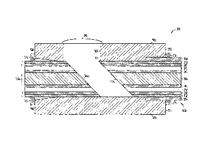
FIGS. 2A-C are a cross-sectional side views illustrating one example of how
an optical cable assembly is separated into two sections, so as to then.
provide
substantially mating or mirrored beveled ends 211A and 211B, which provide the
respective proximal ends 111A and 111B of FIG. 1A. In the example of FIGS. 2A-
2C, only two optical fibers 150 of the optical cable assembly are illustrated,
for ease
in understanding and not by way of limitation.
FIG. 2A illustrates an example of the optical cable assembly 200 before
separation into the probe portion 110A and the lead portion 11013. In this
example,
the then-unitary optical cable assembly 200 typically includes center body
260,
RECTIFIED SHEET (RULE 91)
13
CA 02630662 2008-05-21
WO 2007/062050 PCT/US2006/045080
optical fiber claddings 240, optical fibers 250, and a sheath 230 that
encloses the
optical fibers 250, center body 260 and claddings 240. Cladding 240 or sheath
230
may use the same or different material as center body 260. Center body 260,
cladding 240, and sheath 230 may be formed at substantially the same time, or
may
be formed separately and later assembled to form the optical cable.
FIG. 2B illustrates an example of the optical cable after it has been
angularly
sawed into two sections, such as by using a thin dicing wheel or circular
blade with
a diamond edge blade, for example, or by using any other separation method,
such
as ultrasonic cutting, for example. After sawing, the probe portion 210A and
the
external instrumentation lead portion 210B will have substantially similar,
mating or
mirrored beveled surfaces. Variation in saw blade width may produce a small
anti-
parallel deviation at the beveled ends 211A and 211B. The beveled ends 211A
and
211B may be further polished to reduce or remove surface damage or latent saw
damage or subsurface defects, such as due to sawing, or to produce more
parallel
surfaces to further improve optical coupling, such as by reducing or
minimizing
scattering from the surfaces of such beveled ends 211A-B.
FIG. 2C illustrates the beveled end 211A of probe portion 210A in contact
with the beveled end 211B of the external instrumentation lead portion 210B,
and
positioned within an ergonomically-shaped coupler housing 205 forming an
optical
coupler for coupling light between the probe portion 210A and the lead portion
210B. In certain examples, the external instrumentation lead portion 210B is
permanently affixed to the coupler housing 205, such as by being inserted into
the
coupler housing 205 so as to obtain an interference fit, or by using an
adhesive. The
probe portion 210A is then inserted into the coupler housing 205 until it butt-
couples in self-alignment against the external instrumentation lead portion
210B.
Such convenient self-alignment promotes coupling of light between adjoining
optical fibers 250 in respective probe and external instrumentation lead
portions.
The coupler housing 205 is typically formed of plastic, but in certain
examples, may
include an inner surface that is composed of precision fabricated straight
wall metal,
glass, or ceramic tubing.
14
CA 02630662 2008-05-21
WO 2007/062050 PCT/US2006/045080
In certain examples, an antireflective surface coating is used at the beveled
ends 211A-B, or index matching fluid is used between the beveled ends 211A-B,
such as for further improving the amount of light coupled between the ends of
the
optical fibers 250 of the probe portion 210A and the external instrumentation
lead
portion 210B. Index matching fluid typically has substantially the same
refractive
index as the optical fiber 250 at the desired wavelength of light used. It
typically
reduces or eliminates the likelihood of a fiber-air-fiber interface, which
would likely
cause undesirable reflections of light transmitted to and from the probe
portion
210A or the external instrumentation portion 210B. A fiber-air-fiber interface
may
occur if the beveled ends 211A-B do not butt against each other in perfect
mechanical contact when otherwise in optical alignment.
FIG. 3 is a cross sectional side view schematic diagram illustrating one
example of a connector 300 for aligning beveled ends of a probe portion 310A
and
an external instrumentation lead portion 310B. In one example, the beveled end
311B of the external instrumentation lead portion 310B is secured to
protective
sleeve 308, and may be further secured to a coupler housing 305 near the
external
instrumentation lead end of the housing 305 at 312, such as by using adhesive
or
other suitable material. In another example, the external instrumentation lead
portion 310B may be secured to the housing 305, with or without being securing
to
the protective sleeve 308, such as by a compression clamp 315. The housing 305
may be metal, plastic, or other suitable material, and may be formed from more
than
one component.
In certain examples, the external instrumentation lead portion 310B is
directly or indirectly secured to the housing 305 with the tip 313 of the
beveled end
311B positioned within a perimeter of a view hole or port 307, such that it
can be
oriented toward a view lens 380, which is attached over the view hole 307,
such as
by using an adhesive or other suitable technique. The lens 380 may use one or
more
antireflective surface coatings to increase light transmission through the
lens 380.
The probe portion 310A is inserted into the housing 305; this is aided by a
beveled
housing surface 306, which forms a funnel-like structure to reduce or minimize
any
CA 02630662 2008-05-21
WO 2007/062050 PCT/US2006/045080
potential damage to the beveled end 311A of the probe portion 310A during such
insertion into the housing 305. In certain examples, for aligning the beveled
ends
311A-B, visible light (e.g., red light emitted from a diode, etc.) may be
transmitted
from the instrumentation lead portion 310B while the probe portion 310A is
inserted
into the housing 305. Such visible light exiting an optical fiber 350 at the
beveled
end 311B of the external instrumentation lead 310B is reflected by at least
one
optical fiber 350 at the beveled end 311A of the probe portion 310A through
the
view hole 307 toward the view lens 380. A user looking at the view lens 380
will
observe maximum intensity of the reflected light when the probe portion 310A
is
properly oriented and aligned with respect to the external instrumentation
lead
portion 310B. In another example, light striking lens 380 is coupled to a
photodetector, and the resulting signal from the photodetector similarly used
for
aligning the beveled ends 311A-B. In yet another example, lens 380 is omitted,
and
light propagating through view hole 307 is instead coupled directly to an
external
photodetector where the corresponding photodetector output signal is used for
aligning the beveled ends 311A-B. In another example, the alignment light is
coupled to an external photodetector by a lens 380 that is unsecured to the
housing.
The circumferential surface of the view hole 307 surface may be polished or
coated
with a reflective film to improve surface reflectivity of light used for
aligning the
beveled ends 311A-B.
During insertion of the probe portion 310A into the housing 305, the probe
portion 310A may be rotated to obtain maximum alignment light reflected toward
view lens 380 from the beveled end 311B of the external instrumentation lead
portion 310B until the probe portion 310A and external instrumentation lead
portion
310B butt in mechanical contact. More light is reflected toward the view hole
307
when the optical fibers 350 of the probe portion 310A and the external
instrumentation lead portion 310B are best aligned. Then, when the beveled
ends
311A-B of the probe portion 310A and the lead portion 310B are in mechanical
contact with each other, maximum optical alignment is achieved and
substantially
all alignment light transmitted from external instrumentation lead portion
310B is
16
CA 02630662 2008-05-21
WO 2007/062050
PCT/US2006/045080
coupled into the probe portion 310A, leaving no light for reflection towards
the
view hole 307. As discussed above, index matching fluid may be used between
the
beveled ends 311A-B to improve light coupling between the beveled ends 311A-B,
The end of the probe portion 310A may be secured to the housing 305, such as
by a
compression clamp 316 secured to housing 305, or even by using an adhesive, if
desired.
In the example of Fla 3, such alignment of the probe portion 310A and the
external instrumentation lead portion 310B using the view hole 307 is
generally
possible if the angle of the beveled end 311B is less than the critical angle
for total
internal reflection.
FIG. 4 is a cross-sectional side view schematic illustrating one example of a
connector 400 for aligning beveled ends 411A and 411B of a respective probe.
portion 410A and an external instrumentation lead portion 410B at a stop 414.
In
certain examples, the external instrumentation lead portion 410B is secured to
the
coupler housing 405, such as with adhesive or other suitable technique near
the
beveled end 411B at stop 414 or at another suitable location. If necessary, a
suitable
solvent may be used to remove any stray adhesive from the optical surfaces of
the
beveled end 411B of the external instrumentation lead portion 4108. Then., the
probe portion 410A is inserted into housing 405 until its beveled end 411A
butts in
mechanical contact with the beveled end 4118 of the external instrumentation
lead
potion 410B. Because the external instrumentation lead portion 410B is secured
at
414 to the inner surface of the housing 405, such as near the beveled end
4118, the
beveled end 411A of the probe portion 410A is prevented from further traveling
beyond the stop 414. In such an example, maximum optical alignment is achieved
and substantially all light is coupled between the probe portion 410A and the
external instrumentation lead portion 410B when their respective beveled ends
411A-B butt in. mechanical contact at the stop 414. In certain examples, a
beveled
surface 406 of the housing 405 is provided to reduce the potential for damage
to the
beveled end 411A of the probe portion 410A during insertion. The probe portion
410A is secured to the housing 405, such as by a compression clamp that is
RECTIFIED SHEET (RULE 91)
17
CA 02630662 2008-05-21
WO 2007/062050 PCT/US2006/045080
secured to the housing, or even by an adhesive or other suitable technique, if
desired. The ends of the optical fibers 450 may use an antireflective surface
coating
or index matching fluid between their beveled ends to improve light coupling
between the probe and external instrumentation lead portions 410A-B.
A number of beneficial features can be incorporated into any of the coupler
housings described in this document, such as the coupler housings 205, 305, or
405.
In one example, a soft fabric or other cleaning device is placed at the
receptacle of
the coupler housing that receives the probe portion to clean its end as it is
received
into the coupler housing. In another example, the coupler housing includes a
flushing port (which may be the same or different from the viewing hole 307)
for
removing blood or other debris that may be accumulated during use, such as by
flushing with saline or the like. In another example, the coupler housing
includes an
attachable syringe or other injection device for injecting index matching
fluid
(which could even include injecting medical grade silicone gel) into the
connector
cavity where the probe and external instrumentation lead portions come
together. In
yet another example, the coupler housing includes a gripping mechanism that
attaches to the probe portion along its length without causing damage to its
optical
fibers. In another variation, the angular beveled ends of the probe portion
and the
external instrumentation lead portion is replaced by a longitudinal cut that
creates
semicircular or like mating sections that overlap between the probe portion
and the
external instrumentation lead. For example, FIG. 9 illustrates an example of a
keyed connection in which the beveled end 900 of the probe portion 410A is
separated into semicircular beveled portions 901 and 902, which are separated
by a
longitudinal edge 903. Similarly, the beveled end 904 of the instrumentation
lead
portion 410B is separated into semicircular beveled portions 905 and 906
separated
by a longitudinal edge 907, such that the beveled end 904 mates to the beveled
end
900. This example would provide a more discernable alignment that can be
"felt"
by the user. In another variation, the proximal end of the probe portion is
conical
(male/female) and self-aligning with a conical (female/male) end of the
external
instrumentation lead at the coupler housing.
18
CA 02630662 2008-05-21
WO 2007/062050 PCT/US2006/045080
Finally, the distal end of the external instrumentation lead (i.e., away from
the coupler housing) will be interfaced to an opto-electronic imaging console.
This
can be achieved by using a commercially available multiple fiber connector,
such as
the MTP multi-fiber connector available from US Conec, Ltd. of Hickory, North
Carolina (see
http://www.usconec.com/pages/product/connect/mtpcon/mainfitn.html). This
connector can be customized to accept different diameter and numbers of
optical
fibers. The termination may be achieved by selectively removing the plastic
matrix
coating at the distal end of the external instrumentation lead. The individual
fibers
can be separated from the external instrumentation lead center body and
individually placed in the holes in the connector. A hole may also be provided
for
the center body of the external instrumentation lead, such as to stabilize the
connection.
2. Example of a Guide-Aligning Optical Imaging Device
FIGS. 5A and 5B are respective cross-sectional end and side views
illustrating an example of an optical connector 500 for an optical imaging
device
using a guide 509 at an interior portion of a coupler housing 505. In this
example,
the guide 509 axially receives and accepts each of the probe portion 510A and
the
external instrumentation lead portion 510B in a particular orientation such
that the
optical fibers 550 of each such portion abut in alignment. For example, FIG.
5A
illustrates an example of a guide 509 with a square cross-section sized to
receive at
a first end¨in a particular orientation¨a probe portion 510A that includes a
probe
body 560 with its four optical fibers 550 distributed thereabout at 0 degrees,
90
degrees, 180 degrees, and 270 degrees. Similarly, a second end of the guide
509
would receive¨in an aligned orientation¨an external instrumentation lead
portion
511B that includes an external instrumentation lead body 560 with four optical
fibers 550 similarly distributed thereabout at 0 degrees, 90 degrees, 180
degrees,
and 270 degrees. The square cross-section of the guide 509 and the four
optical
fibers 550 is presented for illustrative purposes only; the underlying idea of
using a
guide 509 that is shaped to fix and align the radial position of the optical
fibers 550
19
CA 02630662 2008-05-21
WO 2007/062050 PCT/US2006/045080
can be extended to any number of one or more optical fibers located on a
circumferential surface of a body portion. Moreover, the coupler 509 need not
be a
unitary piece, but could instead be made of two separate sections that are
keyed
together, if desired.
In certain examples, the guide 509 is part of (or attached to) an interior
portion of a coupler housing 505, and may be plastic, metal, or other suitable
material. The housing 505 and the guide 509 may be integrally formed, or may
instead be assembled from multiple components. In another example, the guide
509
is separate from the housing 505 and is secured in the housing 505, such as by
using
adhesive or other suitable material, and the guide 509 may be the same or a
different
material than the housing 505.
In this example, the external instrumentation lead portion 510B and the
probe portion 510A may be made from the same optical cable assembly, such as
by
sawing the optical cable assembly using a thin dicing wheel or circular
diamond-
edge blade with a diamond edge blade, or by using ultrasonic cutting. The
external
instrumentation lead 510B portion and the probe 510A portion may be formed
from
the same optical cable assembly, or formed from different optical cable
assemblies.
The sawn ends 511A and 511B of the optical fiber 550 may be further polished,
such as to remove surface damage or latent saw damage or subsurface defects
due to
sawing or to produce substantially parallel surfaces to further improve light
coupling between probe and external instrumentation lead portions 510A-B.
FIG. 5B is a cross-sectional side view illustrating an example of the
connector 500 for an optical imaging device using a guide. In this
illustrative
example, only two optical fibers 550 are illustrated, but this is for ease in
understanding and not by way of limitation. This example includes a center
guide
560, fiber claddings 540, optical fibers 550, and a sheath 530 enclosing the
optical
fibers 550, the center guide 560, and the fiber claddings 540. The fiber
cladding
540 may the same material as the center guide 560, or it may be a different
material.
Similarly, the sheath 530 may be the same material as the cladding 540 or
center
guide 560, or it may be a different material. The center guide 560, the
cladding 540
CA 02630662 2008-05-21
WO 2007/062050 PCT/US2006/045080
and the sheath 530 may be formed at substantially the same time, or they may
be
formed separately and later assembled to form the optical cable assembly.
The external instrumentation lead portion 510B is positioned inside the
housing 505, conforming to the guide 509, and secured to the housing 505, such
as
by a compression clamp 516 secured to the housing, or by using adhesive or
other
suitable material. If necessary, a suitable solvent may be used to remove
stray
adhesive from the sawn ends. The probe portion 510A is positioned in the
housing
505, conforming to the guide 509 with the sawn ends 511A and 511B in
mechanical
contact and in maximum optical alignment to couple light between the ends 511A-
B. The probe portion 510A may be secured to the housing 505, such as by a
compression clamp 516 that is secured to the housing, or by adhesive or other
suitable material. The ends of the optical fiber 550 may use an antireflective
surface
coating or an index matching fluid between the ends 511A and 511B to improve
light coupling between the probe and external instrumentation lead portions
510A-
B.
3. Example Using a Lens Such as A GRIN Lens
Figure 6 is a cross-sectional side view illustrating one example of a
connector 600 for an optical imaging device using a lens such as a graded
refractive
index (GRIN) lens (or, alternatively, at least one of: a ball lens; a half
ball lens; a
holographic lens; and a Fresnel lens). In this example, two optical fibers 650
are
shown, but this is for ease in understanding and not by way of limitation. A
center
guide spacer 617 may be used for positioning the GRIN lens 651 with respect to
the
external instrumentation lead end 611B and the probe end 611A inside the
housing
605. In this example, the housing 605 is formed in two separable sections.
This
allows for positioning of the GRIN lens 651 and the spacer 617. The housing
605
and the spacer 617 may be made from plastic, metal or any other suitable
material.
In certain examples, the GRIN lens 651 is secured to the spacer 617, such as
by
adhesive or any other suitable material inside one or more spacer slots 618.
The
spacer slots 618 are cut or otherwise formed from the spacer 617 to accept a
portion
of one or more GRIN lenses 651. In another example, the GRIN lens 651 may be
21
CA 02630662 2008-05-21
WO 2007/062050 PCT/US2006/045080
positioned partially within the spacer slot 618 without using an adhesive.
Similarly,
the GRIN lens 651 may also be positioned inside a housing slot 619 cut from
housing 605 that is sized to accept one or more GRIN lenses. GRIN lens may be
further secured by adhesive or other suitable material or may be positioned
inside
slot 619 without adhesive.
In this example, the external instrumentation lead portion 610B is secured to
the housing 605 such that the external instrumentation lead portion 610B is in
contact with a first end of the spacer 617, such as by using a compression
clamp 615
that is secured to the housing, or by using adhesive or other suitable
technique. The
probe portion 610A is inserted into the housing 605 such that the end 611A of
the
probe portion 610A is in contact with a second end of the spacer 617. The
probe
portion 610 A can be secured to the housing 605 using a compression clamp 616,
which is secured to the housing 605, or by using an adhesive or other suitable
material. The spacer 617 is typically sized for positioning ends of the
optical fibers
650 to obtain increased or maximum light coupling between probe and external
instrumentation lead portions 610A-B by the GRIN lens 651 when the center body
660 of the probe and external instrumentation lead ends 611A and 611B,
respectively, are in contact with the spacer 617. The ends of the optical
fibers 650
may use antireflective surface coatings or an index matching fluid between
ends
611A and 611B of respective probe and external instrumentation lead portions
610A-B. This will improve light coupling between the probe and external
instrumentation lead portions 610A-B.
,
FIG. 7 is a cross-sectional side view illustrating an example of a connector
700 using an integrated or monolithic GRIN lens 751, 1000 with multiple radial
partitioned refractive index regions such as 1002A-H as shown in FIG. 10 (for
the
case of eight optical fibers 750). In the example of FIG. 7, two optical
fibers 750
are shown, but this is for ease in understanding, and not by way of
limitation. FIG.
7 shows center guide spacers 717A-B are used for positioning, inside a housing
705,
the GRIN lens 751 with respect to the ends 711A-B of the probe and external
instrumentation portions 710A-B, respectively. In certain examples, the
housing
22
CA 02630662 2008-05-21
WO 2007/062050
PCT/US2006/045080
705 is provided in two separatable sections for easier positioning of the GRIN
lens
751 and the spacers 717A-B. The housing 705 may be plastic, metal, or other
suitable material. The spacers 717A-B may be plastic, metal, or other suitable
material. In certain examples, the spacers 717A-B are secured to the GRIN lens
751, such as by adhesive or other suitable material positioned inside a
housing slot
719 cut from the housing 705 and sized to accept the GRIN lens 751. The GRIN
lens 751 may be secured to the housing 705, such as by adhesive or other
suitable
material, or may be positioned inside the slot 719 without using such adhesive
In
another example, the spacers 717A-B are secured to the center body portions
760A-
B, respectively, such as by adhesive or other suitable material, and the GRIN
lens
751 is secured to the housing 705.
In the example of FIG. 7, the external instrumentation lead portion 710B is
positioned in contact with the spacer 717B at the external instrumentation
lead end
711B and secured to the housing 705, such as by a compression clamp 715, or by
using adhesive or other suitable material. The probe portion 710A is inserted
into
the housing 705 such that the end 711A of the probe portion 710 is in contact
with
the spacer 717A. The probe portion 710 is then secured to the housing 705,
such as
by the compression clamp 716, or by using an adhesive or other suitable
material.
In certain examples, the spacers 717A-B are sized for positioning the sawn
ends
711A-B to obtain increased or maximum light coupling between probe and
external
instrumentation lead portions 710A-B by the GRIN lens 751 when the center body
760 of the ends 711A-B are in contact with respective spacers 717A-B. The ends
of
the optical fibers 750 may use an antireflective surface coating or an index
matching
fluid between ends 711A-B to improve light coupling between the probe and
external instrumentation lead portions 710A-B.
4. Example
of a Aligning Optical Imaging Catheter with blazed Fiber Bragg
Gratings
FIGS. 8A and 8B are respective cross-sectional end and side views
illustrating an example of a connector 800 using at least one lens 851 that is
positioned between a pair of blazed fiber Bragg gratings (FBGs). In the
example of
23
CA 02630662 2008-05-21
WO 2007/062050
PCT/US2006/045080
FM. 8A-B, two pairs of optical fibers 850 are shown, but this is for ease in
understanding, and not by way of limitation, The optical fibers 850 are
concentrically located along the probe and external instrumentation lead
portions
810A and 810B, respectively. In certain examples, the probe portion 810A is
sized
to allow for insertion over the external instrumentation lead portion 810B at
ends
811A and 811B. In other examples, the probe portion 810A sized to allow for
insertion inside the external instrumentation lead portion 81013. The lens 851
is
sized and positioned by one or more lens mounts, such as the lens mounts 817
and
818, to couple light between the probe and the external instrumentation lead
portions 810A-B when blazed FBGs 852A-B are aligned.
FIG. 8B is a cross-sectional side view further illustrating this example of a
portions of a connector 800 using the lens 851 located between pairs of blazed
FBGs. In the example of FIG_ 8B, one pair of optical fibers 850 is shown, but
this is
for ease in understanding, and not by way of limitation_ FIG. 8B illustrates a
blazed
FB0 832B that is patterned into the optical fiber 850B near the end 811B of
the
external instrumentation lead portion 810B. The external instrumentation lead
portion 810B is secured to the housing 805, such as by using a compression
clamp
that is secured to the housing 805, or by using adhesive or other suitable
material.
In this example, the lens mounts 817 and 818 are secured to the lead portion
810B
near the blazed FBG 852B. The lens mounts 817 and 818 are sized to accept the
lens 851 to couple light between the FBGs 852A-B_ The lens mount 817 may be
configured as a stop for the probe portion. 81OA. In one example, the lens
mounts
817 and 818 are annular rings secured to the inner surface of the external
instrumentation lead portion 810B. In another example, the lens mount 818 is
shaped as a cap that is secured to the external instrumentation lead portion
810B at
its end 811B. In certain examples, the probe portion 810A is positioned inside
the
lead portion 810B against a stop portion of the lens mount 817. This aligns
the
FBGs 852A-B for coupling light between the FBGs 852A-B by the lens 851. The
probe portion 810A is secured to the housing 805, such as by a compression
clamp
that is attached to the housing 805, as discussed above, or by using adhesive
or other
RECTIFIED SHEET (RULE 91)
24
CA 02630662 2008-05-21
WO 2007/062050 PCT/US2006/045080
suitable material. The end 811A of the probe portion 810A may otherwise be
secured to the stop portion of the lens mount 817, such as by using an plug
and
receptacle arrangement. In an example in which the probe portion 810A is sized
to
allow for its insertion over the external instrumentation lead portion 810B,
the lens
mounts 817 and 818 can be secured to the probe portion 810A, and may be
configured as annular rings or as an end cap as shown at 818. The lens mounts
817
and 818 may be metal, plastic, or other suitable material. The lens 851 may
use an
antireflective surface coating to improve light coupling between blazed FBGs.
It is to be understood that the above description is intended to be
illustrative,
and not restrictive. Although the above description has described medical
imaging
applications, the present systems and methods are also useful for imaging any
other
animate or inanimate objects, and are not necessarily limited to imaging a
human or
animal. For example, the above-described embodiments (and/or aspects thereof)
may be used in combination with each other in various permutations or
combinations. Many other embodiments will be apparent to those of skill in the
art
upon reviewing the above description. The scope of the invention should,
therefore,
be determined with reference to the appended claims, along with the full scope
of
equivalents to which such claims are entitled. In the appended claims, the
terms
"including" and "in which" are used as the plain-English equivalents of the
respective terms "comprising" and "wherein." Also, in the following claims,
the
terms "including" and "comprising" are open-ended, that is, a system, device,
article, or process that includes elements in addition to those listed after
such a term
in a claim are still deemed to fall within the scope of that claim. Moreover,
in the
following claims, the terms "first," "second," and "third," etc. are used
merely as
labels, and are not intended to impose numerical requirements on their
objects.
The Abstract of the Disclosure is provided to comply with 37 C.F.R.
1.72(b), requiring an abstract that will allow the reader to quickly ascertain
the
nature of the technical disclosure. It is submitted with the understanding
that it will
not be used to interpret or limit the scope or meaning of the claims. In
addition, in
the foregoing Detailed Description, various features may be grouped together
to
CA 02630662 2008-05-21
WO 2007/062050
PCT/US2006/045080
streamline the disclosure. This method of disclosure is not to be interpreted
as
reflecting an intention that the claimed embodiments require more features
than are
expressly recited in each claim. Rather, as the following claims reflect,
inventive
subject matter may lie in less than all features of a single disclosed
embodiment.
Thus the following claims are hereby incorporated into the Detailed
Description,
with each claim standing on its own as a separate embodiment.
26