Note : Les descriptions sont présentées dans la langue officielle dans laquelle elles ont été soumises.
CA 02634675 2008-06-20
WO 2007/075364
PCT/US2006/047799
- 1 -
SYSTEMS AND METHODS
FOR CLOSING A VESSEL WOUND
BACKGROUND OF THE INVENTION
Field of the Invention
[0001] The invention generally relates to vessel wound closure techniques.
More
particularly, the invention relates to systems and methods for seaJing
puncture wounds in
a blood vessel such as those that result from certain interventional
procedures.
Related Art
[0002] A large number of therapeutic and diagnostic procedures involve the
percutaneous
introduction of instrumentation into a blood vessel, for example, percutaneous
transluminal coronary angioplasty (PTCA). Such procedures most often involve
accessing an intended site through the femoral artery. Ideally, closing and
healing of the
resultant vascular puncture wound successfully completes the procedure.
[0003] Traditionally, the application of external pressure to the skin at the
entry site of
the instrumentation into the patient has been employed to stem bleeding from
the wound.
A nurse or physician, for example, applies pressure to the wound site until
clotting and
tissue rebuilding has occurred sufficiently to seal the perforation. In some
situations, the
external pressure is maintained for an hour or more, during which time the
patient is
uncomfortably immobilized. Thus pati.ent comfort and physician efficiency are
impaired
where such external pressure techniques are employed.
[0004] Additionally, the risk of hematoma exists while bleeding from the
vessel occurs.
Such hematoma risk continues until sufficient clotting of the wound site
occurs.
Moreover, external pressure devices, such as femoral compression systems, are
often
unsuitable for some patients, such as those with substantial amounts of
subcutaneous
adipose tissue, as the skin surface may be a considerable distance away from
the
vasculature puncture site. Inaccurate skin compression, and thus less
effective wound
healing, tends to occur as a result.
[0005] U.S. Patent No. 5,383,896 to Gershony, et al. discloses a device that
applies
pressure to a puncture site internally for a limited period of time, after
which the device is
removed. The device in Gershony includes a shaft with an expandable balloon
and a
guidewire tip at its distal end. The distal end of the device is introduced
into a blood
CA 02634675 2008-06-20
WO 2007/075364
PCT/US2006/047799
- 2 -
vessel through an introducer sheath that is typically used in percutaneous
interventional
procedures. The balloon is then inflated and withdrawn until the balloon
hemostatically
engages the inner surface of the blood vessel, after which the introducer
sheath is
removed. A fixation collar on the shaft applies tension to the balloon for a
medically
sufficient time and thereafter the balloon is deflated and the entire device
is removed
from the body.
[0006] U.S. Patent No. 5,645,566 to Brenneman, et al. discloses a device that
applies
pressure to the outside wall of a punctured blood vessel from a distance using
a balloon, a
sheet and a foam pad. The pressure applying device is located using a balloon
in the
vessel (similar to that of Gershony) and a radiopaque marker.
[0007] PCT Application WO 98/11830, published March 26, 1998, S.Barak,
Inventor,
discloses various embodiments of an apparatus for hemostasis. Among them is a
device
that positions an anchor against an inner surface of an artery wall and a
balloon outside
the wall. The balloon is inflated to pinch the artery wall, after which the
anchor is
withdrawn. The balloon is maintained against the puncture until hemostasis is
achieved.
The anchor and balloon are removed after hemostasis is achieved.
[0008] Other arterial closure devices include bioabsorbable materials intended
to remain
in the body until they are absorbed as in related U.S. Patent Nos. 5,282, 827
and
5,441,517, which disclose an anchor inserted into a vessel and urged against
an inner wall
of the vessel as a collagen plug is deployed externally of the puncture site
to expand and
fill the tissue tract leading to the puncture site. A filament attaches the
plug to the anchor
and moves the plug and anchor relative to one another in pulley-like fashion
to effect a
seal at the puncture site. After emplacement, a tamping member may be used to
urge the
plug against the external puncture site to help seal the same.
[0009] U.S. Patent No. 5,662,681 discloses an arterial closure device in which
an anchor
and plug are attached to one another via a filament. The anchor is inserted
into the vessel
and urged against the interior wall of the vessel as the plug is urged against
the exterior
wall of the vessel at a puncture site. A separate locking means moves the plug
and
anchor relative to one another to maintain the plug and anchor in sealing
position at the
puncture site.
CA 02634675 2008-06-20
WO 2007/075364
PCT/US2006/047799
- 3 -
[0010] U.S. Patent No. 5,391,183 to Janzen, et al. describes a device that
inserts
hemostatic material through a tissue channel and against the outside wall of
the vessel
around the puncture site.
[0011] U.S. Patent No. 5,690,674 to Diaz discloses a biodegradable plug that
has two
Substantially parallel disks joined at their centers by a waist. The plug is
positioned so
that the distal disk is on the interior wall of the blood vessel, the proximal
disk is on the
exterior wall, and the waist is in the wound of the vessel wall.
[0012] Another known closure device includes U.S. Patent No. 5,741,223 to
Janzen, et
al.. This '223 patent discloses the placement of a plug to seal a puncture
site.
[0013] U.S. Patent No. 5,354,271 to Voda discloses suture threads with barbed
ends,
wherein the suture threads are deployed into a vessel and then the barbed ends
penetrate
through the vessel wall and expand to prevent retraction thereof back into the
vessel. The
suture threads are then tied or otherwise secured across the puncture site.
[0014] U.S. Patent No. 5,324,306 discloses a mass of hemostatic material
pushed against
the outside wall of a vessel at a puncture site. Manual pressure is applied to
ensure blood
flow has stopped.
[0015] U.S. Patent No. 5,868,778 discloses a balloon used in combination with
a
procoagulant injected at the puncture site in order to seal a puncture site of
a vessel.
[0016] U.S. Patent No. 5,792,152 discloses a flexible needle with suture
attached thereto
that is deployed across a puncture site of a vessel. The flexible needle and
suture are
introduced into the vessel via an entry lumen, proceed through a U-shaped
return lumen,
and exit the vessel through an exit lumen. Thereafter the suture is drawn
further outward
from the vessel and tied or otherwise secuied across the puncture site. =
[0017] U.S. Patent Publication No. 201)4/0006352 discloses an arterial closure
device
comprising an assembly in which clasp arms, to which a suture is initially
secured, are
deployed within a vessel. Penetrating members including suture catches are
then
separately deployed to snag or capture the sutures associated with a
respective clasp arm.
The sutures are then pulled taught by pulling the penetrating member with
'suture catches
out from the vessel, and then tied or otherwise secured to close the puncture
site.
Thereafter the assembly is withdrawn from the body.
[0018] Current vessel closure devices thus tend to provide vessel wound
closure devices
and techniques after an interventional procedure has been performed. A.need
exists
CA 02634675 2008-06-20
WO 2007/075364 PCT/US2006/047799
- 4 -
therefore for vessel wound closure systems and methods that apply a vessel
wound
closure device prior to performance of an interventional procedure within the
target
vessel.
.SUMMARY OF THE INVENTION
[0019] The various embodiments described herein comprise vessel wound closure
systems and methods for closing a puncture Wound in a target vessel. The
vessel wound
closure system generally comprises at least a biocompatible/biodegradable,
viscoelastic
self-sealing septum material disposed onto the adventitia of a target vessel
prior to
performance of an interventional procedure within the target vessel. The
septum material
may be disposed directly onto the adventitia of the target vessel, or may be
disposed
within a balloon comprised of natural or bio-degradable polymeric materials of
sufficient
porosity that permits slow dispersion of the septum material therefrorrao
adhere to the
adventitia of the target vessel. The septum material disposed on the
adventitia Of the
target vessel may further be preformed and comprise a hemostatic valve
incorporated
therein that closes and seals the vessel wound after an interventional
procedure has been
performed within the target vessel.
[0020] In some embodiments of the vessel wound closure system, access to the
target
vessel is obtained by piercing or cutting through the skin, followed by blunt
dissection to
the adventitia, or outer wall, of the target vessel. The septum material is
thereafter
injected onto the adventitia of the target vessel or into a balloon in
proximity to the
=
adventitia from which balloon the septum material seeps to adhere to the
adventitia. An
introducer is inserted into the target vessel through the septum material,
through which
introducer various instruments are passed to perform an interventional
procedure. After
completion Of the interventional procedure, the various instruments and the
introducer or
other components are removed and the septum material remains to seal the
vessel wound.
[0021] Alternatively, blunt dissection to the adventitia of the target vessel
may be
omitted where the septum material is injected through a needle having holes
aligned to
dispose the septum material onto the adventitia of the target vessel. An
interventional
procedure is then performed through an introducer that has been inserted
through the
septum material and into the target vessel. After completion of the
interventional
procedure, the various instruments associated therewith and the introducer or
other
CA 02634675 2015-09-18
-5-
components are removed while the septum material remains and closes to seal
the puncture
wound of the target vessel.
[0022] In other embodiments, septum material is preformed and disposed on the
adventitia of the target vessel prior to performance of an interventional
procedure within
the target vessel. An introducer is inserted through the preformed septum
material and
into the target vessel. Various instruments are passed through the introducer
to perform
the interventional procedure. After the interventional procedure is complete,
the various
instruments, the introducer, and other components are removed and the
preformed
septum material remains and closes to seal the puncture wound of the target
vessel. The
preformed septum material may further comprise a hemostatic valve incorporated
therein
through which the introducer or other components are disposed to accommodate
performance of the interventional procedure.
[0022a] In an aspect of the present invention, there is provided a vessel
wound closure
system comprising: a self-sealing septum material disposed onto adventitia of
a target
vessel and at a vessel wound prior to performance of an interventional
procedure in the
target vessel.
[0022b] In another aspect of the present invention, there is provided a method
of sealing a
vessel wound in a target vessel, comprising: locating the target vessel;
disposing a self-
sealing septum material onto adventitia of the target vessel; placing an
introducer through
the self-sealing septum material and into the target vessel; performing an
interventional
procedure in the target vessel through the introducer; removing the
introducer; and sealing
the vessel wound by closure of the self-sealing septum.
[0022c] In another aspect of the present invention, there is provided a vessel
wound closure
system comprising: a self-sealing septum material disposed onto adventitia of
a target
vessel and at a vessel wound prior to performance of an interventional
procedure in the
target vessel, wherein the self-sealing septum material is a
biocompatible/biodegradable,
viscoelastic material.
[0022d] In another aspect, there is provided a vessel wound closure system
used prior to
performance of an interventional procedure in a target vessel, comprising: a
self-sealing
septum material adapted to be disposed onto adventitia of the target vessel;
and a stepped
needle comprising a smaller diametered distal portion and a larger diametered
proximal
portion, the smaller diametered portion comprising a pointed tip configured to
pierce
through the adventitia of the target vessel, the larger diametered portion
comprising at
CA 02634675 2015-09-18
-5a-
least one opening formed in a side wall thereof, through which the self-
sealing septum
material is disposed onto the adventitia of the target vessel.
[0023] The above and other features of the invention, including various novel
details of
construction and combinations of parts, will now be more particularly
described with
reference to the accompanying drawings and claims. It will be understood that
the
various exemplary embodiments of the invention described herein are shown by
way of
illustration only and not as a limitation thereof. The principles and features
of this
invention may be employed in various alternative embodiments without departing
from
the scope of the invention.
BRIEF DESCRIPTION OF THE DRAWINGS
[0024] These and other features, aspects, and advantages of the apparatus and
methods of
the present invention will become better understood with regard to the
following
description, appended claims, and accompanying drawings where:
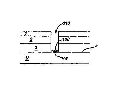
[00251 Figure 1 schematically illustrates a biocompatiblefbiodegradable,
viscoelastic,
septum material disposed onto the adventitia of a target vessel to close a
vessel wound
according to the description herein.
[0026] Figure 2 illustrates a needle and guidewire penetrating into the target
vessel prior to
performance of an interventional procedure according to the description
herein.
[0027] Figure 2a illustrates a stepped needle penetrating into the target
vessel prior to
performance of an interventional procedure according to the description
herein.
CA 02634675 2008-06-20
WO 2007/075364
PCT/US2006/047799
- 6 -
[0028] Figure 3 illustrates the guidewire in place after removal of the needle
of Figure 2
according to the description herein.
[0029] Figures 3a and 3b illustrate various guidewire anchors according to the
.description herein.
[0030] Figure 4 illustrates disposition of septum material onto the adventitia
of the target
vessel according to the description herein.
[0031] Figures 4a-4c illustrate various other techniques of disposing septum
material
onto the adventitia of the target vessel according to the description herein.
[0032] Figure 5 illustrates insertion of an introducer over the guidewire,
through the
septum material, and into the target vessel according to the description
herein.
[0033] Figure 6 illustrates removal of the introducer, any instruments, and
the guidewire
as the septum material closes the vessel wound according to the description
herein.
[0034] Figure 7 illustrates a first needle and a guidewire penetrating into
the target vessel
prior to an interventional procedure according to the description herein.
[0035] Figure 8 illustrates the guidewire in place after removal of the first
needle of Fig.
7 according to the description herein.
[0036] Figure 9 illustrates disposition of preformed septum material onto the
adventitia
of a target vessel according to the description herein.
[0037] Figure 10 illustrates insertion of an introducer over the guidewire,
through the
preformed septum material, and into the target vessel according to the
description herein.
[0038] Figure 11 illustrates removal of the introducer, any instruments, and
the guidewire
as the septum material closes the vessel wound according to the description
herein.
DETAILED DESCRIPTION OF THE INVENTION
[0039] As used herein the term proximal, or variants thereof, is understood as
closest to a
medical practitioner operator, and the term distal, or variants thereof, is
understood as
furthest from a medical practitioner operator.
[0040] Fig. 1 illustrates generally a biocompatible/biodegradable,
viscoelastic, self-
sealing septum material 100 disposed onto the adventitia (a) of a target
vessel (V) to
close a vessel wound (vw) after performance of an interventional procedure in
a target
vessel (V) according to the description herein. The septum material 100 is
disposed onto
the adventitia prior to performance of an interventional procedure within the
target vessel
CA 02634675 2008-06-20
WO 2007/075364
PCT/US2006/047799
- 7 - =
(V). Various systems and techniques may be used to dispose the septum material
100
onto the adventitia (a) of the target vessel (V), as will be described in
greater detail
below. The septum material 100 is a biocompatible/biodegradable, viscoelastic,
self-
sealing material and may be comprised of degradable polyesters, degradable PEG-
esters
(e.g., polyethylene glycol)-initiated lactones such as caprolactone,
glycolide, lactide, p-
dioxanone, and trimethylene carbonate, and copolymers thereof), degradable
polyurethanes, or poly(vinylpyrrolidinone) based functional polymers, for
example. Of
course, other known or later developed biocompatible/biodegradable,
viscoelastic, self-
sealing materials may be used to comprise the septum material 100 provided it
accommodates the closure of the vessel wound as otherwise described herein.
[0041] Figs. 2- 6 illustrate an embodiment of a vessel wound closure system
and method
wherein the septum material 100 is disposed onto the adventitia (a) of a
target vessel (V)
prior to performance of an interventional procedure within the target vessel
(V).
[0042] In particular, Fig. 2 illustrates a tissue tract 110 created by
piercing or cutting
through the skin layers (epidermis 1, dermis 2 and subcutaneous 3), followed
by blunt
dissection to the adventitia (a) of the target vessel (V). In practice, a
first needle 120 may
be inserted through the tissue tract 110 and into the target vessel (V) to
locate the target
vessel (V). Thereafter, a guidewire 130 is inserted through the first needle
120 and into
the target vessel (V). Next, as illustrated in Fig. 3, the first needle 120 is
removed and
the guidewire 130 remains in place within the target vessel (V). The guidewire
130
preferably comprises an expandable member, such as a balloon 131, or other
anchor 132
(Figs. 3a & 3b), that is held against an inside surface of the target vessel
(V) during blunt
dissection. The guidewire anchor 132 may instead comprise a nitinol mesh 132a
or
nitinol anchor 132b, for example, that is held against an inside surface of
the target vessel
(V) during blunt dissection, as in Figs. 3a and 3b, respectively.
[0043] Fig. 4 illustrates the disposition of the septum material 100 onto the
adventitia (a)
of the target vessel (V). In particular, Fig. 4 illustrates a second needle
140 inserted over
the guidewire 130 such that a distal tip 141 of the second needle 140 abuts,
but does not
enter, the target vessel (V). Septum material 100 may then be injected through
the
second needle 140 and onto the'adventitia (a) of the target vessel (V) through
holes 142
(see Fig. 4 inset). After disposition of the septum material 100, the second
needle 140 is
then removed and an introducer 150 is inserted through the septum material 100
and into
CA 02634675 2008-06-20
WO 2007/075364
PCT/US2006/047799
- 8 -
the target vessel (V) for performance of the interventional procedure as
described further
with respect to Figs. 5 & 6 further below.
[0044] Alternatively, the first needle 120 could instead be a stepped needle
1120 as
shown in Fig. 2a and the septum material 100 could be injected onto the
adventitia (a) of
the target vessel (V) through holes 1122 provided on a portion of the stepped
needle
1120. In practice, the target vessel could be located with the stepped, or
graduated,
needle 1120 (Fig. 2a) rather than the needle 120 and guidewire 130
configuration
otherwise depicted in Figs. 2-6. Blunt dissection may not be necessary where
the stepped
needle 1120 locates the target vessel (V) and delivers the septum material 100
to the
adventitia (a) of the target vessel (V). The smaller diametered portion at the
distal end
1121 of the stepped needle 1120 helps insertion of the needle 1120 into the
target vessel
(V). The smaller diametered distal tip 1121 steps, or graduates, to a larger
diametered
portion 1123 that abuts the adventitia (a) of the target vessel (V) and
resists entry
thereinto the target vessel (V). Ideally, the larger diametered portion 1123
of the needle
1120 includes holes 1122 through which septum material may be delivered. onto
the
adventitia (a) of the target vessel (V). After the septum material is injected
through the
needle 1120 and onto the adventitia (a) of the target vessel (V) through holes
1122, the
needle 1120 is removed, leaving the septum material 100 in place. A guidewire
130 and
introducer 150 are inserted into and removed from the target vessel to
accommodate
performance and completion of the interventional procedure as otherwise
described
above with respect to Figs. 5 and 6, for example.
[0045] Still further alternatively, as shown in Figs. 4a-4c, disposition of
the septum
material 100 may occur through a catheter 160 delivery tool, rather than
through a needle
as described above. The catheter 160 delivery tool comprises a balloon 161 or
rigid
prongs 162 deployable from a distal end thereof. The catheter 160 is inserted
over the
previously inserted guidewire 130. The guidewire 130 may be inserted through
needle
120, for example, as described above with respect to Figs. 2-3. The balloon
161 or
prongs 162 deploy at the distal end of the catheter 160 to help dissect tissue
further from
the site of the vessel wound (vw) at the adventitia (a) of the target vessel
(V). Septum '
material is then injected through the catheter 160 and onto the adventitia (a)
of the target
vessel (V) or into the balloon 161. The balloon 161 is preferably comprised of
a natural
material such as intestine or a bio-degradable polymer whose porosity permits
the septum
CA 02634675 2008-06-20
WO 2007/075364
PCT/US2006/047799
- 9 -
material 100 to slowly seep therethrough and adhere to the adventitia (a) of
the target
vessel (V). Of course, such a natural or bio-degradable polymer balloon 161
could be
used with various of the systems and methods described herein to help contain
the septum
material 100 when disposed onto the adventitia (a) of the target vessel (V).
[0046] A mold 163 (Figs. 4c) may further be provided at the distal end of the
catheter
160 to help contain and form the septum material 109 when disposed through the
catheter
to the site of the vessel wound (vw). After the septum material 100 is
disposed onto the
adventitia (a) of the target vessel (V), the catheter 160 is removed while the
septum
material 100, or balloon 161 containing the septum material 100, remains.
[0047] Yet further alternatively, the septum material 100 may be injected onto
the
adventitia (a) of the target vessel (V) by a syringe (not shown) as the
artisan should
readily appreciate, rather than through any of the septum material delivery
tools
otherwise described herein.
[0048] Fig. 5 illustrates an introducer 150 inserted over the guidewire 130
and into the
target vessel (V) after the septum material .100 has been disposed onto the
adventitia (a)
and any septum material delivery toot, i.e., the first needle 120, the
syringe, the second
needle 140, or the catheter 160, as the case may be, has been removed. A
dilator 155
may precede insertion of the introducer 150 in conventional manner if desired,
in order to
aid the insertion of the introducer 150 through the tissue tract 110, the
septum material
100 and into the target vessel (V). If used, the dilator 155 may be removed
after the
introducer 150 has penetrated into the target vessel (V). Once. the introducer
150 is
inserted, then various instruments may be inserted therethrough and an
interventional
procedure within the target vessel (V) is performed.
[0049] Upon completion of the interventional procedure, as shown in Fig. 6,
the various
instruments, the introducer 150, and the guidewire 130 are removed from the
target
vessel (V) through the septum material 100, which remains in place on the
adventitia (a)
of the target vessel. Because of the.viscoelasticity properties of the septum
material 100,
which ideally exhibits at least 800 ¨ 900 % deformation, the septum material
100 readily
recovers to close and seal the opening through which the introducer 150 and
guidewire
130 were emplaced during the interventional procedure.
[0050] Figs. 7-11 illustrate another embodiment of a vessel wound closure
system and
method wherein the septum material 100 is preformed and disposed onto the
adventitia
CA 02634675 2008-06-20
WO 2007/075364
PCT/US2006/047799
- 10 -
(a) of a target vessel (V) prior to performance of an interventional procedure
within the
target vessel (V), wherein like reference numerals or characters are used to
refer to like
parts. The preformed septum material 100,may include a hemostatic valve 101
incorporated therein, through which valve an introducer 150 or other
instruments are
passed through and into the target vessel (V) to perform an interventional
procedure
within the target vessel (V). After completion of the interventional
procedure, the
introducer 150 and other instruments are removed through the valve 101, which
closes
and seals the vessel wound (vw).
[0051] In particular, Fig. 7 illustrates a first needle 120 that locates the
target vessel (V)
by penetrating through the skin and into the target vessel (V). A guidewire
130 is then
inserted through the first needle 120 and into the target vessel (V).
Thereafter, as shown
in Fig. 8, the first needle 120 is removed, leaving only the guidewire 130 in
place within
the target vessel (V).
[0052] Fig. 9 illustrates the disposition of the preformed septum material 100
onto the
adventitia (a) of the target vessel (V). In particular, Fig. 9 illustrates a
catheter 170
inserted over the guidewire 130 such that a distal end of the catheter 170
approaches, but
does not enter, the target vessel (V). The preformed septum material 100 is
then pushed
through the catheter 170 and onto the adventitia (a) of the target vessel (V)
(see Fig. 9
inset). Preferably, a biocompatible/biodegradable bonding agent is applied to
one or both
of the distal surface of the preformed septum material 100 and the exposed
adventitia
surface to aid adherence of the preformed septum material 100 thereto the
adventitia (a)
when disposed thereon from the catheter 170.. Of course, the preformed septum
material
100 could be contained within a balloon 161, as described above with respect
to Figs. 4a-
4c, in which case the septum materials seeps slowly out from the balloon 161
and adheres
to the adventitia. A pusher 171 may be provided through the catheter to aid in
disposing
the preformed septum material 100 onto the adventitia. In any case, after the
preformed
septum material 100 is disposed onto the adventitia, the catheter 170 and the
pusher 171,
if used, are removed.
[0053] Although the preformed septum material 100 may be penetrated to access
the
target vessel (V), it is preferable to provide the preformed septum material
with a
hemostatic valve 101, through which the introducer 150, the guidewire 130, or
other
instruments may access the target vessel (V).
CA 02634675 2015-01-23
- 11 -
[0054] Fig. 10 illustrates an introducer 150 inserted over the guidewire 130
and into the
target vessel (V) through the valve 101 of the preformed septum material 100
after the
preformed septum material 100 has been disposed onto the adventitia (a) and
the catheter
170 has been removed. As in earlier described embodiments, a dilator 155 may
precede
insertion of the introducer 150 in conventional manner if desired, in order to
aid the
insertion Of the introducer 150 through the valve 101 and the septum material
100, and
into the target vessel (V). If used, the dilator 155 may be removed after the
introducer
150 has penetrated into the target vessel (V). Once the introducer 150 is
inserted, then
various instruments may be inserted therethrough and an interventional
procedure within
the target vessel (V) is performed.
[0055] Upon completion of the interventional procedure, as shown in Fig. 11,
the various =
instruments, the introducer 150, and the guidewire 130 are removed from the
target
vessel (V) through the valve 101 and the septum material 100, which remain in
place on
the adventitia (a) of the target vessel. Due to the valve 101 and the
viscoelastic properties
of the septum material 100, the access hole into the target vessel (V) is
readily closed and
sealed.
[0056] The various exemplary embodiments of the invention as described
hereinabove do
not limit different embodiments of the systems and methods of the invention.
The
materials described herein are not limited to the materials, designs or shapes
referenced
herein for illustrative purposes only, and may comprise various other
materials, designs
or shapes suitable for the systems and methods described herein, as should be
appreciated
by the artisan.
[0057] While there has been shown and described what is considered to be
Preferred
embodiments of the invention, it will, of course, be understood that various
modifications
and changes in form or detail could readily be made without departing from the
scope of the invention. It is therefore intended that the invention be not
limited to the
--EX-at-forms described-and illustrated herein; but should bevoustrued-to
cover all
modifications that may fall within the scope of the appended claims.