Note : Les descriptions sont présentées dans la langue officielle dans laquelle elles ont été soumises.
CA 02637816 2008-07-21
WO 2007/092739
PCT/US2007/061476
MICROSURGICAL INSTRUMENT
Field of the Invention
The present invention generally pertains to microsurgical instruments. More
particularly, but not by way of limitation, the present invention pertains to
microsurgical instruments having a port for aspirating and cutting tissue.
Description of the Related Art
Many microsurgical procedures require precision cutting and/or removal of
various body tissues. For example, certain ophthalmic surgical procedures
require the
cutting and/or removal of the vitreous humor, a transparent jelly-like
material that fills
the posterior segment of the eye. The vitreous humor, or vitreous, is composed
of
numerous microscopic fibers that are often attached to the retina. Therefore,
cutting
and removal of the vitreous must be done with great care to avoid traction on
the
retina, the separation of the retina from the choroid, a retinal tear, or, in
the worst case,
cutting and removal of the retina itself.
The use of microsurgical cutting probes in posterior segment ophthalmic
surgery is well known. Such vitrectomy probes are typically inserted via an
incision
in the sclera near the pars plana. The surgeon may also insert other
microsurgical
instruments such as a fiber optic illuminator, an infusion cannula, or an
aspiration
probe during the posterior segment surgery. The surgeon performs the procedure
while viewing the eye under a microscope.
Conventional vitrectomy probes typically include a hollow outer cutting
member, a hollow inner cutting member arranged coaxially with and movably
1
CA 02637816 2013-11-18
disposed within the hollow outer cutting member, and a port extending radially
through the outer cutting member near the distal end thereof Vitreous humor is
aspirated into the open port, and the inner member is actuated, closing the
port. Upon
the closing of the port, cutting surfaces on both the inner and outer cutting
members
cooperate to cut the vitreous, and the cut vitreous is then aspirated away
through the
inner cutting member. U.S. Patent Nos. 4,577,629 (Martinez); 5,019,035
(Missirlian
et al.); 4,909,249 (Akkas et al.); 5,176,628 (Charles et al.); 5,047,008 (de
Juan et al.);
4,696,298 (Higgins et al.); and 5,733,297 (Wang) all disclose various types of
vitrectomy probes.
Conventional vitrectomy probes include "guillotine style" probes and
rotational probes. A guillotine style probe has an inner cutting member that
reciprocates along its longitudinal axis. A rotational probe has an inner
cutting
member that reciprocates around its longitudinal axis. In both types of
probes, the
inner cutting members are actuated using various methods. For example, the
inner
cutting member can be moved from the open port position to the closed port
position
by pneumatic pressure against a piston or diaphragm assembly that overcomes a
mechanical spring. Upon removal of the pneumatic pressure, the spring returns
the
inner cutting member from the closed port position to the open port position.
As
another example, the inner cutting member can be moved from the open port
position
to the closed port position using a first source of pneumatic pressure, and
then can be
moved from the closed port position to the open port position using a second
source of
pneumatic pressure. As a further example, the inner cutting member can be
electromechanically actuated between the open and closed port positions using
a
conventional rotating electric motor or a solenoid. U.S. Patent No. 4,577,629
2
CA 02637816 2008-07-21
WO 2007/092739
PCT/US2007/061476
provides an example of a guillotine style, pneumatic piston / mechanical
spring
actuated probe. U.S. Patent Nos. 4,909,249 and 5,019,035 disclose guillotine
style,
pneumatic diaphragm / mechanical spring actuated probes. U.S. Patent No.
5,176,628
shows a rotational dual pneumatic drive probe.
In many conventional vitrectromy probes, the cutting stroke of the inner
cutting member is limited by contact with the closed, distal end of the probe
at the end
of the cutting stroke. Such actuation may dull the cutting surfaces of the
probe. In
many conventional vitrectomy probes, the return stroke of the inner cutting
member is
limited by the actuating piston or diaphragm contacting a stopping ring. This
arrangement reduces the diaphragm area exposed to actuating pressure at the
beginning of the cutting stroke. In conventional pneumatic piston (or
diaphragm)!
mechanical spring actuated probes, the use of a pre-loaded return spring
requires
relatively large actuating pressures to initiate the cutting stroke. Spring-
returned
probes also exhibit increasing spring return force as the cutting stroke
progresses,
which requires increased pneumatic pressure to complete the cutting stroke.
This
limitation is exacerbated in modern probes with higher cutting speeds because
greater
spring pre-load forces require correspondingly greater pneumatic actuation
pressures.
Therefore, a need exists for an improved vitrectomy probe that exhibits more
efficient cutting. Such efficiency should facilitate the minimization of the
total air
consumed during probe actuation, operation at lower pneumatic pressures, and
operation at higher cutting speeds. Minimizing the total air consumed is
particularly
important for applications where pneumatic pressure is delivered via a
pressurized
tank that is periodically replaced. Operating at higher cutting speeds reduces
the
aspiration time between cuts and the turbulence of vitreous and retinal issues
during
cutting.
3
CA 02637816 2008-07-21
WO 2007/092739
PCT/US2007/061476
Summary of the Invention
In one aspect, the present invention is a microsurgical instrument having a
cutting member and a base. The cutting member has a tubular outer cutting
member
with a port for receiving tissue and a tubular inner cutting member disposed
within the
outer cutting member. The base has an actuating mechanism for reciprocating
actuation of the inner cutting member so that the inner cutting member opens
and
closes the port and cuts tissue disposed in the port. The actuating mechanism
includes
a diaphragm chamber having a first wall portion and a second wall portion, a
rigid
center support disposed in the diaphragm chamber and having a first limiting
surface
and a second limiting surface, and a flexible diaphragm coupled to the center
support
and the base. Upon actuation of the inner cutting member, the first limiting
surface
contacts the first wall portion at an end of a cutting stroke of the inner
cutting
member, and the second limiting surface contacts the second wall portion at an
end of
a return stroke of the inner cutting member.
Brief Description of the Drawings
For a more complete understanding of the present invention, and for further
objects and advantages thereof, reference is made to the following description
taken in
conjunction with the accompanying drawings in which:
Figure 1 is a perspective view of a microsurgical instrument according to a
preferred embodiment of the present invention;
Figure 2 is a top view of the microsurgical instrument of Figure 1;
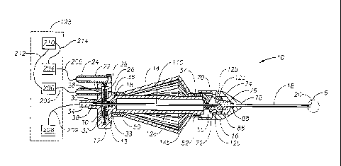
Figure 3 is a side, sectional view of the microsurgical instrument of Figure 1
shown operatively coupled to a microsurgical system;
4
CA 02637816 2008-07-21
WO 2007/092739
PCT/US2007/061476
Figure 4 is an enlarged, perspective view of the cam member of the
microsurgical instrument of Figure 1;
Figure 5 is a cross-sectional view of the cam member of Figure 4;
Figure 6 is an enlarged, fragmentary, side, sectional view of the portion of
the
microsurgical instrument of Figure 1 shown in circle 6 of Figure 2; and
Figure 7 is an enlarged, fragmentary, side, sectional view of a portion of the
actuating mechanism of the microsurgical instrument of Figure 1.
Detailed Description of Preferred Embodiments
The preferred embodiments of the present invention and their advantages are
best understood by referring to Figures 1 through 7 of the drawings, like
numerals
being used for like and corresponding parts of the various drawings.
Microsurgical instrument 10 preferably includes a base 12, an actuating handle
14, a nose member 16, and a cutting member 18 having a distal tip 20. As shown
in
the Figures, microsurgical instrument 10 is a vitrectomy probe. However,
microsurgical instrument 10 may be any microsurgical cutting, aspiration, or
infusion
probe.
Base 12 includes an actuating mechanism 13 for actuating a tubular inner
cutting member 110 of cutting member 18 in a reciprocating manner. Actuating
mechanism 13 preferably includes a first pneumatic port 22, a second pneumatic
port
24, a diaphragm chamber 26, a flexible diaphragm 28, and a rigid center
support 30.
Flexible diaphragm 28 is coupled to center support 30 and base 12. As shown in
the
Figures, flexible diaphragm 28 is frictionally coupled to both center support
30 and
base 12. Alternatively, flexible diaphragm 28 may be frictionally coupled to
base 12
and over-molded onto center support 30. Center support 30 has limiting
surfaces 31a
5
CA 02637816 2008-07-21
WO 2007/092739
PCT/US2007/061476
and 3 lb for interfacing with wall portions 33a and 33b of diaphragm chamber
26,
respectively. Base 12 further includes an aspiration port 34 and a distal
portion 12a
having an aperture 12b and a distal tip 12c. A collar 36 couples distal
portion 12a to
actuating handle 14. Inner cutting member 110 is coupled to center support 30
and is
slidaby and fluidly coupled to base 12 via o-rings 38.
Actuating handle 14 preferably includes a proximal base 50, a distal base 52,
and a plurality of flexible appendages 14a coupled to both base 50 and 52.
Flexible
appendages 14a may be made from any suitable springy material having a memory,
such as titanium, stainless steel, or a suitable thermoplastic. Handle 14
surrounds
distal portion 12a of base 12. Proximal base 50 is coupled to collar 36.
Distal base 52
is received within a slidable collar 54. A user grasps microsurgical
instrument 10 via
handle 14. When a user exerts an inward pressure on flexible appendages 14a,
flexible appendages 14a bend at or near 14b, straightening and elongating
flexible
appendages 14a, and moving collar 54 toward distal tip 20. When such pressure
is
removed, spring 55 returns flexible appendages 14a to the position shown in
Figure 2.
Nose member 16 preferably includes cam chamber 70 for receiving a cam
member 72, a base chamber 74 for receiving distal tip 12c of base 12, a
bushing 76 for
receiving inner cutting member 110 of cutting member 18, and an outlet 78 for
receiving a tubular outer cutting member 100 of cutting member 18. Cam member
72
is rotationally coupled to nose member 16 within aperture 12b of base 12 via
dowel
pins (not shown) inserted into each end of a bore 79. Cam member 72 preferably
has
a first stopping surface 80 for interfacing with collar 54, a second stopping
surface 82
for interfacing with base 12, a clearance slot 84 for receiving inner cutting
member
110 of cutting member 18, and a cam surface 86 for interfacing with bushing
76. An
o-ring 88 slidaby and fluidly seals nose member 16 to inner cutting member
110.
6
CA 02637816 2008-07-21
WO 2007/092739
PCT/US2007/061476
As described above, cutting member 18 preferably includes tubular outer
cutter member 100 and tubular inner cutting member 110. Outer cutting member
100
has an inner bore 102, a closed end 104, a port 106 for receiving tissue, and
cutting
surfaces 108. Inner cutting member 110 has an inner bore 112, an open end 114,
and
a cutting surface 116.
In operation, vitrectomy probe 10 is operatively coupled to a tnicrosurgical
system 198. More specifically, pneumatic port 22 is fluidly coupled to a
pneumatic
pressure source 200 via a fluid line 202, pneumatic port 24 is fluidly coupled
to a
pneumatic pressure source 204 via fluid line 206, and aspiration port 34 is
fluidly
coupled to vacuum source 208 via fluid line 209. Inner bore 112 and fluid line
209
are primed with a surgical fluid. Microsurgical system 198 also has a
microprocessor
or computer 210, which is electrically coupled to pneumatic pressure sources
200 and
204 via interfaces 212 and 214, respectively.
A surgeon inserts distal tip 20 into the posterior segment of the eye using a
pars plana insertion. The surgeon selects a desired vacuum level for vacuum
source
208. Tissue is aspirated into inner bore 112 via port 106. The surgeon selects
a
desired cut rate for probe 10 using microprocessor 210 and optionally a
proportional
control device (not shown), such as a foot controller. More specifically,
microprocessor 210 uses pressurized gas sources 200 and 204 to create a cyclic
pressure differential across diaphragm 28 so as to move center support 30, and
thus
inner cutting member 110, in a reciprocating manner at the desired cut rate.
When the
pressure provided to pneumatic port 22 is greater than the pressure provided
to
pneumatic port 24, inner cutting member 110 is moved toward distal tip 20
until open
end 114 is past cutting surface 108, as shown in Figure 6. This actuation
closes port
106, allowing cutting surfaces 108 and 116 to cut the tissue within inner bore
112.
7
CA 02637816 2008-07-21
WO 2007/092739
PCT/US2007/061476
The cut tissue is aspirated through inner bore 112, aspiration port 34, fluid
line 209,
and into a collection chamber (not shown). When the pressure provided to
pneumatic
port 24 is greater than the pressure provided to pneumatic port 22, inner
cutting
member 110 is moved away from distal tip 20, opening port 106 and allowing the
further aspiration of tissue.
During actuation of inner cutting member 110, limiting surface 31a of center
support 30 contacts wall portion 33a of diaphragm chamber 26 to precisely end
the
cutting stroke. Limiting surface 31b of center support 30 contacts wall
portion 33b of
diaphragm chamber 26 to precisely end the return stroke. When limiting surface
31a
contacts wall portion 33a, cutting surface 116 of open end 114 of inner
cutting
member 110 is preferably disposed at or just past distal cutting surface 108
of outer
cutting member 100. When limiting surface 3 lb contacts wall portion 33b, open
end
114 is preferably disposed at or near proximal cutting surface 108 of outer
cutting
member 100. Such precision control of the actuation of inner cutting member
110
greatly increases the cutting efficiency of probe 10.
From the above, it may be appreciated that the present invention provides
significant benefits over conventional vitrectomy probes. The present
invention is
illustrated herein by example, and various modifications may be made by a
person of
ordinary skill in the art. For example, although the present invention is
described
above in connection with a vitrectomy probe, it is equally applicable to
aspiration
probes, infusion probes, and other cutting probes.
It is believed that the operation and construction of the present invention
will
be apparent from the foregoing description. While the apparatus and methods
shown
or described above have been characterized as being preferred, various changes
and
8
CA 02637816 2013-11-18
modifications may be made therein.
9
