Note : Les descriptions sont présentées dans la langue officielle dans laquelle elles ont été soumises.
CA 02641105 2008-07-31
WO 2007/102829
PCT/US2006/008546
CANNULATED SUTURE ANCHOR SYSTEM
BACKGROUND OF THE INVENTION
1. Field of the Disclosure
The present disclosure relates to orthopedic surgery and, more particularly,
relates to a system and method for performing arthroscopy shoulder repair.
'
2. Description of the Related Art
Shoulder arthroscopy involves the repair of tissue inside or around the
õ
shoulder joint. The procedure is typically performed under endoscopic
visualization with,
e.g., an arthroscope, which is introduced within a small incision in the skin.
Various narrow
diameter instruments are positioned within the tissue to perform the desired
surgical
procedure. A saline solution may be pumped into the shoulder to expand the
joint to enhance
visualization and facilitate manipulation of the instruments during the
procedure.
Common shoulder injuries requiring arthroscopy include a torn or damaged
cartilage ring (labrum) or ligaments causing shoulder instability, a torn
rotator cuff or a torn
or damaged biceps tendon. Each of these injuries necessitates the reattachment
of soft tissue,
e.g., the ligaments or tendons, to bone. Various fixation devices and
methodologies including
sutures, screws, staples, wedges and plugs are known to effectuate the
attachment. Most of
these fixation devices have proven to be generally adequate for their intended
purposes.
1
CA 02641105 2008-07-31
WO 2007/102829
PCT/US2006/008546
SUMMARY OF THE INVENTION
Accordingly, the present disclosure is directed to further improvements in
arthroscopic repair, particularly, repair of the shoulder. In one embodiment,
a system and
associated method for arthroscopic repair is particularly adapted in
reattaching a ligament
and/or tendon to cortical bone of the shoulder. In accordance with this
preferred
embodiment, a suture anchor system includes a suture anchor, preferably, a
screw anchor,
and an installation tool for installing the suture anchor in tissue. The
suture anchor defines a
longitudinal axis and has a longitudinal cannulation for reception and passage
of a guide
wire. The suture anchor has trailing and leading ends, and an anchor head
adjacent the
trailing end thereof. The anchor head includes at least one eyelet for
reception of a suture and
an internal bore therein.
The installation tool includes a main body and a driver head extending from
the main body. The main body includes an outer surface having a longitudinal
recess therein
to accommodate the suture. The driver head is correspondingly dimensioned to
be received
within the internal bore of the anchor head of the suture anchor whereby
movement of the
installation tool about a longitudinal axis, e.g., rotational movement,
thereof causes
corresponding movement, e.g., rotational, of the suture anchor. The
installation tool may
also define a longitudinal cannulation for reception and passage of the guide
wire.
In one preferred embodiment, the anchor head preferably includes first and
second eyelets for reception of respective sutures. With this arrangement, the
outer surface of
the installation tool includes first and second longitudinal recesses for
receiving respective
sutures extending from the respective first and second eyelets of the anchor
head. The first
and second longitudinal recesses of the installation tool are in general
alignment with the first
2
CA 02641105 2008-07-31
WO 2007/102829
PCT/US2006/008546
and second eyelets of the anchor head when the suture anchor is mounted to the
installation
tool. In addition, the driver head of the installation tool may be dimensioned
to define first
and second clearances between respective outer surfaces of the driver head and
internal
surfaces of the internal bore of the anchor head when the driver head is
mounted within the
anchor head. The clearances accommodate suture portions of the sutures and are
in general
alignment with respective longitudinal recesses in the outer surface of the
installation tool.
The arrangement of the eyelets, longitudinal recesses of the installation tool
and sutures within the recesses significantly reduces the profile of the
system to thereby
facilitate maneuvering of the system within the restricted surgical area. In
addition, with the
sutures accommodated within the recesses, the potential of entanglement of the
sutures
during manipulation and/or rotation of the insertion tool is greatly
minimized.
A method for attaching soft tissue to bone tissue within a bone area of a
patient is also disclosed. The method includes the steps of:
accessing an internal target of a bone area of a patient, preferably, the
shoulder
area;
positioning a guide wire in relation to the internal target of the shoulder
area;
mounting a cannulated anchor, preferably, a screw anchor onto the guide wire,
the cannulated anchor having at least one suture connected thereto;
advancing the cannulated anchor along the guide wire to the internal target;
securing the cannulated anchor within bone tissue of the internal target; and
securing soft tissue to the cannulated anchor with the at least one suture.
3
CA 02641105 2008-07-31
WO 2007/102829
PCT/US2006/008546
BRIEF DESCRIPTION OF THE DRAWINGS
Preferred embodiments of the present disclosure will be more readily
appreciated by reference to the drawings wherein:
FIGS. 1-3 are perspective views of the suture anchor system of the present
disclosure;
FIG. 4 is a cross-sectional view taken along the lines 4-4 of FIG. 1
illustrating
the arrangement of the insertion tool within the screw head of the screw
anchor; and
FIGS. 5-6 illustrate a preferred method of use of the suture anchor system in
shoulder repair.
DETAILED DESCRIPTION OF THE PREFERRED EMBODIMENTS
The suture anchor system of the present disclosure is intended for use in
arthrosopic procedures and has particular application in securing soft tissue
including
tendons, ligaments etc. to hard cortical bone. The system may be used in
conjunction with
surgery performed on the knee, back, ankle, elbow etc. and has particular
application in
shoulder repair, particularly, arthroscopic shoulder repair. Such shoulder
repair operations
are inclusive, but, not limited to, reattachment of a torn or damaged
cartilage ring (labrum) or
ligaments, reattachment of a torn rotator cuff or reattachment of a torn or
damaged biceps
tendon. Other procedures are also envisioned.
Referring now to the drawings wherein like reference numerals identify
similar or like elements throughout the several views, FIGS. 1-3 illustrate,
each in perspective
view, the novel suture anchor system in accordance with the principles of the
present
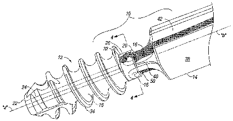
disclosure. Suture anchor system 10 generally includes three components,
namely screw
4
CA 02641105 2008-07-31
WO 2007/102829
PCT/US2006/008546
anchor 12, insertion tool 14 for mounting the screw anchor 12 into bone and a
pair of sutures
16 connected to the screw anchor 12 for securing the soft tissue to the screw
anchor 12.
Screw anchor 12 includes anchor body 18 defining longitudinal axis "a" and
having leading and trailing ends 20, 22. Anchor body 18 includes longitudinal
cannulation
24 which extends the length of the anchor body 18. Cannulation 24 is
dimensioned to receive
a guide wire. Anchor body 18 further includes screw head 26 adjacent leading
end 20.
Screw head 26 includes inner wall portions 28 defining internal bore 30 which
communicates
with cannulation 24 (FIG. 4). Screw head 26 further has first and second
diametrically
opposed eyelets 32 which extend through the outer wall of the screw head 26.
Internal bore
30 is dimensioned to cooperate with insertion tool 14. Although internal bore
30 may take
various geometrical shapes including, e.g., square, rectangular, triangular or
any other
polygonal arrangement, in a preferred embodiment, the internal bore 30 is
generally of
hexagonal configuration. First and second eyelets 32 are adapted to receive
sutures 16.
With reference again to FIGS. 1-3, anchor body 18 has an external thread 34
commencing adjacent screw head 26 and terminating in leading end 22. External
thread 34
may be continuous along the length of anchor body 18 or alternatively be
interrupted to
define a plurality of thread segments. External thread 34 is preferably self-
tapping although
it is envisioned that the external thread may be configured for advancement
within a pre-
tapped bore in bone. External thread 34 further includes a plurality of flutes
or cut-outs 36 in
the thread. Flutes 36 collect bone tissue during the initial self-tapping
advancement of the
anchor body to facilitate the anchoring process.
CA 02641105 2008-07-31
WO 2007/102829
PCT/US2006/008546
Sutures 16 may be fabricated from any biocompatible material. The preferred
materials for sutures 16 include synthetic bioabsorbable materials such as
polymers or
copolymers of glycolide, lactide, trimethylene carbonate, dioxanone,
caprolactone or blends
thereof. Other suitable materials for the components of sutures 16 include
nonabsorbable
materials such as polycarbonate, polyester, polyethylene, polyamide,
polypropylene,
polytetrofluoroethylene (PTFE), polysulfone and acrylic.
Referring still to FIGS. 1-3, insertion tool 14 will be discussed. Insertion
tool
14 includes main body 38 defining longitudinal axis "b" and having driver head
40 at the end
of the main body 38. It is noted that in the Figures only the distal end
portion of main body
38 is illustrated. Main body 38 includes a pair of longitudinal recesses 42
within the outer
surface of the main body 38 and extending from driver head 40 along at least a
portion of the
length preferably, the entire length of the main body 38. Longitudinal
recesses 42 define an
arc section removed from the outer surface of main body 38. The radius of the
arc section
preferably at least approximates the diameter of the sutures 16 to ensure that
the sutures are
fully accommodated within the longitudinal recesses during use of the system
10. Preferably,
longitudinal recesses 42 are arranged in diametrical opposed relation as shown
and are in
alignment with eyelets 32 of anchor screw 14 when the anchor screw 12 is
mounted to
insertion tool 14 as depicted in FIG. 1.
As best depicted in FIGS. 3-4, driver head 40 defines a general rectangular
cross-section having first and second cross-sectional dimensions "dl, d2" each
being
transverse to longitudinal axis "b". Second cross-sectional dimension "d2"is
greater than
first cross-sectional dimension "dl". Driver head 40 includes opposed outer
surfaces 44 and
opposed outer surfaces 46, and chamfered surfaces 48 interconnecting the
surfaces 44, 46.
6
CA 02641105 2008-07-31
WO 2007/102829
PCT/US2006/008546
When driver head 40 is mounted within screw head 26, a clearance or gap 50 is
defined
between outer surfaces 44 and inner surface portions 30 of the screw head 26.
(FIG. 4) The
clearances 50 are in general longitudinal alignment with respective eyelets 32
of screw head
26 and respective longitudinal recesses 42 of insertion tool 14. The distances
between inner
surface portions 30 and surfaces 44 within clearances 50 are each preferably
dimensioned to
at least be equal to, preferably, slightly greater than, the diameters of
sutures 16. With this
arrangement, the sutures 16 may slide within the clearance area 50 during
manipulation of the
system 10. Furthermore, the overall axial profile of system 10 is reduced by
virtue of
longitudinal recesses 42 to facilitate use during a minimally invasive or
laparoscopic
procedure.
Insertion tool 14 further defines longitudinal cannulation 52 extending along
the length of the insertion tool 14. Longitudinal cannulation 52 is
dimensioned for receiving
a guide wire.
The use of the system 10 during repair of a detached soft tissue in the
shoulder
will now be discussed. The following discussion of the use of the system will
be described in
terms of the performance of an arthroscopic procedure within the shoulder,
particularly, a
procedure utilizing reattaching soft tissue, e.g., a tendon or ligament to
cortical bone in the
shoulder. Such tendon or ligament may be the labrum, rotator cuff or bicep
tendon.
Referring now to FIG. 5, an internal target area is accessed through a small
incision in the tissue adjacent the shoulder. An arthroscope may be utilized
and introduced
through a cannula as is conventional in the art to visualize the target area.
Saline solution
may then be pumped within the shoulder joint to expand the joint to provide
more room to
7
CA 02641105 2008-07-31
WO 2007/102829
PCT/US2006/008546
perform the procedure. A guide wire 100 is advanced through the shoulder joint
to contact
the cortical bone 150. The guide wire 100 may be at least partially embedded
within the
cortical bone 150 to positively fix the guide wire 100 to facilitate
advancement of the
remaining instruments. The guide wire 100 may be driven into the cortical bone
100 or
alternatively, a drill may be introduced through a cannula to drill a bore for
reception of the
distal end of the guide wire. The guide wire 100 may optionally be driven
through the
tendon/ligament to be reattached and then secured to the cortical bone as
discussed
hereinabove.
With the guide wire 100 secured within the cortical bone 150, a cannulated
drill (not shown) may be advanced along the guide wire 100 to core a hole in
the targeted
cortical bone 150 for subsequent positioning of screw anchor 12. The hole
within the cortical
bone may be tapped if desired with a tapping instrument. The screw anchor 12
with mounted
insertion tool 14 are positioned over the guide wire 100 with the guide wire
100 being
accommodated within cannulations 24, 52 of the screw anchor 12 and the
insertion tool 14,
respectively.
With reference now to FIG. 6, the screw anchor 12 is positioned within the
hole in the cortical bone 150 by rotating the insertion tool 14 to cause
corresponding
rotational movement of the screw anchor 12 to advance the screw anchor 12
within the
cortical bone 150. Once the screw anchor 12 is secured in place within the
bone, the guide
wire 100 is removed. Sutures 16 are then passed through the tendon/ligament
200 and tied
off (by knotting) to secure the ligament 200 to the screw anchor 12 and
cortical bone 150. It
is appreciated that sutures 16 may be passed through the tendon/ligament 200
and then loaded
onto suture anchor 16 followed by placement of the screw anchor 12 in the
cortical bone if
8
CA 02641105 2012-10-26
desired. As a further alternative, the screw anchor 12 with mounted sutures 16
may be
punched through the tendon/ligament 200 and advanced within the cortical bone
150
followed by subsequent tying-off of the suture 16. Over time, sufficient
tissue
growth/regrowth occurs to affix the natural tendon/ligament 200 to the
cortical bone.
As appreciated, during advancement and rotation of insertion tool 14 and screw
anchor 12, sutures 16 are accommodated within longitudinal recesses 42 of the
insertion tool 14. Thus, the overall profile of the system is reduced.
Moreover, with
the sutures 16 accommodated within the longitudinal recesses 42, the potential
of
entanglement of the sutures 16 is significantly reduced during rotational
movement of
the insertion tool 14.
While the invention has been particularly shown, and described with reference
to the preferred embodiments, it will be understood by those skilled in the
art that
various modifications and changes in form and detail may be made therein. For
example, the system and method for shoulder repair may incorporate a screwless
anchor, i.e., an anchor devoid of an external screw thread. Anchors suitable
for this
purpose are disclosed in commonly assigned U.S. Patent Nos. 5,720,753 to
Sander et
al. and 5,948,000 to Larsen et al. The anchors disclosed in the '753 and '000
patents
incorporate expandable legs with anchoring means to engage the bone and may be
deployed through non rotational longitudinal movement of a drive element.
Another
anchor which may be adapted for use in the system and method of shoulder
repair of
the present invention is disclosed in U.S. Patent No. 5,980,558 to Wiley. The
anchor
disclosed in the '558 patent incorporates a rigid spear for driving into the
bone and a
plurality of wings which engage the bone upon deployment with a drive
instrument.
9
CA 02641105 2012-10-26
The scope of the claims should not be limited by the preferred embodiments set
forth herein, but should be given the broadest interpretation consistent with
the
description as a whole.
10
20