Note : Les descriptions sont présentées dans la langue officielle dans laquelle elles ont été soumises.
CA 02644483 2008-09-02
WO 2007/103744 PCT/US2007/063096
METHODS FOR QUANTIFYING THE RISK OF CARDIAC DEATH
USING EXERCISE INDUCED HEART RATE VARIABILITY
METRICS
CROSS-REFERENCE TO RELATED APPLICATION
[0001] This application claims the benefit of U.S. Provisional Patent
Application
No. 60/779,313 filed March 3, 2006, which is herein incorporated by reference.
TECHNICAL FIELD
[0002] The present invention relates to methods and apparatus for assessing
the risk of death from cardiovascular causes using spectral and temporal
characterizations of heart rate variability from heart rate measurements made
during
cardiac stress testing.
BACKGROUND
[0003] Sudden cardiac death (SCD) accounts for approximately 300,000 -
400,000 deaths per year in the United States. Although the individual risk of
SCD in
the adult U.S. population is only about 0.1 - 0.2 % per year, when applied to
the
large population base, SCD is often the first and only manifestation of the
presence
of a cardiovascular disease in a majority of cardiovascular related deaths.
Deaths
associated with recovering from large myocardial infarctions actually
represent the
minority of the total cardiovascular related deaths per year. As a result, a
low cost
screening tool that would provide early detection of patients at risk for SCD
would be
tremendously valuable for early treatment and intervention.
[0004] However, it can be difficult to accurately predict or assess the risk
of
SCD because many underlying pathologies support or trigger the events leading
to
SCD instead of any single condition. Of these various conditions, most data
suggests that regulation of the heart through the sympathetic and
parasympathetic
(vagal) branches of the autonomic nervous systems is extremely important in
maintaining stable rhythms. In particular, it appears that vagal stimulation
mitigates
the development of ventricular arrhythmias in a variety of experimental
studies.
1
CA 02644483 2008-09-02
WO 2007/103744 PCT/US2007/063096
[0005] One promissory marker related to SCD is the variability of the heart
rate
under various conditions. For example, studies using Holter records have shown
that low heart rate variability (HRV) is a marker for SCD. Holter studies
predominately follow individuals over the course of an average day, mostly
reflecting
low exercise conditions.
[0006] In 1993, a study by van Ravenswaaji et al. reviewed four years of
published HRV papers and summarized the various time and frequency domain
methods for computation of HRV, which remain largely the same today. This
study
concluded that HRV is an important surveillance tool for post infarction and
diabetic
patients to prevent SCD. Although HRV was noted as having a higher association
with risk for death than other variables obtained by Holter monitoring, this
study also
concluded that HRV has a rather low positive predictive value in mass
screening
(less than 20%). Nonetheless, other studies establish that reduced HRV
obtained
from 24 hour Holter recordings is an independent predictor of death in chronic
heart
failure patients.
[0007] Another study by Arai et al. analyzed HRV in a cohort of patients
undergoing exercise testing and found that the power in the low frequency band
[0.03 - 0.15 Hz] systematically decreased with an increase in exercise and
rebounded during recovery after exercise. The low frequency band may be
modulated by both the sympathetic and parasympathetic nervous system related
to
baroreflex activity, temperature regulation and maintenance of homeostasis.
The
low frequency response to exercise testing was found to be muted in patients
with
severe congestive heart failure. Conversely, this study found that power in
the high
frequency band [0.15 - 0.8 Hz] increased with exercise, decreased through
recovery
and was highly correlated to respiration - the respiration sinus arrhythmia
effect.
[0008] Many of the HRV studies have been predicated upon an assumption that
a balance between the operation of the parasympathetic (vagal) and sympathetic
arms of the autonomic nervous system controls heart rate. For example, as the
heart rate increases it has been assumed that sympathetic control increases
and
vagal influence decreases. Additionally, the low and high frequency bands have
been assumed to be related to sympathetic and vagal influence, respectively.
Based
on these assumptions, the concept of a spectral ratio of these two bands,
indicative
of this implied balance, was adopted as a potentially useful metric for risk
-2-
CA 02644483 2008-09-02
WO 2007/103744 PCT/US2007/063096
stratification. Because of the low predictive value of the ratio, teachings of
Verrier et
al. in US Patent No. 5,437,285 are predicated upon this ratio of low and high
frequency components in combination with other metrics for assessing
myocardial
instability.
[0009] Although the concept of a balance between the two components of the
autonomic system has been a widely embraced, and presumed to be quantified
through a HRV spectral ratio, some studies show that calculations of such a
balance
of control may not be useful. One study by Eckberg (1997), for example, finds
that
vagal contributions to baseline low frequency RR-interval fluctuations are
great, and
evidence that baseline low frequency RR-interval spectral power is related
quantitatively to sympathetic-cardiac nerve traffic is nonexistent. This same
study
concludes that calculations of sympathovagal balance may obscure rather than
illuminate human physiology and pathophysiology.
[0010] As noted by Kannankeril et al. (2002), risk of SCD is about 17 times
higher during or immediately following exercise than at rest. Kannankeril et
al. also
finds that the vagal influence of heart rate decreases with exercise, and that
it
appears likely that poor return of vagal control in the post exercise recovery
period
may be a very critical factor in the progression from instability to fatal
arrhythmia.
[0011] Although the above described methods for measuring heart rate
variability are well known to practitioners of the art and it also is
recognized that the
patient risk profile may be substantially unveiled during vigorous exercise
and
recovery, there is no effective method based on HRV for quantifying patient
risk from
heart rate data collected during exercise and recovery. Therefore, existing
methods
and apparatus for quantifying risk of SCD based on HRV do not provide an
accurate
low cost screening tool for mass screening.
SUMMARY
[0012] The invention is directed to methods and apparatus that assess the risk
of death from cardiovascular causes using information based on variabilities
in the
heart rate of a patient. Although much progress has been made in using the
tools of
heart rate variability to characterize records obtained from Holter recording,
little
work has been done with exercise testing where the effects of the autonomic
nervous system are most pronounced. Research has shown that vagal stimulation
-3-
CA 02644483 2008-09-02
WO 2007/103744 PCT/US2007/063096
has a strong anti-arrhythmic effect on the heart, and conversely poor vagal
regulation of the heart, particularly during the recovery period following
exercise, is a
significant risk factor for patients. It is estimated that the risk of SCD is
17-20 times
greater during exercise than during the resting phases that dominate Holter
recordings, which indicates that the characterization of the risk of
cardiovascular
death is better unveiled during exercise testing. Several examples of this
invention
provide new methods and apparatus for (a) characterizing the temporal and
spectral
characteristics of the variability of the heart rate, and (b) integrating or
otherwise
using disparate metrics for risk stratification.
[0013] One example of a method in accordance with the invention for assessing
cardiac risk in a specific patient based on the heart rate variability
comprises
providing heart activity data of a specific patient including a windowed time
series
related to heart rate variability during a heart rate test. The windowed time
series
includes ectopic beats. The method can further include determining a frequency
domain value based on either relative energy values of frequency bands or
slope of
the spectrum across selected frequency ranges of the heart rate variability in
the
windowed time series, and assessing the risk of a cardiac event based on the
frequency domain value.
[0014] Another example of a method for assessing cardiac risk in accordance
with the invention comprises providing heart activity data of a specific
patient
including a windowed time series related to heart rate variability during a
heart rate
test in which the windowed time series includes ectopic beats. This method
further
includes determining an aggregate power for a frequency band of the windowed
time
series, and assessing the risk of a cardiac event based on the aggregate
power.
The aggregate power can be determined by computing a root-means-square value
of the windowed time series. In another embodiment, the aggregate power can be
determined by performing a Fourier transform of the windowed time series into
a
spectrum for a frequency domain analysis and then summing the power of the
spectral components within a selected frequency band.
[0015] Still another method for assessing cardiac risks in a specific patient
in
accordance with the invention comprises providing heart rate activity
including a
windowed time series relating to heart rate variability during a heart rate
test. This
method further includes determining a frequency domain value based on either
-4-
CA 02644483 2008-09-02
WO 2007/103744 PCT/US2007/063096
relative energy values of frequency bands or slope of the spectrum across
selected
frequency ranges of the heart rate variability in the windowed time series,
and
determining an aggregate power for a frequency band of the windowed time
series.
This method further includes assessing the risk of a cardiac event based on
the
frequency value and the aggregate power.
[0016] Apparatus in accordance with the invention can include computers
and/or computer operable media containing instructions that cause the computer
to
receive heart activity data of a specific patient including a windowed time
series
related to heart rate variability during a heart rate test. The windowed time
series
can include ectopic beats. The computer also determines (a) a frequency domain
value based on either relative energy values of frequency bands or slope of
the
spectrum across selected frequency ranges of the heart rate variability in the
windowed time series, and/or (b) an aggregate power for a frequency band of
the
windowed time series. In this apparatus, the computer operable medium can
further
cause the computer to asses the risk of a cardiac event based on the frequency
value and/or the aggregate power and output the assessed risk.
BRIEF DESCRIPTION OF THE DRAWINGS
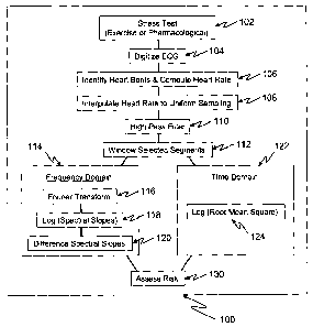
[0017] Figure 1 is a flow chart illustrating a method for determining the risk
of
cardiovascular death from analysis of heart rate variability in accordance
with an
embodiment of the invention.
[0018] Figure 2 is a graph illustrating an ECG and the reference points
corresponding to activation and recovery of the Atria (P); the ventricle
activation
phases Q, R and S, forming the QRS complex; the recovery or re-polarization
phase
T of the ventricles; and the R-R time interval between consecutive beats as
measured between the peaks of the R phase.
[0019] Figure 3 is a flow chart illustrating the processing stages starting
with a
raw instantaneous heart rate determined over the course of an exercise stress
test
(A), through a resampling to a uniform time basis (B), followed by selected
band
pass filtering or detrending (C), resulting in a final time series for heart
rate variability
analysis (D).
[0020] Figures 4A and 4B illustrate two examples of the spectral trends for
three frequency bands (low, mid and high) over the course of the exercise test
for
-5-
CA 02644483 2008-09-02
WO 2007/103744 PCT/US2007/063096
two populations: Figure 4A - patients that remained alive for more than five
years
after the stress test; and Figure 4B - patients that died of cardiovascular
causes
within 5 years of the stress test.
[0021] Figures 5A and 5B illustrate the spectral slopes associated with peak
exercise and mid recovery for a surviving cohort (Figure 5A) and a CV
mortality
cohort (Figure 5B). Figure 5C illustrates two example spectra and an
alternative
method for determining spectral slope across a selected frequency band.
[0022] Figure 6 illustrates the root mean square (RMS) amplitude, in
beats/minute, of the processed heart rate time series for both the alive and
CV
cohorts throughout the exercise test.
[0023] Figure 7 illustrates the Kaplin-Meier assessment of survival for
patients
with abnormal values for the Duke Treadmill Score and for the HRV slope (MR-
PE)
metrics.
[0024] Figure 8 illustrates the Kaplin-Meier assessment of survival for
patients
with abnormal values for the Duke Treadmill Score and for the HRV RMS metrics.
[0025] Figure 9 illustrates the Kaplin-Meier assessment of survival for
patients
with abnormal values for the Duke Treadmill Score in both the HRV Slope (MR-
PE)
and HRV RMS metrics.
DETAILED DESCRIPTION
[0026] The following discussion is presented to enable a person skilled in the
art to practice the invention. Various modifications to the disclosed
embodiments will
be apparent to those skilled in the art, and the generic principles herein may
be
applied to other embodiments and applications without departing from the
spirit and
scope of the present invention as defined by the appended claims. Thus, the
present invention is not intended to be limited to the embodiments presented,
but is
to be accorded the widest scope consistent with the principles and features
disclosed herein.
A. Overview
[0027] Figure 1 is a flow chart of a method 100 for quantifying the risk of
cardiovascular death using exercise induced heart rate variability metrics.
The
-6-
CA 02644483 2008-09-02
WO 2007/103744 PCT/US2007/063096
method 100 includes a stage 102 comprising increasing the heart rate of the
patient.
This may be accomplished through both exercise and pharmacological protocols.
Method 100 continues to a stage 104 comprising digitizing and recording the
electrocardiographic (ECG) signals representative of the electrical signal of
the
beating heart, and a stage 106 comprising analyzing the digitized ECG signal
to
identify each heart beat. The time of ventricular depolarization is recorded
for each
beat and the heart rate is computed. The resulting heart rate time series is
composed of irregularly spaced heart rate measures, corresponding to the
irregular
nature of the time between beats. Stage 108 comprises smoothly interpolating
this
irregular time series into a uniform sampling rate, and stage 110 comprises
removing
the long period (low frequency) components through high-pass filtering or
polynomial
based detrending. The high-pass filtered heart rate time series is next
analyzed over
several time periods corresponding to different phases of the exercise test.
This is
aided by stage 112 which selects segments of the filtered trace from different
times
during the test, including both the exercise and recovery phases.
[0028] Method 100 includes a frequency domain analysis stage 114 and/or a
time domain analysis stage 122 of the heart rate variability. The frequency
analysis
stage 114 comprises performing a Fourier transform of the windowed trace into
the
frequency domain (stage 116), determining the slope of the resulting spectrum
through a least squares fit to the logarithm of the spectral power (stage
118), and
combining the spectral slope from different time windows of the exercise test
(stage
120). The time domain analysis in stage 122 comprises computing the logarithm
of
the square root of the average of the sum of the squares of the windowed trace
(log
of the root mean square - Log RMS).
[0029] Both time domain (RMS) and frequency domain estimates of heart rate
variability naturally have different means and standard deviations. These are
independent metrics of heart rate variability and either can be used in
assessing
patient risk. However, it is advantageous to use the two metrics in an
integrated
estimate of risk. Stage 130 includes methods for assessing patient risk based
upon
either or both metrics.
-7-
CA 02644483 2008-09-02
WO 2007/103744 PCT/US2007/063096
B. Stimulating the Heart and Measuring Heart Rate
[0030] Stage 102 of method 100 is used to stimulate the heart to beat at a
faster rate and is well known in the field of cardiac stress testing. The
heart rate can
be elevated to maximum capacity via exercise on a treadmill, ergometer, or
other
exercise devices or through administration of drugs that elevate the heart
rate.
Cardiac stress tests are typically done using 10 electrodes placed across the
chest
in order to obtain spatial resolution of distinct aspects of the ECG waveform.
However, a single trace measuring the ECG voltage can be used to determine the
time of each beat. Time resolution of the heart beats is important and the ECG
voltage(s) should be digitized at a diagnostic resolution of 500 or more
samples per
second in stage 104. Stages 102 and 104 are generally performed in the
clinical
environment of a cardiac stress test.
[0031] Figure 2 illustrates an example ECG with the key phases identified. A
normal heart beat starts in the upper chambers of the heart (atria) and the
initial
ECG phase that records this activation is termed the P-wave portion of the ECG
signal. Following the activation of the atria the blood moves into the lower
chambers
of the heart (ventricles) and activation of the ventricle muscle pumps the
blood to the
body and generates the ECG phases Q, R and S, often referred to as the QRS
complex. Finally, the ventricle muscles recover (repolarize) in anticipation
of the
next beat, creating the T-wave portion of the ECG signal. Stage 106 can
include
determining the time interval between adjacent beats to identify the heart
beats and
compute the heart rate. For example, the time interval between adjacent beats
can
be measured by measuring the time between the peaks of the R wave (the R-R
interval). A more robust measure of R-R intervals, particularly when the peak
of the
R wave is not sharp, can be obtained by cross-correlating the QRS complex from
an
average or median beat with each subsequent beat and noting the time of
maximum
correlation. Stage 106 can be performed using either approach to determining R-
R
intervals.
[0032] For every beat detected the instantaneous heart rate, measured in beats
per minute, for HRV analysis is computed from the R-R interval between the
current
and proceeding beats by the simple equation HR = 60/(R - R) , where the R-R
interval is measured in seconds. Figure 3A is an example of a graphical
representation of the output of stage 106 showing a typical plot of continuous
heart
-8-
CA 02644483 2008-09-02
WO 2007/103744 PCT/US2007/063096
rate during a stress test. In this example, the heart rate starts at time 0 at
about 90
beat/sec and remains low during the start of exercise (SE), increases
throughout the
middle exercise period (ME) until it climbs to a peak of over 160 beat/sec at
peak
exercise (PE), and then rapidly declines during the start of recovery (SR) as
the
patient recovers until it returns to the low range at the end of recovery
(ER).
C. Signal Conditioning and Windowinci
[0033] Because the beats occur irregularly in time, the resulting
instantaneous
heart rate time series is not uniformly sampled. As most signal processing
techniques are more efficient when the series is uniformly sampled, stage 108
is
useful because it interpolates or transforms the heart rate data onto a
uniformly
sampled series. Figure 3B is an example of a graphical representation
illustrating
the process of stage 108. The vertical bars represent the location in time of
the R
wave from each beat and the height of each bar represents the instantaneous
heart
rate computed from the R-R interval between one beat and its proceeding beat.
In
one example, stage 108 includes using a cubic spline under tension curve to
interpolate the instantaneous heart rate sequence to a uniformly sampled time
series
represented by the small circles on the smooth curve. Although the
interpolated
sample rate is not critical, it should be above the Nyquest frequency
corresponding
to the highest heart rate or above the shortest R-R interval in the data. For
example,
a sampling rate of 10 samples/sec is expected to be sufficient and convenient.
The
time series for HRV analysis can be computed from either the measured heart
rates
or from the measured R-R intervals. When using R-R intervals for HRV, the
vertical
bars in Figure 3B represent the R-R interval time instead of the reciprocal
metric
heart rate.
[0034] Traditional HRV analysis focuses on R-R intervals between normal
beats, where "normal" is the dominant beat in the series. Ectopic beats and
the
adjacent R-R intervals are excluded from the irregular time series and any
subsequent interpolated series and analysis in traditional HRV analysis.
However,
ectopy may introduce feedback to heart rate through the baroreceptor
mechanisms
that may last as long as approximately 10 seconds. Conventional HRV analyses
that merely remove ectopic beats accordingly remove the stimulus while leaving
the
response. This can vitiate the value of the spectral analysis. Furthermore,
some
. .,,.,, ~,.., . . . . -9-
CA 02644483 2008-09-02
WO 2007/103744 PCT/US2007/063096
methods that remove ectopy effectively time-shift the subsequent beat pattern
by the
missing intervals, which can destroy the phase information and alter the
spectral
amplitude information in unpredictable ways. Although it may be ideal to have
perfect records without ectopy, methods in accordance with many examples of
the
present invention include the ectopic beats and the fidelity of the temporal
position
and response of the subsequent beats. This accordingly avoids the downfalls of
excluding such data.
[0035] Stage 110 compensates for such irregularities so that the data can
include ectopic beats. In many examples, stage 110 includes reducing the heart
rate
data via filtering.the heart rate time series over selected periods, such as
at peak
exercise (PE) and start of recover (SR), using a selected band filter. The
filtered
heart rate time series can contain very long signal periods representing the
progression of the heart rate to a peak value at the limits of physical
exercise (PE)
and a rapid return to baseline as the patient recovers (SR). The shorter
frequencies
of the heart rate intervals are of principal interest for HRV analysis, and
thus a high-
pass filter can be used to select shorter frequencies for the windowed time
series. A
single or multi-pole infinite or finite impulse response filter may be used in
effecting
the filtering. A two-pole Butterworth high-pass filter with a corner at 0.015
Hz has
been found to be effective.
[0036] The heart rate signal does not reflect a stationary process. The time
series around peak exercise (PE) is particularly important for the HRV
analysis, and
a filter that extended the filter impulse response from the exercise phase
into the
sharply contrasting recovery phase would distort the true frequency
characteristics of
the recovery phase. As the amplitude characteristics are important in the
spectral
analysis, and the phase information less significant, it is useful to apply
the high pass
filter in a forward direction from the start of the time series to peak
exercise (PE) and
in a reverse direction from the end of the record to the same sample at the
peak.
Figure 3D is an example of a graphical representation showing the two segments
representing filtered exercise and recovery joined for display purposes, but
HRV
analysis should not extend over this discontinuity. This procedure isolates
the
distinct non-stationary aspects of the exercise and recovery phase of the test
and
preserves the temporal amplitude information in the heart rate data.
-10-
CA 02644483 2008-09-02
WO 2007/103744 PCT/US2007/063096
[0037] Stage 112 includes setting a window for segments of the filtered heart
rate time series. Any segment of the filtered time series may be used for HRV
analysis for stage 112. However, a representative characterization of the
changing
HRV signal can be obtained through analysis of six segments corresponding to
the
start (SE), middle (ME) and peak (PE) of exercise, and the start (SR), middle
(MR)
and end (ER) of recovery shown by the boxed areas in Figure 3A and 3D. The
window length of the boxed areas can be adjusted depending upon several
considerations. For spectral analysis, it is desirable to choose window
lengths that
are powers of 2 (e.g.: 512, 1024, etc). Because the longest periods of
interest are
around 25 seconds, corresponding to a frequency of 0.04 Hz, it is generally
useful to
extend the window to a length of one or more multiples of the longest periods
of
interest. Conversely, temporal resolution of vagal changes is diminished as
the
window length extends. In practice a window length of 102.4 seconds,
corresponding to 1024 samples, has proven effective.
[0038] Alternatively, the procedure for reducing the heart rate data in stage
110
can include detrending via fitting a low-order polynomial curve to the heart
rate data
over the selected window segment and subtracting the resulting curve from the
heart
rate data. This alternative reducing procedure via detrending the heart rate
data
may be employed using either the raw heart rate beat data (results from stage
106),
or the uniformly interpolated data from stage 108. In practice, a second order
polynomial has been found to do an excellent job of detrending stress test
heart rate
data over a window length of 102.4 seconds, but in still additional
embodiments of
the invention higher or lower order polynomials may be used to detrend stress
test
heart rate data over other window lengths.
D. Freguency Domain Analysis (Stage 114)
[0039] The windowed time series from method 112 are multiplied by a Hanning
window and Fourier transformed using standard methods familiar to those
skilled in
the art of signal processing, method 116. Several specific frequency bands are
described below to provide examples of useful frequencies, but other
frequencies
may be used. The frequency domain analysis provides a frequency value that can
be used to assess the risk of SCD. One unique finding of the present invention
is
-11-
CA 02644483 2008-09-02
WO 2007/103744 PCT/US2007/063096
that the spectral slopes of the average power in various spectrums is a
diagnostic of
risk stratification for CV death.
[0040] Although the resulting spectrum can be analyzed as a whole, distinctly
different physical processes have been found to correlate with distinct
frequency
bands in the signal (see van Ravenswaaji et al, 1993). The high frequency
spectral
band [0.4 - 1.0 Hz] has been found to capture the respiration induced HRV. At
the
low frequency end, the spectral band [0.04 - 0.15 Hz] has been found to be
modulated by both the sympathetic and parasympathetic nervous system related
to
baroreflex activity, temperature regulation and maintenance of homeostasis.
The
remaining middle band [0.15 - 0.4 Hz] provides a transition between the low
and
high bands. The power in each band, in decibels (db), can be computed by
integrating the logarithm of the spectrum over the defined frequency range of
each
band, method 118.
[0041] Figure 4A shows the average power for the three spectral bands for the
six windowed phases of the exercise test from 1,783 patients, from a total
cohort of
1,959 patients, that were still alive after a 5 year follow-up period. The
high
frequency band shows a progressive increase to peak exercise, corresponding to
increasing respiration induced sinus arrhythmia, that decays over the course
of
recovery. Conversely, the longer period bands show a distinct decay in HRV
power
as exercise progresses, reaching a minimum at peak exercise and rebounding
dramatically in recovery. The first analysis in recovery (SR) for the low
frequency
band, corresponding to the first 102.4 sec of recovery, is characterized by
HRV
power greater than that recorded at the initial stage of exercise (SE). The
mid-band
follows the low band, but somewhat muted in overall response. This signal
shape is
interpreted to represent the process of reduced vagal mediation of heart rate
and
heart rate variability as exercise progresses, followed by a very rapid return
of vagal
control in the early stage of recover.
[0042] Figure 4B shows a similar plot, but for the average power from of 55
patients, from the cohort of 1,959 patients, that died of cardiovascular (CV)
causes
over the following 5 year period. It is important to note the muted overall
spectral
response of these patients relative to the spectra in Figure 4A.
-12-
CA 02644483 2008-09-02
WO 2007/103744 PCT/US2007/063096
[0043] The spectral differences between the alive population (Figure 4A) and
those dying of CV causes (Figure 4B) can be quantified in several ways. For
instance, at the start of exercise the low frequency band shows significant
augmentation, relative to the mid or high bands. The difference in separation
between the low and high bands is much reduced in the CV population. At peak
exercise this relationship is reversed, with the high band containing more
energy
than the lower bands for both populations but again muted in the CV
population.
The recovery phase is characterized by a rebound of the low band and a falling-
off of
the high band, but again muted for the CV population. These trends are readily
apparent when the spectrum is examined relative to different points in the
exercise
test. Figure 5 shows the power in the spectral bands for peak exercise and mid
recovery; the slope of the line fit to the three points across the bands, for
each time
interval, provides an effective characterization of the changing spectral
throughout
the test. The slope difference between peak exercise and other times has
proven to
provide an effective metric for risk stratification. The prognostic optimal
difference
has been found to occur between mid recovery and peak exercise: Slope(MR) -
Slope(PE), method 122.
[0044] It is important to note that the slope can be calculated via several
methods. In Figure 5 A and B the slope is computed via a least squares fit of
a line
to the average power in the three spectral bands. This is effectively the fit
of a line in
Log (power) and Log (frequency) space. Conversely a line could be fit to the
raw
spectrum across a broader frequency range, for instance 0.04 - 1.0 Hz. Another
method would include dividing the spectra from the two time intervals before
taking
the logarithm of the power and fitting the line to the resulting log(spectra).
[0045] Figure 5C illustrates an alternative procedure for spectral slope heart
rate variability analysis by fitting a line to the Log (power) versus Log
(frequency)
over a selected frequency range. Spectral slope estimates from different time
windows of the stress test may also be combined to improve signal to noise. In
one
example of this alternative procedure, the average of the spectral slope
estimates
over the frequency range 0.04 - 0.4 Hz, for the time intervals of mid-exercise
and
start recovery, has proven to provide an effective prognostic score for risk
stratification for CV death. As ectopy tends to introduce high frequency
energy into
-13-
CA 02644483 2008-09-02
WO 2007/103744 PCT/US2007/063096
the heart rate variability spectrum, lowering the highest frequency used in
analysis
from 1 Hz to 0.4 Hz tends to lower the potential noise associated with ectopy.
E. Time Domain Analysis (Stage 116)
[0046] In examining Figures 4A and 4B it is also apparent that the aggregate
power, or level, in the alive population is lower than for the CV population.
For
instance, the spectral value for the low frequency band for the alive
population is
about 1.85 and nearly 2.1 for the CV population. There is no a priori or
physical
reason why the shape of the spectral trends should be tied to the absolute
level of
the power (e.g., the aggregate power), and another unique finding of the
present
invention is that the power level is diagnostic for risk stratification for CV
death. The
total or aggregate power contained in the spectrum for any time windowed data
can
be obtained by integrating the power over the entire spectrum. Conversely, as
noted
by Parseval in 1799, the power contained in the spectrum is exactly equivalent
to the
square root of the sum of the squares (RMS) of the original time series, a
simple
variant in the fundamental principle of equivalent energy in either the time
or
frequency representation. The RMS computation of spectral power for each
windowed and high pass filtered or detrended series is a low cost method to
execute
and has proven useful for risk stratification, method 124. Figure 6 shows the
RMS
values (in units of beats/min) for the alive and CV death patients in the
studied
cohort for all six windows of the exercise test. Note that the RMS value for
the alive
patients is systematically below the CV death patients; the difference at peak
exercise has been found to be most prognostic.
[0047] For statistical analysis, the windowed time series has a zero mean
value
as a necessary result of the high-pass filtering and the RMS values will not
be
normally distributed due to the hard limit of zero on the low side of the RMS
distribution. Following the use of decibels (log of the spectrum) for the
spectral
estimates, it is statistically useful to use the log of the RMS for analysis,
which
transforms this energy metric into a more normal distribution.
F. Risk Assessment
[0048] The frequency value based on the spectral slope (HRV Slope) and the
aggregate power based on the RMS computation provide estimates of HRV that are
both highly predictive of CV death in the cohort discussed above and portrayed
in
-14-
CA 02644483 2008-09-02
WO 2007/103744 PCT/US2007/063096
Figure 4A and 4B. The correlation coefficient between these two parameters,
for this
large cohort, is relatively low (r=0.2), indicating they are independent. To
improve
risk stratification it is useful to combine these parameters into a single
unified metric,
method 130.
[0049] Risk assessment methodologies using Cox proportional hazard and
Kaplan-Meier survival analysis are well known to those familiar with
prognostic
statistical analysis in the medical industry. The HRV Slope and the aggregate
power
parameters discussed in methods 114 and 122 have been assessed relative to the
existing Duke Treadmill Score (TMS), the current industry "gold" standard
exercise
based prognostic metric for risk stratification. Figure 7 shows the survival
analysis
for the four combinations of normal and abnormal measures for the Duke TMS and
HRV Slope metric. An abnormal HRV Slope metric increases a patient's risk of
CV
death by -2.6 times over a normal score. An abnormal Duke TMS score increases
a
patients risk to -3.9 times greater than normal. A Cox proportional hazard
analysis
shows that the Duke TMS metric is an independent parameter, distinct from HRV
Slope, and the combined Kaplan-Meier hazard ratio when both metrics are
abnormal
is -10.8 times greater, a significant increase in risk over the estimate based
upon the
current gold standard Duke TMS.
[0050] Figure 8 shows a similar analysis for HRV aggregate power at peak
exercise combined again with the Duke TMS for comparison. The HRV aggregate
power metric is independent of the Duke TMS and the HRV Slope metrics. HRV
aggregate power metric increases a patient's risk of CV death by -1.8 times
over a
normal score. In this combination, an abnormal Duke TMS score increases a
patients risk to -3.6 times greater than normal. The combined Kaplan-Meier
hazard
ratio when both metrics are abnormal is -8.9 times greater, a significant
increase in
risk over the estimate based upon either parameter alone.
[0051] Finally, the combination of the two HRV metrics, along with the Duke
TMS is shown in Figure 9. An abnormal score in both HRV Slope and the
aggregate
power increases a patient's risk of CV death -3 times greater than normal,
compared
with an increase in risk to -3.2 when just the Duke TMS is abnormal. When both
HRV metrics and the Duke TMS metric are all abnormal, the risk of CV death
increases to -13.4 - a very dramatic increase over the current gold standard
Duke
-15-
CA 02644483 2008-09-02
WO 2007/103744 PCT/US2007/063096
TMS. As such, the new HRV metrics are complimentary to the Duke TMS and
provide a significant improvement in risk assessment when used together.
[0052] From the foregoing, it will be appreciated that specific embodiments of
the invention have been described herein for purposes of illustration, but
that various
modifications may be made without deviating from the spirit and scope of the
invention. Accordingly, the invention is not limited except as by the appended
claims.
-16-