Note : Les descriptions sont présentées dans la langue officielle dans laquelle elles ont été soumises.
CA 02645186 2008-07-21
WO 2007/084957 PCT/US2007/060715
Transplantation of Neural Cells
[Field]
The present invention relates to transplantation of neural cells to increase
inhibitory neuron activity in brain. It relates in particular to treatment of
disorders
that would benefit from increased inhibitory neuron activity - this could
include, but
is not limited to disease characterized by loss of inhibitory neuron function -
and
to compositions useful therefor and further relates to treatment of human
disease
including epilepsy and Parkinson's disease.
[Background to the Invention]
Many neural disorders are characterised by abnormal inhibitory neuron
signalling
and, in particular a lack of the neuro-transmitter y-aminobutyric acid (GABA),
secreted by inhibitory neurons. GABA, a metabolite of glutamate, is an
inhibitory
neurotransmitter which counteracts the effects of excitatory
neurotransmitters.
Excitatory neurotransmitters (typically acetylcholine, glutamate, or
serotonin) open
cation channels, causing an influx of Na+ that depolarises the postsynaptic
membrane toward the threshold potential for firing an action potential and
hence
cause the propagation of a signal across the synapse. Inhibitory
neurotransmitters, by contrast, open either CI' channels or K+ channels, and
this
suppresses firing by making it harder for excitatory influences to depolarise
the
postsynaptic membrane.
Abnormal inhibitory function may contribute to symptoms of Parkinson's Disease
and is fundamental to the pathology of several other neural disorders
including
Huntington's Disease, Schizophrenia, autism, chronic pain and many forms of
Epilepsy. Epilepsy, in common with most such disorders, has no known cure and
is treated with a range of drugs aimed at managing the symptoms. Therefore
Epilepsy and its treatment result in a severe degradation of quality of life,
measured in days of activity, pain, depression, anxiety, reduced vitality and
insufficient sleep or rest (similar to arthritis, heart problems, diabetes,
and cancer).
CA 02645186 2008-07-21
WO 2007/084957 PCT/US2007/060715
Epilepsy affects 50,000,000 people worldwide and sufferers have a mortality
rate
two to three times higher than that of the general population with the risk of
sudden death being 24 times greater. In addition to personal suffering,
epilepsy
imposes an annual economic burden of $15.5 billion in the USA alone, in
associated health care costs and losses in employment, wages, and
productivity.
Therefore any alternative or new therapy, especially one with the potential to
be
curative, would have very far reaching benefits.
Research aiming to enhance inhibitory neuron function by cell transplantation
has
focused on the use of multi-potent cells and immortalised neurons that have
been
genetically engineered to produce GABA (Bosch et al., (2004) Exp Neurol 190,
42-58.; Thompson, (2005) Neuroscience 133, 1029-37).
In order for the grafted cells to effectively reach affected regions and
functionally
integrate, it is necessary that the cells migrate away from the site of the
graft and
intermix with the host cells establishing inhibitory synapses with local
excitatory
neurons. A lack of migratory activity of the transplanted cells has been a
flaw of
previous attempts to derive new neural tissue from precursor cells, such as in
the
case of embryonic stem cell (ES)-derived neurons (Wernig et al., (2004) J
Neurosci 24, 5258-68; Ruschenschmidt et al., (2005) Epilepsia 46 Suppl 5, 174-
83) and genetically engineered GABA-producing cells (Bosch et al., supra.;
Thompson, supra). ES-derived cells or other neural precursors transplanted
into
postnatal brains do not migrate extensively but form clumps of graft-derived
cells
in, or near, the site of transplantation (Bosch et al., supra; Ruschenschmidt
et al.,
supra; Thompson, supra) and thus their value as a therapy is restricted, since
usage would require 'multiple graft sites and only a limited volume of brain
parenchyma can be modified. It is also unlikely that the grafted cells could
be
adequately positioned to effectively increase inhibition if the position of
their cell
body is constrained to the site of transplantation.
During development, cells from the medial ganglionic eminence form inhibitory
interneurons. Studies on MGE cells are contradictory. One recent study (Olsson
M
et al. Neuroscience 69(4) 1169-82 (1995)) concluded that MGE cells have a
relatively low migratory capacity, compared with other neural precursor cells,
2
CA 02645186 2008-07-21
WO 2007/084957 PCT/US2007/060715
when transplanted into a host brain and that they would not be able to cross
regions of the brain affected by neural disease, whereas the paper by Butt et
al.
(Neuron 48, 591-604, 2005) reported rapid migration.
ES cells have been shown to produce differentiated neurons in a host brain and
so appear to be an excellent prospect for restoration of inhibitory neuron
function
in the diseased brain. However, ES-derived transplants also form a
heterogenous
population of cells - although roughly 14% of ES-derived cells grafted into
the
postnatal brain express GAD67 (a marker of GABA-containing interneurons),
another 44% exhibit a glutaminergic phenotype, and so would be likely to have
an
excitatory function (the opposite to that desired), and an unknown number are
presumably astrocytes (Wernig et al., supra). Also transplantation of ES-
derived
progenitor cells in order to increase GABAergic activity of the brain is
fundamentally flawed, not only because of the limited migratory capacity of
the
cells, as mentioned above, but also because, following transplantation,
formation
of tumours is a common problem (Wernig et al., supra; Ruschenschmidt et al.,
supra).
[Qbjects of the Invention]
An object of the present invention is to provide increased inhibitory neuron
function in the brain, and another object is to ameliorate or at least provide
an
alternative therapy for diseases characterized by abnormal inhibitory
interneuron
activity or function. In addition an object of particular embodiments is to
increase
inhibition in cases where inhibitory interneuron function is normal, but
excess
excitation may cause pathological symptoms. An object of specific embodiments
of the invention is to treat disease by transplantation of cells and for
transplanted
cells or their progeny to disperse through disease-affected areas and
differentiate
into mature neurons expressing appropriate neurotransmitters or neuropeptides.
These cells should functionally integrate and directly influence circuitry in
the
damaged host brain. Preferably, grafted cells should be able to disperse
through
the affected area and differentiate into neurons that contribute to
restoration (or
modulation) of existing neural circuit deficits. As such, transplantation of
neuronal
precursors can then be used as a therapeutic strategy for brain repair or
circuit
modification in which increase of inhibitory neuron function is required.
These
3
CA 02645186 2008-07-21
WO 2007/084957 PCT/US2007/060715
cells can also be used as vehicles to deliver the expression of molecules for
a
wide range of disorders including, but not limited to, cancer, infectious
diseases,
neurodegenerative diseases, traumatic brain injury, and psychiatric disorders.
[Summary of the invention]
According to the present invention there is provided a method of increasing
inhibitory neuron activity in the host central nervous system in a mammal,
comprising transplanting MGE cells into the brain of that mammal. In
particular,
the method is for modifying inhibition in the brain, such as a diseased brain.
The invention also provides a method of delivery of an inhibitory interneuron
into a
first portion of a mammalian brain, comprising transplantation of MGE cells
into a
second portion of the brain, distal from the first.
The invention further provides a method of creating an inhibitory interneuron,
comprising obtaining an MGE cell and treating that cell so as to create an
inhibitory interneuron with potential for functional integration in the host
CNS.
Compositions of the invention are provided, comprising isolated human MGE
cells
in a carrier, suitable for transplantation into a human brain.
The invention still further provides use of an MGE cell in manufacture of a
composition for increasing inhibitory interneuron activity in a mammal.
The invention hence provides methods and compositions to increase inhibitory
interneuron function in the central nervous system and can provide methods and
compositions and uses for treatment of disease characterised by abnormal
inhibition, especially such diseases as epilepsy and in particular such
diseases in
humans. In addition, the invention hence provides methods and compositions and
uses for treatment of disease characterised by abnormal excitation, which can
be
characteristic of diskinesias or neuropathic pain, and in particular such
diseases in
humans.
4
CA 02645186 2008-07-21
WO 2007/084957 PCT/US2007/060715
The invention additionally provides in certain embodiments a method to deliver
therapeutic molecules for the treatment of disease, specifically by expressing
these molecules in transplanted MGE cells so that they are then expressed in
the
functional interneurons produced.
[Detailed description of the Invention]
The present invention is based upon the transplantation of MGE cells into
adult or
immature brain so as to form new, functional inhibitory interneurons that can
restore or modify neural circuits. A first aspect of the invention is a method
of
enhancing inhibition in a mammal, comprising transplanting MGE cells into the
brain of that mammal. The method is of use in diseased brain, in which such
interneurons have been functionally impaired, damaged or destroyed, and so the
invention advantageously provides for restoring inhibitory interneuron
function in
the brain. Diseases which may benefit from increased inhibitory function in
the
CNS can thus be treated such as those characterised by abnormal excitatory
neuron function.
In use, an MGE cell is transplanted and forms or creates an inhibitory
interneuron
de novo in the brain. Typically a plurality of cells is used, forming a
plurality of
interneurons. In examples described in more detail below, these are found to
have dispersed from the location of transplantation and to have differentiated
from
the original MGE cells.
A second aspect of the invention is a method of delivery of an inhibitory
interneuron into a first portion of a mammalian brain, comprising
transplantation of
MGE cells into a second portion of the brain, distal from the first. Migration
and
subsequent differentiation of the MGE cell delivers the functional
interneuron.
Lack of inhibitory interneuron circuitry is commonly seen across many areas of
diseased brain, and it is an advantage that the invention comprises
transplantation
into one location from which cells and progeny disperse, providing interneuron
populations in many distal locations. It is hence not necessary to transplant
cells
into multiple loci. The interneuron can be genetically engineered to express a
heterologous gene. In an example, the interneurons expressed GFP and other
5
CA 02645186 2008-07-21
WO 2007/084957 PCT/US2007/060715
cells of the invention can be modified to express other proteins to be
delivered to
the brain.
The interneuron can also be genetically engineered to express a heterologous
gene of therapeutic value. For example, MGE cells could be used to deliver
proteins selected from: proteins for combating CNS malignancies; proteins for
treatment of epilepsies, e.g. by modifying specific signalling pathways;
proteins for
treatment of neurodegenerative disorders, e.g. Alzheimers, including molecules
that contribute to the clearance of neurotoxic substances; and proteins for
treatment of neuropsychiatric disorders, e.g. autism and schizophrenia.
A further aspect of the invention is a method of creating an inhibitory
interneuron,
comprising obtaining an MGE cell and treating that cell so as to create an
inhibitory interneuron. The inhibitory interneuron is preferably part of a
neural
circuit in which it provides inhibitory feedback via secretion of inhibitory
neurotransmitters such as GABA. A suitable treatment is to transplant the cell
into
mammalian brain, especially diseased brain.
The invention is of application generally to mammals, and in particular
wherein the
mammal is selected from the group consisting of mouse, rat, human, livestock
animals and domestic animals. Preferably, the mammal is a human and the
invention provides compositions containing human cells and methods and uses
for treatment of human disease.
MGE cells are described in a number of reports. For use in the present
invention
MGE cells from a variety of different sources may be used. The cells may be
obtained from foetal or embryo brain, for example by dissection of tissue and
then
dissociation of cells to yield a composition comprising dissociated cells. MGE
cells
may also be obtained by differentiation of a neural stem cell. Thus a neural
stem
cell is treated so as to differentiate into an MGE cell. The neural stem cell
may be
obtained directly from tissue of a patient. It may be obtained by
differentiation of a
pluripotent cell, such as an ES cell.
6
CA 02645186 2008-07-21
WO 2007/084957 PCT/US2007/060715
In an embodiment of the invention, MGE cells are transplanted into a region of
the
brain selected from hippocampus, cerebral cortex, subthalamic nuclei, other
thalamic or hypothalamic regions, cerebellum, striatum and spinal cord.
Preferably, the method is for treatment of disease and the patient brain being
treated comprises one or more lesions, such as a region with damaged or
destroyed inhibitory interneurons, the patient typically being a mammal,
especialiy
a human, having consequent reduced inhibitory interneuron activity, or
abnormal
excitatory activity.
In an example set out in more detail below, dissociated MGE cells are injected
into the brain, preferably in association with a carrier, this carrier
preferably being
an air-buffered cell culture media.
Methods described herein are suitable for treatment of a patient afflicted by
a
disease characterised by inadequate inhibitory interneuron activity or
increased
excitatory neuron function and such diseases include Epilepsy, Parkinson's
disease, Huntington's disease, Schizophrenia and chronic pain.
A further aspect of the invention is a composition, comprising isolated human
MGE cells in a carrier, suitable for transplantation into a human brain. The
composition can easily be loaded into a syringe for administration to the
recipient.
Various carriers are suitable for the purpose, including tissue culture
medium.
Preferably the carrier would have an appropriate osmolarity and pH in order to
maintain the viability of the cells. In a typical administration from about
105 to 107 ,
preferably from about 3 x 10⁵ to 3 x 10⁶, cells are used, generally in from
0.5 to 20
pl of medium, and at a concentration of from 5000 to 2 x 10⁶ cells /Ni,
preferably
from 5 x 10⁴ to 106 /NI. It will be appreciated by one of reasonable skill in
the art
that the number of cells and cell density may be optimized per host (e.g.,
human)
through routine experimentation.
Still further aspects of the invention lie in the use of an MGE cell in
manufacture of
a composition for enhancing inhibition in a mammal, the composition being
7
CA 02645186 2008-07-21
WO 2007/084957 PCT/US2007/060715
preferably for restoring inhibitory interneuron function or counteracting
elevated
excitatory neural function e.g. for treatment of a disease characterised by
inadequate inhibitory interneuron activity or over activity of excitatory
neurons
such as neuropathic pain. Another aspect is the use described for de novo
creation of an inhibitory interneuron, in particular in a human.
Referring to specific embodiments of the invention such as are described in
detail
below, transplanted cells are MGE cells, or have the characteristic phenotype
of
MGE cells. Following transplantation, these cells contribute to the inhibitory
neuron function of the host brain, integrating into the host's brain whilst
not being
tumorigenic. The migration is generally found to be fairly rapid, typically 5-
10
pm/hour, facilitating distribution of the cells and progeny neurons throughout
the
brain. This migration allows delivery of interneurons into regions of the
brain
distinct from the site of transplantation, e.g. transplantation into the
cerebral cortex
can result in an inhibitory interneuron creation in the hippocampus.
Transplanted
cells may be tracked following implantation using molecular markers (e.g.
GFP).
Transplanted MGE-like precursors form differentiated interneurons in the
host's
brain, adopting the morphology of inhibitory interneurons, and have been found
to
have the ability to migrate across the lesions in the brain which can occur in
neural disease. Transplanted cells adopt the phenotype of inhibitory
interneurons,
such that they express molecules characteristic of mature inhibitory neurons,
and
are found to alter neural function within the host brain, preferably in a
permanent
manner. Preferably transplanted cells do not form cortical pyramidal neurons
and
do not increase excitatory neuron activity in the brain, but cause a net
increase in
inhibitory neuron function in the brain relative to excitatory function.
Transplanted
cells hence are used to restore inhibitory neural function to normal levels in
diseases characterised by a lack of inhibitory neural function or pathological
excitation. The cells, after integration into the host brain, receive synaptic
inputs.
The cells, after integration into the host brain, also receive excitatory
inputs.
An advantage of the invention is that, following transplantation of an MGE
cell,
there is migration of the cell and formation in situ of a functioning
inhibitory
interneuron. As a result, and referring to the examples subscribed herein,
there is
8
CA 02645186 2008-07-21
WO 2007/084957 PCT/US2007/060715
enhanced inhibitory interneuron activity in the recipient due to formation of
a
functional inhibitory interneuron. This interneuron can be a replacement for
one
lost due to disease or could be an additional interneuron. This interneuron
not
only receives synaptic inputs but also excitatory inputs. A consequence of the
inhibitory outputs, that the cells are capable of producing, is an increase in
GABA
mediated synaptic events in the vicinity of the MGE cell derived inhibitory
neuron.
A further advantage is that the cells produce mature GABA-secreting
interneurons
in situ and there is no need artificially to modify transplanted cells so as
to secrete
GABA.
The invention can thus provide treatment for diseases, such as Epilepsy and
other
diseases discussed herein, where lack of inhibitory interneuron function and
consequent over-activity or inadequate regulation of excitatory interneurons
forms
an underlying element to the disease.
In an example of the invention discussed in more detail below, MGE cells are
obtained by mechanical disruption of a dissected portion of foetal and I or
embryonic brain. MGE cells can thus be obtained for transplantation into
humans.
it is preferred that any MGE cell-containing composition is relatively pure in
that
other contaminating cells are substantially removed. In certain embodiments of
the invention the cellular component of the MGE cell-containing composition
comprises at least 85%, at least 90%, or at least 95% MGE cells. In some
embodiments at least 98% of the cells are MGE cells.
In the art, drug-based therapies are known in which levels of
neurotransmitters
such GABA in the brain are increased, sometimes leading to a generalised
increase in inhibitory activity. A feature of the present invention is that
inhibitory
interneurons are formed de novo and in situ in the brain, typically forming
functional synapses so as to restore neural circuits - in the case, for
example, of
Epilepsy by restoring normal regulation of neural circuits with formation an
inhibitory interneuron. Rather than simply treating a symptom of these
diseases,
an advantage of the invention is that an underlying cause of the disease is
directly
addressed. It also provides a method to target inhibition to an area
restricted by
9
CA 02645186 2008-07-21
WO 2007/084957 PCT/US2007/060715
the migration of grafted cells. This is in contrast to therapies that increase
inhibition throughout the nervous system.
[Description of the Drawings]
The invention is now described in the following specific examples, with
reference
to the accompanying drawings, in which:
Fig. 1 shows MGE cells migrate rapidly following graft and so distribute
throughout
the host's brain;
Fig. 2 shows MGE cells distributed throughout the host's brain adopt a mature
interneuron morphology;
Fig. 3 shows integrated MGE cells in the somatosensory and cingulate cortex
express molecules that characterize interneurons;
Fig. 4 shows grafted MGE derived cells are present in the dentate gyrus of the
hippocampus 60 DAT.
Fig. 5 shows integrated MGE-derived cells function in a manner characteristic
of
inhibitory interneurons;
Fig 6 shows recording configuration for analysis of inhibitory current in host
brain;
Fig 7: shows MGE grafted cells alter synaptic function in the host brain;
Fig. 8 shows synaptic inhibitory current is increased in the hippocampus from
grafted mice;
Fig. 9 shows glutamatergic synaptic excitation is not altered in neocortex of
MGE
grafted mice; and
Fig. 10 shows cortical brain slices prepared from Dlx mutant mice transplanted
with MGE progenitor cells early in development (P0-P2) exhibit a level of
inhibition
CA 02645186 2008-07-21
WO 2007/084957 PCT/US2007/060715
(measured as spontaneous and miniature IPSCs on postsynaptic pyramidal cell
targets in regions containing MGE-GFP interneurons) that is comparable to that
observed in control Dlx heterozygote mice.
[Detailed description of drawings]
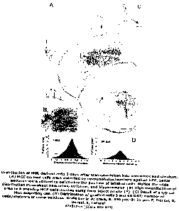
In more detail, Figure 1 shows distribution of MGE derived cells 3 days after
transplantation into neocortex and striatum. (A) MGE derived cells were
detected
by immunohistochemistry against GFP. Serial sections were utilized to
determine
the position of labelled cells. Notice the wide distribution throughout
neocortex,
striatum, and hippocampus. (B) High magnification of area in A showing MGE
cells moving away from injection site (*). (C) Detail of a typical MGE
migrating cell.
(D) Distribution of grafted cells 3 and 60 DAT; number of cells/distance of
serial
sections. Scale bar in A: 1mm; B: 250 pm; D: 25 pm. F, frontal; D, Dorsal; L,
Lateral;
Figure 2 shows acquisition and distribution of mature interneuron morphology
at
60 DAT. (A) Camera lucida maps indicating the position of MGE graft-derived
cells at three rostrocaudal levels after transplantation into neocortex (Ctx),
hippocampus (Hp), and striatum (St). (B) Detection of grafted cells by
immunohistochemistry against GFP in the ipsilateral somatosensory cortex. Note
the wide distribution of grafted cells in multiple cortical layers. Compare
the dark
background in layers l-II and V of the injected hemisphere (B) versus the
contralateral hemisphere (C). (E-K) GFP detection by immunohistochemistry
provides a Golgi-like staining of grafted cells. MGE-derived cells in cortex
differentiated into neurons presenting typical morphology of interneuron
subtypes
e.g., bitufted or bipolar cells (E), chandelier cells (F) with synaptic
boutons
resembling candlesticks (arrowheads), basket cells (H), neurons with small
body
11
CA 02645186 2008-07-21
WO 2007/084957 PCT/US2007/060715
(I), and multipolar cells (J). In hippocampus, grafted cells accumulated in
CAl (D)
and dentate gyrus (G). In striatum, the vast majority of cells differentiated
into
medium aspiny interneuron (K). Scale bars in B, C, D, F, H and I: 100 p m; E,
G, J
and K: 50 pm;
Figure 3 shows molecular characterization of MGE graft-derived cells in
somatosensory (A-F, J-O), and cingulate cortex (G-1), 60 DAT.
Immunohistochemical co-localization of grafted GFP+ cells with GABA,
Parvalbumin (PV), Calretinin (CR), Somatostatin (SOM), and Neuropeptide-Y
(NP-Y). Arrowheads show double positive cells for GFP and specific marker.
Scale bar 50 pm for A-O;
Figure 4 shows grafted MGE derived cells in the dentate gyrus of the
hippocampus 60 DAT. Immunohistochemical co-localization of MGE derived cells
expressing GFP with GABA (A-C), Parvalbumin (PV) (D-F), and Somatostatin
(SOM) (G 1). Arrowheads show double positive cells. Scale bar 100 pm for A-I;
Figure 5 shows MGE-derived cells exhibit interneuronal firing properties. (A)
IR-
DIC image overlayed with an epifluorescence image of an acute coronal slice (4
weeks post-grafting) containing GFP+ MGE-derived cells; epifluorescence image
at right of a cell filled with Alexa red during the patch recording. (B)
Membrane
potential of the GFP+ cell shown in panel A recorded under current clamp at
the
resting potential (--71 mV). Note the small degree of inward rectification
with
hyperpolarizing current steps (200 ms) the lack of spike frequency adaptation
with
long depolarizing current steps (1000 ms) typical of mature cortical
interneurons.
12
CA 02645186 2008-07-21
WO 2007/084957 PCT/US2007/060715
(C) Graph of firing frequency of recorded GFP+ cells at depolarizing step of
0.2 nA
(n = 14). Note the linear frequency-current relationship (inset graph);
Figure 6 shows recording configuration for analysis of inhibitory current in
the host
brain (A) Left panel shows a representative example of an acute coronal slice.
Box indicates region in which electrophysiological recordings were obtained.
(B)
Panel shows the acute coronal slice with GFP"' cells in Layers I-III
visualized
under IR-DIC and epifluorescence microscope. A recording was obtained from a
pyramidal neuron (asterisk) in the vicinity of GFP+ cells. (C) Panel shows a
higher
magnification of the recording site with GFP' MGE cells (green arrows) and a
Lucifer yellow filled pyramidal neuron (yellow asterisk);
Figure 7 shows MGE grafted cells alter synaptic function in the host brain.
(A)
Sample traces of si PSCs recorded from pyramidal cells (control brain and
grafted
brain); 4 weeks post-grafting. Note the increase in IPSC amplitude and
frequency
for grafted animals vs. age-matched controls. (B) Cumulative data plots for
all
IPSC recordings from control (light gray bars) and grafted (black bars)
animals are
shown. Recordings were made at 2, 3, and 4 weeks following grafting. Data
represent 7-10 cells for each bar; data presented as mean S.E.M.;
significance
taken as p < 0.05 using one-way ANOVA. (C, D) Measurement of the total charge
transfer for pyramidal cells from control and grafted brain. Note the
significant
increase for grafted brains at 4 weeks. (E) Cumulative probability plots of
sIPSCs
inter-event intervals show higher frequency values for grafted brains (p <
0.05);
13
CA 02645186 2008-07-21
WO 2007/084957 PCT/US2007/060715
Figure 8 shows synaptic inhibitory current is increased in the hippocampus
from
grafted mice. (A) Spontaneous IPSCs of hippocampal pyramidal cells from
control
grafted mice with plots of frequency and amplitude of sIPSCs of hippocampal
pyramidal cells from control (light gray bars; n= 10) and grafted mice (black
bars;
n =10). (B) Measurement of the total charge transfer of IPSCs recorded from
CAl
hippocampal pyramidal cells from control and grafted brain. Note the
significant
increase values for grafted brains at 4 weeks. (C) Cumulative probability
plots of
sIPSCs inter-event intervals shown higher frequency values for grafted brains
(p <
0.05). Error bars indicate SEM.; *p < 0.001; ** p < 0.05(ANOVA);
Figure 9 shows glutamatergic synaptic excitation is not altered in neocortex
and of
MGE grafted mice. (A) Plots of all cortical pyramidal cells sampled for
spontaneous EPSC data. sEPSC amplitude, decay-time and frequency show no
significant difference between controls (light gray bars) and grafted (black
bars)
brains (B) Representative traces of sEPSCs recorded from a GFP"' grafted cell
at
4 weeks post-grafting. sEPSCs were abolished by application of CNQX and APV
(bottom trace) (C) Sample of eEPSC recording from GFP} grafted cells at
different
holding potentials showing the reversal membrane potential at 0 mV (see inset
graph); and
Figure 10 shows cortical brain slices prepared from Dix mutant mice
transplanted
with MGE progenitor cells early in development (P0-P2) exhibit a level of
inhibition
(measured as spontaneous and miniature IPSCs on postsynaptic pyramidal cell
targets in regions containing MGE-GFP interneurons) that is comparable to that
observed in control Dix heterozygote mice.
14
CA 02645186 2008-07-21
WO 2007/084957 PCT/US2007/060715
Examale I
MGE cells were transplanted from mice expressing green fluorescent protein
(GFP) into the postnatal brain. The time course of migration and
differentiation of
these neuronal precursors was determined. Also the molecular phenotype of
transplanted MGE precursors was analysed using antibodies directed against
GABA, somatostatin (SOM) and neuropeptide Y (NPY). Using cortical slices from
grafted animals we showed that MGE-GFP neurons exhibit intrinsic firing
properties similar to fast-firing basket-type cortical interneurons.
Electrophysiofogical measurements demonstrate that MGE-derived neurons
increase the level of GABA-mediated synaptic inhibition, and therefore appear
to
modify neocortical inhibitory tone.
Materials and Methods
Tissue Dissection and Cell Dissociation. Ventricular and subventricular layers
from the anterior part of the medial ganglionic eminence, where a sulcus
clearly
divides medial and lateral ganglionic eminences, were dissected from E12.5-
E13.5 embryonic GFP+ transgenic mice (Hadjantonakis et al., (1998) Mech Dev
76, 79-90). The day when the sperm plug was detected was considered E0.5.
Bordering tissue between adjacent regions was discarded during dissection to
avoid contamination. Tissue explants were mechanically dissociated by repeated
pipetting through 200 pl yellow plastic tip (10-20 times). Dissociated cells
were
washed with 1 ml of L-15 medium containing DNase 1(10-100 Ng/ml) and pelleted
by centrifugation (2 minutes, 800 g). Cells were resuspended in 4-5 ial of L-
15
medium and kept on ice until further use.
Transplantation. Highly concentrated cell suspension (_106 cells/pl) was front-
loaded into beveled glass micropipettes (-50 pm diameter) that were pre-filled
with mineral oil and L-15 medium. Micropipettes were connected to a
microinjector mounted on a stereotactic apparatus specially adapted for
neonatal
mice. 3-4 days old CD-1 mice (Charles River) were anesthetized by exposure to -
C until pedal reflex was abolished. Anesthesia was maintained by performing
surgery on a cold aluminum plate. 5 x 10⁴ cells/mouse in a 50-100 nl volume
were injected using a 45 inclination angle and the following coordinates from
Bregma: Striatum (3.3 mm A, 2.5mm L, 2.6 mm D); Cortex (2.2 mm A, 3.5 mm L,
CA 02645186 2008-07-21
WO 2007/084957 PCT/US2007/060715
1.2 mm D); Hippocampus (1.2 mm A, 1.7 mm L, 2.0mm D). For survival and
migration distance estimations, 5 x 10³ cells were grafted in a single point
(2.5 mm
A, 3.0 mm L, 2.5-1.5 mm D). Grafted pups were returned to their mothers and
analyzed after 3 days, 1, 2, 3, 4 weeks and 3 months. All experimental animals
were treated in accordance with UCSF Laboratory Animal Research Center
guidelines.
Immunostaining. Animals were transcardially perfused with 4%
paraformaidehyde at different ages. Brains were removed, postfixed overnight
in
the same solution, and sectioned coronally (50 pm) using a Vibratome. Floating
brain sections were immunostained with the following antibodies: rabbit anti-
GABA (1:2500, Sigma), mouse anti-parvalbumin (1:4000, Sigma) and rabbit anti-
calretinin (1:4000, Swant Swiss Abs), rat anti-somatostatin (SOM) (1:500,
Chemicon), rabbit anti-neuropeptide Y (1:5000, lmmunoStar), and mouse anti-
GFP (1:200, Q-Biogene). The following secondary antibodies were used: cy3-
conjugated donkey anti-mouse, cy3-conjugated donkey anti-rabbit, cy2-
conjugated donkey anti-mouse and biotin-conjugated donkey anti-mouse (1:400,
all from Jackson lmmunoResearch, PA). Sections were washed in PBS, blocked
for 1 h in PBS containing 10% donkey serum and 0.1% Triton X-100 at room
temperature. Sections were then incubated overnight at 4 C in primary
antibodies
diluted in PBS containing 10% donkey serum and 0.1% Triton X-100, then were
washed three times in PBS and incubated with secondary antibodies for 1-2 h at
room temperature in the dark. For GABA immunostaining, Triton X-100 was
eliminated from the protocol. Biotinylated secondary antibodies and ABC kit
(Vector) were used for peroxidase reaction with diaminobenzidine (DAB).
Cell Counts and Quantification. Quantifications of cell bodies stained with
immunohistochemistry or GFP were counted on digitized images obtained with a
DFC480 digital camera and iM500/FW4000 image manager software (Leica
Microsystems Imaging Solutions, Cambridge, UK) on a DM6000B microscope
(Leica Microsystems, Wetzlar, Germany). Survival percentage of grafted cells
was estimated counting all GFP' cells in 10 coronal sections (300 pm apart, 1
section with injection site, 4 forward to the injection, and 5 backward). A
representation of cell number vs. distance to injection site was obtained on
graph
16
CA 02645186 2008-07-21
WO 2007/084957 PCT/US2007/060715
paper. Quantification of area under the graph was estimated as total number of
survived cells.
The percentage of grafted GFP+ cells expressing GABA, PV, CR, SOM or NPY
after transplantation was calculated in 3 coronal sections through each of the
following regions: somatosensory cortex, striatum and hippocampus. For
somatosensory each section was 500 pm apart, using stereotaxic coordinates
(bregma levels +0.50 and -0.50 mm; Paxinos and Franklin, 2001); striatum
sections were 400 pm apart, (bregma levels +1.60 and +0.80 mm); and for
hippocampus, sections were 300 pm apart, (bregma levels -1.50 and -2.10 mm).
At least 100 GFP+ cells (-50 in cortical layers II-iV, and -50 in layers V-VI,
visualized using DAPI) were analyzed for each marker in each animal. Brains (n
=
5) were analyzed at 1, 3 and 6 months after transplantation. Statistical
analysis
was performed using the Student's t- test.
Quantifications of neuronal bodies stained by immunohistochemistry for
interneuron markers in grafted and contralateral hemispheres were obtained as
follows: In somatosensory cortex, 5 coronal sections (400 pm apart) per mouse
between septum (bregma level +0.75 mm) and dorsal hippocampus (bregma level
-1.25 mm) were selected. A 1 mm strip of cortex from the white matter to pial
surface was analyzed in each section (1.2 mm2 each). In hippocampus, the
numbers of positive interneurons in the hilus and CAl areas were determined in
3
coronal sections (300 pm apart, between bregma levels -1.50 and -2.10 mm) per
mouse. In striatum, positive cells were counted in 3 coronal sections (400 pm
apart, between bregma levels +1.60 and +0.80 mm) per mouse. Brains from at
least 5 different grafted mice were counted and averaged. To compare results
between grafted and contralateral hemisphere statistical analysis using the
Student's t-test was applied and contralateral results were referred as 100%.
Results are presented as mean SEM. Significance level was taken as p < 0.05.
Electrophysiology. Acute tissue slices were prepared from male or female CD-1
mice 2, 3, and 4 weeks after grafted with MGE cells or saline (control) as
previous
described (Calcagnotto et al., (2002) J Neurosci 22, 7596-605). Whole-cell
recordings were obtained from visually identified neurons (pyramidal cells and
17
CA 02645186 2008-07-21
WO 2007/084957 PCT/US2007/060715
GFP+ cells) using an infrared differential interference contrast (IR-DIC)
video
microscopy system and epifluorescence microscopy (Molecular Devices).
Intracellular patch pipette solution used for whole-cell voltage-clamp
recordings to
study inhibitory postsynaptic current (IPSC) contained (in mM) 120 Cs-
gluconate,
10 HEPES, 11 EGTA, 11 CsC1z, 1 MgCIZ, 1.25 QX314, 2 Na2-ATP, 0.5 Na2-GTP,
(pH 7.25; 285-290 mOsm); for excitatory postsynaptic current (EPSC) solution
contained (in mM) 135 CsCIZ, 10 NaCI, 2 MgC1z, 10 HEPES, 10 EGTA, 2 Na2ATP,
0.2 Na2GTP, and 1.25 QX-314, adjusted to pH 7.2 with CsOH (285-290 mOsm).
To isolate GABAergic currents, slices were perfused with nACSF containing 20
M 6-ciano-7-dinitroquinoxaline-2,3-dione (CNQX) and 50 M d-(-)-2-amino-5-
phosphonovaleric acid (D-APV) and IPSCs were recorded at a holding potential
of
0 mV; for excitatory postsynaptic currents (EPSC), slices were perfused with
nACSF containing 10 M bicuculline methiodide (BMI) and recorded currents at a
holding potential of -75 mV unless otherwise noted. Miniature inhibitory
synaptic
currents (mlPSCs) were recorded in nACSF containing 1 pM tetrodotoxin (TTX).
IPSCs/EPSCs were recorded on "aged-matched" pyramidal neurons (MGE graft-
derived or sham-operated) either in the same slice or in a different one. Age-
matched refers to slices obtained from mice within a three day time period.
Evoked currents were elicited using a monopolar electrode placed in the white
matter. Pyramidal cells were filled with biocytin and analyzed post hoc. To
study
the intrinsic firing properties of GFP+ cells in current-clamp intracellular
patch
pipette solution contained (in mM) 120 KMeGluconate, 10 KCI, 1 MgC12, 0.025
CaC12, 10 HEPES, 0.2 EGTA, 2 Mg-ATP, 0.2 Na-GTP, pH 7.2, (285-290 mOsm).
Cells were depolarized and hyperpolarized, via direct current injection (5 -
1000
ms, duration); cells were filled with Alexa red and analyzed post hoc. Voltage
and
current were recorded with an Axopatch 1 D amplifier (Axon Instruments), and
monitored with an oscilloscope and with pClamp 8.2 software (Axon
Instruments),
running on a PC Pentium computer (Dell Computer Company, Round Rock, TX).
Whole-cell voltage-clamp data were low-pass filtered at 1 kHz (-3 dB, 8-pole
Bessel), digitally sampled at 10 kHz. Whole-cell access resistance was
carefully
monitored throughout the recording and cells were rejected if values changed
by
more than 25% (or exceeded 20 MS2); only recordings with stable series
resistance of <20 Mo were used for analysis (Mini Analysis 5.6.28 software;
18
CA 02645186 2008-07-21
WO 2007/084957 PCT/US2007/060715
Synaptosoft, Decatur, GA). Results are presented as the mean SEM. To
compare results between different cell types, we used a one-way ANOVA with
significance level of p < 0.05.
Results '
Embryonic MGE Cells Grafted in Juvenile Brain Rapidly Disperse Long
Distances. To establish an efficient method for the transplantation and
functional
assessment of MGE progenitors in a host brain, the MGE was dissected from
transgenic E12.5-E13.5 mice expressing green fluorescent protein (GFP)
(Hadjantonakis et al., supra) GFP expression was used to track the migration
and
differentiation of grafted cells in live or fixed tissue. After mechanical
dissociation,
GFP+ MGE cells were loaded into a glass micropipette and grafted into the
neocortex and dorsal striatum in the brain of postnatal day 3 or 4 (P3-P4)
mice
(Lois and Alvarez-Buylla, (1994) Science 264, 1145-8). Host animals were then
sacrificed at 3 days, 1, 2, 3 and 4 weeks post-grafting. Representative
examples
of the injection sites and post-migratory behaviors of GFP} cells are shown in
Figures IA and 2A.
Three days after transplantation (DAT) many GFP+ cells had migrated away from
the injection site (Fig. 1 B) into most of the neocortex, striatum and
hippocampus
(Fig. 1A). Survival rate of grafted cells at this time point was 38.9 7.3%
(n =10).
At 3 DAT most GFP+ cells had the typical morphology of tangentially migrating
interneurons, with a small-elongated cell soma and a forked leading process
(Fig.
1C). GFP+ cells spread extensively around the injection site in all
directions.
Grafted cells covered a linear distance of 336 82 pm/day (n = 20), with a
maximum of 525 pm/day, analyzed 3 DAT; this speed of migration is greater than
reported in adults (-120 pm/day) and similar to that measured in vitro (280
pm/day on matrigel) (Wichterie et al., (1999) Nat Neurosci 2, 461-6)_ A
representation of cell number versus migration distances at 3 DAT results in a
bell-shape curve (Fig. 1 D). These data suggest that cells did not have a
strong
preference for a particular migratory route and disperse in all directions
from the
injection site.
19
CA 02645186 2008-07-21
WO 2007/084957 PCT/US2007/060715
Differentiation of Grafted MGE Cells in the Host Brain. Analysis of grafted
brains 7 DAT revealed a widespread distribution of GFP+ MGE cells. At 7 DAT,
most grafted cells no longer exhibited a migratory morphology; instead they
had
multiple processes and some cells had a thin and longer axon-like process
(data
not shown). This indicates that initiation of differentiation of grafted MGE-
derived
neuronal precursors occurs between three and seven days after transplantation.
Fourteen and 21 DAT, cells acquired progressively a more mature morphology,
showing larger and more elaborated dendritic trees with longer axons. At 30
DAT,
some GFP+ cells were more than 5 mm away from injection site; their
distribution
was similar to that found at 3 DAT (Figs. IC & 2A). However, the survival
percentage was reduced to 19.9 3.9% (n = 10). A similar level of survival,
21.2
4.1 %(n = 10), was observed at 90 DAT. The morphology of the grafted cells
was studied following GFP immunohistochemistry, which provides Golgi-like
staining. Two months after transplantation, GFP+ cells had elaborate dendritic
trees extending profusely through cortical layers (Fig. 2). Axons and their
presynaptic terminals could also be visualized (Figs. 2B-C). Thus grafted
cells
appeared to complete their differentiation into functionally integrated
interneurons
within one month after transplantation.
MGE-derived cells in the cortex differentiated into neurons with morphologies
of at
least five different interneuron subtypes e.g., bitufted or bipolar cells,
chandelier
cells, basket cells, neurons with small body, and multipolar cells (Fig. 2).
For
instance, some neurons displayed synaptic buttons resembling arrays of
candlesticks, suggesting that they differentiated into chandelier cells (Figs.
2B, E,
F, H, I, J). In contrast, grafted cells in the striatum differentiate
primarily to medium
aspiny interneurons (Fig. 2K), and in the hippocampus to interneurons with
morphologies typical for this region (basket, axo-axonic, and bistratified
cells)
(Figs. 2D & G). None of the MGE-derived neurons exhibited morphological
features of cortical pyramidal neurons e.g., triangular cell soma extending a
thick
spiny apical dendrite. Some immature oligodendrocytes were always noted
around the injection site; especially close to the corpus callosum, and
occasionally
in the cortex where they were radially aligned (data not shown). GFPi' cells
with
CA 02645186 2008-07-21
WO 2007/084957 PCT/US2007/060715
an astrocytic morphology were not observed. Therefore, the MGE cells that we
grafted are primarily committed to an interneuronal lineage.
MGE-derived Cells Exhibit Molecular Properties of Cortical Interneurons.
Recent studies suggest that MGE progenitors are the principal source of
cortical
GABAergic interneurons (Lavdas et al., (1999) J Neurosci 19, 7881-8; Sussel et
al., (1999) Development 126, 3359-70; Anderson et al., (2001) Development 128,
353-63; Wichterle et al., (2001) Development 128, 3759-71). Interneurons can
be
classified into several subtypes based on neurochemical markers, such as Ca2+-
binding proteins (parvalbumin (PV), calbindin (CB), and calretinin (CR)),
neuropeptides (e.g., somatostatin (SOM), neuropeptide Y (NPY), cholecystokinin
(CCK), and vasoactive intestinal polypeptide (VIP)) (DeFelipe, (1993) Cereb
Cortex 3, 273-89; Kubota et al., (1994) Brain Res 649, 159-73; DeFelipe,
(1997) J
Chem Neuroanat 14, 1-19; Gonchar and Burkhalter, (1997) Cereb Cortex 7, 347-
58; DeFelipe, (2002) Prog Brain Res 136, 215-38), and recording their
physiological properties (Freund and Buzsaki, (1996) Hippocampus 6, 347-470;
Cauli et al., (1997) J Neurosci 17, 3894-906; Gupta et al., (2000) Science
287,
273-8; Klausberger et al., (2003) Nature 421, 844-8). To evaluate the
interneuronal phenotype and molecular characteristics of transplanted MGE-GFP
cells, we performed a series of immunohistochemical studies 60 DAT. Double-
immunofluorescence revealed that approximately 65-70% of cortical GFP' graft-
derived cells express GABA (Fig. 3; Table 1); a comparable level of GFP' cells
were double-labeled with an antibody against GAD67 (-70%; data not shown).
Subsets of the GFP{ neurons express NPY, SOM, PV, and CR (Fig. 3; Table 1),
at expression levels and in a distribution similar to those of the host
interneurons.
Interestingly, SOM-expressing neurons were enriched in layers I-II of the
cortex,
whereas CR positive cells were almost exclusively found in retrosplenial and
cingulate cortex. This suggests that local environment contributes to the
specification of some interneuron sub-types.
MGE-derived cells were also immunopositive for these neurotransmitters and
markers in the striatum and hippocampus (Fig. 4, Table 1). They were
distributed
in the same areas that usually contain these types of interneurons. GFP+ cells
were immuno-negative for antibodies to glial fibrillary acidic protein (GFAP),
or
21
CA 02645186 2008-07-21
WO 2007/084957 PCT/US2007/060715
choline acetyl transferase (ChAT), indicating that grafted cells did not
differentiate
into astrocytes or cholinergic neurons.
MGE-derived Cells Exhibit Interneuronal Firing Properties. To assess
whether the MGE-derived cells had electrophysiological characteristics of
cortical
interneurons, GFP* cells were targeted for whole-cell current-clamp recording
at 4
weeks post grafting. Diffusion of Alexa Red from the patch pipette permitted
real-
time confirmation of cellular recording site (Fig. 5A). If MGE cells mature
into an
interneuronal phenotype they should exhibit little spike frequency adaptation,
which is a hallmark electrical feature of GABAergic interneurons. In current-
clamp
recordings from fifteen GFP+ cells sampled in cortical layer V, we measured
mean
values of -70.9 0.9 mV for resting membrane potential (RMP) and 101.4 4.1
Ms2 for input resistance (RIN). In fourteen GFP+ cells, depolarizing current
pulses
elicited action potentials (3.0 0.4 ms duration; 69.0 3.3 mV amplitude)
and
hyperpolarizing current pulses evoked a small degree of "sag" current (Fig.
5B).
These intrinsic membrane properties are in the expected range for "mature" non-
accommodating cortical interneurons (Markram et al., (2004) Nat Rev Neurosci
5,
793-807). Most importantly, long duration depolarizing pulses (1000 ms)
clearly
revealed the fast-spiking, little adapting firing activity characteristic of
basket-cell
cortical interneurons. One cell did not exhibit active firing properties
during step
depolarisations, but had a RMP of -70 mV and RIN of 100 MS2. The high firing
frequency typical of GFP} cells sampled is shown in Figure 5B; frequency-
current
relationships were linear as previously reported for fast-spiking hippocampal
interneurons (Fig. 5C) (Smith et al., (1995) J Neurophysiol. 74, 650-72).
Transplanted MGE cells Influence Synaptic Function in the Host Animal. To
determine whether transplanted MGE precursors functionally integrate in the
host
brain, a series of in vitro electrophysiological studies were performed.
Regions of
neocortex containing GFP' cells were identified under epifluorescence (Fig. 6)
and pyramidal neurons in regions surrounded by GFP"' cells were chosen for
patch-clamp recording. Recorded cells were filled with Lucifer yellow for post
hoc
confirmation of cell location and identity (Fig. 6A). Brain slices were
prepared at
various time-points following transplantation (2, 3 and 4 weeks). Spontaneous
IPSCs on pyramidal neurons (Fig. 7A) reflect activation of postsynaptic GABA
22
CA 02645186 2008-07-21
WO 2007/084957 PCT/US2007/060715
receptors following action potential-dependent vesicular transmitter release;
IPSCs were completely abolished by 10 pM BMI a GABAA receptor antagonist
(data not shown). If a significant number of transplanted MGE cells integrate
into
the host micro-circuitry as new GABAergic interneurons, we would expect an
increase in the overall level of GABA-evoked synaptic events onto native
pyramidal neurons. Increments in GABA-, PV- and SOM- expressing neurons
were observed in the cortical hemisphere ipsilateral to the injection site
when
compared to contralateral hemisphere (Table 2). These increments were
significant in a 100 pm area around the graft. In concordance with these
anatomical observations, there were significant increases in IPSC amplitude
and
frequency in slices from transplanted animals 4 weeks following surgery.
Control
cortical slices were obtained from sham-operated mice or from the
contralateral
cortex of transplanted mice (which lacked GFP+ cells) (Figs. 7B-C). IPSC
frequency and amplitude were also increased in the hippocampus of grafted
animals at 4 weeks post-transplantation (Fig. 8). Consistent with an increase
in
the number of GABA-producing neurons, mIPSC frequencies were also increased
in neocortical and hippocampal pyramidal cells 4 weeks after transplantation
(cortex: 2.3 + 0.1 Hz n= 4; CAl: 2.4 + 0.2 Hz, n = 3) when compared with
controls
(cortex: 1.3 + 0.2 Hz, n = 4; CA1: 1.1 0.1 Hz, n = 3; p < 0.05). A
significant
enhancement of GABAergic inhibition was not observed at 2 or 3 weeks following
transplantation; not surprisingly as histological analysis at these times
showed an
immature phenotype of grafted cells. Significant changes in IPSC rise time or
decay-time constant were not observed at any time-point (Fig. 7B) suggesting
that
gross alterations in postsynaptic GABA subunit receptor expression do not
occur
in grafted animals.
To assess the overall level of inhibitory tone in grafted animals, we
performed two
additional analyses. First, measurement of the total charge transfer
(corresponding to total area under the IPSC current over a specified time
period)
indicated that synaptic inhibition was significantly increased in slices
containing
GFP+ cells compared to age-matched controls (Figs. 7C-D). Second, consistent
with an enhancement of GABAergic tone, there was a significant increase in the
frequency of sIPSCs plotted as a cumulative distribution (Fig. 7E).
23
CA 02645186 2008-07-21
WO 2007/084957 PCT/US2007/060715
To test whether the transplanted MGE cells synapse onto existing interneurons,
and thereby modify cortical excitation (through inhibition of interneuron
function),
EPSCs were analyzed. EPSCs recorded from pyramidal neurons (holding
potential of - 75 mV) in regions containing GFPi' cells; spontaneous EPSCs
were
abolished by application of CNQX and APV confirming a role for postsynaptic
glutamate receptors. In comparing spontaneous EPSCs recorded on pyramidal
cells from MGE transplanted animals (n = 4) and controls (n = 4) no difference
in
amplitude, decay-time constant, rise-time or frequency was noted (Fig. 9A).
These findings suggest that overall excitatory tone in the host brain is not
altered
following grafting of MGE precursors. To address whether transplanted neurons
receive excitatory synaptic contact from host axons, we next examined evoked
and spontaneous EPSCs in GFP' neurons. GFP} cells exhibited spontaneous
EPSCs that were blocked by CNQX and APV (n = 4) (Fig. 9B) and evoked EPSCs
with a reversal potential near 0 mV (Fig. 9C). EPSGs exhibited kinetics
similar to
those expected for "normal" glutamate-mediated synaptic currents. These
results
confirm an endogenous excitatory excitation of grafted MGE-GFP neurons. Taken
together, these data suggest that MGE-derived GFP+ cells function as
inhibitory
interneurons receiving excitatory input from local pyramidal neurons and
integrating into cortical synaptic circuitry of the host brain in such a
manner as to
selectively modify inhibition.
The example demonstrates that MGE-derived neuronal precursors grafted into the
early postnatal brain are capable of long distance dispersion across the
neocortex
and other areas of the juvenile brain. These cells then acquire morphological,
molecular and physiological characteristics of mature GABAergic interneurons.
Finally, these grafted MGE-derived cells functionally integrate and
significantly
impact synaptic inhibition in the host brain. Thus, the present example
demonstrates that MGE precursors could be used to modify synaptic circuits in
a
postnatal brain. An ability of these cells to disperse when transplanted into
the
neonatal brain is demonstrated, reaching maximum migration distances of 5 mm
two months after transplantation. As such, a single injection of MGE
precursors
could influence a relatively wide area of the host brain, an important aspect
when
considering the potential clinical usefulness of transplanted cells. The
present
results show that more than 65% of MGE-derived cells express GABA. Grafted
24
CA 02645186 2008-07-21
WO 2007/084957 PCT/US2007/060715
cells also contain SOM and NPY, neuropeptides normally co-localized in
subtypes
of mature cortical interneurons (DeFelipe, (1993) supra; Kubota et aL, supra;
DeFelipe, (1997) supra; Gonchar and Burkhalter, supra). We did not detect
pyramidal-likel neurons or astrocytes that were derived from transplanted MGE
cells. Importantly, tumors were never observed in our MGE grafted mice
although
this is a common problem when ES-derived progenitors are used for
transplantation (Wernig et al., supra; Ruschenschmidt et al., supra). MGE-
derived
cells sampled in layer V exhibit an "electrical fingerprint" typical of mature
GABA-
containing interneurons. For example, MGE-GFP cells consistently fired at a
high
frequency and exhibited very little accommodation. These firing properties are
consistent with a classification as non-accommodating basket-cell interneurons
and it is likely that further current-clamp sampling of GFP' cells across
other
layers of grafted cortex will uncover additional interneuron sub-types. In
previous
analysis of functional integration, single-cell recordings focused exclusively
on
demonstrations that transplanted cells receive synaptic input. Here we also
demonstrate that transplanted MGE-derived cells receive excitatory synaptic
input
(see Fig. 6). Moreover, we present evidence that grafted progenitor cells send
inhibitory outputs, which impact (in a functionally relevant manner) existing
pyramidal neurons. Notably, we found that pyramidal cells in regions
containing
MGE-derived cells exhibit an increased number of GABA-mediated synaptic
events and that GABAergic tone is significantly enhanced in these regions of
the
host brain. Because MGE-derived cells did not alter excitatory cortical
circuitry or
differentiate to neurons with a pyramidal-cell phenotype, these findings
suggest a
method for selective enhancement of inhibitory systems.
Our demonstration that grafted progenitor cells produce functionally
integrated
GABAergic neurons, even in the presence of endogenous GABAergic neurons,
after embryonic stages of neurodevelopment are complete, and in a wide variety
of brain regions, suggests that MGE-derived cells could be useful in
neurological
conditions where increased inhibition would be beneficial e.g., epilepsy or
schizophrenia. MGE precursors may also be used to correct levels of activity
in
deafferented brain regions such as in Parkinson's disease, or in conjunction
with
their inhibitory function, may be used as cellular vectors to deliver
therapeutic
molecules to wide regions of the brain.
CA 02645186 2008-07-21
WO 2007/084957 PCT/US2007/060715
Table 1
MGE graft derived interneuron subtypes (n = 5)
GABA PV SOM CR NPY
CORTEX 68.6 t 4.8qo 38.3 t 5.49"0 43.2 ;t 3.9% 1.9 :k 0.6% 7.8 t 1.29'a
53.1 5.3% a 10.3=1.3% e
33.2 2.4. b
STRIATUM 50.9 2.6% 54.9 t 7.6% 39.5 4.630 6.4 t 1.9% 18.0 t 2.190
HiPPO(DG) 42.8 2.9% 33.7t4.79. 33.8 8.19'0 10.3 1.79"0 13.1=1.93e
Quantifiications were performed in somatosensory cortex except for a) Layers
I-111 of somatosensory cortex, b) Layers IV-Vi of somatosensory cortex, and c)
Retrospienial cortex. DG; Dentate Gyrus
Table 2
Interneuron increment in transplanted somatosensory cortex
GC?RTEXy CORTEX2
(100 Nm) (1200 Nm)
GABA 12.1= 3.7% (P< 0.01) 6.4 3.5 (P< 0.01)
Pv 9.8 2.19'o(P<0.01) 4.8 3.69'o(P=0.23)
S M (1-111) 16.1 _ 2.8 l0 (P < 0.01) 12.9'=- 3.6% (P < 0.05)
Contralat3ral results were taken as 100%. 1) Estimation of cell increment
100Nm around of injection site.
Quantification was performed in 2 slices SOpm forward and backward from
injection site. 2) E.siimation of
cell increment 1200pm around of Injection site. Quantification was performed
in 3 slices forward plus 3
slices backward from injection site. Significance (p) was estimated w~th a T-
student test. N = 10.
EXAMPLE 2: Robust epileptiform burst activity is more difficult to initiate in
slices containing MGE progenitors
Attempts are made to elicit epileptiform burst activity in cortical slices
having
received MGE progenitor cell grafts and control cortical slices that have not
received MGE progenitors. It is determined to be more difficult to initiate
robust
epileptiform burst activity in slices containing MGE progenitors. This finding
26
CA 02645186 2008-07-21
WO 2007/084957 PCT/US2007/060715
supports that MGE progenitors migrate and differentiate into functional
interneurons in the host brain (and thus increase synaptic inhibition).
Neocortical slices are prepared from wild-type mice with MGE grafts and age-
matched controls. Spontaneous seizure activity is initiated in neocortical
slices by
raising the extracellular level of potassium, in a step-wise fashion, from 3
to 6 to 9
mM [Ki']e. Previous studies in our laboratory (Baraban and Schwartzkroin,
Epilepsy Res. 1995 Oct;22(2):145-56) and others (Rutecki et al., J
Neurophysiol.
1985 Nov;54(5):1363-74; Traynelis and Dingledine, J Neurophysiol. 1988
Jan;59(1):259-76), demonstrate this is an efficient method to induce
spontaneous
seizure activity and test anticonvulsant drugs in vitro. The "high K" model
reliably
elicits status-like interictal-like epileptiform activity and is designed to
mimic high
[K+]e observed during clinical seizures. Epileptiform activity is monitored
using
field recording electrodes placed in outer (Layers IV/V) and inner (Layer II)
neocortex. Epileptiform burst discharge amplitude (in mV), duration (in msec)
and
frequency (in Hz) is used to quantitatively compare bursting between
experimental
and control animals. A second method to compare interictal "burst intensity"
in
different [K+]e involves the use of a coastline bursting index (CBI) (Korn et
al., J
Neurophysiol. 1987 Jan;57(1):325-40). CBI is responsive to changes in the
number or amplitude of bursts, and it increases when neuronal synchrony,
firing
frequency or duration changes - thus, it can be considered a sensitive measure
of
whether integrated MGE progenitors influence seizure activity.
A separate series of identical experiments is performed using the zero-Mg2+
acute
seizure model. Removal of Mg2+ from the extracellular bathing medium releases
magnesium blockade of NMDA-type glutamate receptors and initiates epileptiform
activity driven by excess synaptic excitation (Mody et al., J Neurophysiol.
1987
Mar;57(3):869-88). Epileptiform activity elicited in slices from grafted mice
is
compared with age-matched controls. Analysis is performed as described above.
Slices are postfixed and immunostained with an antibody to GFP so the number
of
grafted MGE-GFP+ cells can be assessed. Using these two different mechanisms
of action we reliably determine transplanted progenitors exert anticonvulsant
action in vitro.
27
CA 02645186 2008-07-21
WO 2007/084957 PCT/US2007/060715
Results: It is determined to be more difficult to initiate robust epileptiform
burst
activity in slices containing MGE progenitors. This finding supports that MGE
progenitors migrate and differentiate into functional interneurons in the host
brain
(and thus increase synaptic inhibition). A decrease in burst amplitude,
duration or
frequency or a change in CBI index provides quantitative evidence that
integrated
MGE progenitors, by increasing inhibition, reduce epileptic hyperexcitability.
EXAMPLE 3: Seizures are more difficult to initiate in mice receiving MGE
progenitors. Following bilateral MGE grafting in wild-type mice (and sham
operated controls; young adult P30 and adult P60 ages) EEG electrodes are
implanted bilaterally in neocortex and animals monitored with video-EEG. After
a
1 wk recovery period, following surgery, animals are injected with kainic acid
(KA,
a glutamate receptor agonist) at a concentration previously shown to elicit
status
epilepticus in the mouse e.g., 30-40 mg/kg i.p. (Baraban et al., Brain Res Dev
Brain Res. 1997 Sep 20;102(2):189-96; Baraban et al., J Neurosci. 1997 Dec
1;17(23):8927-36). In analyzing video-EEG traces following initiation of a KA-
induced seizure, the frequency and duration of electrographic seizure events
recorded are quantified. Behaviors that accompany these discharges are fully
characterized by close examination of the video-EEG recordings using an
investigator blind to the status of the animal. Clinician-scientists in the
laboratory
with significant clinical EEG experience assist in analysis of this data.
A second set of identical experiments are performed using pentylenetetrazole
(a
GABA antagonist, 15-20 mg/kg i.p.). Similar to slice electrophysiology
studies,
two separate means of seizure induction are used to adequately assess the
ability
of MGE progenitors to decrease/inhibit seizure activity.
In all animals, euthanasia and transcardial perfusion are performed at the
conclusion of video-EEG experiments. Brains are rapidly removed and fixed in
paraformaldehyde for post hoc confirmation of EEG electrode placement. In
addition, brains are sectioned and stained for analysis of GFP' interneurons.
These anatomical studies allow us to correlate numbers of integrated GFP
progenitors with antiepileptic activity.
28
CA 02645186 2008-07-21
WO 2007/084957 PCT/US2007/060715
Results: It is more difficult to initiate seizures in mice receiving MGE
progenitors.
Electrographic seizure events, if observed, are brief in transplanted animals
and
little or no signs of convulsive behavior are observed. Animals with large
numbers
of integrated MGE progenitors are most resistant to the development of acute
seizure activity.
EXAMPLE 4: MGE progenitors reduce seizure activity in mouse models of
spontaneous epilepsy.
Transplanted MGE progenitor cells are used to enhance synaptic inhibition such
that seizure susceptibility is significantly reduced in the host animal.
Studies are
performed in neocortical tissue sections from wild-type control mice
(following
grafting) and mouse mutants with known cortical interneuron defects. Three
mutants with a demonstrated reduction in synaptic inhibition and
hyperexcitabiiity
are used: particularly, DIx1-/-, GAD65"1" and uPAR`'-. Dlx1 mice show
generalized
electrographic seizures and histological evidence of seizure-induced
reorganization and hence display a phenotype comparable to that of human
epilepsy associated with interneuron loss. GAD65 mutants appear to have normal
numbers of GABAergic cortical interneurons, but a reduced capacity to
synthesize
GABA (Kash et al., Proc Natl Acad Sci U S A. 1997 Dec 9;94(25):14060-5).
uPAR mutants appear to have a reduced density of GABAergic interneurons in
parietal cortex (Powell et al., J Neurosci. 2003 Jan 15;23(2):622-31).
(i) GAD654- and uPAR-/- Mice
Hyperexcitable states have been reported in mutants with abnormal cortical
interneurons (GAD65 KO) and in mutants with reduced numbers of cortical
interneurons (uPAR KO). First, disruption of the GAD65 gene in mice leads to a
50% decrease in cofactor-inducible GAD enzymatic activity (Kash et al.,
supra).
GAD65-deficient mice on a C57BI/6 background are susceptible to infrequent
spontaneous seizures and stress-induced seizures. Second, inactivation of the
urokinase plasminogen activator receptor (uPAR) gene in mice leads to a 50-65%
reduction in cortical GABAergic interneurons (Powell et al., supra). uPAR KO
mice (bred on a C57BI/6 background) are viable, survive into adulthood, and
exhibit overt tonic-clonic seizures or an increased susceptibility to PTZ-
induced
motor convulsions. Both strains of mutant mice are used. Because background
29
CA 02645186 2008-07-21
WO 2007/084957 PCT/US2007/060715
strain can be an important modulator of seizure susceptibility (Schauwecker
and
Steward, Proc Natl Acad Sci U S A. 1997 Apr 15;94(8):4103-8; Schauwecker,
Prog Brain Res. 2002;135:139-48), we are careful to study mutant and wild-type
mice bred on only one background strain e.g., the relatively seizure-resistant
C57BI/6.
Following bilateral MGE grafting in GAD65-/- or uPAR-~ mice, and sham
operated,
strain- and age-matched controls, EEG electrodes are implanted bilaterally in
neocortex and monitored with video-EEG. After a 1 wk recovery period,
following
surgery, animals are monitored each day for 6 hr recording sessions (2 wk
monitoring period). In analyzing video-EEG traces the frequency and duration
of
electrographic seizures recorded are quantified. Behaviors that accompany
these
discharges are fully characterized by close examination of video-EEG
recordings
using an investigator blind to the status of the animal; clinician-scientists
in the
laboratory assist in these studies. The frequency and amplitude of interictal
spikes may vary during sleep-wake cycles (Martins da Silva et al.
Electroencephalogr Clin Neurophysiol. 1984 Jul;58(1):1-13). As such,
interictal
spikes are always analyzed during periods of non-REM sleep. Because mutant
mice can exhibit spontaneous seizure activity (consisting of frequent abnormal
slow waves and interictal discharges with associated convulsive behaviors) it
is
not necessary to induce seizures using kainate or PTZ.
We sacrifice these animals and quantify the number of new GABAergic GFP+
interneurons present in neocortex. We correlate the number of GFP+ cells with
seizure severity (as determined from analysis of behavior and EEG). Detailed
immunocytochemical studies using antibodies to GAD, NPY, parvalbumin,
somatostatin and calbindin are performed. A limited number of slice
electrophysiology studies are also performed to analyze sIPSCs in un-treated
and
grafted animals.
Results: The reduction in functional GABAergic interneurons resulting in a
spontaneous epileptic phenotype observed in uPAR KO mice is alleviated by
grafting MGE progenitors into these animals. Interictal spikes and behavioral
CA 02645186 2008-07-21
WO 2007/084957 PCT/US2007/060715
seizures are reduced (or eliminated) in uPAR KO mice receiving MGE grafts.
Similar results are observed in GAD65 mutant mice.
(ii) DIx-/- Mice
MGE cells were transplanted into the brains of Dix1-1- mice, a murine model of
epilepsy, in a similar manner as described in example 1. For details on Dlx1-/-
mice, see Cobos et al., Nature Neuroscience, 8:1059-1068, 2005, expressly
incorporated herein in its entirety by reference. DIx1 mice show generalized
electrographic seizures and histological evidence of seizure-induced
reorganization and hence display a phenotype comparable to that of human
epilepsy associated with interneuron loss. DIx1 mutant mice transplanted with
MGE progenitor cells appeared to have a reduced epilepsy phenotype, measured
as a reduction in seizure-like behavior upon handling and a lack of EEG-like
seizure activity. Cortical brain slices prepared from Dlx mutant mice
transplanted
with MGE progenitor cells early in development (P0-P2) exhibited a level of
inhibition (measured as spontaneous and miniature 1PSCs on postsynaptic
pyramidal cell targets in regions containing MGE-GFP interneurons) that was
comparable to that observed in control Dlx heterozygote mice. Specifically,
Dix
mutants normally showed reduced IPSC frequency and amplitude and these
values were "rescued" by MGE transplantation. See Figure 10. At the whole
animal level, Dlx mutants normally exhibited handling induced seizures and
spontaneous seizures. Dlx mutants transplanted with MGE cells did not exhibit
handling induced seizures and video-EEG recording confirmed the lack of a
seizure phenotype. This demonstrated that MGE cells can be successfully
transplanted into the diseased brain and demonstrated reduction or ablation of
epileptic symptoms following transplantation.
EXAMPLE 5: MGE precursors increase seizure latencies and reduce mortality in
a rodent seizure model
A commonly used rodent seizure model (e.g., pilocarpine) was used to
investigate
the therapeutic potential of MGE-derived interneurons. MGE cells from e13.5
GFP-expressing mice were transplanted into the postnatal (p4) brain using
procedures described above. After allowing for migration and integration to
occur,
31
CA 02645186 2008-07-21
WO 2007/084957 PCT/US2007/060715
single doses of scopolamine followed by pilocarpine (300 mg/kg) were
administered to induce acute seizure activity. Mortality and seizure latency
were
compared among sham-transplanted mice, MGE cell-transplanted recipients, and
mice pretreated with phenobarbital (PB), a conventional AED (antiepileptic
drug).
Seizure behaviors were scored on a Racine scale by an investigator blind to
the
status of the animal. It was observed that the transplanted mice and PB-
pretreated mice had longer seizure latencies and lower mortality rates
compared
to sham-transpianted littermates. In grafted mice, seizure protection
correlated
with the number of newly generated MGE-GFP cells.
lmmunohistochemistry and electrophysiology were then carried out as described
herein to confirm whether the therapeutic benefit observed in the transplanted
mice was due to the inhibitory activity of MGE-derived interneurons. The
immunohistochemistry revealed that MGE-derived transplanted cells in the
neocortex and hippocampus were mostly neuronal (NeuN+) and GABAergic, as
expected. Whole-cell electrophysiological recordings of presynaptic GFP+ cells
and postsynaptic pyramidal cells confirmed that transplanted cells were able
to
functionally integrate and increase synaptic inhibition, as well as receive
excitatory
inputs from endogenous pyramidal cells.
These results indicate that MGE-derived precursor cells are able to migrate
large
distances and functionally integrate into existing cortical circuitry, thereby
reducing
the harmful effects of induced seizures in transplanted mice. These in vivo
data
provide a strong indication that MGE-derived precursor cells will have
therapeutic
value in seizure disorders, and other disorders of inhibition, including
epilepsy and
other disorders described herein.
All references cited are expressly incorporated herein in their entirety by
reference.
32