Note : Les descriptions sont présentées dans la langue officielle dans laquelle elles ont été soumises.
CA 02646721 2008-09-19
2F07055-PCT 1
DESCRIPTION
BLOOD INSPECTION DEVICE
Technical Field
[0001] The present invention relates to a blood test
apparatus for examining, for example, blood component.
Background Art
[0002] Diabetespatientsneedtomeasurethebloodsugar
level regularly and administer insulin based on the blood
sugar level to maintain a normal blood sugar level. To
maintainthisnormalbloodsugarlevel,diabetespatients
need to measure the blood sugar level regularly, sample
a small amount of blood from fingertips using a blood
test apparatus, and measure the blood sugar level from
this sampled blood.
[0003] The conventional blood test apparatus generally
uses a needle as a means for puncturing skin (see Patent
Documentl,forexample). As shown in FIG. 1, conventional
blood test apparatus 1 which uses a needle as a puncturing
means, includes: housing 2 that forms a chassis;
cylindrical body 3 having opening at one side included
in housing 2; plunger 4 that moves back and forth inside
cylindrical body 3; handle 5, one end of which is connected
to plunger 4; latch part 6 that latches handle 5 at housing
2; spring 7 that urges handle 5 toward opening part 3a
CA 02646721 2008-09-19
2F07055-PCT 2
of cylindrical body 3; lancet 9 which has one end held
by plunger 4 and the other end attached with blood
collection needle (hereinafter "needle") 8; holdingpart
11 that holds blood sensor 10 on the side of opening part
3a; and electrical circuit section 12 to which the output
of sensor 10 is connected.
[0004] Inordertoexaminebloodusingconventionalblood
test apparatus 1, the following preparation woz:ks are
necessary. Blood sensor 10 and needle 8 are replaced to
eliminate the influence of blood which has already been
examined. In order to remove blood sensor 10 after use
and attach new blood sensor 10, holding part 11 is removed
and then sensor 10 after use is removed. Next, new blood
sensor 10 is attached to holding part 11. Then, holding
part 11 is attached to opening part 3a again. If the
neighborhoodof holdingpart 11 is stainedwith, forexample,
blood, it is cleaned.
[0005] These preparation works are troublesome for
diabetespatientswithpooreyesight. Inadditio.n,these
works must be performed several times a day and are
burdensome.
[ 0006] After these preparation works are done, blood te st
apparatus 1 is abutted on the skin of the patient, and
the latching of latch part 6 is released. Then, handle
5, urged by spring 7, is propelled in the direction of
arrow 14. By this release of latching of handle 5, needle
8, connected to this handle 5 via plunger 4 and lancet
CA 02646721 2008-09-19
2F07055-PCT 3
9, is propelled at the same time. Needle 8 breaks through
sensor 10 and punctures skin 13.
[000"7] A small amount of blood flows out from punctured
skin 1 3 . The outflowing blood is guided into inside blood
sensor 10. The blood guided into blood sensor 10 causes
chemical change in sensor 10 according to the blood sugar
levelofthepatient. Thecurrentproducedbythechemical
change is led to electrical circuit section 12, and the
bloodsugarlevelismeasured. Thecalculatedbloodsugar
level is displayed on display section 15. Based on the
calculated blood sugar level, for example, basic data
showing the amount of insulin to administer to the patient
is provided.
[0008 ] On the other hand, an apparatus for samplirig blood
using laser light forthepuncturingmeans, isalsoproposed
(see Patent Documents 2 and 3). Use of laser light provides
an advantage of making unnecessary replacement of:needle
and possibly alleviating the pain of the patient upon
puncturing.
Patent Document 1: Japanese Patent Application
Publication No.2003-524496
Patent Document 2: Japanese Patent Application
Publication No.2004-533866
Patent Document 3: JapanesePatentApplicationLa.id-Open
No.2004-195245
CA 02646721 2008-09-19
2F07055-PCT 4
Disclosure of Invention
Problems to be Solved by the Invention
[ 000 9] A laser apparatus consumes a large amount of power,
and, for example, when a battery is used for the power
supply, the battery capacity may be used up after several
times of puncturing. The battery for the power supply,
which is used up, no longer enables puncturing, much less
the operation of the measuring circuit for examining blood.
The blood test apparatus is an apparatus that influences
life of the patient (for example, diabetes patient) who
is the user, and should be designed so that, even if the
puncturing means does not operate, at least blood test
(for example, measurement of blood sugar level) can be
performed, because puncturing can be performed using a
means other than laser light.
[ 0 0 1 0 ] It is therefore an obj ect of the present invention
to provide a blood test apparatus that makes unnecessary
replacement of a blood collection needle and uses laser
light as the puncturing means, and that can perform blood
test at least, even if laser puncturing is not possible
due to shortage of the battery capacity.
Means for Solving the Problem
[0011] The f.irst aspect of the present invention relates
to the following blood test apparatus.
[1] The blood test apparatus includes : an apparatus body;
a blood sensor that is attached to the apparatus body
CA 02646721 2008-09-19
2F07055-PCT 5
and that analyzes blood; a puncturing section that is
provided inside the apparatus body and that punctures
skinof apatient throughthebloodsensor; andanelectrical
circuit section that is connected to the blood sensor,
and in the blood test apparatus, the puncturing section
includes a laser emitting apparatus; and the blood test
apparatus includes a power supply control circuit that
controls a supply of power from a power supply section
includingapowersupplyfordrivingtheelectricalcircuit
section and a power supply for driving the laser emitting
apparatus.
[2] In the blood test apparatus according to [1], the
power supply section comprises one or more battery power
supplies.
[3] The blood test apparatus according to [2], further
includes: a battery level measuring circuit that measures
a battery level of at least one of the one or more batteries;
and a comparing section that compares a value of them.easured
battery level and a predetermined value of a battery level,
and in the blood test apparatus, the power supply control
circuit controls the supply of power based on an output
result of the comparing section.
[4] The blood test apparatus according to [2], further
includes: a battery voltage measuring circuit that
measures a battery voltage of at least one of the one
or more batteries; and a comparing section that compares
a value of the measured battery voltage and a predetermined
CA 02646721 2008-09-19
2F0~055-PCT 6
value of a battery voltage, and in the blood test apparatus,
the power supply control circuit controls the supply of
power based on an output result of the comparing section.
[0012] The blood test apparatus of [ 3 ] and [ 4] may further
include a display section that displays a test result
of the blood test.
[0013] Thesecondaspectofthepresentinventionrelates
to the following method of controlling the blood test
apparatus.
[5] A control method for controlling the blood test
apparatus according to [3], comprising the steps of: a
pre-operation battery level measuring step of measuring
a battery level of the battery after a power supply of
the apparatus body is started; a post-operation battery
level measuring step of measuring a battery level of the
battery after the pre-operation battery level measuring
step is performed and a laser light is emitted from the
laser emitting apparatus; and a step of measuring battery
consumption level from a difference between the battery
level measured in the pre-operation battery level
measuring step and the battery level measured in the
post-operation battery level measuring step.
[6] A control method for controlling the blood test
apparatus according to [4], comprising the steps of: a
battery level measuring step of ineasuringabatterylevel
of the battery; a step of comparing the measured battery
level and a battery threshold which is a predetermined
CA 02646721 2008-09-19
2F07055-PCT 7
battery level; and a display step of displaying a message
to encourage a change of the battery on the display section
when the measured battery level is equal to or less than
the battery level threshold.
Advantageous Effect of the Invention
[0014] The blood test apparatus of the present invention
can prevent a situation wherein test cannot be performed
due to power shortage of the power supply although laser
lightisusedasthepuncturingmeans. Particularly,even
if the battery with a limited capacity is used for the
power supply, the user can perform a test stably.
Therefore, the present invention is particularly
applicable to a portable blood test apparatus.
Brief Description of Drawings
[0015]
FIG.1 is a cross-sectional view showing an example
of a conventional blood test apparatus;
FIG.2 is an exploded assembly perspective view
showing a first example of the blood test apparatus of
the present invention;
FIG.3 is an exploded assembly perspective view
showing a second example of the blood test apparatus of
the present invention;
FIG.4 is a side view of the blood test apparatus
of FIG.3;
FIG.5 is an exterior perspective view showing an
CA 02646721 2008-09-19
2F07055-PCT 8
example of a laser emitting apparatus in the blood test
apparatus of the present invention;
FIG.6 is a cross-sectional view showing a
configuration example of the laser emitting apparatus
of FIG.5;
FIG.6B is a cross-sectional view showing another
configuration example of the laser emitting apparatus
of FIG.5;
FIG. 7 is a partially broken perspective view showing
another example of the laser emitting apparatus in the
blood test apparatus of the present invention;
FIG.8 is a cross-sectional view showing an example
of a blood sensor in the blood test apparatus of the present
invention;
FIG.9 is a cross-sectional view showing another
example of the blood sensor in the blood test apparatus
of the present invention;
FIG.10 is a cross-sectional view of the blood sensor
of FIG.9 upon puncturing;
FIG.11 is a cross-sectional view showing still
another example of the blood sensor in the blood test
apparatus of the present invention;
FIG. 12 is a transparent plan view of the blood sensor
of FIG.8;
FIG.13 is a transparent plan view showing still
another example of the blood sensor in the blood test
apparatus of the present invention;
CA 02646721 2008-09-19
2F07055-PCT 9
FIG.14 is a transparent plan view showing still
another example of the blood sensor in the blood test
apparatus of the present invention;
FIG. 15 shows exploded plan views of the blood sensor
of FIG.8, where FIG.15A shows a plan view of the cover,
FIG. 15B shows a plan view of the spacer, and FIG. 15C shows
a plan view of the substrate;
FIG.16 is a cross-sectional view showing a blood
sensor unit and its neighborhood in the blood test apparatus
of the present invention;
FIG.17 is an exploded elevation view showing the
primary part of a guide part for attaching the blood sensor
unit to the blood test apparatus of the present invention;
FIG.18 is a perspective view showing an example of
the blood sensor unit in the blood test apparatus of the
present invention;
FIG. 19 is a cross-sectional view of the primary part
of one configuration example showing the neighborhood
of the lower end of a holder in the blood sensor unit
of FIG.18;
FIG. 20 is a cross-sectional view of the primary part
of another configuration example showing the neighborhood
of the lower end of the blood sensor unit in the blood
test apparatus of the present invention;
FIG. 21 is a cross-sectional view of the primary part
of still another example showing the neighborhood of the
lower end of the blood sensor unit in the blood test
CA 02646721 2008-09-19
2F07055-PCT 10
apparatus of the present invention;
FIG.22 is a cross-sectional view of the blood sensor
unit of FIG.18;
FIG.23 is a cross-sectional view showing another
example of the blood sensor unit in the blood test apparatus
of the present invention;
FIG.24 is a cross-sectional view showing still
another example of the blood sensor unit in the blood
test apparatus of the present invention;
FIG.25 is a plan view showing the blood sensor unit
of FIG.24;
FIG.26 is a graph showing the relationship between
the distance from the focal point of a laser light to
the puncturing target (X axis) , and the burn pattern
diameter (Y axis), in the blood test apparatus of the
present invention;
FIG. 27 is an enlarged view of the primary part showing
an example of a negative pressure chamber and a negative
pressure path in the blood test apparatus of the present
invention;
FIG.28 is a cross-sectional enlarged view of the
primary part showing another example of the negative
pressure chamber and the negative pressure path in the
blood test apparatus of the present invention;
FIG.29 illustrates the volume of the negative
pressure chamber shown in FIG.27;
FIG.30 illustrates the volume of the negative
CA 02646721 2008-09-19
2F07055-PCT 11
pressure chamber shown in FIG.28;
FIG.31 is a block diagram showing an electrical
circuit section in the blood test apparatus of the present
invention;
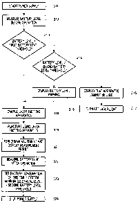
FIG.32 is a flowchart showing an example of steps
of test using the blood test apparatus of the present
invention;
FIG.33Aisacross-sectionalviewshowingindividual
steps in an example of steps of test using the blood test
apparatus of the present invention more specifically;
FIG.33Bisacross-sectionalviewshowingindividual
steps following FIG.33A;
FIG.33Cisacross-sectionalviewshowingindividual
steps following FIG.33B;
FIG.33Disacross-sectionalviewshowingindividual
steps following FIG.33C;
FIG.34 is a flowchart showing another example of
steps of test using the blood test apparatus of the present
invention;
FIG.35 illustrates an example of negative pressure
control in the blood test apparatus of the present
invention;
FIG.36 schematically shows how skin is lifted by
the negative pressure control illustrated in FIG.35;
FIG.37 illustrates another example of the negative
pressure control in the blood test apparatus of the present
invention;
CA 02646721 2008-09-19
2F07055-PCT 12
FIG.38 is an exploded assembly perspective view
showing an example of the laser perforation apparatus
included in the blood test apparatus of the present
invention;
FIG.39 shows an example of laser branch control in
the blood test apparatus of the present invention;
FIG.40 illustrates the laser branch control of
F I G . 3 9 ;
FIG. 41 is a perspective view of a cubic optical device
that can be used in the laser branch control of FIG.39;
FIG.42 shows examples of a cube that can be used
in the laser branch control in FIG.39, where FIG. 42A shows
branchofthelaserlightusingathree-dimensionalimage,
and FIG.42B shows an example of a cube that realizes the
branch;
FIG.43 shows how laser light is emitted from an
oblique direction and punctures skin with the blood test
apparatus of the present invention;
FIG.44 shows variations in the shape of emission
of the laser light;
FIG.45 is a schematic view showing another example
of laser output control in the blood test apparatus of
the present invention;
FIG.46 shows an example of laser pulse corltrol in
the blood test apparatus of the present invention;
FIG. 47 is a cross-sectional view showing apuricturing
state by the laser pulse control in FIG.46;
CA 02646721 2008-09-19
2F07055-PCT 13
FIG.48 shows still another examples of the laser
output control in the blood test apparatus of the present
invention, where FIG.48A shows a circuit diagram, FIG.48B
shows time fluctuation of the current inputted to a
flashlamp over time, and FIG.48C shows time fluctuation
of a laser output;
FIG.49 shows still another examples of the laser
output control in the blood test apparatus of the present
invention, where FIG. 49A shows a circuit diagram, FIG. 49B
shows time fluctuation of the current inputted to the
flashlamp over time, and FIG.49C shows time fluctuation
of the laser output;
FIG.50 is a block diagram showing a first example
of a power supply controlling section of the blood test
apparatus of the present invention;
FIG.51 is a flowchart showing a first example of
control steps in the power supply controlling section
of FIG.50;
FIG.52 is a flowchart showing a second exaniple of
the control steps in the power supply controlling section
of FIG.50;
FIG.53 is a flowchart showing a third example of
the control steps in the power supply controlling section
of FIG.50;
FIG.54 is a flowchart showing a fourth example of
the control steps in the power supply controlling section
of FIG.50;
CA 02646721 2008-09-19
2F07055-PCT 14
FIG.55 is a block diagram showing a second example
of the power supply controlling section of the blood test
apparatus of the present invention;
FIG.56 is a flowchart showing a first example of
control steps in the power supply controlling section
of FIG.55;
FIG.57 is a flowchart showing a second example of
the control steps in the power supply controlling section
of FIG.55;
FIG.58 is a block diagram showing a third example
of the power supply controlling section of the blood test
apparatus of the present invention;
FIG.59 is a flowchart showing a first example of
control steps in the power supply controlling section
of FIG.58;
FIG.60 is a flowchart showing a second exarnple of
the control steps in the power supply controlling section
of FIG.58;
FIG.61A is a graph illustrating a method of setting
a charge level for charging the laser emitting apparatus
stepwise based on the battery level;
FIG. 61B is a graph illustrating a method of setting
the charge level for charging the laser emitting apparatus
continuously based on the battery level;
2 5 FIG.61C is a graph illustrating a method of setting
a charge level for charging the laser emitting apparatus
according to a variable curve based on the battery level;
CA 02646721 2008-09-19
2F07055-PCT 15
FIG.62 shows the relationship between the battery
voltage (Y axis) and the battery level (X axis) when the
charge level is changed;
FIG.63 shows another examples of the laser branch
control in the blood test apparatus with the laser
perforation apparatus of the present invention, where
FIG.63A shows a case where a laser light is divided into
two branches, and FIG. 63B shows a case where a laser light
is divided into four branches;
FIG.64isaschematicviewshowingtheconfiguration
of an optical fiber directional coupler used in the laser
branch control of FIG.63; and
FIG. 65 shows still another example of the laserbranch
control in the blood test apparatus with the laser
perforation apparatus of the present invention.
Best Mode for Carrying Out the Invention
[0016] The present invention provides a blood. test
apparatus using laser light as a means for puncturing
skin and can bring skin in a predetermined position by
sucking force. The focal point of laser light is set
correctly with respect to the skin brought in the
predeterminedposition. In addition, the skin is placed
in close contact with the blood sensor by sucking force,
so that the blood flowing out from the skin punctured
with laser light can be led inside the blood sensor
(detecting section) inareliablemanner. Thebloodtest
CA 02646721 2008-09-19
2F07055-PCT 16
apparatus of the present invention will be described below
with reference to the drawings. Common parts in the
figures will be assigned the same reference numerals
without further explanations.
5[0017] Overall view 1 of the apparatus
FIG.2 is an exploded assembly perspective view
showing the overall configuration of a first example of
the blood test apparatus of the present invention. The
interior of lower case 32 of blood test apparatus 31 shown
in FIG.2 accommodates components including: laser
emitting apparatus 33; negative pressure means 34 which
is configured with suctionpump (negative pressure pump)
34a, pump valve unit 34b and vent switch 34c; battery
35 which supplies power to electrical components;
electrical circuit section 36 which is mounted on these
components; and display section 37 which is mounted on
electrical circuit section 36, and, for example, made
of liquid crystal. Apparatus body 39 is configured so
that upper case 38 covers lower case 32 that accomrnodates
the components. Transparent display window 38a is
provided in upper case 38 in the position corresponding
to display section 37.
[0018] Apparatus body 39 is connected to blood sensor
unit 44 via adapter 40. One end of adapter 40 is a
cylinder-shapedbody, andblood sensor unit 44 is inserted
removably into adapter 40. Blood sensor unit. 44 is
configured with holder 41 and blood sensor 42 attached
CA 02646721 2008-09-19
2F07055-PCT 17
inside holder 41. Window 43 provided in the center of
blood sensor unit 44 is a part for allowing laser light
from the laser emitting hole of laser emitting apparatus
33 to pass through. Window 43 may be a hole or a member
formed with a material that allows laser light to pass
through.
[0019] Overall view 2 of the apparatus
FIG.3 is an exploded assembly perspective view
showing the overall configuration of a second example
of the blood test apparatus of the present invention.
FIG.4 is its side view. Blood test apparatus 31a. shown
in FIG.3 and FIG. 4 is different from blood test apparatus
31 shown in FIG.2 in that the apparatus has a manual pump
that enables manual suction as a negative pressure pump
constitutingnegativepressuremeans140. Thedifference
will be described below.
[0020] Blood test apparatus 31a has negative pressure
means 140 including manual pump (negative pressure pump)
141 and manual pump knob 142 that drives manual pump 141
manually. Vent switch 144 releases the negative pressure
created in pump valve unit 143 to the atmosphere.
[0021] Manual pump knob 142 has the shape of ari arch,
and its one end is made spindle 142a and the other end
is made operating part 142b (see FIG. 4). Manual pump knob
142 can rotate about spindle 142a. Operating part 142b
transmits power to manual pump 141. The patient holds
manual pump knob 142 with apparatus body 39 and can move
CA 02646721 2008-09-19
2F07055-PCT 18
operating part 142b up and down. By this up-and-down
motionmanualpumpl4lstartstocreateanegativepressure.
[0022] To create an adequate negative pressure by the
up-and-down motion of operating part 142b while checking
lift of the skin, the exterior of blood sensor unit 44
is preferably formed with a transparent material so that
the interior of negative pressure chamber 60 (see F'IG. 16,
for example) can be seen. The whole of the exter_ior of
blood sensor unit 44 may be formed with a transparent
material or only the tip 41h side (the negative pressure
chamber 60 side) of blood sensor unit 44 may be formed
with a transparent material. Grip part 142c of manual
pump knob 142 may have finger-shaped pattern with
indentations and projections to prevent the fingers from
slipping.
[0023] Bydriving negative pressure means 140 manually,
it is not necessary to supply power for driving negative
pressure means 140, which extends the life of battery
35 and makes the apparatus suitable for a portable blood
test apparatus.
[0024] The first aspect of the laser emitting apparatus
(including a lens)
The blood test apparatus of the present invention
uses laser light as a means for puncturing skin. When
the skin is irradiated with laser light, the laser light
is absorbed by the OH group of water of the skirl, which
increases heat instantaneously and evaporates the water.
CA 02646721 2008-09-19
2F07055-PCT 19
The surrounding cells also evaporate at this time, to
form a hole in the skin.
[0025] The blood test apparatus accommodates a laser
emittingapparatus. FIG.5isanexteriorperspectiveview
of laser emitting apparatus 33 accommodated in the blood
test apparatus. Further, FIG.6A and FIG.6B are
cross-sectional views of laser emitting apparatus 33.
In FIG.6A, laser crystal 33d is arranged in the internal
part surrounded by the walls where partially reflecting
mirror 33f and total reflection mirror 33g are provided.
In FIG.6B, laser crystal 33d has partially reflecting
mirror 33f and total reflection mirror 33g on both sides
and is attached on the outer wall and the inner wall
(partition) of cylindrical body 33b. That is, in FIG. 6B,
laser crystal (laser rod) 33d is long and extends beyond
the inner wall (partition). Laser emitting apparatus 33
is configured with oscillation tube 33a and cylindrical
body 33b connected to front side of oscillation tube 33a.
Laser emitting port 33c is provided in the center of the
front side of cylindrical body 33b.
[0026] Oscillationtube33aaccommodatesEr:YAG(yttrium
aluminumgarnet) dopedwitherbium, orHo:YAGlasercrystal
33d doped with Holmium, and excitation light source 33e
which includes a xenon flashlamp. Partially reflecting
mirror 33f is attached at one end of oscillation tube
33a (particularly, see FIG.6A). The transmittance of
partially reflecting mirror 33f may be approximately 1
CA 02646721 2008-09-19
2F07055-PCT 20
tol0o. Totalreflectionmirror33gwithatransmittance
of 99 to 100%, is attached to the other end of oscillation
tube 33a (see FIG.6A and FIG.6B). Further, instead of
usingpartiallyreflectingmirror33fandtotalreflection
mirror 33g, films having the same properties may be placed
on the end face of the laser crystal by sputtering. Convex
lens (focus lens) 33h is mounted inside cylindrical body
33b. Convex lens 33h focuses laser light near the surface
of blood sensor 42 (described in detail later). Total
reflection mirror 33g, YAG laser crystal 33d, partially
reflecting mirror 33f, lens 33h and laser emittirlg hole
33c are arranged in this order.
[0027] The process of emitting laser light from laser
emitting apparatus 33 will be described. For example,
theexcitationlightemittedfromexcitationlightsource
33e penetrates to Er:YAG laser crystal 33d and creates
a high energy state by exciting Er (erbium) ion. By this
means, Er:YAG laser crystal 33d becomes a reverse
distribution state, and laser light resonates and is
amplifiedinYAG lasercrystal 33dwhile reflectingbetween
total reflection mirror 33g and partially reflecting
mirror 33f. The same applies to the case of Ho (Holmium) .
Partoftheamplifiedlaserlightpassesthroughpartially
reflecting mirror 33f by stimulated emission. The laser
light passing through partially reflecting mirror 33f
passes through lens 33h and is emitted from laser einitting
port 33c. As described later, the laser light emitted
CA 02646721 2008-09-19
2F07055-PCT 21
from laser emitting port 33c punctures (irradiates) the
skin.
[0028] A second aspect of the laser emitting apparatus
FIG.7 shows another example of the laser emitting
apparatus. Laser emitting apparatus 189 shown in FIG.7
irradiates two kinds of laser crystals with excization
lightusingoneflashlampl85asanexcitationlightsource.
At this time, laser light is outputted from each laser
crystal. Use of two kinds of crystals enables output of
laser lights of different intensities and wavelengths.
[0029] As shown in FIG.7, laser emitting apparatus 189
includes: chassis 188 which has a shape of two overlapping
cylindrical bodies having an elliptical cross section;
flashlamp 185 for exciting laser light, whichisarranged
in the central position of chassis 188; and first crystal
186 and second crystal 187 for oscillating laser light,
which are arranged at the both sides of flashlamp 185.
There are three focal points in elliptical chassis 188.
Chassis 188 has a shape of two overiapping ellipses. Each
ellipse has two focal points and shares one focal point
with the other ellipse, so that there are three focal
points. Out of the three focal points, first crystal 186
is arranged in one of the focal points, and second crystal
187 is arranged in another focal point. Flashlainp 185
is arranged in the center part where two focal points
overlap. Laser lights can be generated from two crystals
186 and 187 using one flashlamp 185, so that it is possible
CA 02646721 2008-09-19
2F07055-PCT 22
to realize a smaller and lower-cost laser emitting
apparatus.
[0030] The intensity of laser light output is
proportional to the light emitting intensity of f lashlamp
185 and is also proportional to the volumes of crystal
186andcrystal187. Therefore,byarrangingtwocrystals
of the same diameter and different lengths, it is possible
to obtain two laser lights of different intensitiesusing
one flashlamp 185.
[0031] Further, by using crystals of the same volume,
it is possible to output two laser lights with the same
intensity at the same time. Therefore, even if a laser
light is not divided into branches (see FIG. 39 and FIG. 40 ),
skin can be punctured with two laser lights of the same
intensity. In this case, energy loss due to branching
by a splitter and mirror is prevented.
[0032] By arranging two crystals with different
compositions (for example, an Er:YAG laser cryst(al with
a wavelength of 2.94 pm and an Nd:YAG crystal with a
wavelength of 1.06 pm), it is possible to obtain laser
lights with different wavelengths. By irradiating the
same position with laser lights having different
wavelengths, it is possible to make pricks of different
depths in skin. For example, Er:YAG and Nd:YAG have
different absorption rates by OH group. Therefore, it
is possible to make a shallower prick using Er:YAG with
a high absorption rate and make a deeper prick using Nd: YAG
CA 02646721 2008-09-19
2F07055-PCT 23
with a lower absorption rate than Er:YAG. By emitting
two laser lights at the same time utilizing these properties,
it. is possible to make a prick on the skin more efficiently.
When the two laser lights are emitted, Er:YAG and Nd:YAG
are preferably emitted in this order with a little time
lag.
[0033] By using laser emitting apparatus 189, it is
possible to use the wavelength of the laser light
selectively. Further, by irradiating the same position
with two types of laser lights using an optical system,
it is possible to improve output intensity.
[0034] The blood test apparatus of the present invention
uses a laser emitting apparatus as a means for puncturing
the skin of the patient that can performpuncturing without
contact with the skin, so that the puncturing needle
required in the conventional blood test apparatus, is
no longer required. Further, the blood test apparatus
uses a puncturing means that does not contact with the
skin of the patient, and so is sanitary. Still further,
although it is necessary to replace the puncturing needle
every test by the conventional blood test apparatus, the
test by the blood test apparatus of the present invention
does not require this replacement. Further, the blood
test apparatus of the present invention does not require
moving component for moving a needle required for
puncturing with a needle, which reduces troubles . Further,
the number of components required in the blood test
CA 02646721 2008-09-19
2F07055-PCT 24
apparatus of the present invention is reduced, so that
componentscontrolbecomessimple. Further,byproviding
a transparent waterproof wall on the front face of laser
emitting port 33c, it is possible to wash the whole of
the blood test apparatus.
[0035] The blood sensor
The blood test apparatus of the present invention
has a blood sensor for taking in the blood flowing out
f rom the punctured skin and examining the bl ood component s.
[0036] The first example of the blood sensor
FIG.8 is a cross-sectional view of the first example
of the blood sensor. Blood sensor 42 shown in FIG.8 has
an outer shape of a round or polygon. Base plate 45
constituting blood sensor 42 has: substrate 46; spacer
47 stacked on the upper face of substrate 46; and cover
48 stacked on the upper face of spacer 47.
[0037] Blood storing part 49 is provided near the center
of base plate 45. Storing part 49 is formed so as to
communicate with hole 46a provided in substrate 46 and
hole 47a provided in spacer 47. Storing part 49 opens
downward to collect blood from the skin. The volume of
storing part 49 is, for example, 0.904 pL, but is by no
means particularly limited. One end of supply channel
50 is connected to storing part 49. The volume of supply
channel 50 is, for example, 0.144 pL, but is by no means
particularly limited. Detecting section 51 is arranged
inside supply channel 50. Blood stored in storing part
CA 02646721 2008-09-19
2F07055-PCT 25
49 flows into supply channel 50 by capillary action and
is led to detecting section 51. The other end of supply
channel 50 is connected to air hole 52. The diameter of
air hole 52 may be approximately 50 um to 250 um. Bymaking
the diameter of air hole 52 small, blood is prevented
fromflowingoutthroughairhole52excessively. F'urther,
in a state where storing part 49 is in close contact with
the skin, air hole 52 operates as a negative pressure
paththroughwhichanegativepressureis createdinstoring
part 49.
[0038] Reagent 53 mounted on detecting section 51 may
be prepared as appropriate according to a test target.
For example, reagent 53 is prepared by dropping a reagent
solution on detecting section 51 arranged on substrate
46, and drying the reagent solution, wherein the reagent
is prepared by adding and dissolving an enzyme (PQQ-GDH)
of 0.1 to 5.0 U/sensor, potassium ferricyanide (10 to
200 mM), maltitol (1 to 50 mM) and taurine (20 to 200
mM) to a 0.01 to 2.0 wt% aqueous solution of CMC.
[0039] Storing part 49 of blood sensor 42 is sealed with
face49a(hereinafter"ceilingface"). Theemittedlaser
light preferably transmits through ceiling face 49a, so
that blood flowing out from the skin punctured with laser
light does not flow out from ceiling face 49a. To allow
the laser light to transmit through ceiling face 49a,
cover 48 may be formed with the material that allows laser
light to transmit (for example, glass or plastic such
CA 02646721 2008-09-19
2F07055-PCT 26
as polyimide).
[0040] Further, if the emitted laser light cannot
transmit through ceiling face 49a, the laser light may
perforate ceiling face 49a. In the case where the laser
light perforates ceiling face 49a, substrate 46, spacer
47 and cover 48 may be formed with the same material.
[0041] The hole formed in ceiling face 49a can serve as
air hole 52, as well as a negative pressure path through
which the negative pressure means creates a negative
pressure in storing part 49.
[0042] A second example of the blood sensor
FIG. 9 is a cross-sectional view of the second example
of the blood sensor. While ceiling face 49a of storing
part 49 of blood sensor 42 shown in FIG.8 is sealed, the
ceiling face of storing part 49 of blood sensor 103 shown
in FIG.9 is open.
[0043] Hole 103b is formed in cover 48 of blood sensor
103. Preferably, the diameter of hole 103b (for example,
1.0 mm) is smaller than the diameter of storing part 49
(for example, 2.0 mm), and is greater than the diameter
of air hole 52 (50 pm to 250 pm) . Fole 103b is preferably
located in the center of the ceiling face of storing part
49. Laser light passes through hole 103b and punctures
the skin. Byprovidinghole 103b, it is possible topr-event
laser light from declining. It is thereby possible to
reduce the energy of laser light to be emitted.
[ 0044 ] Hole 103b and air hole 52 can serve as a neciative
CA 02646721 2008-09-19
2F07055-PCT 27
pressure path through which negative pressure means 34
and 140 create a negative pressure in storing part 49.
[0045] As shown in FIG.10, the surface tension of blood
16 generated inside hole 103b prevents blood 16 collected
by puncturing the skin from overflowing out from the upper
face of the cover. Blood 16 spreads through inside storing
part 49. Therefore, it is possible to collect an adequate
amount of blood 16. Blood 16 that fills storing part 49
flows into supply channel 50 by capillary action.
[0046] By making hole 103b is water-repellent, blood 16
is less likely to overflow through hole 103b. Therefore,
the interior o f the blood test apparatus is not contam.inat ed
with blood.
[0047] Polyethylene terephthalate (PET) can be used as
the material of cover 48 of blood sensor 103, and the
same material can be used as substrate 46 and spacer 47.
Therefore, material control is simple.
[0048] Laser light passes through hole 103b of storing
part 49, and laser light may pass through the center of
hole 103b or pass through a position out of the center
of hole 103b. For example, by making laser light pass
through a position farther from supply channel `D0 than
the center of hole 103b, blood 16 flowing out from skin
13 fills the interior of storing part 49 completely, and
then flows into supply channel 50, so that it is possible
to realize accurate measurement.
[0049] Hole 103b is formed in advance in the ceiling face
CA 02646721 2008-09-19
2F07055-PCT 28
of storing part 49 of blood sensor 103. In this way, hole
103b is formed in advance, so that it is not necessary
to adjust the axis of the laser light to the part to be
perforated. Therefore, blood sensor 103 is easily
attached to blood sensor unit 44. Hole 103b may be made
small, approximately 0.05 to 0.25 mm, and preferably
preventsblood16fromfiowingoutthroughthepuncturing
hole.
[0050] As shown in FIG.8 and FIG.9, the blood sensors
in the blood test apparatus of the present invention
preferably has storing part 49 and supply channel 50.
The inner wall surface of supply channel 50 is preferably
hydrophilic so that blood is transferred smoothly to supply
channel 50 where detecting section 51 is arranged.
Further, the inner wall surface of supply channel 50 is
preferably more hydrophilic than the inner wall surface
of storing part 49 so that blood stored in storing part
49 is supplied to supply channel 50 smoothly.
[0051] Further, as shown in FIG.8 and FIG. 9, the blood
sensor in the blood test apparatus of the present invention
has cover 4 8 , and cover 48 forms the ceiling face of storing
part 49. Upper faces 48a and 103a (faces irradiated with
laser light) of cover 48 are preferablywater- repellent.
More particularly, upper faces 48a and 103a of cover 48
are preferably more water-repellent than the inner wall
surface of storing part 49, so that blood stored in storing
part 49 is prevented from flowing out through the hole
CA 02646721 2008-09-19
2F07055-PCT 29
(the hole perforated with laser light or hole 103b) formed
on cover 48.
[0052] The third example of the blood sensor
The wetness of skin 13 of the patient varies depending
on the environment. On the other hand, skin 13 to be
punctured with laser light preferably has a certain level
of moisture content. Therefore, in order to perform
measurement in a stable, a certain level of wetness is
preferablymaintainedbygiving a certain level of moisture
content to skin 13 by moistening the neighborhood of skin
13 before puncturing with laser light.
[0053] FIG. ll shows blood sensor 42a provided with water
storing part 195 that stores water, on the lower face
side that comes into contact with on skin 13, of blood
sensor 42 (see FIG.8 in detail ). On or before emitting
laser light, water storing part 195 of blood sensor 42a
shown in FIG. 11 breaks to splash a certain amount of water
on skin 13 and moisten the skin which is lifted by negative
pressure means 34 and 140 before laser light is emi_tted.
Water storing part 195 may be, for example, a container
which contains water andwhich is made of a plasticmaterial
such as PET, or a soft bag, a sponge or a spongy ntember
that is soaked with water. Water storing part 195 is
preferably not arranged in transmission part 196 th.rough
which laser light passes, because the intensity of the
laser light is reduced by water.
[0054] Transparent plan view 1 of the blood sensor
CA 02646721 2008-09-19
2F07055-PCT 30
FIG.12 is a perspective plan view of blood sensor
42. In blood sensor 42, detection electrodes 54 to 57
are arranged, and in order from storing part 49 toward
air hole 52, detection electrode 57 (Hct (hematocrit)
electrode) , detection electrode 56 (counter electrode),
detection electrode 54 (active electrode), detection
electrode 56 (counter electrode) and detection electrode
55 (sensing electrode) are arranged. Detection
electrodes 54 to 56 are arranged in detecting section
51.
[0055] Detection electrodes 54 to 57 are connected to
connection electrodes 54a to 57a, respectively.
Connection electrodes 54a to 57a extend up to the outer
periphery of substrate 46. Contact parts 54b to 57b are
provided in connection electrodes 54a to 57a. Further,
in connection electrode 56a, contact part 56c is also
provided in addition to contact part 56b, so that two
contact parts are provided. Reference electrode 56dmay
be provided in connection electrode (54a, 55a and 57a)
other than connection electrode 56a. Contact parts 54b
to 57b and contact part 56c are arranged near the outer
periphery of sensor 42 at virtually regular intervals.
[0056] Among contact parts 54b to 57b and 56c, contact
part 56b and contact part 56c electrically conduct with
each other, and the other contact parts are insalated
from each other. The connection electrodes can be
specified using contact part 56c as a reference contact
CA 02646721 2008-09-19
2F07055-PCT 31
part, that is, reference electrode 56d. That is, the
insulation resistance between the neighboring contact
parts is measured by electrical circuit section 36 (see
FIG. 2), and a contact part where the insulation resistance
is zero is identified as reference electrode 56d. Based
on reference electrode 56d, connection electrodes 56a,
57a, 54a and 55a are specified clockwise.
[0057] In this way, blood sensor 42 has reference
electrode 56d, so that it is possible to specify the
connection electrodes. Therefore, even if the contact
parts (54b to 57b and 56c) are connected casually to the
five connectors arranged in apparatus body 39, it is
possibletospecifytheconnectionelectrodesandperform
measurement. Accordingly, blood sensor 42 (or blood
sensor unit 44 including blood sensor 42) can be made
in a symmetrical shape so that blood sensor 42 can be
attached to apparatus body 39 casually in a very simple
manner.
[ 0058 ] Aligning concave part 46c may be provided on the
outerperipheryofsubstrate46. Ontheouterperipheries
of spacer 47 and cover 48, aligning concave parts 47c
and 48c are provided so as to correspond to aligning concave
part 46c. By using aligning concave parts 46c to 48c,
blood sensor 42 can be attached to blood sensor unit 44
so as to meet a predetermined alignment of blood sensor
unit 44.
[0059] Transparent plan view 2 of the blood sensor
CA 02646721 2008-09-19
2F07055-PCT 32
FIG.13 is a transparent plan view of a round blood
sensor. Blood sensor 101 shown in FIG.13 is different
from blood sensor 42 (see FIG.12) in that reference
electrode 56d is formed via a predetermined pattern from
connectionelectrode 56a. The difference will be mainly
described below.
[0060] Reference contact part 56c is provided in
reference electrode 56d. Reference contact part 56c and
contact parts 54b to 57b are arranged near the outer
periphery at regular intervals. That is, contact parts
54b, 55b, 56b, 56c and 57b are arranged at apexes of a
regular pentagon.
[0061] Connection electrode 56a and reference elF=_ctrode
56d are connected via laser-machined pattern 56e. By
changing the width of pattern 56e, the resistance value
between contact part 56b and reference contact part 56c
can be changed. Reference electrode 56d serves as a
reference for specifying the positions of connection
electrodes 54a to 57a.
[0062] Reference electrode 56d can be utilized to
identify the product specifications of blood sensor 101.
For example, the blood test apparatus is set so that
calibration curve 1 is used when the resistance value
of pattern 56e is 200 to 1000 ohms, calibration curve
2 is used when the resistance value is 1000 to 2000 ohms,
and calibration curve 3 is used when the resistance value
is 2000 to 3000 ohms, the calibration curve of the sensor
CA 02646721 2008-09-19
2F07055-PCT 33
is recognized automatically, and the blood sugar level
is measured using an appropriate calibration curve. The
reference electrode canbe usedto identifyvarious product
specifications, in addition to use in automatic
recognition of the calibration curve. For example, the
reference electrode can be used to identify the users
the product is shipped to, forexample, to identify whether
the product has the specifications for company A or the
specifications for company B.
[0063] By forming pattern 56e with an inductance having
arbitrary property, connecting the inductance to a
resonator constituting an oscillator and changing the
oscillation frequency according to this inductance
property, various information can be provided.
[0064] By providing reference electrode 56d, even when
blood sensor unit 44 is attached to blood test apparatuses
31 or 31a at an arbitrary rotation angle with respect
to the axis of the attaching direction, connection
electrodes 54a to 57a can be specified. Therefore, when
blood sensor unit 44 is attached, the attaching direction
does not have tobe adj usted visually, so that it is possible
to attach blood sensor unit 44 in a simple manner.
[0065] Transparent plan view 3 of the blood sensor
FIG. 14 is a transparent plan view of a square-shaped
blood sensor. Although the outer shape of blood sensor
102 shown in FIG.14 is a square, the outer shape rnay be
a polygonal such as a hexagon and octagon. By forming
CA 02646721 2008-09-19
2F07055-PCT 34
blood sensor 102 in a square or hexagonal shape, the yield
rateofmaterialpreparationimproves. Further, as shown
in FIG.14, aligning concave part 102a for aligning blood
sensor unit 44 may be provided in one of the four sides
of blood sensor 102, in such a case blood sensor 102 has
an asymmetrical shape. Concave part 102a serves as the
reference when blood sensor 102 is attached to blood sensor
unit 44. Further, by alignment blood sensor unit 44 and
adapter 40 by using convex part 130f (see FIG. 25 ) in the
blood sensor unit 44 side that engages with concave part
102a as a reference, detection electrodes 54 to 57 can
be specified even if reference electrode 56d is not
provided.
[0066] Contact parts 54b to 57b are provided in the c:orners
of square-shaped substrate 102b. Spacer 102c and cover
102d are stacked on substrate 102b. Substrate 102b
corresponds to substrate 46, spacer 102c corresponds to
spacer 47, and cover 102d corresponds to cover 48 (see
FIG. 8 ) .
[0067] An exploded plan view of the blood sensor
An assembly and material of blood sensor 42 (see
FIG. 8) provided in the blood test apparatus of the present
invention will be described.
[0068] FIG.15 is an exploded plan view of blood sensor
42. FIG.15A is a plan view of cover 48, FIG.15B is a plan
view of spacer 47, and FIG.15C is a plan view of substrate
46.
CA 02646721 2008-09-19
2F07055-PCT 35
[0069] FIG.15C is a plan view of round substrate 46
constituting blood sensor 42. The diameter of substrate
46 may be approximately 8. 0 mm. The material of substrate
46 is resin such as polyethylene terephthalate (PET),
and its thickness may be 0.075 to 0.250 mm (for example,
0.188 mm).
[0070] On the upper face of substrate 46, detection
electrodes 54 to 57, and connection electrodes 54a to
57a derived from detection electrodes 54 to 57,
respectively, are formed in an integratedmanner. These
detection electrodes and connection electrodes may be
formedbyapplyinglaserprocessingtoaconductivelayer
formed with the sputtering method or the vapor deposition
method. The material of the conductive layer can be gold,
platinum and palladium as materials.
[ 0 0 7 1 ] The diameter of hole 4 6a provided near the center
of substrate 46 may be approximately 2. 0 mm. Preferably,
the wall surface of hole 46a is less hydrophilic than
supply channel 50 or is less water-repellent thar.. upper
face 48a of cover 48.
[0072] Hole 46a is preferably formed by punching press
substrate 46 from the side of detection electrodes 54
to 57, using a convex mold, because it is less likely
to damage detection electrodes 54 to 57. Further, even
if a burr is produced in hole 46a by this punching, the
burr is oriented downward (toward the skin) . Therefore,
blood 16 is prevented from flowing out from storing part
CA 02646721 2008-09-19
2F07055-PCT 36
49. Concave part 46c for aligning provided on the outer
periphery of substrate 46 engages with a aligning convex
part formed in cylindrical body 41e of blood sensor unit
44 (see FIG.16) . The alignment where blood sensor 42 is
attached to blood sensor unit 44 is thereby determined.
[ 0073 ] FIG. 15B is a plan view of spacer 47. The diameter
of spacer 47 may be approximately 5.2 mm. The material
of spacer 47 is resin such as polyethylene terephthalate,
and its thickness may be 0.025 to 0.25 mm (for example,
0.1 mm).
[ 0074 ] The diameter of hole 47a provided near the center
of spacer 47 is 2.0 mm, and hole 47a is provided in the
position corresponding to hole 46a provided in substrate
46. Preferably, the wall surface of hole 47a is less
hydrophilic than supply channel 50 or is less
water-repellent than upper face 48a of cover 48. Storing
part 49 is constituted with hole 46a and hole 47a.
[0075] Slit 47b is formed toward the outer periphery from
hole 47a. Slit 47b serves as blood supply channel 50.
2 0 The wall surface of slit 47b and the upper face of substrate
46 corresponding to the wall surface of slit 47b are
subjected to hydrophilicity treatment. Further, the
width of slit 47b may be approximately 0.6 mm, and the
lengthmaybe approximately 2.4 mm. As a result, the volume
ofsupplychannel50isapproximately0.144uL. Therefore,
by making the volume of supply channel 50 small, test
can be performed with a small amount of blood, so that
CA 02646721 2008-09-19
2F07055-PCT 37
the load on the patient becomes small and the patient
does not feel fear.
[0076] Concave part 47c for aligning provided on the outer
periphery of spacer 47 is formed in the position meeting
concave part 46c for aligning provided in substrate 46.
[0077] FIG.15A is a plan view of cover 48. The diameter
of cover 48 may be approximately 5.2 mm. The thickness
of cover 48 may be approximately 0.050 to 0.125 mm (for
example, 0.075 mm).
[0078] Cover 48 can be made of a material that does not
absorb laser light. Examples of the material of cover
48includeglassandplasticsuchaspolyimide. Whenlaser
light is not absorbed in cover 48, the laser light can
pass through ceiling face 49a of storingpart 49 topuncture
1 5 the skin. The laser light does not perforate ceiling face
49a, and so blood 16 does not flow out through the hole,
and blood 16 does not flow into apparatus body 39.
[0079] Cover 48 may be made of a material that absorbs
laser light. In this case, cover 48 may be perforated
by the emitted laser light, or a hole through which laser
light passes, may be formed in cover 48 before the laser
light is emitted.
[0080] Air hole 52 is provided to meet the tip part of
supply channel 50. The diameter of air hole 52 is 50 pm.
[0081] Upper face 48a (see FIG. 8) of cover 48 that forms
the upper face of substrate 45 is preferably subjected
towater-repellencytreatment. Theceilingfaceofsupply
CA 02646721 2008-09-19
2F07055-PCT 38
channel 50 is preferably subjected to hydrophilicity
treatment. Further, preferably, ceiling face 49a of
storing part 49 is subjected to weaker hydrophilicity
treatment than supply channel 50 or is subjected to weaker
water-repellency treatment than upper face 48a of cover
48.
[0082] Hydrophilicity may be reduced by, for example,
removing the hydrophilic agent applied on a hydrophobic
membertoincreasehydrophobicity. Thehydrophilicagent
is removed by, for example, decomposing the hydrophilic
agent through UV (ultraviolet ray) irradiation. The
hydrophobicmaterial can be directly used as the material
of ceiling face 49a of storing part 49.
[ 0083] The material may be made water-repellent bymixing
a water-repellent agent in the material. Further, the
material may be made water-repellent by applying an
appropriateamountofwater-repellentagentonthesurface
of the hydrophilic material. The levE=_l of
water-repellency may be adjusted by adjusting the amount
of the water-repellent agent to be mixed.
[0084] The hydrophilicity or water-repellency of the
components of blood sensor 42 can be adjusted as follows.
Upper face 48a of cover 48 is subjected towater-repellency
treatmentinadvance. Ontheotherhand,theoveralllower
face of cover 48 is subj ected to hydrophilicity treatment .
The lower face of cover 48 includes the ceiling face of
supply channel 50 . Next, substrate 46, spacer 47 and cover
CA 02646721 2008-09-19
2F07055-PCT 39
48 are stacked. After substrate 46, spacer 47 and cover
48 are stacked, the hydrophilic material of ceiling face
49e may be dissolved and removed by radiating
short-wavelength UV from the opening of storing part 49.
By manufacturing blood sensor 42 as described above, it
is possible to make upper face 48a of cover 48 water
repellent and make the inner face of supply channel 50
hydrophilic. Further, the inner face of storing part 49
may be less hydrophilic than supply channel 50 and less
water repellent than upper face 48a.
[0085] The ratio of the thickness of substrate 46 (0.188
mm) , the thickness of spacer 47 ( 0. 100 mm) and the thickness
of cover 48 (0.075 mm) is approximately, 2.5:1.3:1. By
this means, it is possible to form storing part 49 that
can pool a sufficient amount of blood while making blood
sensor 42 thinner. Further, by the thickness of spacer
47 (0.100 mm) , the effect of capillary action in supply
channel 50 can be obtained sufficiently.
[0086] In blood sensor 42, the ratio of the volume of
storing part 49 ( 0. 904 pL ) and the volume of supply channel
50 (0.144 pL) may be approximately 6:1, but the ratio
is not particularly limited. By this means, test does
not become incorrect, even when the amount of blood 16
is small. Further, the volume of storing part 49 is not
too large with respect to the volume of supply channel
50 required, so that a large amount of blood 16 does not
flow into supply channel 50 and does not wash away reagent
CA 02646721 2008-09-19
2F07055-PCT 40
53 (see FIG.8). Therefore, the rate of flow of blood 16
becomes constant, which does not generate variation in
concentration of reagent 53, so that it is possible to
examine blood 16 accurately.
[ 0087 ] Further, the amount of blood 16 collected is set
a very small amount which is a sufficient amount required
for a test of blood 16. Only blood 16 of approximately
six times the volume of supply channel 50 is collected.
Therefore, it is possible to reduce the load on the patient
significantly. Inviewof thecollectionamountofblood
16 for accurate measurement and the collection amount
of blood 16 for reducing the load on the patient, the
volume of storing part 49 is preferably more than five
times and less than seven times the volume of supply channel
50.
[0088] The blood sensor unit
The blood sensor in the blood test apparatus of the
present invention may be included in the blood sensor
unit. The blood sensor unit can be attached to and removed
from the apparatus body and is a replaceable member.
[0089] FIG.16 is a cross-sectional view of blood sensor
unit 44 and the neighborhood of blood sensor unit 44.
The cross section of blood sensor unit 44 is configured
in the shape of "H" by cylinder-shaped holder 41 that
opens upward and downward, and attaching part 41b that
is provided so as to seal the interior of holder 41.
[0090] The material of holder 41 is preferably resin that
CA 02646721 2008-09-19
2F07055-PCT 41
isapplicable to injection molding, including ABS resin,
AS resin and thermoplastic resin such as polyethylene,
polypropylene, polyvinyl chloride and polyethylene
terephthalate, or thermosetting resin such as pheriol resin,
epoxide resin and silicon resi.n.
[0091] Blood sensor 42 is attacried to attaching part 41b.
Blood sensor 42 can be attached and removed. Although,
in FIG.16, blood sensor 42 is attached to an upper side
(the laser emitting apparatus 33 side) of attaching part
41b, blood sensor 42 may be attached to a lower side (the
punctured skin 13 side) of attaching part 41b.
[0092] In the center of attaching part 41b, window 43
is preferably provided so as to correspond to storing
part 49. The area of the opening part of window 43 is
preferably larger than the area of the opening part of
storing part 49. Further, negative pressure patr 41c is
provided that penetrates the upper side and the lower
side of attaching part 41b. Negative pressure path 41c
maybeprovided,forexample, between the outer periphery
of blood sensor 42 and the inner periphery of holder 41.
[0093] Cylindricalbody4ldlocatedbelowattachingpart
41b forms negative pressure chamber 60 between skin 13
and cylindrical body 41d. Further, the inner wall of
cylindrical body 41e located above attaching pa:rt 41b
of blood sensor unit 44 is latched outside adapter 40.
[0094] Connector 61 is provided inside adapter 40.
Connector 61 includes a plurality of (for example, five)
CA 02646721 2008-09-19
2F07055-PCT 42
individual connectors 61a to 61e. Whenbloodsensorunit
44 is attached to adapter 40, connectors 61a to 61e contact
with contact parts 54b to 57b and 56c of blood sensor
42, respectively. Signals of connectors 61a to 6le are
led to electrical circuit section 36.
[0095] First skin contact sensor 62 provided at tip 41h
of cylindrical body 41d detects skin 13 when blood sensor
unit 44 comes into contact with skin 13. First skin contact
sensor 62 also connects to connection part 62c provided
in adapter 40 via conductor 62a arranged inside holder
41, and further connects to conductor 62b at the adapter
40 side. Conductor 62b is led to electrical circuit
section 36.
[0096] A plurality of (for example, two) first skin
contact sensors 62 made of conductors are preferably
provided in different parts in tip 41h of cylindrical
body 41d (in FIG.16, two first skin contact sensors 62
are provided symmetrically with respect to the center
of cylindrical body 41d). By measuring the resistance
value between two conductors of first skin contact sensor
62, skin 13 is detected when blood sensor unit 44 comes
into contact with skin 13. Therefore, it is possible to
detect skin 13 when the tip of blood sensor unit 44 comes
intocontactwithskinl3completelywithoutspace. Laser
light is preferably not allowed to emit unless fir_st skin
contact sensor 62 detects a contact with the skin. First
skin contact sensor 62 may be a mechanical micro switch
CA 02646721 2008-09-19
2F07055-PCT 43
or a reflection optical switch.
[0097] By emitting laser light from laser emitting
apparatus 33, blood capillaries in skin 13 are damaged
by the laser light, and blood 16 flows out. The outflow
of blood 16 is stored in storing part 49.
[0098] A guide part for attaching blood sensor unit 44
in a simple manner may be provided in cylindrical body
41d and adapter 40 of blood sensor unit 44. FIG.17 is
an exploded elevation view of the primary part of guide
part 63 that guides insertion of blood sensor unit 44
into adapter 40. Convex part 41f is formed inside
cylindrical body 41d, and convex part 40f is formed outside
adapter 40. Tip part 41g and tip part 40g, which are the
tips of convex part 41f and convex part 40f, respectively,
are made sharp. Tip part 41g and tip part 40g face each
other. Convex part 40f and its tip part 40g, and convex
part 41f and its tip part 41g, constitute guide part 63.
[0099] When blood sensor unit 44 is inserted into adapter
40, evenwhentherelativealignmentbetweenbloodsensor
unit 44 and adapter 40 is out of predetermined alignment,
blood sensor unit 44 is inserted along guide part 63 while
correcting the course (see arrow 64) . As a result,
connectors 61a to 61e provided in adapter 40 are sure
to contact with one of contact parts 54b to 57b and 56c
provided in sensor 42. Therefore, blood sensor unit 44
can be inserted without taking into account the rotation
angle with respect to the axis of the insertion direction,
CA 02646721 2008-09-19
2F07055-PCT 44
so that blood sensor unit 44 can be attached in a simple
manner.
[0100] FIG.18 is a diagrammatic perspective view of the
blood sensor unit. Blood sensor unit 110 shown in FIG. 18
may have the same structure as blood sensor unit 44 unless
describedotherwise. Blood sensor unit 110 has the shape
of a cylinder, and its cross section has the shape of
" H . " Five connectors 111 that transmit signals of the
contact part of the blood sensor (one of blood sensors
42, 101, 102 and 103) to electrical circuit section 36
may be provided inside holder 110a of blood sensor unit
110 (in the case of blood sensor 102, four connectors
may be provided). Connector 111 connects to adapter 40
at an upper end of holder 110a and is led to electrical
circuit section 36 via this adapter 40. Connector 111
may be provided in the adapter and may be connected with
the contact part of the blood sensor of blood sensor unit
110.
[01011 Blood sensor 42 is attached on the reverse side
(the lower end 110h side, that is, the side the punctured
skin is arranged) of attaching part 110b provided so as
to seal the opening of holder 110a. Window 110c provided
near the center of attaching part 110b is provided so
as to meet the position of storing part 49 of blood sensor
42. Laser light passes through window 110c and storing
part 49 and punctures skin 13.
[ 01021 Air hole 110d provided in attaching part ?_l Ob is
CA 02646721 2008-09-19
2F07055-PCT 45
provided in the position meeting air hole 52 of blood
sensor 42. Air hole 110d is provided so that blood 16
runs into supply channel 50 of blood sensor 42 or create
a negative pressure in storing part 49.
[0103] Engagingpart110eofbloodsensorunitll0engages
with adapter 40 via the elasticity of engaging part 110e
which engages with adapter 40. Two engaging parts 110e
thatfaceeachotherareprovidedin holder110a. Engaging
parts 110e have slits on both sides so as to have elasticity,
and are formed integrated with holder 110a. Therefore,
engaging parts 110e can be made at a low cost.
[0104] Deodorizing member storage 110f is provided on
the upper face of attaching part 110b in a concentric
fashion. A deodorizing member is placed on deodorizing
memberstoragellOf. Whentheskinispuncturedwithlaser
light, cases occur where skin 13 is carbonized and produces
an odor. This odor can be deodorized with the deodorizing
member (such as deodorant agent). Further, blood pool
110g is provided on the upper face of attaching part 110b
in a concentric fashion. Therefore, even if blood 16
overflows from hole 103b of blood sensor 103 (see F'IG. 10 ),
blood 16 stays in blood pool 110g, so that it is possible
to prevent blood 16 from contaminating the body part of
blood test apparatuses 31 or 31a.
[0105] FIG.19 is a cross-sectional view showing the
primary part of one configuration example of neighborhood
of lower end 11 0h of holder 11 0a . An end part of lower
CA 02646721 2008-09-19
2F07055-PCT 46
end 110h comes into contact with skin 13 of the patient
and forms negative pressure chamber 60. Lower end 110h
needs to closely contact with skin 13. Therefore, lower
end 110h may be formed with two concentric liries llOj
which are made sharp at an acute angle. Since line 110j
comes into contact with skin 13 reliably by line contact,
negative pressure chamber 60 is kept sealed. The number
of lines 110j does not have to be two, and there may be
one or a plurality of lines 110j.
[0106] Further, if a capillary function is given to a
groove formed between two concentric lines 110j,
over-sampled blood 16 after measurement is sucked in the
groove. Therefore, it is not necessary to prepare paper
for wiping off blood flowing out.
[0107] FIG.20 is a cross-sectional view showing the
primary part of another configuration example near_lower
end110hof holder110a. Concentriccontactingpart110k
made of elasticity such as rubber, silicon, urethane and
a sponge, is formed in lower end 110h. Therefore,
contacting part 110k can come into close contact with
skin 13 by its elasticity, and negative pressure chamber
60 is kept sealed. The contact surface of contacting part
110k is preferably a flat to increase the area where
contacting part 110k comes into contact with skin 13.
[0108] By forming contacting part 110k with an absorbing
member, such as a sponge, that has absorbency, it is
possible to wipe off over-sampled blood 16 flowing out
CA 02646721 2008-09-19
2F07055-PCT 47
by puncturing after measurement. Therefore, it is not
necessary to prepare wiping paper. Further, if an
antiseptic is applied to the absorbing member, the
absorbing member becomes sanitary.
[0109] The wetness of skin 13 changes with the external
environmentsuchasseasons. Therefore, the wetness near
skin13tobepuncturedispreferablymaintainedconstant.
Therefore, before puncturing, measurement may be
performed in a stable condition by providing an adequate
level of moisture content to ski_n 13 and moistening the
skin. Therefore, as shown in FIG.21, it is also possible
to provide water storing part 197 which is soaked with
water, throughout the perimeter of lower end 110h of holder
110a of blood sensor unit 110, soak skin 13 near the part
to be punctured with water in advance and puncture skin
13 with laser light . Water storing part 197 may be a porous
body that has elasticity such as a sponge.
[0110] FIG.22 is a cross-sectional view of blood sensor
unit 110. As shown in FIG.22, blood sensor 42 is arranged
in the lower face of attaching part 110b of blood sensor
unit 110 and is held by attaching part 110b. Skin 13 is
lifted by negative pressure means 34 or 140 (see FIG.2
and FIG.3) and is in close contact with blood sensor 42.
Blood sensor 42 is held by attaching part 110b, and so
is less likely to be distorted by skin 13 that is in close
contactwithbloodsensor42. Connectorslllcontactwith
contact parts 54b to 57b and 56c of blood sensor 42. Guide
CA 02646721 2008-09-19
2F07055-PCT 48
part 63 (see FIG.17) matching adapter 40 is preferably
provided in holder 110a.
[0111] Thebloodtestapparatusofthepresentinvention
hasanegativepressuremeans,thenegativepressuremeans
create a negative pressure inside blood sensor unit 110.
As a negative pressure path, groove 110f may be formed
in attaching part 110b of blood sensor unit 110. Groove
110f extends to window 110e formed near the center of
attaching part 110b, from the outer periphery side of
attaching part 110b of holder 110a. When a negative
pressure is created, a negative pressure is also created
i_n groove 110f, and blood sensor 42 is in close contact
with attaching part 110b. When the negative pressure is
released to the atmosphere, blood sensor 42 is removed
from attaching part 110b. Connector 111 contacts with
blood sensor 42 in contact surface llla. Connector 111
is integrated with holder 110a and formed so as to cut
into part of attaching part 110b. By this means, the
contact parts of the connection electrodes formed on the
upper face of blood sensor 42 connect with contact parts
(not shown) provided in connectors 111.
[0112] Second skin contact sensor 110m may be provided
iri the lower face of blood sensor 42. By this means, skin
13 is detected when skin 13 comes into contact with second
skin contact sensor 110m by the negative pressure in
negative pressure chamber 60. Secondskincontactsensor
110m may be, for example, configured with a counter
CA 02646721 2008-09-19
2F07055-PCT 49
electrode. Laserlightemissionispreferablynotallowed
unless second skin contact sensor 110m detects a contact
with the skin. Negative pressure means 34 may stop
creating a negative pressure in negative pressure chamber
60 when second skin contact sensor 1l0m detects the contact
with the skin. By controlling negative pressure means
34 in this way, negative pressure means 34 can be controlled
without wasting a negative pressure power.
[0113] Further, first skin contact sensor 62 may be
provided in lower end 110h of holder 110a.
[ 0 1 1 4 ] FIG.23 is a cross-sectional view of another blood
sensor unit. Blood sensor unit 120 shown in FIG.23 may
have the same structure as blood sensor unit 110 unless
described otherwise. Blood sensor unit 120 is different
from blood sensor unit 110 in that blood sensor 42 is
mounted on the upper side of attaching part 120b formed
so as to seal the opening of holder 120a. Connector 61
connected to electrical circuit section 36 conducts with
contact part (54b to 57b and 56c) of blood sensor 42.
[0115] The upper space and the lower space in attaching
part 120b of blood sensor unit 120 having an H-shaped
cr_osssection,communicatethroughnegativepressurepath
120c. The lower space forms negative pressure chamber
60. First skin contact sensor 62 is provided in lower
end 120h of holder 120a. Further, second skin contact
sensor 120m is provided in the lower face of attaching
part 120b (not shown).
CA 02646721 2008-09-19
2F07055-PCT 50
[0116] By attaching blood sensor 42 on the upper face
of attaching part 120b, it is possible to increase the
contact pressure between connector 61 and the contact
part (54b to 57b and 56c) of blood sensor 42. Further,
it is possible to attach blood sensor 42 to attaching
part 120b in a simple manner.
[ 0 117 ] The space on the side of apparatus body 39 (the
upper space in the figure) and the space on the side of
skin 13 (the lower space in the figure) separated by blood
sensor 42 and attaching part 120b, communicate with each
other via negative pressure path 120c. On creating a
negative pressure on skin 13, it is possible to create
a negative pressure in the space on the side of skin 13
via this negative pressure path 120c. Further, when a
negative pressure is released to the atmosphere, air flows
into space on the side of apparatus body 39 quickly via
negative pressure path 120c. Therefore, it is possible
to prevent blood led in blood sensor 42 from splashing
to apparatus body 39.
[0118] Groove 120f may be formed on the upper side of
attaching part 120b as a negative pressure path. Groove
120f extends from the outer periphery of attaching part
120b of holder 120a to window 120e formed near the center
of attaching part 120b. Providing groove 120f makes it
unnecessary to provide a hole (negative pressure path
120c) which penetrates attaching part 120b.
[0119] FIG. 24 is a cross-sectional view of anotherblood
CA 02646721 2008-09-19
2F07055-PCT 51
sensor unit. Blood sensor unit 130 shown in FIG.24 may
have the same structure as blood sensor unit 44 unless
described otherwise. Here, blood sensor 42 is attached
on the upper face of attaching part 130b of blood sensor
unit 130. The inner diameter of lower end 130d of holder
130a is smaller than the inner diameter of upper end 130c.
[0120] The diameter of opening part 130e of negative
pressure chamber 60 formed on the lower side of attaching
part 130b is preferably 2 to 20 mm, more preferably, 3
to 10 mm, and, even more preferably, 5 to 7 mm, so that
a negative pressure is created on the skin to be punctured
more efficiently. Further, by making the outer shape of
lower end 130d smaller than the outer shape of upper end
130c, it is possible to stack a plurality of blood sensor
units 130 vertically and accommodate blood sensor units
130efficiently. Generally,bloodsensor42needstohave
a certain size, and so the outer shape of upper end 130c
is difficult to be made smaller.
[0121] Further, locking convex part 130g latches blood
sensor 42 and prevents blood sensor 42 from being removed
from holder 130a wherein locking convex part 130g is
provided inside holder 130a so as to project toward blood
sensor 42..
[0122] FIG.25 is a plan view of blood sensor uni7 130.
Two convex parts 130f that fit concave parts 46c and 47c
(see FIG. 15 ) for aligning blood sensor 42 are forrned in
holder 130a of blood sensor unit 130 (at an angle of
CA 02646721 2008-09-19
2F07055-PCT 52
approximately 120 degrees). The alignment where blood
sensor 42 is arranged in blood sensor unit 130 is determined
by convex part 130f of holder 130a and aligning concave
part 46c of blood sensor 42. Blood sensor unit 130 where
blood sensor 42 is arranged adequately is attached to
adapter 40 in a predetermined alignment by guide part
63 (see FIG.17). In this way, signals of detection
electrodes 54 to 57 of blood sensor 42 are transmitted
to electrical circuit section 36. There may be one convex
part 130f, but, inthat case, attachingpart 130bpreferably
has a structure that allows blood sensor 42 to be fit
in.
[0123] The focal point of laser light
The blood test apparatus of the present invention
uses laser light as a puncturing means, and, the laser
emitting apparatus is accommodated in the apparatus body
(see FIG.2, for example). The emitted laser light is
focused by a focus lens and emitted to skin. In the blood
test apparatus of the present invention, laser light is
preferably focused near the surface of the blood sensor.
As described above, skin to be punctured is sucked in
by the negative pressure means and is placed in close
contact with the blood sensor, so that the laser light
focused near the surface of the blood sensor can puricture
the skin effectively.
[0124] The focal point of the laser light may be at the
surface of the blood sensor or may be closer to the skin
CA 02646721 2008-09-19
2F07055-PCT 53
than the surface of the blood sensor or closer to the
laser emitting apparatus than the surface of the blood
sensor. FIG.26 shows a result of examining using a laser
alignment paper (ZAP-IT corporation: Z-48), the
relationship between the "burn pattern diameter (mm)"
(Y axis) and the "distance (mm) from the laser focus to
the target to be punctured (the puncturing target, which
in this case is the laser alignment paper)" (X. axis).
The "burn pattern diameter" is the diameter of the hole
which is opened when laser light is emitted.
[0125] FIG.26 is a graph showing the relationshipbetween
the distance (X axis) from the focal point of laser light
t.o the puncturing target, and the burn pattern diameter
(Y axis) in the blood test apparatus of the present
invention. In the X axis in the graph shown in FIG.2 6,
"0" is the focal point of laser light. The negative ("-")
domain indicates cases where the position of the puncturing
target is set closer to laser emitting apparatus 33 than
the focal point of laser light, and the positive ("+")
domain indicates cases where the position of the puncturing
target is set farther from laser emitting apparatus 33
than the focal point of laser light.
[0126] The laser output intensity includes four types
of 60 mJ, 80 mJ, 100 mJ and 120 mJ. Although the burn
pattern diameter becomes greater proportional to the
output intensity, the relationship between the distance
(X) from the puncturing target to the focal point and
CA 02646721 2008-09-19
2F07055-PCT 54
the burn pattern diameter (Y) is similar between all output
intensities.
[0127] In zone A (when the focus is adjusted near the
puncturing target), even when the position of the
quncturing target shifts somewhat, the burn pattern
diameter does not change significantly. Therefore, it
is possible to puncture the skin reliably. On the other
hand, in zone B or zone C, the burn pattern diameter changes
significantlybytheshiftofthepositionofthepuncturing
t:arget. In a case that the focus position of laser light
shifts, theburnpatterndiameterchange in the samemanner,
because the focus position of laser light has a relative
relationship with the position of the puncturing target.
That is, when the position of the puncturing target is
fixed, for example, in zone A (when the focus is adjusted
near the puncturing target) , even if the focal point of
laser light shifts somewhat, the burn pattern diameter
doesnotchangesignificantly. Therefore, it is possible
to puncture the skin reliably. On the other hand, in the
case of zone B or zone C, when the focal point of laser
light shifts, the burn pattern diameter changes
significantly.
[0128] If the focal point of laser light shifts so as
to increase the burn pattern diameter, the skin is not
punctured, so that safety improves. For example, if the
focal point of laser light is adjusted in zone B, unless
the position of the puncturing target approaches the
CA 02646721 2008-09-19
2F07055-PCT 55
position from which the laser light is emitted, up to
apredeterminedposition,theskinisnotpunctured. That
is, unless enough skin is sucked in and lifted by a negative
pressure, the skin is not punctured. Further, by
adjusting the focal point of the laser light in zone C,
when the position of the puncturing target comes closer
to the position from which the laser light is emitted,
than a predetermined position, the skin is not purictured.
That is, when skin is sucked in and lifted more than
necessarybyanegativepressure, theskinisnotpurctured.
[0129] Further, when a film that is prone to melt is
arranged in blood sensor 42, there is a case where the
focal point is not preferably adjusted on blood sensor
42, because the film melts and energy of laser light is
consumed. Therefore,thereisacasewherethefocalpoint
is preferably adjusted in zone B or zone C.
[0130] The negative pressure chamber
The blood test apparatus of the present invention
has a negative pressure means, and, the apparatus body
accommodates a mechanical suction pump (see 34a in FIG. 2)
or a manual suction pump (see 141 in FIG. 3) as one component
of the negative pressure means. The negative pressure
means creates a negative pressure in a negative pressure
chamber and sucks in and lifts skin, which is the part
to be punctured, thereby bringing the skin intc close
contact with the blood sensor.
[0131] As described above, negative pressure means 34
CA 02646721 2008-09-19
2F07055-PCT 56
is configured with suction pump 34a, pump valve unit 34b
andventswitch34c (seeFIG.2). Negativepressuremeans
140 is configured with manual pump 141 and manual pump
knob 142, in addition to pump valve unit 143 and vent
switch 144 (see FIG.3). In a broad sense, the term
"negativepressuremeans" includes the negative pressure
path in addition to the pump (a suction pump or a negative
pressure pump) and the valve (a negative pressure valve
or an open valve ). Further, here, "driving the negative
pressure means" means driving the pump and the valve,
and "releasing the negative pressure" means opening the
valve and introducing an outside atmospheric pressure
(for example, atmospheric pressure).
[ 0132 ] FIG. 27 and FIG. 28 show a negative pressure chamber
(suction chamber) and a negative pressure path. FIG.27
shows a negative pressure path for the case where the
negative pressure chamber is the largest, and FIG. 28 shows
a negative pressure path for the c-ase where the negative
pressure chamber is the smallest. The negative pressure
chamberofthebloodtestapparatusisexplainedreferring
to apparatus 31 in FIG.2 as an example. Both suction
chamber 60a shown in FIG.27 and suction chamber 60b shown
in FIG.28 are internal space of apparatus body 39, and
provided in space closer to blood sensor 42 than laser
emitting hole 33c of laser emitting apparatus 33.
Negative pressure chamber60widelyreferstospacewhere
skin 13 comes into contact with blood sensor unit 44 and
CA 02646721 2008-09-19
2F07055-PCT 57
a negative pressure is created upon measurem(ent, and
includes internal space of blood sensor unit 44 in addition
to suction chambers 60a and 60b in apparatus body 39.
As shown in FIG.27 and FIG.28, negative pressure chamber
60 (particularly, suction chambers 60a and 60b) is, for
example, vacuumed by pump 34a (that is, a negative pressure
is created) , and a negative pressure is released by valve
34b.
[0133] If negative pressure chamber 60 is small, the
energy required to create a negative pressure is reduced
and the time required for the blood test is also reduced.
Therefore, negative pressure chamber 60 (particularly,
suction chambers 60a and 60b) inside bloodtest apparatuses
31 and 31a of the present invention is preferably
partitioned by a wall provided closer to blood sensor
42 than laser emitting hole 33c of laser emitting apparatus
33.
[0134] To be more specific, wall (partition or dividing
wall for a negative pressure) 70 that partitions suction
chambers 60a and 60b is preferably arranged in the same
position as laser emitting hole 33c or in the same position
as focus lens 33h (that is, the wall and focus lens 33h
are integrated), or focus lens 33h itself may serve as
a wall . Examples shown in FIG. 27 and FIG. 28 show the latter
case. Further, to reduce the volume of negative pressure
chamber 60, the shape of the suction chamber may be a
conically-shaped (see suction chamber 60b in FIG.28).
CA 02646721 2008-09-19
2F07055-PCT 58
Apparatus body 39 has negative pressure path 71 that
communicates with suction chambers 60a and 60b, and this
negative pressure path 71 is connected to the suction
port of pump 34a. As described above, storing part 49,
supply channel 50 and air hole 52 which also function
as negative pressure path 72, are provided inside blood
sensor42. Suction chambers 60a and 60b alsocommunicate
with this negative pressure path 72 in blood ser.sor 42.
Particularly, in a configuration example of FIG.2.8, tiny
negative pressure path 73 that connects suction chamber
60b and air hole 52 is further provided in apparatus body
39. Negative pressure paths 72 and 73 (except part of
storingpart49)aremicro-channels,havingapproximately
zero volume.
[0135] As shown in FIG.29 and FIG.30, in blood test
apparatuses 31 and 31a, there are at least three internal
spaces V1r V2 and V3 as the internal space including the
path of laser light 80. Internal space V1 is the space
between the front surface of laser crystal (laser rod)
33d and focus lens 33h. Internal space V2 is the space
between focus lens 33h and blood sensor 42 (or holder
41) in blood sensor unit 44, and corresponds to suction
chamber 60a or 60b in apparatus body 39 in the configuration
examples in FIG.27 or FIG.28. Internal space V3 is the
space between blood sensor 42 (or holder 41) in blood
sensor unit 44 and skin contacting surface 74 and mainly
corresponds to the internal space of blood sensor unit
CA 02646721 2008-09-19
2F07055-PCT 59
44. Negative pressure chamber 60 shown in FIG.27 is
configured with V2 and V3 and has a volume of approximately
9 cc when the volume of suction chamber 60a is made a
maximum ( see the area surrounded by a dotted line in FIG. 2 9)
Further, negative pressure chamber 60 shown in FIG.28
is also configured with V2 and V3 and has a volume of
approximately 0.45 cc when the volume of suction chamber
60b is made a minimum (see the area surrounded by a dotted
line in FIG.30).
[0136] The electrical circuit
FIG.31 is a block diagram of electrical circuit
section 36. In FIG.31, 54b to 57b and 56c are contact
parts provided in blood sensor 42. Contact parts 54b to
57b and 56c are connected to switch circuit 71 via
connectors 61a to 61e. The output of switch circuit 71
is connected to the input of current/voltage converter
72. The output of current/voltage converter 72 is
connected to the input of calculating section 74 via
analogue/digital converter (hereinafter "A/D
converter") 73. The output of calculating section 74 is
connected to display section 37 formedwith liquid crysta I.
Further, reference voltage supply 78 is connected to switch
circuit 71. Reference voltage supply 78 may be a ground
potential.
[0137] The output and input of controlling section 76
is connected to a control terminal of switch circuit 71,
calculating section 74, puncture button 75, transmitting
CA 02646721 2008-09-19
2F07055-PCT 60
section 77 , timer 7 9, laser emitting apparatus 33, negative
pressure means 34 (particularly, suction pump 34a) and
first skin contact sensor 62, and also connected to a
warning means (not shown) and second skin contact sensor
110m (see FIG.22). Further, the output of calculating
section 74 is also connected to the input of transmitting
section 77. The suction port of negative pressLire means
34 (particularly, pump valve unit 34b) is led inside
negative pressure chamber 60 and blood sensor unit 44
via negative pressure path 71.
[0138] The operation of electrical circuit section 36
will be described. Before a blood test, it is specified
to which of connectors 61a to 61e, contact parts 54b to
57b and 56c of blood sensor 42 are each connected. First,
by the command from controlling section 76, contact part
56c is specified among connectors 61a to 61e, wherein
electrical resistance between contact part 56c and the
neighboring terminals is zero, is specified. The
connectionelectrodeconnectedt.ospecifiedcontactpart
56c is determined as reference electrode 56d. Using
connector 61 connected to contact part 56c as a reference,
connectors 61 connected to connection electrodes 56a,
57a, 54a and 55a, are specified in order. In this way,
connectors 61 connected to connection electrodes 54a to
57a are specified.
[0139] Then, a blood test is conducted. Next, switch
circuit 71 is switched, and detection electrode 54 as
CA 02646721 2008-09-19
2F07055-PCT 61
an active electrode for measuring the amount of blood
component is connected to current/voltage converter 72
viaconnectors6ldeterminedasdescribedabove. Further,
detection electrode 54 which serves as a sensing electrode
for detecting the inflow of blood 16 is connected to
reference voltage supply 78 via connectors 61 determined
as described above. A certain vol.tage is applied between
detection electrode 54 and detection electrode 55. When
blood 16 flows into detecting section 51 in this state,
a current flows between detection electrode 54 and
detection electrode 55. This current is converted to a
voltage by current/voltage converter 72, andthe voltage
value is converted to a digital value by A/D converter
73. Thedigitalvalueisoutputtedtocalculatingsection
74. Calculating section 74 detects a sufficient inflow
of blood 16 based on the digital value.
[0140] When blood 16 is not detected at detecting section
51 after a predetermined time has passed or when the amount
of blood 16 is not adequate, a warning means may be started
for warning, and the treatment may be displayed on display
section 37.
[0141] Next, glucose, which is a blood component, is
measured. The glucose content is measured by, first,
switching switch circuit 71 by the command f romcont:rolling
section 76 and connecting detection electrode 54, which
serves as the active electrode for measuring the glucose
content, to current/voltage converter 72 via connectors
CA 02646721 2008-09-19
2F07055-PCT 62
61. Further, detectionelectrode56, which serves as the
counter electrode for measuring the glucose content, is
connected to reference voltage supply 78 via connectors
61.
[0142] For example, while the glucose in blood and the
oxidation-reduction enzyme are reacted for a certain
period, current/voltage converter 72 and reference
voltage supply 7 8 are turned off . After a certain period
(1 to 10 seconds) has passed, by the command from
controlling section 76, a certain voltage (0.2 V to 0.5
V)isappliedbetweendetectionelectrode54anddetection
electrode 56. The current flowing between detection
electrode 54 and detection electrode 56 is converted to
a voltage by current/voltage converter72. This voltage
value is converted to a digital value by A/D converter
73. This digital value is outputted to calculating
section74. Calculatingsection74calculatestheglucose
content based on this digital value.
[0143] While the glucose content is measured, the Hct
(hematocrit) level is measured. First, by the command
from controlling section 76, switch circuit 71 is switched.
Detection electrode 57, which serves as an active electrode
for measuring the Hct level, is connected to
current/voltage converter 72 via connectors 61. F'urther,
detection electrode 54, which serves as a counter el_ectrode
for measuring the Hct level, is connected to reference
voltage supply 78 via connectors 61.
CA 02646721 2008-09-19
2F07055-PCT 63
[01441 Next, by the command from controlling secti.on 76,
a certain voltage (2V to 3V) is applied between detection
electrode 57 and detection electrode 54. The current
flowing between detection electrode 57 and detection
electrode 54 is converted to a voltage by current/voltage
converter 72 . This voltage value is converted to a digital
value byA/D converter 73. This digital value is outputted
to calculating section 74. Calculating section 74
calculates the Hct level based on this digital value.
[0145] Using the calculated Hct level and the glucose
content, and, with reference to a calibration curve or
a calibration table, which was calculated in advance,
the glucose content is corrected with the Hct level. The
corrected result is displayed on display section 37.
[0146] Further, the corrected result may be transmitted
from transmitting section 77 to an injection apparatus
that injects insulin (an example of a curative drug).
The result may be transmitted by radio, but is preferably
transmitted via optical communication which does not
interfere with medical equipment. If the injection
apparatus can set the dose of insulin automatically based
on themeasureddata transmitted to the inj ection apparatus,
the patient does not have to set the dose of insulin to
be administered in the injection apparatus, which
alleviates the inconvenience of the setting. F'urther,
the dose of insulin can be set in the injection apparatus
without involving an artificial means, so that it is
CA 02646721 2008-09-19
2F07055-PCT 64
possible to prevent setting human errors.
[ 014 7] Although the blood test apparatus of the present
irivention has been described using an example of measuring
glucose,thebloodtestapparatusofthepresentinvention
is also applicable to measurement of the blood component
(such as the lactate level acid and cholesterol) other
than glucose.
[0148] Flow 1 of measurement steps
The flow of a blood test using blood test apparatus
31 shown in FIG.2 will be described with reference to
FIG.32.
First, blood sensor unit 44 is attached to blood test
apparatus 31 (step 81). In this step 81, blood sensor
unit 44 is inserted into adapter 40. By this insertion,
the tip of adapter 40 abuts on attaching part 41b of blood
sensor unit 44 . Blood sensor unit 44 is latched to adapter
40 by the elasticity of holder 41.
[0149] Next, connection electrodes 54a to 57a of blood
sensor 42 are specified (step 82). Here, reference
electrode 56d is specified frorn the resistance values
between neighboring connectors 61a to 61e in electrical
circuit section 36. From specified reference electrode
56d, connection electrodes 56a, 57a, 54a and 55a are
specified clockwise. In this way, each of connection
electrodes 54a to 57a of blood sensor 42 of blood sensor
unit 44 inserted at an arbitrary angle is specified in
step 82, and, as a result, each of detection electrodes
CA 02646721 2008-09-19
2F07055-PCT 65
54 to 57 is specified.
[0150] Next, tip 41h of blood sensor unit 44 is pressed
against skin 13 of the patient and is brought into close
contact with skin 13 (step 83) . When first skin contact
sensor 62 detects a contact between skin 13 and tip 41h,
suction pump 34a of negative pressure means 34 operates
and starts creating a negative pressure. At this time,
it is also possible to detect the load current applied
to suctionpump 34a with controlling section 7 6, anddisplay
on display section 37 whether the negative pressure is
enough for puncturing. Instead of detecting a load
current, it is possible to measure a predetermined time
from the point when a negative pressure is created with
timer 79 and display on display section 37 whether
puncturingispossible. Further, if a second skin contact
sensor (for example 110m in FIG.22) is provided, it is
possible to detect a lift of skin 13 by suction of a negative
pressure. The detection may be displayed on display
section 37.
[0151] In this way, if a negative pressure is created
on skin 13 when skin 13 is punctured with laser light,
skin 13 that is relaxed is brought in a state of tension,
so that it is possible to collect blood 16 efficiently
even if the prick by the puncturing is small. Therefore,
the pain the patient feels is alleviated. Further, by
lifting skin 13 to a predetermined position by a negative
pressure to keep (control) the position of skin 13, it
CA 02646721 2008-09-19
2F07055-PCT 66
is possible to focus the emitted laser light correctly.
[0152] Next, puncture button 75 is pressed (step 84).
A signal of puncture button 75 is recognized in electrical
circuit section 36. When electrical circuit section 36
startslaseremittingapparatus33,laserlightisemitted
toward skin 13. By setting the puncturing voltage of the
laser light approximately 300 V, the pain the patient
feels is alleviated.
[0153] Next, blood is collected (step 85). Blood 16
flowing out from skin 13 of the patient, punctured with
the laser light, is stored in storing part 49 of blood
sensor 42 (see FIG.8, for example) . Blood 16 stored in
storing part 4 9 intrudes into supply channel 50 by capillary
action and is led to detecting section 51. When blood
1.6ledtodetectingsection5lreachesdetectionelectrode
55 as the sensing electrode, detection electrode 55
determines that the amount of blood 16 required for
measurementisobtained. Atthistime,negativepressure
means 34 may be stopped, or negative pressure means 34
rnay be stopped after skin contact sensor 62 detects a
non-contact of the skin.
[0154] On the other hand, when blood 16 is not detected
at detecting section 51 after a predetermined time has
passed or when the amount of blood 16 is not adequate
(whichisdetectedusingtheresistancebetweendetection
electrode54anddetectionelectrode55), awarningmeans
may be started for warning, and the appropriate measures
CA 02646721 2008-09-19
2F07055-PCT 67
may be displayed on display section 37.
[0155] Next, glucose is measured (step 86). After
glucose in blood and glucose oxidation-reduction enzyme
are reacted for a certain period, glucose may be measured
by applying a voltage between detection electrode 54 as
the active electrode and detection electrode 56 as the
counter electrode.
[ 0 1 5 6 ] Further, the Hct level ismeasured (step 87 ). When
a voltage is applied between detection electrode 57 as
the active electrode and detection electrode 54 as the
counter electrode, a current that depends on the Hct level
is measured. The Hct level is measured based on this
current.
[0157] Finally, the blood components are corrected (step
88). That is, the glucose content calculated in step 86
is corrected with the Hct level measured in step 87. .
The corrected result is displayed on display section 37.
When measurement of the blood sugar level is finished
through the above-described steps, blood sensor unit 44
after use is discarded.
[0158] Flow 2 of measurement steps
FIG.33 schematically illustrates a flowchart of
measuring steps in more detail. In FIG. 33, step 151 shows
a state before blood sensor unit 44 is attached to adapter
40 of blood test apparatus 31. Step 152 shows a state
where blood sensor unit 44 is inserted into adapter 40
along guide part 63 (see FIG.7) . Step 153 shows a state
CA 02646721 2008-09-19
2F07055-PCT 68
where connector 61 i s pres sed down and contact s with contact
parts 54b to 57b and 56c of sensor 42.
[0159] Next, in step 154, a main switch of blood test
apparatus 31 is turned on. Electrical circuit section
36 detects reference electrode 56d automatically and
specifieseachof detectionelectrodes54to57. Display
section 37 then displays that preparation for measurement
is completed.
[0160] In step 155, the end part of blood sensor unit
44 of blood test apparatus 31 is abutted on skin 13. In
FIG.33 after step 155, apparatus body 39 of blood test
apparatus 31 is omitted, and only blood sensor 'anit 44
is shown. In step 156, blood test apparatus 31 is abutted
on skin 13 of the patient. First skin contact sensor 62
detects skin 13 when blood test apparatus 31 comes into
contact with skin 13.
[0161] When first skin contact sensor 62 detects skin
13, step 157 is started. Negativepressuremeans 34 starts
operating and vacuums negative pressure chamber 60 as
shown by arrow 157a. As a result of the suction, skin
13 is lifted. In the case of manual negative pressure
means 140 (see FIG. 3) , display section 37 displays start
of manual operation, and the patient starts operating
manual pump knob 142.
[0162] When a negative pressure is created, skin 13 is
further lifted as shown in step 158 and touches second
skincontactsensor (skincontactelectrode) 11 0m. Second
CA 02646721 2008-09-19
2F07055-PCT 69
skin contact sensor 110m may be formed on the reverse
side of blood sensor 42 attached on the lower face of
blood sensor unit 44 (see FIG.22) or formed on the lower
face of attaching part 120b when blood sensor 42 is attached
on the upper face of blood sensor unit 44 (see FIG.23) .
Second skin contact sensor 110m only has to detect a contact
between skin 13 and blood sensor 42, and, for example,
an optical sensor, a mechanical switch or an electrical
resistance detection element may be used instead of an
electrode.
[0163] In step 159, suctioning skin 13 in negative
pressurechamber 60isstopped. Whensecondskincontact
sensor 110m is not provided, the suction may be stopped
after a predetermined time has passed since negative
pressure means 34 started operat ing. The tirae passed may
be measured with timer 79 of electrical circuit section
36.
[0164 ] Next, in step 160, skin 13 is irradiated with laser
light and punctured. By this puncturing, blood 16 flows
out from skin 13. Skin 13 may be punctured automatically
when second skin contact sensor 110m detects skin 13.
Alternatively, it is also possible to allow the patient
to press puncture button 75 (see FIG.29) according to
display on display section 37 that blood sensor unit 44
abutsonskinl3. Whenthepatientpressespuncturebutton
75, the patient can get ready for puncturing.
[0165] As shown in step 161, blood 16 flowing out from
CA 02646721 2008-09-19
2F07055-PCT 70
skin 13 fills storing part 49 and flows into supply channel
50. Blood 16 flows into supply channel 50 by capillary
action in supply channel 50 and the suction through air
hole 52 by negative pressure means 34. As shown in step
162, blood 16 is led to detecting section 51 of blood
sensor 42. When the inflow of blood 16 into detecting
section 51 is detected, the operation of negative pressure
means 34 is stopped (step 163). When blood 16 reaches
detection electrode 55 (see FIG.12) of sensor 42, the
inflow of blood 16 is detected. Then, vent switch 34c
is operated to make the pressure in negative pressure
chamber 60 equal to the outside atmospheric pressure.
[0166] Next, as shown in step 164, blood test apparatus
31 is released from skin 13. Whenmeasurement is finished,
display section 37 displays that the measurernent is
f.inished. Then, in step 165, display section 37 displays
the result of measuring collected blood 16.
[0167] Flow 3 of measurement steps (including
authentication steps)
The f low of a blood test using the blood test apparatus
of the present invention may include the steps of
authenticating the user (i.e., the patient), so that a
party other than the authorized party is not allowed to
use in view of safety, given that the blood test apparatus
is laser equipment.
[0168] FIG.34 shows a flow of a test iricluding
authentication step 261. Whether the patient is a
CA 02646721 2008-09-19
2F07055-PCT 71
predeterminedauthorizedpartymaybeauthenticatedusing
fingerprints, voice prints, iris and vein patterris, for
example.
[0169) When the patient is authorized to use the apparatus,
the flow proceeds to step 262. The patient sets the depth
of puncturing (i.e., laser power) by operating dials.
Then, in step 263, blood sensor unit 44 including blood
sensor 42 is attached to adapter 40 of blood test apparatus
31. Apparatus body 39 automatically starts upon
attachment of blood sensor unit 44 and enters a measurement
standby state. It is also possible to perform step 263
of attaching blood sensor unit 44 before step 261.
Although the measurement operation cannot be performed
unless blood sensor unit 44 is attached, it is possible
to display the measurement result.
[0170] Next, in step 264, first skin contact sensor 62
(see FIG.16, for example) detects whether blooclsensor
unit 44 comes into contact with skin 13. Instead of using
first skin contact sensor 62, the presence or absence
of a blood vessel, the body temperature, the electrical
resistance of the skin, or pulse, may be detected. In
any case, in view of safety, the operation of step 265
and after step 265 is performed when a state where the
contact between blood sensor unit 44 and skin 13 is detected.
The apparatus waits in step 264 until the contact between
blood sensor unit 44 and skin 13 can be detected.
[0171] Whenfirstskincontactsensor62detectstheskin,
CA 02646721 2008-09-19
2F07055-PCT 72
the operation of negative pressure means 34 is started
instep265. Further, laser emitting apparatus 33 starts
being charged with a drive voltage at the same time. In
step 266, the value of the current flowing into negative
pressure means 34 is monitored for 1 to 5 seconds. When
the current value is not normal, display section 37 displays
that the current value is not normal, and the flow returns
to the step before step 264.
[0172] When the current value is normal, step 267 is
started, and whether the negative pressure is adequate
isdetermined. Whetherthenegativepressureisadequate
is determined by comparing the current flowing into
riegative pressuremeans 34 with a predetermined threshold.
When the negative pressure reaches a certain level, step
268 is started, and laser light emission is allowed. When
the negative pressure does not. exceed the threshold,
assuminganairleakage (i.e., poorcontact betweenblood
sensor unit 44 and skin 13) , suction by negative pressure
means 34 is stopped and a retry is commanded, and then
the flow returns to the step before step 264.
[0173] Further, by arranging second skin contact sensor
110m (see FIG.22) , it is possible to detect the lift of
skin 13 sucked in by a negative pressure. When skin 13
is lifted adequately and is in close contact with blood
sensor 42, laser light emission is allowed.
[0174] In step 268, laser light is emitted and punctures
skin 13. In step 269, blood 16 flowing out frora skin 13
CA 02646721 2008-09-19
2F07055-PCT 73
by puncturing is guided into blood sensor 42. At this
time, negative pressure means 34 continues to be driven.
[C)175] Next, in step 270, it is checked whether blood
16 is guided into detecting section 51 (see FIG.8) of
blood sensor 42. Within a certain time (for example, 2
to 10 seconds) after puncturing, it is detected whether
blood 16 reaches detection electrode 55. When blood 16
is not detected within a certain time, the flow returns
to the step before step 264, and the skin is punctured
again. Therefore, blood sensor 42 once attached is not
wasted without being used. In addition, it is possible
to quickly puncture the skin again.
[0176] When blood 16 is detected, step 271 is started,
andthebloodsugarlevelstartsbeingmeasured. Further,
in step 271, the negative pressure starts being released
to the atmosphere by controlling pump valve unit 34b (see
FIG.2) . At this time, negative pressure means 34 still
cperates, so that the patient figures out that measurement
is still in progress from sound and vibration produced
while negative pressure means 34 is driven, and the patient
does not remove blood test apparatus 31 from skin 13.
By this means, blood test apparatus 31 during measurement
of the blood components is prevented from vibration and
shock, and stable measurement is realized. Further,this
prevents the patient from releasing the apparatus from
skin 13 immediately after the negative pressure is released,
and prevents blood 16 from splashing and contaminating
CA 02646721 2008-09-19
2F07055-PCT 74
the surrounding.
[ 0 1 7 7 ] When the measurement is finished in step 271, step
272 is started, and display section 37 displays the
measurementresult. In step 27 3, negative pressure means
34 (particularly, suction pump 34a and pump valve unit
34b) (see FIG.2) stops being driven. Afterward, the
patient removes blood test apparatus 31 from skin 13.
[ 0178 ] Next, in step 274, the patient removes blood sensor
unit 44 from apparatus body 39 of blood test apparatus
31 and discards blood sensor unit 44 . In step 275, removal
of blood sensor unit 44 is detected, and apparatus body
39 automatically turns off.
[0179] As described above, in measurement of the blood
sugar level using blood test apparatus 31, laser emitting
apparatus 33 is driven under the conditions (conjunctive
condition) that blood test apparatus 31 contacts with
skin 13, and so laser light is not emitted for purposes
other than puncturing skin 13, so that it is secure.
Further, prior to use of blood test apparatus 31, the
user is authenticated in step 261. It is therefore also
possible to allow only the authorized party to operate
the apparatus and prevent the unauthorized user to operate
the apparatus, so that the apparatus is secure.
[0180] Blood test apparatus 31 automatically turns on
and off, so that it is possible to make the operation
simple and reduce consumption of battery 35.
[0181] Control of a negative pressure in laser emission
CA 02646721 2008-09-19
2F07055-PCT 75
Blood test apparatus 31 of the present invention
may intermissively create a negative pressure more than
once after puncturing. The timing of creating a negative
pressure and its effect will be described with reference
to FIG.35 and FIG.36. When first skin contact sensor 62
detects skin 13, negative pressure means 34 starts being
driven at time 166a (step 156 in FIG.33). A negative
pressure is created in negative pressure chamber 60, and
skin 13 is tensed and lifted as shown in state 167a (step
157 in FIG.33) . Skin 13 is lifted and comes into contact
with second skin contact sensor llOm at time 166b (step
158 in FIG.33) . At time 166b, skin 13 is as shown in state
167b in FIG.36. Here, the negative pressure supplied to
negativepressurechamber60isstopped (step159inFIG.33)
Then, at time 166c, skin 13 is punctured (step 160 in
FIG.33) . Skin 13 becomes as shown in state 167c, andblood
16 leaks.
[ 0182 ] Then, after the negative pressure supply is once
stopped, a negative pressure is created again at time
166d. By a negative pressure, the opening part of skin
13 widens as shown in state 167d, so that blood 16 flows
out more easily (step 161 in FIG.33) . Therefore, one of
thereasonsforintermissivelycreatingnegativepressure
is to widen the hole punctured in skin 13 and collect
blood 16 more easily. Another reason is to prevent blood
16 from gushing out and being oversampled when suction
iscontinuouslyperformedwithastrongnegativepressure.
CA 02646721 2008-09-19
2F07055-PCT 76
Therefore, negative pressure means 34 is operated
intermissively so that blood 16 does not overflow. In
this way, power is saved by weakening sucking force, and
an adequate amount of blood 16 is collected. When an
adequate amount of blood 16 is obtained and accurate
measurement is f inished, blood test apparatus 31 is removed
from skin 13 (step 164 in FIG.33) . At time 166e when the
measurement is finished, as shown in state 169e, wound
168 widened by a negative pressure, of skin 13, is sealed
again. Therefore, the wound heals faster.
[0183] There may be a case for some patients, littleblood
16 flows out from skin 13 even if skin 13 is punctured
with laser light. In such a case, it is also possible
to make blood 16 flow out easily by increasing the negative
pressure after puncturing compared to the negative
pressure before puncturing. Since the maximum pressure
(negative pressure) is fixed, a negative pressure is
controlled by controlling the period valve 34b is c=Losed.
Further, it is also possible to configure so as to create
a negative pressure continuously, instead of creating
a negative pressure intermissively.
[ 0184 ] Further, blood test apparatus 31 of the present
invention may perform a"rubbing operation" before and
afterpuncturing. Therubbingoperationwillbedescribed
with reference to FIG.37. The rubbing operation is
performed, for example, by driving pump 34a (for example,
an electric suction pump) at a constant voltage and opening
CA 02646721 2008-09-19
2F07055-PCT 77
and closing valve 34b (for example, an electromagnetic
valve) at a predetermined timing. In the operation
example shown in FIG.37, during the period after first
skin contact sensor 62 detects skin 13 andnegativepressure
means 34 starts being driven (starts suction) until the
skin is punctured with laser light, rubbing is performed
as preparation before puncturing. After the skin is
puncturedwithlaserlight,duringtheperiodafterguiding
of blood 16 into detecting section 51 of blood sensor
42 is detected (a drop of blood is detected) until
electromagnetic valve 34b is closed, rubbing is performed
at least once. In FIG.37, air pressure level 90 is a
negative pressure level (for example, -10 kPa) at which
suction is hardly felt by patient, and level 91 is the
maximum pressure (negative pressure) (for example, -70
kPa) when pump 34a is driven at a constant voltage. The
opening and closing operations of valve (electromagnetic
valve) 34b that results in rubbing operation, isperformed
at a timing at which the air pressure in negative pressure
chamber 60 changes between level 90 and level 91 and its
change period is longer (for example, 0. l second or longer)
than the minimum period skin 13 reacts to the change of
the negative pressure. Such opening and cl.osing
operations of valve (electromagnetic valve) 34b are
performed from when suction is started until when a drop
of blood is detected and the electromagnetic valve is
closed. As described above, the electromagnetic valve
CA 02646721 2008-09-19
2F07055-PCT 78
is closed after a drop of blood is detected, so that the
punctured hole in skin 13 is widened and blood 16 is
collected more easily. When blood 16 is collected and
measurement is finished, negative pressure means 34 stops
being driven ( i. e., suction by a pump is stopped and the
valve is released).
[0185] Thisrubbingoperationimprovesbloodcirculation
and makes blood 16 flow out more easily. By rubbing skin
13, the part to be punctured is heated (which irnproves
blood circulation) , so that it is possible to irlcrease
the amount of blood collected compared to a case where
skin 13 is not rubbed. Further, the rubbing operation
alleviates the pain upon puncturing.
[0186] The laser perforation apparatus
Blood test apparatuses 31 and 31a of the present
invention include a laser perforation apparatus shown
in FIG.38. This laser perforation apparatus has a same
structure as blood test apparatuses 31 and 31a without
blood sensor unit 44 and members relating to blood sensor
unit 44 (for example, connectors connected with blood
sensor 42). This laser perforation apparatus has a
function of controlling the laser output of laser em:itting
apparatus 33 so as to puncture the same position to be
puncturedinadividedmannerinonepuncturingoperation.
Here, the "puncturing in a divided manner" refers to
dividing one puncturing operation in space or in time.
To be more specific, the former refers to a case where
CA 02646721 2008-09-19
2F07055-PCT 79
the puncturing operation is performed by dividing a laser
1_ight into a plurality of optical paths (branch control
of the laser output), and the latter refers to a case
where the puncturing operation is performed by emitting
a laser light at several separate times (pulse control
of the laser output).
[0187] Branch of laser light in laser emissiorl
Blood test apparatuses 31 and 31a of the present
invention may puncture skin 13 by dividing one laser light
emittedfromlaseremittingapparatus33intoaplurality
of laser lights. In FIG.39, the reference numeral "33"
is the laser emitting apparatus, and "13" is the skin
of the patient. Further, reference numerals "170a,"
"170b" and "170c" are splitters that distribute incident
li_ght uniformly by allowing half of the incident light
tc> pass and reflecting the other half of the incident
light. These splitters 170a, 170b and 170c are formed
with half mirrors.
[0188] Reference numerals "171a,""171b" and"171c"are
total reflection mirrors that reflect all the incident
light. These total reflection mirrors (hereirlafter
simply "mirrors") 171a, 171b and 171c are paired with
splitters 170a, 170b ad 170c, respectively. In FIG.39,
these splitters 170a, 170b and 170c and mirrors 171a,
171b and 171c are set at predetermined angles with respect
to an incident light so that the same irradiated position
177 is punctured.
CA 02646721 2008-09-19
2F07055-PCT 80
[0189] Laser light 172 emitted from laser emitting
apparatus 33 is branched into laser light 173a and 173b
by splitter 170a . Branched laser light 173b enter.s mirror
171a, and laser light 173b is totally reflected by this
mirror 171a and becomes laser light 174. This laser light
174 is branched into laser light 175a and laser light
175b by splitter 170b. Branched laser light 175a d_irectly
punctures irradiated position 177 in skin 13. Further,
laser light 175b branched at splitter 170b is totally
reflected by mirror 171b, becomes laser light 175c and
punctures irradiated position 177 in skin 13.
[0190] On the other hand, laser light 173a which passes
through splitter 170a is branched into laser light 17 6a
and laser light 176b by splitter 170c. Branched, laser
light 176a directly punctures irradiated position 177
in skin 13. Further, laser light 176bbranchedby splitter
170c is totally reflected by mirror 171c, becomes laser
light 176c and punctures irradiated position 177 in skin
13.
[0191] In this way, one laser light 172 is branched into
a plurality of optical paths and punctures irradiated
position 177 in skin 13, and the skin is punctured with
laser light with a small output, so that it is possible
to alleviate the pain. Therefore, it is possible to focus
laser light on blood capillaries inside skin 13 and perform
puncturing.
[0192] Further, as shown in FIG.40, if mirror 171c is
CA 02646721 2008-09-19
2F07055-PCT 81
arranged farther from laser light 176a than the position
shown in FIG.39, it takes a longer time for laser light
1.76bbranchedby splitter 170cto reach irradiatedposi.tion
177 in skin 13.Laser light 176b is totally reflected by
mirror 171c and becomes laser light. In this way, by
arrangingmirrorsatappropriatepositions,itispossible
to control the laser output so that the same position
can be irradiated with a plurality of branched laser lights
in order.
[0193] Cubic optical device 178 that divides a
rectangular parallelepiped with diagonal line 178a as
shown in FIG.4I is preferably used as splitters 170a,
170b and 170c and mirrors 171a, 171b and 171c. Cubic
splitters 170a, 170band170care formedbystackingmirrors
ofdifferentrefractiveindexesonthematchingsurfaces,
and cubicmirrors 171a, 171b and 171c are formedby stacking
surfaces that totally reflect light and surfaces that
totally transmit light. In this way, optical device 178
formed in a cubic shape does not cause a shift of the
transmitting optical path and ghost, and can maintain
high accuracy against changes such as division and
refraction of the optical path. Further, one cubic
optical device can constitute all or each of spli_tters
170a, 170b and 170c and mirrors 171a, 171b and 171c.
[0194] For example, a case will be described where laser
branches shown in FIG.39 and FIG.40 are configured with
cubic optical devices. Branches of laser light 172 are
CA 02646721 2008-09-19
2F07055-PCT 82
shown in a two-dimensional image in FIG.39 and FIG.40.
Howeverwhentheseareshowninathree-dimensionalimage,
the image becomes as shown in FIG. 42. As shown in FIG. 42A,
laser light 172 emitted from laser emitting apparatus
33 is once branched into a plurality of optical paths
and focused on one position 177 to be irradiated finally.
FIG.42B shows an example of a cube that realizes this
branch. In cube 179 shown in FIG.42B, splitters 170a,
170b and 170c and mirrors 171a, 171b and 171c are arranged
at fixed predetermined positions. In this way, by
accommodating splitters 170a, 170b and 170c and mirrors
171a, 171b and 171c used for laser branch in cube 179,
it is possible to make fine positioning unnecessary and
emit laser light which is controlled in blanches, to the
desired position only by arranging cube 179 on a laser
optical axis.
[0195] As the method of branching a laser light, a laser
light may be divided using an optical fiber. FIG.63A and
FIG.63B show a method of branching a laser light using
an optical fiber. FIG. 63A shows a case where a laser light
from laser emitting apparatus 33 is divided into two
branches by branch fiber cable 421. In this case, laser
light 422 which is divided into two branches is emitted
from this two-branch fiber cable 421 toward the same
positionl77inskinl3tobeirradiated. Two-branchfiber
cable 421 includes one optical f iber directional coupler
423. Further, FIG. 63B shows a case where a laser light
CA 02646721 2008-09-19
2F07055-PCT 83
from laser emitting apparatus 33 is divided into four
branches by branch fiber cable 424. In this case, laser
light 425 divided into four branches is emitted from this
four-branch fiber cable 424 toward position 177 in skin
13tobeirradiated. Four-branchfibercable424includes
three optical fiber directional couplers 423. In this
way, when an optical fiber is used, as in the case shown
in FIG.39, one laser light emitted from laser emitting
apparatus 33 can be divided into a plurality of branches
and puncture skin 13. Particularly, when a fiber cable
is used, laser light does not leak outside, so that handling
is much simpler than the case where a splitter is used.
[0196] FIG.64 is a schematic view showing the
configuration of optical fiber directional coupler 423.
Generally, a directional coupler is an optical device
thatbranchesalight. Opticalfiberdirectionalcoupler
423 is configured by removing clads 428 of two optical
fibers 426 at coupling section 427 and making cores 429
closer to each other. When light enters from one optical
fiber 426, in optical fiber directional coupler 423, the
light transmits to the other core 429 near the part where
two cores 429 come close to each other by the interference
effect of light, and the light can be branched.
[0197] FIG.65 shows a case where branch joint section
430 and fiber cable 431 are used as a means for branching
laser light using an optical fiber. Here, laser light
432 emitted from laser emitting apparatus 33 is divided
CA 02646721 2008-09-19
2F07055-PCT 84
into two branches via branch joint section (T-branch)
430. Branch joint section 430, for example, incorporates
t.riangulartotalreflectionmirror433andbrancheslaser
l ight 432 in an inverted T shape. The branched laser lights
puncture the same position 177 in skin 13 to be irradiated
via fiber cable 431.
[0198] Generally, when skin 13 is irradiated with laser
light, the irradiated part absorbs the light and its
temperature increases rapidly. This increase in the
temperature evaporates blood 16 and lifts skin 13 in a
balloon shape. When skin 13 is further lifted, skin 13
is destroyed and blood 16 flows out. After blood 16 flows
out, the bottom surface punctured with laser light is
carbonized, and a carbonized odor is produced. The
carbonized odor may be deodorized with a deodorizer.
[0199] This laser emitting apparatus 33 is designed so
that laser light punctures skin 13 of the patient
approximately 0.5 mm deep.
[0200] In this case, the type the laser light by laser
emitting apparatus 33 may be Er:YAG or C02 gas, the
wavelength range may be 2.7 to 3.5 pm or 6.5 to 10.5 pm,
the pulse width may be 50 to 400 us, preferably 200 ps,
and the output may be 300 mJ to 3000 mJ. Further, the
diameter of a shot may be 0. 1 mm to 0.5 mm, and the depth
of a shot may be 0. 3 to 0. 7 mm. Further, the charge voltage
falls in a range of 200 to 700 V, preferably 500 V. This
high voltage is obtained by charging electrical charge
CA 02646721 2008-09-19
2F07055-PCT 85
-n a capacitor using a battery and discharging this
electrical charge at a burst.
[0201] The emission angle in laser emission
One laser light may be emitted from an oblique
direction with respect to skin 13 and puncture skin 13.
In FIG.43, a negative pressure is created in negative
pressure chamber 60 of blood sensor unit 44 by negative
pressure means 34, and skin 13 is lifted. The iricident
angle of laser light 181 is less than 90 degrees with
respect to the direction of the tangent to top 180 of
the lifted skin 13. In this way, when the incident angle
of laser light is less than 90 degrees with respect to
the direction of the tangent to top of the lifted skin
compared to a case where laser light is emitted from a
vertical direction, laser light 181 is emitted from an
oblique direction with respect to the surface where blood
capillaries are crowded. Therefore, although the
emission intensity per unit area of laser light 181
decreases, laser light 181 is more likely to damage the
blood capillaries. Therefore, blood collection
efficiency improves. Accordingly, even when the depth
of puncturing is shallow, blood 16 can be collected enough,
and the pain of the patient is alleviated.
[ 0202 ] Further, the shape of emission of laser light 181
does not have to be round, and, as shown in FIG.44, may
be ellipse 183 or rectangle 184 . When the shape of emission
is made ellipse 183 or rectangle 184, laser light 181
CA 02646721 2008-09-19
2F07055-PCT 86
is more likely to damage the crowded blood capillaries,
and blood collection efficiency improves. Therefore,
blood 16 can be collected enough with puncturi.ng only
a shallow depth, and so the pain of the patient can be
alleviated.
[ 0203 ] In blood test apparatuses 31 and 31a of the present
invention, the laser output intensity can be made variable
even with one laser emitting apparatus 33. As shown in
FIG.45, a plurality of types of filters that transmit
different amounts of light, for example, plate 1.93 that
is applied with neutral density (ND) filters l9la to 19ld,
may be provided between laser emitting apparatus 33 and
skin 13. Plate 193 is arranged in the emission path of
laser light 194. By rotating plate 193, the amount of
laser light 194 emitted on skin 13 is controlled. By
controlling the amount of laser light, the depth of
puncturing can be controlled.
[0204] By this means, in addition to the conventional
method which has been performed to control laser intensity,
2 0 of controlling the applied voltage in a case of a flashlamp
and controlling the current in a case of semicorlductor
laser, the output of laser puncturing can be adjusted
using an ND filter. Therefore, more precise laser output
control is possible. Further, in another application,
when laser output intensity is determined by the voltage
applied to the flashlamp, making the voltage variable
may cause deterioration of the stability of the voltage
CA 02646721 2008-09-19
2F07055-PCT 87
value and fluctuation of laser output. To solve this
problem, by a fixed voltage and using ND filters 191a
to 191d that transmit different amount of light even when
the output of laser light 194 decreases (changes), it
is possible to maintain the output of the laser light
constant. Therefore, it is possible to provide stable
laser output.
[0205] Pulse control (time division) in laser emission
To alleviate the pain upon puncturing, the skin may
be punctured a plurality of times up to a certain depth.
Compared to the method of puncturing the skin once using
a large pulse with approximately 320 V as a charge voltage,
as shown in FIG.46, laser light is divided into three
pulses 198a, 198b and 198c, and the skin is purctured
a plurality of times using these small pulses with
approximately 210 V at intervals (i.e., intermission
periods) of 200 }rs to 1 msec. By this means, as shown
in FIG.47, skin 13 can be punctured in three stages of
level 199a, 199b and 199c that match pulses 198a, 198b
and 198c, respectively. In this case, a capacitor is
charged in intermission periods of 200 ps to 1 msec and
a high voltage is obtained.
[0206] Accordingtothecontrolofthispuncturingrnethod,
the depth skin 13 is punctured with one pulse is shallow,
so that it is possible to alleviate the pain and puricture
the skin to a predetermined depth. In this case, it is
important tomaketheintervalsbetweenpulsesl98a, 198b
CA 02646721 2008-09-19
2F07055-PCT 88
and 198c short, between 200 us and 1 msec, and the next
puncturing is preferably performed before blood 16 leaks.
[0207] Further, to alleviate the pain upon puncturing,
as another method for puncturing the skin a plurality
of times up to a predetermined depth, a method of making
laser light variable on a continuous basis and performing
fractionated emission, will be described. The part to
be punctured with the present invention is, for example,
the skin of the finger cushion. The skin is forined, in
order from the surface, with the epidermis that has the
stratum corneum outside and the dermis where pain points
and blood capillaries exist. Therefore, by providing
energythatremovesonlytheepidermisbythefirstemission
or by a plurality of emissions and then puncturing the
dermis with little energy, the pain is alleviated.
[0208 ] For example, when laser rod (laser crystal) 33d
which is formed with Er:YAG doped with erbium and which
is p2.5 mm and 52 mm long, is used, a large pulse of
approximately 450 V is used as the charge voltage to
flashlamp(excitationlightsource)33einonepuncturing.
FIG.48A shows the circuit for causing the operation
described above, FIG.48B shows the current inputted to
flashlamp 33e, and FIG.48C shows the output of the laser
light. In the circuit diagram of FIG.48A, whenthyristor
(SCR 1) 401 is turned on, a boosted voltage of several
kV, is outputted from trigger coil 402, the xenon gas
filling flashlamp 33e is ionized, main discharge of
CA 02646721 2008-09-19
2F07055-PCT 89
electrolytic capacitor 403 is started, andflashlamp 33e
emits light. By this emission of light from flashlamp
33e, laser rod 33d is excited, and laser light is emitted.
'rhe reference numeral "404" is resistance (Rl) . In the
above-described case, the skin is punctured in one time.
[0209] Next, a case will be described where laser light
is emitted at several separate times by charging the
electrolytic capacitor once. FIG.49A, FIG.49B and
FIG.49C show the circuit diagram, the current inputted
to flashlamp 33e and the output of laser light at this
time, respectively.
[0210] Referring to the circuit diagram of FIG.49A, in
order to make flashlamp 33e emits light divided in several
times, when a "high" signal is inputted to transistor
(IGBT) 411 with a large current and high switchinq speed,
the negative terminal of flashlamp 33e is grounded when
transistor (IGBT) 411 is turned on, a voltage from
electrolytic capacitor 412 is applied to flashlamp 33e,
and, at the same time, a boosted voltage of several kV
is outputted from trigger coil 413. By this means, the
xenongas filling flashlamp33e is ionized, maindischarge
of electrolytic capacitor 412 is started, and flashlamp
33e emits light. Next, when a "low" signal is inputted
to transistor (IGBT) 411, transistor (IGBT) 411 is turned
off, and the voltage stops being applied to flashlamp
33e. By this means, flashlamp 33e stops emitting light
and stops outputting laser light. By repeatirig this
CA 02646721 2008-09-19
2F07055-PCT 90
operation, it is possible to divide laser output into
severaltimes. Here,acasehasbeendescribedwherelaser
.light is outputted in two outputs. Thereferencenumeral
"414" is resistance (R1).
[ 0 2 1 1 ] As is clear fromFIG. 49C, laser light canbe emitted
with high power at first and then. emitted with low power.
When laser rod 33d of Er:YAG with cp2.5 mm and 52 mm long
described in the present example is used, the minimum
voltage of flashlamp 33e for emitting laser light is 370
V, and so it is necessary to set the first voltage higher
than 370 V and to shorten the emission time of flashlamp
33e in order to reduce total energy. The second voltage
applied to flashlamp 33e is set a low voltage of 370 V.
By this means, it is possible to puncture skin 13 in two
stages, levels 199a and 199b (see FIG.47) According to
the control of this puncturing method, the epidermis of
skin 13 is removed first and then the dermis is purictured
with little energy, and so laser light does not reach
the deep part underneath the dermis, so that it is possible
to alleviate the pain and puncture the skin to a
predetermined depth. The epidermis ispunctured so that
blood 16 does not leak.
[0212] Power supply control
The blood test apparatus of the present invention
has a laser emitting apparatus that consumes a large amount
of power, and so the management of the power supply is
important. In a case of a portable device that uses a
CA 02646721 2008-09-19
2F07055-PCT 91
battery as the power supply, the capacity is limited,
and so the management of the power supply is particularly
important. Further,whentheapparatusinfluencessafety
of life, for example, measures the blood sugar level,
cases must be avoidedwheremeasurement cannot be performed
due to power exhaustion, and it is important that G.t least
a blood test (for example, measurement of the blood sugar
level) can be conducted.
[ 0213 ] The blood test apparatus of the present invention
preferablyhas apower supplycontrol circuit that controls
a power supply for driving the laser emitting apparatus
included in the apparatus and a power supply for clriving
the electrical circuit section . Further, the power supply
control circuit preferably controls thepower supply for
driving the laser emitting apparatus and the power supply
for driving the electrical circuit section separately.
"C:ontrolling separately" means determining whether to
supply power for driving the laser emitting apparatus
and power for driving the electrical circuit section
according to the remaining power supply (particularly,
battery) level and the voltage, and determining fromwhich
power supply, power is supplied.
[ 0214 ] The power supply in the blood test appara7:~us of
thepresent inventionpreferablyhas a battery power ti;upply,
so that the blood test apparatus can be a portable device.
There may be one battery power supply or two or more battery
power supplies. A battery may be a secondary battery or
CA 02646721 2008-09-19
2F07055-PCT 92
a primary battery, or a combination of both batt:eries.
Examples of the secondary battery include a lithium-ion
battery, lithium polymer battery and nickel hydride
battery. Examples of the primary battery include a
lithium battery, manganese cell, alkali cell and oxyride
dry-cell battery.
[0215] Further, the power supply of the blood test
apparatus of the present inventicn may have a connection
terminal for an emergency power supply in addition to
a battery power supply to connect to other power supplies
in order to use the blood test apparatus when the battery
of the battery power supply runs down. Examples of the
emergency power supply include a dry cell which is easily
available, a USB terminal which is used in, for example,
personal computers, a fuel cell and a hand dynamo. These
power supplies can be connected in a simple manner.
[0216] Further, the power supply of the blood test
apparatus of the present invention may have an external
power supply in addition to a battery power supply. When
the apparatus is connected to an external power supply,
preferably, the external power supply is used
preferentially, and electrical power supply froma battery
is stopped or the battery is charged.
[ 0217 ] The blood test apparatus may have a battery level
measuring circuit for measuring the remaining battery
level of a battery power supply. Further, the blood test
apparatuspreferablyhasacomparingsectionthatcompares
CA 02646721 2008-09-19
2F07055-PCT 93
the remaining battery level measured by the battery level
measuring circuit with a predetermined voltage value
(electrical level), so as to figure out the remaining
battery level and to determine whether it is possible
to perform laser puncturing or a test.
[0218] As described above, the comparing section stores
predetermined electrical levels. The first of the
predetermined electrical levels is the electrical level
required for the predetermined number of times of tests
(including the laser puncturing and measurement). This
value is referred to as the first battery level threshold.
When the remaining battery level is lower than the first
battery level threshold, awarning (battery level warning)
is preferably issued to encourage the user to change the
battery. The first battery level threshold may be set
as appropriate according to designed circuits and is
basically a fixed value.
[0219] Thesecondofthepredeterminedelectricallevels
is the electrical level required for one test (including
the puncturing and measurement) . This value is referred
to as the second battery level threshold. W:nen the
remaining battery level is equal to or higher than the
second battery level threshold, the apparatus determines
that at least one test can be performed, and conducts
a test. As described above, when the remaining battery
level is lower than the first battery level threshold,
a battery level warning is preferably issued. On the other
CA 02646721 2008-09-19
2F07055-PCT 94
hand, when the measured battery level is lower than the
second battery level threshold, a normal test cannot be
performed, and so, preferably, laser puncturing is not
allowed, and the user is informed that a test cannot be
performed (i.e., a message of unavailability is shown).
However, there is a case where, although laser puncturing
isnotpossible,measurementprocesswhichconsumessmall
power can be performed. Therefore, it is possible to
perform measurement after performing puncturing using
means other than laser light.
[0220] The second battery level threshold is preferably
set based on the battery power consumption level consumed
in the previous test. To be more specific, the second
battery level threshold is preferably a sum of the battery
powerconsumptionlevelandtheelectricallevelrequired
to drive the electrical circuit for measurement. The
battery power consumption level changes according to a
change of laser output setting of the laser emitting
apparatus, and so the latest data of the battery power
consumption consumed in the test is stored. In this way,
the second battery level threshold is variable.
[0221] The third of the predetermined electrical levels
is a sum of the electrical level required to charge the
laser emitting apparatus once and the electrical level
required to drive the electrical circuit for measurement .
This value is referred to as the third battery level
threshold. When the power supply for charging the laser
CA 02646721 2008-09-19
2F07055-PCT 95
emitting apparatus and the power supply for driving the
electricalcircuitaredifferent,thethirdbatterylevel
threshold is used as a criterion for determining whether
an emergency power supply for driving the electrical
circuit is used to charge the laser emitting apparatus.
The electrical level required to charge the laser emitting
apparatus once is determined according to the capacity
of the capacitor that is charged for laser excitation,
the charge current and the internal resistance of the
battery.
[0222] Setting of a charge current value according to
the remaining battery level
Further, the charge level for charging the laser
emitting apparatus maybe set ba sed on the rema ining batte ry
level measured in the battery level measuring circuit.
FIG.61A to FIG.61C show examples of setting the charge
levelbasedontheremainingbatterylevel. FIG.61Ashows
a method of changing the charge current stepwise according
to the proportion of the remaining battery level (Y axis ).
For example, when the remaining battery level is 75 to
100 0(first zone) , the charge current is set to a inaximum
value ( 100 0); when the remaining battery level is 50 to
753. (second zone), the charge current value is set to
50% of the maximum value; and when the remaining battery
level is 25 to 50 0(third zone) , the charge current value
is set to 25% of the maximum value. FIG. 61B shows a method
of changing the charge current (X axis) continuously
CA 02646721 2008-09-19
2F07055-PCT 96
proportional to the remaining battery level (Y axis).
FIG.61C shows a method of changing the charge current
(X axis) continuously based on the change curve of the
proportion of the remaining battery level (Y a:xis) so
that the curve shows a variable curve that is a:reverse
of thechangecurve. FIG. 61C shows the control performed
in accordance with a curve symmetric about the proportional
line of "Y=X+a (a: offset)."
[0223] The blood test apparatus of thepresentinvention
preferably has a battery voltage measuring circuit for
measuring the battery voltage of the battery power supply.
Further, the blood test apparatus preferably has a
comparing section that compares the battery voltage
measured by the battery voltage measuring circuit with
a predetermined voltage value.
[0224] Cases occur where, even if the electrical level
required for a test (puncturing and measurement) remains
inthebattery, whenthelaseremittingapparatusischarged
for laser puncturing, the battery voltage becomes lower
than the voltage required to drive the electrical c:ircuit
section for measurement. Therefore, cases occur where,
although there is enough remaining battery level for
measurement,measurementcannotbeperformed. Therefore,
the battery voltage measuring circuit checks whether the
battery can output a sufficient voltage.
[0225] As described above, the comparing section stores
predetermined voltage values. The first of the
CA 02646721 2008-09-19
2F07055-PCT 97
predeterminedvoltagevaluesispreferablyacertainlevel
higher than the minimum voltage required to drive the
electricalcircuitsectionformeasurement. Thisvoltage
value is referred to as the first voltage threshol.d. The
first voltage threshold is set so that, even if a battery
voltage decreases by charging the laser emitting apparatus,
the battery voltage is not lower than the minimum required
voltage . How much the battery voltage decreases by the
charging depends on the property of the battery, and so
thefirstvoltagethresholdissetasappropriateaccording
to the property of the battery.
[0226] When the comparing section determines that the
battery voltage measured by the battery voltage measuring
circuit before the laser emitting apparatus is charged,
is lower than the first voltage threshold, the laser
emitting apparatus is preferably charged with a lower
current than the normal current, because the battery
voltage is less likely to decrease when the laser emitting
apparatus is charged with a lower current.
[0227] FIG. 62 shows the relationship between the battery
voltage (Y axis) and the remaining battery level (X axis)
when the charge level is changed. Curve 410 shows the
relationship between the battery voltage (Y axis) and
the remaining battery level (X axis), when the charge
current is 0(no load) ; curve 42C shows the relationship
between the battery voltage (Y axis) and the remaining
battery level (X axis) when the charge current is I; and
CA 02646721 2008-09-19
2F07055-PCT 98
curve 430 shows the relationship between the battery
voltage (Y axis) and the remaining battery level (X axis)
when the charge current is I' (>I) . Curve 420 and curve
430 show that the battery voltages are lower than curve
410 . This is caused by the resistance (42l and 431) inside
the battery. When the voltage level required to drive
the electrical circuit for measurement is set 440, in
a case where the charge current is I (curve 420), the
battery can drive the electrical circuit until the
remaining battery level is X2. On the other hand, when
the charge current is I' (>I) (curve 430), the battery
can no longer drive the electrical circuit once the
remaining battery level is Xl . In this way, when the charge
current is lowered, a decrease of the battery voltage
is controlled. Significant decrease of the battery
voltage is not preferable, because battery energy which
cannot be used increases as shown by 450.
[0228] The second of the voltage values determined in
advance in the comparing section is equal to or higher
than the first voltage threshold and has primarily a
sufficient margin. This voltage value is referred to as
the second voltage threshold. For example, the second
voltage threshold is approximately 0.5 to 1 V higher than
first voltage threshold.
[0229] When the comparing section determines that the
battery voltage measured by the battery voltage measuring
circuit before the laser emitting apparatus is charged,
CA 02646721 2008-09-19
2F07055-PCT 99
is higher than the second voltage threshold, the laser
emitting apparatus is preferably charged with a higher
charge current.
[0230] The blood test apparatus of the present invention
has a display section (see FIG.l) that displays the test
resultof abloodtest. Theabove-described messagesfor
battery level warning and unavailability are preferably
displayed on the display section.
[0231] A first example of a power supply controlling
section
FIG.50 shows a first example of power supply
controlling section 200-1 of the blood test apparatus.
In FIG.50, outlet 201 connected to a household AC power
supply (used as an example of an external power supply)
is connected to AC adapter 202. The output of AC adapter
202 can be connected to one input of power supply
controlling circuit 203 removably, using a connector.
Battery 210 is connected to battery level and battery
voltage measuring circuit 212. The first output of
circuit 212 is connected to power supply controlling
circuit 203, and the second output of circuit 212 is
connected to comparing section 211. Connectionterminal
204 for an emergency power supply is connected to power
supply control circuit 203.
[0232] Whenpowersupplycontrolcircuit203isconnected
toAC adapter 202, power supplycontrol circuit 203 controls
so that the power supply of AC adapter 202 is preferentially
CA 02646721 2008-09-19
2F07055-PCT 100
used and battery 210 is not used. When the voltage
outputted fromAC adapter 202 is detected and this voltage
is outputted, supply from battery 35 is stopped forcibly
or battery 210 is charged.
5[0233] The first output of power supply control circuit
203 is connected to electrical circuit section 36a. The
second output of power supply control circuit 203 is
connected to the input of boost circuit 205, and the output
of boost circuit 205 is connected to laser emitting
apparatus 33.
[0234] The first output of comparing section 211 is
connectedtopowersupplycontrolcircuit203. Thesecond
output of comparing section 211 is connected to boost
controlling section 208, and the output of boost
controlling section 208 is connected to boost circuit
205. The third output of comparing section 211 is
connected to display controlling section 209, and the
output of display controlling section 209 is connected
tc) display section 37.
[0235] Puncture button 75 is connected to the input of
electrical circuit section 36a, and the signal caused
by pressing puncture button 75 is connected to the input
of boost controlling section 208 via electrical circuit
section 36a. Emergency button 207 is connected to the
otherinputofboostcontrollingsection208. Theoutput
of electrical circuit section 36a is connected to d_isplay
section 37.
CA 02646721 2008-09-19
2F07055-PCT 101
[0236] A first example of the operation of power supply
controlling section 200-1 shown in FIG. 50 will be described
with reference to FIG.51. In step 311, the power supply
isstarted. Next,instep312,theremainingbatterylevel
is measured. In step 313, the measured battery level is
compared with the first battery level threshold, and,
in step 314, the remaining battery level is compared with
the second battery level threshold. The first battery
level threshold refers to the electrical level required
for a predetermined number of times of tests (including
the laser puncturing and measurement), and the second
battery level threshold refers to the electrical level
required for one test (including the puncturing and
measurement).
[0237] When the remaining battery level is determined
to be equal to or higher than the first battery level
threshold in step 313, the flow shifts to step 318, and
the laser emitting apparatus is charged. When the
remaining battery level is determined to be lower than
the firstbattery level threshold in step 313 anddetermined
to be equal to or higher than the second battery level
threshold in step 314, a battery level warning is d_Lsplayed
in step 315 to encourage the user to change the battery,
and, onto step 318, the laser emitting apparatus is charged.
When the remaining battery level is determined to be lower
than the first battery level threshold in step 313 and
determined to be lower than the second battery level
CA 02646721 2008-09-19
2F07055-PCT 102
threshold in step 314, a message of unavailability is
displayed on the display section in step 316 to inform
the user that a normal test cannot be performed, arld power
supply to the laser emitting apparatus is not allowed
in step 317.
[0238] Whenthelaseremittingapparatusischargedwith
a predetermined level, laser light is emitted in step
319 and the skin is punctured. The component in blood
flowing out fromthe skinpunctured in step 321 is measured,
the obtained measurement result is displayed, and then
the blood test is finished.
[0239] After the test, the remaining battery level is
measuredinstep322. Instep323,thebatteryconsumption
.level of this time is calculated, wherein the battery
consumptionlevelisthedifferencebetweentheremaining
batterylevelmeasuredinstep312andtheremainingbattery
level measured in step 322. Further, in step 323, the
secondbattery level threshold is reset, wherein the second
battery level threshold is the sum of the battery
consumption level of this time and the minimum electrical
level required to drive the electrical circuit section..
In step 324, the power supply is stopped.
[0240] A second example of the operation of power supply
controllingsection200-lshowninFIG.50willbedescribed
with reference to FIG.52. In st.ep 311, the power supply
is started. In step 331, the battery voltage is measured,
and, in step 332, the remaining battery level is measured.
CA 02646721 2008-09-19
2F07055-PCT 103
[0241] In step 333, the difference between the voltage
measured in step 331 and the voltage drop value calculated
in step 347 (described later) in the previous test, is
calculated. Further, in step 333, this difference is
compared with the minimum voltage required to drive the
electrical circuit section. In step 313, the remaining
battery level measured in step 332 is compared with the
first battery level threshold, and, in step 314, the
remaining battery level measured in step 332 is compared
with the second battery level threshold. As described
above, the first battery level threshold refers to the
electrical level required for the predetermined number
of times of tests (including the laser puncturing and
measurement), and the second battery level threshold
refers to the electrical level required for one test
(including the puncturing and measurement).
[0242] When the difference is determined to be equal to
or higher than the minimum required voltage in step 333
and the remaining battery level i.s determined to be equal
to or higher than the first battery level threshold in
step 313, step 341 is started, and the laser emitting
apparatus is charged with a normal current. When the
difference is determined to be equal to or higher than
the minimum required voltage in step 333 and the remaining
battery level is determined to be lower than the first
battery level threshold in step 313, a battery level warning
is displayed in step 315 to encourage the user to change
CA 02646721 2008-09-19
2F07055-PCT 104
the battery, the flow shifts to step 341, and the laser
emittingapparatusischargedwithanormalcurrent. When
the difference is determined to be equal to or higher
than the minimum required voltage in step 333 and the
remaining battery level is determined to be lower than
the f irst battery level threshold in step 313 and determined
to be lower than the second battery level threshold in
step 314, a message that the apparatus cannot be used
is displayed in step 316 to inform the user that a normal
test cannot be performed, and power supply to the laser
emitting apparatus is not allowed in step 317.
[0243] On the other hand, if the difference is determined
to be lower than the minimum required voltage in step
333, step 335 is started, and a message that normal charge
cannot be performed is displayed to inform the user that
the laser emitting apparatus cannot be charged normally
(for example, the charging duration becomes long), and
the user is requested to press an emergency button when
a test is performed, in step 336.
[0244] When the emergency button is not pressed in step
336, step 317 is started, and power supply to the laser
emitting apparatus is not allowed. When the emergency
button is pressed in step 336, the laser emitting apparatus
is charged with a lower current than usual in step 337.
A current value for charging is controlled by boost
controlling section 208. In step 338, the laser ernitting
apparatus emits laser light and punctures the skin, and,
CA 02646721 2008-09-19
2F07055-PCT 105
in step 339, measures the component in blood flowing out
from the skin punctured in step 339 and displays the
measurement result. After the test, the power supply is
stopped in step 348.
5[0245] On the other hand, when the laser ernitting
apparatus is charged with a normal current in step 341,
the voltage of the battery charging the laser ernitting
apparatus is measured in step 342 . In step 343, the charged
laser emitting apparatus emits laser light andpunctures
the skin. In step 344, the component in blood flowing
out fromthepuncturedskinismeasured, andthemeasurement
result is displayed. In step 345, the remaining battery
level after the measurement is measured.
[0246] In step 346, the battery consumption level of this
time is calculated, wherein thebatteryconsumptionlevel
of this time is the differencebetween the remainingbattery
level measured in step 332 and the remaining battery level
measured in step 345. Further, in step 346, the second
battery level threshold is reset, wherein the second
battery level threshold is the sum of the battery
consumption level of this time and the minimum electrical
level required to drive the electrical circuit section
for measurement. Further, in step 347, the voltage drop
value is calculated, wherein the voltage drop value is
the difference between the voltage measured in step 331
and the voltage measured in step 342. The voltage drop
value is used in step 333 (described above) in the next
CA 02646721 2008-09-19
2F07055-PCT 106
test. Then, the power supply is stopped in step 348.
[ 0247 ] A third example of the operation of power supply
controlling section 200-1 shown in FIG. 50 will be described
with reference to FIG.53. In step 311, power supply is
started. Next, in step 312, the remaining battery level
is measured. In step 313, the measured battery level is
compared with the first battery level threshold, and,
in step 314, the measured battery level is compared with
the second battery level threshold. As describedabove,
thefirstbatterylevelthresholdreferstotheelectrical
level required for the predetermined number of times of
tests (including the laser puncturing and measurement),
and the second battery level threshold refers to the
electrical level required for one test (including the
puncturing and measurement).
[0248] When the remaining battery level is determined
to be equal to or higher than the first battery level
threshold in step 313, step 351 is started, and the charge
current value (see step 358 described later) for charging
the laser emitting apparatus in the previous test is set
as the charge current value in the present test. When
the remaining battery level is determined to be lower
than the first battery level threshold in step 313 and
determined to be equal to or higher than the second battery
level threshold in step 314, a battery level warning is
displayed in step 315 to encourage the user to change
the battery, step 351 is started, and the charge current
CA 02646721 2008-09-19
2F07055-PCT 107
value (see step 358) for charging the laser emitting
apparatus in the previous test is set as the charge current
value in the present test. When the remaining battery
level is determined to be lower than the first battery
level threshold in step 313 and determined to be lower
than the second battery level threshold in step :314, a
message of unavailability is displayed to inform the user
that a test cannot be performed in step 316, and power
supply to the laser emitting apparatus is not allowed
in step 317.
[0249] In step 352, the laser emitting apparatus is
charged with the charge current value set in step 351.
When the battery is changed or the type of the power supply
is changed, the laser emitting apparatus is chargedwith
a predetermined charge current value. In step 35:3, the
voltage of the battery charging the laser emi_tting
apparatus is measured. In step 354, the voltage of the
battery charging the laser emitting apparatus is conlpared
with the first voltage threshold. In step 356, the voltage
of the battery charging the laser emitting apparatus is
comparedwiththesecondvoltagethreshold. Asdescribed
above, the first voltage threshold is a certain level
higher than the minimum voltage required to drive the
electricalcircuitsectionformeasurement,andthesecond
voltagethresholdprimarilyhasasufficientmargin. This
voltage value is referredto as the secondvoltage threshold.
Forexample,thesecondvoltagethresholdisapproxim.ately
CA 02646721 2008-09-19
2F07055-PCT 108
0.5 to 1 V higher than first voltage threshold.
[ 0250 ] When the voltage of the battery charging the laser
emitting apparatus is equal to or higher than the first
voltage threshold in step 354 and determined to be equal
to or lower than the second voltage threshold in step
356, the charge current value at this time is stored as
the charge current value in the next test (used in step
351 of the next test) in step 358. When the voltage of
the battery charging the laser emitting apparatus is
determined to be lower than the first voltage threshold
in step 354, the charge current value is lowered in step
355. On the other hand, when the voltage of the battery
charging the laser emitting apparatus is determi_ned to
exceed the second voltage threshold in step 356, thecharge
current is increased in step 357.
[0251] In step 359, the laser emitting apparatus emits
laser light and punctures the skin. In step 361, the
component of blood flowing out from the punctured skin
is measured, and the measurement result is displayed.
In step 362, the remaining battery level after the test
is measured. In step 363, the battery consumption level
of this time is calculated, whereinthebatteryconsumption
level of this time is the difference between the rernaining
battery levelmeasured in step 312 and the remainingbattery
level measured in step 362. Further, in step 363, second
battery level threshold is reset, wherein secondbattery
level threshold the sum of the battery consumption level
CA 02646721 2008-09-19
2F07055-PCT 109
of this time and the minimum electrical level required
to drive the electrical circuit section for measurement.
In step 364, the power supply is stopped.
[ 0252 ] A fourth example of the operation of power supply
controlling section 200-1 shown in FIG. 50 will be described
with reference to FIG.54. The flow shown in FIG.54 is
similar to the flow shown in FIG.53, but flow shown in
FIG.54 is different in the method of setting the charge
current value for chargingthe laser emitting apparatus.
That is, in the flow shown in FIG.54, the charge current
value is set in step 350 based on the remaining battery
level. Thespecificsettingmethodisasdescribedabove,
and, basically, when the proportion of the remaining
battery level is higher, the apparatus is charged with
a higher current value. The other steps are the same as
those in the flow shown in FIG.53.
[0253] A second example of the power supply controlling
section will be described. FIG. 55 shows a second example
of power supply controlling section 200-2 of the blood
test apparatus. In FIG.55, outlet 201 connected to a
household AC power (used as an example of the external
power supply) is connected to AC adapter 202. The output
of AC adapter 202 can be connected to one input of power
supply controlling circuit 203 removably, using a
connector. Battery 210a is connected to battery level
and battery voltage measuring circuit 212. The first
output of circuit 212 is connected to power supply
CA 02646721 2008-09-19
2F07055-PCT 110
controlling circuit 203, and the second output of circuit
212 is connected to comparing section 211. Battery 210b
is connected to electrical circuit section 36a.
Connection terminal 204 for an emergency power supply
is connected to power supply control circuit 203.
[0254] Whenpowersupplycontrolcircuit203isconnected
toACadapter202, powersupplycontrolcircuit203controls
so that the power supply of AC adapter 202 is preferentially
used and battery 210a is not used. When the voltage
1 0 outputted fromAC adapter 202 is detected, and this voltage
isoutputted,thesupplyfrombattery35isstoppedforcibly
or charges battery 210a.
[0255] The output of power supply control circuit 203
is connected to boost circuit 205, and the output of boost
circuit 205 is connected to laser emitting apparatus 33.
[0256] The first output of comparing section 211 is
connectedtopowersupplycontrolcircuit203. Thesecond
output of comparing section 211 is connected to boost
controlling section 208, and the output of boost
controlling section 208 is connected to boost circuit
2C)5. The third output of comparing section 211 is
connected to display controlling section 209, and the
output of display controlling section 209 is connected
to display section 37.
[0257] Puncture button 75 is connected to the input of
electrical circuit section 36a, and the signal caused
by pressing puncture button 75 is connected to the input
CA 02646721 2008-09-19
2F07055-PCT 111
of boost controlling section 208 via electrical circuit
section 36a. Emergency button 207 is connected to the
other input of boost controlling section 208. The other
output of electrical circuit section 36a is connected
tc> display section 37.
[0258] A first example of the operation of power supply
controllingsection200-2showninFIG.55willbedescribed
with reference to FIG.56. The flow shown in FIG.56 is
similartotheflowshowninFIG.51. However,powersupply
controlling section 200-2 has two batteries (210a and
210b) , and only battery 210a (laser battery) is used to
charge the laser emitting apparatus. Therefore, the
remaining battery level of the laser battery is measured
in step 312', the remaining battery level measured in
step312' is comparedwiththe firstbatterylevelthreshold
in step 313', and the remaining battery level measured
in step 312' is compared with the second battery level
threshold in step 314 . The other steps are the same as
in the flow shown in FIG.51.
[0259] A second example of the operation of power supply
controlling section 200-1 shown in FIG. 55 will be described
with reference to FIG.57. Although the flow shown in
FIG.57 is similar to the flow shown in FIG.56, the flow
is different in the method of setting the charge current
value for charging the laser emitting apparatus. That
is, in the flow shown in FIG.57, the charge current value
is set in step 350 based on the remaining battery level.
CA 02646721 2008-09-19
2F07055-PCT 112
The specific setting method is as described above, and,
basically, when the proportion of the remaining battery
level is higher, the apparatus is charged with a higher
current value. The other steps are the same as those in
the flow shown in FIG.56.
[0260] A third example of the power supply controlling
section will be described. FIG.58 shows a third example
of power supply controlling section 200-3 of the, blood
test apparatus. In FIG.58, outlet 201 connected to a
household AC power (used as an example of the external
power supply) is connected to AC adapter 202. The output
of AC adapter 202 can be connected to one input of power
supply controlling circuit 203 removably, using a
connector. Battery 210a is connected to battery level
and battery voltage measuring circuit 212a. The first
output of circuit 212 is connected to power supply
controlling circuit 203, and the second output of c:ircuit
212 is connected to comparing section 211. Battery 210b
isconnectedtobatterylevelandbatteryvoltagemeasuring
circuit 212b. The first output of circuit 212b is
connected to power supply controlling circuit 203, and
the second output of circuit 212b is connected to comparing
section 211. Connection terminal 204 for an emergency
power supply is connected to power supply control circuit
203.
[0261] Since both of battery 212a and battery 212b are
connected to power supply controlling section 203, both
CA 02646721 2008-09-19
2F07055-PCT 113
of battery 212a and battery 212b charge laser emitting
apparatus 33 and drive electrical circuit section 36a.
Normally, battery 212a charges the laser emitting
apparatus, and battery 212b drives electrical circuit
section 36a. However, when the remaining battery level
of battery 212a is low and the laser emitting apparatus
cannotbe chargedandthe remainingbattery level of battery
212b is sufficient, battery 212b is used as a means for
emergency to charge the laser emitting apparatus.
[ 0262 ] When power supply control circuit 203 is cor[nected
toAC adapter 202, power supply control circuit 203 controls
so that the power supply of AC adapter 202 is preferentially
used and battery 210a and battery 210b are not used. When
the voltage outputted from AC adapter 202 is detected
and this voltage is outputted, supply from battery 210a
and battery 210b is stopped forcibly or battery 210a and
battery 210b are charged.
[0263] The first output of power supply control circuit
203 is connected to electrical circuit section 36a. The
second output of power supply control circuit 203 is
connected to the input of boost circuit 205, and the output
of boost circuit 205 is connected to laser ernitting
apparatus 33.
[0264] The first output of comparing section 211 is
connectedtopowersupplycontrolcircuit203. Thesecond
output of comparing section 211 is connected to boost
controlling section 208, and the output of boost
CA 02646721 2008-09-19
2F07055-PCT 114
controlling section 208 is connected to boost circuit
205. The third output of comparing section 211 is
connected to display controlling section 209, and the
output of display controlling section 209 is connected
tc> display section 37.
[0265] Puncture button 75 is connected to the ir.iput of
electrical circuit section 36a, and the signal caused
by pressing puncture button 75 is connected to the input
of boost controlling section 208 via electrical circuit
section 36a. Emergency button 207 is connected to the
otherinputof boost controlling section 208. Theoutput
of electrical circuit section 36a is connected to display
section 37.
[0266] A first example of the operation of power supply
controllingsection200-3showninFIG.58willbedescribed
with reference to FIG.59. The flow shown in FIG.59 is
similar to the flow shown in FIG. 56. However, power supply
controlling section 200-3 has two batteries (210a and
210b) , and both batteries are connected to power supply
control circuit 203. Basically, battery 210a (laser
battery) is used to charge the laser emitting apparatus,
and battery 210b (system battery) is used to drive
electrical circuit section 36a. However, there is a case
where, in an emergency, for example, when the remaining
battery level of battery 210a is low, battery 210b charges
the laser emitting apparatus.
[0267] In the same way as in the flow shown in FIG.56,
CA 02646721 2008-09-19
2F07055-PCT 115
the remainingbatterylevel of the laserbattery is coinpared
with the second battery level threshold in step 314',
and, when the remaining battery level of the laser battery
is determined to be lower than the second battery level
threshold, a message for informing the user that the laser
battery cannot be used is displayed in step 371.
[0268] In step 372, the remaining battery level of the
system battery is measured. In step 373, the remaining
battery level measured in step 372 is compared with the
third battery level threshold. The third battery level
threshold may be a sum of the electrical level to be charged
so as to enable the laser emitting apparatus to emit laser
light and the minimum electrical level of the system.
[0269] When the remaining battery level of the system
battery is determined to be lower than the third battery
level threshold, a message that the apparatus cannot be
used is displayed in step 316 to inform the user that
the test cannot be performed. Further, in step 317, power
supply to the laser emitting apparatus is not allowed.
On the other hand, when the remaining battery level of
the system battery is determined to be equal to or higher
than the third battery level threshold in step 373, in
step 374, a message that normal charge cannot be performed
is displayed to inform the user that the laser emitting
apparatus cannot be charged normally (for example., the
charging duration becomes long) , and, if the user still
desires to perform a test, the user is requested to press
CA 02646721 2008-09-19
2F07055-PCT 116
the emergency button.
[0270] When the emergency button is not pressed in step
375, step 317 is started, and power supply to the laser
emitting apparatus is not allowed. On the other hand,
when the emergency button is pressed in step 375, the
charging of the laser emitting apparatus using the system
battery is allowed in step 376, and the laser emitting
apparatus is charged in step 377. Charging in step 377
is preferably performed with a lower current than usual
to avoid a voltage drop of the system battery. A current
value for charging is controlled by boost controlling
section 208.
[0271] In step 378, the laser emitting apparatus emits
laser light and punctures the skin. In step 379, the
component of blood flowing out from the punctured skin
is measured, and the measurement result is displayed.
In step 381, a battery change warning for the systembattery
is displayed to encourage the user to change the system
battery. In step 382, the power supply is stopped.
[0272] A second example of the operation of power supply
controlling section 200-3 shown in FIG. 58 will be described
with reference to FIG.60. The flow shown in FIG.60 is
similar to the flow shown in FIG.59. However the flow
is different in the method of setting the charge current
value for charging the laser emitting apparatus. That
is, in the flow shown in FIG.60, the charge current value
is set in step 350 based on the remaining battery level.
CA 02646721 2008-09-19
2F07055-PCT 117
The specific setting method is as described above, and,
basically, when the proportion of the remaining battery
level is higher, the apparatus is charged with a higher
current value. The rest of the steps are the same as those
in the flow shown in FIG.59.
Industrial Applicability
[0273] The blood test apparatus of the present invention
uses laser light as a means for puncturing skin, and so
a puncturing needle does not have to be replaced, and
a blood sensor can be replaced in a simple manner . Further,
despite the fact that a laser emitting apparatus consumes
a large amount of power, the power supply is managed
adequately, so that cases are less likely to occur where
testcannotbeperformedduetopowershortage. Therefore,
the present invention is applicable to, for example,
portableorhouseholdmedicalequipmentparticularlyused
by diabetes patients, as well as blood test apparatuses
in the field of medicine.
[0274] The disclosures of Japanese Patent Application
No.2006-078416, filed on March 22, 2006, Japanese Patent
Application No.2006-078424, filed on March 22, 2006,
including the specifications, drawings and abstracts,
are incorporated herein by reference in their entirety.