Note : Les descriptions sont présentées dans la langue officielle dans laquelle elles ont été soumises.
CA 02647382 2008-09-25
WO 2007/106402 PCT/US2007/006103
-1-
METHODS AND APPARATUS FOR NEAR FIELD IRRADIATION
Priority
[0001] The present application claims priority to U.S. Provisional Application
Serial No.
60/781,295, filed March 10, 2006, entitled "Methods and Apparatus for Near
Field
Irradiation," which is hereby incorporated herein by reference.
Government Sponsored Research
100021 Some of the research relating to the subject matter disclosed herein
was sponsored
by the United States National Science Foundation, award no. NSF-PHY-0117795,
and the
United States National Institute of Health, award no. NIH-1U54CA119349, and
the United
States government may have certain rights to some disclosed subject matter.
Background
[0003] A host of chemical and/or physical interactions involving a variety of
sample
types (including biological samples) may be enhanced, accelerated or otherwise
affected by
exposure to electric and/or magnetic fields having any of a number of
different field strengths
and frequencies/wavelengths throughout the electromagnetic spectrum.
[0004] For example, microwave enhanced chemistry is a well studied and
accepted tool
in a broad range of biological, medical, and chemistry fields. A great deal of
investigation
has gone into the optimization and study of reactions that use microwave
radiation as an
energy source in fields as far reaching as catalytic chemistry, solvent
extraction, hydrolysis of
proteins and peptides for amino acid analysis, and sample preparation in
pathology.
Microwave irradiation is a fundamentally different technique of inserting
energy into
chemical processes than conventional heating, and as such has added a great
deal of unique
results to many fields over its development.
[0005] An important application of microwave enhanced chemistry is in the
field of
biomedical histology, in which microwave driven fixation and staining is
utilized to speed the
analysis of thin slices of tissue gathered from surgical biopsy. Staining
procedures have been
developed using microwave irradiation which have reduced the processing time
from 24
hours to a half of an hour. In such a procedure, thin slices of tissue may be
fixated in
protective paraffin, cut with a microtone a thickness of several microns, and
stained for
CA 02647382 2008-09-25
WO 2007/106402 PCT/US2007/006103
-2-
cancer cells in under an hour, making it possible to perform real time
biopsies in explorative
surgery.
[0006] The standard laboratory equipment for microwave irradiation is
fundamentally the
same as a conventional microwave oven used for cooking home food. A microwave
oven
works by passing microwave radiation, by convention at 2450 Megahertz (MHz),
from a
magnetron into a cooking chamber. The microwave radiation thusly generated in
the cooking
chamber provides energy to samples in the chamber. Although in many
applications the
samples of interest are very small volumes of fluid or very thin cuts of
biological tissues (e.g.,
on the order of a few micrometers thick), large liter sized conventional
microwave ovens
remain the norm for all fields of microwave enhanced chemistry.
100071 Again, in addition to microwave irradiation, there are several chemical
and/or
physical interactions involving a variety of sample types that may be
enhanced, accelerated or
otherwise affected by exposure to electric and/or magnetic fields at other
frequencies in the
electromagnetic spectrum. One area of the spectrum of particular interest
includes radio
frequency radiation. Whereas microwave (MW) radiation refers generally to
electromagnetic
radiation in the frequency range of approximately 300 MHz - 300 gigahertz
(GHz), radio
frequency (RF) radiation refers generally to electromagnetic radiation in the
frequency range
of approximately 3 kilohertz (kHz) - 300 Megahertz (MHz). Much research
continues on
possible biological effects of exposure to RF/MW radiation from a variety of
sources, such as
radios, cellular phones, the processing and cooking of foods, communications
transmitters,
radar transmitters, and the like.
Summary
[0008] With respect to the example of microwave irradiation discussed above,
Applicants
have recognized and appreciated that there are multiple problems associated
with the use of
conventional microwave ovens for laboratory purposes. For example, most
chemical
reactions require very exact temperature control; however lab microwaves have
inherently
poor power control. The magnetrons typically employed in conventional
microwave ovens
work only at a single power; therefore the power delivered to the sample can
only be
controlled by turning the power to the magnetron on and off. Water loading, in
which a large
cup of water is placed into the microwave to reduce the power delivered to the
sample, is
common practice in microwave-enhanced chemistry. Another inherent problem with
CA 02647382 2008-09-25
WO 2007/106402 PCT/US2007/006103
-3-
microwave ovens is uneven heating. Uneven heating arises due to the complex
standing
wave patterns in which the microwaves fill the cooking chamber. The complex
standing
wave pattems are sensitive to the apparatus that holds the sample, and
therefore expensive
microwave transparent sample holders have become a prevalent laboratory
product.
Additionally, most work on microwave driven chemistry has been performed with
irradiation
at a frequency of 2450 MHz. However, other frequencies within or beyond the
microwave
band, such as radio frequencies, may be of great interest. Finally, the size
of samples of
interest often is significantly smaller than the chamber of a conventional
microwave oven.
[0009] In view of the foregoing, the present disclosure is directed generally
to irradiation
methods and apparatus that, in various embodiments, are configured to deliver
power via
electromagnetic fields at any.of a variety of frequencies (e.g, radio
frequency, microwave,
other bands) and power levels in a localized fashion to a target area, such as
the immediate
vicinity of a sample of interest.
[0010] For example, in one embodiment, an apparatus according to the present
disclosure
comprises an electromagnetic field generator, or "irradiator," disposed on a
substrate. In
various implementations, the substrate may be formed by a variety of rigid or
flexible
materials, and may have a variety of configurations including, but not limited
to, planar,
curved, bent, circular, conical, tubular, well-shaped, and others. In
exemplary aspects, the
apparatus may be configured to deliver on the order of milliwatts of power
(e.g., 0 to
approximately 100 mW) via electromagnetic energy to a thin region (e.g., up to
on the order
of approximately 100 micrometers or greater) proximate to (above) a surface of
the substrate.
However, it should be appreciated that the apparatus is not limited in these
respects, as
different irradiation powers and regions are possible according to various
embodiments.
Generally, the apparatus produces a thin layer of intense electromagnetic
field intensity that
falls off exponentially in distance away from the substrate.
[0011] In various embodiments, different irradiator geometries are configured
to excite
electric and/or magnetic near-field modes. The ability to independently excite
electric and
magnetic modes may be used for selective irradiation of various sample types.
For example,
an irradiator apparatus configured to generate electric fields in a localized
target area (thin
region) proximate to the apparatus may be used to provide dielectric heating
to a sample in
the target area. Peak absorption frequencies of different samples may depend
at least in part
on the nature of the irradiated sample (e.g., organic molecules and tissues
that confine water,
CA 02647382 2008-09-25
WO 2007/106402 PCT/US2007/006103
-4-
aqueous protein solutions, etc.). An irradiator apparatus configured to
generate magnetic
fields in a localized target area may be used to selectively heat materials
impregnated with
magnetic particles (e.g., magnetic nanoparticles).
[0012] Generally, irradiator apparatus and methods according to the present
disclosure
provide local and rapid irradiation of samples disposed in the irradiated
target area. Such
methods and apparatus are particularly useful in a wide variety of processes
involving
chemical and/or physical interactions in connection with the sample of
interest; in particular,
samples with small volumes may be irradiated evenly and efficiently, over a
range of
frequencies and power levels. Moreover, in other aspects, irradiator apparatus
according to
the present disclosure may be made inexpensively, and in some cases may be
implemented as
disposable devices. In yet other embodiments, irradiator apparatus of the
present disclosure
may be used in combination with one or more microfluidic components and/or
sensors, for
example, in a variety of medical diagnostic instrumentation implementations.
[0013] In sum, one embodiment is directed to an apparatus, comprising a
substrate, and at
least one electromagnetic field generator disposed on the substrate, wherein
the at least one
electromagnetic field generator, when energized, is configured to deliver
power only to a
localized area comprising a thin region proximate to the substrate.
[0014] Another embodiment is directed to an electromagnetic irradiation
method,
comprising an act of delivering power only to a localized area comprising a
thin region
proximate to a substrate.
[0015] Another embodiment is directed to a method for accelerating or
enhancing a
chemical process. The method comprises: obtaining a biological sample;
contacting the
biological sample with a reagent or reagents required for performing the
chemical process;
and subjecting the biological sample to an electromagnetic field localized to
the immediate
vicinity of the biological sample, the electromagnetic field providing a level
of power and the
biological sample being subjected for a duration of time sufficient to achieve
such
acceleration or enhancement of the chemical process.
[0016] Another embodiment is directed to a method of accelerating or enhancing
a
binding assay. The method comprises: obtaining a test sample; contacting the
test sample
with a target compound; and, subjecting a mixture containing the test sample
and the target
compound to an electromagnetic field localized to the immediate vicinity of
the mixture, the
CA 02647382 2008-09-25
WO 2007/106402 PCT/US2007/006103
-5-
electromagnetic field providing a level of power and the mixture being
subjected for a
duration of time sufficient to achieve such acceleration or enhancement of the
binding assay.
[0017] Another embodiment is directed to the use of an apparatus for
accelerating or
enhancing a process of intermolecular interaction in a sample, wherein the
apparatus
comprises a substrate; and an electromagnetic field generator deposited on the
substrate for
irradiating a localized region within an immediate vicinity of the sample.
[0018] It should be appreciated that all combinations of the foregoing
concepts and
additional concepts discussed in greater detail below are contemplated as
being part of the
inventive subject matter disclosed herein. In particular, all combinations of
claimed subject
matter appearing at the end of this disclosure are contemplated as being part
of the inventive
subject matter disclosed herein. It should also be appreciated that
terminology explicitly
employed herein that also may appear in any disclosure incorporated by
reference should be
accorded a meaning most consistent with the particular concepts disclosed
herein.
Brief Description of the Drawines
[0019] Fig. 1(a) illustrates various concepts in connection with an irradiator
apparatus
according to one embodiment of the present disclosure.
[0020] Figs. 1(b) and 1(c) are graphs of computed electric field contours for
two
exemplary irradiator apparatus according to embodiments of the present
disclosure.
[0021] Fig.'2(a) illustrates a top view of an irradiator apparatus according
to another
embodiment of the present disclosure having a coiled transmission line
configuration.
[0022] Fig. 2(b) is a cross-sectional side view of a portion of the apparatus
shown in Fig.
2(a).
[0023] Fig. 3 illustrates a top view of an irradiator apparatus according to
another
embodiment of the present disclosure.
[0024] Fig. 4 illustrates a top view of an irradiator apparatus configured to
generate
localized magnetic fields according to another embodiment of the present
disclosure.
[0025] Fig. 5 illustrates a method of irradiating a thin tissue according to
one embodiment
of the present disclosure.
CA 02647382 2008-09-25
WO 2007/106402 PCT/US2007/006103
-6-
[0026] Fig. 6 illustrates an exemplary cross-sectional schematic of a
configuration
involving an irradiator device and sample slide used in the method of Fig. 5.
[0027] Fig. 7 is a schematic showing exemplary experimental steps that may be
enhanced
by the present disclosure.
Detailed Description
[0028] To demonstrate some fundamental concepts underlying an irradiation
apparatus
according to one embodiment of the present disclosure, the behavior of
electric and magnetic
fields is considered for a periodic series of conductors (e.g., electrodes or
wires). For
purposes of illustration, the electrical case is considered in detail, but it
should be appreciated
that the mathematical analysis outlined below is analogous for magnetic
fields.
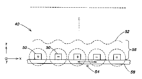
[0029] First, an idealized case for an irradiator apparatus 40 according to
one
embodiment of the present disclosure is considered in Fig. 1(a). The
irradiator apparatus 40
shown in Fig. 1(a) comprises conductors 50 disposed on a substrate 58 and
arranged to form
a parallel array of parallel equally-spaced conductors in an x-y plane defined
by the substrate,
wherein adjacent conductors have an opposite polarity (e.g., an equal and
opposite voltage is
applied to adjacent conductors). To facilitate preliminary analysis, for the
moment the
conductors are considered to be infinitely long in the y-direction and
repeated infinitely in
parallel along the x-direction. As a result of the voltage applied to the
conductors, an electric
field is generated in the vicinity of the conductors above the substrate. If
the generated field
is examined at a large distance in the z- direction normal to the x-y plane,
it is found that the
field is zero. In particular, the conductors with opposite potential cancel
each other such that
there are no electric field lines at large distances. As one moves close to
the array of
conductors, there is a non-zero spatially varying field 52 in a thin region 56
proximate to the
substrate 58 that gets stronger as the distance above the array decreases.
[0030] To calculate the field 52 at distances close to the array, i.e., in the
thin region 56,
due to the periodicity of the array the field 52 may be expressed in terms of
a potential
constituted by a sum of periodic functions in a Fourier series, given by:
O(x, z) = Fõ (z) cos 21rnx , (1)
a
CA 02647382 2008-09-25
WO 2007/106402 PCT/US2007/006103
-7-
where 0 represents the potential as a function of x and z, x denotes position
along the array
parallel to the plane of the array, z denotes the distance from and normal to
the plane of the
array, a is the spacing 54 between adjacent conductors, and n designates the
mode of the
Fourier series. Noting that in the regions above the array there is no net
charge, the potential
must satisfy Laplace's equation:
aZo + aZ0
= 0; (2)
axZ aZ2
47rZnZ 2~rnx d2F 2~tnx
- a? F^ (z) cos a+dZz^ cos a= 0. (3)
With the constraint of Laplace's equation, the harmonics of the Fourier
components of the
field drop off as an exponential with a characteristic distance:
Fn = ,4ne-:lZ, (4)
a
zo (5)
2;rn
Accordingly, with a periodic array of conductors at alternating opposite
potentials, it is
observed that the electrostatic potential drops off at a characteristic
distance based on the
spacing 54 (also referred to as pitch or period) of the conductors. Hence, the
extent of the
thin region 56, normal to the substrate, is determined at least in part by the
conductor spacing
54. In some exemplary embodiments, the value a may be particularly selected
such that the
characteristic distance for the thin region may fall in a range of from
approximately 1
micrometer (beyond which the field falls off sharply), to hundreds of
micrometers (beyond
which the field falls off sharply).
[0031] Thus, according to various embodiments, irradiator apparatus
contemplated herein
operate utilizing the foregoing principals to create oscillating electric or
magnetic fields
whose intensity drops off very sharply beyond a characteristic distance that
delimits a thin
region proximate to a substrate on which the conductors of the apparatus are
disposed.
CA 02647382 2008-09-25
WO 2007/106402 PCT/US2007/006103
-ti-
Hence, a given apparatus irradiates only a thin layer proximate to the
substrate, without
wasting energy by radiating out to the universe.
[0032] In one embodiment, an irradiator apparatus according to the present
disclosure
based on-the concepts illustrated in Fig. 1(a) comprises a number N of
conductors 50 having a
finite length in they-direction and disposed on a substrate 58 in a parallel
equally-spaced
manner along the x-direction. In one exemplary implementation, N may be on the
order of
100, the substrate may be glass, and the overall dimensions of the irradiator
apparatus in the
x-y plane may be on the order of 1 cm2, wherein each conductor has a width
along the x-
direction of approximately 70 mm, a height normal to the substrate in the z-
direction of
approximately 7 mm, and a spacing 54 ("a" in the equations above) of
approximately 200
mm. It should be appreciated that the foregoing examplary parameters are
provided primarily
for purposes of illustration, and that irradiator apparatus according to other
embodiment of
the present disclosure are not limited to the various parameters associated
with this particular
example.
[0033] For the exemplary parameters above, fall-off of the electric field 52
generated by
the irradiator apparatus in the z-direction is shown in a finite-element
simulation in Figs. 1(b)
and 1(c). In particular, Fig. 1(b) shows the computed electric field contours
for an odd
number N of electiodes (in the illustrated example, N= 101), whereas Fig. 1(c)
shows similar
contours for an even number of electrodes (e.g., N = 102). The array of
conductors is
centered at x = 0, and z is the distance from and normal to the plane of the
substrate.
Subsequent contours are the ratio Eõ+,/En 0.8, where n=1 to 20 (n designates
modes of the
Fourier series in the above equations). From Fig. 1(c), it may be appreciated
that for an even
number of electrodes (i.e., for every positive potential there is an opposite
negative potential),
the field goes sharply to zero beyond a localized area comprising a thin
region proximate to
(e.g., above) the substrate (in the figures associated with these particular
examples, on the
order of 50 micrometers).
[0034] In another embodiment, the electric mode variant of an irradiator
apparatus 40
comprises conductors forming a transmission line 60 (two parallel metal lines)
that coils
about in the shape of an octagon, as illustrated in Fig. 2(a). In an exemplary
implementation
of this embodiment, the coiled transmission-line irradiator apparatus 40 may
be fabricated on
a substrate 58 formed by a standard 1" by 3" glass slide, although as
discussed above it
should be appreciated that a variety of other substrates generally may be
suitable. A cross-
CA 02647382 2008-09-25
WO 2007/106402 PCT/US2007/006103
-9-
sectional diagram of such a device is shown in Fig. 2(b). In one aspect, the
octagon-shaped
coil is configured such that the irradiation region is approximately 8
millimeters x 8
millimeters parallel to the plane of the substrate.
[0035] As discussed above in connection with equations (1)-(5), various
spacings a
between the metal lines may be chosen to achieve a desired extent of a thin
region proximate
to the substrate in which power is delivered to a sample. In various
implementations, the
spacing or pitch of the conductors may be selected such that this region in
which power is
delivered ranges from approximately one micrometer to hundreds of micrometers
in a
direction normal to the plane of the transmission line coil. In one example;
metal lines
having a width of approximately 100 micrometers, with a spacing between metal
lines of
approximately 100 micrometers, form an irradiator apparatus similar to that
shown in Fig.
2(a).
[0036] The metal lines may be defined by liftoff of a metal layer (10
nanometers titanium
(Ti), 40 nanometers gold (Au)) following photolithographic patterning. A thick
(5 m) layer
of gold subsequently may be electroplated onto the metal lines with a gold
plating solution,
stirred at 65 C, with a deposition rate of approximately 5 micrometers/hour.
By such
electroplating, the lines are thickened so as to mitigate ohmic heating.
Additionally,
according to another aspect of this embodiment, a thin conformal layer 62
(approximately I
micrometer thick) of Teflon may be spun onto the apparatus to reduce adhesion
between the
sample to be irradiated (or material containing the sample) and the apparatus.
More
generally, any appropriate suface coating may be employed to reduce or prevent
nonspecific
binding or adherence of samples or solutions containing samples to the
apparatus itself.
Other examples of such coatings include, but are not limited to, a thin
film/layer/coating on
the order of micrometers comprising Mylar film, epoxy, nonconductive silicone
rubber, or
silicone grease.
[0037] In other aspects of this exemplary implementation, the irradiator
apparatus shown
in Fig. 2(a) may include electrical contacts in the form of two 1 millimeter
by 1 millimeter
contact pads 64, for example. The apparatus may be driven by a signal
generator 66 that can
provide various signal power levels (e.g., on the order of up to 20 dBm). In
one embodiment,
the signal generator 66 may be implemented as a printed circuit board circuit
that may be
integrated with or coupled to the substrate. A flip-chip pressure connector
may be used to
CA 02647382 2008-09-25
WO 2007/106402 PCT/US2007/006103
-10-
couple the signal generator to the irradiator so as to remove the complication
of wires that
may become a power delivery problem at high frequencies.
[0038] In yet other implementations, a printed circuit (PC) board may be
employed as a
substrate on which the conductors of an irradiator apparatus are formed (e.g.
coiled
transmission line configuration), and the conductors may be formed of
materials other than
titanitum/gold (e.g., copper, lead-coated copper, etc.). As indicated above,
irradiator
apparatus formed on a PC board substrate optionally may be coated with a layer
of epoxy or
other coating to reduce/prevent adhesion between the apparatus and the
sample/solution
containing sample.
[0039] With respect to electrical signals applied to irradiator apparatus
according to the
present disclosure; for signals that have a wavelength much larger than the
size of the field
generating components of an irradiator apparatus (i.e., signals from DC to
approximately
500MHz) a quasi-static approximation may be made, such that the DC analysis
may be
applied to the behavior of the apparatus. For higher frequency signals
approaching the
gigahertz (GHz) range (e.g., microwave radiation), the wavelengths of the
electromagnetic
radiation may approach the same size scale as the dimensions of conductors
used for the
irradiator apparatus, and impedance matching between the signal generator and
the irradiator
apparatus may become important. Accordingly, to improve impedance matching
into the
GHz range, Fig. 3 illustrates the coiled design of Fig. 2(a) implemented with
ground-source
ground terminals.
[0040] According to another embodiment, a magnetic mode variant of an
irradiator
apparatus may comprise a length of wire that coils about itself in a
serpentine pattern, as is
shown in Fig. 4. Magnetic fields do not couple well to electric dipoles, and
as such a
magnetic mode irradiator generally has poor heating efficiency for non-
magnetic materials.
However, such an irradiator can couple very strongly to magnetic particles
(e.g., mangetic
nano-particles), and as such has excellent selectivity for objects impregnated
with magnetic
nano-particles. As above, with respect to an exemplary fabrication process,
the metal lines
may be defined by liftoff of a metal layer (10nm Ti, 40nm Au) following
photolithographic
patterning. A thick (5 m) layer of gold may be electroplated onto the metal
lines with a gold
plating solution, stirred at 65 C, with a deposition rate of --5 m/hr. A thin
conformal layer
(-l m) of Teflon also may be spun onto the apparatus to reduce adhesion of
biomaterial to
the apparatus.
CA 02647382 2008-09-25
WO 2007/106402 PCT/US2007/006103
-11-
[0041] According to yet another embodiment, an additional modality of the
apparatus
disclosed herein includes applying DC offset to the excitation signal applied
frorri the signal
generator to the irradiation apparatus. A DC offset voltage may be applied in
linear
superposition to the AC field, and can be adjusted to a specific proteins
isoelectric point,
tuned to drive antibodies in solution onto tissues or target binding sites.
Similar to isoelectric
focusing based on exact pH characteristics, proteins can be driven out of
solution to their
targets based on the application of an appropriate DC offset.
[0042] In one exemplary application, irradiators according to the present
disclosure may
be used in the enhanced fixation and staining of tissues with bio-markers.
This is illustrated
in Fig. 7 (Act 300). As discussed above, microwave enhanced fixation and
staining is a
common procedure in histology, to date involving large conventional microwave
ovens
which may be replaced by irradiators pursuant to the concepts disclosed
herein, operating at a
variety of possible frequency ranges (e.g., microwave, radio frequency, other
bands). The
illustrations of Figs. 5 and 6 outline how such irradiators may be employed to
deliver power
via electromagnetic radiation to a tissue. In particular, Fig. 5(a) shows an
irradiator apparatus
40 implemented on a glass slide substrate, Fig. 5(b) shows a tissue sample 69
disposed on a
second glass slide substrate 67, and Fig. 5(c) shows the tissue sample/glass
slide overlaying
the irradiator apparatus 40 in a criss-cross manner. Fig. 6 illustrates a
portion of a cross
section of this arragnement, in which one exemplary conductor 50 of the
irradiator apparatus
40 is placed in close proximity to the tissue 69, such that the tissue is
located in the thin
region to which the irradiator apparatus delivers power.
[0043] Accordingly, methods and apparatus according to the present disclosure
are useful
for a wide range of biological and medical procedures. A number of such
applications are
contemplated, including, inter alia, methods directed to biochemical,
histochemical,
histopathological, biomedical, and analytical uses.
[0044] The methods and apparatus disclosed herein are useful for improving one
or more
aspects of a variety of routine analytical and histological procedures
employed in research
and clinical laboratories, as well as in medical/clinical practice. In one
aspect, the various
concepts disclosed herein provide methods for accelerating or enhancing
chemical processes.
As used herein, "chemical processes" shall encompass histological processes,
histochemical
processes, cytochemical processes, immunochemical processes,
immunohistochemical
CA 02647382 2008-09-25
WO 2007/106402 PCT/US2007/006103
-12-
processes, immunocytochemical processes, colometric processes, chemical
processes
involving nanoparticles, electrochemical processes, etc.
.[0045] In typical embodiments, the methods involve obtaining a biological
sample to be
analyzed or histologically processed, performing an appropriate histochemical
process or
processes using a suitable reagent or reagents, and during one or more steps
of such
procedures, allowing the biological sample to be exposed to an electromagnetic
field defined
herein. The degree (intensity/level and duration) to which the biological
sample is subjected
to the electromagnetic field will depend on a number of factors, such as the
type of the '
biological sample, thickness of the sample (e.g., tissue sections), the nature
of the histological
process, intrinsic sensitivity of the assay or procedures being performed, and
so on. In
general, histochemical processes of biological samples include multiple steps,
such as
fixation, staining, incubations, washing, etc. Thus, the present invention may
be applied to
one or more of these steps to improve general outcome of chemical, and/or
related analytical
procedures.
100461 As used herein, the terms "accelerating" "accelerate" and
"accelerating" shall
mean that the amount of time required to obtain reasonably reliable outcome
that is
equivalent in quality as obtained by conventional methods is shortened. For
example, a
staining process that typically requires by conventional methods several hours
to overnight
may be reduced to in an order of seconds to minutes by the methods disclosed
herein.
Similarly, each of multiple incubation and intervening washing periods
associated with a
typical chemical procedure may be shortened significantly using the methods of
the
invention.
(0047] The terms "enhancing" "enhance" and "enhancement" refer to improvement
in the
overall quality of a product, process, and/or data, as compared to
conventional methods that
are available. For example, data acquired according to one or more embodiments
of the
present invention may be enhanced by a heightened signal-to-noise ratio. That
is, the
methods described herein may increase a specific signal and/or reduce
background (or noise)
so that the resulting products, processes and/or data are of better quality.
Using the methods
provided herein, the invention also allows generating comparable results using
significantly
less volume of reagents required for performing one or more steps of these
processes. As a
result, the invention may realize significant cost reduction, particularly in
situations where a
large number of samples are processed, or in cases where reagents are limited
in quantity or
CA 02647382 2008-09-25
WO 2007/106402 PCT/US2007/006103
- 13-
costly. Depending on the particular sample, the nature of the technique, and
also depending
on the type of substrate being used, a typical reaction may require a reagent
volume of in the
order of microlitters - such as 1, 2, 5, 10, 25, 50, 100 microliters. In
certain embodiments of
the invention, the histochemical processes described herein shall embrace
immunohistochemical processes.
100481 Immunohistochemistry involves the localization of antigens in a cell or
tissue
section by the use of labeled antibodies as specific reagents through antigen-
antibody
interactions that are visualized by a marker such as fluorescent dye, enzyme,
radioactive
element, colored dye, marker, stain or colloidal gold. Therefore,
immunohistochemistry has
become a crucial technique and widely used in many medical research
laboratories as well as
clinical diagnostics. The technique offers a wide range of variations and
modified protocols,
which the art is familiar with. The selection of a suitable method should be
based on
parameters such as the type of specimen under investigation and the degree of
sensitivity
required. A skilled partisan will be able to determine a suitable application
in incorporating
the methods and uses taught in the invention as disclosed herein.
[00491 In some embodiments, the methods of the invention are used for
histochemical
processes involving a cross-linking process. Cross-links are covalent bonds
linking one
polymer chain to another. In biology, cross-linking has applications in
forming
polyacrylamide or agarose gels for gel electrophoresis in studies of proteins
and/or nucleic
acids, as well as other matrices including those used as a substrate for cell
culture and tissue
engineering. The term also encompasses cross-linking compounds that are used
to
selectively couple a chemical constituent of a moleule. For example, a variety
of crosslinker
are used to study subunit conformation of proteins. This is deduced since
crosslinkers only
bind surface amino residues in relatively close proximity in the native state.
Examples of
crosslinkers are dimethyl suberimidate and glutaraldehyde. Both induce
nucleophilic attack
of the amino group of lysine and their subsequent covalent bonding via the
crosslinker.
However, the methods described herein may be useful for any other chemical
crosslinkers.
[00501 In yet other cases, however, cross-linking may involve more general
"fixing" such
as fixation of a cell or tissue for primarily preservation purposes. In the
fields of histology,
pathology, and cell biology, fixation is a chemical process by which
biological tissues are
preserved from decay. Fixation terminates any ongoing biochemical reactions,
and may also
increase the mechanical strength or stability of the treated tissues. Thus,
the main purpose of
CA 02647382 2008-09-25
WO 2007/106402 PCT/US2007/006103
-14-
fixation is to preserve a sample of biological material, such as tissue or
cells, to permit stable
storage and analysis. To achieve this goal, several conditions must usually be
met. First, a
fixative usually acts to disable intrinsic biomolecules - particularly
proteolytic enzymes -
which would otherwise digest or otherwise damage the sample. Second, a
fixative will
typically protect a sample from extrinsic damage. Many fixatives are toxic to
most common
microorganisms (bacteria in particular) which might exist in a tissue sample
.or which might
otherwise colonize the fixed tissue. In addition, many fixatives will
chemically alter the fixed
material to make it less palatable (either indigestible or toxic) to
opportunistic
microorganisms. Finally, fixatives often alter the cells or tissues on a
molecular level to
increase their mechanical strength or stability. This increased strength and
rigidity can help
preserve the morphology of the sample as it is processed for further analysis.
Fixation is
usually the first stage in a multistep process to prepare a sample of
biological material for
microscopy or other analysis. Therefore, the choice of fixative and fixation
protocol will
depend heavily on the additional processing steps and final analyses that are
planned. For
example, immunohistochemistry utilises antibodies which bind to a specific
protein target.
The use of the present invention is not limited to a particular fixative or
histochemical
procedure, and thus may be adapted for use in conjunction with any of the
methods described
herein and the like.
[0051] Crosslinking fixatives act by creating covalent chemical bonds between
proteins
in tissue. This anchors soluble proteins to the cytoskeleton, and lends
additional rigidity to the
tissue. Accordingly, the present invention contemplates improving aspects of
such fixation
procedures (by accelerating or enhancing the process) that are commonly
employed. In some
embodiments, the invention is used for histochemical process involving the
crosslinking
fixative, formaldehyde (often sold as a saturated aqueous solution under the
name formalin).
Formaldehyde is thought to interact primarily with the residues of the basic
amino acid
lysine. In some embodiments, the invention is used with glutaraldehyde. While
it is believed
to operate by a similar mechanism to formaldehyde, as a somewhat.larger
molecule,
glutaraldehyde may not penetrate thicker tissue specimens as effectively as
formaldehyde.
On the other hand, glutaraldehyde may offer a more rigid or tightly linked
fixed product-its
greater length and two aldehyde groups allow it to 'bridge' and link more
distant pairs of
protein molecules. In yet other cases, fixation protocols call for a
combination of
formaldehyde and glutaraldehyde, so that their respective strengths complement
one another.
Examples of common fixative solutions used for immunohistochemistry include
the
CA 02647382 2008-09-25
WO 2007/106402 PCT/US2007/006103
-15-
followings: (a) 4% paraformaldehyde in O.IM phosphate buffer; (b) 2%
paraformaldehyde
with 0.2% picric acid in 0.1 M phosphate buffer; (c) PLP fixative: 4%
paraformaldehyde,
0.2% periodate and 1.2% lysine in 0.1 M phosphate buffer; and (d) 4%
paraformaldehyde
with 0.05% glutaraldehyde (electron microscopy immunohistochemistry). However,
it is
understood that a skilled partisan may make modifications to optimize
conditions to suit a
particular use.
[0052] Yet in other embodiments, oxidizing agents are used. The oxidising
fixatives can
react with various side chains of proteins and other biomolecules, allowing
the formation of
crosslinks which stabilize tissue structure. For example, osmium tetroxide is
often used as a
secondary fixative when samples are prepared for electron microscopy.
Potassium
dichromate, chromic acid, and potassium permanganate all find use in certain
specific
histological preparations.
[0053] The invention may be used for fixation procedure involving fixatives
which are
characterized as precipitating fixatives. Precipitating (or denaturing)
fixatives act by
essentially reducing the solubility of protein molecules and often by
disrupting the
hydrophobic interactions which give many proteins their tertiary structure.
The precipitation
and aggregation of proteins is a very different process from the crosslinking
which occurs
with the aldehyde fixatives. The most common precipitating fixatives include
ethanol and
methanol. Acetone is also used.
[0054) Acetic acid is a denaturant that is soinetimes used in combination with
the other
precipitating fixatives. The alcohols, by themselves, are known to cause
shrinkage of tissue
during fixation while acetic acid alone is associated with tissue swelling;
combining the two
may result in better preservation of tissue morphology. In certain
circumstances, the
invention may be also used in a fixation process using fixative agents that
contain picric acid
and mercuric chloride. In any of the above situations, the methods desclosed
herein may
accelerate and/or enhance the process of fixation.
[0055] Similarly, the invention finds applications in improving chemical or
histochemical
processes involving staining. Stains and dyes are frequently used in biology
and medicine to
highlight structures in biological tissues for viewing, often with the aid of
different
microscopes. Stains may be used to define and examine bulk tissues
(highlighting, for
example, muscle fibers or connective tissue), cell populations (classifying
different blood
CA 02647382 2008-09-25
WO 2007/106402 PCT/US2007/006103
-16-
cells, for instance), or organelles within individual cells. Thus, staining is
a biochemical
technique of adding a class-specific (DNA; proteins, lipids, carbohydrates)
dye to a substrate
to qualify or quantify the presence of a specific compound. For example,
biological staining
can be used to mark cells in flow cytometry, and to flag proteins or nucleic
acids in gel
electrophoresis. Thus, the invention in some embodiments embraces methods for
accelerating and/or enhancing these procedures.
[0056] There are a number of staining processes that may benefit frorri the
invention
disclosed herein. Not intending to be limiting, these include: Acid Fast
Bacilli Staining,
Alcian Blue Staining, Alcian Blue/PAS Staining, Alizarin Red Staining,
Alkaline
Phosphatase Staining, Azure A Staining, Bielschowsky Staining, Congo Red
Staining, Diff-
Quik Staining, Diff-Quik II Stain for Helicobacter pylori, Fite Faraco
Staining, Giemsa
Staining, Golgi Staining, Golgi-Cox Staining, Gomori's Trichrome Staining,
Gordon Sweet's
Staining, Gram Staining, Grocott Methenamine Staining, Haematoxylin and Eosin
Staining,
Hyaluronidase Alcian Blue Staining, Luna Staining, Luxol Fast Blue Staining,
Masson
Fontana Staining, Masson Trichrome Staining, Methenamine Sliver Staining,
Microglia
Staining, Miller's Elastic Staining, Nissl Staining, Oil Red 0 Staining, PAS
Staining, PAS
Diastase Staining, Perls Prussian Blue Staining, Pouchet Staining, Prussian
Blue Staining,
Renal Alcian Blue/PAS Staining, Renal Masson Trichrome Staining, Renal PAS
Methenamine Staining, Rhodanine Staining, Safranin 0 Staining, Sirius Red for
Collagen
Staining, Southgate's Mucicarmine Staining, Toluidine Blue Staining, van
Gieson Staining,
von Kossa Staining, VVG Staining, X-Gal Staining and Ziehl Neelsen Staining.
Some are
further discussed below.
[0057] The amount of time required for completing a staining process greatly
varies, but
in any case, the overall process may be accelerated when the present invention
is applied. At
its simplest, the actual staining process may involve immersing the sample
(before or after
fixation and mounting) in dye solution, followed by rinsing and observation.
Many dyes,
however, require the use of a mordant: a chemical compound which reacts with
the stain to
form an insoluble, coloured precipitate. When excess dye solution is washed
away, the
mordanted stain remains. Gram staining is used to determine gram status.to
classify bacteria
broadly. It is based on the composition of their cell wall. Gram staining uses
crystal violet to
stain cell walls, iodine as a mordant, and a fuchsin or safranin counterstain
to mark all
bacteria. Gram status is important in medicine; the presence or absence of a
cell wall will
CA 02647382 2008-09-25
WO 2007/106402 PCT/US2007/006103
-17-
change the bacterium's susceptibility to some antibiotics. Gram-positive
bacteria stain dark
blue or violet. Their cell wall is typically rich with peptidoglycan and lacks
the secondary
membrane and lipopolysaccharide layer found in Gram-negative bacteria. These
differential
characteristics, therefore, may aid a histopathological analysis and
subsequent diangnosis of a
disease or disorder. Accordingly, the present invention may accelerate such
process.
[0058] Haematoxylin and eosin staining protocol is used frequently in
histology to
examine thin sections of tissue, and thus a useful tool in pathology.
Haematoxylin stains cell
nuclei blue, while eosin stains cytoplasm and connective tissue pink or red.
Thus, the
invention includes methods for speeding up a process of Haematoxylin staining
of a patient
specimen, for example, during a surgery. In addition, Eosin is strongly
absorbed by red
blood cells, colouring them bright red. Such property may be used in analyzing
blood
samples. Applying this to the present inevntion, it is possible to greatly
improve such
analytical procedures.
[0059] Papanicolaou staining, or Pap staining, is a frequently used method for
examining
cell samples from various bodily secretions. It is frequently used to stain
the Pap smear
specimens. In general, it uses a combination of haematoxylin, Orange G, eosin
Y, Light
Green SF yellowish, and sometimes Bismarck Brown Y. In some embodiments,
therefore,
the invention contemplates accelerating and/or enhancing the process of Pas
smear tests. For
example, the invention may realize an in-visit Pap smear test, where a patient
may obtain a
result of a test during a single visit to her physician's office, as opposed
to receiving a result
on a later date.
[0060] Similarly, Periodic acid-Schiff staining (PAS staining) is used for
demonstrating
carbohydrates (including glycogen, glycoprotein, proteoglycans). It is used to
distinguish
different types of glycogen storage diseases. Therefore, some embodiments of
the invention
relate to improving the process of diagnosing and/or monitoring such diseases,
based on more
rapid PAS staining.
[0061] In some embodiments, the invention is used for applications involving
staining
protocols for Masson's trichrome, which is a three-colour staining protocol
well-suited to
distinguish cells from surrounding connective tissue. Most recipes will
produce red keratin
and muscle fibers, blue or green staining of collagen and bone, light red or
pink staining of
cytoplasm, and black cell nuclei.
CA 02647382 2008-09-25
WO 2007/106402 PCT/US2007/006103
-18-
[0062] Yet in other embodiments, the methods are provided to enhance staining
process
of the Romanowsky stains, which are all based on a combination of eosinate
(chemically
reduced eosin) and methylene blue (sometimes with its oxidation products azure
A and azure
B). Common variants include Wright's stain, Jenner's stain, Leishman stain and
Giemsa
stain. All can be used to examine blood or bone marrow samples. They are
generally
preferred over H&E for inspection of blood cells because different types of
leukocytes (white
blood cells) can be readily distinguished. All are also suited to examination
of blood to
detect blood-borne parasites like malaria.
[0063] In certain cases, the methods provided herein may be applied to Silver
staining, in
which silver is used to stain histologic sections. This kind of staining is
important especially
to show proteins (for example type III collagen) and DNA. It is used to show
both substances
inside and outside cells. For instance, some cells are argentaffin. These
reduce silver solution
to metallic silver after formalin fixation. This method is based on a reaction
between silver
nitrate and potassium dichromate, thus precipitating silver chromate in some
cells. Other
cells are argyrophilic. These reduce silver solution to metallic silver after
being exposed to
the stain that contains a reductant, for example hydroquinone or formalin.
[0064] Still in other cases, the invention provides methods for improving
Sudan staining.
Sudan staining takes advantage of Sudan dyes to stain sudanophilic substances,
usually lipids.
Sudan III, Sudan IV, Oil Red 0, and Sudan Black B are often used. Sudan
staining is often
used to determine the level of fecal fat to diagnose steatorrhea. Thus, the
methods according
to the present invention can significantly speed up the process.
[0065] In certain embodiments, the invention is useful for improving in vivo
staining. In
vivo staining is the process of dyeing living cells or tissues. By causing
certain cells or
structures to take on contrasting color(s), their morphology or position
within a cell or tissue
can be readily seen and studied. The usual purpose is to reveal cytological
details that might
otherwise not be apparent; however, staining can also reveal where certain
chemicals or
specific chemical reactions are taking place within cells or tissues. As would
be clear to
those skilled in the art, such methods can offer valuable advantage for a
number of clinical
and analytical applications.
[0066] Often these stains are called vital stains. They are introduced to the
organism
while the cells are still living. However, these stains are eventually toxic
to the organism,
CA 02647382 2008-09-25
WO 2007/106402 PCT/US2007/006103
-19-
some more so than others. To achieve desired effects, the stains are used in
very dilute
solutions ranging from 1:5,000 to 1:500,000. Note that many stains may be used
in living
cells, such as primary cells grown in culture.
[0067] There are many effective biological stains available in the art.
Different stains
react or concentrate in different parts of a cell or tissue, and these
properties are used to
advantage to reveal specific parts or areas. Generally, these dyes may be used
with fixed
cells and tissues, and some are particularly suitable for use with living
organisms ("vital
dyes"). Non-limiting examples of biological stains that are commonly used
include:
Bismarck brown, Carmine, Coomassie blue, Crystal violet, DAPI, Eosin, Ethidium
bromide,
Fuchsin, Haematoxylin, Hoechst stains, Iodine, Malachite green, Methyl green,
Methylene
blue, Neutral red, Nile blue, Nile red, Osmium tetroxide, Rhodamine, Safranin.
[0068] Similar to light microscopy, stains can be used to selectively
highlight cellular
structures in transmission electron microscopy, and thus the present invention
also includes
methods of acclerating and/or enhancing one or more steps of preparing
biological samples
for EM analysis. Electron-dense compounds of heavy metals are typically used.
For
example, phosphotungstic acid is a common negative stain for viruses, nerves,
polysaccharides, and other biological tissue materials. Other chemicals used
in electron
microscopy staining include ammonium molybdate, cadmium iodide,
carbohydrazide, ferric
chloride, hexamine, indium trichloride, lanthanum nitrate, lead acetate, lead
citrate, lead(II)
nitrate, osmium tetroxide, periodic acid, phosphomolybdic acid, potassium
ferricyanide,
potassium ferrocyanide, Ruthenium Red, silver nitrate, sodium chloroaurate,
thallium nitrate,
thiosemicarbazide, uranyl acetate, uranyl nitrate, and vanadyl sulfate.
[006.9] Throughout the application disclosed herein, where so desired,
immunodetection
may be carried out by any number of available protocols of choice, which will
benefit when
used in conjunction with the methods provided herein. Target detection used in
any of the
methods of the present invention as described herein, including chemical
assays,
histochemical processes, immuno-affinity assays, binding assays, screenings,
and the like,
generally employs detectable label or labels, which are either colorimetric or
fluorometric in
nature, or combination thereof. In certain circumstances, these detectable
labels are nano-
particles. For example, fluorescent nano-particles conjugated to a primary or
secondary
antibody, for instance, are particularly advantagous reagents since they do
not fade after
CA 02647382 2008-09-25
WO 2007/106402 PCT/US2007/006103
-20-
exposure to fluorescent light, whereas many chemical dyes commonly do. An
exemplary
immunohistochemical procesure is outlined in Fig. 7.
[0070] Common fluorophores include but are not limited to: 1,5 IAEDANS; 1,8-
ANS; 4-
Methylumbelliferone; 5-carboxy-2,7-dichlorofluorescein; 5-Carboxyfluorescein
(5-FAM); 5-
Carboxynapthofluorescein (pH 10); 5-Carboxytetramethylrhodamine (5-TAMRA); 5-
FAM
(5-Carboxyfluorescein); 5-HAT (Hydroxy Tryptamine); 5-Hydroxy Tryptamine
(HAT); 5-
ROX (carboxy-X-rhodamine); 5-TAMRA (5-Carboxytetramethylrhodamine); 6-
Carboxyrhodamine 6G; 6-CR 6G; 6-JOE; 7-Amino-4-methylcoumarin; 7-
Aminoactinomycin
D (7-AAD); 7-Hydroxy-4-methylcoumarin; 9-Amino-6-chloro-2-methoxyacridine;
ABQ;
Acid Fuchsin; ACMA (9-Amino-6-chloro-2-methoxyacridine); Acridine Orange +
DNA;
Acridine Orange + RNA; Acridine Orange, both DNA & RNA; Acridine Red; Acridine
Yellow; Acriflavin; Acriflavin Feulgen SITSA; Aequorin (Photoprotein); Alexa
Fluor 350TM;
Alexa Fluor 430TM; Alexa Fluor 488TM; Alexa Fluor 532TM; Alexa Fluor 546TM;
Alexa Fluor
568TM; Alexa Fluor 594TM; Alexa Fluor 633TM; Alexa Fluor 647TM; Alexa Fluor
660TM;
Alexa Fluor 680TM; Alizarin Complexon; Alizarin Red; Allophycocyanin (APC);
AMC,
AMCA-S; AMCA (Aminomethylcoumarin); AMCA-X; Aminoactinomycin D;
Aminocoumarin; Aminomethylcoumarin (AMCA); Anilin Blue; Anthrocyl stearate;
APC
(Allophycocyanin); APC-Cy7; APTRA-BTC = Ratio Dye, Zn2+; APTS; Astrazon
Brilliant
Red 4G; Astrazon Orange R; Astrazon Red 6B; Astrazon Yellow 7 GLL; Atabrine;
ATTO-
TAGTM CBQCA; ATTO-TAGTM FQ; Auramine; Aurophosphine G; Aurophosphine; BAO 9
(Bisaminophenyloxadiazole); BCECF (high pH); BCECF (low pH); Berberine
Sulphate; Beta
Lactamase; BFP blue shifted GFP (Y66H); Blue Fluorescent Protein; BFP/GFP FRET
Bimane; Bisbenzamide; Bisbenzimide (Hoechst); bis-BTC = Ratio Dye, Zn2+;
Blancophor
FFG; Blancophor SV; BOBOTM -1; BOBOTM -3; Bodipy 492/5 15; Bodipy. 493/5 03;
Bodipy
500/510; Bodipy 505/515; Bodipy 530/550; Bodipy 542/563; Bodipy 558/568;
Bodipy
564/570; Bodipy 576/589; Bodipy 581/591; Bodipy 630/650-X; Bodipy 650/665-X;
Bodipy
665/676; Bodipy Fl; Bodipy FL ATP; Bodipy Fl-Ceramide; Bodipy R6G SE; Bodipy
TMR;
Bodipy TMR-X conjugate; Bodipy TMR-X, SE; Bodipy TR; Bodipy TR ATP; Bodipy TR-
X
SE; BO-PROTM -1; BO-PROTM -3; Brilliant Sulphoflavin FF; BTC - Ratio Dye Ca2+;
BTC-
5N - atio Dye, Zn2+; Calcein; Calcein Blue; Calcium CrimsonTM; Calcium Green;
Calcium
Green-1 Ca2+ Dye; Calcium Green-2 Ca2+; Calcium Green-5N Ca2+; Calcium Green-
C18
Ca2+; Calcium Orange; Calcofluor White; Carboxy-X-rhodamine (5-ROX); Cascade
BIueTM;
Cascade Yellow 399; Catecholamine; CCF2 (GeneBlazer); CFDA; CFP - Cyan
Fluorescent
CA 02647382 2008-09-25
WO 2007/106402 PCT/US2007/006103
-21 -
Protein; CFP/YFP; FRET; Chlorophyll; Chromomycin A; Chromomycin A; CL-NERF
(Ratio Dye, pH); CMFDA; Coelenterazine; Coelenterazine cp (Ca2+ Dye,);
Coelenterazine f;
Coelenterazine fcp; Coelenterazine h; Coelenterazine hcp; Coelenterazine ip;
Coelenterazine
n; Coelenterazine 0; Coumarin Phalloidin; C-phycocyanine; CPM Methylcoumarin;
CTC;
CTC Formazan; Cy2TM; Cy3.1 8; Cy3.5TM; Cy3TM; Cy5.1 8; Cy5.5TM; Cy5TM; Cy7TM;
Cyan
GFP; cyclic AMP Fluorosensor (FiCRhR); CyQuant Cell Proliferation Assay;
Dabcyl;
Dansyl; Dansyl Amine; Dansyl Cadaverine; Dansyl Chloride; Dansyl DHPE; Dansyl
fluoride; DAPI; Dapoxyl; Dapoxyl 2; Dapoxyl 3; DCFDA; DCFH
(Dichlorodihydrofluorescein Diacetate); DDAO; DHR (Dihydorhodamine 123); Di-4-
ANEPPS; Di-8-ANEPPS (non-ratio); DiA (4-Di-16-ASP); Dichlorodihydrofluorescein
Diacetate (DCFH); DiD - Lipophilic Tracer; DiD (DiIC 18(5)); DIDS;
Dihydorhodamine 123
(DHR); Dil (DiIC 18(3 )); Dinitrophenol; DiO (DiOC 18(3)); DiR; DiR (DiIC
18(7)); DM-
NERF (high pH); DNP; Dopamine; DsRed; Red fluorescent protein; DTAF; DY-630-
NHS;
DY-635-NHS; EBFP; ECFP; EGFP; ELF 97; Eosin; Erythrosin; Erythrosin ITC;
Ethidium
Bromide; Ethidium homodimer -1 (EthD-1); Euchrysin; EukoLight; Europium (III)
chloride;
EYFP; Fast Blue; FDA; Feulgen (Pararosaniline); FIF (Formaldehyd Induced
Fluorescence);
FITC; FITC Antibody; Flazo Orange; Fluo-3; Fluo-4; Fluorescein (FITC);
Fluorescein
Diacetate; Fluoro-Emerald; Fluoro-Gold (Hydroxystilbamidine); Fluor-Ruby;
FluorX; FM 1-
43TM; FM 4-46; Fura RedTM (high pH); Fura RedTM/Fluo-3; Fura-2, high calcium;
Fura-2,
low calcium; Fura-2/BCECF; Genacryl Brilliant Red B; Genacryl Brilliant Yellow
10GF;
Genacryl Pink 3G; Genacryl Yellow 5GF; GeneBlazer (CCF2); GFP (S65T); GFP red
shifted
(rsGFP); GFP wild type, non-UV excitation (wtGFP); GFP wild type, UV
excitation
(wtGFP); GFPuv; Gloxalic Acid; Granular Blue; Haematoporphyrin; Hoechst 33258;
Hoechst 33342; Hoechst 34580; HPTS; Hydroxycoumarin; Hydroxystilbamidine
(FluoroGold); Hydroxytryptamine; Indo-1, high calcium; Indo-1, low calcium;
Indodicarbocyanine (DiD); Indotricarbocyanine (DiR); Intrawhite Cf; JC-1; JO-
JO-1; JO-
PRO-1; LaserPro; Laurodan; LDS 751 (DNA); LDS 751 (RNA); Leucophor PAF;
Leucophor
SF; Leucophor WS; Lissamine Rhodamine; Lissamine Rhodamine B; LIVE/DEAD Kit
Animal Cells, Calcein/Ethidium homodimer; LOLO-1; LO-PRO-1; Lucifer Yellow;
Lyso
Tracker Blue; Lyso Tracker Blue-White; Lyso Tracker Green; Lyso Tracker Red;
Lyso
Tracker Yellow; LysoSensor Blue; LysoSensor Green; LysoSensor Yellow/Blue; Mag
Green;
Magdala Red (Phloxin B); Mag-Fura Red; Mag-Fura-2; Mag-Fura-5; Mag-Indo-1;
Magnesium Green; Magnesium Orange; Malachite Green; Marina Blue; Maxilon
Brilliant
Flavin 10 GFF; Maxilon Brilliant Flavin 8 GFF; Merocyanin; Methoxycoumarin;
Mitotracker
CA 02647382 2008-09-25
WO 2007/106402 PCT/US2007/006103
-22-
Green FM; Mitotracker Orange; Mitotracker Red; Mitramycin; Monobromobimane;
Monobromobimane (mBBr-GSH); Monochlorobimane; MPS (Methyl Green Pyronine
Stilbene); NBD; NBD Amine; Nile Red; Nitrobenzoxadidole; Noradrenaline;
Nuclear Fast
Red; Nuclear Yellow; Nylosan Brilliant Iavin E8G; Oregon Green; Oregon Green
488-X;
Oregon GreenTM; Oregon GreenTM 488; Oregon GreenTM 500; Oregon GreenTM 514;
Pacific
Blue; Pararosaniline (Feulgen); PBFI; PE-Cy5; PE-Cy7; PerCP; PerCP-Cy5.5; PE-
TexasRed
[Red 613]; Phloxin B (Magdala Red); Phorwite AR; Phorwite BKL; Phorwite Rev;
Phorwite
RPA; Phosphine 3R; PhotoResist; Phycoerythrin B [PE]; Phycoerythrin R [PE];
PKH26
(Sigma); PKH67; PMIA; Pontochrome Blue Black; POPO-1; POPO-3; PO-PRO-1; PO-PRO-
3; Primuline; Procion Yellow; Propidium Iodid (PI); PyMPO; Pyrene; Pyronine;
Pyronine B;
Pyrozal Brilliant Flavin 7GF; QSY 7; Quinacrine Mustard; Red 613 [PE-
TexasRed];
Resorufin; RH 414; Rhod-2; Rhodamine; Rhodamine 110; Rhodamine 123; Rhodamine
5
GLD; Rhodamine 6G; Rhodamine B; Rhodamine B 200; Rhodamine B extra; Rhodamine
BB; Rhodamine BG; Rhodamine Green; Rhodamine Phallicidine; Rhodamine
Phalloidine;
Rhodamine Red; Rhodamine WT; Rose Bengal; R-phycocyanine; R-phycoerythrin
(PE);
rsGFP; S65A; S65C; S65L; S65T; Sapphire GFP; SBFI; Serotonin; Sevron Brilliant
Red 2B;
Sevron Brilliant Red 4G; Sevron Brilliant Red B; Sevron Orange; Sevron Yellow
L;
sgBFPTM; sgBFPTM (super glow BFP); sgGFPTM; sgGFPTM (super glow GFP); SITS;
SITS
(Primuline); SITS (Stilbene Isothiosulphonic Acid); SNAFL calcein; SNAFL- 1;
SNAFL-2;
SNARF calcein; SNARFI; Sodium Green; SpectrumAqua; SpectrumGreen;
SpectrumOrange; Spectrum Red; SPQ (6-methoxy-N-(3-sulfopropyl)quinolinium);
Stilbene;
Sulphorhodamine B can C; Sulphorhodamine G Extra; SYTO 11; SYTO 12; SYTO 13;
SYTO 14; SYTO 15; SYT; SYTO 17; SYTO 18; SYTO 20; SYTO 21; SYTO 22; SYTO 23;
SYTO 24; SYTO 25; SYTO 40; SYTO 41; SYTO 42; SYTO 43; SYTO 44; SYTO 45;
SYTO 59; SYTO 60; SYTO 61; SYTO 62; SYTO 63; SYTO 64; SYTO 80; SYTO 81;
SYTO 82; SYTO 83; SYTO 84; SYTO 85; SYTOX Blue; SYTOX Green; SYTOX Orange;
Tetracycline; Tetramethylrhodamine (TRITC); Texas RedTM; Texas Red-XTM
conjugate;
Thiadicarbocyanine (DiSC3); Thiazine Red R; Thiazole Orange; Thioflavin 5;
Thioflavin S;
Thioflavin TCN; Thiolyte; Thiozole Orange; Tinopol CBS (Calcofluor White);
TMR; TO-
PRO-1; TO-PRO-3; TO-PRO-5; TOTO-1; TOTO-3; TriColor (PE-Cy5); TRITC;
TetramethylRodamineIsoThioCyanate; True Blue; TruRed; Ultralite; Uranine B;
Uvitex SFC;
wt GFP; WW 781; X-Rhodamine; XRITC; Xylene Orange; Y66F; Y66H; Y66W; Yellow
GFP; YFP; YO-PRO-1; YO-PRO-3; YOYO-1 and YOYO-3.
CA 02647382 2008-09-25
WO 2007/106402 PCT/US2007/006103
-23-
[0071] Apart from the fluorescence-based detection protocols, which are used
to discern
the presence or pattern of a target molecule or molecules (such as an antigen)
either spacitally
(tissue distribution, localization, etc.) or phenotypically (expression
levels, etc.), chromogen-
based visualization protocols are also available, either on their own or in
combination with
fluorescence detection. Preferred chromogens include diaminobenzidine (DAB),
but many
other chromogens are also available. In some cases, staining is intensified by
addition of a
second factor such as heavy metal ions, including nickel and cobalt. One or
more steps of
detection procedures, such as staining (incubation) and color development are
enhanced by
exposing the sample to the irradiation disclosed herein. Commonly used
chromogen
substrate solutions include the following: DAB-Peroxidase Substrate Solution
(Brown);
DAB-Peroxidase Substrate Soluiton (Gray); DAB-Peroxidase Substrate Solution
(Black);
DAB-Peroxidase Substrate Solution (Blue); AEC-Peroxidase Substrate Solution
(Red);
BDHC-Peroxidase Substrate Solution (Blue); TMB-Peroxidase Substrate Solution
(Blue);
New Fuchsin Alkaline Phosphatase Substrate Sulution (Red); BCIP/NBT Alkaline
Phosphatase Substrate Solution (Blue).
[0072] Where the present invention is used in conjunction with a technique or
assay
system that is immuno-affinity-based, the invention may facilitate any such
process by
promoting chemical reactions or molecular intaractions. Typical immuno-
affinity reagents
that are used in the art include: an antibody, an antigen-binding fragment
thereof, and other
engineered derivatives thereof, including so-called Affibody molecules, all
of which are
discussed in further detail elsewhere herein. Furthermore, it should be
appreciated that such
immuno-affinity agents may be used for determining spatial distributions of a
target molecule
of interest (for example, localization of an antigen in a cell or tissue), as
well as for
compositional determination by measuring quantities or comparative levels of a
target
molecule of interest present in a sample (for example, immunoprecipitation or
fluorometric
assays).
[0073] As a more specific example of how the present invention is applied to
facilitate
existing methodology, the methods provided herein can be easily adapted for
steps involved
in identification, detection and/or measurement of a known biological marker
or markers
present in a sample. For example, microwave can be used in immunofluorescence
technique,
such as double immunocytochemical staining. It has been shown that moderate
microwaving
does not elute antibodies, but prevents their reactions with subsequently
applied reagents.
CA 02647382 2008-09-25
WO 2007/106402 PCT/US2007/006103
-24-
Thus, using the methods presented herein, microwaving performed in between the
first and
second staining cycles permits improved double indirect immunofluorescence
staining with
antibodies raised in the same species. Moreover, microwaving also inhibits
reactions with
endogenous immunoglobulins present in extracellular compartments. This
substantially
reduces background in indirect immunostaining of mouse tissues with mouse
monoclonal
antibodies, for instance, and further enhance the results, as compared to
those obtaind using a
conventional microwave oven.
[0074] Diagnostic immunohistology (DIHC), therefore, is an essential
discipline that
provides the accurate identification of infectious organisms, distinction
between
morphologically-similar undifferentiated tumors, separation of benign and
malignant
neoplasms, and prognostication of malignancies. The technology often directly
affects
prognosis, selection of therapy, as well as patients' response to treatment.
Therefore,
improved methods for diagnostic immunohistology that allow faster, more
accurate results
are of much interest, and the methods provided herein embrace such
improvement. Such
methods may involve a variety of tissue types and cell types. For example, the
methods of
the present invention are useful for diagnostic, as well as prognostic
processes of a disease or
disorder, cancer in particular, involving tissues and/or cells including:
nervous system, breast
cancers, skin cancers, renal cell carcinoma, prostate cancer, lung cancers,
gastronintestinal
stromal tumor, bone lesions, nasal and paranasal sinus tumors, melanoma,
hodgkin and non-
hodgkin lymphomas, vascular neoplasms, uterus tumors, thyroid cancer,
pleomorphic
sarcomas, among others.
[0075] Commercially available reagents that may be used to determine cells and
tissues
of epithelial and/or endothelial origin include: CA19-9 antibody [241]; CD166
antibody
[3A6] (FITC); CD 166 antibody [L50]; Cytokeratin 13 antibody [ 1 C7];
Cytokeratin 13
antibody [AE81; Cytokeratin 13 antibody [KS-1A3]; Cytokeratin 13 antibody [KS-
I A3]; Cytokeratin 4 antibody [6B 10]; Cytokeratin 4 antibody [6B 10]; D240
antibody [D2-
40]; prediluted Differentiated Endothelial Cells antibody [1F10 ]; EBP50
antibody; EBP50
antibody [EBP-10]; Endothelial Cell antibody [BW-200 ]; Endothelial Cell
antibody [PAL-
E]; Endothelial Cell antibody [RECA-1]; Endothelium antibody [1.BB.803];
Endothelium
antibody [EN4]; Endothelium antibody [MRC OX43] (FITC); Endothelium antibody
[PAL-
E]; EpCAM antibody [0.N.277] - BSA and Azide free; EpCAM antibody [AUA1];EpCAM
antibody [B29.1 (VU-ID9)]; EpCAM antibody [B29.1 (VU-ID9)] (FITC); EpCAM
antibody
CA 02647382 2008-09-25
WO 2007/106402 PCT/US2007/006103
- 25 -
[B302 (323/A3)]; EpCAM antibody [B302 (323/A3)]; EpCAM antibody [Ber-EP4];
EpCAM
antibody [E144]; EpCAM antibody [HEA125]; prediluted EpCAM antibody [VU-
1 D9]; EpCAM antibody [VU-1 D9]; prediluted Filaggrin antibody; Filaggrin
antibody
[FLGOI]; Filaggrin antibody [SPM181]; Filaggrin antibody [SPM181]; prediluted
Filaggrin
protein (Tagged); Gastric Carcinoma antibody [BY-1 (3H11)]; HMW Cytokeratin
antibody
[34bE12]; HMW Cytokeratin antibody [34betaEl2]; prediluted HMW Cytokeratin
antibody
[DE-SQ]; Junctional Adhesion Molecule C antibody [CRAM-18 F26]; Junctional
Adhesion
Molecule C antibody [CRAM-19 H36]; Kallikrein 6 antibody; Kallikrein 6
antibody -
Catalytic domain; Kallikrein 6 antibody - Kallikrein loop; Kallikrein 6
peptide (Catalytic
domain); Kallikrein 6 peptide (Kallikrein loop); Mammaglobin antibody;
Mammaglobin
antibody; Mammaglobin antibody; Mesothelin antibody [K1]; Mesothelin antibody
[SPM143]; Mesothelin antibody [SPM143]; prediluted MUC1 antibody [E29]; MUC1
antibody [LBS-1 ]; MUC 1 antibody [LH39]; MUC 1 antibody [PR 4D 1]; MUC 1
antibody
[VU-1D9]; PDZK1 antibody; Plakophilin 3 antibody [23E3/4]; Plakophilin 3
antibody
[E612B11F8]; Prostate Secretory Protein/PSP antibody [YPSP-1]; RECAI antibody
[HIS52];
SLC26A3 antibody - Azide free; TEM7 antibody [197C193]; TEM8 antibody; TEM8
antibody; TEM8 antibody [200C1339 ]; TEM8 peptide; TEM8 peptide (551-564);
TEM8
peptide (92-107); THSD1 antibody [TX17.10]; THSD1 antibody [TX17.10] (Biotin);
THSD1
antibody [TX17.10] (FITC); URO10 antibody [T43]; URO2 antibody [S2]; URO4
antibody
[S27]; URO5 antibody [T16]; URO7 antibody [S22]; URO8 antibody [F31 ]; URO9
antibody
[Om5]; Urothelium antibody [LBS 8]; and Vascular Endothelium antibody [10].
[0076] Commercially available reagents for determinating cells and tissues of
brain and
neuronal origin or indicative of some of the neuronally derived diseases
useful for use in the
methods described herein include: 200kDa + 68kDa Neurofilament antibody [SPM
145];
200kDa + 68kDa Neurofilament antibody [SPM 145], prediluted; DYX1C1 antibody
DYX1C1 peptide (408-420); AKAP9 antibody; AKAP9 antibody [17G10]; Arg 3.1
antibody; Arg 3.1 peptide Doublecortin (phospho S28) antibody - Neuronal
Marker; Doublecortin antibody - Neuronal Marker; Doublecortin peptide
Doublecortin
peptide; Doublecortin peptide - phospho S28; Doublecortin peptide - phospho
S297; DYX 1 C 1 antibody;DYX 1 C 1 peptide (408-420); LXN antibody; LXN
protein (T7
Tag); MAP 1 B antibody [3 G5] - Neuronal Marker; MAP 1 B antibody [AA6]; MAP 1
B
antibody [SPM283], prediluted; MAP2a + MAP2b antibody [AP20] (FITC); MAP2a +
MAP2b antibody [AP20] - Neuronal Marker; MAP2a + MAP2b antibody [MT-O1] -
CA 02647382 2008-09-25
WO 2007/106402 PCT/US2007/006103
- 26 -
Neuronal Marker; MAP2a + MAP2b antibody [MT-07]; MAP2a + MAP2b antibody
[SPM284], prediluted; neuron specific beta III Tubulin antibody; neuron
specific beta III
Tubulin antibody [TU-20] - Neuronal Marker; neuron specific beta III Tubulin
antibody
[TUJ-1 I] Neuronal Marker; neuron specific beta III Tubulin peptide; PGP9.5
antibody; PGP9.5 antibody - Neuronal Marker; PGP9.5 antibody [ l 0A 1]-
Neuronal
Marker; PGP9.5 antibody [13C4 / I3C4] - Neuronal Marker; PGP9.5 antibody
[13C4];
PGP9.5 antibody [31A3]; PGP9.5 peptide; PGP9.5 peptide (175-191); Spectrin
(non
erythroid) antibody [D8B7].
[0077] Commercially available tumor-associated reagents useful for use in the
methods
described herein include: ADAMTS I antibody; ADAMTS I antibody - Aminoterminal
end;
ADAMTS I antibody - Carboxyterminal end; ADAMTS 1 antibody - Propeptide
domain;
ADAMTS 1 peptide (Aminoterminal end); AIB 1 antibody [0.T.198]; AIB 1 antibody
[AX 15];
ALK antibody; ALK antibody [5A4]; ALK antibody [SP8]; ALK antibody [SP8],
prediluted;
ALK antibody, prediluted; ALK protein; alpha 1 Fetoprotein Receptor antibody
[2B8]; alpha
1 Fetoprotein Receptor antibody [2B8] (HRP); alpha 1 Fetoprotein Receptor
antibody [5E1];
alpha Lactalbumin antibody; alpha Lactalbumin antibody (Alkaline Phosphatase);
alpha
Lactalbumin antibody [0.N.14]; alpha Lactalbumin antibody [F20.16]; AMACR +
p63
antibody [4A4 (p63)] - Cocktail of mouse monoclonal and rabbit polyclonal;
AMACR
antibody; AMACR antibody [] 3H4]; AMACR antibody, prediluted; Anti-ErbB 2
Affibody
Molecule Imaging Agent; Anti-ErbB2 Affibody Molecule; Anti-ErbB2 Affibody
Molecule (Agarose); Anti-ErbB2 Affibody Molecule (Biotin); Anti-ErbB2
Affibody
Molecule (FITC); Anti-ErbB2 Affibody Molecule (HRP); Anti-HSA Affibody
Molecule;
Anti-HSA Affibody Molecule (Biotin); ASAP1 / DDEF1 antibody; Axin I antibody;
Axin
I peptide (850-862); Axl antibody; Axl antibody; BCA225 antibody [CU18]; BCAR3
antibody; BCAR3 peptide; BCAS2 antibody; BCAS2 protein (T7 Tag); BCRP/ABCG2
antibody [BXP-21] - Hematopoietic/Neural Stem Cell Marker; BCRP/ABCG2 antibody
[BXP-34] - Hematopoietic/Neural Stem Cell Marker; BCRP/ABCG2 antibody [BXP-
53];
BCRP/ABCG2 antibody [BXP-9]; Benzopyrene antibody BRCAAI antibody; c-Kit
(phospho
Y568 + Y570) antibody; c-Kit (phospho Y703) antibody; c-Kit (phospho Y721)
antibody; c-
Kit (phospho Y730) antibody; c-Kit (phospho Y823) antibody; c-Kit (phospho
Y936)
antibody; c-Kit antibody; c-Kit antibody [104D2]; c-Kit antibody [104D2]
(Allophycocyanin); c-Kit antibody [104D2] (Biotin); c-Kit antibody [104D2]
(Phycoerythrin); c-Kit antibody [2B8]; c-Kit antibody [2B8]
(Allophycocyanin/Cy5.5 ); c-
CA 02647382 2008-09-25
WO 2007/106402 PCT/US2007/006103
-27-
Kit antibody [2B8] (Biotin); c-Kit antibody [2B8] (Cy5) Cy5 conjugated; c-
Kit antibody
[2B8] (FITC); c-Kit antibody [2B8] (PE/Cy5) PE/Cy5 conjugated; c-Kit antibody
[2B8]
(Phycoerythrin); c-Kit antibody [B-K15]; c-Kit antibody [B-K15] (Biotin); c-
Kit antibody [B-
K15] (Phycoerythrin); c-Kit antibody [T595]; c-Kit antibody [Y145]; c-Kit
antibody,
prediluted; c-Kit peptide - phospho Y703 (phospho and non-phospho pair); c-Kit
peptide -
phospho Y721 (phospho and non-phospho pair); c-Kit peptide - phospho Y730
(phospho and
non-phospho pair); c-Kit peptide - phospho Y823 (phospho and non-phospho
pair); c-Kit
peptide - phospho Y936 (phospho and non-phospho pair); CA125 antibody [10G12];
CA19-9
antibody [0.N.36]; CA19-9 antibody [192]; CA19-9 antibody [241]; CA19-9
antibody
[BC/121SLE]; CA19-9 antibody [SPM110]; CA19-9 antibody [SPM1 10], prediluted;
Carcino Embryonic Antigen CEA antibody; Carcino Embryonic Antigen CEA antibody
[ 1 C 10] (HRP); Carcino Embryonic Antigen CEA antibody [ 1 C 11 ]; Carcino
Embryonic
Antigen CEA antibody [1 C3]; Carcino Embryonic Antigen CEA antibody [ 1 C7];
Carcino
Embryonic Antigen CEA antibody [26/3/13]; Carcino Embryonic Antigen CEA
antibody
[26/5/1]; Carcino Embryonic Antigen CEA antibody [85A12]; Carcino Embryonic
Antigen
CEA antibody [C6G9]; Carcino Embryonic Antigen CEA antibody [CB30]; Carcino
Embryonic Antigen CEA antibody [CI-P83-1] (FITC); Carcino Embryonic Antigen
CEA
antibody [CLB-139]; Carcino Embryonic Antigen CEA antibody [Col-1]; Carcino
Embryonic Antigen CEA antibody [Col-1], prediluted; Carcino Embryonic Antigen
CEA
antibody [11-7]; Carcino Embryonic Antigen CEA antibody [NCRC16 (AKA 161)];
Carcino
Embryonic Antigen CEA antibody, prediluted; Carcino Embryonic Antigen CEA
antibody,
prediluted; Carcino Embryonic Antigen CEA protein; Carcino Embryonic Antigen
CEA
protein Carcino Embryonic Antigen CEA protein; Carcino Embryonic Antigen CEA
protein
(Mitogen Free); Cathepsin P antibody; CCK4 antibody; CD 15 antibody [0.N.79];
CD15
antibody [28]; Dimethylbenzanthracene antibody; DLC1 antibody;Dysadherin
antibody; Dysadherin peptide (164-178); EMAP II antibody; EMAP II antibody
[546-
2];EpCAM antibody [0.N.277] - BSA and Azide free; EpCAM antibody [AUAI]; EpCAM
antibody [B29.1 (VU-ID9)]; EpCAM antibody [B29.1 (VU-ID9)] (FITC); EpCAM
antibody
[B302 (323/A3)]; EpCAM antibody [Ber-EP4]; EpCAM antibody [E144]; EpCAM
antibody
[HEA125], prediluted; EpCAM antibody [VU-ID9]; EpCAM antibody [VU-ID9],
prediluted; ErbB 2 (phospho T686) antibody; ErbB 2 (phospho Y1221 + Y1222)
antibody; ErbB 2 (phospho Y 1221) antibody; ErbB 2 (phospho Y 1222) antibody;
ErbB 2
(phospho Y1248) antibody; ErbB 2 (phospho Y1248) antibody [PN2A]; ErbB 2
(phospho
Y877) antibody; ErbB 2 antibody; ErbB 2 antibody - BSA and Azide free; ErbB 2
antibody
CA 02647382 2008-09-25
WO 2007/106402 PCT/US2007/006103
- 28 -
[10C7]; ErbB 2 antibody [24D2]; ErbB 2 antibody [24D2] (Allophycocyanin); ErbB
2
antibody [24D2] (FITC); ErbB 2 antibody [24D2] (Phycoerythrin); ErbB 2
antibody
[3B5]; ErbB 2 antibody [9G6]; ErbB 2 antibody [CB 11 ]; ErbB 2 antibody [CB 11
],
prediluted; ErbB 2 antibody [ICR12]; ErbB 2 antibody [ICR52]; ErbB 2 antibody
[ICR55]; ErbB 2 antibody [N24]; ErbB 2 antibody [SP3]; ErbB 2 antibody
[SPM172],
prediluted; ErbB 2 antibody [V2]; ErbB 2 antibody [V2W]; ErbB 2 antibody,
prediluted; ErbB 2 peptide - phospho Y1248; ErbB 3 antibody; ErbB 3 antibody
[RTJ2];
ErbB 3 antibody [SGPI]; ErbB 4 antibody [HFR1]; ErbB2 peptide; EWSR1 antibody;
Factor
XIIIa antibody; Factor XIIIa antibody [AC-1 A 1]; Factor XIIIa antibody [AC-1
A 1],
prediluted; FLJ23603 antibody; Folate Binding Protein antibody; Folate Binding
Protein
antibody (Biotin); Folate Binding Protein antibody (HRP); Folate Binding
Protein antibody
(HRP) - Azide free; Folate Binding Protein antibody [LK26]; Gastric Carcinoma
antibody
[BY-1 (3H11)]; GCDFP 15 antibody; GCDFP 15 antibody [0.N.307]; GCDFP 15
antibody
[23A3]; GCDFP 15 antibody [23A3], prediluted; GCDFP 15 antibody [3G153]; GCDFP
15
antibody [D6], prediluted; GCDFP 15 antibody [SPM135]; GCDFP 15 antibody
[SPM135],
prediluted; GCDFP 15 peptide; GCDFP 15 protein; GPCR GPR124 antibody; GSTM1 +
GSTM2 antibody; GSTM1 + GSTM2 peptide (207-217); GTAM12 antibody
[B43.12]; HAS3 antibody; HAS3 peptide; HE4 antibody; Hepatocellular carcinoma
antibody
[CHALV1]; HHV8 Cyclin antibody [23]; Hippostatin antibody; HL60 antibody [IPO-
M6]; HL60 Whole Cell Lysate; HPV 18 E6 antibody [289-13965]; HPV 18 E6
antibody
[BF7]; Human c-Kit ELISA Kit - 1 x 96 Well Plate; Human c-Kit ELISA Kit - 2 x
96 Well
Plates; Human colorectal adenocarcinoma antibody [C241:5:1:4]; Human Prostate
Tumor
antibody [YPMA-2]; Human Serum Albumin antibody [OCH1E5 ]; Human Serum Albumin
antibody [OCH 1 E5], prediluted; Human sVCAM 1 ELISA Kit - 1 x 96 Well Plate;
Human
VCAMI ELISA Kit - 2 x 96 Well Plates; IFI27 antibody;INSM1 antibody; IRS1
(phospho
S3 07) antibody; IRS I(phospho S312) antibody; IRS 1(phospho S616) antibody;
IRS 1
(phospho S636) antibody; IRS 1(phospho S639) antibody; IRS 1(phospho Y 1179)
antibody; IRS 1(phospho Y1229) antibody; IRS 1(phospho Y612) antibody; IRS
1(phospho
Y869) antibody; IRS 1(phospho Y896) antibody [EP260Y]; IRS I (phospho Y941)
antibody; IRS 1 antibody; Kalinin antibody [GB3]; Kallikrein 14 antibody;
Kallikrein 14
antibody - Catalytic domain; Kallikrein 14 antibody - Kallikrein loop;
Kallikrein 14 peptide;
Kallikrein 14 peptide (Catalytic domain); Kidney tumor (human): renal cell
carcinoma tissue
slides; Kidney tumor (human): Wilm's tumor tissue slides; Kindlin antibody;
LIMA/SIMA
antibody [4A1]; Liver tumor (human): hepatocellular carcinoma tissue slides;
LSM1
CA 02647382 2008-09-25
WO 2007/106402 PCT/US2007/006103
-29-
antibody; LSM1 protein (T7 Tag); Lung Carcinoma antibody [CHLG-26]; Lung
Carcinoma
antibody [T2101 ]; Lung Carcinoma antibody [TFS-4]; MA2 antibody; MAGE 1
antibody;
MAGE 1 antibody [E22-11B2-E9]; MAGE 1 antibody [SPM282]; MAGE 1 antibody,
prediluted; MAGEA3 antibody; MAGEA6 antibody; MAGEA8 antibody; MAGED2
antibody; Mammaglobin antibody; Marek's Disease antibody; MASPIN antibody;
MASPIN
antibody - Aminoterminal end; MASPIN antibody - Helix C-D; MASPIN antibody -
RCL
Shoulder; MASPIN peptide; MASPIN peptide (Aminoterminal end); MASPIN peptide
(Helix C-D); MASPIN peptide (RCL Shoulder); MCSP antibody [LHM 2]; MelanA
antibody; MelanA antibody [A103]; MelanA antibody [DT101 + BC199 ]; MelanA
antibody
[M2-7C 10 + M2-9E3]; MelanA antibody [M2-7C 10]; MelanA antibody, prediluted;
Melanocyte cell surface antigen antibody [Mel.2]; Melanoma antibody [HMB45 +
DT101 +
B C 199 + T311 ]; Melanoma antibody [HMB45 (IgG 1/k) + M2-7C 10 (IgG2b) + M2-
9E3
(IgG2b) + T311 (IgG2a)]; Melanoma antibody [HMB45 + DT101 + BC199]; Melanoma
antibody [HMB45]; Melanoma antibody [HMB45], prediluted; Melanoma antibody
[LHM
3]; Melanoma antibody [PAL-M2]; Melanoma antibody [PNL2]; Melanoma gp100
antibody;
Melanoma gp100 antibody [SPM142]; Melanoma gp100 antibody [SPM142],
prediluted;
Melanoma gp100 antibody, prediluted; Melanoma gp100 protein; Membralin
antibody;
Mesothelin antibody [K1]; Mesothelin antibody [SPM143]; Mesothelin antibody
[SPM143],
prediluted; Mesothelioma antibody [HBME-1]; MTGR1 antibody; MUC1 antibody; N
myc
interactor antibody; N ras + c Ha ras antibody; NABC 1 antibody; NAPSIN A
antibody;
NAPSIN A peptide (408-421); Nck alpha antibody; Nck beta antibody; Nck beta
antibody,
prediluted; NSP 5 alpha 3 alpha antibody; Ovarian Carcinoma-associated Antigen
antibody
[OV632]; Ovarian Carcinoma-associated Antigen OA3 antibody [OVTL- 16]; p15
INK4b
antibody; p15 INK4b antibody - BSA and Azide free; p15 INK4b antibody [15P06];
p15
INK4b antibody [DCS- 114]; pan CEACAM antibody [D14HD11]; pan CEACAM antibody
[TET2]; pan Mucin antibody [bl2]; PHAP3 antibody; PHAP3 peptide; PRAME
antibody;
PRAME peptide; Prostate Secretory Protein/PSP antibody [YPSP-1]; Prostatic
Acid
Phosphatase antibody; Prostatic Acid Phosphatase antibody [ 4LJ]; Prostatic
Acid
Phosphatase antibody [PASE/4LJ], prediluted; Prostatic Acid Phosphatase
antibody
[SPM312], prediluted; Prostatic Acid Phosphatase antibody, prediluted; PRUNE
antibody;
PSA antibody; PSA antibody [2H9]; PSA antibody [5A6] (HRP); PSA antibody
[5G6]; PSA
antibody [8A6]; PSA antibody [A67-B/E3]; PSA antibody [ER-PR8]; PSA antibody
[ER-
PR8], prediluted; PSA antibody [PS1] (HRP); PSA antibody [PS6]; PSA antibody
[PS6]
(HRP); PSA antibody [PSA1]; PSA antibody, prediluted; PSA peptide (244-254);
PSCA
CA 02647382 2008-09-25
WO 2007/106402 PCT/US2007/006103
-3U-
antibody; PSCA antibody, prediluted; PSGR antibody; Psoriasin antibody
[47C1068];
Psoriasin antibody [47C1068] - Azide free; PTP4A3 antibody; PTP4A3 antibody
[02301];
PTP4A3 antibody [111AT714]; PTP4A3 peptide (5-18); PTP4A3 protein (T7 Tag);
Renal
Cell Carcinoma (gp200) antibody [MG38]; Renal Cell Carcinoma (gp200) antibody
[MG38]
(FITC); Renal Cell Carcinoma (gp200) antibody [MG38] (Phycoerythrin); Renal
Cell
Carcinoma (gp200) antibody [PN-15]; Renal Cell Carcinoma (gp200) antibody [PN-
15],
prediluted; Renal Cell Carcinoma (gp200) antibody [RC38]; Renal Cell Carcinoma
(gp200)
antibody [SPM314], prediluted; Reticulon lA antibody [MON160]; Reticulon lA
antibody
[MON161]; Reticulon lA antibody [MON162]; Rituximab antibody [MB2 A4];
Rituximab
antibody [MB2 A4] (FITC); Secretory Component Glycoprotein antibody [O.N.556];
Secretory Component Glycoprotein antibody [SC-05]; Secretory Component
Glycoprotein
antibody [SPM217]; SSX2IP antibody; SSX21P peptide (602-614); STEAP antibody;
Stefin
A antibody; Stefin A antibody, prediluted; TAG72 antibody [0.N.561] - BSA and
Azide free;
TAG72 antibody [0.N.562] - BSA and Azide free; TAG72 antibody [B72.3]; TAG72
antibody [B72.3], prediluted; TAG72 antibody [CC-49], prediluted; TAG72
antibody
[CC49]; TAG72 antibody [SPM148]; TAG72 antibody [SPM148], prediluted; TCR
alpha +
beta + epithelial tumor antibody [R73 + CC52]; TEM7 antibody [ 197C 193]; TEM8
antibody;
TEM8 antibody [200C1339 ]; TEM8 peptide; TEM8 peptide (551-564); TEM8 peptide
(92-
107); TFEB antibody; Thomsen-Friedenreich Antigen antibody [A78-G/A7]; Thomsen-
Friedenreich Antigen antibody [SPM320], prediluted; TM4SF3 antibody; TMEM 16A
antibody; TPD52L1 antibody [dlC5]; TPD52L1 protein (His tag); TRP1 antibody
[Ta99];
TRP1 antibody [Ta99], prediluted; Tyrosinase antibody; Tyrosinase antibody
(Alkaline
Phosphatase); Tyrosinase antibody [T311]; Tyrosinase antibody, prediluted;
Tyrosinase
Related Protein 75 antibody [3F388]; Tyrosinase Related Protein 75 antibody
[TA99];
UBIADI antibody; URO10 antibody [T43]; URO2 antibody [S2]; URO4 antibody
[S27]; URO5 antibody [TI 6]; URO7 antibody [S22]; URO8 antibody [F31 ]; URO9
antibody
[Om5]; Uroplakin III antibody; VIP Receptor I antibody; VIP Receptor 1
antibody
[AS58]; VPAC2 antibody; VPAC2 antibody [AS69]; WHSCl/NSD2 antibody; Wilms
Tumor Protein antibody; Wilms Tumor Protein antibody [WLM04]; Wilms Tumor
Protein
antibody, prediluted; XAGE 1 antibody; XTP4 antibody; YB 1 antibody; YB 1
peptide; ZAP70
(phospho Y292) antibody; ZAP70 (phospho Y315 + Y319) antibody; ZAP70 (phospho
Y319) antibody; ZAP70 (phospho Y319) antibody [E227]; ZAP70 (phospho Y493)
antibody; ZAP70 antibody; ZAP70 antibody [1 E7.2]; ZAP70 antibody [ 1 E7.2]
(Phycoerythrin); ZAP70 antibody [E267]; ZAP70 antibody [SB70] (Alkaline
CA 02647382 2008-09-25
WO 2007/106402 PCT/US2007/006103
-31 -
Phosphatase); ZAP70 antibody [SB70] (Biotin); ZAP70 antibody [SB70] (HRP);
ZAP70
antibody [SBZAP]; ZAP70 antibody [SBZAP] (Alkaline Phosphatase); ZAP70
antibody
[SBZAP] (Biotin); ZAP70 antibody [SBZAP] (FITC); ZAP70 antibody [SBZAP]
(Phycoerythrin); ZAP70 antibody [YE291 ]; ZAP70 antibody [ZAP-03]; ZAP70
antibody,
prediluted; ZNFNIA2 antibody;14-3-3 zeta antibody; 14-3-3 zeta antibody [8C3];
and 14-3-3
zeta antibody, prediluted.
[0078] There are many other useful markers and reagents that may be helpful
for
determining cell types, developmental stages, embryonic origins, lineages,
differentiation
status, disease progression, and so on.
[0079] An exemplary procedure of specimen staining used in a typical
evaluation
scenario for pathology is provided below: (1) After the sample(s) are fixed,
(2) embedded in
paraffin, (3) cut in 8 to 10 micron-thick sections, (4) and placed on a glass
microscope slide.
(5) One slide is stained with H&E (hematoxylin and eosin) for review by a
pathologist, (6)
who makes a preliminary diagnosis. (7) If there is need of more histological
information to
differentiate the origin of the lesion (tumor) (as from lymphoid, epithelial,
nervous, adipose
or mesenchymal, etc.), (8) a panel of differentiating antibodies are applied
to tissue sections,
(9) processed appropriately for label detection, and (10) presented to
pathologist for review.
For example, to identify epithelial origin, CD45 leukocyte common antigen,
cytokeratin and
Epithelial membrane antigen reagents are commonly used. To identify
mesenchymal,
vimentin is commonly used. To identify neural tissues, s-100 reagent is
commonly used. To
identify cycling tumors (prognostic for some tumors), ki-67 is commonly used.
To identify
breast cancer, reagents specific for estrogen or progesterone receptor is
commonly used. To
identify prostate cancer, prostate specific antigen is used. To identify
thyroid or lung
carcinoma, TTFl is used. The samples may be subjected to irradiation as
decribed herein
during the steps (1), (4), (5), (8) and/or (9). Microwave irradiation during
washing steps of
(8) and (9), for instance, may markedly reduce non-specific binding of
antibodies, thereby
enhancing specific signals.
[0080] According to the present disclosure, methods of improving chemical
process that
comprise an intermolecular interaction are provided.
[0081] As used herein, the term "intermolecular interaction" shall encompass
interactions
characterized by a covalent bonding or non-covalent bonding, and shall include
interactions
CA 02647382 2008-09-25
WO 2007/106402 PCT/US2007/006103
-32-
that occur within a molecule, as well as interactions that occur between two
or more
molecules. Examples of interactions that occur within a molecule include, but
are not limited
to: an interaction between two domains of a protein and a palindromic
interaction of a nucleic
acid molecule. Non-covalent interactions may occur between two molecules of
the same
class or two molecules of different classes. The former includes, for example,
an interaction
between two polypeptides (i.e., protein-protein interactions) and an
interaction between two
complementary nucleic acid fragments. iThe latter includes an interaction
between a protein
and a nucleic acid, an interaction between a protein and a lipid, and so on.
100821 According to the invention, intermolecular interactions include
covalent
intermolecular interactions. Covalent bonding is a description of chemical
bonding that is
characterized by the sharing of pairs of electrons between atoms. In short,
attraction-to-
repulsion stability that forms between atoms when they share electrons is
known as covalent
bonding. Intermolecular interactions of the invention also include non-
covalent
intermolecular interactions. Noncovalent bonding refers to a variety of
interactions that are
not covalent in nature between molecules or parts of molecules that provide
force to hold the
molecules or parts of molecules together, usually in a specific orientation or
conformation.
These noncovalent interactions include: ionic bonds, hydrophobic interactions,
hydrogen
bonds, Van der Waals forces (aka London dispersion forces), and Dipole-dipole
bonds. As
used herein, "non-covalent bonding," "non-covalent interactions," and "non-
covalent forces"
all refer to these forces as a whole without specifying or distinguishing
which specific forces
are involved: noncovalent interactions often involve several of these forces
working in
concert. Noncovalent bonds are weak by nature and must generally therefore
work together
to have a significant effect.
[00831 In the context of proteins, intramolecular noncovalent interactions are
largely
responsible for the secondary and tertiary structure of proteins and therefore
the protein's
function in the mechanisms of life. Moreover, intermolecular noncovalent
interactions are
responsible for protein complexes (quatemary structure) where two or more
proteins function
in a coherent mechanism. Therefore, electromagnetic fields provided in the
invention
disclosed herein may provide dielectric energy sufficient to directly or
indirectly alter one or
more aspects of these interactions of biomolecules including proteins and
nucleic acids.
[0084] More specifically, some embodiments of the invention are based on the
premise
that under certain conditions described elsewhere herein dielectric energy may
"drive away"
CA 02647382 2008-09-25
WO 2007/106402 PCT/US2007/006103
-33-
molecules in a solution such that the molecules, an antibody for instance, may
be directed
toward a target, an antigen, for instance, thereby speeding up the reaction
process
significantly. Additional advantage based on the technology is that in some
cases it may
eliminate the need for mechanical agitation of samples during incubation, for
example,
allowing users to carry out assays using a significantly reduced amount of
reagents, and yet
be able to obtain a comparable result. Without intending to be limiting, apart
from direct
thermal effects generated by irradiation, vortex currents (i.e., local
microfluidic circulation)
resulting from the electromagnetic energy field may act to enhance local
mixing of unbound
reagents, enhancing their interaction with the substrate
tissue/epitopes/cells. Accordingly,
these methods may generate a higher yield of detectable signals and lower
background in a
shorter period of time, and at a lower cost.
[0085] In some embodiments, the invention relates to techniques that utilize
hybridization
of nucleic acids. Thus, methods are provided in which one or more steps of
performing such
techniques are improved by the irradiation means of the present invention.
Hybridization
means for DNA or RNA to pair by hydrogen bonds to a complementary sequence,
forming a
double-stranded polynucleotide. The term is often used to describe the binding
(or annealing)
of a DNA probe, or the binding (or annealing) of a primer to a DNA strand
during a
polymerase chain reaction (PCR). The terrn is also often used to describe the
reformation
(renaturation) of complementary strands that were separated by thermal
denaturation.
Hybridization of nucleic acids is used in a variety of assays and screenings
and can take place
in vitro, in situ or in vivo.
[0086] In the context of histochemical applications, perhaps the most common
technique
employing hybridization of nucleic acids is in situ hybridization, as well as
its variations. In
situ hybridization (ISH) is a type of hybridization technique that uses a
labeled
complementary DNA or RNA strand (f. e., probe) to localize a specific DNA or
RNA
sequence in a portion or section of tissue (in situ), or, if the tissue is
small enough (e.g. plant
seeds, Drosophila embryos), in the entire tissue (whole mount ISH). DNA in
situ
hybridization can be used to determine the structure of chromosomes.
Fluorescent DNA in
situ hybridization (FISH) can, for example, be used in medical diagnostics to
assess
chromosomal integrity. RNA in situ hybridization (hybridization
histochemistry) is used to
measure and localize mRNAs and other transcripts within tissue sections or
whole mounts.
For hybridization histochemistry, sample cells and tissues are usually treated
to fix the target
CA 02647382 2008-09-25
WO 2007/106402 PCT/US2007/006103
-34-
transcripts in place and to increase access of the probe. As noted above, the
probe is either a
labeled complementary DNA or, now most commonly, a complementary RNA
(riboprobe).
The probe hybridizes to the target sequence at elevated temperature, and then
the excess
probe is washed away (after prior hydrolysis using RNase in the case of
unhybridized, excess
RNA probe). Solution parameters such as temperature, salt and/or detergent
concentration
can be manipulated to remove any non-identical interactions (i.e. only exact
sequence
matches will remain bound). Then, the probe that was labeled with either radio-
, fluorescent-
or antigen-labeled bases (e.g. dixoygenin) is localized and quantitated in the
tissue using
either autoradiography, fluorescence microscopy or immunohistochemistry,
respectively.
These techniques can also use two oi more probes, labeled with radioactivity
or the other
non-radioactive labels, to simultaneously detect two or more transcripts.
Thus, the present
invention is applicable to any of the foregoing variations of the technique.
While the exact
mechanisms underling the effect of microwave irradiation on hybridization of
nucleic acid
are not entirely understood, it is widely accepted and adapted in routine
laboratory practice
that microwave irradiation exerts desirable effects. That is, microwave
treatment can often
replace protenase K digestion for frozen sections; enhance protenase K
digestion in paraffin
sections; denature mRNA structure to enable better probe access; preserve
tissue and cell
architecture; and inactivate endogenous alkaline phosphatase within sections
to reduce
background when immunohistochemistry-based probe detection is used. For
example, in
fluorescence in situ hybridization (FISH), signals are enhanced by microwave
pulses applied
during the DNA-DNA hybridization process, particularly for a single/low-copy
probe.
Similarly, using microwave irradiation, it is possible to repeatedly carry out
microwave-
assisted fluorescence in situ hybridization. The ability to perform re-
hybridization is
valuable, particularly for pathology archive sections, for instance, or any
other cases where
samples are available only in limited quantities or expensive. For example,
the methods of
the instant invention may be adapted for protocol involving stripping the
probe from the
pathology archive sections with HCI and re-hybridizing with the next probe by
intermittent
microwave irradiation. In addition, these methods may be easily adapted by a
skilled artisan
for use in high throughput screening involving nucleic acid hybridization.
[0087] The invention is useful for histochemical applications involving a wide
range of
biological samples, and may be especially suited for use in processing fixed
samples. As
used herein, "a fixed sample" is a sample that has been treated with a
suitable fixative for
preservation. A number of fixatives are commonly used and are discussed
elsewhere. For
CA 02647382 2008-09-25
WO 2007/106402 PCT/US2007/006103
-35-
instance, in some preferred embodiments, the sample may be dehydrated and/or
paraffin-
embedded. These histological techniques are well known in the art. In some
cases, samples
require antigen retrieval procedure to uncover antigenic sites from tissue
sections. An
exemplary schematic is provided in Fig. 7 (Act 400). Commonly used methods are
either
microwave heat treatment using a conventional microwave oven by boiling the
sections in
0.01M citrate buffer (e.g., pH 6.0) for 10 -20 minutes or enzyme digestion by
incubating
sections with a proteolytic enzyme (such as trypsin (0.05% (v/v) in PBS with
0.1 % CaC12) at
37 C, or at room temperature for 10 - 20 minutes. Therefore, the irradiation
apparatus of the
present invention can be used in lieu of a standard microwave oven and will be
able to
produce superior results. Those skilled in the art can determine the
conditions of
concentration, time and temperature without undue experimentation. Thus, the
methods
disclosed herein can replace most if not all of these methods and produce
superior results.
(0088] In further embodiments, the invention is useful for shortening the
processing time
of samples for scanning electron microscopy. To demonstrate, microwave
irradiation can be
applied for processing microorganisms, such as flagellated bacteria. In the
simplest
methodology, the bacteria are placed on a cover glass, air-dried, and
submitted to
conductivity stain (such as 10 ml of 5% carbolic acid solution, 2 g of tannic
acid, and 10 ml
of saturated aluminum sulfate, and H20). Alternatively, the samples may be
double-fixed
(glutaraldehyde and then osmium, for instance), submitted to conductivity
stain, dehydrated
with ethanol, treated with hexamethyldisilazine (HMDS), and dried at 35 C for
5 minutes. In
either protocol, the steps from fixation to treatment with HMDS is carried out
under
microwave irradiation for 2 minutes in an ice bath. Either of the techniques
provides fast
methods and still preserves the morphology of the bacterial samples
adequately.
[0089] In other embodiments, the biological sample may be a frozen sample. The
frozen
sample may be either previously fixed (such as formalin-fixed) or flash-frozen
without
chemical fixation. For example, flash-frozen food samples, such as produce,
may be
screened for possible contamination, such as bacteria and chemical toxins
(insecticides, etc.).
Yet in certain circumstances, a freshly dissociated samples (i.e., harvested
freshly) may be
used. For example, under certain circumstances, it may be advantageous to
harvest and
process a tissue or cell, for instance during a surgery, for obtaining
immediate results. Thus,
the invention makes it possible to carry out in-surgery, i.e., real-time
analyses of biological
samples.
CA 02647382 2008-09-25
WO 2007/106402 PCT/US2007/006103
-36-
[0090] The invention further includes these methods that are used for chemical
and
histochemical analyses of a living cell or cells. Depending on the cell type
or growth state, as
well as other factors, cells may be in suspension; alternatively, cells may be
adhered to an
appropriate substrate, such as a culture dish or coated glass slide or cover
slip, among others.
Thus, the methods disclosed herein are suitable for biological samples which
are immobilized
or mounted, as well as for biological samples which are present in solution.
[0091] In some embodiments, methods are provided, whereby the present
invention is
used to determine a genotype and/or phenotype of a biological sample.
Typically, these
methods involve subjecting the biological sample to the electromagnetic field
described
herein to enhance the subsequent genotypic and/or phenotypic characterization
of the sample.
[0092] As used herein, the term "genotype" refers to the specific genetic make-
up of an
individual, in the form of DNA, i.g., alleles. An example to illustrate
genotype is the single
nucleotide polymorphism or SNP. An SNP occurs when corresponding sequences of
DNA
from different individuals differ at one DNA base, for example where the
sequence
AAGCCTA changes to AAGCTTA. This contains two alleles : C and T. SNPs
typically
have three genotypes, denoted generically AA Aa and aa. In the example above,
the three
genotypes would be CC, CT and TT. Other types of genetic marker, such as
microsatellites,
can have more than two alleles, and thus many different genotypes. In
contrast, the
"phenotype" of an individual cell or organism, depending on the context, is
either its total
moephological or physical appearance and constitution or a specific
manifestation of a trait,
such as cell type-specific features, or in a case of an individual organism,
size, eye color, or
behavior that varies between individuals. Phenotype is determined to a large
extent by
genotype, or by the identity of the alleles that an individual carries at one
or more positions
on the chromosomes. Many phenotypes are determined by multiple genes and
influenced by
environmental factors. These genetic association studies can be used to
determine the genetic
risk factors associated with a disease. It may also be possible to
differentiate between
populations (both at the cellular and systematic levels) who may or may not
respond
favorably to a particular drug treatment. Such an approach is often referred
to as
personalized medicine or pharmacogenetics, and the present invention finds
applications in
improving many possible steps of genotypic and phenotypic determinations.
[0093] For example, in some embodiments, one or more steps of genotypic or
phenotypic
determination using the methods decribed herein involve detecting at least one
biomarker.
CA 02647382 2008-09-25
WO 2007/106402 PCT/US2007/006103
-37-
[00941 Biomarker (also "bio-marker") is defined as a substance used as an
indicator of a
biologic state. It may be an indication of different things in different
contexts, and non-
limiting examples are shown below.
[0095] In some cases, a bio-marker can be any kind of molecule indicating the
existence
(past or present) of living organisms. In particular, in the fields of geology
and astrobiology
biomarkers are also known as biosignatures. The term is also used to describe
biological
involvement in the generation of petroleum. The methods according to the
present invention
may, therefore, provide a more rapid, sensitive means of detecting and
identifying
biosignatures, as compared to conventional methods.
[00961 In biology and medicine, a biomarker can be a substance whose detection
indicates a particular disease state or risk thereof (for example, the
presence of a particular
antibody may indicate an infection). More specifically, a "biomarker"
indicates a change in
expression or state of a protein that correlates with the risk or progression
of a disease, or
with the susceptibility of the disease to a given treatment. Once a proposed
biomarker has
been validated, its monitoring can be used to diagnose disease risk, presence
of disease in anindividual, or to tailor treatments for the disease in an
individual (choices of drug treatment or
administration regimes). Examples include, but are not limited to, many cancer-
specific or
tumor-specific antigens and viral proteins (such as HIV envelope protein).
More specifically,
and without intending to be limiting, cancer-specific markers that are
commonly used
include, inter alia, CEA, CA 19-9, CA 125, NY-ESO- 1, MAGE-A 1, MAGE-A2, MAGE-
A3,
MAGE-A4, MAGE-A6, MAGE-A9 MAGE-A10, MAGE-A12, MAGE-C2, BAGE, GAGE,
GnTV, HERV-K-MEL, KK-LC-1, KM-HN-1, LAGE, mucin, NA-88, SAGE, Sp17, SSX2,
SSX-4, TRP-2/INT-2.
[00971 In cell biology in particular, biomarkers include cell-specific
molecules that allow
for the detection and isolation of a particular cell type. For example, PSA,
beta-HCG -
(Human chorionic gonadotropin), AFP - (Alpha-fetoprotein), AFP-L3 - (a lectin-
reactive
AFP) and Thyroglobulin all represent some of the known tissue-specific
proteins. For
example, if man has an elevated PSA, a search for prostate cancer will be
undertaken. If an
individual has an elevated level of beta-HCG, AFP or AFP-L3%, a search for a
testicular or
liver cancer, respectively, will be made. In another example, Oct-4 is used as
a biomarker to
identify embryonic stem cells. Similarly, there are numerous proteins whose
expression is
restricted to certain tissue or organ, such as the nervous system, blood
vessels, cardiac tissue,
CA 02647382 2008-09-25
WO 2007/106402 PCT/US2007/006103
-38-
smooth muscles, endothelial tissue, etc. Furthermore, a biomarker can also be
used to
indicate exposure to various environmental substances in epidemiology and
toxicology. In
these cases, the biomarker may be the external substance itself (e.g. asbestos
particles or
NNK from tobacco), or a variant of the external substance processed by the
body (e.g., a
metabolite). In genetics, a biomarker (identified as genetic marker) is a
fragment of DNA
sequence that causes disease or is associated with susceptibility to disease.
Biomarkers may
also be indicated by resistence to certain drugs, such as antibiotics.
Accordingly, the present
invention can be used in conjunction with a number of techniques that are
available in the art
to accelerate or enhance the process of detection and/or analysis based on any
of these
biomarkers, inter alia.
[00981 For example, using the apparatus and methods of the invention, it is
possible to
improve simultaneous imaging of a cellular antigen and the corresponding
chromosomal
locus in samples, including pathology archives. A mutistep procedure for
genotype-
phenotype analysis involves microwave-assisted fluorescence in situ
hybridization combined
with immunofluorescence in the same cell. Microwave irradiation can be
employed for steps
of fixation of a sample; pre-treatment of the sample prior to FISH or CISH for
antigen
retrieval (typically -10 minutes); each washing; probe incubation, etc.
Essentially, any such
protocols that are published and typically used for the technique can be
adapted for better
results using the present invention.
[0099] Similarly, the invention allows improved in situ PCR methods for
detecting
nucleic acids of low abundance. For example, detection of transferred foreign
genes in
-histological sections, for example, has been challenging due to low
transfection efficiency
and a low copy number of vectors present in the sample. In these cases,
localization of
transferred vectors can be sufficiently achieved by using microwave
irradiation, as described
herein, during fixation and/or during proteinase K digestion.
[00100]- Yet another application of the irradiation methods of the invention
is for detecting
chromosomal abnormality, e.g., centromere numerical abnormality, using
microwave-assisted
FISH in various clinicopathological settings. Using intermittent microwave
irradiation,
multiple probes can be effectively employed. Because the methods provided
herein can
enhance specificity of signals and at the same time can reduce non-specific
background,
while speeding up each incubation and waching step involved, superior results
can be
obtained, as compared to those obtained by using a standard microwave oven.
CA 02647382 2008-09-25
WO 2007/106402 PCT/US2007/006103
-39-
[00101] A unique advantage made possible by the present invention is that the
methods
provided herein comprising one or more steps of analyzing a biological sample
and
determining a phenotype of the biological sample may be performed rapidly, in
some cases in
a matter of seconds to minutes, as opposed to hours to days. This offers a
benefit,
part icularly during a surgery or in an emergency situation, where time is
crucial.
[001:02] It should be understood that the aspect of the invention drawn to a
variety of
chemical methods is not limited to use in a particular set of biological
samples, but is widely
applicable to any biological samples. Non-limiting examples include: a tissue
sample, a
bodily fluid sample, a biopsy sample, a cell sample, a blood sample, a serum
sample, a
plasma sample, a urine sample, a hair sample, an airborne sample and a food
sample. Any of
the foregoing biological samples may be collected and used for the methods
described herein
for purposes of: clinical studies, pathological analyses, diagnosis of a
disease or disorder,
prognosis of a disease or disorder, treatment of a disease or disorder,
histological analyses,
morphological analyses, genetic analyses, public health (contamination
analyses for food and
water, bio-defense, epidemiological analyses), and so on.
[00103] Based on the nature of laboratory and clinical techniques, as well as
the type of
analytical or medical instruments of choice, to which the present invention is
to be applied,
those skilled in the art can implement the technology to achieve improved
results.
Accordingly, the methods provided herein may be useful in accelerating or
enhancing the
process of identification, diagnosis and/or prognosis of a disease, disorder,
and other medical
conditions including but not limited to: Allergy; Aspergillosis; B 19
parvovirus; Bacterial
infections; Blastomycosis; various Cancers; Candidiasis; Cardiomyopathy;
Coccidioidomycosis; Cryptococcus; Cryptosporidiosis; Cytomegalovirus (CMV);
Depression; Diabetes; Entamoeba histolytica; Giardia lamblia; Gingivitis;
Guillain-Barre
syndrome; Gynaecomastia (breast enlargement); Hairy leukoplakia; Hepatitis A;
Hepatitis B;
Hepatitis C; Herpes simplex; Histoplasmosis; HIV-associated dementia; HIV-
associated
salivary disease; Hodgkin's disease; Human herpes virus 6; Human papilloma
virus;
Isosporiasis; Kaposi's sarcoma; Lactic acidosis / acidaemia; Leishmaniasis;
Lung cancer;
Lymphocytic interstitial pneumonitis; Malaria; Microsporidiosis; Menopause;
Spontaneous
Miscarriage; Molluscum contagiosum; Multicentric Castleman's disease;
Mycobacterium
avium intracellulare (MAI); Mycobacterium haemophilum; Mycobacterium kansasii;
Neuropathy; Neutropenia; Non-Hodgkin's lymphoma; Osteonecrosis; Osteoporosis;
CA 02647382 2008-09-25
WO 2007/106402 PCT/US2007/006103
-40-
Pancreatitis; Pelvic inflammatory disease; Penicilliosis; Persistent
generalised
lymphadenopathy; Pneumocystis pneumonia (PCP); Pregnancy; Progressive
multifocal
leukoencephalopathy (PML); Psoriasis; Pulmonary arterial hypertension; Q
fever; Renal
(kidney) disease; Salmonellosis; Schistosomiasis and other worm and fluke
infections;
Seborrhoeic dermatitis; Syphilis; Testicular cancer; Testosterone deficiency;
Thrombocytopenia; Thrombotic thrombocytopenic purpura; Tinea; Toxoplasmosis;
Tuberculosis; Ulcers; Vacuolar myelopathy; Varicella zoster virus and Wasting
syndrome.
[00104] According to another aspect of the invention, methods for accelerating
or
enhancing a wide variety of binding assays, are provided. Typically,
embodiments drawn to
binding assays comprise several steps: (1) obtaining a test sample; (2) mixing
together the
test sample with a target compound so as to allow them to come into contact;
and, (3)
subjecting the test mixture containing the test sample and the target compound
to an
electromagnetic field localized to the immediate vicinity of the test mixture;
and finally, (4)
detecting bindings between the test sample and the target compound. As
described in more
detail above, the electromagnetic field can provide a level of power and a
duration of time
sufficient to achieve acceleration or enhancement of the binding assay so as
to produce
overall improvement in the assay system.
[00105] As used herein, the term "binding assay" is intended to include, not
only assays
that examine the level of interaction between at least two molecules by
detecting complex
formation, but also screening assays that are based on binding between
molecules. An array
of libraries are available with which such screening may be performed. The
term, "a test
sample" refers to a molecule or a pool of molecules, defined or undefined,
which are to be
tested for its ability to selectively interact (i.e., bind) with a given
molecule or compound of
choice, which is referred here as "a target compound" and works as a "capture
agent."
Preferably, a target compound is a defined compound. Each of the two
counterparts (a test
sample and a target compound) may consist of a number of different classes of
molecules or
agents, such as polypeptides; nucleic acids, small molecules (such as
hormones, groth factors,
cytokines, chemokines, and various other ligands etc.), lipids, carbohydrates,
synthetic
materials, and so on. The invention in this aspect is not limited for use in
certain classes of
molecules, but rather, the invention is widely applicable to situations, where
an assay is to be
performed and interaction (or binding) between such molecules is to be
detected.
CA 02647382 2008-09-25
WO 2007/106402 PCT/US2007/006103
-41-
[00106] While applicable to an array of biological, biochemical and analytical
assays that
utilize binding or association between a molecule or molecules, the invention
is particularly
suited for a variety of immunoassays, including many variations thereof. Some
examples of
such embodiments are dicussed below.
[00107] As used herein, the term "immunoassay" shall encompass a large
variations of
immuno-affinity-based biochemical tests that measure the level of a substance
in a biological
sample, using the binding of an antigen to an antibody, antibodies, fragments
thereof or
engineered derivatives (e.g., Affibody molecules) there of. These assays take
advantage of
the specific affinity of an antibody to its antigen. Monoclonal antibodies are
often used as
they only usually bind to one site of a particular molecule, and therefore
provide a more
specific and accurate test, which is less easily confused by the presence of
other molecules.
However, polyclonal antibodies may be also used for the immunoassays described
herein.
Both the presence of antigen or antibodies can be measured. For instance, when
detecting
infection the presence of antibody against the pathogen is measured. For
measuring
hormones such as insulin, the insulin acts as the antigen. In certain cases,
use of smaller,
engineered derivatives of immuno-affinity reagents, such as Affibody
molecules, in lieu of
or in combination with an antibody or antibodies, is perferred.
[00108] Antibodies are well known to those of ordinary skill in the science of
immunology. As used herein, the term "antibody" means not only intact antibody
molecules
but also fragments of antibody molecules retaining binding ability. Such
fragments are also
well known in the art and are regularly employed both in vitro and in vivo. In
particular, as
used herein, the term "antibody" means not only intact immunoglobulin
molecules but also
the well-known active fragments F(ab')2, and Fab. F(ab')2, and Fab fragments
which lack the
Fc fragment of intact antibody, clear more rapidly from the circulation, and
may have less
non-specific tissue binding of an intact antibody (Wahl et al., J. Nucl. Med.
24:316-325
(1983)). Preferably the immunoglobulin is selected from the following Ig
isotypes: IgA,
IgM, IgD, IgE and IgG (IgG comprises four sub-classes based on differences in
the H chains,
i.e. IgGI, IgG2, IgG3 and IgG4).
[00109] According to one embodiment, the antibody is an intact soluble
monoclonal
antibody. An intact soluble monoclonal antibody, as is well known in the art,
is an assembly
of polypeptide chains linked by disulfide bridges. Two principle polypeptide
chains, referred
to as the light chain and heavy chain, make up all major structural classes
(isotypes) of
CA 02647382 2008-09-25
WO 2007/106402 PCT/US2007/006103
-42-
antibody. Both heavy chains and light chains are further divided into
subregions referred to
as variable regions and constant regions. As used herein the term "monoclonal
antibody"
refers to a homogenous population of immunoglobulins which specifically bind
to an epitope
(i.e. antigenic determinant).
[00110] Significantly, as is well-known in the art, only a small portion of an
antibody
molecule, the paratope, is involved in the binding of the antibody to its
epitope (see, in
general, Clark, W.R. (1986) The Experimental Foundations of Modern Immunology
Wiley &
Sons, Inc., New York; Roitt, I. (1991) Essential Immunology, 7th Ed.,
Blackwell Scientific
Publications, Oxford). The pFc' and Fc regions of the antibody, for example,
are effectors of
the complement cascade but are not involved in antigen binding. An antibody
from which
the pFc' region has been enzymatically cleaved, or which has been produced
without the pFc'
region, designated an F(ab')2 fragment, retains both of the antigen binding
sites of an intact
antibody. An isolated F(ab')2 fragment is referred to as a bivalent monoclonal
fragment
because of its two antigen binding sites. Similarly, an antibody from which
the Fc region has
been enzymatically cleaved, or which has been produced without the Fc region,
designated an
Fab fragment, retains one of the antigen binding sites of an intact antibody
molecule.
Proceeding further, Fab fragments consist of a covalently bound antibody light
chain and a
portion of the antibody heavy chain denoted Fd (heavy chain variable region).
The Fd
fragments are the major determinant of antibody specificity (a single Fd
fragment may be
associated with up to ten different light chains without altering antibody
specificity) and Fd
fragments retain epitope-binding ability in isolation. The terms Fab, Fc,
pFc', F(ab')2 and Fv
are used consistently with their standard immunological meanings [Klein,
Immunology (John
Wiley, New York, NY, 1982); Clark, W.R. (1986) The Experimental Foundations
ofModern
Immunology (Wiley & Sons, Inc., New York); Roitt, I. (1991) Essential
Immunology, 7th
Ed., (Blackwell Scientific Publications, Oxford)].
[00111] Therefore, antibodies of the invention may be single chain antibodies
or may be
single domain antibodies (intrabodies or intracellular antibodies).
Intrabodies are generally
known in the art as single chain Fv fragments with domains of the
immunoglobulin heavy
(VH) and light chains (VL). Well-known functionally active antibody fragments
include but
are not limited to F(ab')2, Fab, Fv and Fd fragments of antibodies. These
fragments which
lack the Fc fragment of intact antibody, clear more rapidly from the
circulation, and may
have less non-specific tissue binding than an intact antibody (Wahl et al., J.
Nucl. Med.
CA 02647382 2008-09-25
WO 2007/106402 PCT/US2007/006103
- 43 -
24:316-325 (1983)). For example, single-chain antibodies can be constructed in
accordance
with the methods described in U.S. Patent No. 4,946,778 to Ladner et al. Such
single-chain
antibodies include the variable regions of the light and heavy chains joined
by a flexible
linker moiety. Methods for obtaining a single domain antibody ("Fd") which
comprises an
isolated variable heavy chain single domain, also have been reported (see, for
example, Ward
et al., Nature 341:644-646 (1989), disclosing a method of screening to
identify an antibody
heavy chain variable region (VH single domain antibody) with sufficient
affinity for its target
epitope to bind thereto in isolated form). Methods for making recombinant Fv
fragments
based on known antibody heavy chain and light chain variable region sequences
are known in
the art and have been described, e.g., Moore et al., US Patent No. 4,462,334.
Other
references describing the use and generation of antibody fragments include
e.g., Fab
fragments (Tijssen, Practice'and Theory of Enzyme Immunoassays (Elsevieer,
Amsterdam,
1985)), Fv fragments (Hochman et al., Biochemistry 12: 1130 (1973); Sharon et
al., -
Biochemistry 15: 1591 (1976); Ehrilch et al., U.S. Patent No. 4,355,023) and
portions of
antibody molecules (Audilore-Hargreaves, U.S. patent No. 4,470,925). Thus,
those skilled in
the art may construct antibody fragments from various portions of intact
antibodies without
destroying the specificity of the antibodies for their target.
[00112] As is well-known in the art, the complementarity determining regions
(CDRs) of
an antibody are the portions of the antibody which are largely responsible for
antibody
specificity. The CDRs directly interact with the epitope of the antigen. In
both the heavy
chain and the light chain variable regions of IgG immunoglobulins, there are
four framework
regions (FR1 through FR4) separated respectively by three complementarity
determining
regions (CDRI through CDR3). The framework regions (FRs) maintain the tertiary
structure
of the paratope, which is the portion of the antibody which is involved in the
interaction with
the antigen. The CDRs, and in particular the CDR3 regions, and more
particularly the heavy
chain CDR3 contribute to antibody specificity. Because these CDR regions and
in particular
the CDR3 region confer antigen specificity on the antibody these regions may
be
incorporated into other antibodies or peptides to confer the identical
specificity onto that
antibody or molecule.
[00113] Detecting the quantity of antibody or antigen can be achieved by a
variety of
methods which the art is familiar with. One of the most common is to label
either the antigen
or antibody.* The label may consist of an enzyme (i.e., enzyme immunoassay, or
EIA),
CA 02647382 2008-09-25
WO 2007/106402 PCT/US2007/006103
-44-
radioisotopes such as I-125 Radioimmunoassay (RIA) or fluorescence. Other
techniques
include agglutination, nephelometry, turbidimetry and Western Blot, or
immunoblot.
Chemical coupling of such a label or labels to a suitable reagent (such as an
antibody) may
also be enhanced by the present invention, as schematically shown in Fig. 7
(Act 500).
[00114] Furthermore, immunoassays can be competitive or noncompetitive, and
can be
homogeneous or heterogeneous. In a competitive immunoassay, the antigen in the
unknown
sample competes with labeled antigen to bind with antibodies. The amount of
labeled
antigen bound to the antibody site is then measured. In this method, the
response will be
inversely proportional to the concentration of antigen in the unknown. This is
because the
greater the response, the less antigen in the unknown was available to compete
with the
labeled antigen.
[00115] In noncompetitive immunoassays, also referred to as the "sandwich
assay,"
antigen in the unknown is bound to the antibody site, then labeled antibody is
bound to the
antigen. The amount of labeled antibody on the site is then measured. Unlike
the
competitive method, the results of the noncompetitive method will be directly
proportional to
the concentration of the antigen. This is because labeled antibody will not
bind if the antigen
is not present in the unknown sample.
[00116] A heterogeneous immunoassay may require an extra step to remove
unbound
antibody or antigen from the site, usually using a solid phase reagent.
Immunoassays have a
particularly important role in the diagnosis of a number of medical
conditions, diseases and
disorders. Non-limiting examles include the diagnostic applications of the
following: viral
infections (HIV, HPV, HVC, HVB, etc.), bacterial infections (Staphylococcus
aureus; 'Gram
negative' bacteria; methicillin-resistant S. aureus (MRSA); Shigella;
Campylobacterjejuni;
Salmonella; Clostridium; Clostridium dicile; Listeria=, Salmonella;
Campylobacter;
Lymphogranuloma venereum (LGV); Streptococcus pneumoniae; Haemophilus
influenzae;
Pseudomonas aeruginosa; Rhodococcus equi, Nocardia; Bordetella; Bartonella;
Staphylococcus; Mycobacterium avium intracellulare (MAI); Pseudomonas;
Neisseria
gonorrhoeas, etc.), various cancers, blood disorders, liver disorders, kidney
disorders, skin
disorders, allergies, etc. In addition, the invention is useful when combined
with routinely
used techniques to determine or monitor medical conditions such as pregnancy,
miscarriage,
menopause, diabetes, and so on.
CA 02647382 2008-09-25
WO 2007/106402 PCT/US2007/006103
-45-
[00117] In further embodiments, therefore, invention is implemented to improve
ELISA
assays, including variations thereof.
1001181 The Enzyme-Linked ImmunoSorbent Assay, or ELISA, is well known in the
art.
ELISA is a biochemical technique used mainly in immunology to detect the
presence of an
antibody or an antigen in a sample. Generally, it uses two antibodies: one
aritibody is specific
to the antigen; and the other reacts to antigen-antibody complexes, and is
coupled to an
enzyme. This second antibody, which accounts for "enzyme-linked" in the test's
name, can
also cause a chromogenic or fluorogenic substrate to produce a signal. Because
the ELISA
can be performed to evaluate either the presence of antigen or the presence of
antibody in a
sample, it is a useful tool both for determining serum antibody concentrations
(such as with
the Human Immunodeficiency Virus, HIV test or West Nile Virus) and also for
detecting the
presence of antigen. It has also found applications in the food industry in
detecting potential
food allergens, such as milk, peanuts, walnuts, almonds, and eggs.
[00119] The steps of a typical or "indirect" ELISA for determining serum
antibody
concentrations may comprise the following: (1) Apply a sample of known antigen
to a
surface, often the well of a microtiter plate. The antigen is fixed to the
surface to render it
immobile; (2) The plate wells or other surface are then coated with serum
samples of
unknown antibody concentration, usually diluted in another species' serum. The
use of non-
human serum prevents non-specific antibodies in the patient's blood from
binding to the
antigen; (3) The plate is washed, so that unbound antibody is removed. After
this wash, only
the antibody-antigen complexes remain attached to the well; (4) The second
antibodies,
which will bind to any antigen-antibody complexes, are added to the wells.
These second
antibodies are coupled to the substrate-modifying enzyme; (5) Wash the plate,
so that excess
unbound antibodies are removed; (6) Apply a substrate which is converted by
the enzyme to
elicit a chromogenic or fluorescent signal; and (7) View/quantify the result
using a
spectrophotometer or other optical device. Recently, a conventional microwave
oven has
been adapted for ELISA protocols to accelerate the process. In these cases,
typically -2.45-
GHz microwave irradiation was found to be effective. However, problems
persisted due to
uneven heating of within a sample or across samples, added to the fact that
conventional
microwave ovens lack sufficient fine tuning of wavelength, power, the range of
irradiation.
In view of the foregoing, therefore, the technology of this invention brings
about a number of
superior features. Using the above ELISA protocol as an example, the
irradiation means of
CA 02647382 2008-09-25
WO 2007/106402 PCT/US2007/006103
-46-
the present invention may be applied to one or more of the steps (1), (2),
(3), (4), (5) and (6),
to obtain rapid, often superior results.
[00120] ELISA may be run in a qualitative or quantitative format. Qualitative
results
provide a simple positive or negative result for a sample. In certain
circumstances, this is a
prefered mode of detection. These include, for example, certain blood tests
(Rh+/-; A, B,
AB, 00), screening for infections (HIV, Hepatitis, etc), and pregnancy test.
The cutoff
between positive and negative is determined by the analyst and may be
statistical. In some
cases, two or three times the standard deviation is may be used to distinguish
positive and
negative samples. In quantitative ELISA, the optical density or fluorescent
units of the
sample is interpolated into a standard curve, which is typically a serial
dilution of the target.
Enhanced sensitivity of the assay based on the implementation of the methods
described
herein may reduce the number of samples (e.g., dilutions) and may also reduce
the amount
(volume) of the reagensts necessary for each sample. Because of even spatial
distribution of
irradiation across a target surface, deviations across samples are expected to
be significantly
reduced.
1001211 Many modified ELISA assays are used. In a so-called "sandwich ELISA"
protocol, for example, the following steps are typically involved: (1) Plate
is coated with a
capture antibody; (2) sample is added, and any antigen present binds to
capture antibody; (3)
detecting antibody is added, and binds to antigen; (4) enzyme-linked secondary
antibody is
added, and binds to detecting antibody; (5) substrate is added, and is
converted by enzyme to
detectable form.
[00122] A less-common variant of the "sandwich" ELISA technique, is used to
detect
sample antigen. - The steps are as follows: (1) Prepare a surface to which a
known quantity of
antibody is bound; (2) Apply the antigen-containing sample to the plate; (3)
Wash the plate,
so that unbound antigen is removed; (4) Apply the enzyme-linked antibodies
which are also
specific to the antigen; (5) Wash the plate, so that the unbound antibodies
are removed; (6)
Apply a chemical which is converted by the enzyme into a fluorescent signal;
and (7) View
and analyze the result: fluoresce signal means that the sample contained
antigen.
[00123] The image to the right, includes an additional step, the addition of
'detecting
antibody', used to avoid the expensive conjugation process that would be
necessary to create
enzyme-linked antibodies for every antigen one might want to detect. By using
an enzyme-
CA 02647382 2008-09-25
WO 2007/106402 PCT/US2007/006103
-47-
linked antibody that binds the Fc region of other antibodies, this same enzyme-
linked
antibody can be used in a variety of situations.
[00124] Competitive ELISA is a third use of EL1SA, which is based on
competitive
binding. The basic steps for this ELISA may include: (1), Unlabeled antibody
is incubated in
the presence of its antigen; (2) These bound antibody/antigen complexes are
then added to an
antigen coated well; (3) The plate is washed, so that unbound antibody is
removed. (The
more antigen in the sample, the less antibody will be able to bind to the
antigen in the well,
hence "competition."); (4) The secondary antibody, specific to the primary
antibody is added.
This second antibody is coupled to the enzyme; and (5) A substrate is added,
and remaining
enzymes elicit a chromogenic or fluorescent signal. Thus, for competitive
ELISA, the higher
the original antigen concentration, the weaker the eventual signal. Any of the
above steps
that involve incubations of antibody or antibodies, binding reaction (complex
formation) as
well as washing steps can benefit from the electromagnetic irradiation of the
present
invention.
[00125] One widely used clinical application based on the principle of ELISA
is
ELISPOT. The Enzyme-linked immunosorbent spot (ELISPOT) is a common method for
monitoring immune responses in humans and animals. The ELISPOT assay is based
on, and
was developed from a modified version of the ELISA immunoassay. ELISPOT assays
were
originally developed to enumerate B cells secreting antigen-specific
antibodies, and have
subsequently been adapted for various tasks, especially the identification and
enumeration of
cytokine-producing cells at the single cell level. Simply put, at appropriate
conditions the
ELISPOT assay allows visualization of the secretory product of individual
activated or
responding cells. Each spot that develops in the assay represents a single
reactive cell. Thus,
the ELISPOT assay provides both qualitative (type of immune protein) and
quantitative
(number of responding cells) information, and may be improved in sensitivity
by
implimenting the apparatus, methods and uses disclosed herein.
[00126] Yet another example of application of the invention is the secretion
assay.
Secretion assay is a process used in cell biology to identify cells that are
secreting a particular
peptide (often a cytokine). Usually, a cell that is secreting the protein of
interest is isolated
using an antibody-antibody complex that coats the cell and is able to "catch"
the secreted
molecules. And this capture step may be greatly facilitated by subjecting the
sample to
electromagnetic irradiation. For example, reaction time for the capture step
may be
CA 02647382 2008-09-25
WO 2007/106402 PCT/US2007/006103
-48-
effectively reduced by 30%, 40%, 50%, 60%, 70%, 80%, 90% or more. In parallel,
sensitivity of the assay may be enhanced by up to 300%. The cell is then
detected by another
fluorochrome-labelled antibody, and is subsequently extracted using a process
called
fluorescent-activated cell sorting (FACS). The detection step (schematically
shown in Fig. 7,
Act 710), as well as the sorting step (schematically shown in Fig. 7, Act
700), both of which
involve binding of specific labels (schematically shown in Fig. 7, Act 600 &
610), may also
be be accelerated by the use of the invention. The FACS method is broadly
similar to the
ELISA antibody format, except that the encapsulated cells remain intact. This
is
advantageous as the cells are still living after the extraction has taken
place.
[00127] A number of commercial applications exist for secretion assay. One
such
example is the Gel Microdrop (GMD) techonology, developed by One Cell Systems.
One
Cell asserts that GMD typically recovers a higher number of viable secreting
cells than other
methods, whilst ignoring any cells which are not secreting the desired
protein.
[00128] In further embodiments, the invention may be effectively implemented
for use in
techniques and instruments that exploit nano-particles, such as fluorescent or
magnetic nano-
particles. It is, for example, possible to extract the secreting cells using a
magnetic-based
separation system or using a flow cytometer. In other applications fluorescent
inanoparticles
are used as a dye conjugated to antibodies used to identify or decorate
epitopes of interest. In
certain embodiments, the invention is used to accelerate or enhance assays
that are aimed to
determine relative affinity between two molecules or compounds. Generally
speaking, the
term "affinity" denotes preferential interaction between such molecules or
compounds.
Relative affinity may be assayed based on either binding constant or
dissociation constant. In
either case, exposing such a sample mixture to certain levels of irradiation
during a step
comprising complex formation or dissociation, for instance, at a frequency in
the range of 10
megahertz, delivered at 100 vpp, may heighten the sensitivity of the assay and
reduce the
reaction time and reaction volume. Those skilled in the art will thus be able
to use the
invention disclosed herein to perform such assays and obtain superior results
at a lower cost.
[00129] As would be clear to those skilled in the art, interactions between
molecules, or
compounds in the context of the aspect of the invention extend beyond protein-
prtein
interaction, but further include interactions involving nucleic acid
hybridization. "Nucleic
acids" as used herein include DNA, RNA, analogs thereof, combination thereof
and mixture
thereof. For example, "DNA" may be a fragment of genomic DNA, cDNA, plasmid
DNA,
CA 02647382 2008-09-25
WO 2007/106402 PCT/US2007/006103
-49-
oligonucleotides, and so on. Similarly, RNA may include, inter alia, mRNA and
siRNA. A
"substrate" in these cases, may take a variety of forms: for example, nucleic
acid samples
may be disposed onto a microchip (gene chip, etc.), may be contained in a
microtube or well,
may be coupled to the surface of such substrates, or in some cases may be in a
solution.
Samples may be provided as isolated samples, crude samples, extracts, and may
be purified
as is or may be present in a cell or in situ. In addition, nucleic acids may
be obtained from a
cell, tissue, or viral source; alternatively, nucleic sampels may be
chemically synthesized.
These technologies are well known in the art.
[00130] For most of the related applications, the interaction between a test
sample and a
target compound involving nucleic acids represents annealing, i. e.,
hybridization of
complementary base pairs, for example, DNA:DNA, DNA:RNA, and RNA:RNA. However,
the use of the invention in enhancing nucleic acid interactions further
embraces interaction
between a nucleic acid molecule and a second molecule/compound of a different
kind,
particularly polypeptides. Thus, in certain embodiments, the invention
provides methods
for accelerating for enhancing interactions between an aptamer and its target
compond.
1001311 A "target compound", again, may constitute a wide range of molecules.
Aptamers
are oligonucleic acid or peptide molecules that bind a specific target
molecule. Aptamers are
usually created by selecting them from a large random sequence pool, but
natural aptamers
also exist in riboswitches. Aptamers can be used for both basic research and
clinical purposes
as macromolecular drugs. Aptamers can be combined with ribozymes to self-
cleave in the
presence of their target molecule. More specifically, aptamers can be
classified as: DNA or
RNA aptamers and peptide aptamers. The former consist of (usually short)
strands of
oligonucleotides. And the latter consist of a short variable peptide domain,
attached at both
ends to a protein scaffold. Each is descrived in further details below.
[00132] RNA and DNA aptamers are nucleic acid species that have been
evolutionary
engineered through in vitro selection or equivalently, SELEX (systematic
evolution of
ligands by exponential enrichment) to bind to various molecular targets such
as small
molecules, proteins, nucleic acids, and even cells, tissues and organisms.
Aptamers offer the
utility for biotechnological and therapeutic applications as they offer
molecular recognition
properties that rival that of the commonly used biomolecule, antibodies. In
addition to their
discriminate recognition, aptamers offer advantages over antibodies as they
can be
CA 02647382 2008-09-25
WO 2007/106402 PCT/US2007/006103
-50-
engineered completely in a test tube, are readily produced by chemical
synthesis, possess
desirable storage properties, and elicit little or no immunogenicity in
therapeutic applications.
[00133] Peptide aptamers are proteins that are designed to interfere with
other protein
interactions inside cells. They consist of a variable peptide loop attached at
both ends to a
protein scaffold. This double structural constraint greatly increases the
binding affinity of the
peptide aptamer to levels comparable to an antibody's (nanomolar range). The
variable loop
length is typically comprised of 10 to 20 amino acids, and the scaffold may be
any protein
which have good solubility and compacity properties. Currently, the bacterial
protein
Thioredoxin-A is the most used scaffold protein, the variable loop being
inserted within the
reducing active site, which is a -Cys-Gly-Pro-Cys- loop in the wild protein,
the two Cysteins
lateral chains being able to form a disulfide bridge. Peptide aptamer
selection can be made
using different systems, but the most used is currently the yeast two-hybrid
system. Selection
of Ligand Regulated Peptide Aptamers (LiRPAs) has been demonstrated.
[00134] As used herein, a biomolecule refers to a chemical compound that
naturally occurs
in living organisms, fragments thereof, and synthetic analogs and derivatives
thereof.
Biomolecules consist primarily of carbori and hydrogen, along with in some
cases, nitrogen,
oxygen, phosphorus and sulfur. Other elements sometimes are incorporated but
are much
less common. A diverse range of biomolecules exist, including: Small molecules
(Lipid,
Phospholipid, Glycolipid, Sterol, Vitamin, Hormone, Neurotransmitter, etc.);
Carbohydrate
(Monosaccharide, Disaccharide, Polysaccharide, etc.); Monomers (Amino acid,
Nucleotide,
Phosphate, Monosaccharide, etc.); Polymers (Peptide, Oligopeptide,
Polypeptide, Protein,
Nucleic acid, i.e. DNA, RNA, Oligosaccharide, Polysaccharide, etc.);
Macromolecules
(Prion, etc.). In some cases, biomolecules include modified and/or non-natural
amino acids,
modified and/or nucleic acid analogues (such as GNA, PNA, TNA, LNA and
morpholino).
Accordingly, biomolecules include: a hormone, a neurotransmitter, a cytokine,
a chemokine
or a growth factor, and functional analogues thereof; as well as an agonist,
an antagonist, a
ligand, an inhibitor, a blocker and a co-factor.
[00135] In further embodiments, the invention contemplates that the test
sample comprises
a small molecule.
[001361 As used herein, "a small molecule" includes both naturally occurring
small
molecules and synthetic small molecules. These, and other compounds, may be
used to
CA 02647382 2008-09-25
WO 2007/106402 PCT/US2007/006103
-51 -
examine selective or preferential binding to a candidate molecule. Thus,
combining
screening technologies and assay systems that are available in the art, the
present invention
may greatly accelerate the overall process of such assays. In some
embodiments, binding
assays are used to test binding/interactings of two more defined molecules.
Yet in other
embodiments, assays may use a known/defined molecule as a target compound, and
screen
for candidate molecule or molecules that exhibit selective binding.
[00137] Some small molecules are hormones or analogues thereof. Non-limiting
examples
of such molecules that may be used for purposes of screening or binding assays
of the
invention include: Melatonin (N-acetyl-5-methoxytryptamine); Serotonin;
Thyroxine (thyroid
hormone); Triiodothyronine (thyroid hormone); Epinephrine (or adrenaline);
Norepinephrine
(or noradrenaline); Dopamine; Antimullerian hormone (or mullerian inhibiting
factor or
hormone); Adiponectin; Adrenocorticotropic hormone (or corticotropin);
Angiotensinogen
and angiotensin; Antidiuretic hormone (or vasopressin, arginine vasopressin);
Atrial-
natriuretic peptide (or atriopeptin); Calcitonin; Cholecystokinin;
Corticotropin-releasing
hormone; Erythropoietin; Follicle-stimulating hormone; Gastrin; Ghrelin;
Glucagon;
Gonadotropin-releasing hormone; Growth hormone-releasing hormone; Human
chorionic
gonadotropin; Human placental lactogen; Growth hormone; Inhibin; Insulin;
tyrosine kinase;
Insulin-like growth factor (or somatomedin); Leptin; Luteinizing hormone;
Melanocyte
stimulating hormone; Oxytocin; Parathyroid hormone; Prolactin; Relaxin;
Secretin;
Somatostatin; Thrombopoietin; Thyroid-stimulating hormone; Thyrotropin-
releasing
hormone; Cortisol; Aldosterone; Testosterone; Dehydroepiandrosterone;
Androstenedione;
Dihydrotestosterone; Estradiol; Estrone; Estriol; Progesterone; Calcitriol
(Vitamin D3);
Prostaglandins; Leukotrienes; Prostacyclin and Thromboxane.
[00138] Similarly, exemplary ligands include, but are not limited to: 5-
hydroxytryptamine,
acetylcholine, adenosine, noradrenaline, adrenaline, anaphylatoxin C5a, C5a
des Arg74,
anaphylatoxin C3a, angiotensin, apelin, neuromedin B, gastrin-releasing
peptide, bradykinin,
cannabinoid, CXCL1, CXCL2, CXCL3, CXCL4, CXCL5, CXCL6, CXCL7, CXCL8,
CXCL9, CXCL 10, CXCL 11 (eotaxin), CXCL 12, CXCL 13, CXCL 14, CXCL 15, CXCL
16,
macrophage derived lectin, CCL1, CCL2, CCL3, CCL4, CCL5 (RANTES), CCL6, CCL7,
CCL8, CCL9, CCL10, CCL11, CCL12, CCL13, CCL14, CCL15, CCL16, CCL17, CCL18,
CCL19, CCL20, CCL21, CCL22, CCL23, CCL24, CCL25, CCL26, CCL27; CCL28,
CX3CL1, XCL1, XCL2, cholecystokinin, gastrin, dopamine, endothelin 1,
endothelin 2,
CA 02647382 2008-09-25
WO 2007/106402 PCT/US2007/006103
-52-
endothelin 3, long chain carboxylic acids, carboxylic acids, acetate, bile
acids, galanin,
motilin, ghrelin, thyroid-stimulating hormone, luteinizing hormone, chorionic
gonadotropin,
follicle-stimulating hormone, gonadotrophin-releasing hormone, histamine, KiSS-
1 gene
product, leukotriene D4, leukotriene C4, leukotriene B4, 5-oxo-ETE, lipoxin A,
lysophosphatidic acid, sphingosine 1-phosphate, melanin-concentrating hormone,
a-
inelanocyte stimulating hormone, adrenocorticotropic hormone, g-melanocyte
stimulating
hormone, b-melanocyte stimulating hormone, melatonin, neuromedin U,
neuropeptide FF,
Neuropeptide S, neuropeptide W, neuropeptide B, neuropeptide Y, pancreatic
polypeptide,
neurotensin, N-formyl-L-Met-L-Leu-L-Phe (fMLP), nicotinic acid (low affinity),
nicotinic
acid (high affinity), b-endorphin, dynorphin A, b-endorphin,
nociceptin/orphanin FQ, orexin
A, orexin B, ADP, UTP, ATP, UDP, UDP-glucose, RF-amide P518 gene product,
platelet-
activating factor, prokineticins 1, prokineticins 2, prolactin-releasing
peptide, prostaglandin
D2, prostaglandin E2, prostaglandin F2a, prostacyclin, thromboxane A2, 11-
dehydro-
thromboxane B2, thrombin, serine proteases, relaxin, relaxin-3, INSL5, relaxin-
3,
somatostatin, (lyso)phospholipid mediators, substance P, neurokinin A,
neurokinin B, b-
phenylethylamine, tyramine, thyrotropin-releasing hormone, urotensin II,
oxytocin,
vasopressin, sphingosine 1-phosphate, neuropeptide head activator,
lysophosphatidic acid,
succinate, a-ketoglutarate, b-alanine, BAM8-22, cortistatin, RARRES2, resolvin
El, TIG2,
estrogen, obestatin and oleoylethanolamide. Cytokines include, without
limitation,
interleukin (IL)-1, IL-2, IL-3, IL-4, IL-5, IL-6, IL-7, IL-8, IL-10, IL-12, IL-
15, IL-18,
interferon (IFN)-a, IFN-0, IFN-y, transforming growth factor (TGF)-(3, tumor
necrosis factor
(TNF)-a, TNF-J3, and granulocyte macrophage colony-stimulating factor (GM-
CSF).
Immune cells may also upregulate certain molecules on their cell surface upon
activation, for
example, MHC class I, MHC Class II, CD11b, CD20, CD25, CD28, CD40, CD43, CD54,
CD62L, CD69, CD71, CD80, CD86, CD95L, CD106, CD134, and CD134L. growth
factors,
such as platelet-derived growth factor, platelet factor 4, transforming growth
factor-0; tissue
factor VIIa, thrombin, fibrin, plasminogen-activator initiator, adenosine
diphosphate, etc.
[00139] Yet other known compounds which may be commonly used include but are
not
limited to: A23187, Actinomycin D, AG 1295, AG 1478 HC1, Agmatine,
Alamethicin,
Albendazole, Aldosterone, Alsterpaullone, Amantadine HCI, Amiloride HCI,
Aminopyridine, 4- Amiodarone HCI, Amodiaquine, Anandamide, Angiotensin II,
Anisomycin, Anthopleurin C, Antimycin A3, Apamin, Arachidonic Acid,
Artemisinin,
Artemisinin, ATP, ATX II, Aurintricarboxylic Acid, Bafilomycin Al, Baicalein,
BAPTA,
CA 02647382 2008-09-25
WO 2007/106402 PCT/US2007/006103
-53-
Barium, Bcl-x(L) BH4(4-23), Benzamil HCI, Bepridil HC1, Berberine,
Hemisulfate, Bromo-
cAMP Sodium salt, 8-, Bromo-cGMP Sodium salt, 8-Bumetanide, Bungarotoxin A,
Butyric
Acid Na, C6 Ceramide, Caffeine, Calcitriol, Calcium, Calphostin C, Calyculin
A, CaM
kinase II, CAM Kinase II inhibitor, cAMP, Carbachol, Cerulenin, Charybdotoxin,
Chlorophenylthio)-cAMP, 8-(4-, Chlorotoxin, Chlorpropamide, Clofilium
Tosylate,
Coichicine, Cyclopiazonic acid, Cyclosporin A, Cytochalasin B, D60, potassium,
Damnacanthal, Dantrolene sodium salt, Daphnetin, Dapsone, dB-cAMP,
Diethylamine
NONOate/AM, Ditrazine, Dopamine, Doxorubicin HC1, Emodin, Epothilone A.
Epothilone
B, Erbstatin analog, Flecainide, Flufenamic Acid, Forskolin, Fura-2,
Furosemide,
Gadolinium, Galanthamine HBr, Geldanamycin, Genistein, GF-109203X HCI,
Gingerol,
Glibenclamide, Glirriepiride, Glipizide, Go 6976, Guanosine, H-7 diHCl, HA-
1077 diHCl,
HA14-1, Helenalin, HELSS, Heparin, Herbimycin A, Hymenialdisine, Hypericin,
IAA-94
R(+)-, Indirubin-3, InsP3, lonomycin Calcium salt, Isoproterenol, HCI,
Ivermectin, KN-93,
Lappaconitine HBr, Lavendustin A, Licochalcone-A, Synthetic, Linopirdine,
Loperamide
HC1, Mannoheptulose, Melatonin, Merocyanine 540, Methysergide, Mibefradil,
Mosapride,
N-Acetyl-L-cysteine, N-Butyl-DNJ, HCI, Nateglinide, NEM, Nifedipine,
Nitrendipine, NPY,
NS 1619, NSC-65346, Ochratoxin A, Okadaic acid, Okadaic acid sodium, Ouabain,
Pentamidine, Phenylarsine Oxide, Phorbol 12-myristate 13-acetate, Pinitol,
Praziquantel,
Propafenone HCI, Puromycin, Purvalanol A, Quercetin, Quinine, QX-222,
Retigabine,
Ryanodine, S-AMPA, S, 216763, SB 415286, SNAP, Sodium Azide, Staurosporine,
Sumatriptan, Suramin, Taxol, Testosterone, Tetraethylammonium, Tetrodotoxin,
Thapsigargin, Thiabendazole, Tinidazole, Tolazamide, Tolbutamide, TPEN,
Triamterene, U-
37883A, Valinomycin, Verapamil HCI, , Veratridine, Vinblastine, Vitamiti C,
Xylazine, Z-
VAD, 12(S)-HETE, 2-APB, 4-Chloro-m-cresol, 6-Bnz-cAMP Na, 7-Nitroindazole,
A23187,
Adenophostin A, ADMB, Adrenaline, A, 1295, Aldosterone, Alsterpaullone,
Aminoadipic
Acid L-a-, Aminoguanidine, Hemisulfate, AMITU, Anandamide, Anhydroryanodine,
Anisomycin, Apigenin, Arachidonic Acid, ATP, Aurintricarboxylic Acid,
Baicalein, BAPTA
AM, Bastadin 5, Berbamine, Bohemine, Bombesin Free Base, Bradykinin, Bromo-
cAMP
Sodium salt, 8-, Bromo-cAMP, 8-, Bromo-cGMP Sodium salt, 8-; Bromocriptine
Mesylate,
Butyric Acid, Na, C6 Ceramide, Caffeine; Calmidazoliu; chloride, Calmodulin,
Calphostin C,
CaM kinase II, CaM kinase II (290-309), CAM Kinase II inhibitor, CAM Kinase II
selective
substrate, CAM Kinase II substrate, CAM kinase IV substrate, cAMP,
Cardiotoxin, CCCP,
Chelerythrine chloride, Chenodeoxycholic Acid, Chlorophenylthio)-cAMP, 8-(4-,
Chlorpromazine, Cholera toxin, Cholera toxin B subunit, Cilostamide, Compound
48/80,
CA 02647382 2008-09-25
WO 2007/106402 PCT/US2007/006103
-54-
Curcumin, Cyclic ADPR, Cyclosporin A, Cyproterone acetate, Cytochalasin B,
Cytochalasin
D, D-erythro-Sphingosine-l-phosphate, D-IP3, Damnacanthal, Daphnetin, dB-cAMP,
Diacylglycerol kinase inhibitor I, Diacylglycerol kinase inhibitor II,
Diethylamine
NONOate/AM, Diphenyleneiodonium chloride, Dipropyl-7-methylxanthine, 1,3-,
Dipyridamole, Dopamine, Emodin, Enniatin B, Erbstatin analog, Etazolate HCI,
ETPI, FFT,
Fluoxymesterone, Fluphenazine N-mustard diHCl, Flutamide, Forskolin,
Forskolin, 1,9-
Dideoxy-, Forskolin, 6B-[B-(Piperidino)propionyl]-, HCI, Forskolin, 7B-
Deacetyl-7B[a-
(morpholino) butyryl]-, HCI, Furanophostin, Geldanamycin, Genistein, GF-
109203X HCI,
Go 6976, GSK-3, Guanosine, Guanylin, H-7 diHCl, H-8 diHCl, H-89, H-9 diHCl, HA-
1004
HCI, HA-1077 diHCl, Haloperidol HCI, Helodermin, Heparin, Hepoxilin A3,
Herbimycin A,
Hymenialdisine, Hypericin, IBMX, Imperatoxin A, Indirubin-3, Ingenol, Inositol
1,4,5-
trisphosphate, D-myo-, Inositol 1,4,6-trisphosphorothioate, L-chiro-, InsP3
Iodotubercidin, 5-, IP4, Isoproterenol, HC1, Isoquinolinediol, 1,5-, Kemptide,
Kenpaullone,
Ketamine HCI, KN-62, KN-93, KT5720, L-cis-Diltiazem HCI, L-NAME HCI, L-NIL,
DiHCI, L-NIO, L-NMMA, L-NNA, L-Thiocitrulline, 2HC1, Lavendustin A, Lithium
Chloride, LY-294,002 HCI, LY-83,583, Lysophosphatidylcholine, L-alpha-,
Mastoparan,
Melatonin, Melittin, ML-7, ML-9, Molsidomine, Monomethyl-D-arginine acetate,
NG-,
Mycophenolic Acid, NAADP, NADPH, NG-Hydroxy-L-arginine Acetate Salt, Niflumic
Acid, Nitro-D-arginine methyl ester, NG- HCI, NPC-15437 diHCI, NSC-65346, ODQ,
Oleic
Acid, Olomoucine, Ophiobolin A, PACOCF3, Papaverine HCI, Pentamidine,
Pentoxifylline,
Pertussis Toxin B oligomer, Phenoxybenzamine HC1, Phenylarsine Oxide,
Phloretin, Phorbol
12,13-diacetate, Phorbol 12,13-dibutyrate, Phorbol 12-myristate 13-acetate,
PIP2, PKA
substrate, PKC selective substrate, PKC substrate, PPM-18, Progesterone,
Protein kinase C
selective inhibitor, Purvalanol A, Purvalanol B, Quercetin, Resiniferatoxin,
Ribophostin, Ro
20-1724, Ro 31-8220, Rolipram, Rolipram, (R)-(-)-, Rp-cAMP TEA, Ru360,
Ruthenium
Red, Ryanodine, S-Benzylisothiourea HC1, SB 203580, SB 216763, SB 415286, SC-
10,
SC68376, SIN-1 chloride, SMT, SNAP, SNVP, SP 600125, Sp-cAMPS Triethylamine,
Sp-
cGMPS Triethylamine, SPC, Spermine NONOate, Spironolactone, SQ 22536,
Staurosporine,
Tamoxifen citrate, Taxol, Testosterone, Theophylline, TMB-8 HCI,
Trifluoroperazine,
TRIM, Vanadate, Vinpocetine, W-13 HCI, W-, HCI, W-7 HCI, Wortmannin,
Xestospongin
C, Xestospongin D, Xylazine, YC-1, Zaprinast, Zinc, Zinc protoporphyrin I,
AACOCF3,
Ammodytoxin, Antiflammin-1, Antiflammin-2, Aristolochic acid,
Aurintricarboxylic Acid,
Bombesin, Free Base, Bungarotoxin, B, C6 Ceramide, Compound 48/80, D-erythro-
Sphingosine-l-phosphate, D609 potassium, Gelsolin, Helodermin, HELSS,
Herbimycin A,
CA 02647382 2008-09-25
WO 2007/106402 PCT/US2007/006103
-55-
Isoquinolinediol, 1,5-, MAFP, Manoalide, Mastoparan, Melittin, MJ33, NEM
Neomycin Sulfate, Notexin Np, PACOCF3, Pertussis Toxin B oligomer, Phloretin,
Phospholipase A2, Phospholipase D, PIP2, Propranolol HCl (+-), Protopine HCI,
Quercetin,
Suramin, Trifluoroperazine, U-73122, Wortmanni, 2-APB, Adenophostin A,
Agatoxin IVA,
w-, Agatoxin TK, w-, Aminoadipic Acid, L-a-, Antimycin A3, Bastadin 5,
Bepridil HC1,
BHQ, Bombesin, Free Base, C6 Ceramide, Calmidazolium chloride, Calyculin A,
Cantharidic acid, Cerulenin, Chlorpromazine, Clozapine, Cyclopiazonic acid,
Cyclosporin A,
Cytochalasin B, DCCD, Digitonin, Diphenyleneiodonium chloride, DMHV,
Endothall,
Etomoxir, FCCP, Fostriecin, Furanophostin, L-SPD, Luciferin-Luciferase,
Mannoheptulose,
Melatonin, Microcystin LR, Microcystin-LF, N-Acetyl-L-cysteine, NADPH,
Nodularin,
Okadaic acid Okadaic acid sodium, Oligomycin, Phenylarsine Oxide, Pinitol,
Potassium
Cyanide, Rotenone, S-15176, Sodium Azide, Suramin, Tacrolimus, Tautomycin,
Thapsigargin, Trifluoroperazine, Vanadate.
[00140] In certain embodiments, the test sample comprises a biosimilar. As
used herein,
"a biosimilar" is defined as a biopharmaceutical product, e_g., a drug with a
protein as an
active ingredient which is produced by genetically modified cell lines, having
therapeutic
equivalence as compared to original product but a small change in the
manufacturing process
results in an important impact on the efficacy and safety of a product.
[00141] In some embodiments, the target compound is immobilized on supports
(i.e.,
substrates), such as microtiter plates or beads, using procedures known to the
artisan of
ordinary skill in the art. These may take many forms, as deemed suited, for
instance,
microchip (DNA gene chip, etc.), dot blots, tissue blots, and others.
Detection and analytical
methods may also vary, as would be clear to those skilled in the art.
[00142] As used herein, "a high throughput assay" or "a high throughput
screen" (HTS)
refers to a highly parallel, partially or fully automated screening or
assaying system designed
to systematically process a large number of samples for specific biological
activity of interest.
It is sometimes also referred to as "a high throughput screening." Generally,
a high
throughput screen uses robotics to simultaneously test thousands of distinct
compounds in
functional and/or binding assays. Therefore, such screening is often used to
look for drug
candidates.
CA 02647382 2008-09-25
WO 2007/106402 PCT/US2007/006103
-56-
[00143J Through a combination of modem robotics, data processing and control
software,
liquid handling devices, and sensitive detectors, HTS allows a researcher to
effectively
conduct millions of biochemical, genetic or pharmacological tests in a short
period of time.
Through this process one can rapidly identify active compounds, antibodies or
genes which
modulate a particular biomolecular pathway. The results of these experiments
provide
starting points for drug design and for understanding the interaction or role
of a particular
biochemical process in biology. In essence, HTS uses a brute-force approach to
collect a
large amount of experimental data -- usually observations about how some
biological entity
reacts to exposure to various chemical compounds -- in a relatively short
time. A screen, in
this context, is the larger experiment, with a single goal (usually testing a
scientific
hypothesis), to which all this data may subsequently be applied.
[00144] A key piece of HTS equipment is a plate: a small container, usually
made of
plastic, that features a grid of small, open divots called wells. Most of the
wells contain
experimentally useful matter, often a solution of dimethyl sulfoxide (DMSO)
and some other
chemical compound, the latter of which is different for each well across the
plate. (The other
wells are empty, intended for use as optional experimental controls.)
[00145] To prepare for an assay, the researcher fills each well of the plate
with some
biological entity that he or she wishes to conduct the experiment upon, such
as a protein,
some cells, or an animal embryo. After some incubation time has passed to
allow the
biological matter to absorb, bind to, or otherwise react (or fail to react)
with the compounds
in the wells, measurements are taken across all the plate's wells, either
manually or by a
machine. Manual measurements are often necessary when the researcher is using
microscopy
to (for example) seek changes or defects in embryonic development caused by
the wells'
compounds, looking for effects that a computer could not easily determine by
itself.
Otherwise, a specialized automated analysis machine can run a number of
experiments on the
wells (such as shining polarized light on them and measuring reflectivity,
which can be an
indication of protein binding). In this case, the machine outputs the result
of each experiment
as a grid of numeric values, with each number mapping to the value obtained
from a single
well. A high-capacity analysis machine can measure dozens of plates in the
space of a few
minutes like this, generating thousands of experimental datapoints very
quickly.
[00146] Depending upon the results of this first assay, the researcher can
perform follow
up assays within the same screen by "cherrypicking" liquid from the wells that
gave
CA 02647382 2008-09-25
WO 2007/106402 PCT/US2007/006103
-57-
interesting results (known as "hits") into new assay plates, and then re-
running the
experiment to collect further data on this narrowed set, confirming and
refining observations.
[00147] A screening facility typically holds a library of stock plates, whose
contents are
carefully catalogued, and each of which may have been created by the lab or
obtained from a
commercial source. These stock plates themselves are not directly used in
experiments;
instead, separate assay plates are created as needed. An assay plate is simply
a copy of a
stock plate, created by pipetteing a small amount of liquid (often measured in
nanoliters)
from the wells of a stock plate to the corresponding wells of a completely
empty plate.
[00148] Automation is an important element in HTS's usefulness. A specialized
robot is
often responsible for much of the process over the lifetime of a single assay
plate, from
creation through final analysis. An HTS robot can usually prepare and analyze
many plates
simultaneously, further speeding the data-collection process. HTS robots
currently exist
which can test up to 100,000 compounds per day (Hann 2004). Because many of
the
embodiments disclosed herein can be implemented for any such high throughput
screening
assays, such that the irradiation apparatus of the present invention
constitues one or more
units of a high throughput platform, it is possible to facilitate the overall
process and reduce
cost.
[00149] In some embodiments, the invention finds applications for a tissue
microarray
section. In the tissue microarray technique, a hollow needle is used to remove
tissue cores as
smalt as 0.6 mm in diameter from regions of interest in paraffin embedded
tissues such as
clinical biopsies or tumor samples. These tissue cores are then inserted in a
recipient paraffin
block in a precisely spaced, array pattern. Sections from this block are cut
using a
microtome, mounted on a microscope slide and then analyzed by any method of
standard
histological analysis. Each microarray block can be cut into 100 - 500
sections, which can be
subjected to independent tests. Tests commonly employed in tissue microarray
include
immunohistochemistry, and fluorescent in situ hybridization. Tissue
microarrays are
particularly useful in analysis of cancer samples.
[00150] Typically, tissue microarrays (also TMAs) consist of paraffin blocks
in which up
to 1000 separate tissue cores are assembled in array fashion to allow
simultaneous
histological analysis. The major limitations in molecular clinical analysis of
tissues using
traditional histological methodology include the cumbersome nature of
procedures, limited
CA 02647382 2008-09-25
WO 2007/106402 PCT/US2007/006103
-58-
availability of diagnostic reagents and limited patient sample size.
Subsequently, the
technique of tissue microarray was developed to address these issues.
Combining the
features of the present invention with TMA, therefore, the technique can be
further improved.
[00][51] Similar approach can be taken to screen a collection of cells,
proteins or genes.
Thus, the invention also finds applications in the areas of MicroArray and
Gene Expression
(MAGE) and Cytomics, among others.
[00152] As used herein, the tenm "substrate" shall refer to any compartment or
surface of
support within which or onto which a sample or reagent may be placed. Many
different types
and many variations of substrates are contemplated, including but are not
limited to: a
capillary tube, a pipette tip, a needle, a cavity, a well, a chamber, a slide
or a container, which
are in some cases disposable. In some circumstances, especially when a sample
volume is
small, such as in a microlitter range, it may be desirable that the surface of
a substrate that
comes to a direct contact with a sample be coated. Surface coating on the
substrate that coats
or covers the paired conducting transmission lines to prevent nonspecific
binding or
adherence of a reagent, such as antibodies, dispersed in solution to the
apparatus itself. Thin
film/layer of coating on the order of micrometers such as Mylar film, Teflon,
epoxy may be
used to successfully prevent protein binding to the glass or gold aspects of
the near-field
radio frequency delivery applicator (e.g., antenna). Other materials may also
be used, such
as nonconductive silicone rubber, or silicone grease, for the same purpose.
[00153] In certain contexts, substrates may also refer to solid supports, onto
which a
molecule or molecules of interest may be coupled. In these cases, substrates
may include
beads, columns, filters, and the like.
[00154] The invention further contemplates embodimetns involving a staining or
binding
process achieved in a fluid stream of flow cytometry.
[001551 Flow cytometry is well known in the art and is a technique for
counting,
examining and sorting microscopic particles suspended in a stream of fluid. It
allows
simultaneous multiparametric analysis of the physical and/or chemical
characteristics of
single cells flowing through an optical and/or electronic detection apparatus.
The present
invention may be integrated into the technology to significantly improve
results, both interms
of time and quality.
CA 02647382 2008-09-25
WO 2007/106402 PCT/US2007/006103
-59-
[00156] In principle, flow cytometry uses a beam of light (usually laser
light) of a single
wavelength directed onto a hydro-dynamically focused stream of fluid. A number
of
detectors are aimed at the point where the stream passes through the light
beam; one in line
with the light beam (Forward Scatter or FSC) and several perpendicular to it
(Side Scatter
(SSC) and one or more fluorescent detectors). Each suspended particle passing
through the
beam scatters the light in some way, and fluorescent chemicals found in the
particle or
attached to the particle may be excited into emitting light at a lower
frequency than the light
source. This combination of scattered and fluorescent light is picked up by
the detectors, and
by analysing fluctuations in brightness at each detector (one for each
fluorescent emission
peak) it is then possible to extrapolate various types of information about
the physical and
chemical structure of each individual particle. FSC correlates with the cell
volume and SSC
depends on the inner complexity of the particle (i.e. shape of the nucleus,
the amount and
type of cytoplasmic granules or the membrane roughness). Some flow cytometers
on the
market have eliminated the need for fluorescence and use only light scatter
for measurement.
[00157] Modern flow cytometers are able to analyse several thousand particles
every
second, in "real time", and can actively separate and isolate particles having
specified
properties. A flow cytometer is similar to a microscope, except that instead
of producing an
image of the cell, flow cytometry offers "high-throughput" (for a large number
of cells)
automated quantification of set parameters. To analyze solid tissues single-
cell suspension
must first be prepared.
[00158] A conventional flow cytometer has typically five main components: (1)
a flow
cell: liquid stream (sheath fluid) carries and aligns the cells so that they
pass single file
through the light beam for sensing; (2) a light source: commonly used are
lamps (mercury,
xenon); high power water-cooled lasers (argon, krypton, dye laser); low power
air-cooled
lasers (argon (488nrn), red-HeNe (633nm), green-HeNe, HeCd (UV)); diode lasers
(blue,
green, red, violet); (3) a detector and Analogue to Digital Conversion (ADC)
system:
generating FSC and SSC as well as fluorescence signals; (4) an amplification
system: linear
or logarithmic; and (5) a computer for analysis of the signals. Modern
instruments usually
have multiple lasers and fluorescence detectors (the current record for a
commercial
instrument is 4 lasers and 18 fluorescence detectors). Increasing the number
of lasers and
detectors allows for multiple antibody labelling, and can more precisely
identify a target
population by their phenotype. The irradiation apparatus disclosed herein may
be, for
CA 02647382 2008-09-25
WO 2007/106402 PCT/US2007/006103
-60-
instance, incorporated prior to the step (1) shown above. Flow cytometers can
also be
configured as sorting instruments (fluorescent-activated cell sorting or
FACS). As cells or
particles pass through the instrument they can be selectively charged, based
on user defined
parameters, and can be deflected into separate paths of flow directed to
different collection
tubes. It is therefore possible to separate up to 4 defined populations of
cells from an original
mix with a high degree of accuracy and speed, which, in a conventional
instrument is up to
-90,000 cells per second in theory.
[00159] The present invention can further improve the capacities of these
existing
instruments by accelerating and enhancing one or more steps involving
molecular
intaractions discussed herein. Examples of instrument manufacturers include,
but are not
limted to the following: Amnis: ImageStream imaging flow cytometer (PC
Platform); Bay
bioscience corp: JSAN (PC platform); BD Biosciences: (FACS): FACSCalibur,
FACScan,
FACSort, FACSVantage (Mac OS platform) FACSCanto II, BD LSR II, FACSArray,
FACSAria; FACSDiVa (PC Platform); Beckman Coulter (ex-Coulter): Cytomics
FC500/FC500-MPL, Cell Lab Quanta SC, Cell Lab Vi-Cell, Epics XL/XL-MCL; Epics
Altra
(Hypersort) (PC platform); CytoBuoy : an instrument specialized for
oceanographic
applications; Cytopeia: Influx (PC platform); Dako (ex-Dako Cytomation):
MoFlo, Cyan
(1BM-PC platform); Fluid Imaging Technologies: FlowCAM imaging flow cytometer
and
VisualSpreadsheet analysis software (PC platform); Guava Technologies:
Personal Cell
Analysis (PCA) System, Easycyte, Easycyte mini, PCA-96 (PC platform); Partec
(for a
period associated with Dako: PAS; CyFlow; CCA; PA (PC platform); PointCare
Technologies: AuRICA; Accuri Cytometers: C6 Flow CytometerTM System.
[00160] Accordingly, the cytometric technology has applications in a number of
fields,
including molecular biology, pathology, immunology, plant biology, marine
biology and
oceanography. As would be clear to a skilled artisan, in the field of
molecular biology it is
especially useful when used with fluorescence tagged antibodies, for instance,
which provide
information on specific characteristics of the cells being studied in the
cytometer. It has
broad application in medicine (especially in transplantation, heamatology,
tumor
immunology and chemotherapy, genetics). In marine biology, the auto-
fluorescent properties
of photosynthetic plankton can be exploited by flow cytometry in order to
characterise
abundance and community structure. In protein engineering, flow cytometry is
used in
conjunction with yeast display and bacterial display to identify cell surface-
displayed protein
CA 02647382 2008-09-25
WO 2007/106402 PCT/US2007/006103
-61 -
variants with desired properties. Such technology may be used for measuring a
wide range of
parameters, including but are, not limited to: volume and morphological
complexity of cells;
cell pigments such as chlorophyll or phycoerythrin; DNA (cell cycle analysis,
cell kinetics,
proliferation etc.); RNA; chromosome analysis and sorting (library
construction,
chromosome paint); protein expression and localization; transgenic products in
vivo,
particularly the Green fluorescent protein or related fluorescent proteins;
cell surface antigens
(Cluster of differentiation (CD) markers); intracellular antigens (various
cytokines, secondary
mediators etc.); nuclear antigens; enzymatic activity pH, intracellular
ionized calcium,
magnesium, membrane potential; membrane fluidity; apoptosis (quantification,
measurement
of DNA degradation, mitochondrial membrane potential, permeability changes,
caspase
activity); cell viability; monitoring electropermeabilization of cells;
oxidative burst;
characterising multidrug resistance (MDR) in cancer cells; glutathione;
various combinations
(DNA/surface antigens etc.).
[00161] Furthermore, using the technology disclosed herein, it is possible to
facilitate the
process of detecting, identifying and/or measuring a biological marker or
markers, which are
indicative of certain biological state. In some cases, the facilitated process
of marker
detection, identification and/or measurement will aid a prognostic or
diagnostic process.
Using the technology and the mehtods disclosed herein, a disease-assocciated
antigen can be
detected and measured from a biological sample, such as a tumor biopsy sample
and a blood
sample, in a fraction of time required for a conventional method. For example,
the use of
fluorescent label allows spatial determination of antigens or gene loci by
examining
localizations/distruibtions in cells or tissues, as well as compositional
(phenotypic)
information by detection, identification, and measurement by fluometric
techniques,
depending on specific purposes, which, one of ordinary skills in the art will
be able to
discern.
[001621 Additionally, the invention extends to facilitating certain
therapeutic processes.
For example, the process may involve controlled cross-linking of components of
connective
tissue (such as the lung) and smooth muscle constituents (arterial wall,
aorta, etc.), including
collagen and elastin. One exemplary application relates to keratoconus
treatment. At
present, cross linking by means of photosensitizers (Riboflavin) and UV light
is used for the
treatment of keratoconus. Keratoconus is a disease of the cornea that makes
the cornea
become weak and may gradually bulge outward. Approximately half of the
keratoconus
CA 02647382 2008-09-25
WO 2007/106402 PCT/US2007/006103
-62-
patients have significant visual problems beyond corrective lenses. The only
resolution to
keratoconus has been corneal transplantation, with a long healing period and
unpredictable
refractive error. Therefore, Comeal Cross Linking is used to increase the
biomechanical
stability of cornea to avoid corneal transplantation. This treatment generally
involves
Corneal Collagen Crosslinking with Riboflavin (C3-R), a one-time application
of riboflavin
eye drops to the eye. The riboflavin, when activated by approximately 30
minutes
illumination with UV-A light, augments the collagen cross-links within the
stroma and so
recovers some of the cornea's mechanical strength. C3-R, developed at the
Technische
Universitat Dresden, has been shown to slow or arrest the progression of
keratoconus, and in
some cases even reverse it, particularly when applied in combination with
intracorneal ring
segments. The methods presented herein thus may provide greater precision and
fine control
to improve such technique for keratoplasty.
[00163] The technology of the present invention may be incorporated into a
number of
clinical, medical, biochemical or analytical instruments. Non-limiting
examples of such
instruments include: an instrument for analyzing and/or measuring one or more
parameters of
a blood sample, a cardiac instrument or a kidney dialysis instrument.
1001641 The uses described herein are applicable to detect and identify an
array of
molecules of interest present in a variety of samples. Sample sources may
include tissues,
including, but not limited to lymph tissues; bodily fluids (e.g., blood, lymph
fluid, etc.),
cultured cells; cell lines; histological slides; tissue embedded in paraffin;
etc. The term
"tissue" as used herein refers to both localized and disseminated cell
populations including,
but not limited to: brain, heart, serum, breast, colon, bladder, epidermis,
skin, uterus, prostate,
stomach, testis, ovary, pancreas, pituitary gland, adrenal gland, thyroid
gland, salivary gland,
mammary gland, kidney, liver, intestine, spleen, thymus, bone marrow, trachea,
and lung.
Biological fluids include, but are not limited to, blood, lymph fluid,
cerebrospinal fluid, tears,
saliva, urine, and feces, etc. In some embodiments, a sample comprises a blood
or lymph
node sample. Invasive and non-invasive techniques can be used to obtain such
samples and
are well documented in the art. A control cell sample may include a cell, a
tissue, or may be
a lysate of either. In some embodiments, a control sample may be a sample from
a cell or
subject that is free of cancer and/or free of a precancerous condition.
[00165] Apart from cell and tissue samples obtained from a subject, such as a
human
patient, the invention further contemplates its applications in the detection
and identification
CA 02647382 2008-09-25
WO 2007/106402 PCT/US2007/006103
-63-
of airborne pathogens using nucleic acid hybridization techniques or DNA
mapping
technology. In the latter, the technology includes a single molecule detection
technology,
where the irradiation apparatus and the methods for use disclosed herein may
promote "open"
or "elongated" conformation of nucleic acid molecules thereby enhancing the
pathogen
identification process. Such uses offer broad applications in rapid detection
of airborne
pathogens in an environmental sample. Typically, environmental samples are
collected by
filtering, and any airborne particulate matters may be dispersed into a
suitable buffer and/or
organic solvents.
[00166] In some embodiments, the non-covalent interaction comprises a
polymerization
process. As used herein, the term "polymerization process" refers to the
process of forming
or extending a matrix or matrices (e.g., gels that form nanoporous solids)
comprising a
matrix-forming molecule, optionally containing one or more components that
catalyze or
promote the formation process and/or enhance stability of a formed matrix.
Examples of
matrix-forming biomolecules include: certain polysaccharides such as agar, and
certain
proteins such as gelatin and collagen.
[00167] In some cases, the matrix-forming compounds comprise organic and or
inorganic
polymers. For example, the process of forming organic-inorganic polymer
hybrids from
various organic polymers such as poly(ethylene oxide) and poly(N-
vinylpyrrolidone) can be
accelerated with the assistance of microwave heating (such as 500 W, 2.45 GHz
of
microwave irradiation). Conventionally, such application of microwave
irradiation was
carried out with a standard household microwave oven, which in some cases
produces uneven
results, stemming from, presumably, uneven distribution of irradiation and
lack of precise
control. Therefore, the present invention may solve these technical
limitations and provide
faster, and better results.
[00168] In some cases, the polymerization process described above may envolve
one or
more extracellular matrix (ECM) components. The ECM's main components are
various
glycoproteins, proteoglycans and hyaluronic acid. In most animals, the most
abundant
glycoproteins in the ECM are collagens. The ECM also contains many other
components,
including, proteins such as fibrin, elastin, fibronectins, laminins, and
nidogens. Biological
use relating to ECM components, polymerization thereof, in particular, would
be apparent to
those skilled in the art. As illustrated in Fig. 7 (Act 100), cell culture and
tissue culture
techniques often involve preparations and use of ECM as preferred substrates
on which or
CA 02647382 2008-09-25
WO 2007/106402 PCT/US2007/006103
-64-
into which cells and or tissues are grown and maintained. These also present
valuable
opportunities for tissue engineering. In this regard, the invention
contemplates uses of
microwave irradiation for improved polymerization of substrates, which may
include, gelatin,
elastin, collagen, fibrin, heparin and/or laminin. Such uses may relate to
improved
applications in the areas of skin grafting, wound healing, etc.
[00169] It is further contemplated that any of the methods or uses described
herein may
constitute one or more functional units in a high throughput screening
process. High
throughput screening is used to detect or identify spatial or compositional
components of
blood, cells or tissues, drug activity or cellular response to drug activity,
cell identification,
cell sorting, and tissue specific distribution of reagents applied therein.
[001701 Other applications of the present invention also include: use of
microwave
irradiation for enhancing DNA-small molecule interaction; use of microwave
irradiation
during chemical synthesis of DNA/oligonucleotides to achieve higher yields (by
facilitated
coupling of Phosphoramidite on Controlled Pore Glass); use of microwave
irradiation for
enhanced protein folding, such as denaturation and renaturation; use of
microwave irradiation
for improved detection of chromosomal abnormality, such as abnormal numbers of
chromosomes and chromosomal translocations; preparation of improved matrices
for tissue
grafting or tissue engineering.
[00171] Apart from biomedical applications, the irradiators disclosed herein
can find
application in any situation in which a small volume of fluid or a thin tissue
is to be irradiated
by electromagnetic energy. A list of possible examples includes, but is not
limited to,
prototyping in large scale manufacturing processes in which RF energy is used,
the food
industry, electronics, aerospace, and other medical applications. lrradiator
apparatus may be
especially useful in chemical processes in which small, limited reagents are
to be used.
[00172] Having thus described illustrative embodiments, it is to be
appreciated that various
alterations, modifications, and improvements will readily occur to those
skilled in the art.
Such alterations, modifications, and improvements are intended to be part of
this disclosure,
and are intended to be within the spirit and scope of this disclosure. While
some examples
presented herein involve specific combinations of functions or structural
elements, it should
be understood that those functions and elements may be combined in other ways
according to
the present invention to accomplish the same or different objectives. In
particular, acts,
CA 02647382 2008-09-25
WO 2007/106402 PCT/US2007/006103
- 65 -
elements, and features discussed in connection with one embodiment are not
intended to be
excluded from similar or other roles in other embodiments. Accordingly, the
foregoing
description and attached drawings are by way of example only, and are not
intended to be
limiting.