Note : Les descriptions sont présentées dans la langue officielle dans laquelle elles ont été soumises.
CA 02649359 2008-10-15
WO 2007/123345 PCT/KR2007/001947
1
METHOD FOR DIFFERENTIALLY DETECTING MULTIMERIC FORM
FROM MONOMERIC FORM OF MULTIMER-FORMING
POLYPEPTIDES THROUGH THREE-DIMENSIONAL INTERACTIONS
BACKGROUND OF THE INVENTION
FIELD OF THE INVENTION
The present invention relates to methods for differentially detecting a
multimeric form from a monomeric form of a multimer-forming polypeptide
through
three-dimensional interactions and immunoassay kits therefor.
DESCRIPTION OF THE RELATED ART
A multimerization of polypeptides constituting proteins has been generally
known to be required for the function of proteins. However, the multimeric
forms
often cause diseases or disorders in some proteins. In particular, a protein
exists as a
monomer in normal conditions and is converted to a multimer (or aggregate
form) in
abnormal conditions (e.g., by the conversion to a misfolding form).
It has been well established that proteins that are misfolded and ultimately
aggregated (or accumulated), i.e., that are not in their functionally relevant
conformation are devoid of normal biological activity. The failure to fold
correctly, or
to remain correctly folded, gives rise to many different types of biological
malfunctions and hence, to many different forms of diseases (Massimo Stefani,
et al.,
J. Mo% Med, 81:678-699(2003); and Radford SE, et al., Cel% 97:291-298(1999)).
Many diseases ultimately result from the presence in a living system of
protein
molecules with structures that are incorrect, i.e., that differ from those in
normally
functioning organisms.
For instance, the diseases or disorders associated with abnormal aggregation
or misfolding of proteins include Alzheimer's disease, Creutzfeldt-Jakob
disease,
Spongiform encephalopathies, Parkinson's disease, Huntington's disease,
Amyotrophic
lateral sclerosis, Serpin deficiency, emphysema, cirrhosis, Type II Diabetes,
primary
CA 02649359 2008-10-15
WO 2007/123345 PCT/KR2007/001947
2
systemic amyloidosis, secondary systemic amyloidosis Fronto-temporal
dementias,
senile systemic amyloidosis, familial amyloid polyneuropathy, hereditary
cerebral
amyloid angiopathy and haemodialysis-related amyloidosis.
Early diagnosis of the aggregation-associated diseases has been intensively
studied. However, there has not been suggested any process and approach to
differentially detect multimeric (aggregating) forms from their monomeric
(normal)
forms.
Sporadic, variant, iatrogenic, and familial Creutzfeldt-Jakob diseases, kuru,
Familial Fatal insomnia, and Gerstmann-Straussier-Scheinker syndrome in
humans,
scrapie in sheep and goats, feline spongiform encephalopathy in cat, mink
spongiform encephalopathy, Chronic Wasting disease in deer, elk, and moose,
and
bovine spongiform encephalopathy in cattle are the fatal neurodegenerative
diseases,
due to transmissible spongiform encephalopathies (TSE) (Prusiner S.B. Proc.
Natl.
Acad. Sci. USA 95:13363-13383(1998); and Hope J. Curr. Opin. Genet. Dev. 10,
568-
57(2000)). Abnormal isoform or the scrapie form of prion protein (PrPs`) has
been
strongly suggested to the main culprit of TSE (Caughey B. Trends Biochem. Sci.
26:235-42(2001)).
The normal form of the prion protein (PrPc), contains both an a-helical and a
flexibly disordered portion and exists as a monomeric form (Zahn, R., et al.,
Proc.
Natl, Acad. Sci. USA 97:145-150(2000)), where the scrapie form (PrPs`) has
highly P-
sheet conformation and exists as a multimeric (aggregating) or at least dimer
forms
(Caughey, B., et al., J. Biol. Chem. 273:32230-35(1998)). The conformational
change
from a-helical to (3-sheet conformations is the central event of the disease
that seems
to be responsible for its neuropathology.
While PrPc is protease sensitive (PrPsen), PrPs` is partially resistant to
proteolysis
(PrPres) and prone to form high-molecular-weight aggregates (Bolton D. C.
Lancet,
358:164-5 (2001)). This latter feature makes it difficult to analyze the
conformational
transition that leads to the formation of PrPres or to characterize it.
The method of protease K (PK) digestion has been used to discriminate the
resistance of its various forms of PrP (scrapie form) by digesting the
cellular form,
CA 02649359 2008-10-15
WO 2007/123345 PCT/KR2007/001947
3
leaving only the scrapie form to be detected in ELISA. However, the PK
digestion
method is being questioned. PrP conformation, concentration, tissue
antibodies,
digestion time and buffers could influence the PK sensitivity, which
significantly
reduces the reliability of the PK digestion method.
Therefore, there remains a need to develop a novel approach for differentially
detecting multimeric form (e.g., PrPs`, scrapie form of PrP) from their
monomeric
forms (e.g., PrP`, cellular form of PrP) with much higher reliability and
convenience.
Throughout this application, various patents and publications are referenced
and citations are provided in parentheses. The disclosure of these patents and
publications in their entities are hereby incorporated by references into this
application in order to more fully describe this invention and the state of
the art to
which this invention pertains.
SUMMARY OF THE INVENTION
Accordingly, it is an object of this invention to provide a method for
differentially detecting a multimeric form from a monomeric form of a multimer-
forming polypeptide.
It is another object of this invention to provide a kit for differentially
detecting
a multimeric form from a monomeric form of a multimer-forming polypeptide.
Other objects and advantages of the present invention will become apparent
from the detailed description to follow and together with the appended claims
and
drawings.
BRIEF DESCRIPTION OF THE DRAWINGS
Fig. la schematically represents an example of the MDS-3D-Single Bead
(Multimer Detection System-Three Dimensional-Single Bead) System of this
invention.
Fig. lb schematically represents an example of the MDS-3D-Dual Bead System
of this invention.
CA 02649359 2008-10-15
WO 2007/123345 PCT/KR2007/001947
4
Fig. 1c schematically represents an example of the MDS-3D-Dual Bead System
with double label of this invention.
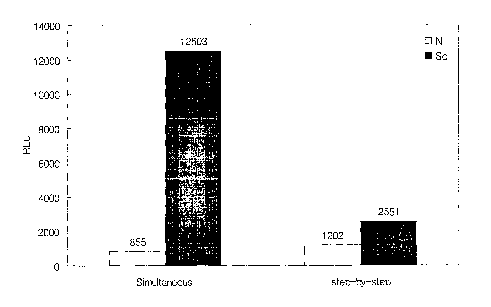
Fig. 2 shows the results of experiments for detecting multimeric prion
proteins
according to the present invention (simultaneous method) and conventional
process
(step-by-step method). "N" and "Sc" denote normal plasma and plasma containing
PrPs`, respectively. "RLU" denotes relative light units.
Fig. 3a represents the results of experiments for selecting a buffer type
suitable in the MDS-3D-Single Bead System.
Fig. 3b shows another presentation of results of Fig. 3a represented by the
ratio of the signal from PrPs` plasma to the signal from normal plasma.
Fig. 4 shows the results of experiments for detecting multimeric prion
proteins
in plasma according to the MDS-3D-Dual Bead System using 3E7 and MA1 750
antibodies. "RFU" denotes relative fluorescence units.
Fig. 5 represents the results of experiments for selecting a buffer type
suitable
in the MDS-3D-Dual Bead System.
Fig. 6 represents the results of experiments for selecting concentrations of
plasma suitable in the MDS-3D-Dual Bead System.
Fig. 7 represents the results of experiments for selecting concentrations of
plasma suitable in the MDS-3D-Singl Bead System.
Fig. 8 shows the results of experiments for detecting multimeric prion
proteins
in plasma according to the MDS-3D-Dual Bead System using T2 and MA1 750
antibodies.
Fig. 9 shows the analysis results for detecting multimeric prion proteins in
plasma according to the MDS-3D-Single Bead System with varying concentrations
and
types of detergent for preparing plasma samples.
Fig. 10 represents the analysis results for detecting multimeric prion
proteins
in plasma according to the MDS-3D-Single Bead System with varying type of
washing
buffer for washing of beads incubated with plasma samples and antibodies.
Fig. 11 represents the analysis results for detecting multimeric prion
proteins
in plasma with concentrations of 30% and 100% according to the MDS-3D-Single
CA 02649359 2008-10-15
WO 2007/123345 PCT/KR2007/001947
Bead System.
Fig. 12 represents the analysis results for detecting multimeric prion
proteins
in plasma with concentrations of 20% and 25% according to the MDS-3D-Single
Bead System.
5 Fig. 13 shows influence of PK digestion on the detection of multimeric prion
proteins in plasma according to the MDS-3D-Single Bead System.
Fig. 14a represents the analysis results for detecting multimeric prion
proteins
in plasma according to the MDS-3D-Single Bead System with varying weight
ratios of
antibodies.
Fig. 14b represents the analysis results for detecting multimeric prion
proteins
in plasma according to the MDS-3D-Dual Bead System with varying weight ratios
of
antibodies.
Figs. 15a and 15b represent the analysis results for detecting multimeric
prion
proteins in plasma according to the MDS-3D-Single Bead System with capturing
antibody cocktail.
Fig. 15c represents the analysis results for detecting multimeric prion
proteins
in plasma according to the MDS-3D-Single Bead System with detection antibody
cocktail. Left and right bars correspond to (i) 3E7-bead as a capturing
antibody and
T2-biotin and MA1-biotin as a detection antibody cocktail and (ii) MA1-bead as
a
capturing antibody and T2-biotin and 3E7-bitoin as a detection antibody
cocktail,
respectively.
Fig. 16 demonstrates the comparison of detection potentials of MDS and MDS-
3D Single Bead System.
DETAILED DESCRIPTION OF THIS INVETNION
In one aspect of this invention, there is provided a method for differentially
detecting a multimeric form from a monomeric form of a multimer-forming
polypeptide in a biosample, which comprises the steps of: (a) preparing a
carrier-
capturing antibody conjugate by binding a capturing antibody to the surface of
a
solid phase carrier in a three dimensional manner, wherein the capturing
antibody is
CA 02649359 2008-10-15
WO 2007/123345 PCT/KR2007/001947
6
capable of recognizing an epitope on the multimer-forming polypeptide; (b)
preparing
a detection antibody, wherein an epitope recognized by the detection antibody
is
present at a position in the multimer-forming polypeptide to cause a steric
hindrance
by the capturing antibody bound to its epitope to prevent the binding of the
detection antibody to the multimer-forming polypeptide; (c) contacting
simultaneously the carrier-capturing antibody conjugate and the detection
antibody
to the biosample; and (d) detecting the formation of a carrier-capturing
antibody-
multimeric form-detection antibody complex.
In another aspect of this invention, there is provided a kit for
differentially
detecting a multimeric form from a monomeric form of a multimer-forming
polypeptide in a biosample, which comprises: (a) a capturing antibody
recognizing an
epitope on the multimer-forming polypeptide and bound three-dimensionally to
the
surface of a solid phase carrier; and (b) a detection antibody recognizing an
epitope
present at a position in the multimer-forming polypeptide to cause a steric
hindrance
by the capturing antibody bound to its epitope to prevent the binding of the
detection antibody to the multimer-forming polypeptide.
The present invention is directed to a method for differentially detecting a
multimeric form from a monomeric form of a multimer-forming polypeptide in a
biosample by immunoassay involving antigen-antibody reactions. Furthermore,
the
present invention uses two types of antibodies, a capturing antibody and a
detection
antibody both of which are competitive in binding to a multimer-forming
polypeptide.
Such competitive antibody binding occurs through steric inhibition. In
particular, the
capturing antibody bound to an epitope on a multimer-forming polypeptide
inhibits
the detection antibody from binding to its epitope on the multimer-forming
polypeptide because of competition to binding sites on the multimer-forming
polypeptide. One of the features of the present invention is to perform the
immunoassay under three-dimensional contacting circumstances. In the present
invention, the capturing and detection antibodies are ensured to have three-
dimensional contacting opportunities to antigens in biosamples.
CA 02649359 2008-10-15
WO 2007/123345 PCT/KR2007/001947
7
The present inventors had already proposed a prototype process for
differentially detecting a multimeric form from a monomeric form of a multimer-
forming polypeptide, called "Multimer Detection System (MDS)" and filed for
patent
application under PCT (PCT/KR2005/004001). The present invention is to improve
the MDS in light of sensitivity and differentiation potential as demonstrated
in
Example XV. The most prominent feature of this invention is that the capturing
and
detection antibodies are three-dimensionally contacted to antigens in
biosamples.
Therefore, the process of this invention is named "MDS-3D (three-dimensional)
system ".
The term "multimer-forming polypeptide" used herein refers to a polypeptide
capable of forming an aggregation (i.e., multimer) form, particularly,
following
conformational change, causing a wide variety of diseases such as Alzheimer's
disease, Creutzfeldt-Jakob disease, Spongiform encephalopathies, Parkinson's
disease, Huntington's disease, Amyotrophic lateral sclerosis, Serpin
deficiency,
emphysema, cirrhosis, Type II diabetes, primary systemic amyloidosis,
secondary
systemic amyloidosis Fronto-temporal dementias, senile systemic amyloidosis,
familial
amyloid polyneuropathy, hereditary cerebral amyloid angiopathy and
haemodialysis-
related amyloidosis. Therefore, the term "multimer-forming polypeptide" will
be
interchangeably used with the term "aggregate-forming polypeptide".
The present method uses two types of antibodies, i.e., capturing antibody and
detecting antibody. As used herein, the term "capturing antibody" means an
antibody
capable of binding to the multimer-forming polypeptide of interest in
biosamples. The
term "detecting antibody" means an antibody capable of binding to the multimer-
forming polypeptide captured by the capturing antibody. By "antibody" is meant
an
immunoglobulin protein which is capable of binding an antigen. Antibody as
used
herein is meant to include the entire antibody as well as any antibody
fragments
(e.g., F(ab`)2, Fab', Fab, Fv) capable of binding the epitope, antigen or
antigenic
fragment of interest.
In the present invention, the epitopes specifically recognized by the
capturing
antibody and detecting antibody are located at positions in multimer-forming
CA 02649359 2008-10-15
WO 2007/123345 PCT/KR2007/001947
8
polypeptides to cause steric hindrance (competitive binding) between
antibodies to
be bound to the epitopes. Preferably, the amino acid sequence of the epitope
recognized by the capturing antibody is identical to, overlapped with or
adjacent to
that of the epitope recognized by the detection antibody. It would be readily
understood that the capturing antibody and detection antibody to be bound to
their
epitopes induce steric hindrance or are competitive in binding where the amino
acid
sequence of the epitope recognized by the capturing antibody is identical to
or
overlapped with that of the epitope recognized by the detection antibody.
The term "overlapped with" used herein with referring to epitopes to capturing
and detecting antibodies encompasses epitopes having completely or partially
overlapped amino acid sequences. For example, the epitopes to T2 and 3E7
antibodies have amino acid sequences spanning amino acid 147-152 and 140-160,
respectively, of a bovine prion sequence. Such epitopes can be described as
completely overlapped epitopes. Furthermore, the epitopes to ICSM35 and 1E4
antibodies have amino acid sequences spanning amino acid 104-113 and 108-119,
respectively, of a bovine prion sequence. Such epitopes can be described as
partially
overlapped epitopes.
As to the adjacent epitopes causing steric hindrance, one epitope (e.g.,
epitope recognized by the capturing antibody) in the multimer-forming
polypeptide
may be located at a position apart from the other epitope (e.g., epitope
recognized
by the detection antibody) so long as two antibodies are competitively bound
to the
adjacent epitopes.
Another feature of this invention is to bind capturing antibodies three-
dimensionally to the surface of a solid phase carrier for preparing a carrier-
capturing
antibody conjugate. For instance, capturing antibodies are bound to the
surface of
three-dimensional beads for preparing a carrier-capturing antibody conjugate.
Therefore, the present invention excludes capturing antibodies bound to the
surface
of plates because such binding is considered to be two-dimensional. The
capturing
antibodies bound three-dimensionally to carriers allows for contacting three-
dimensionally. to antigens in biosamples, ensuring much more opportunities to
CA 02649359 2008-10-15
WO 2007/123345 PCT/KR2007/001947
9
contact to antigens in biosamples.
Solid phase carriers conjugated with capturing antibodies may be any material
having three-dimensional structure, preferably, materials isolatable by
gravity, charge
or magnetic force. Most preferably, the solid phase carrier is a magnetic
bead.
According to a preferred embodiment, the detection antibody has a label
generating a detectable signal or an affinity substance. The label includes,
but not
limited to, an enzymatic (e.g., alkaline phosphatase, peroxidase, P-
galactosidase and
(3-glucosidase), a radioactive (e.g., I125 and C14), a fluorescent (e.g.,
fluorescein), a
luminescent, a chemiluminescent and a FRET (fluorescence resonance energy
transfer) label. The affinity substance includes biotin. Various labels and
methods for
labeling antibodies are well known in the art (Harlow and Lane, eds.
Antibodies: A
Laboratory Manual (1988) Cold Spring Harbor Laboratory Press, Cold Spring
Harbor,
N.Y.).
Where the radioactive label is used for detection antibodies, the antigen-
antibody complex formed in the final step of this invention may be detected by
measuring radioactivity from label. Where the detection antibody is labeled
with
enzymes catalyzing colorimetric reactions, the antigen-antibody complex formed
may
be detected by use of substrates for enzymes. For example, where the detection
antibody is labeled with alkaline phosphatase, bromochloroindolylphosphate
(BCIP),
nitro blue tetrazolium (NBT), naphthol-AS-B1-phosphate and ECF (enhanced
chemifluorescence) may be used as a substrate for color developing reactions;
in the
case of labeled with horseradish peroxidase, chloronaphtol,
aminoethylcarbazol,
diaminobenzidine, D-luciferin, lucigenin (bis-/IFmethylacridinium nitrate),
resorufin
benzyl ether, luminol, Amplex Red reagent (10-acetyl-3,7-
dihydroxyphenoxazine),
TMB (3,3,5,5-tetramethylbenzidine) and ABTS (2,2-Azine-di[3-
ethylbenzthiazoline
sulfonate]) may be used as a substrate.
Where the detection antibody is labeled with affinity substance, the antigen-
antibody complex formed may be detected by use of its binding partner linked
to
label generating a detectable signal (e.g., colorimetric reaction-catalyzing
enzymes).
For example, where the detection antibody is labeled with biotin, enzyme-
conjugated
CA 02649359 2008-10-15
WO 2007/123345 PCT/KR2007/001947
streptavidin may be used for detecting the antigen-antibody complex.
Labels described above make it more feasible to detect qualitatively and
quantitatively the multimeric antigen-antibody complex in the step (d). Where
the
detection antibody is linked to a label, the step (d) may be carried out by
measuring
5 a signal generated from the label.
The preferable antibody set consisting of capturing and detection antibodies
comprises one antibody as a capturing antibody recognizing the epitope of
amino
acids 140-160 of bovine prion sequence and the other antibody as a detection
antibody recognizing the epitope of amino acids 147-152 of bovine prion
sequence.
10 Another preferable antibody set comprises one antibody as a capturing
antibody
recognizing the epitope of amino acids 104-113 of bovine prion sequence and
the
other antibody as a detection antibody recognizing the epitope of amino acids
108-
119 of bovine prion sequence.
The striking feature of this invention is that both the carrier-capturing
antibody
conjugate and detection antibody are contacted to biosamples in a simultaneous
manner. If the capturing antibody is initially contacted and then the
detection
antibody is contacted to biosamples, carrier-capturing antibody conjugates are
bound
to most of epitopes in the multimeric form of a polypeptide to the capturing
antibody,
giving rise to the binding inhibition of the detection antibody. Therefore,
where the
step by step protocol is executed rather than the simultaneous protocol, the
signal
from the detection antibody binding becomes far poor. In contrast, where the
capturing antibody and detection antibody are simultaneously contacted to
biosamples, two types of antibodies are rendered to be under competition
circumstances and their binding to an antigen depends on their concentration.
The term "simultaneously contacting" with reference to carrier-capturing
antibodies and detection antibodies refers to: (i) contacting each of the
carrier-
capturing antibody and detection antibody to biosamples in a simultaneous
manner;
or (ii) contacting a mixture containing both the carrier-capturing antibody
and
detection antibody to biosamples.
According to a preferred embodiment, the capturing antibody bound to the
CA 02649359 2008-10-15
WO 2007/123345 PCT/KR2007/001947
11
carrier and the detection antibody in the step (c) are used at 5:1-1:5 mole
ratio,
more preferably 3:1-1:3 mole ratio, more still preferably 2:1-1:2 mole ratio,
most
preferably about 1:1 mole ratio of the capturing antibody to the detection
antibody.
Where the present method is performed according to the MDS-3D Dual Bead
System, the capturing antibody bound to the carrier and the detection antibody
bound to the carrier in the step (c) are used at 5:1-1:5 mole ratio, more
preferably
3:1-1:3 mole ratio, most preferably about 2:1 mole ratio of the capturing
antibody to
the detection antibody.
The present invention makes it possible to differentially detect a multimeric
form from a monomeric form of any multimer-forming polypeptide. According to a
preferred embodiment, the multimer-forming polypeptide includes AP peptide and
tau protein related to Alzheimer's disease, prion related to Creutzfeldt-Jakob
disease
and Spongiform encephalopathies, a-synuclein related to Parkinson's disease Ig
light
chains related to primary systemic amyloidosis, serum amyloid A related to
secondary
systemic amyloidosis, tau related to Fronto-temporal dementias, transthyretin
related
to senile systemic amyloidosis, transthyretin related to familial amyloid
polyneuropathy, cystatin C related to hereditary cerebral amyloid angiopathy,
RZ-
microglobulin related to haemodialysis-related amyloidosis, huntingtin related
to
Huntington's disease, superoxide dismutase related to Amyotrophic lateral
sclerosis,
serpin related to Serpin deficiency, emphysema, and cirrhosis, and amylin
related to
Type II Diabetes.
Most preferably, the multimer-forming polypeptide is the prion protein causing
Creutzfeldt-Jakob disease and Spongiform encephalopathies.
The present invention is significantly useful in detecting a multimeric prion,
i.e., PrPs`formed by conformational change of prion proteins.
When the present method is applied to the prion protein (PrP), the monomeric
form is PrP` (cellular or normal form of prion) and the multimeric form is
PrPs`
(scrapie or infectious form of prion).
One of the features of this invention is to employ antibodies which are bound
to epitopes having non-repeated sequence in an antigen molecule. Unless
epitopes
CA 02649359 2008-10-15
WO 2007/123345 PCT/KR2007/001947
12
recognized by antibodies have a non-repeated sequence, the present invention
may
not effectively detect a multimeric form from a monomeric form of a multimer-
forming polypeptide.
According to a preferred embodiment, the epitope specifically recognized by
the capturing antibody and/or the epitope specifically recognized by the
detection
antibody are not repeated in the multimer-forming polypeptide.
The antibodies used in this invention could be prepared according to
conventional techniques such as a fusion method (Kohler and Milstein, European
Journal of Immunology, 6:511-519(1976)), a recombinant DNA method (USP
4,816,56) or a phage antibody library (Clackson et al, Nature, 352:624-
628(1991);
and Marks et al, J. Mo% Biol., 222:58, 1-597(1991)). The general procedures
for
antibody production are described in Harlow, E. and Lane, D., Antibodies: A
Laboratory Manual, Cold Spring Harbor Press, New York, 1988; Zola, H.,
Monoclonal
Antibodies: A Manual of Techniques, CRC Press, Inc., Boca Raton, Florida,
1984; and
Coligan, CURRENT PROTOCOLS IN IMMUNOLOGY, Wiley/Greene, NY, 1991. The
preparation of hybridoma cell lines for monoclonal antibody production is done
by
fusion of an immortal cell line and the antibody producing lymphocytes. This
can be
done by techniques well known in the art. Polyclonal antibodies may be
prepared by
injection of the antigen described above to suitable animal, collecting
antiserum
containing antibodies from the animal, and isolating specific antibodies by
any of the
known affinity techniques.
The present invention also encompasses the utilization of cocktails of
antibodies as capturing antibodies or detection antibodies only if the
cocktailed
capturing or detection antibodies are reactive to epitopes identical to or
overlapped
with epitopes for detection or capturing antibodies, respectively. As
addressed in
Examples XIII and XIV, the present invention using a cocktailed capturing or
detection antibody permits to differentially detect PrPs` from PrP`.
The term "Biosample" used herein is an organism-originated sample of
material to be tested. The biosample refers to any cell, tissue, or fluid from
a
biological source, or any other medium that can advantageously be evaluated
CA 02649359 2008-10-15
WO 2007/123345 PCT/KR2007/001947
13
according to this invention, including a sample drawn from human, a sample
drawn
from an animal, a sample drawn from food designed for human or animal
consumption. Preferably, the biosample to be tested is a body fluid sample
including
blood, serum, plasma, lymph, milk, urine, feces, ocular fluid, saliva, semen,
brain
extracts (e.g., brain homogenates), spinal cord fluid (SCF), appendix, spleen
and
tonsillar tissue extracts. More preferably, the biosample is a brain
homogenate or
plasma, most preferably, plasma.
Where a brain homogenate is used as a biosample, it is advantageous that the
present method further comprises the step of pretreating the biosample with
protease K (PK) or trypsin.
Where blood or plasma is used as a biosample, it is preferable that the
biosample is not pretreated with proteases (e.g., PK). The protease treatment
results
in significant decrease in the detection and differentiation potentials of the
present
method to multimeric forms, particularly, PrPs`. Surprisingly, the present
methods
permits to completely eliminate a need of protease (e.g., PK) digestion in the
detection of PrPs` in blood or plasma samples, as demonstrated in Example X.
Where blood or plasma is used as a biosample, it is advantageous that the
present method further comprises the step of pretreating the biosample with
sarkosyl
or Triton series (e.g., Triton X-100) detergent, preferably, Triton series,
most
preferably Triton X-100. For preparation of biosamples (preferably blood, most
preferably plasma), a preferable buffer includes TBST (tris-buffered saline
with
Tween 20) and Tricine. Preferably, the concentration of biosamples (preferably
blood,
most preferably plasma) in the step (c) ranges from 10 v/v% to 70 v/v %, more
preferably, from 20 v/v% to 40 v/v%, most preferably from 23 v/v% to 30 v/v %.
In still another aspect of this invention, there is provided a method for
differentially detecting a multimeric form from a monomeric form of a multimer-
forming polypeptide in a biosample, which comprises the steps of: (a)
preparing a
magnetic bead-capturing antibody conjugate by binding a capturing antibody to
the
surface of a magnetic bead in a three dimensional manner, wherein the
capturing
CA 02649359 2008-10-15
WO 2007/123345 PCT/KR2007/001947
14
antibody is capable of recognizing an epitope on the multimer-forming
polypeptide;
(b) preparing a detection antibody, wherein an epitope recognized by the
detection
antibody is present at a position in the multimer-forming polypeptide to cause
a
steric hindrance by the capturing antibody bound to its epitope to prevent the
binding of the detection antibody to the multimer-forming polypeptide; (c)
contacting
simultaneously the magnetic bead-capturing antibody conjugate and the
detection
antibody to the biosample; (d) applying the resultant of step (c) to isolate a
magnetic
bead-capturing antibody-multimeric form-detection antibody complex; and (e)
detecting the formation of the magnetic bead-capturing antibody-multimeric
form-
detection antibody complex.
Preferably, the method of this invention further comprises the step of washing
the isolated capturing antibody-multimeric form-detection antibody complex
between
the step (d) and the step (e). The washing step may be carried out using
various
washing buffers such as PBS (phosphate-buffered saline), PBST (phosphate-
buffered
saline with Tween 20), PBSX (phosphate-buffered saline with Triton X-100),
TBSX
(tris-buffered saline with Triton X-100) and TBST (tris-buffered saline with
Tween
20). Most preferably, the washing step is performed using TBST.
Fig. la schematically represents an example of this invention using magnetic
beads as carriers. The present invention will be described in more detail with
referring to Fig. la. The capturing antibodies on magnetic beads and the HRP-
detection antibodies are simultaneously contacted to biosamples containing
PrP` and
PrPs`, HRP-conjugated detection antibodies cannot be bound to magnetic bead-
capturing antibody-bound PrP` but bound only to magnetic bead-capturing
antibody-
bound PrPs`. In addition, magnetic bead-capturing antibodies cannot be also
bound
to HRP-conjugated detection antibody-bound PrP`. The epitopes to the capturing
antibody and detection antibody have a non-repeated sequence in the prion
protein.
The amino acid sequence of the epitope recognized by the capturing antibody is
identical to, overlapped with or adjacent to that of the epitope recognized by
the
detection antibody. In Fig. la, epitopes are denoted as triangle. Since the
epitope
recognized by the detection antibody is occupied by the capturing antibody,
the
CA 02649359 2008-10-15
WO 2007/123345 PCT/KR2007/001947
detection antibody cannot be bound to PrP` having only one epitope. However,
since
the multimeric prion protein, PrPs` contains a plurality of certain epitope,
the
detection antibody can be bound to capturing antibody-bound PrPs`. After the
antigen-antibody reaction, a magnetic field is applied to the reaction
resultant to
5 collect magnetic beads, followed by washing the collected beads. The color-,
fluorescence- or luminescence-developing reactions are induced using HRP
substrates
and their results are measured, providing qualitative and quantitative
analysis data to
verify whether the PrPs`-antibody complex is formed.
10 According to a preferred embodiment of this kit, the capturing antibody
bound
to the carrier and the detection antibody are contained at 5:1-1:5 mole ratio,
more
preferably 3:1-1:3 mole ratio, more still preferably 2:1-1:2 mole ratio, most
preferably about 1:1 mole ratio of the capturing antibody to the detection
antibody in
the form of mixture. The kit may further comprise magnetic plate, buffer,
color-
15 developing enzymes and substrates.
The MDS-3D System of this invention is classified into MDS-3D Single Bead
System and MDS-3D Dual Bead System.
The MDS-3D Single Bead System uses capturing antibody-conjugated beads
(Fig. la) and the MDS-3D Dual Bead System uses both capturing antibody-
conjugated beads and detection antibody-conjugated beads (Figs. lb and lc).
The
MDS-3D Single Bead System is described hereinabove with reference to Fig. la.
In the MDS-3D Dual Bead System, the detection antibody is three-
dimensionally linked to the surface of a solid phase carrier. The detection
antibodies
bound to carriers permit to contact to multimeric polypeptides in a three-
dimensional
manner.
Solid phase carriers conjugated with detection antibodies may be any material
having three-dimensional structure, preferably, materials isolatable by
gravity, charge
or magnetic force. Most preferably, the solid phase carrier is a latex bead.
The carrier
may be labeled. Where the carrier has a label (e.g., the carrier is a latex
beads
CA 02649359 2008-10-15
WO 2007/123345 PCT/KR2007/001947
16
containing fluorescent substance), a detectable signal generated from the
carrier
(indicative of the presence of multimeric polypeptides) may be obtained
without
labeling detection antibodies.
The MDS-3D Dual Bead System is further classified into "MDS-3D Dual Bead
System with Single Label" (Fig. lb) and "MDS-3D Dual Bead System with Double
Label" (Fig. lc).
In the MDS-3D Dual Bead System with Single Label, either detection antibody
or carrier has a label generating a detectable signal. Both detection antibody
and
carrier have a label generating a detectable signal in the MDS-3D Dual Bead
System
with Double Label. Such double labeling strategy enables to doubly check a
signal
indicative of the presence of multimeric polypeptides.
The MDS-3D Dual Bead System with Single Label will be described in more
detail with referring to Fig. lb. The capturing antibodies on magnetic beads
and
Fluor-detection antibodies (detection antibodies linked to latex beads
containing
fluorescent substance) are simultaneously contacted to biosamples containing
PrP`
and PrPs`, and Fluor-detection antibodies cannot be bound to magnetic bead-
capturing antibody-bound PrP` but bound only to magnetic bead-capturing
antibody-
bound PrPs`. The epitopes to the capturing antibody and detection antibody
have a
non-repeated sequence in the prion protein. The amino acid sequence of the
epitope
recognized by the capturing antibody is identical to, overlapped with or
adjacent to
that of the epitope recognized by the detection antibody. In Fig. lb, epitopes
are
denoted as triangle. Since the epitope recognized by the detection antibody is
occupied by the capturing antibody, the detection antibody cannot be bound to
PrP`
having only one epitope. However, since the multimeric prion protein, PrPs`
contains
a plurality of certain epitope, the detection antibody can be bound to
capturing
antibody-bound PrPs`. After the antigen-antibody reaction, a magnetic field is
applied
to the reaction resultant to collect magnetic beads, followed by washing the
collected
beads. Measurements are carried out to analyze fluorescence intensities,
verifying
whether the PrPs`-antibody complex is formed.
The MDS-3D Dual Bead System with Double Label will be described in more
CA 02649359 2008-10-15
WO 2007/123345 PCT/KR2007/001947
17
detail with referring to Fig. 1c. The capturing antibodies on magnetic beads
and
Fluor-HRP-detection antibodies (HRP-conjugated detection antibodies linked to
latex
beads containing fluorescent substance) are simultaneously contacted to
biosamples
containing PrP` and PrPs`, Fluor-detection antibodies cannot be bound to
magnetic
bead-capturing antibody-bound PrP` but bound only to magnetic bead-capturing
antibody-bound PrPs`. The epitopes to the capturing antibody and detection
antibody
have a non-repeated sequence in the prion protein. The amino acid sequence of
the
epitope recognized by the capturing antibody is identical to, overlapped with
or
adjacent to that of the epitope recognized by the detection antibody. In Fig.
1c,
epitopes are denoted as triangle. Since the epitope recognized by the
detection
antibody is occupied by the capturing antibody, the detection antibody cannot
be
bound to PrP` having only one epitope. However, since the multimeric prion
protein,
PrPs` contains a plurality of certain epitope, the detection antibody can be
bound to
capturing antibody-bound PrPs`. After the antigen-antibody reaction, a
magnetic field
is applied to the reaction resultant to collect magnetic beads, followed by
washing
the collected beads. Measurements are carried out to analyze fluorescence
intensities
and HRP reactions, verifying whether the PrPs`-antibody complex is formed.
The following specific examples are intended to be illustrative of the
invention
and should not be construed as limiting the scope of the invention as defined
by
appended claims.
EXAMPLES
EXAMPLE I: Detection of Multimeric PrP in Plasma Using MDS-3D-Single
Bead System
2705 pl of sheep plasma, 22.5 ial of recombinant multimeric sheep PrP
(genotype ARQ, 120-fold diluted) and 3605 pl of 2.5 x detergent (including 3%
Triton
X-100, 1.5% Na deoxycholate and 0.25% sarkosyl) were mixed to prepare a
sample.
The negative control was also prepared to contain 22.5 pI of PBS instead of
the
recombinant multimeric sheep PrP. Capturing antibody-conjugated magnetic beads
CA 02649359 2008-10-15
WO 2007/123345 PCT/KR2007/001947
18
were prepared in which 1 pg of capturing antibodies was bound to 2.5 pl of
magnetic
beads. The capturing antibody bound to magnetic beads is 3E7 or MA1-750
monoclonal antibodies. The epitope against the 3E7 monoclonal antibody is an
amino
acid sequence spanning 140-160 of PrP` (with reference to a bovine prion
sequence)
or 132-152 (with reference to a sheep prion sequence), which is found to be a
non-
repeated sequence in prion. The MA1-750 antibody recognizes specifically an
epitope
on PrP`, Ser-Arg-Pro-Leu-Ile-His-Phe-Gly-Ser-Asp-Tyr-Glu-Asp-Arg, which is
found to
be a non-repeated sequence in prion.
The capturing antibody-conjugated magnetic bead and the detection antibody
were incubated with the plasma sample in a simultaneous manner. Then, we
determined whether the recombinant multimeric sheep PrP in plasma samples was
detected by the MDS-Single Bead System of this invention. As detection
antibodies,
3B8/D5-HRP or T2-HRP was used. The T2 antibody is described in Hiroko Hayashi,
et
al., J. l/et. Med. Sci., 66(6):515(2004), recognizing PrP147_152 epitope (with
reference
to a bovine prion sequence) or PrP140_145 epitope (with reference to a sheep
prion
sequence). The epitope against the 3B8/D5 monoclonal antibody is an amino acid
sequence spanning 132-152 (with reference to a sheep prion sequence) or 140-
160
(with reference to a bovine prion sequence).
The capturing antibody-conjugated magnetic bead and the detection antibody
were simultaneously added to the plasma sample and incubated for 1 hr at 37 C.
The
magnetic field was then applied to the reaction mixture to separate magnetic
beads,
followed by washing the beads three times with TBST. The ECL (enhanced
chemiluminescence) detection was performed.
TABLE 1
Experiment Antibodies Amount of Ab
Bead-3E7 7.5 ial
Exp. 1
3B8/D5-HRP 1 pI
Bead-3E7 5 ial
Exp. 2
3B8/D5-HRP 2 lal
CA 02649359 2008-10-15
WO 2007/123345 PCT/KR2007/001947
19
Bead-3E7 2.5 pl
Exp. 3
3B8/D5-HRP 3 pl
Bead-3E7 2.5 ial
Exp. 4
T2-HRP (36 iag/ml) 3.3 ial
Bead-MA1-750 2.5 pl
Exp. 5
T2-HRP (36 pg /ml) 3.3 pl
For comparison, the capturing antibody-conjugated magnetic bead was initially
incubated with the plasma sample and then incubated with detection antibodies.
The plasma sample was prepared as described hereinabove. The capturing
antibody-conjugated magnetic beads were added to the plasma sample and
incubated for 1 hr at 37 C. The magnetic field was then applied to the
reaction
mixture to separate magnetic beads, followed by washing the beads three times
with
TBST. Then, the separated beads were incubated with the detection antibody for
1 hr
at 37 C. The magnetic field was applied to the reaction mixture to separate
magnetic
beads, followed by washing the beads three times with TBST. Finally, the ECL
(enhanced chemiluminescence) detection was performed.
As shown in Fig. 2, where the magnetic bead-capturing antibody (3E7
antibody) and the detection antibody (T2-HRP) were incubated with samples in a
simultaneous manner, the detection signal to multimeric PrP was shown to be
much
higher than that of the step-by-step protocol, demonstrating that the
simultaneous
protocol dramatically increases sensitivity in the detection of multimeric
prion. In
addition, the ratio of signal intensity of multimeric prion to that of normal
prion in the
simultaneous protocol is much greater than that in the step-by-step protocol,
urging
us to reason that the present invention exhibits an excellent differentiation
potential
to multimeric prion.
EXAMPLE II: Determination of Buffer Type Suitable in MDS-3D-Single Bead
System
To determine a suitable buffer type in the MDS-3D-Single Bead System of this
CA 02649359 2008-10-15
WO 2007/123345 PCT/KR2007/001947
invention, the magnetic-bound capturing antibody, 3E7-bead and detection
antibody,
T2-HRP were used at a weight ratio of 1 to 1 (0.8 pg:0.8 pg, substantially
correspond
to a mole ratio of 1 to 1) for performing the MDS-3D-Single Bead System.
Plasma
samples were prepared as follows: 120 pl of 10% Triton X-100 in d-H20, 580 pl
of
5 buffer (TAPS, TBST or Tricine, pH 8.0) and 300 pl of sheep plasma showing
clinical
signs to have PrPs` were mixed to give 1 ml of the total volume of plasma
sample
containing 1.2% Triton X-100 and 30% plasma. 2 pl (0.8 pg) of 3E7-conjugated
magnetic bead as capturing antibodies and 200 lal (0.8 pg) of T2-HRP (4 pg/mI
in
TBST) as detection antibodies were mixed to prepare a mixed antibody. 200 ial
of the
10 mixed antibody were added to 1 ml of the plasma sample and incubated for 1
hr at
37 C. The magnetic field was then applied to the reaction mixture to separate
magnetic beads, followed by washing the beads three times with TBST. The ECL
detection was performed (Figs. 3a and 3b). In Figs. 3a and 3b, "N" and "Sc"
denote
normal plasma and plasma containing PrPs`, respectively. As shown in Figs. 3a
and 3b,
15 Tricine buffer shows the highest signal to plasma containing PrPs` and
lowest signal to
normal plasma.
EXAMPLE III: Detection of Multimeric PrP in Plasma Using MDS-3D-Dual
Bead System
20 120 pl of 10% Triton X-100, 730 lal of TBST buffer (pH 8.0) and 150 ial of
sheep plasma were mixed to give 1 ml of plasma sample containing 1.2% Triton X-
100 and 15% plasma. 4 pl of 3E7-conjugated magnetic bead as capturing
antibodies,
2 lal of MA1 750-conjugated fluorescence latex bead as detection antibodies
and 200
pl of TBST buffer were mixed to prepare a mixed antibody (3E7-bead:MA1 750-
fluorescence bead, 2:1). 200 pl of the mixed antibody were added to 1 ml of
the
plasma sample and incubated for 1 hr at 37 C. The magnetic field was then
applied
to the reaction mixture to separate magnetic beads, followed by washing the
beads
three times with TBST. Finally, the fluorescent signal was detected. As
represented in
Fig. 4, the MDS-3D-Dual Bead System permits to detect differentially PrPs` in
plasma.
CA 02649359 2008-10-15
WO 2007/123345 PCT/KR2007/001947
21
EXAMPLE IV: Determination of Buffer Type Suitable in MDS-3D-Dual Bead
System
4 lal of 3E7-conjugated magnetic bead as capturing antibodies and 1 pl of MA1
750-conjugated fluorescence latex bead as detection antibodies were mixed to
prepare a mixed antibody(3E7-bead:MA1 750-fluorescence bead, 4:1). 120 lal of
10%
Triton X-100, 730 pl of buffer (TBST or Tricine, pH 8.0) and 150 lai of sheep
plasma
were mixed to obtain a plasma sample. The other procedures are the same as
Example III. As shown in Fig. 5, TBST was shown to be higher signal and
differentiation potential than Tricine buffer in detection of PrPs` in plasma
sample
using the MDS-3D-Dual Bead System.
Furthermore, the MDS-3D-Dual Bead System was made using T2 and MA1
antibody as capturing and detection antibodies, respectively, and then the
above-
described procedures were carried out. As represented in Fig. 5, the MDS-3D-
Dual
Bead System using another antibody combination also allows for the detection
of
PrPs` in plasma.
EXAMPLE V: Determination of Plasma Concentration Suitable in MDS-3D-
Dual Bead System
2 ial of 3E7-conjugated magnetic bead as capturing antibodies and 1 pl of MA1
750-conjugated fluorescence latex bead as detection antibodies were mixed to
prepare a mixed antibody(3E7-bead:MA1 750-fluorescence bead, 2:1). Plasma
samples in concentrations of 30%, 15% and 7.5% were prepared. Tricine (pH 8.0)
buffer was used. The other procedures are the same as Example III.
As represented in Fig. 6, the MDS-3D-Dual Bead System of this invention
permits to detect differentially PrPs` in all plasma samples in concentrations
of 30%,
15% and 7.5%. While the ratio of Sc/N decreases upon decreasing the
concentration
of plasma, the intensity of signals increases upon decreasing the
concentration of
plasma. Accordingly, it could be appreciated that the concentration of plasma
suitable
in the MDS-3D-Dual Bead System is around 15%.
CA 02649359 2008-10-15
WO 2007/123345 PCT/KR2007/001947
22
EXAMPLE VI: Determination of Plasma Concentration Suitable in MDS-3D-
Single Bead System
To determine a suitable plasma concentration in the MDS-3D-Single Bead
System, the magnetic-bound capturing antibody, 3E7-bead and detection
antibody,
T2-HRP were used at a ratio of 1 to 1 (0.8 pg:0.8 pg) for performing the MDS-
3D-
Single Bead System. Plasma samples in concentrations of 30%, 15% and 7.5% were
prepared. Tricine (pH 8.0) buffer was used. The other procedures are the same
as
Example II.
As represented in Fig. 7, the MDS-3D-Single Bead System of this invention
permits to detect differentially PrPs` in all plasma samples in concentrations
of 30%,
15% and 7.5%. The ratio of Sc/N, and the signal intensities to both normal
plasma
and PrPs` plasma decreases upon decreasing the concentration of plasma.
Accordingly,
it could be understood that the concentration of plasma suitable in the MDS-3D-
Single Bead System is around 30%.
Furthermore, the 3E7-bead antibody and the T2-HRP antibody were used at a
ratio of 1 to 2 and plasma samples in concentrations of 30% and 100% were
prepared using Tricine (pH 8.0) buffer. The other procedures are the same as
Example II. As shown in Fig. 11, the 100% plasma concentration was observed to
produce much lower signal intensities and differentiation than 30% plasma
concentration.
In addition, the 3E7-bead antibody and the T2-HRP antibody were used at a
ratio of 1 to 1 and plasma samples in concentrations of 20% and 25% were
prepared
using Tricine (pH 8.0) buffer. The volumes of the reactions containing 20% and
25%
plasma samples were 300 ial and 400 ial, respectively. The other procedures
are the
same as Example H. As shown in Fig. 12, the 25% plasma concentration was
observed to produce much higher signal intensities and differentiation than
20%
plasma concentration.
EXAMPLE VII: Detection of Multimeric PrP in Plasma Using MDS-3D-Dual
Bead System
CA 02649359 2008-10-15
WO 2007/123345 PCT/KR2007/001947
23
T2-conjugated magnetic bead as capturing antibodies and MA1 750-
conjugated fluorescence latex bead as detection antibodies were used at a
ratio of
2:1 or 4:1 for performing the MDS-3D-Dual Bead System. 15% plasma sample and
TBST (pH 8.0) buffer were used. The other procedures are the same as Example
III.
As shown in Fig. 8, the Sc/N ratio and signal intensity are found to be higher
at a 4:1
ratio of T2-bead to MA1 750-bead than a 2:1 ratio.
EXAMPLE VIII: Detection of Multimeric PrP in Plasma Using MDS-3D-Single
Bead System with Varying Concentration and Type of Detergent
The multimeric PrP in sheep plasma samples showing clinical signs to have
PrPs` was detected in the MDS-3D-Single Bead System with varying
concentrations
and types of detergent in the same manner as Example II, except for detergent
conditions. As shown in Fig. 9, concentrations of Triton X-100 (1%, 1.5% and
2%)
show similar detection and differentiation results to multimeric PrP. In
addition, Np-40
(USB Corp., USA) also shows considerable detection and differentiation
results,
although the deviation of results is relatively high. Np-40 is one of nonionic
detergents and represents [Octylphenoxy]polyethoxyethanol.
EXAMPLE IX: Detection of Multimeric PrP in Plasma Using MDS-3D-Single
Bead System with Varying Type of Washing Buffer
The multimeric PrP in sheep plasma samples with PrPs` was detected in the
MDS-3D-Single Bead System with varying the type of washing buffers used in
washing of beads incubated with plasma samples and antibodies in the same
manner
as Example II, except for antibody weight ratio (3E7-bead:T2-HRP, 1:2) and
washing
conditions. As shown in Fig. 10, the TBST (tris-buffered saline with Tween 20)
washing buffer exhibits most superior detection and differentiation results to
multimeric PrP compared with other buffers, PBST (phosphate-buffered saline
with
Tween 20) , PBSX (phosphate-buffered saline with Triton X-100) and TBSX (tris-
buffered saline with Triton X-100).
CA 02649359 2008-10-15
WO 2007/123345 PCT/KR2007/001947
24
EXAMPLE X: Influence of PK Digestion on Detection of Multimeric PrP in
Plasma Using MDS-3D-Single Bead System
The multimeric PrP in sheep plasma samples with PrPs` was detected in the
MDS-3D-Single Bead System with or without PK (protease K) digestion in the
same
manner as Example II, except that antibody weight ratio (3E7-bead:T2-HRP,
1:4). As
shown in Fig. 13, the PK digestion greatly decreases signal intensities to
PrPs` in
plasma samples. In this connection, it could be recognized that the MDS-3D-
Single
Bead System of this invention permits to completely eliminate a need of PK
digestion
in the detection of PrPs` in blood or plasma samples.
EXAMPLE XI: Detection of Multimeric PrP in Plasma Using MDS-3D-Single
Bead System with Varying Weight Ratios of Antibodies
The multimeric PrP in sheep plasma samples with PrPs` was detected in the
MDS-3D-Single Bead System with varying weight ratios of antibodies in the same
manner as Example II. The weight ratios of 3E7-bead capturing antibody to T2-
HRP
detection antibody were 4:1, 2:1, 1.33:1 and 1:1. As shown in Fig. 14a, where
two
antibodies were utilized in the same amount, the MDS-3D-Single Bead System of
this
invention exhibits most excellent detection and differentiation potentials to
PrPs` in
plasma samples.
EXAMPLE XII: Detection of Multimeric PrP in Plasma Using MDS-3D-Dual
Bead System with Varying Weight Ratios of Antibodies
The multimeric PrP in sheep plasma samples with PrPs` was detected in the
MDS-3D-Dual Bead System with varying weight ratios of antibodies in the same
manner as Example III. The weight ratios of 3E7-bead capturing antibody to MA1
750 fluorescence bead detection antibody were 4:1 and 2:1. As shown in Fig.
14b,
where the capturing and detection antibodies were utilized at a ratio of 2:1,
the MDS-
3D-Dual Bead System of this invention exhibits most excellent detection and
differentiation potentials to PrPs` in plasma samples.
CA 02649359 2008-10-15
WO 2007/123345 PCT/KR2007/001947
EXAMPLE XIII: Detection of Multimeric PrP in Plasma Using MDS-3D-Single
Bead System with Capturing Antibody Cocktail
The multimeric PrP in sheep plasma samples with PrPs` was detected in the
MDS-3D-Single Bead System with a capturing antibody cocktail in the same
manner
5 as Example II. As a capturing antibody, the capturing antibody cocktail
consisting of
3E7-bead and T2-bead antibodies (Fig. 15a) or 3E7-bead and MA1-bead antibodies
(Fig. 15b) was used. The weight ratio of the capturing antibody cocktail to
the
detection antibody, T2-HRP was 1:1. As shown in Figs. 15a and 15b, the
capturing
antibody cocktail also shows excellent detection and differentiation
potentials to PrPs`
10 in plasma samples.
EXAMPLE XIV: Detection of Multimeric PrP in Plasma Using MDS-3D-Single
Bead System with Detection Antibody Cocktail
The multimeric PrP in sheep plasma samples with PrPs` was detected in the
15 MDS-3D-Single Bead System with a detection antibody cocktail in the same
manner
as Example II. The antibody sets used were (i) 3E7-bead as a capturing
antibody and
T2-biotin and MA1-biotin as a detection antibody cocktail and (ii) MA1-bead as
a
capturing antibody and T2-biotin and 3E7-bitoin as a detection antibody
cocktail. As
shown in Fig. 15c, the detection antibody cocktails also show excellent
detection and
20 differentiation potentials to PrPs` in plasma samples.
EXAMPLE XV: Comparison of Detection Potentials of MDS and MDS-3D-
Single Bead System under Same Conditions
The present inventors had already proposed a prototype process for
25 differentially detecting a multimeric form from a monomeric form of a
multimer-
forming polypeptide, called "Multimer Detection System (MDS)" and filed for
patent
application under PCT (PCT/KR2005/004001).
For comparing PrPs` detection and differentiation potentials of MDS and MDS-
3D Single Bead System in a reliable manner, the multimeric PrP in sheep plasma
samples with PrPs` was detected according each procedure under same
experimental
CA 02649359 2008-10-15
WO 2007/123345 PCT/KR2007/001947
26
conditions. 3E7 and T7 antibodies were used as capturing and detection
antibodies,
respectively.
As represented in Fig. 16, it becomes evident that the MDS-3D Single Bead
System of this invention shows much higher sensitivity and differentiation
potentials
to PrPs` in sheep plasma samples than the MDS.
EXAMPLE XVI: Determination of Plasma Concentration Suitable in MDS-3D-
Single Bead System with Other Antibody Set
The multimeric PrP in sheep plasma samples with PrPs` was detected in the
MDS-3D-Single Bead System in the same manner as Example II, except for the
type
of antibody set and plasma concentration. ICSM35-biotin streptavidin bead as a
capturing antibody and 1E4-HRP as a detection antibody were utilized. The
concentration of sheep plasma was 25%. The ICSM35 antibody (D-Gen, Inc.)
recognizes the epitope corresponding to the amino acid sequence spanning 96-
105
(with reference to a sheep prion sequence) or 104-113 (with reference to a
bovine
prion sequence). The 1E4 antibody (Sanquin Reagents, Inc.) recognizes the
epitope
corresponding to the amino acid sequence spanning 100-111 (with reference to a
sheep prion sequence) or 108-119 (with reference to a bovine prion sequence).
As shown in Fig. 17, another antibody set recognizing partial overlapping
epitopes also exhibits excellent sensitivity and differentiation potentials to
PrPs` in
plasma samples.
Having described a preferred embodiment of the present invention, it is to be
understood that variants and modifications thereof falling within the spirit
of the
invention may become apparent to those skilled in this art, and the scope of
this
invention is to be determined by appended claims and their equivalents.