Note : Les descriptions sont présentées dans la langue officielle dans laquelle elles ont été soumises.
CA 02650223 2008-10-23
WO 2007/117366 PCT/US2007/004726
SYSTEMS AND METHODS FOR STABILIZATION OF BONE STRUCTURES
Field of the Invention
Statement of Related Applications
[0001) This application is a continuation-in-part of co-pending U.S. Patent
Application
Serial No.: 11/362,366, filed February 23, 2006, entitled "Systems And Methods
For
Stabilization of Bone Structures," which claims the benefct of U.S.
Provisional Patent
Application Serial No. 60/701,660, filed July 22, 2005. Each ofthe prior
applications is
incorporated herein by reference in its entirety. The present invention
generally relates to
surgical instruments and methods for using these instruments. More
particularly, but not
exclusively, minimally invasive methods of stabilizing one or more bone
structures is
disclosed.
Background of the Invention
[0002J Systems, methods and devices for stabilizing one or more bone
structures of a patient
have been available for many years. Securing a metal plate is used to
stabilize a broken bone,
maintaining the bone in a desired position during the healing process. These
implanted plates
are either removed when sufficient healing has occurred or left in place for a
long-term or
indefinite, chronic period. A procedure involving the placement of one or more
elongated
rods extending between two bone structures or between two components of a
single bone
structure is often used as a stabilization technique. These rods are placed
alongside the bone
structure or structures and attached to bone via one or more attachment
mechanisms (e.g. -
bone screws, anchors, etc). These procedures typically require large incisions
and also
significant tissue manipulation to adequately expose the areas intended for
the attachment.
The procedures are associated with long recovery times and increased potential
for adverse
events, such as infection, muscle and other tissue trauma and scarring.
100031 Currently available minimally invasive techniques and products are
limited. These
procedures are difficult to perform, especially in spinal applications in
which the attachment
points are deeper in tissue, and damage to neighboring tissue must be avoided.
Many of the
currently available less invasive products remain somewhat invasive due to
component
configurations, and required manipulations to be performed during the
attachment.
[0004J In reference specifically to treatment of the spine, Fig. 1A
illustrates a portion of the
I
CA 02650223 2008-10-23
WO 2007/117366 PCT/US2007/004726
human spine having a superior vertebra 2 and an inferior vertebra 4, with an
intervertebral
disc 6 located in between the two vertebral bodies. The superior vertebra 2
has superior facet
joints 8a and 8b, inferior facet joints 10a and l Ob, posterior arch 16 and
spinous process 18.
Pedicles 3a and 3b interconnect the respective superior facet joints 8a, 8b to
the vertebral
body 2. Extending laterally from superior facetjoints 8a, 8b are transverse
processes 7a and
7b, respectively. Extending between each inferior facet joints l0a and lOb and
the spinous
process 18 are lamina 5a and 5b, respectively. Similarly, inferior vertebra 4
has superior
facet joints 12a and 12b, superior pedicles 9a and 9b, transverse processes I
la and 11b,
inferior facet joints 14a and 14b, lamina 15a and 15b, posterior arch 20,
spinous process 22.
100061 The superior vertebra with its inferior facets, the inferior vertebra
with its superior
facets, the intervertebral disc, and seven spinal ligaments (not shown)
extending between the
superior and inferior vertebrae together comprise a spinal motion segment or
functional spine
unit. Each spinal motion segment enables motion along three orthogonal axis,
both in
rotation and in translation. The various spinal motions are illustrated in
Figs. 2A-2C. In
particular, Fig. 2A illustrates flexion and extension motions and axial
loading, Fig. 2B
illustrates lateral bending motion and Fig. 2C illustrated axial rotational
motion. A normally
functioning spinal motion segment provides physiological limits and stiffness
in each
rotational and translational direction to create a stable and strong column
structure to support
physiological loads.
[0007] Traumatic, inflammatory, metabolic, synovial, neoplastic and
degenerative disorders
of the spine can produce debilitating pain that can affect a spinal motion
segment's ability to
properly function. The specific location or source of spinal pain is most
often an affected
intervertebral disc or facet joint. Often, a disorder in one location or
spinal component can
lead to eventual deterioration or disorder, and ultimately, pain in the other.
[0008] Spine fusion (arthrodesis) is a procedure in which two or more adjacent
vertebral
bodies are fused together. It is one of the most common approaches to
alleviating various
types of spinal pain, particularly pain associated with one or more affected
intervertebral
discs. While spine fusion generally helps to eliminate certain types of
pain,=it has been shown
to decrease function by limiting the range of motion for patients in flexion,
extension, rotation
and lateral bending. Furthermore, the fusion creates increased stresses on
adjacent non-fused
motion segments and accelerated degeneration of the motion segments.
Additionally,
pseudarthrosis (resulting from an incomplete or ineffective fusion) may not
provide the
2
CA 02650223 2008-10-23
WO 2007/117366 PCT/US2007/004726
expected pain-relief for the patient. Also, the device(s) used for fusion,
whether artificial or
biological, may migrate out of the fusion site creating significant new
problems for the
patient.
[00091 Various technologies and approaches have been developed to treat spinal
pain
without fusion in order to maintain or recreate the natural biomechanics of
the spine. To this
end, significant efforts are being made in the use of implantable artificial
intervertebral discs.
Artificial discs are intended to restore articulation between vertebral bodies
so as to recreate
the full range of motion normally allowed by the elastic properties of the
natural disc.
Unfortunately, the currently available artificial discs do not adequately
address all of the
mechanics of motion for the spinal column.
[00101 It has been found that the facet joints can also be a significant
source of spinal
disorders and debilitating pain. For example, a patient may suffer from
arthritic facet joints,
severe facet joint tropism, otherwise deformed facet joints, facet joint
injuries, etc. These
disorders lead to spinal stenosis, degenerative spondylolithesis, and/or
isthmic
spondylotlisthesis, pinching the nerves which extend between the affected
vertebrae.
[0011] Current interventions for the treatment of facet joint disorders have
not been found to
provide completely successful results. Facetectomy (removal of the facet
joints) may provide
some pain relief; but as the facet joints help to support axial, torsional,
and shear loads that
act on the spinal column in addition to providing a sliding articulation and
mechanism for
load transmission, their removal inhibits natural spinal function. Laminectomy
(removal of
the lamina, including the spinal arch and the spinous process) may also
provide pain relief
associated with facet joint disorders; however, the spine is made less stable
and subject to
hypermobility. Problems with the facet joints can also complicate treatments
associated with
other portions of the spine. In fact, contraindications for disc replacement
include arthritic
facet joints, absent facet joints, severe facet joint tropism, or otherwise
deformed facet joints
due to the inability of the artificial disc (when used with compromised or
missing facet joints)
to properly restore the natural biomechanics of the spinal motion segment.
[0012] While various attempts have been made at facet joint replacement, they
have been
inadequate. This is due to the fact that prosthetic facet joints preserve
existing bony
structures and therefore do not address pathologies which affect facet joints
themselves.
Certain facet joint prostheses, such as those disclosed in U.S. Pat. No.
6,132,464, are intended
to be supported on the lamina or the posterior arch. As the lamina is a very
complex and
3
CA 02650223 2008-10-23
WO 2007/117366 PCT/US2007/004726
highly variable anatomical structure, it is very difficult to design a
prosthesis that provides
reproducible positioning against the lamina to correctly locate the prosthetic
facet joints. In
addition, when facet joint replacement involves complete removal and
replacement of the
natural facet joint, as disclosed in U.S. Patent No. 6,579,319, the prosthesis
is unlikely to
endure the loads and cycling experienced by the vertebra. Thus, the facet
joint replacement
may be subject to long-term displacement. Furthermore, when facet joint
disorders are
accompanied by disease or trauma to other structures of a vertebra (such as
the lamina,
spinous process, and/or transverse processes) facet joint replacement is
insufficient to treat
the problem(s).
[0013] Most recently, surgical-based technologies, referred to as "dynamic
posterior
stabilization," have been developed to address spinal pain resulting from more
than one
disorder, when more than one structure of the spine have been compromised. An
objective of
such technologies is to provide the support of fusion-based implants while
maximizing the
natural biomechanics of the spine. Dynamic posterior stabilization systems
typically fall into
one of two general categories: (1) interspinous spacers and (2) posterior
pedicle screw-based
systems.
[00141 Examples of interspinous spacers are disclosed in U.S. Patent Nos. Re.
36,211,
5,645,599, 6,695,842, 6,716,245 and 6,761,720. The spacers, which are made of
either a hard
or compliant material, are placed between adjacent spinous processes. Because
the
interspinous spacers involve attachment to the spinous processes, use of these
types of
systems is limited to applications where the spinous processes are
uncompromised and
healthy.
[00151 Examples of pedicle screw-based systems are disclosed in U.S. Patent
Nos.
5,015,247, 5,484,437, 5,489,308, 5,609,636 and 5,658,337, 5,741,253,
6,080,155, 6,096,038,
6,264,656 and 6,270,498. These types of systems involve the use of screws
which are
positioned in the vertebral body through the pedicle. Certain types of these
pedicle screw-
based systems may be used to augment compromised facet joints, while others
require
removal of the spinous process and/or the facet joints for implantation. One
such system, the
Zimmer Spine Dynesys employs a cord which is extended between the pedicle
screws and a
fairly rigid spacer which is passed over the cord and positioned between the
screws. While
this system is able to provide load sharing and restoration of disc height,
because it is so rigid,
it does not effectively preserve the natural motion of the spinal segment into
which it is
4
CA 02650223 2008-10-23
WO 2007/117366 PCT/US2007/004726
implanted. Other pedicle screw-based systems employ articulating joints
between the pedicle
screws.
[0016] There remains a need for minimally invasive methods and devices for
bone
stabilization procedures, including but not limited to spinal segment
stabilization procedures
such as dynamic spinal segment stabilization procedures. There is a need for
procedures that
are simple to perform and reliably achieve the desired safe and effective
outcome. Goals of
these new procedures and instruments include minimizing the size of the
incision and
reducing the amount of muscle dissection in order to shorten recovery times,
improve
procedure success rates and reduce the number of resultant adverse side
effects.
Brief Description of the Drawings
[0017] The invention is best understood from the following detailed
description when read in
conjunction with the accompanying drawings. It is emphasized that, according
to common
practice, the various features of the drawings are not to-scale. On the
contrary, the
dimensions of the various features are arbitrarily expanded or reduced for
clarity. Included in
the drawings are the following figures:
[0018] Figs. IA and 1B illustrate perspective views of a portion of the human
spine having
two vertebral segments, where the spinous process and the lamina of the
superior vertebra
have been resected in Fig. 1B.
[0019] Figs. 2A, 2B and 2C illustrate left side, dorsal and top views,
respectively, of the
spinal segments of Fig. 1A under going various motions.
[0020] Figs. 3A, 3B and 3C illustrate a side sectional view of a bone
stabilization device,
consistent with the present invention, placed between a first bone location
and a second bone
location and shown in various levels of rotation of a pivoting arm of the
hinged assembly of
the device.
[0021] Fig. 4 illustrates a perspective view of a bone stabilization device
consistent with the
present invention.
[0022] Figs. 4a and 4b illustrate a perspective view of the bone stabilization
device of Fig. 4
shown with the pivoting arm rotating through an arc and engaged with an
attaching cradle,
respectively.
[0023] Fig. 5 illustrates an exploded perspective view of a bone stabilization
device
consistent with the present invention.
CA 02650223 2008-10-23
WO 2007/117366 PCT/US2007/004726
[0024] Figs 6a through 6h illustrate multiple side sectional views of a method
of placing a
bone stabilization device in a minimally invasive percutaneous procedure,
consistent with the
present invention.
[0025] Fig. 7 illustrates a perspective view of a slotted cannula consistent
with the present
invention.
[0026] Fig. 7a illustrates a perspective view of the slotted cannula of Fig. 7
positioned to
access or place a device at a vertebral segment of a patient.
[0027] Fig. 8 illustrates a perspective view of a pivoting tool consistent
with the present
invention.
[00281 Fig. 8a illustrates a perspective view of the pivoting tool of Fig. 8
positioned to rotate
a pivoting arm of a hinged assembly of the present invention.
[0029] Fig. 9 illustrates a side schematic view of a hinged assembly
consistent with the
present invention wherein the pivoting arm includes a functional element along
its length.
[0030] Figs. 9a and 9b illustrate perspective views of hinged assemblies of
the present
invention in which a functional element includes a dynamic motion element, a
tension-
compression spring and a coiled spring respectively.
[0031] Fig. 9c illustrates a side sectional view of the bone stabilization
device of the present
invention with the hinged assembly of Fig. 9b shown in multiple stages of
rotating its
pivoting arm.
[0032] Figs. 10a, 10b and 10c show side sectional views of a stabilization
method consistent
with the present invention in which multiple vertebral segments are
stabilized.
[0033] Figs. 11a and 11b illustrate perspective views of pairs of pivoting
arms consistent
with the present invention, shown with "stacked" and "side-by-side"
configurations,
respectively, for poly-segment (more than two segment) bone stabilization.
[0034] Figs. 12a and I 2b illustrate perspective views of pairs of pivoting
arms consistent
with the present invention, shown with "stacked" and "side-by-side"
configurations,
respectively, for poly-segment bone stabilization, wherein each pivoting arm
includes an
integral coiled spring.
[0035] Fig. 13 illustrates a side sectional view of a poly-segment bone
stabilization system
consistent with the present invention, in which the pivoting arm pair of Figs.
12a or 12b has
been secured to vertebrae and engaged at their midpoint with a receiving
assembly, also
secured to a vertebra.
6
CA 02650223 2008-10-23
WO 2007/117366 PCT/US2007/004726
[0036] Figs. 14a, 14b and 14c illustrate hinged assemblies consistent with the
present
invention including, respectively, a pivoting arm with "snap-in" axle, a
pivoting arm with a
captured axle, and a pivoting arm with a flexible segment.
[0037] Figs. 15a and 15b illustrates perspective views of bone stabilization
devices
consistent with the present invention wherein additional set screws are placed
to secure the
pivoting arm.
[003$] Fig. 16 illustrates a side sectional view of a method consistent with
the present
invention in which an already placed bone stabilization device is accessed for
adjustment,
removal or partial removal.
[0039] Fig. 17 illustrates a side sectional view of a bone stabilization
device consistent with
the present invention in which each bone anchor includes a removable and/or
replaceable
threaded core and the pivoting arm includes a functional element.
[0040] Fig. 18 illustrates a side view of a bone stabilization device
consistent with the
present invention in which the pivoting arm comprises a telescoping assembly
such that the
radius of the arc during rotation of the pivoting arm is greatly reduced.
[0041] Fig. 19 illustrates a top view of a hinged assembly consistent with the
present
invention in which the hinged assembly comprises multiple pivoting arms.
[0042] Fig. 19a illustrates a side sectional view of a bone stabilization
device of the present
invention in which the hinged assembly of Fig. 19 is anchored to a bone
segment, and the
first pivoting arm rotates to a first receiving assembly and the second
pivoting arm rotates to a
second receiving assembly.
[0043] Fig. 20 illustrates an end view of receiving assembly consistent with
the present
invention in which the cradle includes a projection that is configured to
capture a pivoting
arm.
[0044] Figs. 20a and 20b illustrate side and end views, respectively, of a
bone stabilization
device consistent with the present invention using the receiving assembly of
Fig. 20 and
shown with the pivoting arm captured by the cradle of the receiving assembly.
[0045] Fig. 21 illustrates a side sectional view of a hinged assembly
consistent with the
present invention in which two mechanical advantage elements are integral to
the hinged
assembly.
[0046] Fig. 22a and 22b illustrate side sectional and top views of a bone
stabilization device
7
CA 02650223 2008-10-23
WO 2007/117366 PCT/US2007/004726
of the present invention in which two hinged assemblies are secured to bone in
an adjacent,
connecting configuration with a receiving assembly secured at one end.
[0047] Fig. 23 illustrates a perspective view of a bone stabilization device
according to an
embodiment of the present invention in which a mechanism is provided for
driving the screw
despite the presence of the rod.
100481 Fig. 24 illustrates an exploded view of the device of Fig. 23.
[0049] Fig. 25 illustrates a side sectional view of the device of Fig. 23.
[0050] Fig. 26 illustrates a top view of the device of Fig. 23.
[0051] Fig. 27(A) and (B) show a clam-shell capture mechanism for a pivoting
rod to attach
to a bone anchor.
[0052] Fig. 28(A) and (B) show a screw-thread capture mechanism for a pivoting
rod to
attach to a bone anchor.
[0053] Fig. 29 (A) and (B) show top and side views of a frictional-fit
engagement for a
pivoting rod to attach to a seat of a bone anchor.
[0054] Fig. 30 (A) and (B) show top and side views of an alternative
embodiment of a
frictional-fit engagement for a pivoting rod to attach to a seat of a bone
anchor.
[0055] Fig. 31(A)-(D) show assemblies for frictional-fit engagements for a
pivoting rod to
attach to a seat of a bone anchor, where the degree of range of motion is
controllably
adjusted.
[0056] Fig. 32(A)-(C) show assemblies for frictional-fit engagements for a
pivoting rod to
attach to a seat of a bone anchor.
[0057] Fig. 33 (A) and (B) show an alternative embodiment of a rod and bone
anchor
assembly.
[0058] Fig. 34 shows a device that may be employed in an embodiment of a rod
and bone
anchor assembly.
[0059] Fig. 35(A)-(C) show a system for automatic distraction or compression.
[0060] Fig. 36(A) and (B) show an embodiment related to that of Fig. 49(A)-(C)
in which
[0061] one ball end of a pivoting rod is movable.
[0062] Fig. 37 shows a top view of a rod and seat system in which screws are
used to widen a
slot, frictionally securing the rod to the seat.
[0063] Fig. 38(A)-(C) show a dual-pivoting rod assembly for use in multi-level
bone
stabilization or fixation.
8
CA 02650223 2008-10-23
WO 2007/117366 PCT/US2007/004726
[0064] Fig. 39(A)-(D) show details of an embodiment related to that of Fig.
41(A)-(C).
[0065] Fig. 40(A)-(C) show a dual arm system with a unitary hinged assembly
employing
adjustable-length rods.
[0066] Fig. 41(A)-(F) show a dual arm system with a unitary hinged assembly
employing
multiple axles for the pivoting rods.
100671 Fig. 42(A)-(D) show an alternative dual arm system with a unitary
hinged assembly
employing multiple axles for the pivoting rods.
[0068] Fig. 43(A)-(C) show a dual arm system with a unitary hinged assembly
employing
pivoting rods with an offset angle.
[0069] Fig. 44(A)-(E) show a dual arm system with a unitary hinged assembly
employing
pivoting rods, each with a complementary taper.
[0070] Fig. 45 shows top and side views of a bone screw system employing a
partial skin
incision to allow use of a long pivoting rod.
[0071] Figs. 46 and 46(A) show side views of a bone screw system employing a
pivoting rod
with a sharpened edge to assist in skin dissection.
[0072] Fig. 47 shows a side view of a bone screw system employing a pivoting
rod with a
resiliently-biased feature. -
[0073] Fig. 48 shows a side view of a bone screw system employing a pivoting
rod with a
curved feature.
[0074] Fig. 49 shows a side view of a bone screw system employing a receiving
assembly
configured such as to provide confirmation of attachment of the pivoting rod.
[0075] Fig. 50(A)-(B) show views of a bone screw system employing radiopaque
markers to
confirm placement and pivoting rod rotation.
[0076] Fig. 51(A)-(B) show views of a bone screw system employing a hinged
pivoting rod.
[0077] Fig. 52(A)-(B) show a bone screw system with a guidewire lumen through
the
pivoting rod and bone anchor.
[0078] Fig. 53 shows a view of a bone screw system with a custom cannula to
accommodate
a dynamic stabilization element or other custom functional element.
[0079] FIG. 54 shows a target needle that is used to penetrate through the
skin up to and
through the pedicle.
[0080] FIGs 55a-d show various embodiments of a guidewire that is used for
over-the-wire
insertion and exchange of various cannulated devices.
9
CA 02650223 2008-10-23
WO 2007/117366 PCT/US2007/004726
[0081] FIG. 56 shows one of a series of cannulated dilators that may be used
to
sequentially dilate and expand the tissue between the entry site established
by the target
needle and the pedicle.
[0082] FIG. 57 shows an alternative embodiment of the dilator that includes
advancable
grippers such as retractable teeth on their distal ends.
[0083] FIG. 58 shows an alternative embodiment of the dilator that includes
helical
grooves.
[0084] FIG. 59 shows an expandable or tapered dilator.
[0085] FIG. 60a shows a tap device that is used to tap a hole in the bone in
which
the screw will be implanted; FIG. 60(b) shows the handle of the tap device
with an
integrated optical motion sensor and a visual display.
[0086] FIG. 61 shows a screw tower assembly (STA) tool that is used to insert
the
pedicle screw assembly.
[0087] FIG. 62 shows a locking tool having a tubular body that includes
engaging
lugs on its distal end.
[0088] FIGs. 63a and 63b show alternative embodiments of a polyaxial
screwdriver that
includes a handle and a tubular body to which the handle attaches.
[0089] FIGs. 64 and 65 show various perspective views of a primary alignment
guide that
is employed to align the seat of the screw assembly.
[0090] FIG. 66 shows the distal end of the primary alignment guide fitting
over the proximal
end of the STA 1130.
[0091] FIGs. 67a-67d show various perspective views of a secondary alignment
guide
that forms a hinge or pivot with the primary alignment guide.
[0092] FIG. 68 shows a rod length measuring tool that is used to determine the
appropriate rod length that is needed.
[0093] FIG. 69 shows a tissue splitter that is used to dissect the tissue
between the
seats of the screws so that a subcutaneous path is created for the rod to
rotate.
[0094] FIG. 70 shows a rod introducer assembly that is used to implant the rod
after the
screw assemblies have been inserted.
[0095] FIG. 71 shows a rod pusher 1194 to pivot the rod 903 so that it engages
with both
screw assemblies.
CA 02650223 2008-10-23
WO 2007/117366 PCT/US2007/004726
[0096] FIG. 72 shows a cap inserter instrument that is used to place the cap
assembly into
the grooves of the seat to secure the end of the rod.
[0097] FIG. 73 shows a cap reducer that may be used to facilitate advancement
of the cap
assembly in the threads of the cap seat.
[0098] FIGs. 74a-74c show a distraction/compression instrument that is used to
either distract or compress the vertebra to which the bone stabilization
device is attached.
[00991 FIGs. 75a and 75b show the distraction/compression instrument attached
at a
location above and below, respectively, the pivot point formed by the primary
and
secondary alignment guides.FIG. 76 shows a torque indicating driver that is
used to
tighten the setscrew in the cap assembly.
[0100] FIG. 77 shows a torque stabilizer attached to one of the alignment
guides so that
the operator can stabilize the system during the final tightening procedure.
[0101] FIG. 78 shows a guidewire clip that may be used to prevent the
guidewire from
inadvertently advancing during the procedure.
[0102] FIG. 79 shows a rod holder that may be inserted through the cannula of
the rod
introducer assembly shown in FIG. 70 to hold the rod in place.
[0103] FIG. 80 shows a cap release tool that may be used to facilitate the
removal of the
cap inserter instrument.
[0104] FIG. 81(a) shows an exploded view of one embodiment ofthe bone
stabilization
device, which will be used to illustrate the system of tools that may be used
to properly
place the device in a minimally invasive percutaneous procedure; FIG. 81(b)
shows the
screw assembly and FIG. 81(c) shows the cap assembly.
[0105] FIGs. 82a-82c shows an alternative embodiment of the tissue splitter in
which the
blade cuts through tissue by pushing on the handle rather than pulling.
[0106] FIG. 83 shows the target needle as it gains access to the pedicle.
[0107] FIG. 84 shows the target needle being removed while leaving the guide
in place.
[0108] FIG. 85 shows the guidewire being inserted through the guide.
[0109] FIG. 86 shows an over-the-wire "exhange" in which the guide is removed,
leaving
the guidewire in place.
[0110] FIG. 87 shows the first of a series of dilators being placed over-the-
wire.
[0111] FIG. 88 shows a second dilator being placed over the first dilator.
[0112] FIG. 89 shows a third dilator being placed over the second dilator.
11
CA 02650223 2008-10-23
WO 2007/117366 PCT/US2007/004726
[0113] FIG. 90 shows the torque stabilizer being used to exert force on the
dilator.
[0114] FIG. 91 shows the largest diameter dilator after the smaller dilators
have been
removed.
[0115] FIG. 92 shows the tap device being assembled.
[0116] FIG. 93 shows the tap device being placed over-the-wire and through the
largest
diameter dilator.
[0117] FIG. 94 shows the guidewire clip attached to the guidewire to maintain
the
guidewire's position.
[0118] FIG. 95 shows the tapped hole that is created by the tap device.
[0119] FIGs. 96a and 96b show the STA being attached to the screw assembly.
[0120) FIGs. 97a and 97b show the locking tool being connected to the STA.
[0121) FIGs. 98a and 98b show the screw assembly after being locked to the
STA.
[0122] FIG. 99 shows the screw assembly is engaged with the STA after the
locking tool
is removed.
[0123) FIG. 100 shows the polyaxial screwdriver being assembled.
[0124] FIG. 101 shows the polyaxial screwdriver being attached to STA.
[0125] FIG. 102 shows the assembly, STA and screwdriver being inserted over
the wire
into the pedicle.
[0126] FIG. 103 shows the first and second STAs after the screwdriver is
removed.
[0127] FIG.104 shows the primary alignment guide (PAG) being placed over the
first
STA.
[0128] FIG. 105 shows the secondary alignment guide (SAG) being placed over
the
second STA.
[0129] FIG. 106 shows the locking tool being attached to the SAG after the
cross pin of
the SAG and the hook of the PAG have been engaged to create a hinge.
[0130] FIG. 107 shows the rod gauge indicator being attached to the secondary
alignment
guide and the rod gauge measurement device being attached to the primary
alignment
guide.
[0131] FIG. 108 shows the tissue splitter being inserted into the SAG.
[01321 FIG. 109 shows the rod being inserted into the SAG.
[0133) FIG. 110 shows the rod pusher being used to pivot the rod into
position.
12
CA 02650223 2008-10-23
WO 2007/117366 PCT/US2007/004726
[0134] FIG. 111 shows the cap inserter instrument being attached to the cap
assembly.
FIG. 112 shows the cap inserter instrument being secured to the primary
alignment guide.
[0135] FIG. 113 shows the first and second cap inserter instruments secured in
the PAG
and the SAG, respectively.
[0136] FIG. 114 shows both bone stabilization devices after being installed in
the
vertebra.
Detailed Description
[0137] Before the subject devices, systems and methods are described, it is to
be understood
that this invention is not limited to particular embodiments described, as
such may, of course,
vary. It is also to be understood that the terminology used herein is for the
purpose of
describing particular embodiments only, and is not intended to be limiting,
since the scope of
the present invention will be limited only by the appended claims.
[013$] Unless defined otherwise, all technical and scientific terms used
herein have the same
meaning as commonly understood by one of ordinary skill in the art to which
this invention
belongs.
[0139] It must be noted that as used herein and in the appended claims, the
singular
forms "a", "an", and "the" include plural referents unless the context clearly
dictates
otherwise. Thus, for example, reference to "a spinal segment" may include a
plurality of such
spinal segments and reference to "the screw" includes reference to one or more
screws and
equivalents thereof known to those skilled in the art, and so forth.
[0140] Where a range of values is provided, it is understood that each
intervening value,
to the tenth of the unit of the lower limit unless the context clearly
dictates otherwise,
between the upper and lower limits of that range is also specifically
disclosed. Each smaller
range between any stated value or intervening value in a stated range and any
other stated or
intervening value in that stated range is encompassed within the invention.
The upper and
lower limits of these smaller ranges may independently be included or excluded
in the range,
and each range where either, neither or both limits are included in the
smaller ranges is also
encompassed within the invention, subject to any specifically excluded limit
in the stated
range. Where the stated range includes one or both of the limits, ranges
excluding either or
both of those included limits are also included in the invention.
[0141] All publications mentioned herein are incorporated herein by reference
to
13
CA 02650223 2008-10-23
WO 2007/117366 PCT/US2007/004726
disclose and describe the methods and/or materials in connection with which
the publications
are cited. The publications discussed herein are provided solely for their
disclosure prior to
the filing date of the present application. Nothing herein is to be construed
as an admission
that the present invention is not entitled to antedate such publication by
virtue of prior
invention. Further, the dates of publication provided may be different from
the actual
publication dates which may need to be independently confirmed.
[0142] The present invention will now be described in greater detail by way of
the
following description of exemplary embodiments and variations of the systems
and methods
of the present invention. While more fully described in the context of the
description of the
subject methods of implanting the subject systems, it should be initially
noted that in certain
applications where the natural facet joints are compromised, as illustrated in
Fig. 113, inferior
facets l0a and lOb, lamina 5a and 5b, posterior arch 16 and spinous process 18
of superior
vertebra 2 of Fig. lA may be resected for purposes of implantation of certain
of the dynamic
stabilization systems of the present invention. In other applications, where
possible, the
natural facet joints, lamina and/or spinous processes are spared and left
intact for implantation
of other dynamic stabilization systems of the present invention.
[0143] It should also be understood that the term "system", when referring to
a system of
the present invention, most typically refers to a set of components which
includes multiple
bone stabilization components such as a superior or cephalad (towards the
head) component
configured for implantation into a superior vertebra of a vertebral motion
segment and an
inferior or caudal (towards the feet) component configured for implantation
into an inferior
vertebra of a vertebral motion segment. A pair of such component sets may
include one set
of components configured for implantation into and stabilization of the left
side of a vertebral
segment and another set configured for the implantation into and stabilization
of the right side
of a vertebral segment. Where multiple bone segments such as spinal segments
or units are
being treated, the term "system" may refer to two or more pairs of component
sets, i.e., two
or more left sets and/or two or more right sets of components. Such a
multilevel system
involves stacking of component sets in which each set includes a superior
component, an
inferior component, and one or more medial components therebetween.
[0144] The superior and inferior components (and any medial components
therebetween), when operatively implanted, may be engaged or interface with
each other in a
manner that enables the treated spinal motion segment to mimic the function
and movement
14
CA 02650223 2008-10-23
WO 2007/117366 PCT/US2007/004726
of a healthy segment, or may simply fuse the segments such as to eliminate
pain and/or
promote or enhance healing. The interconnecting or interface means include one
or more
structures or members that enables, limits and/or otherwise selectively
controls spinal or other
body motion. The structures may perform such functions by exerting various
forces on the
system components, and thus on the target vertebrae. The manner of coupling,
interfacing,
engagement or interconnection between the subject system components may
involve
compression, distraction, rotation or torsion, or a combination thereof. In
certain
embodiments, the extent or degree of these forces or motions between the
components may
be intraoperatively selected and/or adjusted to address the condition being
treated, to
accommodate the particular spinal anatomy into which the system is implanted,
and to
achieve the desired therapeutic result.
[0145] In certain embodiments, the multiple components, such as superior and
inferior
spinal components, are mechanically coupled to each other by one or more
interconnecting or
interfacing means. In other embodiments, components interface in an engaging
manner,
which does not necessary mechanically couple or fix the components together,
but rather
constrains their relative movement and enables the treated segment to mimic
the function and
movement of a healthy segment. Typically, spinal interconnecting means is a
dorsally
positioned component, i.e., positioned posteriorly of the superior and
inferior components, or
may be a laterally positioned component, i.e., positioned to the outer side of
the posterior and
inferior components. The structures may involve one or more struts and/or
joints that provide
for stabilized spinal motion. The various system embodiments may further
include a band,
interchangeably referred to as a ligament, which provides a tensioned
relationship between
the superior and inferior components and helps to maintain the proper
relationship between
the components.
[0146] Reference will now be made in detail to the present embodiments of the
invention, examples of which are illustrated in the accompanying drawings.
Wherever
possible, the same reference numbers will be used throughout the drawings to
refer to the
same or like parts.
[0147] Referring now to Figs. 3A-3C, there is illustrated a bone stabilization
device 100
operatively implanted into a patient. Device 100 includes hinged assembly 120
which has
been attached to first bone segment 70a, and a receiving assembly 150 which
has been
attached to second bone segment 70b. Bone segments 70a and 70b can take on
numerous
CA 02650223 2008-10-23
WO 2007/117366 PCT/US2007/004726
forms, such as two segments from a broken bone such as a femur, tibia and/or
fibula of the
leg, or the humerus, radius and/or ulna bones of the forearm. In a preferred
embodiment,
bone segments 70a and 70b are vertebrae of the patient, such as adjacent
vertebra or two
vertebra in relative proximity to each other. Device 100 may be implanted to
promote
healing, reduce or prevent pain, restore motion, provide support and/or
perform other
functions. Device 100 may be utilized to stabilize bone segments, to prevent
or limit
movement and/or to dynamically control movement such as to provide restoring
or
cushioning forces. Device 100, specifically applicable to uses wherein the
bone segments
70a and 70b are vertebrae of the patient, may stabilize these segments yet
dynamically allow
translation, rotation and/or bending of these spinal segments, such as to
restore an injured or
diseased spinal segment to a near-healthy state. In an alternative embodiment,
device 100 is
inserted into a patient, such as a healthy or unhealthy patient, to enhance
spinal motion, such
as to increase a healthy patient's normal ability to support large amounts of
weight, such as
for specific military applications, and/or be conditioned to work in unusual
environments
such as the gravity reduced environments of locations outside earth's
atmosphere or at high
pressure locations such as in deep-water scuba diving.
[0148] Device 100 may be implanted for a chronic period, such as a period over
thirty
days and typically an indefinite number of years, a sub-chronic period such as
a period
greater than twenty-four hours but less than thirty days, or for an acute
period less than 24
hours such as when device 100 is both placed and removed during a single
diagnostic or
therapeutic procedure. Device 100 may be fully implanted under the skin of the
patient, such
as when chronically implanted, or may exist both outside the skin and in the
patient's body,
such as applications where the stabilization components reside above the
patient's skin and
anchoring screws pass through the skin and attach these stabilization
components to the
appropriate bone structures.
[01491 Referring back to Figs. 3a through 3c, hinged assembly 120 is anchored
to bone
segment 70a with two screws 121, such as bone screws or pedicle screws when
bone segment
70a is a vertebra, passing through base 124. Screws 121 may be inserted in a
pre-drilled hole,
such as a hole drilled over a pre-placed guidewire with a cannulated bone
drill and/or the
screws may include special tips and threads that allow the screws to self-tap
their insertion.
The screws may include one or more treatments or coatings, such as including a
Teflon layer
that supports long-term removal of the screw from the bone, such as to replace
an implanted
16
CA 02650223 2008-10-23
WO 2007/117366 PCT/US2007/004726
component. In a preferred embodiment, screw 121 includes threads that include
a surface
configured to prevent anti-rotation or loosening, such as an adhesive surface
or a grooved
surface whose grooves are aligned to support rotation in a single direction
only. In another
preferred embodiment, the screws include expansion means, such as hydraulic or
pneumatic
expansion means, which allow the diameter of the thread assembly to slightly
increase or
decrease on demand to facilitate secure long-term attachment, as well as ease
of removal.
Base 124 includes recess 123, which is a countersink that allows the tops of
screws 121 to
reside below the top surface of base 124 when anchored to bone segment 70a.
[0150] In an alternative embodiment, an articulating element, not shown, is
included allowing
hinged assembly 120 to move relative to bone segment 70a. Attached to base 124
is hinge
130, which rotatably attaches base 124 to pivoting arm 140. Hinge 130 shown is
a pin and
bushing construction similar to a door hinge. Numerous alternatives may be
employed,
additionally or alternatively, some of which are described in detail in
reference to subsequent
figures, without departing from the spirit can scope of this application.
Hinge 130 may
include a ball and socket construction, or may simply consist of a flexible
portion integral to
pivoting arm 140, base 124 and/or a flexible element that couples base 124 to
pivoting arm
140. Hinge 130 may be configured to allow one or more degrees of freedom of
motion of
pivoting arm 140 relative to base 124. Hinge 130 may be an attachable hinge,
such as a hinge
that is assembled by an operator during the surgical procedure but prior to
passing hinged
assembly 120 through the skin of the patient. Alternatively hinge 130 may be
preattached,
and may not be able to be disassembled by the operator during or subsequent to
the
implantation procedure.
[0151] Also depicted in Figs. 3a through 3c is receiving assembly 150, which
is
configured to be securely attached to second bone segment 70b with attachment
screws 151,
which are preferably similar to attachment screws 121. Screws 151 are
similarly passed
through base 154 such that the head of screw 151 resides entirely within
recess 153. In an
alternative embodiment, an articulating element, not shown, is included
allowing receiving
assembly 150 to move relative to bone segment 70b. Securedly attached to base
154 is cradle
170, configured to attach to the distal end of pivoting arm 140. Cradle 170
may be fixedly
attached to base 154, or may include an articulating member, not shown, to
allow a limited
range of motion between cradle 170 and base 154. Cradle 170 includes threads
175 which
17
CA 02650223 2008-10-23
WO 2007/117366 PCT/US2007/004726
are configured to receive a securing element, such as a set screw, to maintain
pivoting arm
140 in a secured connection with receiving assernbly 150.
[0152] Referring specifically to Fig. 3b, pivoting arm 140 has been rotated
approximately forty-five degrees in a clockwise direction, such that the
distal end of arm 140
has traversed an arc in the direction toward cradle 170. Referring
specifically to Fig. 3c, arm
140 has been rotated approximately an additional forty-five degrees, a total
of ninety degrees
from the orientation shown in Fig. 3a, such that the distal end of arm 140 is
in contact or
otherwise in close proximity with cradle 170. A securing device, locking screw
171 has been
passed through a hole in the distal end of arm 140 and threaded into threads
175 of cradle
170, such that a stabilizing condition has been created between first bone
segment 70a and
second bone segment 70b. This stabilizing condition, as has been described
above, can take
on a number of different forms, singly or in combination, such as fixed
stabilization and
dynamic stabilization forms. Dynamic stabilization provides the benefit of
allowing motion
to occur, such as normal back or other joint motions that a fixed
stabilization device may
prevent or compromise.
[0153] Cradle 170 of Figs. 3a through 3c includes a "U' or "V" shaped groove,
end view
not shown, which acts as a guide and accepts the distal end of arm 140. Arm
140 is securedly
attached in a fixed connection shown through the placement of screw 171
through arm 140
and in an engaged position with threads 175 of cradle 170. In an alternative
embodiment,
dynamic stabilization between first bone segment 70a and second bone segment
70b is
achieved by the creation of a dynamic or "movable" secured connection between
the distal
end of arm 140 and cradle 170. In an alternative or additional embodiment,
dynamic
stabilization between first bone segment 70a and second bone segment 70b is
achieved via a
dynamic secured connection between hinge 130 and base 124 of hinged assembly
120. In yet
another additional or alternative embodiment, dynamic stabilization of first
bone segment 70a
and second bone segment 70b is achieved via pivoting arm 140, such as an arm
with a spring
portion, such as a coil or torsional-compress spring portion, or by an
otherwise flexible
segment integral to arm 140. Arm 140 may take on numerous forms, and may
include one or
more functional elements, described in detail in reference to subsequent
figures. Arm 140
may include multiple arms, such as arms configured to perform different
functions. In an
alternative embodiment, described in detail in reference to Fig. 14c, arm 140
may include a
hinge-like flexible portion, performing the function of and obviating the need
for hinge 130.
18
CA 02650223 2008-10-23
WO 2007/117366 PCT/US2007/004726
[0154] Cradle 170 may also take on numerous forms, in addition or alternative
to the
grooved construction of Figs. 3a through 3c. Cradle 170 performs the function
of securing
arm 140 to receiving assembly 150, such as via screw 171 engaging threads 175.
In
alternative embodiments, numerous forms of attaching a rod to a plate may be
used, with or
without a guiding groove, including retaining rings and pins, belts such as
flexible or
compressible belts, and other fixed or dynamic stabilization means. Screw 171
is placed by
an operator, such as a clinician inserting and rotating screw 171 through a
dilating cannula
used in a minimally invasive percutaneous procedure, such that when screw 171
engages
threads 175, pivoting arm 170 stabilizes hinged assembly 120 and receiving
assembly 150
relative to each other, thus stabilizing first bone segment 70a and second
bone segment 70b
relative to each other. Insertion and engagement of screw 171 into threads 175
provides
stabilization of hinged assembly 120 and receiving assembly 150 in two ways.
First, motion
between arm 140 and receiving assembly 150 is stabilized. Also, motion between
arm 140
and base 124 of hinged assembly 120 is stabilized. In an alternative or
additional
embodiment, when pivoting arm 120 is pivoted, such as to the location shown in
Fig. 3c, an
automatic locking tab, not shown, is automatically engaged with further
operation of the
operator, such that pivoting arm 140 is prevented from pivoting back (in a
counterclockwise
direction as depicted in Fig. 3c). In another alternative or additional
embodiment, described
in detail in reference to Figs. 20, 20a and 20b, an automatic engaging
assembly is integral to
cradle 170, such as a "U" shaped groove with a projection at the top of the.
`U" that allows
arm 140 to snap in place into a secured configuration. Numerous other
automatic or semi-
automatic engaging mechanisms, such as those that limit rotation of arm 140
and/or secure
the distal end of arm 140, may be employed in hinged assembly 120 and/or
receiving
assembly 150.
[0155] The components of system 100 of Figs. 3a are configured to be used in
an open
surgical procedure as well as a preferred minimally invasive procedure, such
as an over-the-
wire percutaneous procedure. Hinged assembly 120 and receiving assembly 150
preferably
can each be inserted through one or more cannulae previously inserted through
relatively
small incisions through the patient's skin. Devices and methods described in
reference to
Figs. 4a, 4b and 4c, as well as Figs. 6a through 6h include components with
cannulated
(including a guidewire lumen) bone anchors and other components with lumens
and or slots
that allow placement over a guidewire as well as one actions that can be
completed with a
19
CA 02650223 2008-10-23
WO 2007/117366 PCT/US2007/004726
guidewire in place, such actions including but not limited to: securing to
bone, rotation of the
pivoting arm, and securing of the pivoting arm to the receiving assembly.
[0156] Referring now to Figs. 4, 4a and 4b, a preferred embodiment of a bone
stabilization device of the present invention is illustrated in which each of
the hinged
assembly and the receiving assembly include cannulated bone screws that are
configured to
anchor into bone as rotated (while placed over a guidewire), and the hinged
assembly
pivoting arm hinge comprises a ball and socket configuration. Device 100
includes hinged
assembly 120 comprising pivoting arm 140 and a bone anchoring portion
including screw
head 125 and bone threads 126. Screw head 125 includes one or more surfaces
configured to
engage with a tool, such as a percutaneously inserted socket wrench or
screwdriver, to engage
and rotate hinged assembly 120. Screw head 125, and all the similar screws of
the present
invention, are preferable polyaxial screw heads, such as the heads included in
polyaxial
pedicle screws commonly used in spine surgery. A lumen, not shown, passes
through arm
140 and inside the tube surrounded by threads 126 such that hinged assembly
120, in the
orientation shown in Fig. 4, can be placed into the patient through a cannula
and over a
previously placed guidewire, such as a"K-wire commonly used in bone and
joint
procedures.
[0157] At the end of arm 140 is ball end 141, which is rotationally received
and captured
by screw head 125. Arm 140 can be inserted into screw head 125 by an operator,
or may be
provided in a pre-attached state. Arm 140 can be removable from screw head
125, or may be
permanently, though rotatably, attached, whether provided in a"to-be-assembled
' or a pre-
assembled state. The ball and socket design of Fig. 4 allows multi-directional
rotation of
pivoting arm 140. Alternative designs, may allow a single degree of freedom,
and/or may
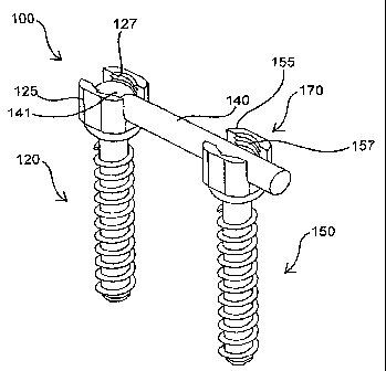
allow more sophisticated trajectories of travel for the distal end of arm 140.
[0158] System 100 further includes receiving assembly 150, which similarly
includes a
bone anchor comprising screw head 155, preferably a polyaxial screw head, and
bone threads
156. Within the tube surrounded by bone threads 156 is a guidewire lumen that
is configured
to allow carrier assembly 150 to be placed through a cannula and over a
guidewire that has
previously been placed into the bone of a patient. Screw head 155 includes one
or more
surfaces configured to engage with a tool, such as a percutaneously inserted
socket wrench or
screwdriver, to engage and rotate receiving assembly 150. Cradle 170 comprises
a` U"
shaped groove that is sized and configured to accept and capture the distal
end of pivoting
CA 02650223 2008-10-23
WO 2007/117366 PCT/US2007/004726
arm 140. Cradle 170 may include positive engagement means such as threads 157,
or other
securing means such as a projecting member that is configured to provide a
snap fit, magnetic
holding means, pivoting engagement means such as a rotatable holding arm,
adhesive holding
means, or other retention elements all not shown.
[0159] Referring specifically to Fig. 4a, pivoting arm 140 is shown in
multiple stages of
rotation, including the starting position of Fig. 4 in which pivoting arm 140
and threads 126
are linearly aligned to allow over-the-wire insertion. After threads 126 are
properly engaged
with bone, pivoting arm 140 is rotated, in a clockwise direction as shown, to
a point in which
it engages with receiving assembly 150, preferably a near ninety degree
rotation as shown,
but alternatively a smaller or greater angle as determined by the orientation
of the two bone
segments to be stabilized. Arm 140 may be rotated with the guidewire removed,
or may
include a slot, not shown, that allows arm 140 to "separate" from the
guidewire as arm 140 is
rotated. In an alternative embodiment, hinged assembly 120 includes a
cannulated screw, but
arm 140 is not cannulated, traveling along side the guidewire during
insertion, and rotating
about the guidewire during rotation and bone anchoring of threads 126. In this
alterative
embodiment, a slot is not required to rotate arm 140, in a direction away from
central axis of
the in-place guidewire.
[0160] Referring now specifically to Fig. 4b, pivoting arm 140 has been
rotated and
engaged with cradle 170 of receiving assembly 150. In the preferred method of
placing
system 100 components through cannulae and over previously placed guidewires,
pivoting
arm 140 distal end passes through an arc that resides under the skin of the
patient. Rotation
of arm 140 is preferably accomplished with one or more pivoting tools, such as
a
percutaneous tool placed through the in-place cannula through which hinged
assembly 120
was inserted. Detailed descriptions of a preferred percutaneous insertion
method is described
in reference to Figs. 6a through 6h described herebelow. In the embodiment of
Fig. 4b, both
screw head 125 and screw head 155 include securing means, threads 127 and 157
respectively, into each of which a set screw, not shown, is placed to "lock in
place" pivoting
ann 140 and provide high levels of stabilization forces, including axial
forces, radial forces
and torsional forces. Threads 127 and 157 as well as the corresponding set
screws, are
configured to provide sufficient anti-rotation properties to prevent loosening
over time, such
as anti-rotation achieved with specific thread patterns and/or included
adhesive. In an
alternative embodiment, the engagement shown in Fig. 4b, without additional
set screws into
21
CA 02650223 2008-10-23
WO 2007/117366 PCT/US2007/004726
either threads 127 or threads 157, provides the necessary stabilization
forces. In another
alternative embodiment, an automatic anti-rotation mechanism engages when
sufficient
rotation of arm 140 is achieved, simplifying the procedure for the operator,
such as by
simplifying the placement of a set screw into threads 157 with an already
locked in place
pivoting arm 140.
[0161] Referring now to Fig. 5, an exploded view of a preferred construction
of the bone
stabilization device of the present invention is provided. Hinged assembly 120
includes
multiple components captured by the dashed line of Fig. 5. Pivoting arm 140
includes ball
end 141 at its proximal end. Ball end 141 is sized and configured to be
received by screw
head 125 such that a rotatable hinge is formed, allowing the distal end of arm
140 to be
rotated in numerous directions. Ball end 141 may be inserted by the operator,
such as during
a sterile procedure prior to insertion into the patient, or be provided pre-
assembled by the
manufacturer. Hinged assembly 120 further includes a bone anchor comprising an
elongate
tube with bone threads 126, ball end 128 and thru lumen 161, a lumen sized and
configured to
facilitate placement of hinged assembly 120 over a guidewire, such as a
guidewire placed into
a bone segment to be stabilized. Ball end 128 is sized and configured to be
securedly
engaged with pivoting element 129, which in turn securedly engages with screw
head 125,
such that polyaxial rotation of screw head 125 is achieved, such as rotation
which simplifies
insertion of hinged assembly 120 in a vertebra or other bone structure during
an over-the-
wire, through-a-cannula, percutaneous procedure.
[0162] The bone stabilization device of Fig. 5 further includes receiving
assembly 150, also
including multiple components captured by the dashed line of Fig. 5. Receiving
assembly
150 includes cradle 170, an attachment point for the distal end of pivoting
arm 140 of hinged
assembly 120. Cradle 170 comprises screw head 155 that includes a `U" shaped
groove for
slidingly receiving the distat end of arm 140. In a preferred embodiment, the
geometry of the
"U" shape groove provides a snap fit to (permanently or temporarily) maintain
the pivoting
arm in place such as behind held in place during a further securing event.
Receiving
assembly 150 further includes a bone anchor comprising an elongate tube with
bone threads
156, ball end 158 and thru lumen 162, a lumen sized and configured to
facilitate placement of
receiving assembly 150 over a guidewire, such as a guidewire placed into a
bone segment to
be stabilized. Ball end 158 is sized and configured to be securedly engaged
with pivoting
element 159, which in turn securedly engages with screw head 155, such that
polyaxial
22
CA 02650223 2008-10-23
WO 2007/117366 PCT/US2007/004726
rotation of screw head 125 is achieved, such as rotation which simplifies
insertion of hinged
assembly 120 in a vertebra or other bone structure during an over-the-wire,
through-a-
cannula, percutaneous procedure.
[0163] Screw head 155 of receiving assembly 150 includes means of securing the
distal end
of pivoting arm 140, threads 157 which are configured to accept a set screw
after arm 140 is
slidingly received by the groove of screw head 155, thus locking the distal
arm in place. Set
screw 171 can be inserted and engaged by an operator into threads 157, such as
in an over-
the-wire placement procedure through the lumen of screw 171 shown, Additional
stabilization can be attained by inserting an additional set screw, set screw
142, into threads
127 of screw head 125 of the hinged assembly. Set screw 142 is also configured
to be
delivered in an open surgical procedure, or preferably an over-the-wire
percutaneous
procedure as placed through a similar lumen in screw 142. When threads 126 of
hinged
assembly 120 and threads 156 of receiving assembly 150 are anchored in bone,
and pivoting
arm 140 is secured within cradle 170, stabilization between hinged assembly
120 and
receiving assembly 150 is achieved. In a preferred embodiment, pivoting arm
140 is
configured to provide one or more of numerous parameter of stabilization,
including but not
limited to: rigid or fixed stabilization, and dynamic stabilization such as
stabilization that
allows controlled or limited motion in one or more directions. Pivoting arm
140 may be
rigid, or have some degree of flexibility. Pivoting arm 140 may include one or
more
functional elements, such as a spring to resists but permits motion.
Functional elements may
include one or more engaging surfaces, such as surfaces that permit motion in
one or more
directions, yet limit motions in other directions, or surfaces which allow
motion in a
particular direction within a finite distance. Functional elements may provide
other functions,
such as an agent delivery element which provides an anti-infection agent or an
agent targeted
at reducing bone growth that otherwise would limit motion. These and other
functions of
pivoting arm 140 are described in detail in reference to subsequent figures
herebelow.
[0164] Referring now to Figs. 6a through 6h, a preferred method of stabilizing
one or more
patient bone segments, specifically vertebral segments, is illustrated.
Referring to Fig. 6a, a
guidewire placement procedure is illustrated in which a puncture has been made
through the
patient's skin 80, and into the pedicle 3a of patient vertebra 2. A guidewire
212, such as a K-
wire, is shown in place, allowing subsequent devices to be passed over
guidewire 212, using
standard over-the-wire techniques. Referring now to Fig. 6b, a sequential
dilation is being
23
CA 02650223 2008-10-23
WO 2007/117366 PCT/US2007/004726
performed for the purpose of having a sufficiently sized cannula, dilating
cannula 220, in
place over guidewire 212. Dilating cannula 220 is positioned above, and with
its central axis
aligned with, vertebra 2 such that additional devices can be inserted over
guidewire 212 and
within a lumen of cannula 220 to access pedicle 3a and surrounding areas. The
sequential
dilation is performed to minimize tissue trauma that would result from initial
insertion of the
final, large sized cannula to be. used.
[0165] Referring now to Fig. 6c, a cannulated drill bit 231 has been placed
through cannula
220, over guidewire 212 and is in operable connection with cannulated drill
230. Drill bit
231 is near completion of drilling an appropriately sized hole into pedicle 3a
of vertebra 2,
such that an anchoring screw can be placed in a subsequent step. Referring now
to Fig. 6d,
cannulated drill bit 231 has been removed, using an over-the-wire removal or
exchange
technique, and receiving assembly 150 of the bone stabilization device of the
present
invention has been placed through cannula 220 and over guidewire 212.
Receiving assembly
150 has been inserted with its bone anchoring portion and its attaching cradle
170 in an
aligned, linear configuration. Guidewire 212 has been passed through a lumen,
not shown
but within both the anchoring portion and attaching cradle 170 of receiving
assembly 150. In
an alternative embodiment, guidewire 212 passes through a lumen of the
anchoring portion,
but then passes alongside attaching cradle 170 of receiving assembly 150.
Receiving
assembly 150 has been rotated, such as with a screwdriver tool or socket
wrench tool passed
through cannula 220 and engaging one or more portions of receiving assembly
150, tool not
shown, such that its threads 156 are fully engaged with pedicle 3a of vertebra
2. In a
preferred embodiment, these rotating tools include a thru lumen and are also
inserted and
manipulated over-the-wire.
[0166] Referring now to Fig. 6e, an adjacent vertebra, patient vertebra 4, has
undergone
similar access techniques, including guidewire placement, sequential dilation
and pedicle
drilling. As shown, receiving assembly 150 remains in place with threads 156
anchoring
receiving assembly 150 to vertebra 2, and cradle 170 positioned to receive one
or more
pivoting arms of the present invention. Dilating cannula 220b has been
inserted, such as the
same cannula as previous figures or an additional cannula with cannula 220
remaining in
place, not shown but as depicted in Fig. 6d. Guidewire 212b, preferably a K-
wire, passes
within cannula 220b, through the patient's skin 80 and into pedicle 3b of
patient vertebra 4.
Vertebra 4 is shown as an adjacent vertebra but in an alternative embodiment,
vertebra 4 may
24
CA 02650223 2008-10-23
WO 2007/117366 PCT/US2007/004726
be separated from vertebra 2 by one or more additional vertebrae, with the
associated
pivoting arm sized accordingly.
[0167] Referring back to Fig. 6e, cannula 220b is positioned above, and with
its central axis
aligned with, vertebra 4 such that additional devices can be inserted over
guidewire 212b and
within a lumen of cannula 220b to access pedicle 3b and surrounding areas.
Hinged assembly
120 has been inserted with its bone anchoring portion, its pivoting arm 140
and hinge 130 in
an aligned, linear configuration as shown. Prior to its insertion, hinged
assembly 120 may
have been assembled by the operator, such as an operator in the sterile field
connecting the
pivoting arm to the anchor portion, or may have been provided by the
manufacturer in an
assembled state. Guidewire 212b has been passed through a lumen, not shown but
within
both the anchoring portion and pivoting arm 140 of hinged assembly 120. In an
alternative
embodiment, guidewire 212b passes through a lumen of the anchoring portion,
but then
passes alongside attaching pivoting arm 140 of hinged assembly 120. Hinged
assembly 120
has been rotated, such as with a screwdriver tool or socket wrench tool passed
through
cannula 220b and engaging one or more portions of hinged assembly 120, tool
not shown,
such that its threads 126 are fully engaged with pedicle 3b of vertebra 4. In
a preferred
embodiment, these rotating tools include a thru lumen and are also inserted
and manipulated
over-the-wire. In another preferred embodiment, the rotating tool includes an
open lumen on
its distal end sized to slide over the distal end of pivoting arm 140 and
engage one or more
engagable surfaces integral to hinged assembly 120 and located at or near
hinge 130.
[0168] Referring now to Fig. 6f, hinged assembly 130 is securely attached to
vertebra 4, an
pivoting arm 140 is being rotated, such that the distal end of arm 140 forms
an arc that
remains under patient's skin 80, and is slidingly received into a groove of
attaching cradle
170 of receiving assembly 150. Pivoting arm 140 may rotatably pass through a
slot in
cannula 220b, not shown but described in detail in reference to Figs. 7 and
7a. Alternatively,
cannula 220b can be retracted a sufficient distance to allow pivoting arm 140
to swing below
the distal end of cannula 220b. In the embodiment shown in Fig. 6f, guidewire
212b has been
removed to allow pivoting arm 140 to freely swing toward cradle 170. In an
alternative
embodiment, pivoting arm 140 includes a slot from its thru lumen to it's outer
surface such
that arm 140 can be pivoted away from a guidewire. In another altemative
embodiment,
hinged assembly 120 is inserted such that pivoting arm 140 is not over-the-
wire, i.e. does not
CA 02650223 2008-10-23
WO 2007/117366 PCT/US2007/004726
include a guidewire lumen and is inserted with pivoting arm alongside the
guidewire. In this
embodiment, arm 140 can also be rotated with the guidewire in place.
[0169] Referring now specifically to Fig. 6g, a percutaneous screwdriver 240
of the present
invention has been inserted within the lumen of cannula 220b and is rotatably
engaging a set
screw, now shown but as has been described in reference to Fig. 5 hereabove,
to secure
pivoting arm 140 to prevent or limit rotation. In a preferred embodiment,
screwdriver 240
and inserted set screws include lumens such that each can be inserted over an
in-place
guidewire. In another preferred embodiment, not shown, percutaneous
screwdriver 240 is
similarly inserted within the lumen of cannula 220, not shown but aligned with
receiving
assembly 150 as shown in Fig. 6d, such that another engaging set screw can be
inserted, into
cradle 170, to securedly attach pivoting arm 140 to cradle 170. Referring now
to Fig. 6h, the
cannulae and guidewires have all been removed, and bone stabilization device
100 is
implanted in the patient. Receiving assembly 150 is securedly attached to
vertebra 2, and
hinged assembly 120 is securedly attached to vertebra 4. Pivoting arm 140 is
securedly
attached to receiving assembly 150 thus providing stabilization between
vertebra 2 and
vertebra 4. The type and amount of stabilization achieved between the two
vertebrae can take
on the various forms described throughout this application, including but not
limited to: fixed
or fused stabilization, and dynamic stabilization.
[0170] Referring now to Fig. 7, a slotted cannula of the present invention is
illustrated.
Slotted cannula 300, preferably a sequential dilating cannula, additional
sliding tubes not
shown, includes a longitudinal slot, starting from its distal end, the end
that is inserted into
the patient, and extending proximally. Slot 301, and any additional slots
included in any
slidingly received tubes not shown, are sized and positioned such that a
device contained
within cannula 300 can be passed through the slot, such as to a location
within the body of a
patient. Referring now to Fig. 7a, slotted cannula 300 is shown passing
through the skin of a
patient, skin not shown, and aligned with vertebra 4 of the patient. Hinged
assembly 120 of
the present invention is included within the lumen of cannula 300 and has been
securedly
attached to vertebra 4. Also shown is the receiving assembly of the present
invention with
attaching cradle 170 having been securedly attached to vertebra 2 of the
patient. Slot 301 of
cannula 300 has been aligned such that pivoting arm 140 of hinged assembly 120
can be
rotated to the orientation in which the distal end of arm 140 is slidingly
received by the
groove of cradle 170 without having to reposition cannula 300. In a preferred
embodiment,
26
CA 02650223 2008-10-23
WO 2007/117366 PCT/US2007/004726
the proximal end of slotted cannula 300 includes one or more markings that
indicate the
location of slot 301 such than when inserted in the body, slot 301 position
can be oriented
and/or confirmed. In an alternative embodiment, dilator 300 includes multiple
slots along its
length.
[0171] Referring now to Fig. 8, a pivoting tool of the present invention is
illustrated.
Pivoting tool 400 includes engagement end 401, configured to operably engage a
pivoting
arm of the present invention, such as to rotate the pivoting arm through one
or more cannulae
during a percutaneous procedure. Referring now to Fig. 8a, slotted cannula 300
is shown
passing through the skin of a patient, skin not shown, and aligned with
vertebra 4 of the
patient. Hinged assembly 120 of the present invention is included within the
lumen of
cannula 300 and has been securedly attached to vertebra 4. Also shown is the
receiving
assembly of the present invention with attaching cradle 170 having been
securedly attached to
vertebra 2 of the patient. Slot 301 of cannula 300 has been aligned such that
pivoting arm
140 of hinged assembly 120 can be rotated using pivoting tool 400 to the
orientation in which
the distal end of arm 140 is slidingly received by the- groove of cradle 170.
Pivoting arm 140
is rotated by first engaging end 401 of pivoting tool 400 with arm 140, and
then advancing
and potentially pivoting end 401 until arm 140 is engaged with cradle 170. In
a preferred
embodiment, the proximal end of pivoting tool 400 includes one or more
markings that
indicate the orientation of engaging end 401, such as when engaging end 401
has an non-
symmetric geometry.
[0172] Referring now to Fig. 9, another preferred embodiment of the bone
stabilization
device of the present invention is illustrated. Fig. 9 depicts a schematic
view of bone
stabilization device 100 comprising hinged assembly 120 and receiving assembly
150.
Hinged assembly 120 includes a bone anchoring portion including bone threads
126, that is
fixedly or rotatably attached to hinge 130. Hinge 130 provides a rotatable
connection, such
as a single or multi-axis rotatable connection, to pivoting arm 140. Receiving
assembly 150
includes a bone anchoring portion including bone threads 156, that is fixedly
or rotatably
attached to cradle 170. Cradle 170 is configured to be securedly attached,
intraoperatively, to
pivoting arm 140 to achieve stabilization between a first bone location and a
second bone
location. The type and amount of stabilization can be greatly specific and
customized as is
provided in the multiple embodiments of the present invention.
[0173] As depicted in the schematic representation of Fig. 9, pivoting arm 140
includes
27
CA 02650223 2008-10-23
WO 2007/117366 PCT/US2007/004726
functional element 145, depicted at the midpoint of pivoting ann 140 but
existing anywhere
along its length or comprising the entirety of pivoting arm 140. Also included
in pivoting arm
140 is adjustment means 144, shown as part of functional element 145 but
alternatively a
separate component or components of functional element 145. Adjustment means
144 is an
engageable assembly, preferably engageable via cannulae as has been described
in reference
to Figs. 6a through 6h, placed during the procedure implanting bone
stabilization device 100
or a subsequent procedure in which bone stabilization device 100 is to be
adjusted.
Numerous parameters of device 100 may require adjustment, at the time of
implantation or
thereafter, including but not limited to: force adjustments such as forces
resisting translation,
rotation and bending of vertebral segments; length adjustments; position
adjustments; and
combinations thereof. In a preferred embodiment, pivoting arm 140 is slidable
within a
component of device 100 or includes two slidable arms, and adjustment means
144 is a screw
driven assembly that causes controlled sliding and resultant length adjustment
of pivoting arm
140. In another preferred embodiment, device 100 includes one or more springs
which
provide compressive forces for stabilization, and adjustment means 144 is a
screw driven
assembly to adjust the forces exerted by the springs. In yet another preferred
embodiment,
device 100 includes one or more pneumatic or hydraulic assemblies and
adjustment means
144 is a screw driven assembly to adjust those assemblies.
[01741 Functional element 145 can provide functions that enhance therapeutic
benefit and/or
reduce complications and adverse side effects. In a preferred embodiment,
functional
element 145 comprises one or more flexible joints and provides dynamic
stabilization to
mimic a health joint such as a vertebral segment. In another preferred
embodiment,
functional element 145 comprises an artificial facet or partial facet, and
serves the function of
replacing or supporting a facet of a patient's vertebral segment. In yet
another preferred
embodiment, functional element 145 provides a function selected from the group
consisting
of: single axis flexion; multi-axis flexion; force translation such as
providing a force to hinder
motion in or more directions; motion limiting such as limiting a maximum
relative motion
between the first location and the second location; agent delivery such as
anti-bone
proliferation drugs; radiation delivery percutaneous access; facet
replacement; facet
enhancement; and combinations thereof. In yet another preferred embodiment,
functional
element 145 provides multiple functions such as those described above. Drug
delivery or
radiation exposure might be advantageous to limit the body's reaction to the
surgery and/or
28
CA 02650223 2008-10-23
WO 2007/117366 PCT/US2007/004726
the implant, such as bone proliferation which may limitjoint movement that has
been
dynamically stabilized. Drug delivery, such as a coating on one or more
components of
device 100, or an eluding drug depot such as a refillable drug depot integral
to functional
assembly 145 or another component, may alternatively or additionally be used
to deliver an
agent such as an anti-biotic delivered to prevent infections not uncommon to
implants and
implant procedures. In another preferred embodiment, functional element 145 is
a flexible
band, such as a band that provides a tensioning force between the two bone
locations to be
stabilized. In another preferred embodiment, the band is included to provide a
ligament
function. In yet another preferred embodiment, functional element 145 provides
multiple
functions, such as two or more functions selected from the numerous functions
described
immediately hereabove.
[01751 In another preferred embodiment, device 100 includes a valve assembly,
such as a
valve assembly integral to adjustment means 144. The valve assembly can be
used to provide
one-way fluid access to one or more components of device 100, such as to
refill a drug depot,
adjust a hydraulic or pneumatic assembly, or other valve function. In an
alternative
embodiment, a valve is included which opens at a pre-determined pressure, such
as a pressure
relief valve which opens to prevent undesirable forces from being generated by
device 100.
[0176] Referring now to Fig. 9a, a bone stabilization device of the present
invention is
depicted with a functional element configured to provide dynamic
stabilization. Hinged
assembly 120 includes axle 122, a pin projecting from pivoting arm 140 that is
captured and
rotatably received a receiving hole in screw. head 125 to form a single degree
of freedom
hinge. Pivoting arm 140, shown secured with set screws to cradle 170 of
receiving assembly
150, includes a functional element along its length, torsion-compression
spring 146a that is
configured to provide appropriate torsion and compressive forces for dynamic
stabilization of
two bone structures.
[0177] Referring now to Fig. 9b, another preferred hinge assembly of the
present invention is
depicted. Hinge assembly 120 includes hinge 130, of similar construction to
the hinge of Fig.
9a, and pivoting arm 140, which includes a functional element, compression
spring 146b
along its length. Compression spring 146b is configured to provide appropriate
forces for
dynamic stabilization of two bone structures when Hinge assembly 120 and
pivoting arm 140
are securedly attached to a receiving assembly of the present invention.
[0178] Referring now to Fig. 9c, device 100, consisting of the hinge assembly
120 of Fig. 9b,
29
CA 02650223 2008-10-23
WO 2007/117366 PCT/US2007/004726
is shown secured to vertebra 4 of a patient. Also implanted is receiving
assembly 150 shown
secured to vertebra 2 of the patient. Pivoting arm 140 is shown in various
rotational
positions, rotating clockwise, as shown, until fully engaged with cradle 170.
Pivoting arm
140 includes compression spring 146b along its length to provide dynamic
stabilization
between vertebra 4 and vertebra 2 of the patient.
[0179] Referring now to Figs. 10a, 10b and l Oc, another preferred device and
method of the
present invention is illustrated in which three vertebral segments are
stabilized relative to
each other. Referring specifically to Fig. 10a, a hinged assembly 120 has been
securedly
attached to vertebra 4 and a receiving assembly 150 has been securedly
attached to adjacent
vertebra 2, such as by using similar percutaneous tools and techniques
described in reference
to Figs. 6a through 6h. Pivoting anm 140 is being rotated in a clockwise
direction, as shown,
via hinge 130, to a location in which it's distal end resides within cradle
170 of receiving
assembly 150. In the preferred embodiment of Figs. l0a and I Ob, the distal
end of pivoting
arm 140 includes a reduced segment, recess 143, which is configured to
geometrically mate
with an end portion of a separate pivoting arm. Referring now to Fig. 10b, a
second hinged
assembly, hinged assembly 120' has been inserted into a vertebra 30, a
vertebra adjacent to
vertebra 2 but opposite the side adjacent to vertebra 4, such as by using
similar percutaneous
tools and techniques described in reference to Figs. 6a through 6h. Hinged
assembly 120' is
shown with its pivoting ann 140' being rotated in a counterclockwise
direction, as shown, via
hinge 130' to a location in which it's distal ends also resides within cradle
170 of receiving
assembly 150. The distal end of pivoting arm 140' also includes a reduced
segment, recess
143', which is configured to geometrically mate with the end portion of recess
143 of
pivoting arm 140 of hinged assembly 120.
[0180] Referring now specifically to Fig. l Oc, poly-segment (more than two
segments) bone
stabilization device 1000 includes first hinged assembly 120, second hinged
assembly 120'
and receiving assembly 150. Receiving assembly 150 has slidingly receiving and
is not
securedly attached to the distal ends of pivoting arm 140 and pivoting arm
140' or hinged
assembly 120 and hinged assembly 120' respectively. Stabilization, such as
dynamic
stabilization or fixed stabilization, has been achieved between vertebra 4 and
vertebra 2 and
vertebra 30. The numerous enhancements, such as functional elements including
one or more
spring included in a pivoting arm, or other enhancements, can be included in
first hinged
CA 02650223 2008-10-23
WO 2007/117366 PCT/US2007/004726
assembly 120, second hinged assembly 120' and/or receiving assembly 150 to
provide more
therapeutic benefit, improve safety and/or longevity of the implanted device.
[0181] The distal ends of the pivoting arms 140 and 140' each have a reduced
segment such
that the combined cross-sections is relatively equivalent to the cross-section
of either arm
prior to the reduction. This mating portion allows a similar cradle 170 to be
used that would
be used to securedly engage a single pivoting arm without a reduced segment.
Various
geometries of the reduced cross sections can be employed. In a preferred
embodiment, a
fixation means, such as a set screw, not shown, is placed through each reduced
portion and
into cradle 170 to secure both pivoting arms to the receiving assembly.
[01821 Referring now to Figs. 11 a and 11b, two preferred geometries of the
reduced portions
of Figs. l0a through l Oc are illustrated. A pair of pivoting arms is shown,
pivoting arm 140
and pivoting arm 140'. On each proximal end, a pin, axle 147 and axle 147'
extends radially
out from the tubular structure, each pin configured to rotate in a bushing of
the appropriate
hinge assembly to perform a hinge function. Fig. 11 a represents a geometry
including two
half-circular cross sections that are stacked on top of each other, when
engaged, as viewed
from the top of the cradle (looking down on the anchoring means). Fig. 11b
represents a
geometry also consisting of two half-circular cross sections, these sections
aligned in a side-
by-side orientation as viewed from the top of the cradle.
[0183] Referring now to Figs. 12a and 12b, two additional preferred geometries
of pairs of
pivoting arms are illustrated. The cross sectional geometries of pivoting arms
140 and 140'
are the same as those of arms 140 and 140' of Figs.'11 a and 1 1 b
respectively. The pivoting
arms of Figs. 12a and 12b further each include a functional element, coil
springs 146b and
146b', along their length, to provide dynamic stabilization forces when a poly-
segment
stabilization device of the present invention is implanted. Referring now to
Fig. 13, poly-
segment bone stabilization device 1000 includes first hinged assembly 120 and
second hinged
assembly 120' which include the pivoting arms 140 and 140' of Figs. 12a and/or
12b. In the
preferred embodiment of Fig. 13, multiple caps are placed on engagable
portions of
components of device 1000, such as cap 134 placed on top of the hinge of
hinged assembly
120, cap 174 placed on top of cradle 170 of receiving assembly 150, and cap
134' placed on
top of the hinge of hinged assembly 120'. These caps are made of a
biocompatible metal or
plastic, and prevent tissue in-growth and other contamination from entering
engagement
means such as slots and other engagable surfaces. The caps are preferably a
pressure fit or
31
CA 02650223 2008-10-23
WO 2007/117366 PCT/US2007/004726
screw cap, and can be easily removed with minimally invasive means. In an
alternative
embodiment, one or more of the caps are biodegradable.
[0184] Referring now to Figs. 14a, 14b and 14c, hinge mechanisms of the hinged
assemblies
of the present invention are illustrated. Referring specifically to Fig. 14a,
an operator
assembled hinge is illustrated. Hinge 130 includes a projecting pin, axle 147,
that extends
from pivoting arm 140. Axle 147 is configured to be snapped in place into slot
131 of screw
head 125. Screw head 125 is fixedly or rotatably connected to an anchoring
portion of hinge
assembly 120, anchor portion not shown. Screw head 125 further includes
threads 127,
which are configured to accept a set screw to prevent inadvertent disassembly
of hinge 130.
Threads 127 can also be used to lock-down, or otherwise prevent rotation of
arm 140. A set
can be partially inserted to capture the pin yet allow rotation, such as prior
to implantation in
the patient, or a set screw can be inserted after insertion into the body of
the patient.
[0185] Referring specifically to Fig. 14b, another preferred embodiment of a
hinge of the
present invention is illustrated. Hinged assembly 120 includes pivoting arm
140, which is
pivotally attached to base 124 via hinge 130. Pivoting arm 140 includes a
projecting pin 147,
which is permanently captured by a bushing included in housing 132. Pivoting
arm 140 can
be fixed in place by one or more mechanisms described in detail throughout
this application.
[0186] Referring specifically to Fig. 14c, an alternative embodiment of a
hinge is provided in
which a portion of pivoting arm 140 includes a flexible portion, such as two
metal rods
connected with a elastic or otherwise deformable section. Pivoting arm 140 is
fixedly
mounted to base 124, and hinge 130 consists of flex point 139 of arm 140.
Pivoting arm 140
and flex point 139 may be resiliently biased, either in the final secured
position, or starting
(linearly aligned with the anchor portion) position, or a position in between.
Alternatively,
pivoting arm 140 may be plastically deformable, changing its biased position
as it is rotated.
[0187] Referring now to Figs. 15a and 15b, means of securing the pivoting arrn
of the present
invention are illustrated. Fig. 15a illustrates sets screws 142 and 171,
configured to be
operatively engaged with threads 127 and 157 respectively. Threads 127 are
integral to screw
head 125 of hinged assembly 120 and threads 157 are integral to screw head 155
of receiving
assembly 150. Both screw 142 and 171 include a thru-lumen, which allows over-
the-wire
insertion, such as insertion performed by an operator using an over-the-wire
screwdriver of
the present invention. Referring now to Fig. 1 Sb, an alternative securing
means is illustrated,
including a two-piece assembly comprising a screw and an expandable ring. Ring
133 is
32
CA 02650223 2008-10-23
WO 2007/117366 PCT/US2007/004726
inserted to screw head 125 of hinge assembly 120 after which sorew 142 is
rotatably engaged
with ring 133, causing ring 133 to radially expand and provide a high
compression, reliable
connection. Similarly, ring 173 is inserted into screw head 155 of receiving
assembly 150
after which screw 171 is rotatably engaged with the threads of ring 173,
causing ring 173 to
radially expand and provide high compression, reliable connection.
[0188] Referring now to Fig. 16, a method of accessing a bone stabilization
device is
illustrated. Two cannula, cannula 220a and 200b are shown as having been
inserted through
the patient's skin 80 at locations directly above vertebra 4 and vertebra 2
respectively. A
poly-segment hinged assembly device 1000 of the present invention has been
planted at an
earlier date, such as a time period of months or more earlier. Device 1000 is
configured to
stabilize vertebra 4, vertebra 2 and vertebra 30 in a fixed or fused
configuration, or in a
dynamically stabilized configuration. Device 1000 includes a first hinged
assembly 120
securedly attached to vertebra 4, a receiving assembly 150 securedly attached
to vertebra 2
and a second hinged assembly 120 securedly attached to vertebra 30. Pivoting
arm 140' of
hinged assembly 120' is shown in secure attachment with cradle 170 of
receiving assembly
150. Hinge 130' is covered with cap 134' attached during the original
implantation
procedure of device 1000. Caps that were originally attached in the original
implantation
procedure, such as a cap on hinge assembly 130 and cradle 170 have been
removed in the
accessing procedure of Fig. 16. Percutaneous grasping and ply tools, as well
as percutaneous
rotational tools such as screwdrivers are preferably used to detach these caps
and extract
through either cannula 220a or 220b.
[0189] The method depicted in Fig. 16 involves the unsecuring of pivot arm
140, already
completed, and the reverse rotation of pivot arm 140, depicted as partially
rotated by using
lifting tool 233 inserted through cannula 220b. Screwdriver 232 has been
inserted through
cannula 220a and used to loosen and/or remove engagement means such that
pivoting arm
140 can rotate, engagement means already removed and not shown. Subsequent
steps may
include the complete removal of hinge assembly 120, and reinsertion of a new
hinged
assembly, such as when hinged assembly 120 is damaged or when a hinged
assembly with
different properties, such as a differently configured pivoting arm 140 is
desirable. In an
alternative embodiment, hinge 120 is adjusted, and pivoting arm 140 again
secured to cradle
170. Numerous combinations of adjustments and replacements of one or more
components
of system 1000 can be accomplished utilizing the percutaneous tools and
methods depicted in
33
CA 02650223 2008-10-23
WO 2007/117366 PCT/US2007/004726
Fig. 16. Use of one or more caps, such as cap 134', make subsequent engagement
of tools
with system 1000 components easier to accomplish since the covered surfaces
are free from
material that would compromise engagement.
[0190] Referring now to Fig. 17, another preferred embodiment of bone
stabilization device
of the present invention is illustrated wherein anchor portions consist of an
outer tube and a
removable core. Device 100 includes hinged assembly 120 including a bone
anchor and
pivoting arm 140 which attaches to the bone anchor portion via hinge 130.
Pivoting arm 140
includes function element 145, such as a spring or other flexible element that
provides a
flexion point for dynamic stabilization of two bone structures. Device 100
further includes
receiving assembly 150 which includes a bone anchor portion which is attached
to surface
170. Surface 170 is configured to securedly attach to the distal end of
pivoting arm such as
via a screw, not shown, but preferably inserted through the distal end of arm
140 and into
threads 175. Both hinged assembly 120 and receiving assembly 150 include
anchor portions
which have external threads for engaging and securing in bone, and a removable
inner core,
configured to be removed via one or more means such as the threaded engagement
depicted
in Fig. 17. Internal threads 126a and internal threads 156a of the hinged
assembly and
receiving assembly anchor portions respectively, allow the remaining portion
of these
assemblies to be removed, such as after a period of implantation, while
leaving the outer
threaded portions in place, such as for insertion of a subsequent assembly or
otherwise.
[0191] Referring now to Fig. 18, another preferred embodiment of the bone
stabilization
device of the present invention is illustrated wherein the pivoting arm can be
telescopically
extended or retracted, such as to rotate with a minimal radius of curvature.
Device 100
includes hinged assembly 120 including a bone anchor and pivoting arm 140
which attaches
to the bone anchor portion via hinge 130. Device 100 further includes
receiving assembly
150 which includes a bone anchor portion which is attached to cradle 170.
Cradle 170 is
configured to securedly attach to the distal end of pivoting arm such as by
the various
engagement means described throughout this application. Both hinged assembly
120 and
receiving assembly 150 include anchor portions which have external threads for
engaging and
securing in bone, external threads 126 and 156 respectively. Pivoting arm 140
consists of a
series of interlocking slidable tubes configured to telescopically be
advanced, such as to be
long enough to engage with cradle 170. In a preferred embodiment, hinged
assembly 120 is
percutaneously inserted into the body, and pivoting arm 140, in a
telescopically retracted
34
CA 02650223 2008-10-23
WO 2007/117366 PCT/US2007/004726
state, is pivoted an amount such that it's axis is pointing at the engagement
portion of cradle
170, such as a ninety degree rotation in the configuration shown.
Subsequently, using a push
tool, an integral extending assembly such as a hydraulic or pneumatic
extending assembly, or
other means, the distal end of an inner, such as the innermost, telescopic
section is advanced
until properly seated for engagement in cradle 170. The telescoping tubes of
pivoting arm
170 are preferably made of a rigid metal, sufficient to provide sufficient
force to achieve the
desired stabilization.
[0192J Referring now to Fig. 19, a preferred embodiment of the hinged assembly
of the
present invention is illustrated wherein multiple pivoting arms are included.
Hinged
assembly 120 includes thru lumen 148, such as a lumen for a guidewire and/or
bone screw,
and recess 149 which can accommodate the screw head of such a bone screw.
Hinged
assembly 120 further includes hinge 130, which rotatably attaches base 124 to
two pivoting
arms, 140a and 140b. In an alternative embodiment, more than two pivoting arms
are
rotatably attached by hinge 130. These multiple arms can be used to stabilize
the particular
bone segment to which hinged assembly 120 is attached to a single additional
bone segment,
or multiple bone segments wherein each arm is connected by an operator to a
component on
the different bone segments. Referring now to Fig. 19a, a preferred
configuration of a poly-
segment stabilization device 1000 and attachment method is illustrated. Device
1000
includes the dual arm hinged assembly 120 of Fig. 19, and two receiving
assemblies 150a and
150b. Hinged assembly 120 is securedly attached via screw 121 to second bone
segment 70b,
such as a fractured bone in the patient's arm or leg, or a vertebra of the
patient's spine.
Receiving assembly 150a is securedly attached to bone segment 70a with screw
151 a and
receiving assembly 150b is securedly attached to bone segment 70c with screw
151 b, the
three bone segments aligned as shown. Hinged assembly 120, preferably inserted
in the over-
the-wire percutaneous technique described in reference to Figs. 6a through 6h,
such as
wherein one or none of the pivoting arms includes a thru lumen for advancement
of the
percutaneous guidewire. As shown,- pivoting arm 140a is rotated such that it
can be securely
engaged with cradle 170a of receiving assembly 150b and pivoting arm 140b is
rotated such
that it can be securely engaged with cradle 170b of receiving assembly 150b.
Upon dual
engagement of each pivoting arm, fixed or dynamic stabilization is achieved
between the
three bone segments, 70a, 70b and 70c. Additional dual arm and single arm
hinged
assemblies, as well as dual or single cradle receiving assemblies, can be
added, in the linear
CA 02650223 2008-10-23
WO 2007/117366 PCT/US2007/004726
arrangement shown, and/or with hinged assemblies and/or receiving assemblies
placed in a
side-by-side configuration. These poly-component (more than 2) devices and
methods can be
useful in treating complex bone fractures and other poly-location
stabilization procedures. In
an alternative embodiment, the multiple arms of the hinged assembly have
different lengths,
such as to securedly engage with components separated from the hinged assembly
by
different displacements. Each of the multiple arms can rotate to a single
receiving assembly,
or different receiving assemblies.
[0193] Referring now to Figs 20, 20a and 20b, a preferred embodiment of the
present
invention is illustrated wherein the receiving assembly automatically engages
the pivoting
arm of the hinged assembly. Referring specifically to Fig. 20, an end view of
hinged
assembly 150 is shown wherein cradle 170 is securedly mounted to plate 154,
via fixed or
movable engagement means. Cradle 170 includes a circular notch for maintaining
a pivoting
arm of the present invention, the diameter chosen to be slightly larger than
the diameter of the
appropriate pivoting arm. At the top of the notch is projection 176, wherein
the size of notch
176 and the materials of construction of cradle 170 are chosen such that the
distal end of a
pivoting arm can snap into place, being maintained in place by projection 176
under certain
load conditions. In a preferred embodiment, the forces are chosen such that no
additional
securing means are required to achieve the desired therapeutic function
(stabilization of bone
structures). In an alternative, also preferred embodiment, an additional
securing function is
included, such as the retraining set screws described throughout this
application. Referring to
Fig. 20a, pivoting arm 140 of hinged assembly 120 is shown rotating in a
clockwise direction
about hinge 130. Receiving assembly 150, of Fig. 20, is included and provides
a snap-fit
function that retains the distal end of arm 140 when full rotated to be
constrained within
cradle 170 as shown in Fig. 20b.
[0194] Referring now to Fig. 21, a preferred embodiment of the hinged assembly
of the
present invention is illustrated wherein assemblies are included that provide
a mechanical
advantage to perform one or more functions, such as functions performed during
or post
implantation. Hinged assembly 120 includes pivoting arm 140, which is
rotatably attached to
hinge 130. Pivoting arm 140 is also rotatably attached to piston 193 via pin
192. Piston 193
is a hydraulically or pneumatically driven piston of piston assembly 190.
Piston assembly
190 includes engagable activation means 191, shown in operable attachment to
screwdriver
232b, such as a percutaneous screwdriver than can be advanced through a
percutaneous
36
CA 02650223 2008-10-23
WO 2007/117366 PCT/US2007/004726
cannula. Rotation of means 191 is used to advance and retract piston 193,
which in turn
causes pivoting arm 140 to rotate in counterclockwise and clockwise
directions, respectively.
Hydraulic and pneumatic assemblies can be used to generate large amounts of
force, perform
precise movements, and provide other mechanical advantages.
[0195] Hinged assembly 120 further includes another mechanical advantage
assembly, a
precision, high-torque screw advancement and/or screw retraction assembly
including linear
advancement element 182, rotational element 183, and engagement means 181. The
screw
advancement assembly is shown as engaged by percutaneous screwdriver 232a on
its input
end, and engages screw 121, preferably a screw configured for advancement into
bone, such
as a screw with polyaxial head pedicle screw construction. Linear advancement
element 182
includes an expandable bellows construction, expandable via an internal gear
train
mechanism, not shown, such that as screwdriver 232a is engaged and rotated,
the bottom
surface of element 182 expands in the direction opposite the surface including
hinge 130.
Rotation element 182 is operably engaged with a circular array of teeth
integral to screw 121,
teeth 184. Rotation of screwdriver 232a when engaged with engagement means 181
causes
both downward expansion of element 182, and rotation of screw 121 via
rotational element
182's engagement with teeth 184. Configuration of the included gear train can
provide
numerous benefits, including but not limited to: high levels of torque;
precise advancement
and/or rotation of screw 121; and other advantages.
[0196] It should be appreciated that numerous forms and varied configurations
of mechanical
advantage assemblies can be incorporated, to provide one or more functions,
especially to
overcome the limitations imposed by small implantable assemblies that are
preferably
accessed with miniaturized tools. Hydraulic and pneumatic assemblies can be
employed to
generate large forces and provide other benefits. Gear trains and lever arm
assemblies can be
employed to create precision control of motion and also provide other
benefits. These
mechanical advantage assemblies of the present invention can be integrated
into one or more
components of the bone stabilization device, such as the hinged assembly, the
receiving
assembly, or a separate component also configured to be implanted. These
mechanical
advantage assemblies can perform numerous functions including but not limited
to: rotation
of the pivoting arm; extension such as telescopic extension of the pivoting
arm such as a
hydraulically advanced pivoting arm; rotation and/or longitudinal advancement
of a bone
anchoring component such as a bone screw, application of one or more forces to
a bone
37
CA 02650223 2008-10-23
WO 2007/117366 PCT/US2007/004726
segment, such as a variable force stabilizing function such as a shock
absorber for two bone
segments; and combinations thereof.
[01971 Referring now to Figs 22a and 22b, another poly-segment bone
stabilization device
and method.ofthe present invention is illustrated, in which two hinged
assemblies are
implanted at adjacent locations, and at least one hinged assembly includes an
attaching cradle
for receiving a pivoting arm of the other hinged assembly. System 1000
includes first hinged
assembly 120a securedly attached to first bone segment 70a via attachment
screw 121a,
second hinged assembly 120b attached to second bond segment 70b via attachment
screw
121b, and receiving assembly 150 attached to third bone segment 70c via
attachment screw
151. Bone segments 70a, 70b and 70c, such as three adjacent vertebra of a
patient, receive
device'1000 in order to provide stabilization between the segments. Both
hinged assembly
120a and 120b include means of receiving a pivoting arm, the receiving means
comprising
cradles 137a and 137b respectively. In the figure shown, hinged assembly 120b
receives, in
cradle 137b, the pivot arm of hinged assembly 120a. Cradle 137a of hinged
assembly 130a is
implanted with no secured pivoting arm, an acceptable configuration especially
as it would
result in fewer variations of components (hinged assemblies with and without
cradles).
[0198] The pivoting arm of hinged assembly 120b is received by cradle 170 of
receiving
assembly 150 as shown. Each of the receiving arms can provide fixed or dynamic
stabilization, through inclusion of one or more flexing means as has been
described in detail
hereabove. In an alternative embodiment, a single component, a universal
component
consisting of a hinged assembly with a cradle, and a detachable (or
attachable) pivoting arm,
can be used, in multiplicity, to recreate the three-segment scenario depicted
in Figs. 22a and
22b, as well as any other two-segment or poly-segment stabilization scenario
such as the
other embodiments described hereabove. In a preferred embodiment, this
universal
component includes multiple types of pivoting arms, such as arms that provide
different
amounts and/or directions of stabilizing forces and or limit ranges of motions
in varied
distances and orientations.
[0199] It should be understood that numerous other configurations of the
systems, devices
and methods described herein may be employed without departing from the spirit
or scope of
this application. The pivoting arm of the stabilization device can be attached
to bone anchors
at its proximal, hinged end, and/or at its translating distal end, with a
secured connection that
is static (fixed), or it can be secured with a movable, dynamic connection.
The pivoting arm
38
CA 02650223 2008-10-23
WO 2007/117366 PCT/US2007/004726
and securing connections can be configured to prevent motion of the bone
segments, limit
motion such as limiting a specific direction or type of motion, or apply
specific resistive
forces to motion.
[0200] The components of the devices of the present invention are preferably
configured for
percutaneous placement, each device sized for placement through a percutaneous
cannula.
Each device preferably includes a lumen or sidecar through which a guidewire
can be placed,
or allowing placement along side a percutaneously placed guidewire. The
pivoting arm of
the present invention can preferably be rotated, such as with the inclusion of
a slot allowing
the guidewire to exit a lumen, while a guidewire is in place. The pivoting arm
and attached
components are preferably configured such that the pivoting arm can be
secured, such as with
insertion of multiple set screws, also with a guidewire in place. Other
components may
include slot exits from guidewire lumens such as to allow over-the-wire
delivery and
subsequently escape the guidewire while leaving the guidewire in place. The
devices and
methods of the present invention are configured to be inserted without
resection of tissue,
however procedures including or requiring resection are also supported.
[0201] The pivoting arm of the present invention preferably includes one or
more functional
elements. In a preferred embodiment, an artificial facet or facet portion is
included and built
into the pivoting arm or other component of the bone stabilization device.
Each component
may include one or more articulating surfaces, such as one located at the end
of the pivoting
arm and one on either the receiving assembly or hinged assembly of the present
invention,
such that pre-defined motion between the two attached bone segments can be
achieved.
[0202] One difficulty occasionally associated with driving bone screws
according to certain
embodiments of the present invention is that the pre-assembly of the rod onto
the head of the
screw eliminates or severely limits the use of current driving mechanisms, as
the head of the
screw is generally rendered difficult to access or non-accessible.
[0203] Certain other embodiments of the invention address this difficulty. It
should be noted
that such embodiments may in particular refer to assemblies such as element
100 of Fig. 4,
but that the same may also be employed in the receiving assembly of element
150.
[0204] Referring in particular to Figs. 23-26, a device 500 includes a
pivoting arm 540 and a
bone anchoring portion including a seat 525. Seat 525 may be a polyaxial seat,
such as the
seats included in polyaxial pedicle screws commonly used in spine surgery. A
lumen 561
(shown in Fig. 24) passes through arm 540 and inside the tube surrounded by
screw 526 such
39
CA 02650223 2008-10-23
WO 2007/117366 PCT/US2007/004726
that the assembly may be passed, in the orientation shown in Fig. 24, into a
patient through a
cannula and over a previously-placed guidewire, such as a"K-wire ' commonly
used in bone
and joint procedures.
[0205] At the end of arm 540 is ball end 541, which is rotationally received
and captured by
seat 525. The arm 540 can be inserted into seat 525 by an operator, or may be
provided in a
pre-attached state. The arm 540 can be removable from seat 525, or may be
permanently,
though rotatably, attached, whether provided in a"to-be-assembled' or a pre-
assembled state.
The ball and socket design of Fig. 23 allows multi-directional rotation of
pivoting arm 540.
Alternative designs may allow a single degree of freedom, or may allow more
sophisticated
trajectories of travel for the distal end of arm 540. "U"-shaped grooves 542
are provided to
allow the rod 540 to be pivoted in a perpendicular (or other angular) fashion
relative to screw
526.
[0206] Referring now to Fig. 24, an exploded view of a construction of the
bone stabilization
device is shown. The system 500 includes screw 526 with screw head 528 which
matingly
engages with a pivoting element or coupler 529 in, e.g., a ball-and-socket
arrangement. The
pivoting element 529 engages with the seat 525 via a friction-fit, as seen in
Fig. 25. Other
ways in which the pivoting element 529 can engage the seat 525 include a snap-
fit or other
such clearance fit. The pivoting element 529 can also be captured by other
means, including
a C-ring. In general, any geometric features which can cooperatively engage
may be
employed, including lugs, recesses, etc. The pivoting element 529 is provided
with a hole
therethrough to accommodate a guidewire within lumen 561. The pivoting element
529 has
two partially-spherical voids formed within, as seen in Fig. 25, to
accommodate the base 541
of the rod 540 and the screw head 528.
[0207] After the rod has been pivoted to a position for use in a patient, the
rod may be held in
that position by use of a closure element or cap 542 and a set screw 547. The
closure element
542 may be snap-fitted into the seat 525 by interaction of closure element
tabs 551 and seat
grooves 549. Instead of grooves and tabs, lugs may also be employed. Lugs have
the benefit
of preventing the seat from splaying and releasing the rod. Furthermore,
besides the snap-fit
of closure element 542, the same may also be dropped in and captured with set
screws or
other capture devices. One particular other such capture device includes an
integral locking
nut/plug combination, which eliminates the need for a plug and set screw set.
[0208] A closure element slot 545 may be disposed in the closure element 542
so that the
CA 02650223 2008-10-23
WO 2007/117366 PCT/US2007/004726
same may be further tightened along the groove 549. Of course, various other
techniques
may also be used to keep closure element 542 within seat 525. The set screw
547 may then
be tightened to secure the rod 540 against movement.
[0209] The screws such as screw 526 are generally driven into place in the
bone when the
rod 540 is in the position shown in Fig. 25, that is, coaxial with respect to
the axis of the
screw thread. The top of the screw head 528 is then rendered inaccessible,
although that is
where slots for the driving of such screws are generally disposed. For this
reason, at least one
peripheral slot 565 may be disposed so that a driver with a cooperating
element may be used
to rotate the screw 526. As even peripheral slots 565 would be rendered
inaccessible by the
above-described assembly, one or more corresponding pivoting element slots 555
may be
disposed in the pivoting element 529.
[0210] In use, the screw 526, the pivoting element 529, the seat 525, the rod
540, and the
corresponding intermediate elements, e.g., couplers or rod-capturing elements,
are assembled
prior to implantation in the patient. The device is inserted over the
guidewire. The screw is
then driven into the desired bone by use of a driver (not shown) generally
having one or more
protrusions which are long enough to pass through the seat 525, through
intermediate
elements, couplers, or rod-capturing elements, and to cooperatively engage
with peripheral
slots 565. The configuration of the driver protrusions is such that the same
can cooperatively
engage or mate with corresponding peripheral slots 565. Any number of
protrusions and slots
may be employed. In certain embodiments, 2, 3, 4, or 5 slots 565 and a
corresponding
number of protrusions on the driver may be employed. The slots 565 may be
equidistantly
disposed about the screw head 528 or may be otherwise disposed arbitrarily.
Once the screw
is driven into the bone, the rod 540 may be pivoted and the closure element
542 and set screw
547 applied.
[0211] Further details of the above embodiment may be seen by reference to the
previously-
described embodiments, in which similar elements have similar descriptions and
functions.
In particular, over-the-wire drivers may be employed such as described above
in connection
with Fig. 6.
[02121 In some of the embodiments shown in Figs. 3-22 above, the bone
stabilization system
was seen to include a first bone anchor with a pivoting rod pre-attached. It
should be noted
that in some embodiments, the first bone anchor may be inserted without the
pivoting arm
41
CA 02650223 2008-10-23
WO 2007/117366 PCT/US2007/004726
attached. Once the bone anchor is installed, or at a point during the
installation thereof, the
pivoting arm may be attached.
[02131 Attachment of the pivoting arm may be accomplished using any of the
configurations
described above. Generally, such attachment is preferably performed in a
manner in which
minimal force is applied to the bone anchor. One method is to employ a"snap-
ring ' disposed
into the seat to retain the pivoting rod after the same is installed in the
seat. In this method,
application of the snap-ring into the seat should not put undue or an
otherwise significant
amount of pressure on the bone anchor.
[0214] Various advantages inure to this non-pre-attached pivoting rod
embodiment. In
particular, the same allows customization of various properties of the
assembly, including:
length, diameter, curvature, dynamic stabilization performance
characteristics, etc., to meet
the requirements of the patient's spine.
[0215] Besides snap-fit or other sorts of frictional attachment mechanisms to
connect the
pivoting arm to the first bone anchor, a"clam-shell capture mechanism may
also be
employed. Referring to Fig. 27, a system 610 is shown with a bone screw 604, a
seat 602
having a void 614 formed therein, and a pivoting rod 606 having a distal end
608. Prior to,
during, or following installation of the bone screw 604 into the desired bone
segment, the
distal end 608 is inserted into the void 614 and more particularly into a clam-
shell capture
mechanism 612. Clam-shell capture mechanism 612 includes a first shell 611, a
second shell
613, and a hinge 615 for connecting the first shell 611 and the second shell
613. The first
shell 611 and the second shell 613 are coupled to the seat 602 within its void
614.
[0216] The shells may be attached to the seat via various means. There may be
a cap over
the shell. The shell may be slitted to allow expansion for a snap-fit. The
shell may also be
attached via a friction-fit or hinge, or via a combination of these techniques
and devices.
[0217] Fig. 27(A) shows the system during installation of the pivoting rod 606
into the clam-
shell capture mechanism 612, and Fig. 27(B) shows the system following
installation. To
allow a degree of pivot, the clam-shell capture mechanism 612 may have a
varying shape and
size of the outlet 603 through which the pivoting rod 606 extends. The overall
shape of the
interior of the clam-shell capture mechanism 612, when closed, must be such
that the pivoting
rod 606 is held in a secure fashion. However, the same may be provided with a
slit (seen as
dotted line 605) through which the rod can pivot. The outlet 603 may also be
somewhat
42
CA 02650223 2008-10-23
WO 2007/117366 PCT/US2007/004726
larger than the diameter of rod 606 so a degree of movement is provided in the
plane of the
figure, if desired.
[0218] In another system, shown in Fig. 28(A) and (B), a system is shown with
a bone screw
616, a seat 617 having a void 619 formed therein, and a pivoting rod 618
having a threaded
distal end 621. Prior to, during, or following installation of the bone screw
616 into the
desired bone segment, the threaded distal end 621 is inserted into the void
619 and more
particularly into a threaded receiving assembly 622. Threaded receiving
assembly 622
includes receiving threads 623, bearings 626, and an axle 624 about which the
assembly
rotates on the axle. Alternatively, lugs which mate with recesses may be
employed. The
threaded receiving assembly 622, and in particular bearings 626, are coupled
to the seat 617
within its void 619 in known fashion.
[0219] Fig. 28(A) shows the system prior to installation of the pivoting rod
618 into the
threaded receiving assembly 622, and Fig. 28(B) shows the system following
installation.
Following installation, the pivoting arm 618 may rotate and its distal end
captured by a
receiving assembly as described above.
[0220] Fig. 29 (A) and (B) show top and side views of a frictional-fit
engagement for a
pivoting rod 634 to attach to a seat 628 of a bone anchor (not shown).
Pivoting rod 634 is
shown with a small axle 636 therethrough. Of course, axle 636 could also be
constituted of
two small pins (or one pin which passes all the way through) disposed on
opposing sides of
the pivoting rod 634. Seat 628 has a void 632 formed therein, with press-fit
slots 638 on two
sides thereof. Pivoting arm 634, and in particular axle 636, press-fits into
the slots 638 and is
held in place by the frictional engagement of the axle and the slots. Despite
being held in
place, the placement of the axle and the slots allows a rotational degree of
freedom, in this
case out of the plane of the figure. The pivoting arm may then be captured by
a receiving
assembly as described above.
[0221] The slots may have a larger separation opening at the bottom to allow
the rod to
"snap-in '. In addition, the slots may have a larger separation at the top for
ease of insertion.
In either case, the slots may be tapered to the larger separation. Both of
these tapering may
be employed in combination or separately.
[0222] Fig. 30(A) and (B) show top and side views of a related embodiment of a
bayonet-fit
engagement for a pivoting rod 644 to attach to a seat 642 of a bone anchor
(not shown).
Pivoting rod 644 is shown with a small axle 646 therethrough, the nature of
which is similar
43
CA 02650223 2008-10-23
WO 2007/117366 PCT/US2007/004726
to axle 636 above. The seat 642 has two entry slots 645 and 647, which are
respectively
adjacent receiving ramps 641 and 643. Pivoting arm 644, and in particular axle
646, is
disposed in the entry slots 645 and 647 and then twisted to securedly engage
the seat 642, in a
bayonet-fit fashion. Despite being held in place, the placement of the axle
and the slots
allows a rotational degree of freedom, in this case out of the plane of the
figure. The pivoting
arm may then be captured by a receiving assembly as described above (the ramps
have a hole
in the middle to accommodate rotation of the rod).
[0223] Fig. 31(A)-(D) show assemblies for frictional-fit engagements for a
pivoting rod to
attach to a seat of a bone anchor, where the degree of range of motion is
controllably
adjusted. The degree of range of motion may be in travel, angle, or other sort
of motion.
[0224] In particular, referring to Fig. 31(A), pivoting rod 654 is shown with
a small axle 658
through a distal end 656 thereof. In a manner similar to that of Figs. 29 and
30, the pivoting
rod is securedly attached to a seat 652, within a groove 650, which in turn is
attached to bone
screw 648. The side walls 651 of groove 650 may be closely fit to the distal
end 656 of the
pivoting rod 654 or they may be spaced more apart. If they are closely-fit, as
shown in Fig.
31(A) and (C), then the swing of pivoting rod 654 is substantially limited to
a single plane.
On the other hand, if the side walls 651 of groove 650 are spaced apart to
form a void 662 in
which sits the distal end 656 of the pivoting rod 654, as shown in Fig. 31(B)
and (D), then the
swing of pivoting rod 654 has considerably more movement or motion. In this
case, the
swing of pivoting rod 654 is defined by an arc 653. A set-screw 664 may be
disposed to
control the size of arc 653. Note that the void 662 may be generally
trapezoidal in shape, and
that the size of the slots in which the axle 658 is disposed may also be
somewhat enlarged to
accommodate movements of the axle and rod.
[0225] Further, while production of an arc-allowed movement for a pivoting rod
is shown,
analogous alterations in the side walls and axles and slots would allow
additional movements
such as: flexion, extension, axial rotation, lateral bending, etc.
[0226] Referring ahead to Fig. 32(A)-(C), another way of frictionally engaging
a pivoting rod
to a seat of a bone anchor is shown, as well as a way of frictionally engaging
a seat to a bone
anchor.
[0227] Referring to Fig. 32(A), a system 960 is shown where a bone screw 962
has a guide
lumen 964. Following, during, or before installation of the bone screw 962, a
snap-in tapered
screw retainer 966 is attached to the bone screw 962, in particular by
frictionally engaging the
44
CA 02650223 2008-10-23
WO 2007/117366 PCT/US2007/004726
screw head 963 to a first screw void 972 formed in screw retainer 966. In one
embodiment,
slots (not shown) may be formed in the screw retainer 966 around first screw
void 972 in
order to allow a portion of the screw retainer 966 to "flare" outwards to
accept and
frictionally engage the screw head 963. A second screw void 974 is formed in
the screw
retainer 966 generally opposite the first void. The second screw void 974 is
configured to
accept a pivoting rod following, during, or before installation of the bone
screw 962. The
second screw void 974 includes an elastic member 968 to assist the securing of
the pivoting
rod.
[0228] Following installation of the screw head 963 into the screw retainer
966, the screw
retainer 966 is inserted into a seat 976. Seat 976 includes two lips, lip 981
for securing the
screw retainer and lip 982 for securing the pivoting rod. The top end of the
screw retainer
966, due to its inherent elasticity, compresses somewhat as it passes lip 981.
Following
insertion, the top end springs back to its original configuration. The screw
retainer 966 outer
diameter is greater than the inner diameter of the seat 976, preventing the
screw retainer from
coming out of the seat. Moreover, a force pulling the screw downward would
likewise cause
the first void to tighten around the screw head because the first void would
itself be caused to,
decrease in radius due to the inner diameter of the seat. In other words, a
force pulling the
screw downward also prevents the screw from coming out because any such force
pulls the
capturing element in such a way as to make the capturing element tighten
around the head of
the screw, preventing removal.
[0229] Once the seat is installed, the pivoting rod 984 with guide lumen 986
and ball end 985
can then be snap-fit into the second void 974. A clearance or space is
provided adjacent the
second void such that the same can flare out and securely accept the rod.
[0230] Fig. 33 (A) and (B) show an alternative embodiment of a rod and bone
anchor
assembly. In particular, referring to Fig. 33(A), a bone screw 961 is shown
with a seat 967
having a void 965 therein. Referring to Fig. 33(B), a pivoting rod 984 with
ball end 969 has
been disposed into the void 965 of the seat 967. A plug 988, which may have
threads that
engage corresponding threads on the opening of the void, is used to secure the
pivoting rod in
place. The rod is disposed such that a space 990 is left within void 965 which
allows the rod
to slide back and forth once the rod is rotated into position, approximately
at a 90 degree
angle with the screw 961.
[0231] Fig. 34 shows a device that may be employed in the above embodiments of
a rod and
CA 02650223 2008-10-23
WO 2007/117366 PCT/US2007/004726
bone anchor assembly. In particular, a connector 991 is shown having a tip 992
for capturing
a rod (not shown) or a screw retainer which then in turn connects to a rod
(not shown).
Connector 991 also has a tip 994 having ridge 996 that connects to a bone
screw. The ridge
996 allows a rotational force to be transmitted through to the bone screw if
desired.
[0232] Systems according to the invention may also include those that can
provide a degree
of flexibility to allow a more convenient capture of a pivoting rod. Referring
to Figs. 35(A)-
(C), a system 920 includes two bone screws 922 and 924 that are shown with
respective
screw heads 926 and 928. Each screw head is disposed in a first void formed in
respective
retaining members 932 and 934. Retaining members or seats 932 and 934 each
have a second
void formed therein substantially opposite the first void. The second void
contains the ball-
shaped ends 942 and 944 of rod 946. Seats 936 and 938 contain respective
retaining
members 932 and 934. Seats 932 and 934 perform functions similar to those
shown in Fig.
32.
[0233] The ability of the retaining members or seats to pivot and rotate about
the. screw head
allows the retaining members or seats to be disposed in a number of different
positions
relative to the axis of the screws. This is important as the screw axes are
generally non-
parallel as the same depends on the orientation of the pedicle in which they
are installed. The
retaining members or seats can thus be oriented arbitrarily and independently,
and can in
particular be oriented such that the pivoting rod can be conveniently
installed. In so orienting
the retaining members or seats, a degree of compression or distraction is
often imparted to the
spinal segments.
[0234) In an actual installation, typically the rod would be disposed between
the retaining
members or seats, and a set screw would be started in each to retain the rod.
Then a degree of
distraction or compression would be imparted to better seat the rod, and the
set screw would
then be tightened. 'In this way, the set screw is always properly placed in
the retaining
members.
[0235] Fig. 36(A) and (B) show an alternative embodiment 950 of a rod 956 that
may be
employed in the system of Fig. 35. Rod 956 has a stationary ball end 952 and a
movable ball
end 954. Movable ball end 954 can slide back-and-forth along rod 956. The same
can be
secured by methods and devices described here, including set screws, friction-
fits, crimping,
etc. As the ball end 954 must still be disposed in the void within retaining
member 934
(which in turn sits within seat 938), retaining member 934 and seat 938 may be
configured
46
CA 02650223 2008-10-23
WO 2007/117366 PCT/US2007/004726
with a slot substantially opposite to the slot facing seat 936. This slot,
opposite to the slot
facing seat 936, allows an excess rod portion 955 to exit the retaining member
934 and seat
938 in the case where the ball end 954 is not at the extremity of the rod 956.
[0236] It should be noted with respect to this embodiment that the ball end
954 may be
deployed such that it can slide easily along rod 956, or can slide with effort
along rod 956, or
cannot slide along rod 956. Moreover, a universal joint-type end may be
situated at either
ball end, or may also be disposed at an intermediate position along rod 956.
[02371 While numerous varieties of pivoting rod have been disclosed above,
even more types
may also be employed. For example, a locking cone system, as shown in Fig. 18
above, may
allow a single device to accommodate a continuous range of sizes of pivoting
rods.
[02381 Further, while numerous varieties of capture and receiving assemblies
have been
disclosed above, even more types may also be employed. For example, the
pivoting rod may
be swaged into place or otherwise captured. In any case, the initial
attachment of the pivoting
rod to the initial seat may be permanent or detachable. Moreover, the
secondary attachment
of the pivoting rod to the capture seat or other receiving assembly may also
be permanent or
detachable. Following rotation of the pivoting rod, the same may be fixed in
place with, e.g.,
set screws or other means.
[0239] As another example, referring to Fig. 37, a system is shown with a
pivoting rod 684
which pivots about axle 686 such that the pivoting rod 684 extends from a seat
682 to a seat
682'. Slots 692 and 692' are provided in the pivoting rod 684 at extremities
thereof. A screw
688 is disposed which intersects slot 692, and correspondingly a screw 688' is
disposed
which intersects slot 692'. When the pivoting rod 684 is in a deployed
configuration, as
shown, screws 688 and 688' may be tightened, which in turn widens slots 692
and 692'
respectively. As the slots widen, the extremities of rod 684 bow outward and
are forced
against sidewalls 691 and 691', frictionally engaging the same. Once the
frictional
engagement is great enough, pivoting rod 684 is secured between the seats, and
bone
stabilization occurs. Again, it is noted that the screws 688 and 688' need not
provide a force
normal to the plane of the figure, frictionally securing the rod against the
seat. Rather, the
screws bow the rod ends outward, parallel to the plane of the figure,
frictionally securing the
rod against the sidewalls.
[0240] Of course, a set screw may also be used that does provide a force
normal to the plane
of the figure, frictionally securing the rod against the seat.
47
CA 02650223 2008-10-23
WO 2007/117366 PCT/US2007/004726
[0241] As noted above in connection with the discussion corresponding to Figs.
10-13, 16,
19, and 22, embodiments of the invention may not only be used to provide
stabilization to
two adjacent vertebrae, but indeed can be used in a multi-level fashion to
stabilization three
or more vertebrae. Additional details concerning these designs may be seen by
reference to
Figs. 38-43.
10242] Referring to Fig. 38 (A)-(C), a system is shown in which two bone
screws 770 and
772 are shown, each with an associated respective seat 770' and 772'. Seat
770' houses one
pivoting rod 773, while seat 772' houses dual pivoting rods 774 and 774'. Seat
772' with
dual pivoting rods further has an axle 776 about which each rod pivots. Rod
773 also has an
axle (not shown). The dual rod system can be loaded into the seat at any time,
before, during,
or after installation of the bone anchor, to allow connection to adjacent
screws, e.g. at seat
77Q'.
[0243] Referring to Fig. 38(B), a system is shown in which the dual-rod system
of Fig. 38(A)
(right hand side) is shown between two bone anchors. These two bone anchors
are not shown
with their own rods, but the same may also be incorporated. To the right of
bone anchor 770'
and seat 772' is bone anchor 770" and seat 772". To the left of bone anchor
770' and seat
772' is bone anchor 770"' and seat 772"'. In Fig. 38(B), the dual rod system
is connected to
the seat at their distal end, in which case the rods rotate down to be
captured by receiving
assemblies, one rotating clockwise and the other counter-clockwise.
[0244] Referring to Fig. 38(C), a system is shown in which a related dual-rod
system is
shown between two bone anchors. As before, these two bone anchors are not
shown with
their own rods, but the same may also be incorporated. The dual-rod system has
a bone
anchor 770', seat 776, and two rods 778 and 778'. To the right of bone anchor
770' and seat
776 is bone anchor 770" and seat 772". To the left of bone anchor 770' and
seat 772' is
bone anchor 770"' and seat 772' '. In Fig. 38(C), the dual rod system is
configured such that
the rods slide outward, from their distal ends, such that the distal ends then
become the
portions captured by receiving assemblies.
[02451 Fig. 39(A)-(D) show an embodiment related to that of Fig. 38(A)-(C). In
particular,
referring to Fig. 39(A), a bone screw 782 is shown with a seat 784 and a dual-
rod assembly
having rods 786 and 786'. On the left side of bone screw 782 is a bone screw
782' with a
seat 784', and on the right side of bone screw 782 is a bone screw 782" with a
seat 784".
Rod 786' rotates in a clockwise=direction to engage a capture mechanism (not
shown) within
48
CA 02650223 2008-10-23
WO 2007/117366 PCT/US2007/004726
seat 784", and rod 786 rotates in a counter-clockwise direction to engage a
capture
mechanism (not shown) within seat 784'.
[0246] Fig. 39(B) shows additional details. In particular, the figure shows a
rotation
mechanism 788 through which rods 786 and 786' rotate. In particular, referring
to Fig.
39(C), rotation mechanism 788 has a first half 788' and a second half 788".
First half 788'
and second half 788 ' matingly engage, e.g., each can form half of a sphere,
and the two
combined can approximately form a complete sphere. Fig. 39(D) shows a plug 794
formed
on an interior wall of half-sphere 792 of second half 788" which can matingly
engage a
corresponding hole (not shown) in 788'. Other rotation mechanisms can also be
employed.
[0247] Other systems can also provide multilevel stabilization. Figs. 40-44
show additional
embodiments of systems employing dual arms on a single hinged assembly.
[0248] In particular, Fig. 40(A)-(C) show a dual arm system with a unitary
hinged assembly
employing adjustable-length rods. In this embodiment, pivoting rods 802 and
804 meet at a
rotation mechanism having first half 806 and second half 808. The rotation
mechanism may
be like that disclosed above. The rotation mechanism snaps into place in a
seat like those
disclosed above. A first ball 812 is disposed at an end of rod 802 opposite
that of first half
806, and a second ball 814 is disposed at an end of rod 804 opposite that of
second half 808.
[0249] In some of the above-described capture mechanisms, a pivoting rod is
that which is
captured, and the same is secured by a threaded plug, set screw, or other such
retainer.
Accordingly, the system is per se adjustable because the rod may be captured
at any point
along its length. In Fig. 40(A)-(C), if the ball is that which is to be
captured, then the length
of the rod becomes much more important. Accordingly, in Fig. 40(A)-(C), the
ball 814 is
attached to an inner rod 822 (see Fig. 40(C)) which is slidably and
telescopically disposed
within rod 804. Inner rod 822 may become immovable with respect to rod 804 in
a number
of ways, including via use of a set screw, by rotation of inner rod 822 on
which a cam is
biased to engage the inner wall of rod 804, etc. Alternatively, the same may
be left to
slidably move relative to rod 804, depending on the desires of the physician.
[0250] Fig. 41(A)-(F) show a dual arm system with a unitary hinged assembly
employing
multiple axles for the pivoting rods. Referring to Fig. 41 (A)-(F), a bone
screw 830 is shown
with a seat 832 and a dual-rod assembly having rods 824 and 826. On the left
side of bone
screw 830 is a bone screw 830" with a seat 832", and on the right side of bone
screw 830 is
a bone screw 830' with a seat 832'. Rod 826 rotates in a clockwise direction
to engage a
49
CA 02650223 2008-10-23
WO 2007/117366 PCT/US2007/004726
capture mechanism (not shown) within seat 832', and rod 824 rotates in a
counter-clockwise
direction to engage a capture mechanism (not shown) within seat 832".
[0251] Fig. 41(B) shows additional details. In particular, the figure shows a
rotation
mechanism 828 through which rods 824 and 826 rotate. In particular, the
rotation mechanism
includes dual parallel axles, each attached to one of rods 824 and 826.
[0252] Fig. 41(B) shows the rods in a parallel alignment, such as during
insertion. Fig. 41(C)
shows the rods in an anti-parallel alignment, such as following deployment.
[0253] Fig. 41(F) shows the same set of bone screws and seats, this time being
engaged by
pivoting rods 824' and 826' which are coupled together via rotation mechanism
828'. In this
embodiment, the step of pushing the rod assembly down acts to automatically
open the rods,
swinging the same into position where they may be captured by an appropriate
receiving
assembly. In a manner similar to that of Fig. 41(B) and (C), Fig. 41(D) shows
the rods in a
parallel alignment, such as during insertion, while Fig. 41(E) shows the rods
in an anti-
parallel alignment, such as following deployment.
[0254] In all of these embodiments, it should be noted that the rod can be pre-
attached to the
seat or alternatively the same can be installed in the seat following
installation of the bone
screws into the spine of the patient.
[0255] Fig. 42(A)-(D) show an alternative dual arm system 850 with a unitary
hinged
assembly employing multiple axles for the pivoting rods. In particular, rods
852 and 854 are
shown with distal ends 852' and 854' (see Fig. 42(C)), respectively. These
distal ends each
have a groove into which a flat extension 856 is disposed. Flat extension 856
(and a
corresponding flat extension (not shown) within rod 854 are attached to
central assembly 860.
Moreover, through the flat extensions axles 858 and 862 are disposed, which
extend from one
side of the distal ends 852' and 854' to a side diametrically opposite. In
this way, rods 852
and 854 are hingedly attached to central assembly 860.
[0256] The distal ends of the rods are disposed within a seat 864 attached to
a bone screw
866 having a guidewire lumen 864 disposed therein.
[0257] Fig. 42(A) shows the rods in a position for insertion and Fig. 42(B)
shows the rods in
a deployed configuration.
[0258] Fig. 43(A)-(C) show a dual arm system 870 with a unitary hinged
assembly
CA 02650223 2008-10-23
WO 2007/117366 PCT/US2007/004726
employing pivoting offset rods. In particular, rods 872 and 874 are shown with
distal ends
having indentation features 878. Indentation features 878 allow for secure
connection to
other seats on a multilevel system.
[0259] Rods 872 and 874 are joined at a rotation mechanism 876 which includes
an axle 877
about which both rods rotate. Multiple axles may also be employed. When the
rods are in an
insertion configuration, they are generally parallel to each other. When the
rods are
deployed, they are anti-parallel to each other. A guide lumen 875 may be
employed for
placement.
[0260] Fig. 44(A)-(E) show a dual arm system 880 with a unitary hinged
assembly
employing pivoting rods, each with a complementary taper. In particular, rods
882 and 884
are shown joined within seat 886 attached to bone screw 888. The rods may
rotate relative to
each other via an axle or other mechanism (not shown). For example, referring
to Fig. 44(C),
the rod 884 may have a plug 889 formed on a end 882' which matingly engages a
hole 881
formed on an end 884' of rod 882. When the plug 889 engages the hole 881, the
ends 882'
and 884' of rods 882 and 884 adjacent the plug and hole form a substantially
spherical head
which may be securely and rotatably inserted within seat 886. A slot 886' may
be formed
within the seat 886 into which the rods rotate when deployed. To allow the
rods to align in a
substantially parallel manner during, e.g., insertion, each rod may be formed
with a
cooperating taper. In the figures, rod 882 is formed with a taper 883 and rod
884 is formed
with a taper 885. The tapers are formed in a manner such that the face each
other when the
rods are disposed in the seat, either before, during, or after installation of
the bone screw.
[0261] When the rods are in an insertion configuration, they are generally
parallel to each
other, as shown in Figs. 44(A) and (D). When the rods are deployed, they are
generally anti-
parallel to each other, as shown in Fig. 44(E). Of course, they are still
deployed through the
cannula.
102621 Other multi-level systems have been disclosed above, in particular,
dual attaching
cradles on a single receiving assembly are shown in Figs. 12 and 13, and a
sequential
arrangement, having a hinged assembly and an attaching cradle coupled to a
bone anchor, is
shown in Fig. 22.
[0263] Many of the dual arms disclosed above show two arms attached to a
single seat on a
bone screw, i.e., dual pivoting rods on a unitary hinged assembly, these rods
then linking to
two receiving assemblies diametrically opposed from each other. However, it is
noted that a
51
CA 02650223 2008-10-23
WO 2007/117366 PCT/US2007/004726
receiving assembly itself may also include a rotatably attachable pivoting
rod. In this case,
clearance should be allowed for the rotation, typically via a ball-and-socket
or hinge, while
still allowing secure attachment of the first pivoting rod. One way of
configuring this is for
each bone anchor to include a receiving assembly (for a first pivoting rod)
and a separate seat
for attachment of a second pivoting rod (which is then received by another
receiving
assembly). An advantage of this configuration is that the bone
screw/seat/pivoting
rodlreceiving assembly systems can all have the same or a similar
construction, easing
manufacture. There is no need to have a separate construction for the hinged
assembly vis-a-
viz the receiving assembly. Such an embodiment is shown above in Fig. 22b with
particular
reference to assemblies 70a and 70b.
[0264] The above description has disclosed devices and methods for minimally-
invasive
surgery. Certain additional complementary features may apply to many or all of
the above.
[0265] For example, referring to Fig. 45, two bone screws 666 and 666' are
shown below
skin 678. Seats 668 and 668' are attached, or integral with, respectively,
bone screws 666
and 666. A pivoting rod 672 has a proximal end attached to seat 668 and when
deployed
extends to and is captured by seat 668'. Insertion cannulae 674 and 674' are
shown above
their respective seats and bone screws. As may be seen, when in the insertion
configuration,
and due to the length of the pivoting rod 672, pivoting rod 672 extends a
distance above skin
678. A shorter pivoting rod would not extend above the skin, and could be
immediately
rotated into the receiving assembly. However, due to the length, the pivoting
rod cannot be
rotated into seat 668'. In this case, a partial incision 676 may be made to
accommodate a
partial amount of the rotation of the pivoting rod 672. The first part of the
rotation of the
pivoting rod passes through the skin 678 through the partial incision 676. In
this way, the
partial incision 676 allows use of a longer pivoting rod, as may be desired
for certain
procedures. The same may also accommodate sites that are located closer to the
skin.
[0266] Systems may also be employed that nearly-automatically perform a level
of dissection
per se. Referring to Fig. 46, a system is seen with two bone screws 694 and
694', respective
seats 696 and 696', and pivoting rod 698. The pivoting rod 698 is constructed
with an
anterior facing edge 700 that is sharpened to reduce the forces required to
pass through tissue
during the rotation of the pivoting rod 698 into the receiving assembly such
as seat 696'. In
other words, during rotation, sharpened edge 700 can improve dissection to
allow passage of
the pivoting rod 698 through the skin and surrounding tissues.
52
CA 02650223 2008-10-23
WO 2007/117366 PCT/US2007/004726
[0267] In an alternative embodiment to Fig. 46, sharpened edge 700 may be
blunted prior to
the closing procedure. Alternatively, the sharpened edge itself, though not
the pivoting rod,
may be made biodegradable such that, over time, it would dissolve in the body.
The
sharpened edge could also be filed off or otherwise dulled by the physician,
or a collar may
be slid onto the edge so that the sharpened edge is not unsheathed while
maintained in the
body.
[0268] To assist in insertion and installation or in maintenance in a deployed
position, the
pivoting rod can be combined with a torsional spring to bias the pivoting arm
in various
positions. Referring to Fig. 47, a system is seen with two bone screws 702 and
702',
respective seats 704 and 704', and a pivoting rod 703. The end of pivoting rod
703 that is
initially disposed within a seat, i.e., seat 704, is also coupled to a
torsional spring 706. The
torsional spring 706 may resiliently bias the pivoting rod 703 in a position
parallel to bone
screw 702, perpendicular to the axis of the bone screw 702, or at any angle in
between as may
be desired.
[0269] In the case where the torsional spring 706 resiliently biases the
pivoting rod 703 in a
position perpendicular to bone screw 702, the rotation procedure may be
simplified as the
pivoting rod will naturally move to the "captured" or "received"
configuration. In the case
where the torsional spring 706 resiliently biases the pivoting rod 703 in a
position parallel to
bone screw 702, the insertion procedure may be simplified as the pivoting rod
will move
more easily down the cannula. The parallel position will also result in a more
convenient
removal or readjustment following the pivoting action, if necessary or
desired. The angular
position of torsional spring 706 may be reset at any time to change the bias,
i.e., the "rest"
position. This bias may be adjustable by the physician. For example, the
spring may be
attached to the seat with a screw such that rotation of the screw alters the
rest position of the
spring.
[0270] Of course, the torsional spring 706 may be biased at any point between
the two
extremes discussed above, and many different functional elements may be
employed to
resiliently bias the spring in one or more positions. For example, different
types of springs or
other elastic members may be employed.
[02711 Other systems which may maintain a pivoting rod in one configuration or
another are
shown above. In particular, the above-described Fig. 31(A)-(D) show a system
in which the
frictional engagement between the rod 654 and the groove walls 651 allow a
degree of
53
CA 02650223 2008-10-23
WO 2007/117366 PCT/US2007/004726
maintenance of the rod in a desired position. In other words, if the groove
walls 651 fit the
rod 654 tightly, the same is resiliently held in a given position. This
embodiment has an
advantage that the any position may be the "resiliently-biased" position, as
placement of the
rod in any rotational position naturally becomes the "rest" position (or which
may be set by
the physician via an adjustment), and any movement out of that position is met
with a return
force, unless and until the movement out of that position becomes so great
that a new "rest"
position is attained. This embodiment also has the advantage that the rod is
secured against
small movements, as may occur if the connection between the seats is not
tight.
[0272] The pivoting rod may be curved or otherwise contoured to approximately
mimic the
curvature of the spine. Referring to Fig. 48, a system is seen with two bone
screws 708 and
708', respective seats 712 and 712', and a pivoting rod 714. The pivoting rod
714 has a
curved shape 716, which somewhat matches the curve of the spine. However, a
guidewire
lumen 710 may be provided that is maintained straight throughout the bone
screw 708, the
seat 712, and the pivoting rod 714. The straightness of the guidewire lumen
710 allows use
of even a relatively stiff K-wire. The guidewire lumen can form a slot, open
on one side,
rather than a hole, so that the guidewire can be left in place even during
rotation of the rod
into the capture or receiving assembly.
[02731 In a related embodiment, the guidewire lumen may also be curved, but
may be curved
such that the same has a larger radius of curvature than the radius of
curvature of the rod.
That is, the guidewire lumen is straighter than the rod. In this way, a
guidewire may more
easily pass through, i.e., with less bending. In another related embodiment,
the guidewire
lumen may have a greater inner diameter than usual, i.e., much larger than the
guidewire
diameter, and again this would result in minimized bending of the guidewire as
the same
passes through.
[0274] Embodiments may include assistance or confirmation of proper engagement
with the
receiving assembly or attaching cradle. Referring to Fig. 49, a system is
shown with a bone
screw 718 capped by a seat 722. This system has a flared opening 726 leading
to a capture
void 720 that receives the pivoting rod (not shown). The taper of the flared
opening 726
provides a snap-fit for the pivoting rod that in turns lead to audible and/or
tactile feedback for
the physician. An optional magnet 724 may also be employed to assist in the
alignment of
the rod, which would include a magnetic element in this embodiment. The flared
opening
54
CA 02650223 2008-10-23
WO 2007/117366 PCT/US2007/004726
further has the advantage of serving to self-align the pivoting rod as the
same is guided into
place.
[0275] In this embodiment the magnetic material may either be a separate piece
attached to
the rod, or the rod itself may have some magnetic character. Stainless steel
has only very low
ferromagnetic properties, and titanium lacks any. Thus, suitable design
considerations must
be employed in this design.
[0276] Other systems may employ radiopaque markings or markers to identify
placement of
the bone screws and the pivoting rod, and to confirm proper alignment of the
distal end of the
pivoting rod and the receiving assembly or cradle. In this case, of course,
the other
components would preferably be made of polymers to make the markers distinct.
Referring
to Fig. 50(A)-(B), a system is shown with two bone screws 728 and 728', each
with a
respective seat 732 and 732'. A pivoting rod 734 extends between the seats. A
radiopaque
marker 738 is shown on the pivoting rod 734 which, when in a deployed
configuration, is
disposed substantially in the center of seat 732'. Another radiopaque marker
736 is disposed
in the center of the top face of seat 738. Each of the radiopaque markers
extends linearly a
predetermined distance. When viewing the system from the top, proper
deployment of the
pivoting rod is seen by co-linearity of the two radiopaque markers 736 and
738. If the
radiopaque markers are parallel but not collinear, as seen in Fig. 50(B), the
pivoting rod may
be determined to be not in a properly-deployed configuration. Of course,
numerous other
arrangements of radiopaque markers may be envisioned by those of ordinary
skill in the art
given this teaching.
[0277] The radiopaque markings or markers may include radiopaque fillers or
dyes, tantalum
beads or strips, etc. Alternative types of markers may also be employed,
including those that
are evident on MRI or ultrasound scans. These may include magnetic markers and
ultrasonically reflective markers, respectively. Such markers may be employed
to confirm
proper placement, configuration, etc.
[0278] Several of the above systems describe configurations in which a hinge
for a pivoting
rod is provided in the seat attached to a bone screw. However, such a hinge
may also form a
part of the pivoting rod. Referring to Fig. 51(A)-(B), two bone screws 740 and
740' are
shown with respective seats 742 and 742'. Seat 742 has a receiving assembly
744 including a
threaded section 746. Of course, the threaded section could be integral with
the seat 742 in
an alternative embodiment.
CA 02650223 2008-10-23
WO 2007/117366 PCT/US2007/004726
102791 Hinges in the embodiment of Fig. 51(A)-(B) may be designed with one
degree of
freedom or multiple degrees of freedom, and can include elements that limit
travel such as
various restricting devices. Such hinges can be adjustable by the physician,
e.g., via a sliding
rigid collar or partial collar, etc. In general, other hinge designs
described, where the hinge
forms part of a base or is formed in the attachment of the rod to the base or
seat, may be
carried over into this design.
[0280] A pivoting rod 748 is shown with an integral hinge 756. The pivoting
rod has a
pivoting section 752 and a threaded rod section 754. The threaded rod section
754 screws
into the threaded section 746 to secure the rod into the seat. Following the
securing, the
pivoting rod may be pivoted and captured by a receiving assembly within seat
742'.
[0281] In an alternative embodiment, as noted above, the threaded rod section
754 could
screw directly into the seat 742 or into a portion of the bone screw 740 (not
shown). In this
case, the threading of the threaded rod section 754 into the bone screw 740
could serve to
further expand the bone screw, further anchoring the same into the pedicle.
[0282] The embodiment of Fig. 5 l(A)-(B) has the manufacturing advantage that
the same
screw design may be used for all pedicle screw and seat systems.
[0283] In all of the above systems, a guidewire lumen such as for a K-wire may
be employed
to assist in the installation of the system. Referring to Fig. 52(A)-(B), a
system 900 is shown
with a bone screw 902, a seat 906, a rod 912 coupled to a ball end 908 that is
rotatably but
fixedly installed in the seat 906, and a guidewire lumen having a distal end
904 and a
proximal end 904'. The guidewire is shown as guidewire 914 in Fig. 52(B).
[0284] In this system, the guidewire lumen extends from the proximal tip of
the pivoting rod
912 to the distal tip of the screw 902. In other words, the assembled device
is cannulated to
allow the acceptance of a guidewire such as a K-wire. Generally, the lumen may
have a
uniform inner diameter through its length.
[0285] Systems as have been described may employ pivoting rods that have
dynamic
stabilization elements. Certain such "dynamic rods" may incorporate non-
cylindrical or
otherwise non-uniform shapes, such as a bulge, and as such may encounter
difficulty when
rotating out of an installation cannula for deployment. For example, referring
to Fig. 53, a
bone screw 758 is shown with a seat 762 having an axle 768 for rotation of a
pivoting arm
761 having disposed within a dynamic stabilization element 763. While pivoting
arm 761
and dynamic stabilization element 763 are shown with cylindrical cross-
sections, the dynamic
56
CA 02650223 2008-10-23
WO 2007/117366 PCT/US2007/004726
stabilization element 763 `bulges" with respect to pivoting ann 761, and thus
would be
difficult to slide down a cannula in a secure fashion. To address this
situation, a cannula 760
is shown that has a void section 764 for a rod and a void section 766 that is
substantially in
the shape of the "bulge" of the dynamic stabilization element 763. Enough
clearance should
be provided between the dynamic stabilization element 763 and the void section
766 such that
the pivoting rod 761, along with the dynamic stabilization element 763, may be
rotated out of
the cannula. In this case, the pivoting rod 761 would be rotated into or out
of the plane of the
figure for deployment.
[0286] The nature of dynamic stabilization element 763 may vary, and may
include any
functional such element. Of course, the system may be used with any pivoting
rod that has a
nonuniform part - it is not limited to dynamic rod systems.
[0287] It should be noted that the description above refers to specific
examples of the
invention, but that the scope of the invention is to be limited only by the
scope of the claims
appended hereto. Moreover, the sizes and materials shown for the components of
the system
may vary, but certain ranges of sizes and materials have been shown to be of
particular use.
[0288] For example, the bone anchors, i.e., pedicle screws, shown may have
exemplary
lengths ranging from 25 to 80 mm, and may, e.g., be available within that
range in 5 mm
increments. The diameters of the same may be, e.g., 5.5 mm, 6.0 mm, 6.5 mm,
etc. They
may be made of metal, such as a titanium alloy, e.g., Ti-6Al-4V, ELI, etc.
They may also be
made of stainless steel, e.g., 316LSS or 22-13-5SS. The holes into which the
same are
inserted may be pre-tapped, or alternatively the pedicle screws may be self-
tapping. If the
bone anchor has a receiving slot, such as a hex head or other such head, then
a screwdriver
may be used to attach to the bone anchor directly. Once the pivoting rod is in
place, a
screwdriver may attach to the pivoting rod for further rotation. The pivoting
rod itself may be
used to further drive the screw.
10289] The bone anchors may further have either fixed or polyaxial heads.
Their threads may
be standard, may be cutting threads, may incorporate flutes at their distal
end, or may be any
other type of thread.
[0290] The bone anchors need not be purely of a screw-type. Rather they may
also be soft-
tissue-type anchors, such as a cylindrical body with a Nitinol barb.
[0291] The pivoting rods or arms shown may have exemplary lengths ranging from
30 to 85
57
CA 02650223 2008-10-23
WO 2007/117366 PCT/US2007/004726
mm, and may, e.g., be available within that range in 5 mm increments. The
diameters of the
same may be, e.g., 5.5 mm, etc. They may be made of metal, such as CP Titanium
Grade 2,
stainless steel, etc..
[0292] The pivoting rods may be rigid or may also include a dynamic element,
as is shown in
Figs. 9, 12, 13, 15, 17, and 18. In many of these embodiments, a spring or a
spring-like
mechanism forms a portion of the dynamic rod.
[0293] Moreover, the rod, whether dynamic or rigid, may be contoured prior to
insertion. In
other words, to more closely match the curvature of a spine, or for increased
strength, i.e., to
accommodate the geometry of the pedicle bone screws, or to accommodate the
geometry of
the spinal segment in which it is installed, a curve or other contour may be
designed into the
rod prior to insertion. Alternatively, a physician may bend the rod or put
another such
contour into the rod, either manually or with the aid of a device, prior to
insertion.
[0294] While the multi-level systems have been shown with rods that are
substantially the
same size and shape, there is no inherent need for such similarity. The rods
can vary in
length, diameter, or both. Moreover, the rods can be non-dynamic or can employ
dynamic
elements.
[0295] Further, systems according to the disclosed embodiments may be disposed
not only
on multiple levels of the vertebrae but also on different sides of the spinous
process. In other
words, two systems may be disposed in a single segment, one on each pedicle.
Moreover, the
use of the disclosed pedicle-screw-based systems may be employed in
combination with
various spacer systems, such as are disclosed in
, assib ied to the assignee
of the present invention and herein incorporated by reference in its (their)
entirety. The
guidewire lumen configuration of Fig. 52 can be used with other spinal
systems, such as facet
devices, dynamic linking devices, etc.
[0296] Cannulae such as those described in connection with Fig. 53, or indeed
any cannulae,
should generally be such that the last, largest, cannula, is as small as
possible but large
enough to accommodate passage of the large OD device within. A large dilator
such as this
may have a outer diameter of, e.g., 13.0 mm. The first cannula, that initially
slides down the
K-wire or other guide, may have an inner diameter of, e.g., 1.6 mm.
[0297] The first or a later cannula may be configured to mate with the hinged
assembly, i.e.,
58
CA 02650223 2008-10-23
WO 2007/117366 PCT/US2007/004726
the pivoting rod assembly, in order that the cannula can be used to direct the
slot (for the
pivoting rod) into the proper orientation. To this end as well, the cannulae
may have
markings on their proximal end to indicate the orientation of the slot. The
second or later-
used cannulae need not have a slot to allow movement of the pivoting rod -
rather they may
be withdrawn a short distance, e.g. a distance slightly greater than the
length of the pivoting
rod, to allow the rod to pivot through the tissue and into a deployed
configuration and into a
receiving assembly.
[0298] FIG. 81(a) shows an exploded view of one embodiment of the bone
stabilization
device, which is similar to the embodiment depicted in FIG. 24. The bone
stabilization
device includes a screw assembly 901, pivoting rod 903 and cap assembly 905.
As shown
in FIG. 81(b), the screw assembly includes a screw 911 with screw head 919
which
matingly engages with a pivoting element or coupler 913. The coupler 913
engages with
the seat 915 using retaining ring 917. The seat 915 has two partially-
spherical voids
formed within to accommodate a hinge pin 921 located at the base of the rod
903. After
the rod is pivoted into position for use in a patient, the rod is held in that
position by a cap
assembly 905 shown in FIG. 81(c), which is defined by cap 907 and setscrew
909. The
cap assembly 905 may be fitted into seat 915 using grooves or the like.
Further details of
the embodiment shown in FIG. 81 may be seen by reference to the previously
described
embodiments, in which similar elements have similar descriptions and
functions. Prior to
installing the bone stabilization device into a patient, the cap assembly 905
and the.screw
assembly 901 are pre-assembled for each of the pedicles in which they are to
be installed.
102991 FIGs. 54-82 illustrate a system of tools that may be used to place the
bone
stabilization device of FIG. 81 in a minimally invasive percutaneous
procedure. A
procedure using these tools will then be presented to further facilitate an
understanding of
the systems, tool, and procedures of the present invention.
[0300] The procedure begins with a guidewire placement procedure depicted in
FIGs. 54-
55. FIG. 54 shows a target needle 1102 that is used to penetrate through the
skin up to and
through the pedicle. The target needle 1102 has an inner needle portion that
is removable
while leaving an outer guide in place. A Guidewire 1104 is inserted through
the outer
guide of the target needle 1102. In an alternative embodiment, the inner
needle portion of
the target needle 1102 may be cannulated, allowing the guidewire to be
inserted through
it without removal. In this alternative embodiment, the needle may be
partially
59
CA 02650223 2008-10-23
WO 2007/117366 PCT/US2007/004726
withdrawn, e.g. to retract the sharp tip, prior to guidewire advancement The
guidewire
1104, shown in FIG. 55a, may be similar to a conventional guidewire that is
used for
over-the-wire insertion and exchange of various cannulated devices. The
guidewire 1104
may include a depth marker 1106 (e.g., a groove or band such as depicted in
FIGs. 55b
and 55c, respectively) to indicate how far it has penetrated. Alternatively or
additionally,
markers may be included in guidewire 1104 or target needle 1102, such as
visible
markers, radiopaque markers, ultrasonically reflective markers, magnetic
markers and
other markers. In one alternative embodiment, depicted in FIG. 55d, the
guidewire 1104
may include an expandable tip 1108 such as a balloon or cage. The expandable
tip 1108
serves as an anchor in the vertebra, thereby preventing the guidewire 1104
from
advancing through the anterior side of the vertebra and/or pulling out of the
vertebra. If a
balloon is employed, the guidewire 1104 may employ a thru-lumen with a valve
1110 on
its proximal end to releasably maintain the pressure in the balloon. The
guidewire 1104
may also have a flexible tip to prevent advancement through the anterior side
of the
vertebra and a retractable sharp tip for purposes of advancement. In an
alternative
embodiment, guidewire 1104 includes a retractable, sharpened tip, which can be
selectively advanced to assist in penetration through bone. After the
guidewire 1104 has
been properiy placed, the target needle 1102 can be removed from the patient.
[03011 A series of cannulated dilators are employed to sequentially dilate and
expand the
tissue between the entry site established by the target needle 1102 and the
pedicle. An
example of such a dilator is shown in FIG. 56. The dilator 1112 may be
provided with a
knurled end 1114 for the operator to grip. The dilators fit one over the other
in increasing
order of diameter. For instance, if three dilators are employed, the dilator
with the
smallest diameter advances over the guidewire 1104, the dilator with the
intermediate
diameter advances over the smallest diameter dilator and the dilator with the
largest
diameter advances over the intermediate diameter dilator. Each dilator has an
ID/OD
selected so that it mates with both the corresponding smaller and larger
dilators. As
shown in FIG. 56 and 57, some or all of the dilators 1112, particularly the
largest dilator,
may have advancable grippers such as retractable teeth 1116 on their distal
ends to
provide a gripping force when pushed against bone or other tissue. In an
alternative or
additional embodiment, the teeth 1116 can be used to cut through tissue as the
dilator
1112 is advanced. The grippers are preferably configured to be deployed only
when
CA 02650223 2008-10-23
WO 2007/117366 PCT/US2007/004726
needed. In some embodiments, depicted in FIG. 58, the dilators 1112 may have
helical
grooves 1118 on their outer diameters to assist in advancement through tissue.
The
dilators 1112 may also be provided with depth, tip or other markings, which
may include,
for example, visible, radiopaque, ultrasonically reflective, or magnetic
markers.
Alternatively, the markers may be formed from grooves or bands formed in the
dilator
1112. In other embodiments, an expandable or tapered dilator is provided. As
shown in
FIG. 59, the expandable dilator 1120 increases in diameter from its distal end
to its
proximal end. The expandable dilator can be formed from a rolled sheet such as
a
flexible metal (e.g.,nitinol, spring-steel, etc ), which has preferably been
rolled into a
tube that may or may not be tapered. During or after insertion, the tube is
"unrolled",
manually or with an end-gripping, torque tool (not shown) that causes the
outside end of
the sheet to rotate relative to the inside end of the sheet), thus increasing
the diameter of
the tube. This embodiment allows insertion of a small diameter dilator, OD
increase of
the dilator and further dilation of tissue while the dilator is in place,
which transforms to a
larger dilator without insertion of a 2 d dilator. The expandable dilator 1120
may include
any of the aforementioned features such as advancable grippers, retractable
teeth and the
like.
[0302] FIG. 60a shows a tap device 1122 that is used to tap a hole in the bone
in which
the screw 901 will be implanted. The tap device is placed over-the-wire and
through the
large diameter dilator and positioned up to the pedicle surface. The tap
device 1122 is a
two part assembly comprising a handle 1124 and a tap drive 1126. A variety of
different
handle types may be employed such as a T-handle, axial and ratchet, for
example.Alternatively, the handle 1124 and tap drive 1126 may be formed as an
integral
unit. The tap 1126, which may be available in multiple sizes, is cannulated
for over-the-
wire use. Alternatively, the tap 1126 may be a solid structure so that it can
be used with
smaller size screw e.g.,, 4.0-5.0mm). Rotation of the tap device 1122 creates
a threaded
hole for insertion of the pedicle screw assembly 901. The tap 1126 contains a
trocar style
point. The trocar creates a slightly undersized hole in the bone to help ease
the cutting
flutes into the bone to start the tapping process. This way bone is removed
incrementally
in a way that reduces stress so the bone or pedicle is not fractured. This
provides a snug
and secure fit between the bone and the screw. The thread of the tap may be
slightly
undersized so that the self tapping flute of the screw cuts the final path
into the bone for a
61
CA 02650223 2008-10-23
WO 2007/117366 PCT/US2007/004726
snug and secure fit. Alternatively, instead of the tap 1126, self-tapping
pedicle screws
may be employed. The tap device 1126 may include an operator releasable clamp
to
prevent undesired movement of the guidewire and avoid the need for a separate
guidewire
clamp. In some embodiments the tap handle 1124 and/or tap device may include a
measurement assembly such as an optical motion sensor and a visual display to
indicate
the relative movement of the device relative to the guidewire. Among other
things, the
measurement assembly can allow measurement of the drilled hole to determine an
appropriate pedicle screw length. In FIG. 60(b) the handle 1124 is shown with
an
integrated optical motion sensor 1126 and a visual display 1128. The tap 1126
may also
be provided with markings such as to indicate the depth to which the tap has
been
inserted, which can be correlated to the appropriate pedicle screw length. The
markers
may include, for example, visible, radiopaque, ultrasonically reflective, or
magnetic
markers.
[0303] FIG. 61 shows a screw tower assembly (STA) tool 1130 that is used to
insert the
pedicle screw assembly 901. The STA effectively becomes a working channel
through
which the remaining components (e.g., rod 903, cap) of the bone stabilization
device will
be inserted. The STA 1130 has a generally tubular configuration with an
externally
threaded bushing 1132 in its proximal end and extendable/retractable tangs
1134 on its
distal end to which the screw assembly 901 is secured. The proximal end of the
tower and
the bushing 1132 has two or more notches 1137 (four are shown in FIG. 61) that
allow for
the keyed insertion of various other devices such as a locking tool and a
screwdriver, both
of which will be described below. Alternative attachment mechanisms may be
included
on the proximal end of STA 1130, such as an internally threaded bushing,
frictional
engagement collar, bayonet lock, magnetic attachment assemblies, and other
mechanisms
used to attach a hand-held device to the tubular structure of the STA 1130.
The bushing
1132 and tangs are arranged in a mechanically cooperative manner so that
rotation of the
collar 1132 extends and retracts the tangs 1134, which secure the screw to STA
1130. The
distal end of the STA 1130 may also be sharpened, include grippers, or the
like. A rod
channel 1138 is formed in the tubular body of the STA 1130 and extends to the
distal end
of the STA 1130. The rod channel 1138 provides an exit pathway for the rod 903
so it can
be pivoted about its base 921 from a location within the STA 1130 and into the
adjacent
screw assembly 901. The rod channel 1138 can also serve as an alignment marker
and is
62
CA 02650223 2008-10-23
WO 2007/117366 PCT/US2007/004726
preferably oriented in a cephalad-caudal alignment through the procedure. A
vertical line
1140 or other marker may be provided on the proximal end of the STA 1130 that
allows
the rod channel 1138 to be properly aligned with the primary and secondary
alignment
guides 1154 and 1160, which are described below.
[0304] FIG. 62 shows a locking tool 1142 having a tubular body that includes
engaging
lugs 1144 on its distal end. The engaging lugs 1144 mate with the notches in
the STA
1130 (see FIG. 61) so that the locking tool 1142 is operatively attached to
the STA 1130.
The locking tool 1142 serves as a rotational device that allows relatively
large torsional
forces to be exerted on various tubular devices to which it connects. The
locking tool
1142 can also be operatively attached to the primary and secondary access
guides and the
rod introducer, all of which will be described below. In some case the locking
tool 1142
may be integrally formed with the STA 1130 or any of the other devices to
which it
connects. Instead of the engaging tangs 1144 the locking tool 1142 may employ
other
attachment means such as threads, a male-female slip fit engagement
arrangement, or the
like so that it can be operatively attached to the various other devices. The
locking tool
1142 may also be provided with markings to indicate depth, orientation,
alignment or
other information. The markers may include, for example, visible, radiopaque,
ultrasonically reflective, or magnetic markers.
[0305] FIGs. 63a and 63b show a polyaxial screwdriver 1146 that includes a
handle 1148
and a tubular body 1150 to which the handle 1148 attaches. The engagement
mechanism
employed by the screwdriver 1146 may comprise tangs (FIG. 63a) or a hex driver
1153
(FIG. 63b). The tubular body 1150 can act as an operator grip location, which
allows the
operator to hold screwdriver 1146 while the handle 1148 and tubular body 1150
are being
turned. Gripping along the tubular body 1150 allows the operator to
independently orient
the channel in the STA while turning the handle 1148 and shaft to insert the
screw. The
handle 1148 may include an operator engageable/releasable clamp to prevent
movement
of the guidewire, thereby avoiding the need for a separate guidewire clamp.
The polyaxial
screwdriver 1146 is inserted through the proximal end of the STA 1130 and
engages with
the screw assembly 901 that is held in place at the distal end of the STA 1130
by the
tangs 1144 (see FIG. 62). The screwdriver 1146 is inserted over-the-wire with
the STA
1130 and the screw assembly 901. Rotation of the screwdriver 1146 inserts the
screw
assembly 901 into the pediele. The tubular body 1150 has a proximal end that
allows for
63
CA 02650223 2008-10-23
WO 2007/117366 PCT/US2007/004726
quick connect with the handle 1148 and a mid-portion that can serve as an
operator grip
point that also is used to orient the channel of the STA, such as to pivot the
rod from
screw to screw. The tubular body 1150 is cannulated. The distal end of the
screwdriver
1146 has an inner diameter sized to slidingly receive the proximal end of the
STA 1130.
A tang may be provided so that the distal end of the screwdriver 1146 mates
with the
notches 1137 in the proximal end of the tubular body 1132 of the STA 1130. A
locking
mechanism may be provided to lock the screwdriver 1146 to the STA 1130. The
locking
mechanism can hold the STA 1130 to prevent it from disengaging as the
screwdriver
1146 is passed over the guidewire. The distal end of the screwdriver 1146 has
a generally
smaller diameter than its proximal end. The engagement mechanism (e.g., tangs
1152 or
hex dirver 1153) located on the distal end of the screwdriver 1146 pass though
the
coupler 913 of the screw assembly 901 (see FIG. 81 a). The engagement
mechanism
engages with the spherical head 919 of the screw 911. Both the handle 1148 and
the
tubular body 1150 may include linear markers so that after the final rotation
of the screw
assembly there is proper alignment with the rod channel 1138 of the STA 1130.
That is,
the linear markers can be used to confirm that the screw heads are
appropriately aligned
with the spine such that when the pivoting rod is inserted into the first
screw assembly it
903 will pivot towards the second screw assembly. The screwdriver 1146 may
also be
provided with depth, tip and other markings. The markers may include, for
example,
visible, radiopaque, ultrasonically reflective, or magnetic markers.
[0306] FIGs. 64 and 65 show perspective views of a primary alignment guide
1154 that
is employed to align the seat 915 of the screw assembly 901 so that the rod
903 can be
received by the coupler 913 using a rod introducer assembly. . It is also used
to receive
the rod measuring instruments (described below), tissue splitter (described
below), rod
introducer (described below) to introduce and insert the rod 903,, rod pusher
(described
below) to pivot the rod once inserted, cap inserter (described below) to
insert and
provisionally tighten the cap assembly 905, to mount the
distraction/compression tool
(described below), The distal end of the primary alignment guide 1154 fits
over the
proximal end of the STA 1130, as shown in FIG. 66, and is secured thereto with
the
locking tool 1142 The primary alignment guide 1154 may also have an internal
bushing at
its proximal end, with notches that are used to secure it to the proximal end
of the STA
1130. Markers may be provided to ensure that the primary alignment guide 1154
and the
64
CA 02650223 2008-10-23
WO 2007/117366 PCT/US2007/004726
STA 1130 are properly aligned. The proximal end of the primary alignment guide
1154
has internal threads 1156 to receive the rod length measuring tool, the torque
indicating
driver, the tissue splitter, rod pusher and the cap inserter, which are
described below. A
mechanical alignment mechanism (e.g. notches, lugs, tangs, etc.) may be
provided to
ensure that the aforementioned tools are properly aligned. A hook 1158 extends
outward
from a mid-portion of the primary alignment guide 1154. The hook 1158 mates
with a
cross pin in the secondary alignment guide 1160, described below, to form a
hinge
therewith. The hinge allows the alignment guides to be coupled so the seats of
the
polyaxial screws are aligned to accept the rod during insertion. The hinge
also allows for
distraction or compression forces to be applied to the instruments to adjust
the distance
between the vertebra segments such as to restore proper disc height and
relieve
impingement of soft tissue structures.
[0307] FIGs. 67a-67d show various views of a secondary alignment guide 1160.
The
secondary alignment guide 1160 fits over the proximal end of a second STA 1130
that is
positioned with a screw in the pedicle of a vertebra either above or below the
vertebra in
which the first STA 1130 is positioned. The locking tool 1142 is used to
secure the
second alignment guide 1160 to the STA 1130. An internal bushing in the
secondary
alignment guide 1160has notches 1161 that mate with the locking tool 1142. The
lugs
1144 of the locking tool 1142 engage with the notches of the bushing. Rotation
of the
locking tool causes the bushing to advance and lock the secondary alignment
guide 1160
to the STA 1130. The Secondary Alignment Guide 1160 has an elongated hexagonal
shape with a cannula extending through its body. The distal end of the through
cannula is
designed to accept and attach to the proximal end of the screw tower assembly
1130. As
described in more detail below, at the hex points located at the mid-point of
the body a
cross pin 1164 is provided that engages with the hook 1158 of the primary
alignment
guide so that the seats 915 of the screw assemblies are pivotably aligned with
one another
to accept the rod The proximal end of the secondary alignment guide 1160
includes
internal threads that mate with the tissue splitter, the rod introducer, the
rod pusher and
the cap inserter. A mechanical key is also provided so that the tissue
splitter, the rod
introducer, the rod pusher and the cap inserter are properly aligned when
mated with the
secondary alignment guide 1160. The secondary alignment guide 1160 may also be
provided with depth, tip and other markings. The markers may include, for
example,
CA 02650223 2008-10-23
WO 2007/117366 PCT/US2007/004726
visible, radiopaque, ultrasonically reflective, or magnetic markers.
Horizontal or vertical
linear markers may also be provided to align or orient with other tools such
as the rod
channel 1138 of the STA 1130.
[0308] As previously mentioned, the secondary alignment guide 1160 is
pivotably
attached to the primary alignment guide 1154. In the particular embodiment of
the
secondary alignment guide shown in FIGs. 67a-67d, a cross pin 1164 is provided
at the
mid-point of the secondary alignment guide body. The cross pin 1164 extends
through the
body from one end face to the other in a direction perpendicular to the
longitudinal axis of
the body. The cross pin 1164 fits over the hook 1150 of the primary alignment
guide 1154
to define a pivot or hinge that allows rotational movement of the secondary
alignment
guide 1160 relative to the primary alignment guide 1154 (see FIG. 68).
[0309] In some embodiments of the invention the proximal ends of primary and
second
alignment guides 1154 and 1160 [may include alternative attachment mechanisms
such
as, without limitation, external threads or externally threaded collars,
internally threaded
collars, frictional engagement collars, bayonet locks, magnetic attachment
assemblies,
keyed (rotationally oriented) attachment mechanisms, and other mechanisms used
to
attach a hand-held device to the primary and second alignment guides 1154 and
1160.
[0310] In some embodiments of the invention the primary and second alignment
guides
1154 and 1160 may be formed as a single unit.
[0311] FIG. 68 shows a rod length measuring tool that is used to determine the
appropriate rod length that should be used. The rod length measuring tool
measures the
pivot angle of the pivot or hinge formed between the primary and secondary
alignment
guides 1154 and 1160. Based on the angle that is measured, the appropriate rod
length
that is needed can be determined. The rod length measuring tool includes a rod
gauge
indicator 1168 that is attached to the secondary alignment guide 1160 and a
rod gauge
measurement device 1166 that attaches to the primary alignment guide 1154. The
rod
gauge measurement device 1166 and rod gauge indicator 1168 slidingly engage
with the
primary alignment guide 1154 and the secondary alignment guide 1160,
respectively,
using the mechanical keys that are provided. The rod gauge indicator 1168
includes a
gauge 1170 on which the pivot angle is indicated by a pointer 1172. In some
cases the rod
gauge indicator 1168 may include a mechanical or electronic rotary encoder
that converts
the angle into a value that represents the rod length that is required. If the
rotary encoder
66
CA 02650223 2008-10-23
WO 2007/117366 PCT/US2007/004726
is electronic, the value for the length of the rod may be converted into an
electronic
signal. An electronic module may be provided to receive the electronic signal
from the
rotary encoder and convert it into information representing the appropriate
rod length..
The rod gauge measurement device 1166 and rod gauge indicator 1168 may also be
provided with depth, tip and other markings. The markers may include, for
example,
visible, radiopaque, ultrasonically reflective, or magnetic markers.
[0312] FIG. 69 shows a tissue splitter 1174 that is used to dissect the tissue
between the
seats of the screws so that a subcutaneous path is created for the rod to
rotate into position
between the screws once one end of the rod is secured in one end of the screw
seats. The
tisue splitter 1174 is passed through the primary alignment guide 1154 and/or
the
secondary alignment guide 1160 and is secured by threads. A button 1176 or
other
actuator located on the proximate end of the device is provided to extend a
blade 1178
that is located on the distal end of the device. As seen in FIG. 69, the
handle 1180 is
attached to an elongate shaft 1182. A rotatable collar 1184 located on the
proximate end
of the shaft 1182 has external threads that engage with the primary or
secondary
alignment guide 1154 and 1160. The distal tip 1178 is shaped so that it can
pass through
the screw tower assembly 1130 in a single orientation. That is, the distal tip
is a lug. The
tip of the tissue splitter 1174 fits into the polyaxial seat 915 of the screw
assembly 901 to
determine the correct orientation of the instrument for actuation.
Alternatively, the shaft
1182 may include a projection or lug that serves to orient the instrument by
mating with
the primary or second alignment guides 1154 and 1160 for proper alignment. The
shaft
1182 slides through the rotatable collar 1184 to move the blade 1178 so that
it cuts the
tissue when pulled upward. As shown in FIG. 82, when the blade is extended it
is
oriented at 45 degrees with respect to the axis of the shaft 1182 (FIG. 82b).
When the
handle 1180 of the tissue splitter 1174 is pulled the blade 1178 is pulled
upward along the
axis of the shaft 1184 while maintaining the 45 degree angle to create
friction along the
edge of the blade 1178 to split the tissue (Fig 82c) to create the path for
the rod 903. An
indicator may be provided to depict the position of the blade 1178. The blade
1178 itself
may be provided with markers such as holes or the like that serve as a
reference for
determining the distance between the screw assemblies 901. Since the blade can
be seen
on fluoroscopy during the procedure, the blade outline can acts as a marker
for the
operator. The shaft 1182 may be provided with depth, tip and other markings.
The
67
CA 02650223 2008-10-23
WO 2007/117366 PCT/US2007/004726
markers may include, for example, visible, radiopaque, ultrasonically
reflective, or
magnetic markers. In some cases the tissue splitter 1174 may be energy
assisted using, for
example, RF energy, to facilitate cutting. In some alternative embodiments the
blade may
cut through tissue by pushing on the handle I 180 rather than pulling. This
can be
accomplished, for instance, by orienting the sharp side of the blade 1178 away
from the
operator instead of towards the operator as in FIG. 82a-c. In other
embodiments the shaft
1182 may be flexible with a trocar point that pushes down. When the shaft
bends and
extends toward the other screw assembly tissue is cut with the trocar edges
during the
advancement process.
[03131 FIG. 70 shows a rod introducer assembly 1186 that is used to implant
the rod 903
after the screw assemblies have been inserted. The rod 903 is slidingly
received by the
distal end of the assembly 1186 and held in place by a frictional fit,
possibly with the use
of an o-ring that surrounds and compresses the rod 903. Alternatively, the
distal end of
the assembly 1186 may include threads that engage with the rod to hold it in
place. In
other cases the distal end of the assembly may be magnetized to hold the rod
in place. In
yet another alternative, shown in FIG. 79, a separate rod holder 1232 may be
inserted
through the cannula of the rod introducer assembly 1186 to hold rod 903 in
place. The rod
introducer assembly 1186 is inserted through the primary or secondary
alignment guides
1154 and 1160 and the screw tower assembly 1130 and into the coupler 913 of
the screw
assembly 901. The proximal end of the introducer assembly 1186 includes a
rotating
collar 1188 having external threads received by the threads of the primary and
secondary
alignment guides 1154 and 1160. The rotating collar 1188 includes notches 1192
that
mate with the locking tool or other driving and/or pushing tool(s). By
rotating the collar
1188 the rod is pushed into the coupler 913. The rod 903 is advanced until it
engages with
the seat/coupler 915/913 of the screw assembly 911. Once the rod 903 is
secured the rod
introducer assembly 1186 is removed. (The assembly 1186 is configured so that
it can
only be inserted through the STA 1130 in a single orientation so that the lugs
on the base
921 of the rod 903 properly engages with the coupler and secures the rod to
the screw
assembly. The rod introducer assembly 1186 may also be provided with depth,
tip and
other markings. The markers may include, for example, visible, radiopaque,
ultrasonically
reflective, or magnetic markers. Other markers or the like may be provided on
the shaft
of the rod introducer assembly 1186 to align it with the primary or secondary
alignment
68
CA 02650223 2008-10-23
WO 2007/117366 PCT/US2007/004726
guides 1154 and 1160 before it is pushed into the coupler 913. FIG. 71 shows
the rod
pusher 1194, which is used to pivot rod 903 into position so that the rod is
engaged with
both screw assemblies 901. The rod pusher 1194 fits into the cannula of either
the
primary or secondary alignment guide 1160. A handle 1196 is rotated to pivot
the rod
toward the second screw assembly. The shaft of the rod pusher 1194 is keyed so
that it
only fits into the cannula with the proper orientation. A threaded collar 1198
secures the
rod pusher 1194 to the secondary alignment guide 1160 during the operation.
Rotation of
the handle 1196 turns a pinion to engage and actuate a rack that pushes on a
shaft or
piston. As the shaft advances it pivots a member on a linkage at the distal
tip to drive and
pivot the rod into the adjacent screw assembly. This pivoting causes rod 903
to pass
through the rod channel in the second alignment guide 1160 so that it is
received into the
coupler of the opposite screw assembly. An indicator 1195 in the handle 1196
is attached
or etched to the rack to show the actuation of the rod pusher 1194. In one
embodiment,
when the indicator is fully extended toward the proximal end of the handle
1196 the rod
pusher is fully open. When the indicator is retracted toward the distal end of
the handle
1196 the rod pusher is fully actuated Once the rod is in place the rod pusher
1194 can be
removed by depressing a spring loaded level that unlocks on the rack (Fig7l).
Once the
release lever is depressed the rack can be retracted to pull and release the
rod pusher
1194. At this point the collar 1198 can be disengaged so that the rod pusher
1194 can be
removed. In some embodiments the rod introducer assembly 1186 is included with
the
rod pusher 1194. In this way the rod introducer assembly 1186 does not have to
be
removed before the rod is pivoted toward the second screw assembly. The rod
pusher
1194 may also be provided with depth, tip and other markings. The markers may
include,
for example, visible, radiopaque, ultrasonically reflective, or magnetic
markers. In some
embodiments of the invention extensions and/or additional tools may be used to
apply an
additional mechanical advantage , such as to assist the rod in passing through
tissue when
the rod is pivoted. For example, a vibrational transducer may be provided
which applies
micro-pushes or taps to the rod.
[0314] FIG. 72 shows a cap inserter instrument that is used to place the cap
assembly 905
into the grooves of the seat 915 to secure the end of the rod. As shown, the
distal end of
the cap inserter 1200 has tangs 1202 that mate with recesses in the cap
assembly 905 to
ensure proper orientation so that the cap lugs properly engage with the mating
groove in
69
CA 02650223 2008-10-23
WO 2007/117366 PCT/US2007/004726
the seat 915. The tangs 1202 may be spring loaded so that they exert a force
on the cap
assembly 905 to retain it during the insertion. Once the lugs of the cap are
in the seat the
knob at the proximal end of the instrument is turned to engage the lugs into
the grooves of
the seat. The knob 1204 on the proximal end of the inserter may be knurled for
ease in
handling and it may also contain a slot for a screwdriver or the like A
threaded collar
1206 fits into the top of the secondary alignment guide 1160 and must be fully
secured in
place to ensure that the cap assembly 905 is properly seated for engagement
with the seat
915 of the screw assembly 901 Instead of a threaded collar 1206, a seating
collar with a
lug may be used which drops into slots across the top or proximal ends of the
primary and
secondary alignment guides.. The collar 1206 also provides mechanical
advantage to push
the cap before it engages with the screw assembly 901. The cap assembly 905 is
inserted
with the setscrew 909 in its remote, fully-retracted position to maximize the
room that is
available for the rod 903. The setscrew 909 is dropped into the seat 915 of
the screw
assembly 901, where it engages with the grooves prior to being tightened. The
knob 1204
is rotated (thereby rotating the shaft of the cap inserter instrument 1200)
until the cap
'assembly 905 is engaged into the grooves of the seat 915, which engagement
may be
indicated to the operator by an audible and/or tactile click. If the cap
assembly 905 does
not readily engage with the seat 915 (because of tissue that may be in the
way, for
instance), an optional cap reducer 1205 may be employed as shown in Fig. 73.
By
pressing on the arm of the cap reducer 1205 while rotating knob 1204, a
downward force
is applied that helps to engage the cap assembly 905 with the seat 915 so that
the cap
assembly may advance in the grooves in the seat. In an alternative embodiment,
the cap
reducer 1205 is included in cap inserter 1200.
[0315] To facilitate the removal of the cap inserter instrument 1200, an
optional cap
release tool 1234 such as shown in FIG. 80a may be employed. The cap release
tool 1234
can be inserted into the cannula of the instrument 1200. An actuator such as a
button 1236
is located on the proximal end of the instrument 1234. Fins 1238 (see FIG.
80b) are
located on the distal end of the instrument 1234. A plunger extends through
the shaft of
the instrument 1224 and is operatively coupled to the actuator 1236 and the
fins 1236.
When the button 1236 is actuated the fins 1238 extend radially outward. The
fins 1236
exert a force on the tangs 1202 of the cap inserter instrument 1200, which
spread the
CA 02650223 2008-10-23
WO 2007/117366 PCT/US2007/004726
tangs 1202 radially outward and releases the cap inserter instrument 1200 from
the cap
905 so that the cap,instrument 1200 can be removed.
[0316] FIGs. 74a-74c show a distraction/compression instrument 1208 that is
used to
either distract or compress the vertebra to which the bone stabilization
device is attached.
The distraction/compression instrument 1208 attaches to the primary or
secondary
alignment guides 1154 and 1160. Specifically, a recess 1209 (FIG. 74c) on the
back of the
distraction/compression instrument 1208 slides over and onto a corresponding
mating
mount on the alignment guides 1154 and 1160. A ball detent device provides
just enough
force or resistance to keep the instrument 1208 from coming off. That is, the
distraction/compression instrument 1208 is fixedly attached to one of the
alignment
guides at 1154 and/or 1160. When attached to one of the guides and actuated,
the
instrument 1208 can pull the other guide around the pivot point (i.e., the
hook and cross
pin) via a lateral post 1210 when the rack and pinion are actuated..
Alternatively, the
instrument 1208 can be pivotally attached to both alignment guides 1154 and
1160, or
even integrally formed with either or both of the alignment guides 1154 and
1106. The
instrument 1208 includes a rack and pinion 1212 or other linear drive
mechanism that is
translatable along a rack 1214. Of course, other types of drive mechanisms may
be
employed such as hydraulic/pneumatic or magnetic drives, jack screw drives and
rotary
gears, for example. The rack 1214 then pulls the opposite alignment guide in
such a way
around the pivot point formed by the hook and cross pin to either distract or
compress the
vertebra. Depending on whether the distraction/compression instrument 1208 is
mounted
above the pivot point or below the pivot point determines whether distraction
or
compression is performed
[0317] As shown in FIG. 75a, the instrument 1208 is attached at a location
above the
pivot point formed by the primary and secondary alignment guides 1154 and 1160
when
it is used to distract the vertebra (by pulling together the rack and pinion
1212) or
compress the vertebra (by pushing apart the rack and pinion 1212) Likewise, as
shown
in FIG. 75b, the instrument 1208 is attached at a location below the pivot
point formed by
the primary and secondary alignment guides 1154 and 1160 when it is used to
contract
the verebra (by pulling together the rack and pinion 1212)or distract the
verebra (by
pushing apart the rack and pinion 1212). The linear drive mechanism 1212
includes an
adjustment screw 1216 to extend or retract the rack 1214. Rotation of the
screw 1216 with
71
CA 02650223 2008-10-23
WO 2007/117366 PCT/US2007/004726
the screwdriver in one direction causes distraction and rotation in the
opposite direction
causes compression. By extending or retracting the rack 1214 in this way a
force is
applied between the primary and secondary alignment guides 1154 and 1160. A
linear or
rotary encoder, or a force measuring transducer, may be provided to increase
the
precision of the force that is applied and/or the actual measurement of the
distraction or
compression that is achieved. The force is translated through the STAs 130 to
the screw
assemblies 901, which then impart the force to extend or retract the vertebra
to restore
disc height to the degenerated or collapsed disc. Once the desired degree of
compression
or distraction is achieved, the setscrew 1216 of the cap assembly is tightened
down on the
rod to secure the relative position of the screw assemblies 901. A spring
loaded lever
1211 serves as a lock and release mechanism on the distraction/compression
instrument.
The lever 1211 engages with the drive mechanism 1212 so that it can slide to
release the
pressure so that the instrument 1208 can be removed. In some embodiments of
the
invention the instrument 1208 may also exert a force directly on the STAs 1130
by
gripping each STA 1130 and applying a relative torsional forces between them .
For
instance, the instrument 1208 may include its own hinge portion in addition to
the linear
drive mechanism.
[03181 FIG. 76 shows a torque indicating driver 1218 that is used to tighten
the setscrew
909 in the cap assembly 905 while the distraction/compression instrument 1208
is still in
place. The shaft of the torque indicating driver 1218 is configured so that it
can be
inserted through the cannulae of the primary and secondary alignment guides
1154 and
1160 and engage with the setscrews 909. One setscrew 909 is first
provisionally tightened
and then the other setscrew 909 is fully tightened. The torque indicating
driver 1218
includes a torque measurement gauge or strain gauge to tighten the setscrews
909 to the
desired torque. Alternatively, the driver 1218 may be configured to strip or
shear at a
known torque so that a safety threshold is provided to prevent excessive
forces from
being applied to the implanted components and/or the patient. After the second
setscrew
909 is fully tightened, the first setscrew 909 is then fully tightened to the
desired torque.
[0319] In some cases a torque stabilizer may be used to provide a counter
torque to
reduce or prevent undue stress from being placed on the construct (implants
and vertebral
bodies, etc.)such as during final tightening of the setscrews with the torque
indicating
driver 1218. As shown in FIG. 77, torque stabilizer 1220 attaches to the
primary and/or
72
CA 02650223 2008-10-23
WO 2007/117366 PCT/US2007/004726
second alignment guides 1154 and 1160 so that the operator can stabilize the
system
during the final tightening procedure. The torque stabilizer 1220 includes a
handle 1222
from which extends a fork that slides over a corresponding lug on the primary
and
secondary alignment guides 1154 and 1160. In an alternative embodiment, the
torque
stabilizer may be included in primary and/or secondary alignment guides 1154
and 1160
such that a stabilizing force can be applied at any time without the need to
attach a
separate tool. In some cases the torque stabilizer also may be used to apply a
force to one
or more of the dilators (e.g., the largest diameter dilator) to advance the
dilator as it is
inserted through tissue. To accomplish this, a dilator insert is press fit
into the end of the
torque stabilizer 1220. The insert slips over the diameter of the dilator and
advances to its
end top surface.
[0320] The torque stabilizer handle provides a grip to help apply force to the
proximal
end of the dilator, such as to advance the dilator through tissue when
significant
resistance is met.
[0321] The torque stabilizer 1220 may include a lumen to accommodate a
guidewire,
thereby allowing over-the-wire placement when force is exerted on the proximal
end of
the dilator.
[03221 FIG. 78 shows a guidewire clip 1226 that may be used to prevent the
guidewire
from inadvertently advancing during the procedure. If the guidewire were to
improperly
j0323] advance it could perforate through the anterior vertebral wall. The
guidewire
could also puncture one of the major arteries along the anterior column of the
spine. The
clip 1226 may also serve as a visual reference to the operator that indicates
if there is any
movement of the guidewire, either forward or backward, during the procedure.
In some
embodiments the guidewire clip 1226 may include a slip sensor 1228 that is
operatively
coupled to alarm transducer 1230. If the guidewire should slip out of the clip
1226, the
slip sensor 1228 will activate the alarm transducer 1230 to inform the
operator.
[0324] Many of the tools described above include one or more engagement means
such
as matched sets of internal and external threads. Of course, various other
types of
engagement means may be employed instead, such as press-fits, frictional fits
(e.g.,
tapered fits), bayonet locks and the like. Since a downward force is often
applied to the
tools (including the engagement means), the tools should be configured to
provide a
significant mechanical advantage so that a large force can be generated, while
allowing
73
CA 02650223 2008-10-23
WO 2007/117366 PCT/US2007/004726
the operator to precisely control the force and the distance over which the
force is applied.
Although it has only been specifically noted with respect to some of the tools
described
above, any or all of the tools may include markers, which may be visible
either with or
without equipment. The markers may be used for a variety of purposes, such as
to
facilitate rotational alignment or orientation(within a single tool, between
different tools,
and/or between one or more tools and the patient's spine), to measure
insertion depth or
rod length, to determine engagement or deployment status, or any combination
thereof.
[0325] The previously described tools can be used to operatively implant the
bone
stabilization device 100. One illustrative procedure using such tools to
implant the device
will now be presented below.
[0326] As shown in FIG. 83 the surgical procedure begins by gaining access to
the
pedicle 1300 using the target needle 1102 under fluoroscopy. The entry point
is generally
3-4 cm lateral of the midline of the spine. The target needle is inserted
about two-thirds of
the way through the vertebral body while avoiding penetration of the anterior
wall. The
target needle 1102 is carefully removed (FIG. 84) while leaving the guide in
place. Next,
in FIG. 85 the guidewire 1104 is inserted through the guide. The distal end of
the
guidewire 1104 extends into vertebral body, about 10 mm from the anterior
wall. The
proximal end of the guidewire 1104 resides outside the patient so that it can
accept over-
the-wire devices.
[0327] An over-the-wire "exhange" is shown in FIG. 86 in which the guide is
removed,
leaving the guidewire 1104 in place. Tissue dilation is next performed (FIG.
87) by .
placing the first of a series of dilators over-the-wire, starting with the
smallest diameter
dilator 1112t, to expand/dilate the tissue residing between the entry site and
the pedicle
1300 so that a safe pathway can be provided for inserting instruments and
implants to the
surgical site. As shown in FIGs. 88-89, the second dilator 11122 is placed
over the first
first dilator 11121, and the third dilator 11123 is placed over the second
dilator 11122. In
some cases the torque stabilizer 1220 may be placed over-the-wire and used to
exert force
on the dilator (FIG. 90). The tip of the final dilator (e.g., dilator 1 i 123)
may have "teeth"
to exert a force that grips the pedicle 1300, which can be helpful during the
tapping and
screw insertion steps so that there is no slippage or the like. The dilator
may be
manipulated (e.g. back-forth rotation) to enhance this grip force. As
previously noted, a
single expandable dilator (e.g., a rolled tube that unfolds to expand) may be
used instead
74
CA 02650223 2008-10-23
WO 2007/117366 PCT/US2007/004726
of the series of dilators. The tissue dilation steps are completed by removing
all but the
largest diameter dilator by an over-the-wire exchange, leaving only the
largest diameter
dilator in place (FIG. 91).
[0328J As shown in FIG. 92, the tap device 1122 is assembled by snap fitting
any one of
the handles 1124 onto the tap drive 1126 of the appropriate size. The tap
device 1122 is
placed over-the-wire and through the largest diameter dilator 11123 and
extends up to the
pedicle surface (FIG. 93). Optionally, as shown in FIG. 94, the guidewire clip
1226 may
be attached to the guidewire 1104 to maintain the guidewire's position. In
this case the
handle of the tap device 1122 provides a visual reference during the tapping
process to
prevent inadvertent advancement of the guidewire 1104, thereby avoiding
penetration of
the vertebral body. The guidewire clip 1226, in addition to or instead of
being integral to
the tap as previously described, may be integral to the dilator 11123. The
tapped hole
1304 that is created by rotating the handle 1124 under fluoroscopy is shown in
FIG. 95.
At this stage the guidewire 1104 should be visually checked to ensure that it
has not
advanced. If the guidewire clip 1126 is present, the distance between it and
the point to
which the handle 1124 is advanced is indicative of the screw length that is
needed. The
guidewire clip 126, if present, may also be incrementally advanced to prevent
undesired
guidewire advancement. As indicated in FIG. 95, the distal end of the tap
generally
should be advanced to within about 10-15 mm of the distal end of the guidewire
1104, as
can be seen under fluoroscopy.
[03291 The procedure continues by attaching the STA 1130 to the screw assembly
901
while the STA 1130 is in its open or advanced position (See FIGs. 96a and
96b). Next, as
indicated in FIGs. 97a and 97b, the locking tool 1142 is connected to the STA
1130 by
engaging the tangs 1144 of the locking tool 1142 with the notches 1137 of the
STA 1130.
The screw assembly 901 is locked to the STA 1130 by rotating the locking tool
1142 until
the tangs 1134 of the STA 1130 are closed or retracted (FIGs. 98a and 98b).
The locking
tool 1142 engages with the bushings of the STA 1130 so that rotation of the
locking tool
1142 causes the tangs to retract. Once the screw assembly 901 is properly
engaged with
the STA 1130 the locking tool is removed (FIG. 99).
[0330J The polyaxial screwdriver 1146 is assembled by attaching the handle
1148 to the
tubular body 1150 (FIG. 100) and the screwdriver 1146 is in turn attached to
STA 1130
by passing the body 1150 though the proximal opening in the STA 1130 (FIG.
101). The
CA 02650223 2008-10-23
WO 2007/117366 PCT/US2007/004726
hexagonal end of the screwdriver 1146 engages with the hexagonal opening in
the
spherical head 919 of the screw 911.
[0331] Next, the screw assembly 901, STA 1130 and screwdriver 1146 are
inserted over
the wire into the pedicle. As shown in FIG. 102, this is accomplished by
placing the
guidewire 1104 through the cannulas of the screw assembly 901, STA 1130 and
screwdriver 1146. During this process the operator should hold the STA 1130 to
prevent
the screwdriver 1146 from disengaging. Alternatively, the screwdriver 1146 and
STA
1130 may have locking collars so that it is not necessary to hold the STA
1130. Such
locking collars may also facilitate transmission of torsional forces. At this
point the lugs
of the screwdriver 1146 should be fully engaged with the notch on the STA 1130
to
ensure that torsional forces will be transmitted from the screwdriver 1146 to
the screw
assembly 901. The operator then rotates the handle 1148 while holding the mid-
point of
the tubular body 1150 to drive the screw assembly 901 to the appropriate
depth. The
screw assembly 901 should not be advanced so far that the seat 915 contacts
the pedicle
1300. In this way the seat 915 has sufficient freedom of movement to allow
self-
alignment with the rod 903 when the rod 903 is inserted. During insertion of
the screw
assembly 901, as well as during the remaining steps of the procedure, it is
important that
the orientation of rod channel 1138 of the STA 1130 be maintained in the
cephalad-
caudal direction so that the screw assembly 901 will be properly aligned with
the
subsequently installed second screw assembly, thereby allowing the rod 903 to
be
properly connected to both screw assemblies. Proper alignment can generally be
verified
under fluoroscopy using any of the various markings or indicators located on
the STA
1130 and/or on the instruments inserted into the STA 1130. Once the screw
assembly 901
is installed, the screwdriver 1146 and the guidewire 1104 are removed.
[0332] The previously described steps are repeated for the adjacent vertebra
pedicle (or in
some cases a non-adjacent vertebra pedicle) to install the second screw
assembly. The
first and second STAs 1130, and 11302 are shown in FIG. 103 after the
screwdriver 1146
is removed.
[0333] After both screw assemblies have been installed the primary alignment
guide
(PAG) 1154 is placed over the first STA 11301 so that it is slidingly received
by the
proximal end of the first STA 1130, (FIG. 104). The markings or other
indicators on the
PAG 1154 should be properly aligned with the marking on the first STA 11301 so
that the
76
CA 02650223 2008-10-23
WO 2007/117366 PCT/US2007/004726
seats 915 and couplers 913 of the screw assemblies 901 are correctly aligned
to receive
the rod 903. Similarly, as shown in FIG. 105, the secondary alignment guide
(SAG) 1160
is placed over the second STA 11302 so that it is slidingly received by the
proximal end
of the second STA 11302. At this point the cross pin 1164 of the SAG 1160
drops over
the hook 1158 of the PAG 1154 to create a hinge. Once the cross pin 1164 and
the hook
1158 are engaged, the locking tool 1142 is attached to the SAG 1160 by
engaging the
tangs 1144 with the notches in the bushings of the SAG 1160 (FIG. 106). The
locking
tool 1142 is rotated by the operator so that the SAG 1160 is locked to the STA
11302.
[0334] Next, to determine the proper rod length that is to be used, the rod
gauge indicator
1168 is attached to secondary alignment guide 1160 and the rod gauge
measurement
device 1166 is attached to the primary alignment guide 1154 (FIG. 107). The
screw
length can be directly read off the scale of the rod gauge measurement device
1166. It
will generally be sufficient to round up the rod length to the nearest whole
value indicated
on the scale. As previously noted the rod gauge indicator 1168 (or another
tool that
measures the angle of the hinge 1164) may be integrally formed with the SAG
1160 and
the rod gauge measurement device 1166 (or another tool that measures the angle
of the
hinge 1164) may be integrally formed with the PAG 1154, thereby avoiding the
need to
separately insert these two instruments. In some cases the rod length
measuring tool may
not even be used. Instead, the appropriate rod length can be determined simply
using
fluoroscopy.
10335] In preparation for inserting the rod 903, In FIG. 108 the tissue
splitter 1174 is
inserted into and properly aligned with the PAG 1154 and/or the SAG 1160. The
tissue
splitter 1174 is only used when tissue separation is needed. The collar 1184
of the tissue
splitter 1174 is rotated so that it engages with the threads of the PAG 1154
and/or the
SAG 1160. The blade 1178 is deployed by depressing the button 1176 on handle
1180. In
this way the tissue is dissected between the seats 915 of the screw assemblies
901. To
facilitate dissection, the tissue splitter may be energy assisted. In some
embodiments the
deployed blade is used to measure the proposed screw length under fluoroscopy.
For
these purposes, the blade may include radiopaque markers or holes indicative
of the
desired rod length. Instead of using a dedicated tissue splitter tool, tissue
separation may
be accomplished by other means. For example, the rod 903 may have a sharpened
surface
77
CA 02650223 2008-10-23
WO 2007/117366 PCT/US2007/004726
that dissects the tissue while it is being pivoted into position and/or energy
may be
delivered to cut or ablate tissue.
j0336] After the appropriate length rod 903 is selected based on the
information obtained
from the rod length measuring too] and/or other means, the rod 903 is attached
to the rod
introducer assembly 1186 as previously shown in FIG. 70. Next, as shown in
FIG. 109,
the rod 903 is inserted into the PAG 1154 or the SAG 1160 and properly aligned
using
any of the alignment mechanisms that are provided. The rod 903 is advanced
through the
PAG 1154 or SAG 1160 until the base 921 of the rod 903 engages with the seat
915 and
coupler 913 of the screw assembly 901. The collar 1188 is rotated to push the
rod 903
into its proper position. If needed, the locking tool 1142 may be used to help
rotate the
collar 1188. Once the rod is properly positioned and it has been confirmed
that the rod
903 is properly secured to the seat 915 and the coupler 913, the rod
introducer 1186 is
removed.
[0337] The rod 903 is next pivoted into position using the rod pusher 1194.
The rod
pusher 1196 is inserted into the cannula of the PAG 1154 (or the SAG 1160 if
the rod 903
was inserted therethrough) and properly aligned using any of the alignment
mechanisms
that are provided (FIG. 110). Once properly engaged with the PAG 1154, the
handle 1196
is rotated to advance the piston and apply force onto the rod 903 so that it
pivots toward
the second screw assembly. The rod pusher 1194 is then removed.
[03381 After the rod is in place, the cap inserter instrument 1200 is used to
place the cap
assembly 905 over the end of the rod and fit it into the grooves of the seat
915. As shown
in FIG. 111, the tangs 1202 mate with recesses in the cap assembly 905 to
ensure proper
orientation so that the cap lugs properly engage with the mating groove in the
seat 915.
The threaded collar 1206 of the cap inserter instrument 1200 is advanced
through the
primary alignment guide 1154 and secured in place (FIG 112). It should be
confirmed
that the cap assembly 905 is inserted with the setscrew 909 in its remote,
fully-retracted
position to maximize the room that is available for the rod 903. The setscrew
909 is
oriented with the lugs in position to be dropped into the seat 915 of the
screw assembly
901, where it engages with the grooves prior to being tightened. The knob 1204
is rotated
(thereby rotating the shaft of the cap inserter instrument 1200) until the cap
assembly 905
is engaged into the grooves of the seat 915, which engagement may be indicated
to the
operator by an audible and/or tactile click. If the cap assembly 905 does not
readily
78
CA 02650223 2008-10-23
WO 2007/117366 PCT/US2007/004726
engage with the seat 915 (because of tissue that may be in the way, for
instance), the
optional cap reducer 1205 may be employed as shown in Fig. 73. By pressing on
the arm
of the cap reducer 1205 while rotating knob 1204, a downward force is applied
that helps
to engage the cap assembly 905 with the seat 915 so that the cap assembly may
advance
in the seat threads. In an alternative embodiment, the cap reducer 1205 is
included in cap
inserter 1200.
[0339] A second cap inserter instrument 1200 is used to install a second cap
assembly
905 through the SAG 1160 in a process similar to that used to insert the
previous cap
assembly through the PAG 1154. FIG. 113 shows both the first and second cap
inserter
instruments 1200, and 12002 in the PAG 1154 and SAG 1160, respectively.
[0340] Next, the distraction/compression instrument 1208 is attached to the
primary and
secondary alignment guides 1154 and 1160 in the manner discussed above in
connection
with FIGs. 74a and 74b so that the vertebra can be either distracted or
compressed by an
appropriate amount. Finally, the torque indicating driver 1218 is used to
tighten the
setscrews 909 in the two cap assemblies 905 while the distraction/compression
instrument
1208 is in place. If needed, the torque stabilizer 1220 may be used to
facilitate the
process. In general, a mechanical advantage is achieved by placing the
instrument 1208
above the hinge formed by the cross pin 1164 and hook 1158 since large forces
can be
generated. On the other hand, if the instrument 1208 is placed below the
hinge, finer
control and precision can be achieved.
[0341] Finally, the bone stabilization device installation process is
completed by
removing the various instruments. First, the cap inserter instruments 1200,
and 12002 are
removed. If needed, the cap remover instrument 1234 shown in FIGs. 80a and 80b
may
be used to assist in the removal of the cap inserter instruments 1200, and
12002.
Next, the locking tool 1148 is used to disengage the PAG 1154 and 1160 from
the STAs
1130. Once the STAs are loosened by the locking tool 1148 they can be removed
by
gripping them at their knurled ends.
[0342] FIG. 113 shows the bone stabilization device 1500 installed in one side
of the
vertebral segment. A second bone stabilization device will generally be
installed on the
other side of the spine to achieve bilateral bone stabilization. The second
bone
stabilization device can be installed by the same procedure presented above.
FIG. 114
shows both bone stabilization devices 1500, and 15002 installed in the
vertebra. Some or
79
CA 02650223 2008-10-23
WO 2007/117366 PCT/US2007/004726
all of the tools presented above may be suitably modified to achieve
simultaneous or
partial simultaneous bilateral construction by simultaneously installing some
or all of the
components of the two bone stabilization devices. (I'd like to add a little
text associated
with repeating one or more steps and/or reversing one or more steps, for
example:
remove/replace pedicle screw <e.g. with larger one>, pivoting rod back up
<e.g. to
reorient spinal alignment which may require additional tissue dissection>,
remove/replace
rod <e.g. with longer or shorter rod>, remove an existing system of the
present invention
<e.g. with similar tools or in an open procedure>, etc. )
[0343] Although various embodiments are specifically illustrated and described
herein, it
will be appreciated that modifications and variations of the present invention
are covered
by the above teachings and are within the purview of the appended claims
without
departing from the spirit and intended scope of the invention. For example,
while the
present invention has been described in terms of systems, methods and tools
for
implanting a stabilization device between two vertebra, the systems, methods
and tools
described herein more generally may be used to implant bone stabilization
devices in
other locations such as an arm or leg, for example, to treat a bone fracture.
[0344] The preceding merely illustrates the principles of the invention. It
will be appreciated
that those skilled in the art will be able to devise various arrangements
which, although not
explicitly described or shown herein, embody the principles of the invention
and are included
within its spirit and scope. Furthermore, all examples and conditional
language recited herein
are principally intended to aid the reader in understanding the principles of
the invention and
the concepts contributed by the inventors to furthering the art, and are to be
construed as
being without limitation to such specifically recited examples and conditions.
Moreover, all
statements herein reciting principles, aspects, and embodiments of the
invention as well as
specific examples thereof, are intended to encompass both structural and
functional
equivalents thereof. Additionally, it is intended that such equivalents
include both currently
known equivalents and equivalents developed in the future, i.e., any elements
developed that
perform the same function, regardless of structure. The scope of the present
invention,
therefore, is not intended to be limited to the exemplary embodiments shown
and described
herein. Rather, the scope and spirit of present invention is embodied by the
appended claims.