Note : Les descriptions sont présentées dans la langue officielle dans laquelle elles ont été soumises.
CA 02650318 2008-10-23
WO 2007/123451 1 PCT/SE2007/000173
TITLE
MECHANICAL BARRIER IN WOUND HEALING
DESCRIPTION
Technical field
The present invention relates to a device to be used in topical negative
pressure treatment
of wounds in particular sternotomy wounds.
Background of the invention
In cardiac surgery, e.g., by-pass operation of the heart, the sternum is cut
lengthwise, and
quite often the left pleura is opened as well. This generates a so called
sternotomy wound.
Following surgery, the sternotomy wound is closed with sternal wires and left
to heal. In a
number of patients, about 1 to 5 % of those undergoing cardiac surgery
including
sternotomy, an infection called mediastinitis occurs. Such poststernotomy
mediastinitis
occurs in particular in a risk group of patients, such as those suffering from
diabetes
mellitus, low left ventricular ejection fraction, obesity, renal failure, and
three-vessel
disease.
Established treatment of poststernotomy mediastinitis includes debridement
with frequent
postoperative irrigation, change of wound dressings and direct secondary
closure or
secondary closure by use of vascularized muscle flaps. The reported early
mortality using
these established techniques in poststernotomy mediastinitis following
coronary bypass
surgery is between 8 and 25%. However, the introduction of a technique for
using topical
negative pressure (TNP) to treat poststernotomy mediastinitis has essentially
reduced the
mortality due to mediastinitis to 0% (Sjogren, J., et al. Ann Thorac Surg. 80:
1270, 2005).
The TNP technique entails applying negative pressure to a wound in a
controlled manner.
A wound dressing in the form of a sterile polyurethane foam is placed between
the sternal
edges. but not below the level of the sternum, in order not to affect
hemodynamic and
respiratory function. A second layer of foam is often placed subcutaneously
and secured
with a running suture to the surrounding skin. This facilitates the
application of the adhesive
drape and reduces the risk of accidental movement of the device. Drainage
tubes are
insertedinto the foam. The wound is then sealed with a transparent adhesive
drape. The
drainage tubes are connected to a purpose-built vacuum pump and a canister for
collection
of effluents. Initially, a low pressure (e.g. -50 mmHg) is applied to allow
adjustment of the
foam as the air is evacuated. If the wound geometry and foam contraction are
considered
satisfactory, a pressure of -125 mmHg is applied. Air leakage is known to dry
out the
CA 02650318 2008-10-23
WO 2007/123451 2 PCT/SE2007/000173
wound and can be prevented by additional draping. Most of the patients can be
extubated
and mobilized immediately after TNP application. Revisions and dressing
changes are
performed regularily , e.g. three times a week, under aseptic conditions and
general
anesthesia. The sternal wound can be closed and rewired when the infection has
resolved,
typically after 1-3 weeks of TNP treatment. The method is simple and effective
and is
believed to combine the benefits of closed and open wound treatment to create
an
environment that promotes wound healing.
However, a very serious potential complication of TNP therapy of sternotomy
wounds is the
risk for serious damage to the heart and surrounding structures, in particular
rupture of the
right ventricle of the heart. Two cases of right ventricular rupture have been
described in
the literature (Abu-Omar, Y., et al. Ann Thorac Surg. 76: 974; author reply
974, 2003). A
total of 36 cases of heart rupture have been reported as of February 2006
(unpublished
data).
It is established that poststernotomy mediastinitis can be effectively treated
using TNP, but
it is a major concern that the method is not completely reliable and can cause
heart
rupture.
Summary of the present invention
The present invention discloses a device as well as a method for eliminating
this problem,
i.e., eliminating the risk for serious damage to underlying tissue, including
heart rupture, at
TNP treatment of different wounds, including sternotomy wounds.
Detailed description of the present invention
The present invention in particular relates to a barrier disc to be placed
underneath the
opening of a wound, i.e. the underneath the sternum, preferably a rigid
barrier disc,
preferably a perforated barrier disc, preferably in an attached relationship
to a, preferably
foam, wound interface dressing.
By means of the present invention the underlying tissues, i.e. the heart and
surrounding
structures, are hindered from becoming sucked up in between the edges of the
wound, i.e.
the sternal edges, thereby preventing the underlying tissues from being
damaged by the
wound edges, i.e. right ventricular rupture from being wedged by the, many
times, sharp
edges of the sternum. In the case of a sternotomy wound, the heart, in
particular the right
ventricle, lung tissue and the by-pass grafts will be protected from the
sternal edges.
Furthermore, the barrier disc can protect the impairment of heart function via
suction of the
right ventricular free wall up into the gap between the sternal edges.
CA 02650318 2008-10-23
WO 2007/123451 3 PCT/SE2007/000173
The present invention will now be described more in detail with reference to
the following
and the accompanying drawings showing preferred embodiments of the invention.
In the
drawing
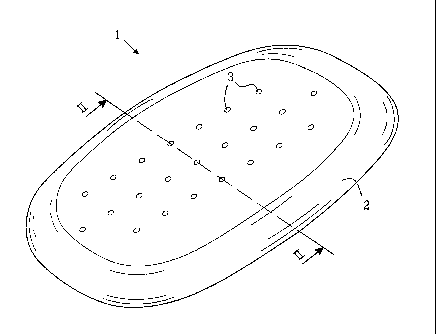
FIG. 1 shows a perspective view of a first embodiment of the invention,
FIG. 2 shows a cross-section of the embodiment of FIG. 1 along line II-II
therein,
FIG. 3 shows a perspective view of a second embodiment of the invention,
FIG. 4 shows a photograph of a sternum to which a spongy material is applied,
FIG. 5 shows the spongy material of FIG. 4 provided with suction tubes,
FIG. 6 shows the spongy material and tubes of FIG. 5 covered with a non-air
permeable
adhesive drape,
FIG. 7 shows a magnetic resonance (MR) image of a clinical test on pig before
application
of negative pressure,
FIG. 8 shows a MR image of a clinical test of FIG. 7 at application of
negative pressure,
FIG. 9 shows a MR image of a clinical test of FIG. 7 at application of
pressure amounting to
-75 mmHg, whereby the heart is sucked up into the space between the sternal
edges,
FIG. 10 shows a MR image of clinical test using a device of the present
invention before
application of negative pressure,
FIG. 11 shows a MR image of a clinical test using a device of the present
invention of FIG.
10 at application of negative pressure,
FIG. 12 shows a MR image of a clinical test using a device of the present
invention of FIG.
10 at application of pressure amounting to -175 mmHg, whereby the device
prevents the
heart from being sucked up between the sternal edges,
FIG. 13 shows a MR image of a clinical test in the absence of a device of the
present
invention and during the application of -125 mmHg pressure, whereby the
sternal edge
protrudes into the heart (white arrow),
FIG. 14 shows a MR image of a clinical test with a device of the present
invention of FIG.
13 during the application of -125 mmHg pressure, whereby the heart is
protected from the
sternal edges (white arrow),
1 denotes generally a substantially rectangular flat barrier disc made of a
biocompatible
material. The barrier disc is preferably made of a polymeric silicon material
having a rigid
structure. In order to fit the wound the barrier disc has a width of 10 to 15
cm and a length
of 15 to 25 cm depending of the size of the patient. The barrier disc has
preferably a
thickness of I to 3 mm. Barrier discs for use with other wounds can be sized
appropriately.
CA 02650318 2008-10-23
WO 2007/123451 4 PCT/SE2007/000173
The barrier disc as such may be flexible but so rigid that it does not become
bent by a
pressure amounting to -200 mm Hg. I.e. the material shall be so rigid that the
barrier disc
cannot be sucked up in between the sternal edges, or become deformed in any
other way.
The edges 2 of the barrier disc 1 are preferably of a less rigid structure.
Thus these more
flexible edges are allowed to adapt themselves to the inner side of the deep
wound, i.e. the
inner part of the sternum, and to provide a sealing of the barrier disc
between the wound
edges and the deeper structures inside the wound. The barrier disc 1 is
perforated by
means of a number of through going holes 3. These holes 3 have the function of
allowing
for passage of wound fluid being sucked from the interior of the wound to the
drainage of
the wound into drainage tubes. The drainage is made possible by the vacuum
applied onto
the top of the barrier disc by means of one or more suction tubes applied to a
vacuum
source, such as a vacuum pump.
FIG. 3 shows a second embodiment of the invention where a wound interface
dressing
material 4, such as a spongy foam polymer material has been attached to the
top surface
of the barrier disc 1. The barrier disc is attached to the wound dressing in
order to insure
that the barrier disc remains fixed in relation to the wound geometry. Hereby
the wound
dressing 4 has been attached via a thread 5 having a length of about the
thickness of the
sternum. The foam material has an open pore structure of 400 to 600 m.
After surgery, the barrier disc 1 is applied underneath the sternum to cover
the sternal
edges and anterior of the barrier disc is a wound interface dressing that
distributes the
negative pressure to the wound surface, or as being a part of the barrier disc
assembly on
top of and over the sternal wound. Non-collapsible evacuation tubes are
connected to the
wound and the wound is sealed with adhesive drape is inserted into the center
of the
sternal foam layer (FIG. 4) and sutured in place. The superficial foam layer
is sutured to the
surrounding subcutaneous tissue (FIG. 5) and a skin protector (FIG. 6) is
applied. The
tubes are positioned 5 cm apart to facilitate application of adhesive draping
around the
tubes.
In a relaxed state the foam should protrude 1 to 2 cm over the edge of skin to
allow volume
reduction during vacuum therapy. The foam layer is then secured subcutaneously
with a
running suture to the surrounding skin edge. A second tube is normally
inserted into the
middle of this foam layer and sutured. A skin barrier disc protector (such as
Cavilon; 3M
HealthCare, St. Paul, MN) is applied (FIG. 5) and the open wound is sealed
with a
transparent adhesive drape (KCI, Copenhagen, Denmark). The drape overlaps the
wound
CA 02650318 2008-10-23
WO 2007/123451 5 PCT/SE2007/000173
margins by 5 cm. The two drainage tubes are positioned 5 cm apart to
facilitate application
of the draping (FIG. 6). The two drainage tubes from the closed wound are
connected to a
vacuum source (VAC pump unit; KCI, Copenhagen, Denmark). This vacuum source
set to
deliver a continuous or intermittent negative pressure of -25 to -250mmHg.
Initially -50
mmHg is applied as it allows adjustment of the foam as the air is evacuated.
If the wound
geometry and foam contraction are considered to be satisfactory the pump unit
is
programmed to deliver -125 mm Hg continuous negative pressure. At this
pressure no
further adjustment can be carried out since the compressed foam will be firm.
A canister in
the pump unit collects exudate from the wound. The wound dressings are changed
regularly, e.g every.3rd day, under aseptic conditions and under general
anesthesia. Bone
and soft tissue necrosis is demarked by lack of granulation tissue on the
sternal edges and
complementary revisions are made during dressing change surgery.
FIG. 7 shows an image generated using Magnetic Resonance Imaging (MRI) in a
clinical
test on pig before application of negative pressure. The arrow in the figures
points at the
opening in the sternum. After having applied the negative pressure, FIG. 8,
the sternum
starts to close. The figure 8 also shows that the heart starts to turn
sidewise.
FIG. 9 shows the clinical test of FIG. 7 at application of pressure amounting
to -75 mmHg,
As shown at the point of the white arrow, the heart is sucked up into the
space between the
sternum edges and which might lead to impaired heart function.
FIG. 10 shows a clinical test, on a pig, using a device, the present invention
before
application of negative pressure. The device is present but not directly
visible in the image
because MRI only depicts structures containing water. In FIG. 11 the device of
the present
invention of FIG. 10 is shown at application of negative pressure, whereby it
should be
noted that the heart now starts to turn round, left arrow. FIG. 12 shows the
clinical test
using a device of the present invention of FIG. 10 at application of pressure
amounting to -
175 mmHg, whereby it is evident, vertical arrow, that the barrier disc
prevents the heart
from being sucked up between the sternum parts, left inclined arrow.
FIG. 13 shows a clinical test, on a pig, in the absence of a device, the
present invention,
during the application of -125 mmHg pressure. One sternal edge protrudes
markedly into
the heart (white arrow).
FIG. 14 shows a clinical test, on the same pig as in FIG. 13, using a device,
the present
invention, during the application of -125 mmHg pressure. The sternal edge no
longer
protrudes into the heart.