Note : Les descriptions sont présentées dans la langue officielle dans laquelle elles ont été soumises.
CA 02652131 2009-01-29
1
TISSUE INHIBITOR OF MATRIX METALLOPROTEINASES TYPE-1
(TIMP-1) AS A CANCER MARKER
This application is a division of co-pending Canadian Patent Application No.
2,366,682 filed April 10, 2000.
BRIEF DESCRIPTION OF THE INVENTION
The present invention relates to a test to be used to screen large populations
for the
occurrence of cancer. The method is based on the measurement of tissue
inhibitor of
metalloproteinases 1 (TIMP-1) in body fluids. The invention permits the early
identification of patients having colorectal cancer. The method is highly
specific, and
patients with non-malignant conditions, such as inflammatory bowl diseases,
are not
detected. Measurement of another similar inhibitor, TIMP-2, does not
demonstrate
equivalent clinical value, indicating an additional level of specificity of
the invention.
The test is based on the measurement of tissue inhibitor of
inetalloproteinases type 1
(TIMP-1), in various body fluids, including plasma, serum, stool and urine.
TIMP-1
concentrations can be determined either as the total TIMP-1 concentration, the
free
TIMP-1 concentration, the concentration of complexes between TIMP-1 and MMP's
and/or ratios and fractions thereof, hereafter referred to as TIMP-1 levels.
According
to the invention, individuals with a high likelihood of having cancer, e. g.
colon
cancer, can be identified by elevated TIMP-1 levels in their body fluids,
while
individuals with low TIMP-1 levels are unlikely to suffer from cancer, e. g.
colon
cancer. Thus, the invention can be used to identify individuals with a high
probability
of having early stage, non-symptomatic cancer, e. g. colon cancer. The
identified
individuals should be further examined and if cancer is found, the patients
should be
offered surgery, irradiation, and/or adjuvant anti-neoplastic therapy, thereby
increasing the chance of cure of cancer for the individual.
BACKGROUND
Colorectal cancer is the fourth most frequent cancer in the Western world,
with about
130,000 new cases yearly in the US. Forty to 50% of all colorectal cancer
patients
will be diagnosed with early stage disease (Dukes' stage A or B). Most of
these
patients with early stage colorectal cancer can be cured by surgery alone.
Thus, risk
of recurrence is closely related to stage of disease at time of primary
surgery, with
about a 10% relapse rate in Dukes' stage A and 25-30% in Dukes' stage B.
Patients
with Dukes' stage C colorectal cancer have a five-year relapse rate of 70%
following
surgery and are offered adju-
CA 02652131 2009-01-29
2
vant chemotherapy. Following relapse, the risk of dying of the disease is-
significant. Thus,
one way to improve survival is to increase the number of patients being
diagnosed with
early stage disease. Screening for colorectal cancer has been shown to improve
survival,
however, current tests suffer from a lack of compliance, from low sensitivity,
and from the
need for strict dietary restrictions. Thus, the development of new and
improved tests for
the early detection of colorectal cancer is needed.
Because metastatic disease is the main cause of cancer patient morbidity and
mortality,
molecules involved in the regulation of tumor invasion and metastasis are
attractive as
potential diagnostic/prognostic targets. It is well established that
proteolytic enzymes pro-
duced by cancer cells or by cells in the tumor stroma are involved in
extraceNular tissue
degradation, leading to cancer cell invasion and metastasis. A number of
enzymes have
been associated with this process, the most thoroughly investigated being the
metallo-
proteinases, such as the collagenases and stromelysins, and the serine
proteases such
as plasmin. Recently, data have been published indicating that these
molecules, free or
bound in complexes, are released from tumor tissue and find their way into the
circulation.
Matrix metalloproteinases (MMP's) play a pivotal role in cancer growth and
spread, contri-
buting to enzymatic degradation of the extracellular matrix (Liotta et al,
1991; Stetler-
Stevenson et al, 1993; MacDougall & Matrisian, 1995). The naturally occurring
inhibitors
of MMP's, tissue inhibitors of MMP's (TIMP's), form tight 1:1 stoichiometric
complexes
with the activated forms of the MMP's (Welgus et al, 1985; Kleiner et a/,
1993), thereby
inhibiting the catalytic activity of these enzymes (Stetler-Stevenson et a!,
1996; Goldberg
et al, 1992; Birkedal-Hansen et al, 1993).1Nhile the balance between the
matrix-degrad-
ing properties of MMP's and the inhibitory effect of TIMP's is closely
regulated under nor-
mal physiological conditions (Matrisian, 1992; Thorgeirsson et al, 1993;
Birkedal-Hansen
et al, 1993), this balance might be disnipted in malignant tissue.
A number of enzyme-linked immunoassays for the detection of TIMP-1 (Kodama et
al,
1989; Cooksley et al, 1990; Clark et al, 1991) and TIMP-2 (Fujimoto et al,
1993) have
been described. These assays have been applied to body fluids, e.g. serum,
plasma, am-
niotic fluid, cerebrospinal fluid, urine, but the number of samples tested has
not been suf-
ficient to establish normal ranges for TIMP levels in healthy individuals
(Kodama et al,
1989; Clark et al, 1991). Furthermore, none of these assays has been
sufficiently vaii-
dated for technical performance or for clinical use.
CA 02652131 2009-01-29
3
In a study by Mimori et al (Mimori et al, 1997) in which tumor tissue levels
of TIMP-1
mRNA were studied in patients with gastric carcinoma, high tumor/normal tissue
ratios of TIMP-1 mRNA were found to be associated with increased invasion and
poor prognosis. However, TIMP-1 protein levels in sera from prostate cancer
patients and healthy donors (Baker et al, 1994) showed a high degree of
overlap.
Similarly, a separate study of plasma from prostate cancer patients and
healthy
donors showed no difference in TIMP-1 levels between the two groups (Jung et
al,
1997).
Studies of TIMP-1 complexed with MMP-9 in plasma of patients with advanced
gastrointestinal and gynaecological cancer (Zucker et al, 1995) demonstrated
significantly higher levels in blood samples from cancer patients with
metastatic
disease compared to healthy control individuals, and that patients with high
levels of
TIMP-1: MMP-9 complex had a shorter survival (Zucker et al, 1995 and US
5,324,634). However, this study did not include measurements of total or free
TIMP-
1, only the complex between TIMP-1 and one of the up to now approximately 24
identified MMP's. Furthermore, in this study, no differences in complex levels
were
found between patients with breast cancer and healthy donors. Also, this study
did
not include patients with early stage cancer.
SUMMARY OF THE INVENTION
In accordance with one aspect of the present invention, there is provided a
method
for determining whether a patient who has been treated for primary breast
cancer is
likely to have metastatic breast cancer, comprising determining a first
parameter
representing the concentration of TIMP-1 in a plasma samples, and indicating
the
individual as having a high likelihood of having metastatic breast cancer if
the
parameter is at or beyond a discriminating value and indicating the individual
as
unlikely of having metastatic breast cancer if the parameter is not at or
beyond the
discriminating value, said discriminating value being determined by measuring
said at
least one first parameter in both a healthy control population and a
population with
known metastatic breast cancer, thereby determining the discriminating value
which
identifies the cancer population with a predetermined specificity or a
predetermined
sensitivity.
CA 02652131 2009-01-29
3a
DETAILED DESCRIPTION OF THE INVENTION
In a number of cancer types, there is a critical and unmet need for highly
sensitive
and specific markers for screening large populations for the presence of
malignant
disease. Such markers should be able to identify individuals with a high
probability of
early stage cancer. These individuals should be further examined, and if
cancer is
found, they should subsequently be offered surgery, radiation, or adjuvant
anti-
neoplastic therapy.
Since proteinases and their receptors and inhibitors seem to play a pivotal
role in the
basic mechanisms leading to cancer invasion, these molecules may be expressed
at
a very early time point in the carcinogenic process. As many of these
molecules exert
their biological action extracellularly, they may be present at elevated
levels in body
fluids, even in patients with early stage invasive malignant disease.
Moreover, since
these molecules are involved in the more basic features of malignant
progression, e.
g. invasion and metastasis, it should be investigated whether which forms of
cancer
that display an increase in these molecules.
CA 02652131 2009-01-29
4
The present invention relates to a method to aid in the diagnosis of
colorectal cancer in a
patient, said method comprising determining the amount of total, complexed
and/or free
TIMP-1 levels and ratios and fractions thereof in body fluids such as blood,
serum,
plasma, urine, faeces or cerebrospinal fluid.
An aspect of the present invention relates to a method for determining whether
an
individual is likely to have cancer, the method comprising determining a first
parameter
representing the concentration of TIMP-1 in body fluid samples, and indicating
the
individual as having a high likelihood of having cancer if the parameter is at
or beyond a
discriminating value and indicating the individual as unlikely of having
cancer if the
parameter is not at or beyond the discriminating value.
The parameter representing the concentration of TIMP-1 may be the
concentration proper
of TIMP-1. TIMP-1 exists both in free fonn and in the form of complexes with
metalloproteinases, and it has been found that an important parameter is the
total
concentration of T1MP-1, that is, the sum of the TIMP-1 in free form and the
TIMP-1 in
complex forms. It will be understood that the other expressions than the
concentration
proper can represent the concentration, such as, e.g., the concentration
muttiplied by a
factor, etc. etc., and that such other representations can be used equally
well for the
purpose of the present invention provided the corresponding adjustments are
made.
The discriminating value is a value which has been determined by measuring the
pa-
rameter in both a heatthy control population and a population with known
cancer thereby .
determining the discriminating value which identifies the cancer population
with either a
predetermined specificity or a predetermined sensitivity based on an analysis
of the
relation between the parameter values and the known clinical data of the
healthy control
population and the cancer patient population, such as it is apparent from the
detailed
discussion in the examples herein. The discriminating value determined in this
manner. is
valid for the same experimental setup in future individual tests.
Specificity is defined as the proportion of positives (i.e. individuals having
a parameter
representing the concentration of TIMP-1 in body fluid samples higher than a
predefined
diagnostic level) that are correctly identified by the described method of the
invention.
Sensit'rvity is defined as the proportion of negatives (i.e. individuals
having a parameter
CA 02652131 2009-01-29
representing the concentration of TIMP-1 in body fluid samples lower than-a
predefined
diagnostic level) that are correctly identified by the described method.
The invention may be used both for an individual and for an entire population.
5
In the specific experimental setups described herein, the concentration
threshold of total
TIMP-1 useful as a discriminating value was found to be in the range of 50-160
microgram/L of tqtal TIMP-1 at a specificity of 90%. Other experimental setups
and other
parameters will result in other values which can be determined in accordance
with the
teachings herein.
The method can be applied to an unselected population, but more appropriately
to a
population already identified as having an increased risk of developing
cancer, e.g. indi-
viduals with a genetic disposition, individuals who have been exposed to
carcinogenic
substances, or individuals with cancer-predisposing non-malignant diseases. In
the case
of colorectal cancer, the population selected for the invention could
represent individuals
with a prior polyp, individuals with Crohn's disease or ulcerative colitis,
individuals with
one or more family members with colorectal cancer, or individuals with a
prior.resection of
an early colorectal cancer.
When an individual has been identified as having high TIMP-1 levels in his or
her body
fluid, the individual should be referred for further examination. If a cancer
is found, the pa-
tient could be offered surgery, radiation or adjuvant anti-neoplastic therapy
aiming at cur-
ing the patient of cancer.
Example I describes the preparation and validation of an assay that measures
total
TIMP-1 with high analytical sensitivity and specificity. It is described that
healthy blood
donors have a very narrow range of plasma total TIMP-1.
In Example 2, the formatting of a TIMP-1:MMP-9 ELISA is described. The format,
execu-
tion, and validation of this assay are similar to those for total TIMP-1,
except that a poly-
clonal antibody against MMP-9 is used in the capture step. By substituting the
MMP-9 an-
tibody with an antibody against another MMP, complexes between TIMP-1 and
other
MMP's can be quantitated.
CA 02652131 2009-01-29
6
In Example 3, the formatting of a TIMP-1 assay which exclusively measurEs free
TIMP-1,
is described. This assay utilizes a monoclonal anti-TIMP-1 antibody (MAC 19)
which only
recognises TIMP-1 in its uncomplexed form. Thus, this assay will measure the
amount of
free TIMP-1 in a sample. The execution and validation of this assay are
similar to the as-
say for total T1MP-1
By subtracting the free TIMP-1 concentration from the total TIMP-1
concentration in a
biological sample, the concentration of all complexed forms of TIMP-1 can be
determined.
It should be emphasized that TIMP-1 can form complexes with many of the MMP's
and
therefore, subtracting one type of complex from the total amount of TIMP-1
will only pro-
vide information on the fraction of TIMP-1 not being complexed to this
specific MMP.
In Example 4, it is shown that patients suffering from colorectal cancer have
significantly
elevated total TIMP-1 levels in their preoperative plasma samples. A
percentile plot of the
total TiMP-1 levels in plasma from all colorectal cancer patients and from
heatthy blood
donors shows that a total TIMP-1 concentration of 119.1 g/L is the 90'"
centile of the
heafthy donors. Using this cut-off, 68% of the colorectal cancer patients were
identified as
having elevated plasma TIMP-1 levels. When analyzing the colon cancer patients
sepa-
rately, it was shown that total TIMP-1 measurements in plasma identified 75%
of the colon
cancer patients (diagnostic sensitivity) with a 90% diagnostic specificity
(10% of the
healthy blood donors were classified as being high). Similarly, it was shown
for the rectal
cancer patients alone, that total TIMP-1 measurements in plasma identified 60%
of the
rectal cancer patients with a 90% diagnostic specificity. lf a higher or lower
sensitivity or
specificity is desired, the cut-off value can be changed. This is illustrated
in Figure 13
showing ROC curves of total TIMP-1 in plasma from colorectal cancer patients.
In addi-
tion, ROC curves are included for the individual groups of colon and rectal
cancer pa-
tients. Any other information which can be derived from these ROC curves falls
within the
scope of the present invention.
An independent prospective study, including preoperative plasma samples from
64 pa-
tients with colon (n=43) or rectal (n=21) cancer confirmed the data obtained
from the
above described study (Figure 15). An additionat study of 180 healthy blood
donors and
20 colorectal cancer patients, using different antibodies (Anti TIMP-1 11
E/C6, Anti-T1MP-1
RRU-T6) in an automated immunoassay, further corroborated the previous
clinical results.
CA 02652131 2009-01-29
7
Moreover, the absolute values generated from the automated assay showed a high
de-
gree of correlation to those obtained by the assay described in Example 1(r-
0.9).
The clinical value of a marker for cancer screening is related to its ability
to detect early
stages of disease, potentially impacting survival. It was shown that total
TIMP-1 was as
efficient a screening marker in early stage colorectal cancer (Dukes' stages A
and B) as it
was in the total population of colorectal cancer patients (Figure 14), Thus,
screening with
total TIMP-1 will result in more patients being diagnosed with early stage
cancer. fn a
similar manner, any information that can be derived from Figure 14 falls
within the scope
of the present invention.
By dividing the colon cancer patients into two groups, patients with left-
sided and right-
sided tumors respectively, it was evident that measurement of total TIMP-1 was
especially
useful in identifying those patients with early stage, right-sided colon
cancer lesions (Fig-
ure 14).
The specificity of a given cancer screening test is based on the efficiency of
the test to
identify only those patients suffering from cancer while patients suffering
from non-malig-
nant diseases should not be identified as false positive subjects. In the case
of colorectal
cancer, it is important that the test in question can distinguish between
malignant and
non-malignant diseases of the colon and rectal. This is particularly important
for diseases
like Crohn's disease and ulcerative colitis, since patients with these
diseases are at higher
risk of developing cancer.
In Example 5 it is shown that total TIMP-1 levels are significantly higher in
patients with
colorectal cancer than in patients with inflammatory bowl diseases (IBD),
showing that
total TIMP-1 can be used to screen for colorectal cancer in a population of
patients with
IBD. That TIMP-1 is not increased in non-malignant diseases is supported by a
recent pa-
per, (Keyser et a/, 1999), demonstrating that patients with rheumatoid
arthritis do not have
increased plasma TIMP-1 levels. Also, by comparing total TIMP-1 levels among
patients
with IBD (excluding patients with clinically assessed acute active disease,
n=4) and
healthy blood donors, no significant differences in total plasma TIMP-1 levels
were found
(p=0.56), showing that these non-malignant diseases do not give false positive
test re-
sufts.
CA 02652131 2009-01-29
8
In Example 6, the additive effect of the measurement of an additional
colvfectal cancer
marker is described. Carcino Embryonic Antigen (CEA) was measured in aU cancer
pa-
tient and healthy blood donor samples. Combining CEA and the TIMP-1 levels
measured
by the assay described in Example 1, it could be shown that while CEA alone
gives 35%
sensitivity at a 98% specificity, the sensitivity of the combination of CEA
and total TIMP-1
as determined by logistic regression analysis increased to 57%, without
sacrificing speci-
ficity.
In Example 7 it is shown that patients suffering from colorectal cancer have
significantly
elevated free TIMP-1 levels in their preoperative plasma samples. A percentile
plot in-
cluding the free TIMP-1 levels from the colorectal cancer patients as well as
free TIMP-1
levels from healthy blood donors, showed that colorectal cancer patients had
significantly
elevated free TIMP-1 levels as compared to blood donors p= 0.02. A ROC curve
was cre-
ated (Figure 18) for these patients and donors and it was seen that free TIMP-
1 gave an
area under the curve (AUC) of 0.61.
In Example 8, the use of the TIMP-1:MMP-9 complex assay as an aid for the
diagnosis of
colorectal cancer is described.
TIMP-1 is known to exist either as the free molecule or in complex with MMP's,
preferen-
tially MMP-9. Measuring total TIMP-1, complexed TIMP-1 and free TIMP-1 will
make it
possible to validate each of these species for their potential diagnostic
value. In addition, it
will be possible to calculate ratios or any derived algorithm between the
different species
which might provide additional diagnostic value.
In Example 9, the diagnostic value of the combination of total and free TIMP-1
is de-
scribed.
The data shows that combining by logistic regression analysis the free TIMP-1
measure-
ments with the total TIMP-1 measurements, a significant increase in the AUC
was dem-
onstrated. Any information that can be derived from Figure 18 falls within the
scope of the
present invention.
The specificity of a given cancer screening test is based on the efficacy of
the test to iden-
tify only those patients suffering from cancer while patients suffering from
non-malignant
CA 02652131 2009-01-29
9
diseases should not be identified as false positive. However, it would be
desirable that the
test was specific for a specific type of cancer, e.g. colon cancer, instead of
being a pan-
cancer marker.
In Example 10, total plasma TIMP-1 values in preoperative blood samples from a
cohort
of 322 patients with primary breast cancer (stage I and II) as compared with
total TIMP-1
levels in 108 healthy blood donors are described. lt was shown that the breast
patients
had a median, total TIMP-1 level of 88.3 Fzg/L, while the healthy donors had a
median
plasma concentration of total TIMP-1 of 88.9 pg/L. The difference between
these values is
not clinically significant, supporting the specificity of TIMP-1 levels for
the diagnosis of
colorectal cancer. However, it should be studied whether elevated plasma TIMP-
1 levels
are found in patients with early stage non-colorectal cancer.
In Example 11, total TIMP-1 levels in patients with metastatic breast cancer
are de-
scribed.
Of note, total TIMP-1 in plasma from women with metastatic breast cancer had
rMedian
value of 236 pg/L, significantly higher than levels in healthy blood donors.
This shows the
potential of using plasma TIMP-1 levels for the management of breast cancer
patients.
In Example 12, the concentrations of TIMP-2 in preoperative plasma samples
from pa-
tients with colorectal cancer are described.
TIMP-2 is another tissue inhibitor of metalloproteinases with a high degree of
homology to
TIMP-1. Using a specific immunoassay for TIMP-2, concentrations of this
inhibitor were
determined in plasma samples from colorectal cancer patients and in healthy
blood do-
nors. No significant differences in plasma TIMP-2 levels were found between
the two
populations, supporting the unique value of TIMP-1 as an aid for the early
diagnosis of
colorectal cancer.
FIGURE LEGENDS
Figure 1: Kinetic assay for TIMP-1. Progress curves for the change in
absorbance at 405
nm produced by hydrolysis of p-nitrophenyl phosphate by solid-phase bound
alkaline
phosphatase immunoconjugate. The data shown are generated by 4 individual
assay
CA 02652131 2009-01-29
wells treated with 4 different concentrations of purified recombinant TIMP-'1;
10 g/L (o-
D), 2.5 g/L (0-A), 0.63 pg/L ( D- D) and 0.16 g/L (0-0). The lines shown
have been
fitted by simple linear regression.
5 Figure 2: TIMP-1 standard curve. Absorbance unitsfor triplicate TIMP-1
standards in the
range of 0 to 5 g/L are collected automatically over 60 minutes, with
readings taken at
405 nm every 10 min. Progress curves are computed for each well and the rates
obtained
are frtted to a standard curve using a four-parameter equation of the form y =
d+[(a-
d)/(1+(x/c))j. In the example shown, the four derived parameters had the
following vat-
10 ues: a=1.87, b=1.11, c=3.35, d=73.5. The correlation coefficient for the
fitted curve is
>0.999.
Figure 3: Recovery of signal from standard TIMP-1 added in increasing
concentration to
assay dilution buffer ( D- D), a 1:100 dilution of EDTA plasma pool (d-d), a
1:100 dilution
of citrate plasma pool (V-V) and a 1:100 dilution of heparin plasma pool (0-
0). The val-
ues shown are the means of triplicates. The correlation coefficient for each
fitted curve is
greater than 0.99.
Figure 4: Westem blotting of immunoabsorbed patient plasma sample. Lane 1:
standard
TIMP-1; lane 2: eluate of patient citrate plasma sample diluted 1:10 and
immunoabsorbed
with sheep polyclonal anti-TIMP-1. Bands of non-reduced standard TIMP-1 and
TIMP-1
isolated from plasma sample both appear just below 30 kDa.
Figure 5a: Percentiles plot for the level of TIMP-1 (pg/L) measured in citrate
plasma (0)
and EDTA plasma (A) from the same individual in a set of 100 volunteer blood
donors.
Figure 5b: Linear regression plot for the level of TIMP-1 in citrate plasma
samples com-
pared with EDTA plasma samples from the same 100 individuals. The equation of
the fit-
ted line is y = 0.93x, with a regression coefficient of 0.99.
Figure 6: Percentiles plot for the levels of TIMP-1 ( g/L) measured in two
sets of citrate
plasma samples obtained by the same procedure from volunteer blood donors at
different
times. 100 samples from May =97 (A) and 94 samples from Sept =96 ( 0
).
CA 02652131 2009-01-29
11
Figure 7: Percentile plot for the levels of TIMP-1 (pg/L) measured in 194 c-
Iirate plasma
samples from volunteer blood donors and stratified by sex into 107 males (0)
and 87 fe-
males (0).
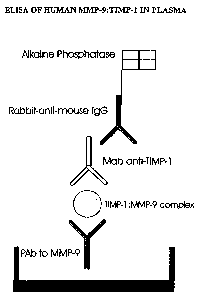
Figure 8: Graphical illustration of the TIMP-1:MMP-9 complex ELISA.
Figure 9: Graphical illustration of the free TIMP-1 ELISA.
Figure 10: A plate was coated with the polyclonal anti-MMP-9 antibody.
Different concen-
trations of TIMP-1:MMP-9 complex, free MMP-9, free TIMP-1 and a blank control
were
added. Only TIMP-1:MMP-9 complexes and free MMP-9 were bound by the capture
poly-
clonal anti-MMP-9 antibody. MAC19 was then added for antigen detection.
Neither TIMP-
1:MMP-9 complex nor free MMP-9 were detected by MAC19, defining the
specificity of
this antibody for free TIMP-1.
Figure 11: A plate was coated with the polyclonal anti-MMP-9 antibody.
Different concen-
trations of TIMP-1:MMP-9 complex, free MMP-9, free TIMP-1 and a blank control
were
added. Only TIMP-1:MMP-9 complexes and free MMP-9 were bound by the capture
poly-
clonal anti-MMP-9 antibody. MAC 15 was then added for antigen detection. Only
TIMP-
1:MMP-9 complex bound by the capture polyclonal anti-MMP-9 antibody was
detected by
MAC15. Free MMP-9 was not detected by MAC15.
Figure 12: Recovery of signal in the free TIMP-1 assay from standard TIMP-1
added in
increasing concentration to assay dilution buffer ( 0- 0), a dilution of EDTA
plasma pool
(e-e), a dilution of citrate plasma pool (V-V), and a dilution of heparin
plasma pool (0-0).
The values shown are the means of triplicates. The correlation coefficient for
each fltted
curve is greater than 0.99.
Figure 13: ROC curves for total TIMP-1 in all colorectal cancer patients, in
rectal cancer
patients separately and in colon cancer patients separately. As healthy
control subjects, a
cohort of 108 healthy blood donors was used. (Number in parenthesis = Area
under
curve)
CA 02652131 2009-01-29
12
Figure 14: ROC curves for total TIMP-1 in all colorectal cancer patients wlth
Dukes' A or B
disease. In addition, ROC curves for Dukes' A or B patients with colon cancer
or with right
sided colon cancer is included. (Number in parenthesis = Area under curve)
Figure 15: ROC curve for total TIMP-1 from an independent set of 64 colorectal
cancer
patients compared to 108 healthy blood donors is shown. (AUC = Area under
curve)
Figure 16: Box plot showing total TIMP-1 concentrations in plasma from healthy
blood do-
nors, from patients with Crohn's Disease, from patients with ulcerative
colitis and from pa-
tients wi#h colorectal cancer. Medians, 10th, 25th, 75th and 90th centiles are
shown.
Figure 17: ROC curves for total TIMP-1, CEA and for the combination of total
TIMP-1 and
CEA in patients with right colon cancer patients and 108 healthy blood donors.
(Number in
parenthesis = Area under curve)
Figure 18: ROC curves for plasma free TIMP-1, for plasma total TIMP-1 and for
the com-
bination hereof in 64 CRC patients and 108 donors. (Number in parenthesis =
Area under
curve)
Figure 19: ROC curve for 322 primary breast cancer patients and 108 blood
donors. (AUC
= Area under curve)
Figure 20: Percentiles plot for the level of total TIMP-1 ( g/L) measured by
ELISA in 19
EDTA plasma samples from female breast cancer patients (0) and 87 healthy
blood do-
nors (A).
EXAMPLES
Example 1
Preparation of an ELISA to quantitate total TIMP-1 concentrations in human
plasma.
CA 02652131 2009-01-29
13
This example describes the preparation and validation of an ELISA that m-
easures total
TIMP-1 levels in plasma. In addition, this example provides information on
TIMP-1 levels
in different plasma preparations as well as in healthy blood donors of both
sexes.
Materials and methods:
Blood donors
Blood samples were initially obtained from 94 apparently healthy volunteer
blood donors,
comprising 51 males aged 19 to 59 years (median: 41 years) and 43 females aged
20 to
64 years (median: 36 years). In a subsequent collection, 100 donor samples
were ob-
tained, comprising 56 males aged 19 to 59 years (median 42: years) and 44
females aged
to 60 years (median: 36.5 years). Informed consent was obtained from all
donors, and
permission was obtained from the local Ethical Committees.
15 Blood collections and plasma separation
Peripheral blood was drawn with minimal stasis (if necessary a maximum of 2
min stasis
with a toumiquet at maximum +2 kPa was acceptable) into pre-chilled citrate,
EDTA, or
heparin collection tubes (Becton-Dickinson, Mountain View, CA), mixed 5 times
by inver-
sion, and immediately chilled on ice. As soon as possible (no later than 1.5 h
after collec-
20 tion) the plasma and blood cells were separated by centrifugation at 4 C at
1,200 x g for
min, and stored frozen at -80 C prior to assay. Plasma pools were made with
freshly
collected samples from at least ten donors, aliquoted and stored frozen at -80
C. For
analysis, the samples were quickly thawed in a 37 C water bath at and then
placed on ice
until needed.
TOTAL TIMP-1 ELISA
A sensitive and specific sandwich ELISA was prepared, using TIMP-1 antibodies
develop-
ed at the Strangeways Laboratories (Hembry et al, 1985). A sheep polyclonal
anti-TIMP-1
antiserum (Hembry et a!, 1985; Murphy et al, 1991) was used for antigen
capture, and a
murine monoclonal anti-TIMP-1 IgG1 (MAC-15) (Cooksley ef al, 1990) for antigen
detec-
tion. A rabbit anti-mouse immunoglobulin/alkaline phosphatase conjugate
(Catalog num-
ber D0314, Dako, Glostrup, Denmark) was the secondary detection reagent. The
latter
conjugate was supplied preabsorbed against human IgG, thus eliminating cross-
reactivity_
with IgG in the plasma samples. As the monoclonal detection antibody MAC-15
recog-
CA 02652131 2009-01-29
14
nises both free TIMP-1 and TIMP-1 in complex with MMP's (Cooksley efal, 1990),
the
total TIMP-1 content captured by the sheep polyclonal anti-TIMP-1 antiserum
was quan-
titated by the ELISA.
96-well microtiter plates (Maxisorp, Nunc, Roskilde, Denmark) were coated for
1 h at 37 C
with 100 pLlwell of polyclonal sheep anti-TIMP-1 (4 mg/L) in 0.1 mol/L
carbonate buffer,
pH 9.5. The wells were then rinsed twice with 200 Uwell of SuperBlockJ
solution (Pierce
Chemicals, Rockford, IL) diluted 1:1 with phosphate-buffered saline (PBS). The
microtiter
plates were stored for up to 14 days at -20 C. On the day of analysis, the
plates were
thawed at room temperature and washed 5 times in PBS containing 1 g/L Tween.
A series of purified, recombinant human TIMP-1 standards were used to
'calibrate each
plate. Standards were prepared by serially diluting a stock solution of
purified TIMP-1.
Standard concentrations were 10, 5, 2.5, 1.25, 0.625, 0.313 and 0.156 g/L.
Included on
each plate was a blank containing only sample dilution buffer, and 2 controls
made from a
1:100 dilution of a citrate plasma pool. One control was added as the first
sample on the
plate and the second control was added as the last. All plasma samples were
diluted
1:100 in sample buffer consisting of 50 mol/L phosphate, pH 7.2, 0.1 mot/L
NaCt, 10 g/L
bovine serum albumin (Fraction V, Boehringer-Mannheim, Penzberg, Germany), and
1
g/L Tween 20. A total of 100 pUwell of each standard, blank, control, and
patient sample
was incubated on the plate for 1 h at 30 C. All standards, blanks, controls,
and samples
were run in triplicate on each plate for every assay. After primary
incubation, the wells
were washed 5 times, then treated for I h at 30 C with 100 pUwell of purified
MAC-15
monoclonal antibody (0.5 mg/L) in sample dilution buffer. After another 5
washes the wells
were incubated for 1 h at 30 C with 100 pUwell of rabbit anti-mouse
immunoglobu-
lins(Ig)/alkaline phosphatase conjugate diluted 1:2000 in sample dilution
buffer. Following
5 washes with washing solution and 3 washes with distilled water, 100 L of
freshly made
p-nitrophenyi phosphate (Sigma, St. Louis, MO) substrate solution (1.7 g/L in
0.1 moUL
Tris.HCI, pH 9.5, 0.1 moUL NaCI, 5 mmol/L MgCI2) was added to each well. The
plate was
placed in a Ceres 900J plate reader (Bio-Tek Instruments, Winooski, VT) at 23
C with the
yellow color development automatically monitored. Readings were taken at 405
nm
against an air blank every 10 min. for one hour.. KinetiCaic 11 software was
used to ana-
lyze the data by calculating the rate of color formation for each well (linear
regression
analysis), generating a 4-parameter fitted standard curve, and calculating the
TIMP-1
concentration of each plasma sample.
CA 02652131 2009-01-29
Recovery experiments
The recovery of TIMP-1 signal was measured following addition to 1:100
dilutions of cit-
rate, EDTA or heparin plasma pools.Purified TIMP-1 was added to plasma pools
to give
5 final concentrations in the range of 0 to 10 g/L. The recovery in each case
was calcu-
lated from the slope of the line representing TIMP-1 signal as a function of
concentration,
where 100% recovery was defined as the slope obtained when T1MP-1 was diluted
in
sample dilution bUffer.
10 Immunoblotting
Citrate plasma from a patient with a high level of TIMP-1 in blood (634 g/L,
determined
by ELISA), was diluted 1:10 and added to a protein A-Sepharose column pre-
incubated
with polyclonal sheep anti-TIMP-1. Following 5 cycles, bound proteins were
eluted from
the column and 50 pL of the resulting eluate run on 12% SDS-gel
electrophoresis (Ready
15 GeIJ, Bio-Rad). A mixture of low molecular weight (Pharmacia) and high
molecular weight
markers (Bio-Rad) and 50 L of TIMP-1 standard (100 pg/L) in Laemmli Sample
Buffer
were also run on the gel. Proteins were transferred electrophoretically from
the gel onto a
polyvinylidene difluoride (PVDF) membrane (Millipore). The membrane was
incubated for
1 h at room temperature with 1% skim milk powder in TBS. Following washing,
the mem-
brane was incubated for 1 h at room temperature with 20 ml of MAC-15 (5 mg/L).
The
membrane was then washed and incubated for an additional hour at room
temperature
with 20 ml of rabbit anti-mouse lg/alkaline phosphatase conjugate diluted
1:1000. Finally
the membrane was washed and color developed by the addition of a phosphate
substrate
solution (NBT/BCIP).
Results:
ELISA performance
Development of color in each well progressed as a linear function of time for
all concen-
trations of total TIMP-1 in these experiments (Figure 1), with correlation
coefficients for
the fitted lines typically greater than 0.99. The standard curve for the rates
plotted against
the TIMP-1 concentration consisted of the linear and upper curved regions
(over the
range of 0 to 5 g/L) of a sigmoidal curve, and the correlation coefficient
for the 4-pa-
rameter fit was typically better than 0.999 (Figure 2). The rate with no TIMP-
1 (read
CA 02652131 2009-01-29
16
against an air blank) was 1.21 0.15 (mean SD) milliabsorbance units/miTt
(n=29), while
the rate with 10 pg/L standard TIMP-1 was 50.3 6.01 milliabsorbance units/min
(n=29).
The limit of detection for the assay, defined as the concentration of TIMP-1
corresponding
to a signal 3 SD above the mean for the TIMP-1 blank, was 0.089 g/L. This
value was
13% of the mean of the measured concentrations of TIMP-1 in healthy citrate
plasma
samples. The intra-assay coefficient of variation (CV) for 16 replicates of a
control citrate
plasma pool was 5.3%, and the inter-assay CV for 29 successive assays of the
plasma
pool (run on different days) was 6.2%. This plasma pool had a TIMP-1
concentration of
57.8 pg/!., corresponding to the 22nd centile of the normal individuals.
.~
Recovery of recombinant TIMP-1 after dilution in plasma Specific recovery was
deter-
mined by addition of increasing concentrations of purified TIMP-1 to a panel
of plasma
pool replicates, followed by subsequent measurement of signal. Recovery was
104% in
citrate plasma, 101% in diluted EDTA plasma, and 87% in diluted heparin plasma
(Figure
3). Thus the recovery of TIMP-1 signal from an intemal standard was acceptable
for all
preparations of plasma
Dilution curves for total plasma TIMP-1 signal
Serial dilutions of citrate, EDTA and heparin. plasma pools were made to test
for linear re-
duction in TIMP-1 signal. Citrate, EDTA and heparin plasmas all gave good
linearity of
signal as a function of dilution. The 1 % plasma dilution which was chosen for
subsequent
determinations lay well within the range of this linear dilution curve.
Immunoblotting of total plasma TIMP-1
A Western blot of an immunoabsorbed patient plasma sample showed a clear band
of 28
kDa (Figure 4, lane 2), corresponding to free, uncomplexed TIMP-1 (Figure 4,
lane 1). No
bands were found at the expected higher molecular weights corresponding to
complexes
between MMP's and TIMP-1, e.g. MMP-2:TIMP-1, 100 kba. This indicates either
that the
major part of TIMP-1 was ptesent in the plasma as the free form, or that
complexes were
dissociated during SDS-PAGE. Although the sample was left both unreduced and
un-
heated in order to preserve any complexes present in the plasma sample, it has
been re-
ported that MMP:TIMP complexes may be unstable in SDS-PAGE (Wilhelm et a!,
1989;
Stetier-Stevenson et a!, 1989; Moll et a!, 1990), even under non-reducing
conditions
(Moutsiakis et al, 1992).
CA 02652131 2009-01-29
17
Total TIMP-1 in citrate and EDTA plasma from the same healthy donor
A collection of citrate and EDTA plasma samples taken simultaneously from 100
heatthy
donors was available for this study. These samples were not specifically
collected as
platelet-poor plasma. However, a srriall, representative number of samples,
prepared as
platelet-poor plasma, did not differ significantly in total TIMP-1 values-.
The percentile plots
for total TIMP-1 levels in these samples are shown in Figure 5a. The values in
each set
approximated a normal distribution. Citrate plasma TIMP-1 levels ranged
between 55.0
and 90.3 g/L (10th to 90th percentile) with a mean of 69.2 13.1 }-g/L.
Similarly, EDTA
plasma TIMP-1 levels ranged from 58.0 to 91.8 pg/L with a mean of 73.5 14.2
g/1.. For
both citrate and EDTA plasma, the mean TIMP-1 levels were in close proximity
to the me-
dian levels (Table 1). A paired means comparison showed that the level of TIMP-
1 in cit-
rate plasma was significantly lower by 4.34 g/L (95% Cl 2.34-6.33; (p<0.0001)
than the
EDTA plasma level from the same individual. However, it is likely that this
difference may
be due to the variability in sampiing procedure during plasma collection. EDTA
plasma
tubes contained dry anticoagulant material, while citrate plasma tubes
contained a small
amount of liquid citrate buffer which gave a small and variable systematic
dilution error (x
9/10). The level of TIMP-1 in citrate plasma correlated with that in EDTA
plasma from the
same individuals. The linear regression plot in Figure 5b shows a regression
coefficient of
0.99 with a slope of the fitted line of 0.93, perhaps illustrating this small
dilution error. A
non-parametric Spearman's rank test for the data set gave a rho value of 0.62
and
p<0.0001.
Total TIMP-1 levels in citrate plasma
A total of 194 citrate plasma samples from healthy blood donors were assayed,
compris-
ing 94 samples taken during one collection and 100 samples taken 9 months
later from a
different set of donors. Figure 6 shows the percentile plots for TIMP-1 levels
measured in
these two independent groups. The reference range for TIMP-1 levels in citrate
plasma
from the first collection was 53.3 to 77.7 g/L (10th to 90th percentile) with
a mean of
65.4 10.1 pg/L which was indistinguishable from the median (Table 1). and
approximating
a normal distribution. The mean TIMP-1 level for the second collection was
69.2 13.1
g/L (reference range 55.0 to 90.3 pg/L). An unpaired means comparison showed
that
TIMP-1 levels in the two sets of samples taken during two different periods-
differed only
by 3.82 g/L (95% .Cl: 0.50-7.14 g/L; p=0.024). Moreover, no significant
difference was
apparent between the controls (n=8) included in each set of assays (mean
difference 0.36
CA 02652131 2009-01-29
18
pg/L; 95% Cl: 1.71-2.44 pg/L; p=0.69). The mean TIMP-1 level for all 194
citrate plasma
samples was 67.3 11.8 pg/L, close to the median of 66.1 pg/L, with levels
again approxi-
mating a normal distribution (reference range 54.0 to 82.7 g/L).
TABLE 1:
SUMMARY OF'TOTAL TIMP-1 LEVELS DETERMINED IN BLOOD FROM HEALTHY
DONORS:
Blood fraction Date of Number of MeanfSD Median Reference range*
sampling samples (ug/L) (Eeg1L) ( g/L)
Citrate plasma Sept. 96 94 65.4 10.1 65.6 53.3-77.7
Citrate plasma May 97 100** . 69.2 13.1 67.0 55.0-90.3
Citrate plasma 96+97 194 67.3 11.8 66.1 54.0-82.7
EDTA plasma May 97 100** 73.5 14.2 71.2 58.0-91.8
*The reference range is defined as the 10th to 90th percentile.
**These samples were collected from the same donors.
+.~
Tests for correlations to gender and age of the donor
In these studies, the control values measured in each assay had a CV of 2.7%.
Percen-
tiles for TIMP-1 levels in 194 citrate plasma samples calculated according to
gender are
shown in Figure 7. The mean TIMP-1 value for 107 male donors was 70.4 12.0
g/L
(median 69.4 pg/L) vAth a reference range from 56.2 to 86.6 g/L, while the
mean TIMP-1
value for 87 female donors in this set was 63.5t10.5 g/L (median: 62.0 pg/L)
with a ref-
erence range from 51.8 to 77.0 pg/L. There was a signiricant difference
(p<0.0001) in
TIMP-1 mean levels between the two groups, with males having higher TIMP-1
levels
than females. There was a trend towards an increase in plasma TIMP-1 with
increasing
age (Spearman's rho=0.33, P=0.001 1), but this did not increase with gender
(females:
Spearrnan's rho=0.29, P=0.006; males: Spearman's rho=0.35, P=0.0003). In EDTA
plasma, the mean TIMP-1 value for 56 males was 76.9 15.0 pg/L (median: 75.1
g/L)
with a range from 58.8 to 96.9 g/L, while 44 female donors had a mean TIMP-1
level of
CA 02652131 2009-01-29
19
69.3 11.8 pg/L (median: 67.9) with a reference range from 56.1 to 85.5 gIL.
Again, a sig-
nificant difference appeared between males and females (p=0.0076, unpaired
means
comparison).
Discussion:
The assay described above enables accurate determination of total TIMP-1 in
human
plasma samples. Kinetic rate assays of the bound antigen were easily
accomplished,
permitting automated fitting of rate curves, which has proven considerably.
more reliable
than single end-point measurements. The use of a rapid blocking agent and a
dilution
buffer with high buffering capacity also contributed to reproducible assays.
incorporating
all these elements in the final assay fulfilled the requirements of
sensitivity, specificity,
stability, and good recovery of an intemal standard.
The quantitative studies in blood from healthy donors showed that both citrate
and EDTA
plasma samples are suitable for TiMP-1 determination. Compared to other
published
studies of TIMP-1 i from healthy donors (Jung et a/, 1996; Jung et a/, 1996),
levels in the
present study fell within a very narrow range. Some studies have reported
resutts in se-
rum, but plasma was selected for the present study to avoid the variable
contribution of
platelet activation to the measured TIMP-1 values (Cooper et a/, 1985). While
the plasma
samples used in this study were not specifically prepared as platelet-poor
plasma, it was
shown, based on tests carried out in the lab, that this does not change the
values. The
donor material was large enough to demonstrate that TIMP-1 levels in healthy
individuals
(both EDTA and citrate) approximated a normal distribution, for females as
well as for
males. Mean TIMP-1 levels were approx. 10% higher in males than in females for
both
EDTA and citrate plasma. One explanation for this is a higher release rate of
TIMP-1 into
blood from activated platelets, reflecting a tendency towards higher incidence
of thrombo-
embolic disease in the male population. When males and females were considered
sepa-
rately, there was a weak correlation between TIMP-1 and age as seen for the
whole
population (see above).
Example 2
Preparation of an ELISA to quantitate TIMP-1:MMP-9 complexes in plasma.
The following example describes an assay to determine the concentration of
TIMP-
1:MMP-9 complexes in body fluids. The assay is used with plasma samples of
heafthy
CA 02652131 2009-01-29
blood donors in order to establish normal ranges of this complex (Holten
Andersen et al.,
1999).
Materials and methods:
5
TIMP-1:MMP-9 complex ELISA
A sensitive and specific sandwich ELISA was prepared using the above-descr'bed
TIMP-1
antibody, MAC-15, and a rabbit MMP-9 polyclonal antibody developed in the
Hematologi-
cal Department, Rigshospitalet, Denmark (Kjeldsen et al, 1992). The MMP-9
antibody was
10 used for antigen capture and MAC-15was used for antigen detection. A rabbit
anti-mouse-
Ig/alkaline phosphatase conjugate (Dako, Glostrup, Denmark) enabled a kinetic
rate as-
say (Figure 8). The latter conjugate was supplied preabsorbed against human
lgG, thus
eliminating cross-reactivity with IgG in the plasma samples. The MMP-9
antibody cap-
tured both free MMP-9 and MMP-9 complexed with TIMP-1, while MAC-15 only recog-
15 nised TIMP-1. Therefore only T1MP-1:MMP-9 complexes were quantitated by
this reagent
pair.
To prevent spontaneous, ex vivo TIMP-1:MMP complex formation during sampling
and
assay procedures, a protease inhibitor (ie. Galardin, Batimastat, Marimastat)
was added
20 to the plasma sample after thawing. The addition of the protease inhibitor
blocked in vitro
complex formation by inhibition of the catalytic activity of the
metalloproteinases. The TIMP-1:MMP-9 assay was prepared and validated by a
method similar to that de-
scribed above for the total TIMP-1 assay. The TIMP-1:MMP-9 standard was
prepared by
incubating equimolar amounts of purified recombinant TIMP-1 and MMP-9
(activated by
adding APMA) in PBS for 1 hour at 37 degrees Celsius.
Briefly, 96-well micotiter plates were coated overnight at 4 C with 100
L/well of rabbit
polyclonal anti-MMP-9 antiserum in 0.1 mol/L carbonate buffer, pH 9.5. Prior
to use, as-
say wells were rinsed twice with 200 LJwell of SuperBlock solution diluted
1:1 with phos-
phate-buffered saline (PBS), and then washed 5 times in PBS containing 1 g/L
Tween 20.
Wells were then incubated for 1 h at 30 C with 100 lJwell of plasma diluted
in sample
buffer A series of purified TIMP-1:MMP-9 standards were used to calibrate each
plate.
Standards were prepared by serially diluting a stock solution of purified TIMP-
1:MMP-9
complex. Included on each plate was a blank containing only sample dilution
buffer, and 2
CA 02652131 2009-01-29
21
controls made from a citrate plasma pool. One control plasma pool was aoed as
the first
sample on the plate and the second control was added as the last. All
standards, blanks,
controls, and samples were run in triplicate on each plate for every assay.
After sample
incubation and TIMP-1:MMP-9 complex binding, the wells were washed 5 times,
followed
by treatment for 1 h at 30 C with 100 Uwell of MAC-15 in sample dilution
buffer. After
another 5 washes, the wells were incubated for 1 h at 30 C with 100 pLlwell of
rabbit anti-
mouse lg/alkaline phosphatase conjugate diluted in sample dilution buffer.
Following 5
washes with washing solution and 3 additional washes with distilled water, 100
L of
freshly made p-nitrophenyl phosphate substrate solution was added to each
well. The
plate was placed in a Ceres 900J plate reader at 23 C with the yellow color
development
= monitored automatically. Readings were taken at 405 nm against an air blank
every 10
., .
min for one hour. KinetiCaic II software was used to analyze the data by
calculating the
rate of color formation for each well (linear regression analysis), generating
a 4-parameter
fitted standard curve, and calculating the TIMP-1:MMP-9 concentration of each
plasma
sample.
Recovery experiments
Specific recovery was determined by addition of TIMP-1:MMP-9 complex to a
series of
citrate, EDTA or heparin plasma pools. The recovery in each case was
calculated from
the slope of the line representing TIMP-1:MMP-9 complex signal as a function
of concen-
tration, where 100% recovery was defined as the slope obtained when TIMP-1:MMP-
9
complex was diluted in the sample dilution buffer.
Results
ELISA performance
Development of color in each well was a linear function of time for all
concentrations of
TIMP-1:MMP-9 complexes measured in these experiments, with correlation
coefficients
for the automatically fitted lines typically greater than 0.9. The correlation
coefficient for
the 4-parameter fit was typically greater than 0.999.
Recovery of TIMP-1:MMP-9 complex after dilution in plasma
Specific recovery was determined by addition of increasing concentrations of
TIMP-
1:MMP-9 to a plasma pool and subsequent measurement of the specific signal.
CA 02652131 2009-01-29
22
Dilution curves for plasma TIMP-1:MMP-9
Serial dilutions of citrate and EDTA plasma pools were made and complex levels
quanti-
tated to determine the linearity of the assay. Citrate and EDTA plasmas all
gave good
linearity of signal as a function of dilution.
Example 3
Preparation of an ELISA to quantitate free TIMP-1 levels in plasma.
The following example describes an assay that determines the concentration of
free
TIMP-1 levels in body fluids. The assay is applied to plasma samples of
healthy blood do-
nors in order to establish normal ranges of free TIMP-1.
Materials and methods:
FREE TIMP-1 ELISA
A sensitive and specific sandwich immunoassay was prepared, using a T1MP-1
mono-
clonal IgG1 antibody (MAC-19) developed at the Strangeways Laboratories,
England
(Cooksley et al, 1990) and a sheep polyclonal anti-TIMP-1 antibody. The sheep
polyclonal
anti-TIMP-1 antibody was used for antigen capture and the murine monoclonal
MAC-19
was used for antigen detection. A rabbit anti-mouse-Ig/alkaline phosphatase
conjugate
was the secondary detection reagent (Figure 9). The latter conjugate was
supplied preab-
sorbed against human IgG, thus eliminating cross-reactivity with IgG in the
plasma sam-
ples. The MAC-19 monoclonal antibody is completely specific for free TIMP-1,
which
therefore is the only form quantitated in this assay.
In order to test that MACi 9 does not react with complexes between TIMP-1 and
MMP-9,
the rabbit polyclonal anti-MMP-9 antibody described in Example 2 was used for
antigen
capture and the mouse monoclonal antibody MAC19 for antigen detection. A
rabbit anti-
mouse-Ig/alkaline phosphatase conjugate was used as the secondary labelled
reagent.
Standard TIMP-1:MMP-9 complex, free MMP-9, free TIMP-1, and a blank control
were
assayed. Figure 10 shows that TIMP-1:MMP-9 complexes bound by the polyclonal
anti-
MMP-9 antibody are not detected by MAC19. An equivalent experiment was
performed,
where MAC19 was substituted with MAC15. Figure 11 shows the results of this
experi-
CA 02652131 2009-01-29
23
ment. It is seen that MAC15 detects TIMP-1:MMP-9 complex bound by the-
polyclonal
anti-MMP-9 antibody.
To prevent ex vivo formation of TIMP-1:MMP complexes during the sampling and
assay
procedures, a protease inhibitor (ie. Galardin, Batimastat, Marimastat) was
added to the
plasma sample after thawing. The addition of the protease inhibitor prevented
in vitro
complex formation by inhibition of the catalytic activity of the
metalloproteinases.
96-well micortiter plates were coated for 1 h at 37 C with 100 pL/well of
polyclonal sheep
anti-TIMP-1 (4 mg/L) in 0.1 moUL carbonate buffer, pH 9.5. The assay wells
were then
rinsed twice with 200 Vwell of SuperBlock solution diluted 1:1 with phosphate-
buffered
saline (PBS). The microtiter plates were stored for up to 14 days at -20 C. On
the day of
use, the plates were thawed at room temperature and washed 5 times in PBS
containing
1 g/L Tween 20. Wells were then incubated for 1 h at 30 C with 100 L/well of
triplicate
1:25 dilutions of plasma made in a sample buffer consisting of 50 mol/L
phosphate, pH
7.2, 0.1 moVL NaCI, 10 g/L bovine serum albumin (Fraction V. Boehringer-
Mannheim,
Penzberg, Germany) and 1 g/L Tween 20. Standards were prepared by serially
diluting a
stock solution of purified free TIMP-1 to yield concentrations of 10, 5, 2.5,
1.25, 0.625,
0.313 and 0.156 g/L. Included on each plate was a blank containing only
sample dilution
buffer, and 2 controls made from a citrate plasma pool diluted 1:25. One
control plasma
pool was added as the first sample on the plate and the second control was
added as the
last. All standards, blanks, controls, and samples were run in triplicate on
each plate for
every assay. After TIMP-1 binding, the wells were washed 5 times, then treated
for 1 h at
C with 100 Uwell of the purified murine monoclonal anti-TIMP MAC-19 (0.5
mg/L) in
25 sample dilution buffer. After another 5 washes the wells were incubated for
1 h at 30 C
with 100 Uwell of rabbit anti-mouse immunoglobulins/alkaline phosphatase
conjugate
diluted 1:2000 in sample dilution buffer. Following 5 washes with washing
solution and 3
washes with distilled water, 100 L of freshly made p-nitrophenyl phosphate
(Sigma, St.
Louis, MO) substrate solution (1.7 g/L in 0.1 mol/L Tris.HCI, pH 9.5, 0.1
mol/L NaCl, 5
30 mmol/L MgCI2) was added to each well, and the plate was placed in a Ceres
900J plate
reader (Bio-Tek Instruments, Winooski, VT).at 23 C. The yellow color
development was
monitored automatically, with readings taken at 405 nm against an air blank
every 10 min
for one hour. KinetiCalc lI software was used to analyze the data, to
calculate the rate of
color change for each well (linear regression analysis), and to compute a 4-
parameter fit-
CA 02652131 2009-01-29
24
ted standard curve, from which the free TIMP-1 concentration of each plasma
sample was
calculated.
Dilution curves and recovery experirnents
These experiments were performed essentially as described in Example 1, but
using
MAC19 instead of MAC15 for detection of free TIMP-1 (as desc(bed above).
Healthy donors
108 donor plasma samples were obtained. Donors gave blood on a volunteer basis
and
were all apparently healthy. Informed consent was obtained from all blood
donors, and
permission was obtained from the local Ethical Committees. The blood sampling
and
handling were performed as described in Example 1.
Results
ELISA performance
Development of color in each well was a linear function of time for all
concentrations of
free TIMP-1 measured in these experiments, with correlation coefficients for
the automati-
cally fitted lines typically better than 0.9. The correlation coefficient for
the 4-parameter frt
was typically better than 0.999. The intra-assay variation for 24 triplicate
measurements of i
the control plasma pool was 9.6%.
Recovery of recombinant TIMP-1 after dilution in plasma
Specific signal recovery was determined by addition of increasing
concentrations of puri-
fied TIMP-1 standard to a plasma pool and subsequent measurement of the ELISA
signal.
In the diluted citrate plasma pool 105% recovery was obtained, while 96%
recovery was
obtained in the diluted EDTA plasma pool (Figure 12).
Dilution curves for free plasma T1MP-1 signal
Serial dilutions of citrate and EDTA plasma pools were made and free TIMP-1
levels as-
sayed to test for linear reduction in ELISA signal. Citrate and EDTA plasmas
all gave
good linearity of signal as a function of dilution.
CA 02652131 2009-01-29
Healthy blood donors
Free TIMP-1 was measurable in all plasma samples. The median free TIMP-1
concentra-
tion was 70.9 g/L (range: 32.3-169.7 g/L).
5 Discussion
This assay directly measures plasma free TIMP-1 levels. When comparing free
TIMP-1
levels with total TIMP-1 levels (the latter measured with assay described in
Exafnple 1) in
the 108 healthy blood donors, a correlation coefficien of 0.46 (Rho, Spearman
Rank,
p<0.0001) was obtained.
Example 4
Diagnostic value of total TIMP-1 in patients with colorectal cancer.
Total TIMP-1 levels in plasma from 591 colorectal cancer patients (338 colon
and 253
rectal) and in plasma from 108 age and gender matched healthy individuals were
meas-
ured vvith the TIMP-1 assay described in Example 1. The TIMP-1 values were
analyzed
and compared using standard biostatistical parameters.
Materials and methods:
Patients
591 patients undergoing elective surgery for pathologically confirmed
colorectal cancer
were included in the study. Blood samples were obtained preoperatively with
informed
consent from all patients in accordance with the Helsinki declaration, and
permission was
granted by the local ethical committee of Hvidovre Hospital, Denmark. All
patients had
pathologically verified adenocarcinoma of the colon or rectum. It was found
that 59 (1(r/0)
patients could be classified as having Dukes' stage A disease, 219 (37%)
patients Dukes'
stage B, 170 (29%) patients Dukes' stage C and 143 (24%) patients Dukes' stage
D. 338
tumors were colon cancers and 253 were rectal cancers. Clinical data such as
age, sex
and survival after surgery were collected. The median age of the patients was
69 years
(range 33-90 years) with 237 females and 354 males represented in the patient
cohort.
CA 02652131 2009-01-29
26
A second patient cohort was collected prospectively. This cohort consisted of
21 rectal
cancer and 43 colon cancer patients. There were 11 patients with Dukes' stage
A, 27 with
Dukes' stage B, 14 with Dukes' stage C, and 13 with Dukes' stage D disease.
Healthy donors
The same donor population as described in Example 3.
Blood samples
Blood samples (5 mt) were collected preoperatively from all patients on the
day of their
surgery. To ensure valid TIMP-1 measurements, peripheral blood was drawn with
minimal
stasis and collected in EDTA anticoagulant tubes (Becton-Dickinson, Mountain
View, CA)
in accordance with a previously described protocol (Example 1).
TIMP-1 ELISA
TIMP-1 levels were measured in EDTA plasma samples using the assay described
in Ex-
ample 1.
Results:
Total TtMP-1 levels in plasma
Using a kinetic rate assay, total TIMP-1 levels were determined in all patient
and healthy
donor plasma samples. Every plasma sample had measurable levels of TIMP-1,
with a
median total TIMP-1 value for the 591 colorectal cancer patients of 141.1 pg1L
(range
53.7 - 788.7 glL). When stratified into colon and rectal cancer, the median
values were
158.6 pg/L (range: 53.7-788.7 g/L) for colon and 126.3 (range: 64.1-640.1
pgJL) for rec-
tal cancer. There was a statistically significant difference in TIMP-1 levels
when the pa-
tient material- was stratified according to Duke's stage, with Dukes' A being
the lowest and
Dukes' D the highest (Kruskal-Wallis test, P<0.0001). However, the highest
TIMP-1 levels
were not restricted to advanced disease, and no significant difference in
total plasma
TIMP-1 levels was seen among patients with Dukes A-C disease. A relatively
weak cor-
relation between plasma TIMP-1 and age was found (r=0.35; p<0.0001). There was
no
significant difference in TIMP-1 levels between males and females (p=0.97).
CA 02652131 2009-01-29
27
The median TIMP-1 level in plasma from healthy donors was 88.6 pg/L with.a
range of
51.0-156.2 g/L. There was a highly statistical difference in the total plasma
TIMP-1 val-
ues between the colorectal cancer patients and the heafthy blood donors.
The median total TIMP-1 value for the 64 colorectal cancer patients was 138.2
g/L
(range: 80.7-790.6 pg/L). Stratifying the patients into colon and rectal
cancer, the median,
total TIMP-1 values were 152.2 glL (range: 80.7-626.2 g/L) for colon and
133.6 (range:
84.3-790.6) for rectal cancer. There was a highly statistical difference in
the total plasma
TIMP-1 values between the colon and rectal cancer patients each compared with
the
healthy blood donors.
Diagnostic value of total TIMP-1
Using the measured total TIMP-1 levels in plasma from healthy donors and the
591 colo-
rectal cancer patients, Receiver Operating Characteristics (ROC) curves were
generated
to evaluate the diagnostic value of total TIMP-1. As seen in Figure 13, the
ROC curve was
initially steep, indicating a high sensitivity and specificity of total TIMP-1
as a marker for
colorectal cancer. It appears that the AUC is greater for colon cancer than
for rectal can-
cer. Figure 14 shows a similar ROC curve now including only patients with
early stage
colorectal cancer, i.e. Dukes' stage A or B disease. Also shown is the ROC
curve for earty
stage (Dukes' stage A and B) right- sided colon cancer.
Using the total TIMP-1 levels in plasma from healthy donors and in the second
cohort of
64 colorectal cancer patients, ROC curves were again constructed to confirm
the diag-
nostic value of TIMP-1. As seen in Figure 15, the curve was again initially
steep, indicat-
ing a high sensitivity and specificity of total TIMP-1 as a marker for
colorectal cancer.
An additional study of 180 healthy blood donors and 20 colorectal cancer
patients, using
different antibodies (Anti TIMP-1 11 E/C6, Anti-TIMP-1 RRU-T6) (these two
clones produ-
cing the antibodies were deposited on 10 April 2000 with ATTC) in an automated
immuno-
assay, further corroborated the previous clinical results. Moreover, the
absolute values
generated from the automated assay showed a high degree of correlation to
those ob-
tained by the assay described in Example 1(r-0.9).
CA 02652131 2009-01-29
28
Discussion:
These data suggest that total TIMP-1 measurements in plasma can be used as a
screen-
ing procedure to aid in identifying patients with a high risk of having
colorectal cancer. In
particular, total TIMP-1 was as effective in identifying patients with early
cancer (Duke's
stage A+B) as identifying patients with more advanced disease. Also, total
T1MP-1 was
even more effective in identifying patients with early stage, right-sided
colon cancer, a di-
agnosis that is difficult with conventional diagnostic procedures. Right-sided
colon cancer
cannot be visualized by flexible sigmoidoscopy, a standard colon cancer
screening meth-
odology. )t has a more insidious onset than do left-sided lesions, and
clinical symptoms
develop only in late stage disease. Eariy diagnosis of right sided colon
cancer has the
potential to reduce the mortality of this disease.
Moreover, the smaller, prospective trial corroborated the results of the
larger retrospective
study, further confirming the diagnostic value of total TIMP-1 in patients
suffering from
colorectal cancer.
Example 5
Quantitation of total TIMP-1 in plasma from patients with inflammatory bowel
diseases.
Patients
46 patients with IBD (inflammatory Bowel Disease) were included in the study.
22 patients
had ulcerative colitis and 24 patients had Crohn's Disease. Total TIMP-1
levels in EDTA
plasma from healthy blood donors and colorectal cancer patients (Examples 3
and 4)
were included for comparison.
TOTAL TIMP-1 VALUES
Total TIMP-1-leveis were measured in the EDTA plasma samples using the
sandwich as-
say described in Example 1.
Resutts:
The measured total TIMP-1 values are shown in Table 2.
Table 2
CA 02652131 2009-01-29
29
Ulcerative colitis Crohn's disease FBD total
n=22 n=24 n=46
Median total TIMP-1 ( g/L) 79.5 78.3 84.8
Range total TIMP-1 ( g/L) 54.9-189.9 49.0-156.2 38.7-154.5
Healthy donors Colorectal Cancer
n=108 n=591
Median total TIMP-1 ( g/L) 89.1 141
Range total TIMP-1 (f-g/L) 51.0-156.2 53.7-789
There was no significant difference when total TIMP-1 values from patients
with IBD and
healthy blood donors were compared (Mann-Whitney; p=0.45). There was a highly
signifi-
cant difference between total plasma TIMP-1 levels between patients with IBD
and the
591 colorectal cancer patients (Mann-Whitney; p<0.0001). A graphical
representation of
these resutts is depicted in Figure 16.
Discussion:
These results demonstrate that patients with colorectal cancer have
significantly higher
total TIMP-1 plasma levels than do patients with IBD. Moreover, patients with
IBD had
total TIMP-1 levels equivalent to those found in healthy blood donors, showing
that
plasma total TIMP-1 can be used as a highly sensitive and specific marker to
distinguish
between non-malignant and malignant diseases of the gastrointestinal tract.
Example 6
Diagnostic value of total TIMP-1 in combination with CEA in patients with
colorectal can-
cer.
Total TIMP-1 in plasma from 591 colorectal cancer patients (338 colon'and 253
rectan
and in plasma from 108 age and gender matched healthy individuals was measured
using
the TIMP-1 assay described in Example 1. In addition, CEA was measured in the
corre-
sponding patient and donor serum samples using a commercially available,
chemilumi-
nescent CEA EIA kit (Immulite CEA, DPC , Los Angeles, Califomia, USA). The
TiMP-1
CA 02652131 2009-01-29
and CEA values from healthy donors and cancer patients were combinedLby
logistic re-
gression analysis and ROC curves were generated.
Results:
5
Diaqnostic value of total TIMP-1 and CEA
Calculating the sensitivity and specificity of CEA in the 591 colorectal
cancer patients
when including the 108 healthy donors a cut-off providing 98% specificity gave
a sensitiv-
ity of 35%. When stratifying patients into colon or rectal cancer, the
sensitivity was 37%
10 and 33%, respectively at the same level of specificity. Including only
patients with right-
sided colon cancer, it is demonstrated in Figure 17 that the sensitivity
increased to 45%.
When the total TIMP-1 values from Example 4 are included together with CEA,
the sensi-
tivity of the marker combination was found by logistic regression analysis to
be 52%. The
15 additional diagnostic sensitivity obtained by the addition of CEA
measurements in serum
is highly significant (p<0.0001).1Nhen stratifying the patient cohort into
colon and rectal
cancer, the sensitivity was 61% and 39%, respectively at the 98% specificity
level. In-
cluding only patients with right-sided colon cancer, the sensitivity was 74 h.
A graphical
illustration of these resutts appears from Figure 17.
Discussion:
These data show that by adding an additional marker, an improvement in the
diagnostic
sensitivity of total TIMP-1 can be obtained, while maintaining a high
speciricity of 98%.
Thus, the combination of CEA and TIMP-1 could be useful as a screening
procedure to
identify patients with a high risk of having colorectal cancer. In particular,
this combination
was efficient in identifying patients with early stage cancer (Duke's stage
A+B). Also, this
combination was highly effective in identifying patients with early stage,
right-sided colon
cancer.
Example 7
Lack of diagnostic value of plasma free TIMP-1 levels in patients with
colorectal cancer.
Materials and Methods
CA 02652131 2009-01-29
31
Free TIMP-1 levels in plasma from 64 colorectal cancer patients (43 colorrand
21 rectal)
and in plasma from 108 age matched, healthy individuals were measured using
the TIMP-
1 assay described in Example 3. The free TIMP-1 values were analysed and
compared
using standard biostatistical parameters.
Results:
Free TIMP-1 levels in plasma
Using the kinetic rate ELISA described in Example 3, free TIMP-1 levels were
measured
in all patient and healthy donor plasma samples. All samples had measurable
levels of
free TIMP-1, with a median free TIMP-1 value of 82.0 g/L (range: 44.7-424.0
g/L) for
the colorectal cancer patients. The median free TIMP-1 level in plasma from
healthy do-
nors was 70.9 t-g/L, with a range of 32.3-169.7 g/L. While no significant
difference in free
TIMP-1 levels was found among patients with Dukes' stage A-C disease, patients
with
Dukes' stage D had significantly elevated free plasma TIMP-1 levels compared
to the pa-
tients with Dukes' stage A and B disease. When comparing total TIMP-1 values
with free
TIMP-1 values in plasma from these 64 colorectal cancer patients a correlation
coefficient
of 0.91 (Rho, Spearman Rank, p<0.0001) was found.
Lack of diagnostic value of free TIMP-1
Figure 18 shows the ROC curves generated from the plasma measurements of free
TIMP-1. The AUC is 0.61 when determining the diagnostic performance of free
TIMP-1.
Discussion:
These data show that free TIMP-1 alone is not likely to be useful as a
screening marker to
identify patients with a high risk of having colorectal cancer.
Example 8
Diagnostic value of TlMP-1:MMP-9 complex measurements in patients with
colorectal
cancer.
TIMP-1:MMP-9 complex levels in plasma from cotorectal cancer patients and in
plasma
from age and gender matched healthy individuals can be measured using the TIMP-
CA 02652131 2009-01-29
32
1:MMP-9 assay described in Example 2. TIMP-1:MMP-9 complex values'from healthy
do-
nors and cancer patients can be compared and the diagnostic value determined.
Using the measured values of free, total, and TIMP-1:MMP-9 complex levels, the
diag-
nostic value of ratios or fractions can be calculated. In addition other
molecules e.g. se-
rum CEA can be added to these calculations to generate mathematical algorithms
to in-
crease the overall diagnostic value.
Example 9
Diagnostic value of the combination of plasma total TIMP-1 and plasma free
TIMP-1 in
patients with colorectal cancer
Total TIMP-1 levels in plasma from 64 colorectal cancer patients and in plasma
from 108
age and gender matched healthy individuals were measured with the TIMP-1 assay
de-
scribed in Example 1. Using the assay described in Example 3, plasma free TIMP-
1 levels
were measured in the same individuals as described above. The total and the
free TIMP-1
values were analyzed and compared using logistic regression analysis.
Materials and methods:
Patients
64 patients undergoing elective surgery for pathologically confirmed
colorectal cancer
were included in the study. This cohort consisted of 21 rectal cancer and 43
colon cancer
patients. There were 11 patients with Dukes' stage A, 27 with Dukes' stage B,
14 with
Dukes' stage C, and 13 with Dukes' stage D disease. Blood samples were
obtained pre-
operatively with informed consent from all patients in accordance with the
Helsinki decla-
ration, and permission was granted by the local ethical committee of Hvidovre
Hospital,
Denmark. All patients had pathologically verified adenocarcinoma of the colon
or rectum.
Clinical data such as age, sex and survival after surgery were collected.
Healthy donors
The same donor population as described in Example 3.
CA 02652131 2009-01-29
33
Blood samples
Blood samples (5 mL) were collected preoperatively from all patients on the
day of their
surgery. To ensure valid TIMP-1 measurements, peripheral blood was drawn with
minimal
stasis and collected in EDTA anticoagulant tubes (Becton-Dickinson, Mountain
View, CA)
in accordance with a previously described protocol (Example 1).
Total and free TIMP-1 plasma measurements
TIMP-1 levels were measured in EDTA plasma samples using the assays described
in
Example 1 and Example 3.
Results:
TIMP-1 levels in plasma
The total TIMP-1 levels in these 64 patients with colorectal cancer are
described in Ex-
ample 4, and the free TIMP-1 levels in these patients are described in Example
7.
Using the total and the free TIMP-1 levels in plasma from healthy donors and
from the 64
colorectal cancer patients, ROC curves were constructed for each of these TIMP-
1 forms.
The total TIMP-1 values obtained confirm the diagnostic value of total TIMP-1
measure-
ments in patients with colorectal cancer. As seen in Figure 15, the curve was
again ini-
tially steep, indicating a high sensitivity and specificity of total TIMP-1 as
a marker for co-
lorectal cancer. The ROC curve for free TIMP-1 was discussed in Example 7.
The corresponding data for total TIMP-1 in this patient population gave an AUC
of 0.88.
However, an increase in diagnostic value was obtained when combining free and
total
TIMP-1 (AUC=0.94). This increase is highly statistically significant, p<0.0001
and shows
the value of the important embodiment of using both free and total TIMP-1 in
the same
analysis.
Discussion:
These data suggest that the combination of total TIMP-1 and free TIMP-1
measurements
in plasma can be used as a screening procedure to aid in identifying patients
with a high
risk of having colorectal cancer.
CA 02652131 2009-01-29
34
In a similar manner as described in the present example, other ratios and/or
combinations
or mathematical permutations between free and total TIMP-1 values can be
calculated
and used for diagnostic purposes.
Calculations of relationships among all the various forms of TIMP-1, including
total and
free TIMP-1, total concentration of complexes (total TIMP-1 - free TIMP-1),
and/or the
concentration of TIMP-1:MMP-9, might be extremely useful in the management,
and
especially the differential diagnosis of patients with non-malignant diseases
and patients
with cancer.
Example 10
Lack of diagnostic value of total TIMP-1 measurements in patients with primary
(stage I
and II) breast cancer.
Materials and Methods
Using the total TIMP-1 assay described in Example 1, pre-operative plasma
samples from
322 patients with primary breast cancer were analysed for total TIMP-1
content. In addi-
tion, 108 plasma samples from healthy blood donors were evaluated. The median
total
TIMP-1 for the breast cancer patients was 88.3 g/L (range: 45.5-289.3 g/L),
while the
median total TIMP-1 level for the healthy blood donors was 88.9 g/L (range:
51.0-156.2
g/L). There was no statistical significant difference in total TIMP-1 plasma
levels between
the two groups (Mann-Whitney, p=0.87). Figure 19 shows a ROC-curve including
the
above mentioned data.
Discussion
These data demonstrate that patients with primary breast cancer have total
TIMP-1 val-
ues that are not significantly different from those of healthy blood donors.
Thus, these
data support the specificity of TIMP-1 measurements in the diagnosis of
patients vvith co-
lorectal cancer.
Example 11
Diagnostic value of plasma total TIMP-1 in patients with metastatic (stage IV)
breast can-
cer.
CA 02652131 2009-01-29
This example describes total TIMP-1 determinations from patients with stage IV
breast
cancer.
Materials and methods:
5
Patients and blood donors
Blood was collected from 19 stage IV breast cancer patients (aged 45 to 70
years) at the
Oncology Department, Herlev University Hospitai, Copenhagen, Denmark, and from
87
heatthy female blood donors (Example 1).
Total TIMP-1 plasma levels
Total TIMP-1 levels were measured in all EDTA plasma samples using the assay
de-
scribed in Example 1.
Results:
Total TIMP-1 levels in plasma from patients with advanced breast cancer
Total TIMP-1 was measured in EDTA_plasma samples from 19 breast cancer
patients
with stage IV disease. These levels were compared with total TIMP-1 levels in
87 healthy
,~, := 20 female donors. The mean, total TIMP-1 level measured in the 19
breast cancer patients
was 292 331 pg/L (median: 236 g/L), compared with a mean, total TIMP-1 level
of
63.5#10.5 pg/L (median: 62.0 g/L) in 87 healthy female donors. A Wald-
Wolfowitz runs
test indicated a highly significant difference (p<0.0001) between patient
total TIMP-1
levels and those of healthy donors. Figure 20 shows a percentile plot of total
TIMP-1
levels in from the 19 breast cancer patients and the TIMP-1 levels found in
plasma from
the 87 healthy female donors.
Discussion:
These data show that plasma TIMP-1' measurements can be used to monitor breast
can-
cer patients for recurrence of disease.
Example 12
CA 02652131 2009-01-29
36
Diagnostic value of TIMP-2 measurements in patients with colorectal cancer
Materials and Methods
TIMP-2 levels were measured with an in-house TIMP-2 assay in plasma from 64
colorec-
tal cancer patients (43 colon and 21 rectal) and in plasma from 108-age
matched heafthy
individuals. The measured TIMP-2 values from healthy donors and cancer
patients were
compared.
Results:
TIMP-2 was measurable in all samples. No significant differences in TIMP-2
levels were
found between healthy blood donors and colorectal cancer patients.
Discussion
These data support the specificity of TIMP-1 measurements in the diagnosis of
patients
with colorectal cancer, supporting the unique value of TIMP-1 as an aid in the
early diag-
nosis of colorectal cancer.
CA 02652131 2009-01-29
37
REFERENCES
Baker T, Tickle S, Wasan H, Docherty A, Isenberg D & Waxman J (1994) Serum
metallo-
proteinases and their inhibitors: markers for malignant potential. Br.J.
Cancer 70, 506-512.
Birkedal-Hansen H, Moore WG, Bodden MK, Windsor U, Birkedal-Hansen B, DeCarto
A
& Engler JA (1993) Matrix metalloproteinases: a review. Crit.Rev.Oral
Biol.Med. 4, 197-
250.
Clark IM, Powell LK, Wright JK & Cawston TE (1991) Polyclonal and monoclonal
anti-
bodies against human tissue inhibitor of metalloproteinases (TIMP) and the
design of an
enzyme-linked immunosorbent assay to measure TIMP. Matrix 11, 76-85.
Cooksley S, Hipkiss JB, Tickle SP, Holmes IE, Docherty AJ, Murphy G & Lawson
AD
(1990) Immunoassays for the detection of human collagenase, stromelysin,
tissue inhibi-
tor of metalloproteinases (TIMP) and enzyme-inhibitor complexes. Matrix 10,
285-291.
Cooper TW, Eisen AZ, Stricklin GP & Welgus HG (1985) Platelet-derived
collagenase in-
hibitor. characterization and subcellular localization.
Proc.Natl.Acad.Sci.U.S.A. 82, 2779-
2783.
DeClerck YA, Perez N, Shimada H, Boone TC, Langley KE & Taylor SM (1992)
Inhibition
of invasion and metastasis in cells transfected with an inhibitor of
metalloproteinases.
Cancer Res. 52, 701-708.
Fujimoto N, Zhang J, Iwata K, Shinya T, Okada Y & Hayakawa T (1993) A one-step
sand-
wich enzyme immunoassay for tissue inhibitor of inetalloproteinases-2 using
monocional
antibodies. CIin.Chim.Acta 220, 31-45.
Goldberg Gl, Strongin A, Collier IE, Genrich LT & Marmer BL (1992) Interaction
of 92-kDa
type IV collagenase with the tissue inhibitor of metalloproteinases
prevents'dimerization,
complex formation with interstitial collagenase, and activation of the
proenzyme with
stromelysin. J.Biol.Chem. 267, 4583-4591.
CA 02652131 2009-01-29
38
Hembry RM, Murphy G & Reynolds JJ (1985) Immunolocalization of tissue
inhibitor of me-
talloproteinases (TiMP) in human cells. Characterization and use of a specific
antiserum.
J. Cell Sci. 73, 105-119.
Holten-Andersen M, Murphy G, Nielsen HJ, Pedersen AN, Christensen IJ, Hmyer-
Hansen
G, Brunner N and Stephens RW (1999) Quantitation of TIMP-1 in plasma of
healthy blood
donors and patients with advanced cancer. Br J Cancer 80, 495-503.
Jung K, Nowak L, Lein M, Priem F, Schnorr D, Loening SA (1997) Matrix
metallopro-
teinases 1 and 3, tissue inhibitor of inetalloproteinase-1 and the complex of
inetalloprotei-
nase-1ltissue inhibitor in plasma of patients with prostate cancer. Inf. J.
Cancer 74, 220-
223.
Jung K, Nowak L, Lein M, Henke W, Schnorr D & Loening SA (1996)1Nhat kind of
speci-
men should be selected for determining tissue inhibitor of metalloproteinase-1
(TIMP-1) in
blood? [letter]. Clin. Chim.Acta 254, 97-100.
Jung K, Nowak L, Lein M, Henke W, Schnorr D & Loening SA (1996) Role of
specimen
collection in preanalytical variation of metalloproteinases and their
inhibitors in blood.
Clin. Chem. 42, 2043-2045.
Keyszer G, Lambiri I, Nagel R, Keysser C, Keysser M. Gromnica-lhle E, Franz J,
Burmester GR, Jung K (1999) Circulating levels of matrix metalloproteinases
MMP-3 and
MMP-1, tissue inhibitor of inetalloproteinases 1(T1MP-1), and MMP-1rTIMP-1
complex in
rheumatic disease. Correlation with clinical activity of rheumatoid arthritis
versus other
surrogate markers. J. Rheum. 26, 251-258.
Kjeldsen L, Bjerrum OW, Askaa J, Borregaard N (1992) Subcellular localization
and re-
lease of human neutrophil gelatinase, confirming the existence of separate
gelatinase-
containing granules. Biochem. J. 287,603-610. Khokha R & Waterhouse P (1993)
The role of tissue inhibitor of inetaqoproteinase-1 in
specific aspects of cancer progression and reproduction. J.NeurooncoL 18, 123-
127.
CA 02652131 2009-01-29
39
Khokha R, Zimmer MJ, Graham CH, Lala PK & Waterhouse P (1992a) Suppression of
invasion by inducible expression of tissue inhibitor of metalloproteinase-1
(TIMP-1) in
B16-F10 melanoma cells. J.Natl.Cancer lnst. 84, 1017-1022.
Khokha R, Zimmer MJ, Wilson SM & Chambers AF (1 992b) Up-regulation of TIMP-1
ex-
pression in B16-F10 melanoma cells suppresses their metastatic ability in
chick embryo.
Clin.Exp.Metastasis 10, 365-370.
Kleiner Jr DE, Tuuttila A, Tryggvason K & Stetler-Stevenson WG (1993)
Stability analysis
of latent and active 72-kDa type IV collagenase: the role of tissue inhibitor
of metallopro-
teinases-2 (TIMP-2). Biochemistry 32, 1583-1592.
Kodama S, Yamashita K, Kishi J, Iwata K & Hayakawa T (1989) A sandwich enzyme
im-
munoassay for collagenase inhibitor using monoclonal antibodies. Matrix 9, 1-
6.
Liotta LA, Steeg PS & Stetler-Stevenson WG (1991) Cancer metastasis and
angiogene-
sis: an imbalance of positive and negative regulation. Cell 64, 327-336.
MacDougail JR & Matrisian LM (1995) Contributions of tumor and stromal matrix
metallo-
proteinases to tumor progression, invasion and metastasis. Cancer Metastasis
Rev. 14,
351-362.
Matrisian LM (1992) The matrix-degrading metalloproteinases. Bioessays 14, 455-
463.
Mimori K, Mori M, Shiraishi T, Fujie T, Baba K, Haraguchi M, Abe R, Ueo H &
Akiyoshi T
(1997) Clinical significance of tissue inhibitor of inetalloproteinase
expression in gastric
carcinoma. Br.J.Cancer76, 531-536.
Moll UM, Youngleib GL, Rosinski KB & Quigley JP (1990) Tumor promoter-
stimulated Mr
92,000 gelatinase secreted by normal and malignant human cells: isolation and
charac-
terization of the enzyme from HT1080 tumor cells. Cancer Res. 50, 6162-6170.
Moutsiakis D, Mancuso P, Krutzsch H, Stetler-Stevenson WG & Zucker S (1992)
Char-
acterization of metalloproteinases and tissue inhibitors of
inetalloproteinases in human
plasma. Connect.Tissue Res. 28, 213-230.
CA 02652131 2009-01-29
Murphy G, Houbrechts A, Cockett MI, VYrlliamson RA, O'Shea M & Docherty AJ
(1991)
The N-terminal domain of tissue inhibitor of metalloproteinases retains
metalloproteinase
inhibitory activity [published erratum appears in Biochemistry 1991 Oct 22;
30(42):10362). 5 Biochemistry 30, 8097-8102.
Stetler-Stevenson WG, Hewitt R & Corcoran M (1996) Matrix metalloproteinases
and tu-
mor invasion: from correlation and causality to the clinic. Semin.CancerBiol.
7, 147-154.
10 Stetier-Stevenson WG, KnJtzsch HC & Liotta LA (1989) Tissue inhibitor of
metallopro-
teinase (TIMP-2). A new member of the metailoproteinase inhibitor family.
J.Biol.Chem.
264, 17374-17378.
Stetler-Stevenson WG, Liotta LA & Kleiner Jr DE (1993) Extracellular matrix 6:
role of
15 matrix metalloproteinases in tumor invasion and metastasis. FASEB J. 7,
1434-1441.
Thorgeirsson UP, Lindsay CK, Cottam DW & Gomez DE (1993) Tumor invasion, prote-
olysis, and angiogenesis. J.NeurooncoL 18, 89-103.
20 Welgus HG, Jeffrey JJ, Eisen AZ, Roswit WT & Stricklin GP (1985) Human skin
fibroblast
collagenase: interaction with substrate and inhibitor. Coll.Relat.Res. 5, 167-
179.
Wilhelm SM, Collier IE, Marmer BL, Eisen AZ, Grant GA & Goldberg GI (1989)
SV40-
transformed human lung fibroblasts secrete a 92-kDa type IV coliagenase which
is identi-
25 cal to that secreted by normal human macrophages [published erratum appears
in J Biol
Chem 1990 Dec 25; 265(36):22570]. J.Biol.Chem. 264, 17213-17221.
Zucker S, Lysik RM, DiMassimo Bi, Zarrabi HM, Moll UM, Grimson R, Tickle SP &
Do-
cherty AJ (1995) Plasma assay of gelatinase B: tissue inhibitor of
metalloproteinase com-
30 plexes in cancer. Cancer 76, 700-708.