Note : Les descriptions sont présentées dans la langue officielle dans laquelle elles ont été soumises.
= CA 02654417 2009-01-28
, 60285-1129
1
SPATIALLY RESOLVED MEASUREMENT METHOD FOR THE DETECTION OF
MELANIN IN FLUOROPHORE MIXTURES IN A SOLID SAMPLE
FIELD
5 [0001] The present invention relates to a spatially resolved
method for the detection of melanin in fluorophore mixtures
in a solid sample by means of fluorescence excitation of the
melanin present in the fluorophore mixture by means of
photon absorption using at least one pulse of a laser light
source and for the identification of the melanin present in
the fluorophore mixture on the basis of its emitted spectral
fluorescence response by evaluating the numbers of emitted
photons.
BACKGROUND
[0002] Fluorescence examinations for the identification of
specific substances have been known for a long time. The
ability to emit light after photon absorption, i.e. to
luminesce, is substance-specific. This is the basis of
conventional luminescence analysis. Several million
luminescent, i.e. fluorescing and/or phosphorescing organic
compounds are known today, and it is often the case that
several luminescent substances are present in a material
that is to be examined. This often applies, for example, to
measured samples and to issues encountered in biosciences
and medicine. For example, human skin tissue contains at
least ten different endogenous fluorophores, along with
exogenous fluorophores, and consequently the
autofluorescence spectrum of the skin is the result of many
individual fluorescence bands. A number of methods are
known, which generally have to be used in combination in
order to yield a component analysis with fluorophore
mixtures, for example, by varying the excitation wavelength,
by turning to excitation spectra as a function of the
CA 02654417 2009-01-28
. 60285-1129
2
fluorescence wavelengths, fluorescence decay behavior and
polarization spectra, although employing combined methods is
not only time-consuming but, for example, in cases where the
fluorophore mixture is present in a matrix, might only be
useable to a limited extent due to the optical properties of
the matrix itself, such as self-absorption and scattering.
Another complication of the analysis of fluorophore mixtures
in matrices arises if the latter are non-homogeneous in
terms of their optical properties and if the composition of
the fluorophore mixture in these non-homogeneous matrices is
additionally itself a function of the location. Such a
situation exists in the matrix of human skin tissue, in view
of the mixture of endogenous and exogenous fluorophores that
is present there. The fluorophore component analysis with
this matrix is also made more difficult in that it has a
penetration depth for visible light that decreases sharply
from the long-wave to the short-wave range. This drawback
can be countered by non-linear fluorophore excitation by
means of simultaneous two-photon absorption in the long-wave
spectral range, but this considerably limits not only the
above-mentioned broad combination of methods for the
fluorophore component analysis and makes it extremely
complicated, but above all, it also calls for the use of
ultra-short, intense high-repeating laser light pulses in
the femtosecond range (fs). This entails the well-known risk
of photochemical bleaching of the fluorophores and,
especially with in-vivo applications, there is also a risk
of affecting the cell division rate caused by the requisite
high photon flux densities of typically 1029 photons per
cm2 and per second, and by the high-repeating radiation
regime.
[0003] However, it is precisely the fluorophore component
analysis in human tissue that is of considerable interest,
e.g. in conjunction with medical-diagnostic, pharmaceutical
CA 02654417 2009-01-28
=. 60285-1129
3
and cosmetic issues. In particular, the focus of attention
is directed at the endogenous fluorophore melanin. Melanin
occurs, among other places, in the skin, hair and eyes; it
is responsible, for example, for skin and hair color, and it
especially plays a central role, on the one hand, as a
"sunscreen" and, on the other hand, in the degeneration of
skin tissue into malignant melanoma, the black skin cancer.
According to S. P. Nighswander-Rempel et. al. in "A quantum
yield map for synthetic eumelanin" in J. Chem. Phys. 123,
2005, 194901-1-6, when it comes to fluorescence analysis,
melanin has the serious drawback of an extremely small
fluorescence quantum yield in the order of magnitude of 10-4
at the maximum; even a specific fluorescence quantum yield
derived from the unusual absorption of melanin is only in
the order of magnitude of 10-6. The absorption spectrum of
melanin differs from that of almost all other organic
fluorophores. Whereas the latter exhibit only individual
discrete absorption bands between the near ultraviolet and
the near infrared spectral ranges, melanin exhibits a
monotonously decreasing absorption curve in the cited
spectral range. Thus, when two-photon absorption in the red
or near infrared spectral range is applied to fluorophore
mixtures containing melanin, the results do not even come
close to achieving a selective excitation of the melanin
spectrally because every light wavelength that excites any
fluorophore also excites melanin. German patent
specification DE 199 39 706 C2 discloses that an
accumulation of the excited melanin in comparison to all
other fluorophores can be achieved by two-photon excitation
with femtosecond pulses, meaning that, so to speak, a
certain compensation for the low fluorescence quantum yield
is possible. This is based on the fact that two-photon
excitation of melanin takes place as a stepwise process of
two consecutive one-photon absorptions via a real
CA 02654417 2009-01-28
. 60285-1129
4
intermediate level (see K. Teuchner et. al. in "Femtosecond
Two-photon Excited Fluorescence of Melanin" in Photochem.
Photobiol. 70(2), 1999, pp. 146-151), in contrast to the
usual simultaneous two-photon excitation with an only
virtual intermediate level in the case of the other relevant
fluorophores. However, the fluorescence-spectroscopic
significance and analytical usefulness of this accumulation
of excited melanin are limited by its extremely low
fluorescence quantum yield in comparison to the other
relevant fluorophores. It is known from the publication by
K. Hoffmann et. al. "Selective Femtosecond Pulse-Excitation
of Melanin Fluorescence in Tissue" in J. Invest. Dermatol.
116 (2001), 629-630 that, with this two-photon excitation
based on femtosecond pulses, a red shift of the fluorescence
can be measured in malignant melanoma ex-vivo in comparison
to healthy skin tissue and a shortening of the fluorescence
decay occurs (also see German patent application DE
102 39 028 B4).
100041
U.S. Pat. No. 5,034,613 describes a laser microscope
with a simultaneous two-photon fluorescence excitation that,
in order to examine cell material, uses excitation
wavelengths in the range from red to near infrared, i.e.
between 640 nm and 1200 nm, with pulse lengths in the sub-
picosecond range, i.e. <10-12 seconds, here at 100
femtoseconds (fs), at a repetition rate of 80 MHz. A very
high local light intensity arises due to the focusing
at 1 pm. This very narrow focusing is meant to limit the
bleaching of the fluorophores to the immediate observation
area. Moreover, the two-photon excitation is supposed to
suppress the so-called background fluorescence to a greater
extent. German patent specification DE 44 14 940 C2
describes a luminescence scanning microscope using two-
photon excitation that works with laser pulses that are
greater than 1 picosecond (ps) in order to avoid the use of
= CA 02654417 2009-01-28
. 60285-1129
expensive femtosecond lasers. With an eye towards offsetting
the low pulse power that is used so as to treat the
examination objects gently, a greater measuring duration,
i.e. a longer pulse sequence is used for the luminescence
5 excitation. German patent application DE 197 19 344 Al
discloses an arrangement for the optical micromanipulation,
analysis and processing of objects, said arrangement working
with a wavelength spectrum for the excitation in the range
between 400 nm and 1200 nm and pulse lengths in the
nanosecond, picosecond and femtosecond ranges. The
arrangement relates mainly to the use of a laser that can be
tuned over the entire spectral range and less to the
fluorescence excitation intended for the actual substance
analysis. Nevertheless, this publication explicitly points
out that only the pulse durations in the femtosecond range
are used for the analysis. Pulse lengths in the range of
picoseconds or longer are used exclusively for the
micromanipulation.
10005] German patent application DE 199 35 766 Al describes
a method for the optical excitation of fluorophore-marked
DNA and RNA in which a simultaneous non-resonant multi-
photon fluorescence excitation is used preferably at
wavelengths in the range between 760 nm and 820 nm, and with
power densities between 100 MW/cm2 and 10 TW/cm2. It is noted
that the simultaneous two-photon or three-photon excitation
is not known yet in the DNA/RNA .analysis under discussion
here. An example is presented in which various fluorophores
with a wavelength of 770 nm, a pulse duration of 200 fs, a
pulse frequency of 76 MHz and a power density of 500 GW/cm2
could be excited to a high-contrast fluorescence spectrum
with maxima between 480 nm and 650 nm. German patent
specification DE 199 39 706 C2 describes the selection of
fluorophores for substance marking in multi-photon laser
scanning microscopy, comprising a stepwise resonant
CA 02654417 2009-01-28
= 60285-1129
6
absorption with real intermediate levels. Here, a much lower
laser intensity, i.e. photon flux density, is said to be
necessary for the excitation, so that, on the one hand, less
equipment is needed and, on the other hand, the risk of
electric disruptive discharges and the photochemical effect
of bleaching of the substance sample can be minimized after
the one-photon absorption. In particular, mention is made of
synthetic melanin as such a fluorophore in which the
mechanism of action of the stepwise resonant multi-photon
absorption is systematically utilized, i.e. the excitation
is not achieved via virtual but rather via real intermediate
levels. Concretely speaking, a wavelength of 800 nm, a pulse
duration of 120 fs and a pulse energy of 1 J are used for
the fluorescence excitation. The emitted fluorescence is in
the blue-green-red spectral range at a maximum of 610 nm.
German patent application DE 100 65 146 Al describes a
method and an arrangement for non-invasive three-dimensional
optical examination and treatment of the skin that, for the
multi-photon excitation of the body's own fluorophores, use
pulsed laser radiation in the near infrared range at
wavelengths of 700 nm to 1200 nm as well as pulse lengths of
less than 20 ps with light intensities in the order of
magnitude between gigawatts per cm2 and terawatts per cm2 at
a pulse sequence frequency of 80 MHz. In particular, it is
said that melanoma of the skin can be located and
irreversibly damaged. It is described that resonant and non-
resonant multi-photon fluorescence excitation of specific
endogenous fluorophores, especially melanin, occurs, as a
result of which it is said to be possible to distinguish
between certain pathological tissue and healthy tissue on
the basis of the ascertained arrangement of the fluorescence
intensity and of the fluorescence lifetime. The exact
mechanisms of action of the multi-photon excitation in
conjunction with the excitation parameters as well as the
CA 02654417 2009-01-28
. 60285-1129
7
interpretation of the fluorescence response for purposes of
precisely locating pathological tissue are not discussed.
[0006] International patent publication WO 02/069784
describes a portable fluorescence lifetime spectrometer
(FLS) for the simultaneous in-vivo analysis of the spectral
and temporal fluorescence properties of tissue or cells in
terms of their carcinogenic or pre-carcinogenic tissue
components. The time-dependent fluorescence response of
endogenous fluorophores such as collagen, elastin, NADPH and
tryptophan is highly dependent on the biochemical
environment and on its pH value and oxygen content, as a
result of which a conclusion can be drawn as to whether the
tissue is said to be healthy or diseased. In less than one
second, the FLS can process the data about the transient
decay behavior of a certain frequency band of the
fluorescence of the examined tissue over periods of time
averaging 360 picoseconds and consequently, it is suitable
for in-vivo use. This publication does not present a new
measuring method but rather a measuring device that has been
optimized for a specific purpose.
[0007] German patent application DE 102 39 028 B4 describes
a method for identifying naturally occurring or
synthetically produced types of melanin. The occurring
melanin is selectively excited - relative to other
fluorophores present in the sample - by one-photon
excitation and by stepwise, resonant two-photon excitation
with laser pulses having a wavelength of 800 nm and at a
pulse length in the femtosecond range, and the fluorescence
spectrum obtained as the response to this is evaluated after
spectral distribution and after being temporally resolved.
On the basis of the spectral distribution of the obtained
fluorescence intensities and of the decay behavior, it
becomes possible to selectively distinguish among the
various types of melanin and thus to draw a conclusion about
CA 02654417 2009-01-28
. 60285-1129
8
the presence of tissue that is suspected of having a
malignant melanoma.
[0008] In the state of the art, fluorophores in general and
melanin in particular are regularly detected with laser
pulses having pulse lengths in the femtosecond range, but at
the most of less than 20 ps. The wavelength range is
specified as being from 700 nm to 1200 nm, a wavelength of
800 nm being commonly used. The high-energy pulses are
radiated highly repetitively at frequencies of, for example,
80 MHz, and they generate photon flux power densities that
lie between 100 GW/cm2 and several TW/cm2.
SUMMARY
[0009] In an embodiment, the present invention provides a
method of obtaining a spatially resolved measurement for the
detection of melanin in fluorophore mixtures of a solid
sample wherein at least one pulse of light is emitted from a
laser light source onto the solid sample so as to provide
fluorescence excitation of the melanin by photon absorption.
The pulse of light has a photon flux density between 1026 to
1028 photons per cm2 per second, a pulse length
between 0.5 ns and 5 ns and an excitation wavelength between
300 nm and 1000 nm. An emitted spectral fluorescence
response of the melanin is provided by evaluating a number
of photons emitted at fluorescence wavelengths between
400 nm and 700 nm. Based on this fluorescence response, the
melanin may be identified.
[0010] In addition to the above-mentioned local bleaching
effects with still-unknown subsequent reactions and in
addition to the risk of affecting the cell division rate at
pulse power densities of more than 100 GW/cm2, there is yet
another source of danger stemming from non-linear
fluorophore excitation in-vivo using intense, ultrashort
light pulses, which has been almost completely ignored up
CA 02654417 2009-01-28
. 60285-1129
9
until now: the dangerously high effect of an undesired
three-photon excitation of fluorophores that results from a
one-photon absorption from the excitation state that follows
the simultaneous two-photon absorption. In the literature, a
three-photon absorption that is possible in the case of non-
linear fluorophore excitation has only been rarely
mentioned, and then, it was incorrectly interpreted as a
simultaneous three-photon absorption because, due to its
extremely small effective cross section, it was said to have
an infinitesimally small effect. Owing to this
misinterpretation, the concrete risk that stems from the
actual occurrence of two-photon absorption with subsequent
one-photon absorption is not recognized. Energetically, this
process corresponds to an excitation in the UV-B or even
UV-C range. Such a high carcinogenic potential makes the use
of the method of non-linear fluorophore excitation with
femtosecond pulses in human tissue very risky and increases,
for example, the risk of DNA protein cross-links. In view of
the described effects, this form of melanoma diagnosis on
the basis of femtosecond pulses appears to be too risky for
in-vivo applications.
(011011]
Pulse lengths in the femtosecond range call for more
complex equipment, making it impossible to use handy and
easily operable devices, for example, for melanoma
diagnosis. If the benefit of selective melanin detection
does not outweigh the drawbacks and risks described above,
then the diagnostic methods aimed at in-vivo use have to
operate at power densities well below 100 GW/cm2, the
threshold for cell damage. The latest findings on the risk
of an actual three-photon absorption with carcinogenic UV-C
potential that occurs unnoticed instead of what was assumed
to be a two-photon absorption call for a markedly more
stringent stipulation of power densities 5_ 1 GW/cm2. The high
energy input into the tissue matrix, caused by the measured
CA 02654417 2009-01-28
60285-1129
=
systems based on high-repeating laser systems, should be
avoided if the fluorophore mixtures to be examined contain
melanin, since practically all of the energy absorbed in the
melanin remains in the tissue and is necessarily converted,
5 for example, into heat or into photochemical subsequent
processes. This is an aspect of the extremely low
fluorescence quantum yield of melanin. Other fluorophores
typically release most of the absorbed excitation energy in
the form of fluorescence radiation.
10 10012] An aspect of the present invention is thus to provide
a generic spatially resolved method for the detection of
melanin in fluorophore mixtures in a solid sample of in such
a way as to minimize or avoid one or more of the drawbacks
having to do with complex equipment, complicated handling,
multi-step methods and ambiguous detection results. It is
another alternative aspect of the present invention to
minimize or avoid risks associated with high irradiation
intensities that trigger bleaching, affect cell division
mechanisms, cause burns and can be carcinogenetic in in-vivo
detection procedures.
BRIEF DESCRIPTION OF THE DRAWINGS
[0013] The spatially resolved measuring method according to
the invention for the detection of melanin in fluorophore
mixtures in a solid sample is described in greater detail
below with reference to the schematic figures. The following
is shown:
Figure 1A fluorescence spectra of a melanoma, taken with
excitation pulses at 810 nm and 2 ns; with photographs of
the measurement sites,
Figure 1B fluorescence spectra of a melanoma, taken with
excitation pulses at 880 nm and 2 ns; with photographs of
the measurement sites,
CA 02654417 2009-01-28
. 60285-1129
11
Figure 2 comparison of two fluorescence spectra of a
melanoma and of healthy skin, taken with excitation pulses
at 810 nm and 0.7 ns,
Figure 3A photograph of a skin region with a malignant
melanoma,
Figure 3B grayscale overview photograph of the skin region
mentioned under 3A in the light of the fluorescence
generated with 337 nm-excitation pulses,
Figure 3C fluorescence spectra at the measurement sites
shown in the photograph of Figure 3B at 810 nm and 2 ns two-
photon excitation pulses,
Figure 4A arrangement for taking images in the light of the
fluorescence using two-photon excitation, and
Figure 4B measuring arrangement for fluorescence spectra
using two-photon excitation.
[0044] The spatially resolved method according to an
embodiment of the present invention for the detection of
melanin in fluorophore mixtures in a solid sample is
provided for through fluorescence excitation of the melanin
only which is present in the fluorescence mixture by photon
absorption with at least one pulse of a laser light source
that, at a pulse length of 0.5 ns to 5 ns, generates an
excitation wavelength in the range between 300 nm
and 1000 nm having a photon flux density of 1026 to 1028
photons per cm2 and per second. The detection is made by
counting the photons irradiated in the fluorescence spectrum
between 400 nm and 700 nm.
[0011.5] In a particularly surprising manner, it has been
found that, in contrast to the current knowledge according
to the literature (K. Teuchner et al. J. Fluor. 10/3, 2000,
275-281 J. Fluor. 10/3, pp. 275-281, 2000), the fluorescence
spectrum of melanin caused by two-photon excitation can also
be excited with laser pulses having pulse lengths of more
than about 100 fs, e.g., with pulses in the nanosecond
CA 02654417 2009-01-28
= 60285-1129
=
12
range; preferably, this is done at a pulse duration of 2 ns.
The present invention offers significant advantages which
can be demonstrated theoretically and practically on the
basis of the fact that the occupation density of the
fluorescence level occupied by means of two-photon
absorption is dependent on the pulse duration. A
prolongation of the pulse duration by 4 orders of magnitude
(e.g., 100 fs to 1 ns) - with otherwise unchanged parameters
in the simultaneous two-photon absorption for the usual
fluorophores - likewise raises the occupation density in the
fluorescence level by 4 orders of magnitude, whereas for
stepwise two-photon absorption in melanin, it raises the
occupation density by 8 orders of magnitude. Since this
occupation density is dependent by a square function on the
excitation intensity for both types of two-stage absorption,
the laser pump intensity of the pulses in the nanosecond
range could theoretically be reduced by 4 orders of
magnitude in comparison to the pulses of about 100 fs in
order to obtain the same fluorescence intensity for melanin
for excitation in the nanosecond range as is obtained in the
method with pulses of about 100 fs. In contrast, the
fluorescence of the other usual fluorophores would have been
reduced by 4 orders of magnitude, i.e. it would be
relatively unmeasurable. This is approximately confirmed in
actual practice as well. In order to obtain a clear
selective fluorescence response of the melanin with the same
detection system in case of excitation in the nanosecond
range, the photon flux density can be reduced by almost 3
orders of magnitude in comparison to the excitation by about
100 fs and, in an embodiment of the method according to the
present invention, can lie at 1027 photons per cm2 and per
second, corresponding to approximately 300 MW/cm2 for photons
of the red to near infrared spectral range. At this
excitation and under identical detection conditions, the
CA 02654417 2009-01-28
60285-1129
13
other usual fluorophores are unmeasurably weak, i.e. a
reliable selective detection of the melanin fluorescence
from the fluorophore mixture on hand is achieved. In this
process, at the same time, the requisite laser intensities
have been reduced by several orders of magnitude, i.e. the
risk of the above-mentioned radiation damage is drastically
diminished. In order to detect the melanin fluorescence,
according to other embodiments of the method according to
the invention, averaging over an accumulation number of 2 to
100 individual pulses of the laser light source can be
carried out with low energy input and the spectral
fluorescence response can be ascertained in wavelength
increments of about 25 nm.
100161 According to other embodiments of the method
according to the present invention, the fluorescence
excitation can be carried out by excitation wavelengths in
the range between 300 nm and 350 nm exclusively by one-
photon absorption and the fluorescence response can be
ascertained over the entire solid sample in the form of a
visual depiction. Preferably, the excitation wavelength can
be 337 nm and the visual depiction can be made by direct
optical photography of the fluorescence emitted in the
visible range. It has surprisingly been found that skin
tissue regions with malignant melanoma have a characteristic
structuring in the intensity distribution of the
fluorescence image of the entire region if this image is
generated with one-photon excitation and is created with a
highly sensitive, gated detection system. For this purpose,
excitations with a nanosecond pulse nitrogen laser (337 nm)
and spectrally selected fluorescence within the range from
400 nm to 650 nm are suitable. It has also surprisingly been
found that such a reduction of the fluorescence of the
fluorophores that are usually dominant in case of
conventional one-photon excitation occurs in malignant
CA 02654417 2009-01-28
. 60285-1129
14
melanoma, and also that, at excitation and detection
wavelengths selected according to the embodiments of the
method, this fluorescence extinction can be used as a first
indication of a malignant degeneration. Therefore, with an
embodiment of the method according to the present invention,
the spatially resolved detection of skin tissue regions of
interest can be carried out in the fluorescence light of
spectrally filtered fluorophores excited by suitable UV-
photons from nanosecond pulses in order to recognize regions
that are suspected of having a malignant melanoma.
[00171 According to further embodiments of the method
according to the present invention, the fluorescence
excitation can be effectuated by excitation wavelengths in
the range between 600 nm and 1000 nm exclusively by stepwise
two-photon absorption and the fluorescence response can be
ascertained locally and selectively at the fluorescence
wavelength of or near 475 nm that is characteristic for the
melanin type eumelanin, and at the fluorescence wavelength
of or near 575 nm that is characteristic for the melanin
type pheomelanin. Preferably, the excitation wavelength can
be between 800 nm and 900 nm and the local spatial
resolution can lie at measuring spots in the range between
40 pm and 100 pm. In order to further minimize risk during
in-vivo fluorescence measurements, the excitation wavelength
can be shifted from the otherwise normally employed 800 nm
to wavelengths of about 900 nm. In this manner, the
prescribed laser wavelength already precludes that the
critical UV range below 300 nm for non-linear three-photon
absorption will be reached. The energy of 3 hv for k = c/v =
900 nm corresponds to a wavelength of 300 nm. This
energetically essential shift of the excitation wavelength
for melanin to the range of about 900 nm is described here
for the first time and used according to the invention. It
has also surprisingly been found that melanin can also still
= CA 02654417 2009-01-28
= 60285-1129
be excited at a wavelength of about 900 nm to yield a
measurable stepwise excited fluorescence. Such a melanin
fluorescence, for example, in human skin tissue in a
paraffin section ex-vivo, exhibits a spectrally wide,
5 asymmetrical profile with a maximum in the blue-green
spectral range at about 475 nm and a gradually tapering
flank into the red spectral range, implicitly depicting a
second component. It has also surprisingly been found that,
in case of degeneration of the skin tissue into a malignant
10 melanoma, the spectral profile of the melanin fluorescence
changes significantly. It now clearly exhibits two bands, in
the paraffin section at 475 nm and 575 nm, i.e. the yellow-
red spectral fraction of the fluorescence is much more
pronounced. This situation stems from the significant
15 decrease in the total fluorescence of the melanin in the
malignant melanoma as compared to healthy tissue, and this
decrease comes at the expense of the short-wave component.
These pure fluorescence spectra of melanin, which were
obtained for the first time through the selection of the
process parameters according to an embodiment of the present
invention, make it clear during the excitation of the
fluorophore mixture in the skin tissue that all of the
fluorescence spectra of skin tissue that were previously
measured exclusively with pulses in the femtosecond range do
not reflect a pure melanin fluorescence but rather also
fractions of fluorophores with simultaneous two-photon
absorption. This can be seen explicitly in the fluorescence
detection of skin tissue with a malignant melanoma in
paraffin, in which one and the same tissue region was
excited with 800 nm pulses in the femtosecond range as well
as with pulses in the nanosecond range. The minimum that
occurs at 525 nm during the fluorescence excitation in the
nanosecond range is concealed by the FAD fluorescence
(flavin adenine dinucleotide). Hence, the method according
= CA 02654417 2009-01-28
, 60285-1129
16
to an embodiment of the present invention provides greater
detection sensitivity to melanin fluorescence in the skin
tissue caused by pathological changes. This detection
sensitivity benefits the recognition of the onset of
malignant degeneration in nevi and it provides other
advantages for malignant degenerations, along with the
advantage of non-invasiveness, such as the elimination of
the various above-mentioned risks of radiation load as well
as the advantage of the much simpler equipment requirements
for pulses in the nanosecond range in comparison to pulses
in the femtosecond range.
100181 In
another embodiment of the method according to the
present invention, both versions of the method described
above are combined. For this purpose, the fluorescence
detection of skin regions that are to be examined in their
entirety is carried out in the light of a fluorescence that
is excited by one-photon absorption and the selection of
sample regions of special interest is made by ascertaining
fluorescence extinctions in the fluorescence response.
Subsequently, in these selected regions, a fluorescence
response excited by means of the stepwise two-photon
absorption is ascertained locally and selectively, and the
occurring fraction especially of the melanin type
pheomelanin is ascertained by determining the appertaining
number of emitted photons. The extent of the fluorescence
extinction to be determined in the first part of. the method
can advantageously be ascertained on the basis of the color
or grayscale gradations that occur in the visual depiction
proportionally to said fluorescence extinction, whereby the
darkest regions with the greatest fluorescence extinction
are selected for further examination in the second part of
the method. The ratio of the occurring fractions of the
melanin types eumelanin and pheomelanin can also be
ascertained. Moreover, the method can be used for solid
= = CA 02654417 2009-01-28
60285-1129
17
samples of tissue parts of the human skin, of the human
fundus of the eye or of human hair, whereby the method can
be carried out either ex-vivo in solid samples stabilized in
paraffin and fixed in formalin, or else in-vivo in solid
samples fixed in their natural environment. Preferably, the
spatially resolved measuring method for the detection of
melanin in fluorophore mixtures in a solid sample can be
used for the technical objective of early detection of
malignant melanomas in human skin tissue as a solid sample.
[0019] All of the numerical values and diagrams showing
fluorescence spectra indicated in the figures described
below refer to spectrally uncorrected fluorescence spectra
that were obtained under comparable test conditions using
the measuring arrangements shown in Figure 4.
[0020] Figure lA shows three fluorescence spectra of a
malignant melanoma using two-photon excitation and
excitation pulses at a wavelength of 810 nm and at a pulse
length of 2 ns, with photographs of the measurement sites.
The photographs on the right-hand side show a section made
perpendicular to the skin surface through a malignant
melanoma that is 7.7 mm thick and that is embedded in
paraffin. The light-colored spot in it shows the specific
measurement site at a measuring depth of 0 mm at the top,
3 mm in the middle and 6 mm at the bottom. The fluorescence
spectra obtained with the above-mentioned parameters are
shown on the left-hand side. The measuring device, is set in
such a way that each measuring region has a diameter of
70 pm. The fluorescence spectra at the top and bottom,
directly on the skin surface and directly at the lower end
of the malignant melanoma show the spectrum of healthy skin
tissue with the characteristic form of eumelanin at 475 nm.
The fluorescence spectrum in the middle, in the center of
the malignant melanoma shows the relative form of the
fraction that is characteristic of pheomelanin, i.e. of the
= CA 02654417 2009-01-28
= 60285-1129
18
malignant melanoma, at 575 nm and the gap at 525 nm as
evidence of the suppression of the flavin fluorescence as
well as the decrease in the intensity of the total
fluorescence in the malignant melanoma. Figure 1B shows two
examples of fluorescence spectra of a malignant melanoma
using two-photon excitation and excitation pulses at a
wavelength that is shifted to 880 nm and at a pulse length
of 2 ns, with photographs of the measurement sites at a
measuring depth of 0 mm at the top and 2 mm at the bottom,
and having a diameter of 70 pm. The ascertained fluorescence
spectra are shown again on the left-hand side. The measured
results demonstrate that the malignant melanoma can also be
reliably identified even when the excitation is at a
wavelength close to 900 nm, with the decisive advantage of a
risk-free measurement, especially for in-vivo applications,
since even three-photon absorption, which has a negligible
probability under the present measuring conditions anyway,
does not lead to the UV-C range. As a result of the low-
energy excitation that results from the lengthening of the
excitation wavelength, the autofluorescence spectrum is
altogether shifted somewhat bathochromically, the
characteristic maxima are shifted from 475 nm to 525 nm, or
from 574 nm to 600 nm. The fluorescence spectrum at the top
again shows the spectrum of healthy skin tissue with the
bathochromically shifted form of eumelanin at 525 nm. The
fluorescence spectrum at the bottom again shows the spectrum
for the malignant melanoma with the bathochromically shifted
form of pheomelanin at 600 nm as well as once again the
decrease in the intensity of the total fluorescence in the
malignant melanoma.
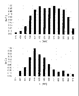
[0021] Figure 2 compares the fluorescence spectra of skin
tissue with a malignant melanoma and of healthy skin tissue.
The excitation parameters are: two-photon excitation at a
wavelength of 810 nm and at a pulse duration of 0.7 ns, with
= CA 02654417 2009-01-28
= 60285-1129
19
a diameter of the measurement site of 70 pm. The samples
stem from several measuring procedures and consequently, the
two fluorescence spectra are only qualitatively but not
quantitatively comparable. The lower fluorescence spectrum
stems from a sample of healthy skin and shows the
characteristic spectral distribution of the fluorescence
with a clear focal point at 475 nm for eumelanin. The upper
fluorescence spectrum stems from a sample with a malignant
melanoma and shows a clear increase in the fluorescence at
575 nm for pheomelanin, the indicator of the malignant
melanoma, and the characteristic gap at 525 nm for the
flavin fluorescence suppression. In this example, the
reduction in the total fluorescence that occurs in such
cases is not visible, since the two fluorescence spectra
cannot be standardized with respect to each other because
they stem from different measuring procedures. For example,
the thicknesses of the paraffin layers over the skin samples
that are to be overcome can differ. Irrespective of that,
the resulting fluorescence spectra of the two samples of
qualitatively clearly diseased and healthy skin tissue can
be identified and they show that, already on the basis of
the spectral effect, a clear-cut conclusion can be drawn
about the condition of the sample at the measurement site,
even with detections using completely different conditions
while the same pulse parameters for the fluorescence
excitation are retained.
[0022]
Figure 3A shows a photograph of a skin tissue region
with a malignant melanoma under normal light and embedded in
paraffin as shown in Figure 1. The cross identifies a
striking point on the skin surface of the sample. For
purposes of a size comparison, the distance from the cross
to the light spot is 3 mm. In order to make an assessment of
suspicious regions, Figure 3B shows a grayscale image as an
overview of the same measured object as in Figure 3A, here
= CA 02654417 2009-01-28
= 60285-1129
=
in the light of fluorescence excited at 337 nm. For purposes
of making a comparison with Figure 3A, the cross is entered
at the identical place. Ten measurement sites are marked
whose fluorescence spectra are also shown below in Figure
5 3C. The dark region that is suspected of having a malignant
melanoma is especially clearly visible around the
measurement site 2. The fluorescence spectra at the ten
measurement sites were excited with pulses at a wavelength
of 810 nm and at a pulse duration of 0.7 ns. Each
10 fluorescing region has a diameter of 70 Am. The ordinate of
the spectra is standardized for the maximum of the entire
measurement series, i.e. in addition to the spectral
variation, the intensity variation over the measurement
sites is also visible. The measurement site 8 located
15 outside of the suspicious region that can be seen in the
overview image, especially at measurement site 2, also
proves to be quite unsuspicious in the spectral analysis
and, with its undisturbed fluorescence typical of healthy
skin tissue, serves as a standardization reference for all
20 of the other nine fluorescence spectra in this figure. At
the measurement site 9, the undisturbed spectral
distribution typical of healthy skin tissue is still
detected with the practically continuous course of the
measured value decrease between 475 nm and 675 nm, but with
a marked weakening of the total fluorescence, as a result of
which a fundamental suspicion of an irregularity exists, but
not of a skin region affected by a malignant melanoma in the
early stage. The measurement sites 1 and 6 show a small but
clearly recognizable deviation from this continuous course,
in each case at 550 nm, and consequently, they give rise to
a greater suspicion that the appertaining skin region is
affected with a malignant melanoma at an early stage. The
fluorescence spectra at the other measurement sites 2, 3, 4,
5, 7 and 10 show the forms of the spectral distribution with
CA 02654417 2009-01-28
= = 60285-1129
21
maxima around 575 nm that are typical for disease with a
malignant melanoma, and also show the characteristic gap
around 525 nm for the flavin fluorescence suppression as
well as the marked weakening of the total fluorescence.
100231 Figure 4A shows a measuring arrangement 1.1 for
taking images of objects to be measured in the light of
their fluorescence excited with one-photon excitation. The
excitation laser 1.2 emits pulses 1.3 of laser light having
a wavelength of, for example, 337 nm, at a pulse length of
2.5 ns. The pulses 1.3 are guided unfocussed through a
bundle of optical waveguides 1.4 onto a measuring region 1.5
having a diameter of, for instance, 1 cm. The fluorescence
light created by the pulses 1.3 is then passed through a
filter 1.8 to an imaging camera 1.6, where it is converted
into a grayscale or color-coded image that is proportional
to the intensities of the imaged wavelengths. The image is
depicted on an evaluation unit 1.7, stored and kept ready
for further processing. The images thus taken can be used to
scan, for example, larger skin regions for a preliminary
assessment of the regions suspected of having a malignant
melanoma. According to the unambiguous evaluation criteria
cited in the description and shown in Figures 1 to 3, this
is a purely technical measuring method that can be carried
out by a trained technician or, in the future, even by an
appropriately configured program, and it yields completely
objective results.
[0024] Figure 4B shows a measuring arrangement 2.1 for the
imaging and processing of the fluorescence spectra using
two-photon excitation. The excitation laser 2.2 that can be
tuned in the wavelength range between 600 nm and 1000 nm
emits pulses 2.3 of laser light at a wavelength of, for
example, about 850 nm at a pulse length in the range from
0.7 ns to 2.5 ns. The pulses 2.3 are deflected by a
dielectric mirror 2.8 that functions as a mirror for certain
CA 02654417 2009-01-28
= = 60285-1129
22
wavelengths and that is permeable for other wavelengths, and
said pulses are focused by a lens system 2.9 onto the
measuring spot 2.5 having a diameter, for example, of 70 Am.
Consequently, in the measuring spot 2.5, the melanin
fraction in the fluorophore mixture - selectively and
spatially resolved - is excited to fluorescence. The emitted
fluorescence spectrum in the wavelength range between 400 nm
and 700 nm is then guided through the lens system 2.9 back
to the dielectric mirror 2.8, allowed to pass through the
latter and focused by another lens system with a filter 2.10
onto the input of a bundle of optical waveguides 2.4. The
bundle 2.4 guides the fluorescence light into a spectrometer
2.6 in which the acquired fluorescence spectrum is resolved
and the intensity of the wavelengths is detected in
increments of, for example, 25 nm. A secondary electron
multiplier 2.11 amplifies the measured result and
subsequently feeds it into an evaluation unit 2.7, which
depicts it, stores it and keeps it ready for further
processing. With the fluorescence spectra thus acquired, for
example, measuring spots from the suspicious regions
ascertained with the measuring arrangement shown in
Figure 4A can be tested concretely and objectively for the
presence of skin regions diseased with the malignant
melanoma. Here, too, according to the unambiguous evaluation
criteria cited in the description and shown in Figures 1 to
.3, this is a purely technical measuring method that can be
carried out by a trained technician or by a program. It
yields completely objective results that can be evaluated by
computers. Therefore, the measuring methods described are in
their entirety of a purely technical nature and are
fundamentally subject to clear and objective evaluation
standards employed by trained operating personnel.