Note : Les descriptions sont présentées dans la langue officielle dans laquelle elles ont été soumises.
CA 02657841 2009-01-14
WO 2008/014162 PCT/US2007/073769
POSITIONING SYSTEM FOR MANIPULATING A TREATMENT INSTRUMENT
AT THE END OF A MEDICAL DEVICE
DESCRIPTION OF THE INVENTION
Cross-Reference to Related Applicaitons
[001] This international application claims the priority of earlier filed
United
States Provisional Application No. 60/832,594, filed July 24, 2006. The entire
content of that provisional application is expressly incorporated by reference
herein.
Field of the Invention
[002] The invention relates to an endoscope system for accessing a
patient's body portion and used for diagnosis and treatment of medical
conditions.
For example, embodiments of the invention may include a particular endoscopic
positioning mechanism for placing an endoscope and an additional treatment
device within desired body portions in order to assist in diagnosis and
treatment of
anatomical diseases and disorders.
Background of the Invention
[003] Endoscopes for medical use have been adopted for various
diagnostic and medical treatment procedures. Endoscopes have been used for the
diagnosis and treatment of a wide range of diseases and disorders that often
require a physician to access the tortuous and relatively small cross-
sectional areas
of a patient's internal anatomical body lumens. A patient's pancreaticobiliary
system (including the anatomical regions of the gall bladder, pancreas, and
the
biiiary tree), for example, is accessed for diagnosis, and/or treatment of
disorders of
certain portions of the digestive system.
[004] During treatment of the digestive system, endoscopes are often used
to access and visualize a patient's pancreaticobiliary system. Once the
endoscope
is positioned in the desired body portion, a treatment instrument can be
advanced
through the working channel of the endoscope to the desired body portion. The
-1-
CA 02657841 2009-01-14
WO 2008/014162 PCT/US2007/073769
endoscope and treatment instrument may then be manipulated as desired for
visualization and treatment respectively.
[005] Endoscopic retrograde cholangiopancreatography (ERCP) is one
example of a medical procedure that uses an endoscope. ERCP enables the
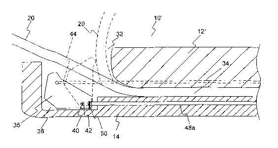
physician to diagnose problems in the liver, gallbladder, bile ducts, and
pancreas.
The liver is a large organ that, among other things, makes bile that helps
with
digestion. The gallbladder is a small, pear-shaped organ that stores bile
until it is
needed for digestion. The bile ducts are tubes that carry bile from the liver
to the
gallbladder and small intestine. These ducts are sometimes called the biliary
tree.
The pancreas is a large gland that produces chemicals that help with digestion
and
hormones such as insulin.
[006] The biliary system delivers bile produced by the liver to the duodenum
where the bile assists other gastric fluids in digesting food. The biliary
system
includes the liver, as well as a plurality of bodily channels and organs that
are
disposed between the liver and the duodenum. Within the liver lobules, there
are
many fine "bile canals" that receive secretions from the hepatic cells. The
canals of
neighboring lobules unite to form larger ducts, and these converge to become
the
"hepatic ducts." They merge, in turn, to form the "common hepatic duct." The
"common bile duct" is formed by the union of the common hepatic and the cystic
ducts. It leads to the duodenum, where its exit is guarded by a sphincter
muscle.
This sphincter normally remains contracted until the bile is needed, so that
bile
collects in the common bile duct and backs up to the cystic duct. When this
happens, the bile flows into the gallbladder and is stored there.
[007] ERCP is used primarily to diagnose and treat conditions of the bile
ducts, including gallstones, inflammatory strictures (scars), leaks (from
trauma and
surgery), and cancer. ERCP combines the use of x-rays and an endoscope.
Through the endoscope, the physician can see the inside of the stomach and
duodenum, and inject dyes into the ducts in the biliary tree and pancreas so
they
can be seen on x-rays.
[008] An ERCP is performed primarily to identify a problem in the bile ducts
or pancreas. Other applications are directed more towards therapy rather than
only
diagnosis. For example, other procedures include using endoscopes for stone
-2-
CA 02657841 2009-01-14
WO 2008/014162 PCT/US2007/073769
removal and sphincterotome. In addition, combined diagnostic and therapeutic
procedures may be performed. For example, if a gallstone is found during the
exam, it can often be removed by means of a treatment instrument, eliminating
the
need for major surgery. If a blockage in the bile duct causes yellow jaundice
or
pain, it can be relieved through the use of a treatment instrument inserted
through
the endoscope.
[009] Since endoscopes are often used to access the tortuous and
relatively small cross-sectional areas of a patient's internal anatomical body
lumens, repeated manipulation and positioning of an endoscope during a medical
procedure can cause problematic side-effects. For example, repeated
manipulation
and positioning of the endoscope can cause unnecessary trauma to a patient's
internal tissues. Improper placement and repeated attempts to access a desired
treatment region can exacerbate tissue trauma as well as unnecessarily prolong
the
medical procedure. Accordingly, there is a need for more precise endoscope
manipulation as well as manipulating an underlying treatment instrument
through
an access channel of an endoscope.
[010] Thus, it is desirable to have an endoscope assembly that can more
precisely access the tortuous and relatively small cross-sectional areas of
certain
anatomical body lumens, and more precisely manipulate a treatment device
provided within an access channel of an endoscope.
SUMMARY OF THE INVENTION
[011] Embodiments of the present invention are directed to an improved
endoscope system and a positioning device for manipulating a treatment device
that obviates one or more of the limitations and disadvantages of prior
medical
devices.
[012] In one embodiment, a medical device comprises an elongated
flexible tube including a distal end and a proximal end and defining a lumen
extending from the proximal end to an aperture at the distal end. A
positioning
mechanism is positioned at the distal end of the flexible tube proximate the
aperture. The positioning mechanism is configured for movement through at
least
two degrees of freedom to transmit force to a treatment instrument extending
-3-
CA 02657841 2009-01-14
WO 2008/014162 PCT/US2007/073769
through the lumen and to control a direction at which a treatment instrument
extends from the aperture.
[013] In various embodiments, the device may include one or more of the
following additional features: wherein the positioning mechanism is housed
within a
recess at the distal end of the flexible tube, the positioning mechanism being
configured for rotation about a pin within the recess; wherein the positioning
mechanism is configured for lateral displacement within the recess and along
the
pin; wherein the positioning mechanism is configured for longitudinal
displacement
within the recess; wherein the positioning mechanism includes an elongated
slot
extending therethrough that receives the pin such that the positioning
mechanism is
configured for longitudinal movement relative to the pin; wherein a resilient
sponge
material is included within a portion of the elongated slot such that the
positioning
mechanism returns to a resting longitudinal position when longitudinally
directed
actuation forces are no longer applied to the positioning mechanism; wherein
the
positioning mechanism is configured for angular displacement through combined
lateral and longitudinal displacement of the positioning mechanism; wherein
the pin
comprises a resilient, flexible material such that the positioning mechanism
is
configured for further angular displacement through combined lateral and
longitudinal displacement of the positioning mechanism; further comprising a
spring
connected at one end to a second side of the positioning mechanism, opposite
the
first side of the positioning mechanism, and connected at another end to the
flexible
tube such that after actuation of the pull wire the positioning mechanism
returns to
a resting position; wherein the positioning mechanism comprises a movable
positioning sleeve having a roller positioned on the distal end thereof, the
roller
being rotatable relative to the sleeve and including a lumen therethrough
configured
for receiving a treatment instrument extended distally beyond the lumen;
wherein
the positioning mechanism is configured for lateral displacement in a first
direction
through actuation of a pull wire connected to a first side of the positioning
mechanism; wherein the positioning mechanism is configured for lateral
displacement in a second direction, opposite the first direction, through
actuation of
a pull wire connected to a second side of the positioning mechanism, opposite
the
first side of the positioning mechanism; wherein the pull wires connected to
the first
-4-
CA 02657841 2009-01-14
WO 2008/014162 PCT/US2007/073769
and second sides of the positioning mechanism extend laterally away from the
positioning mechanism then wrap around and extend proximally away from force
transmission posts located within the recess; wherein the positioning
mechanism
includes a concave surface configured to maintain contact with a treatment
instrument extended distally beyond the lumen; wherein the aperture is a side
facing aperture opening laterally along the flexible tube; wherein the
positioning
mechanism is configured for movement through at least three degrees of
freedom;
wherein the positioning mechanism is rotatable about three orthogonal axes;
wherein the positioning mechanism comprises a roller rotatable relative to the
aperture, the roller including a lumen therethrough configured for receiving a
treatment instrument extended distally beyond the lumen; wherein a proximal
end
of the lumen through the roller is configured to maintain alignment with the
lumen of
the elongated flexible tube; wherein the lumen through the roller exhibits a
cone
shape having a distal opening more narrow than a proximal opening; further
comprising a sleeve extending within the lumen of the roller and movable
within
and distally beyond the lumen of the roller; wherein the sleeve is configured
for
receiving a treatment instrument and imparting rotation to the treatment
instrument
upon rotation of the sleeve; wherein the roller is configured for rotation
about three
orthogonal axes; wherein rotation of the roller relative to the aperture is
achieved
through the actuation of pull wires, each fixedly attached to a predetermined
location along the roller; further comprising a wedge having an inclined
surface
positioned distally of the roller and wherein the rotation of the roller
relative to the
aperture is achieved through proximal movement of the base beneath the roller;
wherein attachment of each pull wire to the roller occurs at a constant
predetermined distance from a distal point of exit of the lumen of the roller;
wherein
at least three pull wires are fixedly attached to the roller; wherein the
medical
device is an endoscope that includes visualization components therein; wherein
the
medical device is an endoscope that includes illumination components therein;
wherein the medical device is an endoscope that includes an additional
positioning
mechanism for achieving controlled deflection of the elongated flexible tube.
[014] In another embodiment, a medical device comprises an elongated
flexible tube including a distal end and a proximal end and defining a lumen
-5-
CA 02657841 2009-01-14
WO 2008/014162 PCT/US2007/073769
extending from the proximal end to an aperture at the distal end. A deflection
mechanism is housed within the distal end of the flexible tube opposite the
aperture, the deflection mechanism being configured for rotation about a pin
extending within the recess and for lateral displacement along the pin.
[015] In various embodiments, the device may include one or more of the
following additional features: wherein the deflection mechanism is configured
for
longitudinal displacement within the recess; wherein the deflection mechanism
includes an elongated slot extending therethrough that receives the pin such
that
the deflection mechanism is configured for longitudinal movement relative to
the
pin; wherein a resilient sponge material is included within a portion of the
elongated
slot such that the deflection mechanism returns to a resting longitudinal
position
when longitudinally directed actuation forces are no longer applied to the
deflection
mechanism; wherein the deflection mechanism is configured for angular
displacement through combined lateral and longitudinal displacement of the
deflection mechanism; wherein the pin comprises a resilient, flexible material
such
that the deflection mechanism is configured for further angular displacement
through combined lateral and longitudinal displacement of the deflection
mechanism; wherein the deflection mechanism includes a concave surface
configured to maintain contact with a treatment instrument extended distally
beyond
the lumen; wherein the aperture is a side facing aperture opening laterally
along the
flexible tube; wherein the deflection mechanism is configured for lateral
displacement in a first direction through actuation of a pull wire connected
to a first
side of the deflection mechanism; wherein the deflection mechanism is
configured
for lateral displacement in a second direction, opposite the first direction,
through
actuation of a pull wire connected to a second side of the deflection
mechanism,
opposite the first side of the deflection mechanism; and wherein the pull
wires
connected to the first and second sides of the deflection mechanism extend
laterally away from the deflection mechanism then wrap around and extend
proximally away from force transmission posts located within the recess.
[016] In another embodiment, a medical device comprises an elongated
flexible tube including a distal end and a proximal end and defining a lumen
extending from the proximal end to an aperture at the distal end. A roller is
-6-
CA 02657841 2009-01-14
WO 2008/014162 PCT/US2007/073769
positioned at the distal end of the flexible tube and rotatable relative to
the aperture,
the roller including a lumen therethrough configured for receiving a treatment
instrument extended distally beyond the lumen.
[017] In various embodiments, the device may include one or more of the
following additional features: further comprising a movable sleeve and wherein
the
roller is positioned on the distal end thereof, the roller being rotatable
relative to the
sleeve; a sleeve extending within the lumen of the roller and movable within
and
distally beyond the lumen in the roller; wherein the sleeve is configured for
receiving a treatment instrument and imparting rotation to the treatment
instrument
upon rotation of the sleeve; wherein the roller is configured for rotation
about three
orthogonal axes; wherein rotation of the roller relative to the aperture is
achieved
through the actuation of pull wires, each fixedly attached to a predetermined
location along the roller; further comprising a wedge having an inclined
surface
positioned distally of the roller and wherein the rotation of the roller
relative to the
aperture is achieved through proximal movement of the inclined wedge surface
beneath the roller; further comprising a movable base positioned distally of
the
roller and wherein the rotation of the roller relative to the aperture is
achieved
through longitudinal and lateral movement of the base beneath the roller;
wherein
attachment of each pull wire to the roller occurs at a constant predetermined
distance from a distal point of exit of the lumen of the roller; wherein at
least three
pull wires are fixedly attached to the roller; wherein the medical device is
an
endoscope that includes visualization components therein; wherein the medical
device is an endoscope that includes illumination components therein; wherein
the
medical device is an endoscope that includes an additional positioning
mechanism
for achieving controlled deflection of the elongated flexible tube.
[018] Additional objects and advantages of the invention will be set forth in
part in the description which follows, and in part will be obvious from the
description, or may be learned by practice of the invention. The objects and
advantages of the invention will be realized and attained by means of the
elements
and combinations particularly pointed out in the appended claims.
-7-
CA 02657841 2009-01-14
WO 2008/014162 PCT/US2007/073769
[019] It is to be understood that both the foregoing general description and
the following detailed description are exemplary and explanatory only and are
not
restrictive of the invention, as claimed.
[020] The accompanying drawings, which are incorporated in and
constitute a part of this specification, illustrate several embodiments of the
invention
and together with the description, serve to explain the principles of the
invention.
BRIEF DESCRIPTION OF THE DRAWINGS
[021] FIG. 1 is a perspective view of a prior art endoscope system.
[022] FIG. 2 is a cross-sectional view illustrating the structure of a known
elevator device.
[023] FIG. 3 illustrates an exemplary coordinate system for designating
translational and rotational displacement of elements in a system of connected
bodies.
[024] FIG. 4 is cross-sectional view of a distal portion of an endoscope
according to an embodiment of the present invention.
[025] FIG. 5 is a top view of components of an instrument positioning
device according to an embodiment of the present invention.
[026] FIG. 6A is a perspective view of components of an instrument
positioning device according to an embodiment of the present invention.
[027] FIG. 6B is a top view of components of an alternative instrument
positioning device according to an embodiment of the present invention.
[028] FIG. 7A is a perspective view of a distal part of an endoscope
according to an embodiment of the present invention.
[029] FIG. 7B is a front view of a distal part of an endoscope according to
an embodiment of the present invention.
[030] FIG. 7C is a side view of a distal part of an endoscope according to
an embodiment of the present invention.
[031] FIG. 8A is a side view of components of an alternative instrument
positioning device according to an embodiment of the present invention.
[032] FIGS. 8B-8D are top views of components of alternative instrument
positioning devices according to embodiments of the present invention.
-8-
CA 02657841 2009-01-14
WO 2008/014162 PCT/US2007/073769
[033] FIG. 9 is a perspective view of a distal part of an endoscope
according to another embodiment of the present invention.
[034] FIG. 10 is a perspective view of a distal part of an endoscope and a
treatment instrument according to another embodiment of the present invention.
[035] FIG. 11 is a side view of a distal part of an endoscope according to
another embodiment of the present invention.
[036] FIG. 12 is a side view of components of an alternative instrument
positioning mechanism according to an embodiment of the present invention.
[037] FIG. 13 is a top view of components of an alternative instrument
positioning mechanism according to an embodiment of the present invention.
[038] FIG. 14 illustrates the positioning of an endoscope and treatment
device within a patient's body portion.
DESCRIPTION OF THE EMBODIMENTS
[039] Reference will now be made in detail to the exemplary embodiments
of the invention, examples of which are illustrated in the accompanying
drawings.
Wherever possible, the same reference numbers will be used throughout the
drawings to refer to the same or like parts. The drawing figures of this
application
are intended to provide a general understanding of the working elements of the
underlying system. Accordingly, unless explicitly stated, the figures do not
represent a literal depiction of proportional dimensions or the precise
locations for
the illustrated inter-related components.
[040] According to exemplary embodiments, the invention relates to a
medical device for positioning a treatment device and/or viewing a patient's
internal
body portion. In embodiments that use a treatment device in an endoscopic
medical procedure, the treatment device can be advanced through a working
channel of an endoscope, including an endoscope specifically designed and/or
sized for use with the treatment device, and into a tissue tract. For purposes
of this
disclosure, "treatment device" or "treatment instrument" includes, for
example, any
working medical device advanced through a working channel of an endoscope and
for use during an endoscopic procedure. Exemplary treatment instruments
include,
but are not limited to, guide wires, cutting or grasping forceps, biopsy
devices,
-9-
CA 02657841 2009-01-14
WO 2008/014162 PCT/US2007/073769
snare loops, injection needles, cutting blades, scissors, retractable baskets,
retrieval devices, ablation and/or electrophysiology catheters, stent
placement
devices, surgical stapling devices, and balloon catheters.
[041] FIG. 1 illustrates a known endoscope system. For purposes of this
disclosure, "distal" refers to the end further from the device operator during
use and
"proximal" refers to the end closer to the device operator during use. FIG. 1
depicts
an endoscope 10 including a flexible outer tube 12 extending between a distal
end
14 and a proximal end 16 of the device. Endoscope 10 includes a treatment
device
insertion port 11 for receiving a treatment device 20 into a working channel
of the
endoscope 10. The distal end 14 of the endoscope system 10 includes a side
facing operation window 18 that can include visualization and lighting
components
for viewing during a treatment procedure. In addition, a working channel (not
shown) extends within the endoscope 10 and terminates at the operation window
18, thereby allowing the treatment instrument 20 to be extended from the
distal end
of the endoscope 10. The extension of the treatment instrument 20 at a desired
treatment site can be then be viewed through the visualization components,
which
transmit images to the proximal end of the endoscope 10, as in known in the
art.
While FIG. 1 illustrates a side facing operation window 18, both front/forward
facing
and oblique/intermediate angled windows are known.
[042] FIG. 2 illustrates a cross-sectional view of a distal portion of a known
endoscope system including a deflecting lever/elevator device for deflecting a
treatment instrument as the instrument is extended beyond a working channel of
an
endoscope. As seen in FIG. 2, a deflecting lever 22 is rotated clockwise about
a
pin 24 by means of a pull wire 26 connected to an upper portion of the
deflecting
lever 22. Upon actuation of the pull wire 26 through proximal movement
thereof,
the deflecting lever 22 deflects the treatment device 20 in order to alter the
angle at
which the treatment device 20 exits the endoscope's working channel, resulting
in
the position of device 20 shown by the dashed lines in FIG. 2. By means of
pull
wire 26, the endoscope operator can control the placement of the treatment
instrument 20 as it is positioned during a medical procedure.
[043] As seen in FIG. 1, a handle 28 at the proximal end 16 of the device
can include various positioning controls 30 to effectuate bending and rotation
of the
-10-
CA 02657841 2009-01-14
WO 2008/014162 PCT/US2007/073769
flexible outer tube 12 for positioning of the device during a medical
procedure. In
addition, the handle can include a distinct positioning control for actuation
of the
deflection lever pull wire 26. During a medical procedure such as, for
example, an
ERCP procedure, the treatment instrument 20 must be precisely inserted into a
particular duct in the biliary tree. While the use of a deflection lever 26 is
capable of
altering the angle at which the treatment device exits the endoscope, precise
positioning often requires repeated manipulation of the distal end of the
endoscope
including the operation window in order to achieve proper placement of the
treatment device 20. As noted above, this repeated manipulation of the
underlying
endoscope 10 can lead to tissue trauma and unnecessarily prolong the entire
medical procedure.
[044] As seen in the embodiment of FIG. 2, the deflection lever 26 is
displaceable about a single axis (i.e. the axis coincident with the pin 24).
Accordingly, lever 26 is movable about and only effectuates movement of the
treatment device 20 through one degree of freedom. Precise manipulation of a
treatment instrument is increased when manipulation is afforded along or about
an
additional particular coordinate axis. A degree of freedom describes
flexibility of
motion added due to displacement along or about a particular coordinate axis.
[045] FIG. 3 illustrates a known Cartesian coordinate system illustrating
the three orthogonal axes of X, Y, and Z. A linkage or any system of connected
bodies that has complete freedom of motion (even if only in a limited area)
has six
degrees of freedom. Three modes are translation (i.e. the ability to move in
each of
three dimensions in a direction parallel to each of the three orthogonal
axes). An
additional three modes are rotation, i.e. the ability to change an angular
position
around the three orthogonal axes. Only three degrees of freedom are necessary
to
move a structure anywhere in space, but additional degrees of freedom provide
more versatility. For example, each of the following is one degree of freedom:
moving up and down along the Y axis (heaving); moving left and right along the
X
axis (swaying); moving forward and back along the Z axis (surging); tilting up
and
down (rotation Rx about the X axis); turning left and right (rotation Ry about
the Y
axis); and tilting side to side (rotation Rz about the Z axis). Accordingly, a
-11-
CA 02657841 2009-01-14
WO 2008/014162 PCT/US2007/073769
positioning mechanism that effectuates movement through more than one degree
of freedom will allow for more precise positioning of an underlying treatment
device.
[046] FIG. 4 illustrates a cross-sectional view of a distal portion of an
endoscope according to an embodiment of the present invention. FIG. 4 depicts
a
cross-sectional view of a distal end 14 of an improved endoscope 10'. The
distal
portion of endoscope 10' includes an exterior flexible outer tube 12', a side
facing
operation window aperture 32, and a working channel 34 forming a lumen within
the endoscope 10' and extending from the proximal end of the endoscope 10' and
terminating at the operation window aperture 32. A deflection elevator in the
form
of a positioning block 35 is housed within a recess 36 at the distal end of
the
endoscope 10' at a position opposite the operation window aperture 32.
[047] FIGS. 5-6B illustrate top and perspective views, respectively, of
exemplary displacement mechanisms which control movement of the positioning
block 35. As seen in FIG. 6A, positioning block 35 includes a curved concave
surface 38 configured to maintain contact with a treatment instrument extended
beyond the endoscope's working channel (see FIG. 4). The curved surface 38 of
the positioning block 35 acts as the surface for transferring a deflection
force
against a treatment instrument 20 during extension of the treatment instrument
20.
Alternatively, the positioning block 35 may include a closed top surface
thereby
forming an internal lumen for receiving a treatment instrument therein. As
another
alternative the positioning block can be provided with a notch or channel
formed in
the concave surface 38. The notch can be provided with a "v" shaped trough
sized
to releasable engage a treatment instrument therein in a passive friction fit
engagement.
[048] The positioning block 35 is disposed for operative connection within
the distal end of the endoscope through a pin 40, which extends laterally
within the
endoscope's distal end 14 and perpendicular to the longitudinal axis of outer
tube
12'. The pin 40 extends laterally within a pin aperture 42 formed in the body
of
positioning block 35. The pin 40 is fixed to the flexible tube 12' such that
the
positioning block 35 is configured to rotate about and translate laterally
relative to
the pin 40. Pin 40 extends through the aperture 42 but is not fixedly attached
to
positioning block 35. Accordingly, the positioning block 35 is configured to
deflect a
-12-
CA 02657841 2009-01-14
WO 2008/014162 PCT/US2007/073769
treatment instrument, such as, for example, device 20 extending within working
channel 34. Positioning block 35 is configured for clockwise rotation about
rotation
pin 40 through actuation of a pull wire 44, illustrated in dashed lines in
FIG. 4. Pull
wire 44 is connected at an upward offset distal position along the positioning
block
35 such that proximal movement of pull wire 44 rotates the positioning block
35
about rotation pin 40. As seen in dashed lines in FIG. 4, the pull wire 44
extends
proximally within a pull wire channel (not shown) of the endoscope where it
extends
for connection with a positioning control device at a handle at the
endoscope's
proximal end. As pull wire 44 is displaced in a proximal direction, the
positioning
block 35, and in turn, the treatment instrument 20 (as seen in dashed lines in
FIG.
4) are rotated such that the angle at which treatment instrument 20 extends
from
the endoscope 10' is increased.
[049] Pull wire 44, for example, can extend for connection to a bending
lever or rotation wheel control device where proximal actuation can be
effected by
an operator. While a pull wire element is illustrated as the mechanism for
deflection
of the positioning block 35, alternative deflection mechanisms can be used,
including, but not limited to, forward acting push wires, or stylets,
electronic
piezoelectric bending transducers, and an inflatable cuff element underlying
the
positioning block 35.
[050] With combined reference to FIGS. 4-6B, in addition to the deflection
control pull wire 44, endoscope 10' is equipped with a lateral displacement
mechanism. As seen in FIG. 4, the pin 40 extends a lateral distance L within
the
recess 36 across the distal end of endoscope 10. As noted above, the pin 40
extends through the pin aperture 42 within the positioning block 35. In
addition to
the deflection capability through rotation about pin 40, positioning block 35
is also
configured for lateral displacement relative to the pin 40 along the distance
L
between left and right sides of recess 36 within the distal end of endoscope
10'.
[051] Positioning block 35 includes surfaces 46a and 46b along opposite
lateral sides of the block 35. Lateral displacement pull wires 48a and 48b are
each
connected at a point along the lateral side surfaces 46a and 46b of the
positioning
block 35. Pull wires 48a and 48b extend laterally away from the positioning
block
35 where they wrap around and extend proximally away from force transmission
-13-
CA 02657841 2009-01-14
WO 2008/014162 PCT/US2007/073769
posts 50, which extend upwardly within the endoscope recess 36. As seen in
FIGS. 5-68, proximal actuation of pull wire 48a results in rightward lateral
displacement of the positioning block 35 along the guide of pin 40.
Conversely,
proximal actuation of pull wire 48b results in leftward lateral displacement
of the
positioning block 35 along the guide of pin 40. The placement of left and
right force
transmission posts 50 permit the transfer of a proximally directed force along
either
of pull wire 48a and 48b into a laterally transmitted force for displacement
of the
positioning block along the lateral distance L. Pull wires 48a and 48b
therefore will
exhibit some degree of flexibility in order to bend about posts 50 and allow
for slack
during rotation of positioning block 35.
[052] The point of connection for lateral pull wires 48a and 48b should be
selected in order to result in the least amount of interference with the
rotation
deflection of the positioning block 35 about rotation pin 40 through actuation
of the
deflection control wire 44. For example, as seen in FIGS. 4-6A, connection of
lateral pull wires 48a, 48b and positioning block 35 may occur at a point just
proximal of the aperture 42. The illustrated connection point is intended to
be non-
limiting and alternative connection locations are permitted with a focus on
reducing
any interference with the free actuation of deflection wire 44. In addition,
the pull
wire arrangement illustrated for lateral displacement is also intended to be
non-
limiting and alternative mechanisms for achieving lateral displacement of
positioning block 35 are possible. Any alternative mechanical force transfer
mechanism which transfers a back and forth force into a laterally directed
force,
such as, for example, a rack and pinion gear mechanism, can be utilized.
[053] For example, FIG. 6B depicts a top view of an alternative positioning
block 35'. As seen in FIG. 6B, the arrangement for the positioning block 35'
only
requires a single pull wire 49 instead of the two lateral pull wires 48a and
48b
required by the arrangement of FIG. 6A. The single pull wire 49 connects to
one
side of the positioning block 35' and a spring 51 connects to another side of
positioning block 35', opposite the surface of connection for pull wire 49.
The end
of spring 51 that is not attached to the positioning block 35' can be secured
to an
internal surface of the underlying endoscope within the recess 36. In
addition, the
arrangement of FIG. 6B, differs from that of FIG. 6A, in that it includes only
a single
-14-
CA 02657841 2009-01-14
WO 2008/014162 PCT/US2007/073769
force transmission post 50 for interaction with pull wire 49. During a
procedure, the
positioning block 35' can then be manipulated and laterally displaced upon
proximal
actuation of the pull wire 49. Upon removal of an actuation force on
positioning
block 35' through the pull wire 49, the spring 51 acts on the positioning
block 35' to
return it to an initial resting position.
[054] FIGS. 7A-7C illustrate perspective, front, and side views,
respectively, of a distal part of an endoscope 10" utilizing a combined
lateral
displacement and deflection controlled positioning block, according to an
embodiment of the present invention. FIG. 7A, for example, illustrates a
perspective view of a distal portion of the endoscope 10" including the
operation
window 32 including positioning block 35 for manipulation of a treatment
instrument
as well as a visualization device 52 and a lighting device 54 for viewing an
internal
body portion. Referring to the front view of FIG. 7B, lateral displacement of
positioning block 35 between left and right ends of the length L is
illustrated. As
explained above, actuation of lateral pull wires 48a and 48b allow more
precise
manipulation of an extended treatment instrument 20 without trauma-causing
movement of the underlying endoscope 10". In particular, the combined lateral
movement and rotation of positioning block 35 allows for precise manipulation
of a
treatment instrument through two degrees of freedom as opposed to the single
positioning degree of freedom afforded by past elevator rotation systems.
[055] FIG. 7C depicts a side view of the distal portion of endoscope 10"
and in particular, the deflection of a treatment instrument 20 as it extends
from a
working channel of the endoscope 10". Actuation of deflection pull wire 44
causes
rotation of positioning block 35 in order to increase or decrease the
deflection angle
(3 (as shown in FIG. 7C) at which the treatment instrument extends from the
working channel of underlying endoscope 10". For example, rotation of
positioning
block 35 about pin 40 can cause deflection of treatment instrument 20 between
an
angle of about 30 degrees to about 135 degrees relative to the longitudinal
axis of
the endoscope 10".
[056] FIG. 8A is a side view of components of an alternative instrument
positioning device according to an embodiment of the present invention. FIG.
8A
depicts an alternative positioning block 35" similar to the positioning block
35 as
-15-
CA 02657841 2009-01-14
WO 2008/014162 PCT/US2007/073769
previously described, with the feature of an elongated pin slot (or channel)
45
replacing the pin aperture 42 described above. The inclusion of the elongated
pin
slot 45 allows for a predetermined amount of controlled longitudinal (both in
a distal
and a proximal direction) displacement of the positioning block 35" relative
to the
underlying endoscope.
[057] The length of elongated pin slot 45 dictates the extent of longitudinal
displacement for positioning block 35". At the distal-most and proximal-most
displacement positions for positioning block 35", further movement of the
positioning block 35" is prevented due to the engagement between an internal
surface of the pin slot 45 and the rotation pin 40, housed therein. Back and
forth
movement of the positioning block 35" within a recess 36 of an underlying
endoscope can be caused by any force actuation mechanism capable of displacing
the positioning block 35". Examples include, but are not limited to, pull
wires,
pushable stylets, fluid pressure actuated force transmission mechanisms, and
expandable balloons. The slot 45 may be filled with a compliant, self-healing
material, such as a sponge material, for example. The inclusion of a sponge
material within the slot 45 allows for stabilization of the pin 40 therein
such that the
pin returns to a centered rest position once a displacement force is no longer
transmitted to the positioning block 35".
[058] Rotation of the positioning block 35" relative to the pin 40 (in order
to
achieve deflection of a treatment instrument as illustrated in FIG. 4, for
example)
can be achieved by maintaining the longitudinal position of the positioning
block
35" within the recess 36 and then causing controlled rotation of the
positioning
block 35" in the manner described above. Maintaining the longitudinal position
of
the positioning block 35" can be achieved through any type of known active of
passive position locking mechanism.
[059] FIGS. 8B and 8C illustrate partial cross-sectional views of the
positioning block 35" depicting the position of pin 40 within the slot 45. As
seen in
FIGS. 8B and 8C, the area of the slot 45 allows for the capability of partial
angular
displacement of the positioning block 35" within the housing recess.
Accordingly, in
addition to the pure lateral and longitudinal displacement capability for the
displacement block 35", the area of slot 45 allows for partial angular
displacement
-16-
CA 02657841 2009-01-14
WO 2008/014162 PCT/US2007/073769
(as seen in FIG. 8C) that allows for greater range of movement for the
positioning
block 35".
[060] FIG. 8D illustrates a partial cross-sectional view of the positioning
block 35" depicting an alternative flexible rotation pin 40' disposed within
the slot
45. The use of the flexible rotation pin 40' allows for further controlled
angular
displacement of the positioning block 35". As seen in FIG. 8D, the flexible
characteristics of pin 40' allow for further angular displacement of the
positioning
block 35" beyond what is capable in an arrangement where the rotation pin is
rigid.
Control of the angular displacement of the positioning block 35" can be
effectuated
though the use of any known force transmission mechanism.
[061] FIG. 9 is a perspective view of a distal part of an endoscope
according to another embodiment of the present invention. FIG. 9 depicts a
distal
portion of an endoscope 10"' including an operation window 56 in part forming
an
aperture 62 that houses a roller 60. For example, the size of roller 60 can be
selected to be retained within an operating window aperture 62. Roller 60
includes
a lumen 64 therethrough that forms an extension of a working channel (not
shown)
of endoscope 10"', such that a treatment instrument can be extended through
the
distal opening of lumen 64 during a medical procedure. The roller 60 can be
provided in any shape so long as it is rotatably housed within the aperture
62.
Roller 60 may be housed within aperture 62 such that a ball and socket type
connection joint is formed. For example, roller 60 can be formed of a
spherical
shape as illustrated in FIGS. 9 and 10. Alternatively, roller 60 can be formed
to
exhibit a cylindrical shape, an oblong, curved football shape, for example, or
any
three dimensional structure exhibiting a partially curved exterior surface
configured
for moving the opening of lumen 64 relative to the endoscope 10"' while housed
within aperture 62. Accordingly, the relative shapes of roller 60 and aperture
62
should be coordinated in order to facilitate the housing and movement of
roller 60
therein.
[062] As noted above, roller 60 is configured for rotation within aperture 62
such that the opening of lumen 64 can be directed for more precise
manipulation of
a treatment instrument extending therethrough. Lumen 64 extending through the
roller 60 is configured for receiving a treatment instrument as the treatment
-17-
CA 02657841 2009-01-14
WO 2008/014162 PCT/US2007/073769
instrument extends distally through an interior working channel of endoscope
10"'.
Since lumen 64 is configured to movably direct and adjust the direction at
which the
treatment instrument extends out of the endoscope 10"', the proximal end of
lumen
64 must maintain communication with the distal opening of an interior working
channel of endoscope 10"' that houses the treatment instrument. In one
arrangement, for example, lumen 64 exhibits a cone shape 65, illustrated in
FIG. 9.
Accordingly, lumen 64 extends distally from a large diameter opening at the
proximal end to a relatively narrow diameter at the distal point of exit of
lumen 64.
Since the proximal end of lumen 64 exhibits a greater diameter opening,
alignment
and communication is maintained between an interior working channel of
endoscope 10"' and lumen 64 as roller 60 is moved relative to the aperture 62.
[063] Roller 60 can be manipulated relative to the housing aperture 62
through a system of pull wires. FIG. 9, for example, illustrates a system of
four pull
wires 66-69 for manipulation of roller 60. Pull wires 66-69 can be fixedly
attached
to the roller 60, each at a predetermined distance from the distal exit point
of lumen
64. Pull wires 66-69 can each be spaced relative to the distal exit point of
lumen
64, such that selective manipulation of each of the pull wires 66-69 allows
for a
predetermined degree of rotation of roller 60 about at least two orthogonal
axes.
For example, proximal actuation of wire 68 coupled with a release of tension
in wire
66 permits a controlled rotation of roller 60 relative to an axis extending
upward in
FIG. 9. Tension within some of wires 66-69 may need to be selectively loosened
in
cooperation with selective tightening of others in the unit in order to permit
controlled rotation of roller 60. In one embodiment, the point of connection
of each
pull wire to roller 60 occurs at a constant predetermined distance from the
distal
point of exit of lumen 64 through roller 60.
[064] Pull wires 66-69 can be connected for operator manipulation through
any type of known wire actuation device at the endoscope handle at the
proximal
end of the system. As is apparent from FIG. 9, selective manipulation of each
of
the pull wires 66-69 allows for a predetermined degree of rotation of sphere
60
about three axes, like an eyeball. For example, with reference to FIGS. 3 and
9,
controlled manipulation of pull wires 66-69 allows for three degrees of
freedom.
While a system of four pull wires is disclosed as the manipulation mechanism
for
-18-
CA 02657841 2009-01-14
WO 2008/014162 PCT/US2007/073769
roller 60, any alternative mechanism for controlled displacement of the roller
can be
used. For example, alternative mechanisms for rotation of roller 60 (some of
which
are more particularly described below, with reference to FIGS. 11-13) include
specifically positioned and controllable track rollers, an arrangement of
three pull
wires, or controlled actuation of selectively placed piezoelectric
transducers.
[065] FIG. 10 depicts an arrangement of a distal portion of an endoscope
similar to that of FIG. 9 and further including an additional positioning
mechanism
for manipulation of a treatment instrument 20. In FIG. 10, a treatment
instrument
20 is extended through an opening of a lumen 64 that extends through roller
60.
Within lumen 64 of FIG. 10, extends a slidable sleeve 70 configured for
movement
relative to the lumen 64 within which it is housed. Sleeve 70 can be
configured to
exhibit a predetermined level of rigidity such that a treatment instrument 20
extended therethrough will be reliably directed coincident with the direction
sleeve
70 extends from lumen 64. For example, during a treatment procedure, sleeve 70
can be used to position the point in space at which the distal end of a
treatment
instrument 20 is located within a patient's body. This further positioning
adjustment
mechanism is advantageous in that the distal end of a treatment instrument can
be
precisely located without requiring repeated manipulation and trauma-casing
movement of the entire underlying endoscope body. If the extended sleeve 70 is
easily deflected and collapsible during contact with internal body tissues,
proper
control and repeatable placement of sleeve 70 (and in turn, the treatment
instrument 20 extended therethrough) may not be possible. Accordingly,
construction of sleeve 70 with a predetermined level of rigidity is
advantageous.
[066] Forward and backward movement of sleeve 70 within lumen 64 and
the internal working channel of endoscope 10"', in combination with controlled
rotation of roller 60, allows for more precise positioning of treatment
instrument 20
during a medical procedure. Sleeve 70 may be configured for back and forth
movement within lumen 64 through a pushable actuation wire (not shown)
proximally extending through endoscope 10"'. For example, the actuation wire
could be configured for connection to the proximal end of sleeve 70 such that
back
and forth movement of the actuation wire through endoscope 10"' is translated
into
back and forth movement of sleeve 70.
-19-
CA 02657841 2009-01-14
WO 2008/014162 PCT/US2007/073769
[067] The addition of slidable sleeve 70 within lumen 64 also affords an
added two degrees of freedom to the endoscope system. As noted above, sleeve
70 can be manipulated by an operator to move forward and backward within lumen
64. In addition, sleeve 70 can be sized to receive and engage the exterior
surface
of the treatment instrument 20 through a friction fit, such that controlled
rotation of
sleeve 70 within lumen 64 effectuates rotation of a treatment instrument 20
extending therein. In addition, sleeve 70 can be configured to engage the
treatment instrument 20 in a friction fit such that back and forth movement of
sleeve
70 effectuates back and forth displacement of instrument 20. Alternatively,
the
controlled rotation of treatment instrument 20 by rotation of sleeve 70 can be
effectuated through a complimentary groove and recess arrangement between the
interior surface of sleeve 70 and the exterior surface of the treatment
instrument 20.
Accordingly, a treatment instrument 20 can be precisely manipulated through
controlled rotation of roller 60, through forward and backward movement of
sleeve
70, and through rotation of sleeve 70, to impart rotation to treatment
instrument 20.
[068] FIG. 11 depicts a side view of a distal part of an endoscope
according to another embodiment of the present invention. In FIG. 11, a
generic
endoscope 10 is depicted housing a positioning sleeve 71 therein. The
positioning
sleeve 71 includes a roller 60 positioned at the distal end thereof. The
positioning
sleeve 71 can itself be manipulated and positioned relative to the underlying
endoscope 10. In addition, the roller 60 at the distal end of the positioning
sleeve
71 can also be precisely rotated and positioned relative to the sleeve 71.
Just as in
the embodiments of FIGS. 9-10, the roller 60 includes a lumen 64 for receiving
a
treatment instrument therein. The angular position of a treatment instrument
can
then be precisely controlled through controlled rotation and positioning of
the roller
60 relative to the sleeve 71. Such controlled rotation can be effectuated
through a
system of pull wires, as described above, or through any other force
transmission
mechanism capable of moving roller 60.
[069] FIG. 12 depicts a side view of components of an alternative
instrument positioning mechanism for the roller 60 described in FIGS. 9-11. As
seen in FIG. 12, rotation of roller 60 can be effectuated through proximal
movement
of a wedge 90 connected to a pull wire 92. The wedge 90 includes an inclined
-20-
CA 02657841 2009-01-14
WO 2008/014162 PCT/US2007/073769
surface 91. Interaction between the inclined surface 91 of the wedge 90 and
the
exterior surface of the roller 60 leads in turn to controlled rotation of the
roller 60
upon proximal actuation of the pull wire 92. For example, due to the
interaction of
the roller 60 with the inclined surface 91, proximal movement of the wedge 90
and
the pull wire 92 in the direction of arrow 93 results in rotation of roller 60
in the
direction of arrow 94. The particular materials for the exterior surface of
roller 60
and the inclined surface 91 can be selected to decrease the amount of sliding
therebetween.
[070] FIG. 13 depicts a top view of components of an alternative
instrument positioning mechanism for the roller 60 described in FIGS. 9-11.
Instead of the moveable wedge 90 described in FIG. 12, FIG. 13 depicts a
movable
base component 94, upon which roller 60 rests. Due to the interaction between
roller 60 and the surface of base component 94, controlled lateral and
longitudinal
displacement of the base component 94 within an endoscope recess 36 results in
controlled rotation of roller 60. Movement of the base component 94 can be
effectuated in both longitudinal directions designated by arrow 95 as well as
lateral
directions designated by arrow 96.
[071] In all of the embodiments described above, the particular positioning
mechanism for a treatment instrument can be equipped with any type of known
locking mechanism for the purpose of releasably maintaining a particular
position of
a treatment instrument relative to an endoscope.
[072] FIG. 14 illustrates the positioning of an endoscope 10', 10", or 10"'
and treatment device 20 within a patient's body portion. In particular, FIG.
14
depicts the extension of a treatment instrument 20 within a particular bile
duct 80
during an ERCP procedure. As seen in FIG. 14, the endoscope 10"', for example,
is inserted and extended through a patient's stomach 82 such that the distal
end
and aperture 62 (not shown) of endoscope 10"' are positioned is close relation
to a
particular bile duct 80 leading to, for example, gall bladder 84. As seen in
FIG. 14,
treatment instrument 20 is extended beyond the internal working channel of
endoscope 10"'. The treatment instrument can then be precisely manipulated,
for
example, by controlled rotation of roller 60 and/or the additional extension
of sleeve
-21 -
CA 02657841 2009-01-14
WO 2008/014162 PCT/US2007/073769
70 beyond endoscope 10"', described above. In addition, further manipulation
of
instrument 20 can be effectuated through rotation of sleeve 70, for example.
[073] Precise manipulation of treatment instrument 20 allows for more
precise positioning and location of instrument 20 such as, for example, during
placement of instrument 20 within a particular bile duct 80 of interest. More
precise
manipulation of a treatment device 20 can result in shortened treatment
procedures
by reducing the amount of time necessary to effectuate proper position of the
treatment device 20. In addition, controlled deflection of the angle at which
treatment device 20 exits the underlying endoscope 10"' can reduce internal
tissue
trauma caused during endoscopic procedures requiring repeated repositioning
and
manipulation of the entire endoscope during location of the endoscope. For
example, the positioning mechanisms described above facilitate the location of
treatment instrument 20 within a particular bile duct 80 such that the
duration of,
and occurrence of tissue trauma during, a treatment procedure can be reduced.
[074] While the above described positioning system has been depicted as
utilizing pull wire manipulation mechanisms, the invention it not intended to
be
limited to this particular structure. Therefore, alternative actuation devices
are
intended to be within the scope of this invention, including all equivalent
structures
known for transferring endoscopic manipulation forces along the longitudinal
axis of
an endoscope. Furthermore, unless expressly stated as otherwise, all
components
and elements of one of the various disclosed embodiments can be used, either
via
substitution, or in addition with the components and elements of any of the
other
embodiments.
[075] Other embodiments of the invention will be apparent to those skilled
in the art from consideration of the specification and practice of the
invention
disclosed herein. It is intended that the specification and examples be
considered
as exemplary only, with a true scope and spirit of the invention being
indicated by
the following claims.
- 22 -