Note : Les descriptions sont présentées dans la langue officielle dans laquelle elles ont été soumises.
CA 02658168 2013-08-26
1
APPARATUS AND METHOD FOR SECURING OCULAR TISSUE
CROSS-REFERENCE TO RELATED PATENT DOCUMENTS
[0001] This application is related to the following U.S.
patent applications and issued patents:
(1) U.S. Patent No. 6,007,578 entitled "Scleral
Prosthesis for Treatment of Presbyopia and Other
Eye Disorders" issued on December 28, 1999;
(2) U.S. Patent No. 6,280,468 entitled "Scleral
Prosthesis for Treatment of Presbyopia and Other
Eye Disorders" issued on August 28, 2001;
(3) U.S. Patent No. 6,299,640 entitled "Scleral
Prosthesis for Treatment of Presbyopia and Other
Eye Disorders" issued on October 9, 2001;
(4) U.S. Patent No. 5,354,331 entitled "Treatment of
Presbyopia and Other Eye Disorders" issued on
October 11, 1994;
(5) U.S. Patent No. 5,465,737 entitled "Treatment of
Presbyopia and Other Eye Disorders" issued on
November 14, 1995;
(6) U.S. Patent No. 5,489,299 entitled "Treatment of
Presbyopia and Other Eye Disorders" issued on
February 6, 1996;
(7) U.S. Patent No. 5,503,165 entitled "Treatment of
Presbyopia and Other Eye Disorders" issued on
April 2, 1996;
(8) U.S. Patent No. 5,529,076 entitled "Treatment of
Presbyopia and Other Eye Disorders" issued on
June 25, 1996;
(9) U.S. Patent No. 5,722,952 entitled "Treatment of
Presbyopia and Other Eye Disorders" issued on
218435092
CA 02658168 2013-08-26
2
March 3, 1998;
(10) U.S. Patent No. 6,197,056 entitled "Segmented
Scleral Band for Treatment of Presbyopia and
Other Eye Disorders" issued on March 6, 2001;
(11) U.S. Patent No. 6,579,316 entitled "Segmented
Scleral Band for Treatment of Presbyopia and
Other Eye Disorders" issued on June 17, 2003;
(12) U.S. Patent No. 6,926,727 entitled "Surgical
Blade for Use with a Surgical Tool for Making
Incisions for Scleral Eye Implants" issued on
August 9, 2005;
(13) U.S. Patent No. 6,991,650 entitled "Scleral
Expansion Device Having Duck Bill" issued on
January 31, 2006;
(14) U.S. Patent No. 7,189,248 entitled "System and
Method for Making Incisions for Scleral Eye
Implants" issued on March 13, 2007;
(15) U.S. Patent Publication No. US2004/0073245
entitled "System and Method for Determining a
Position for a Scleral Pocket for a Scleral
Prosthesis" published on April 15, 2004;
(16) U.S. Patent Publication No. US2005/0283233
entitled "Scleral Prosthesis for Treatment of
Presbyopia and Other Eye Disorders" published on
December 22, 2005;
(17) U.S. Patent Publication No. US2006/0036269
entitled "Surgical Blade for Use with a Surgical
Tool for Making Incisions for Scleral Eye
Implants" published on February 16, 2006;
(18) U.S. Patent Publication No. US2006/0095126
entitled "Scleral Expansion Device Having Duck
Bill" published on May 4, 2006;
21843509.2
CA 02658168 2013-08-26
3
(19) U.S. Patent Publication No. US2006/0106408
entitled "Surgical Blade for Use with a Surgical
Tool for Making Incisions for Scleral Eye
Implants" published on May 18, 2006;
(20) U.S. Patent Publication No. US2006/0106409
entitled "System and Method for Making Incisions
for Scleral Eye Implants" published on May 18,
2006;
(21) U.S. Patent Publication No. U52006/0106457
entitled "Segmented Scleral Band for Treatment of
Presbyopia and Other Eye Disorders" published on
May 18, 2006; and
(22) U.S. Patent Publication No. US2006/0111775
entitled "Segmented Scleral Band for Treatment of
Presbyopia and Other Eye Disorders" published on
May 25, 2006.
TECHNICAL FIELD
[0002] This disclosure is generally directed to surgical
devices and more specifically to an apparatus and method
for securing and modifying ocular tissue.
21843509.2
CA 02658168 2013-08-26
4
BACKGROUND
[0003] It is often desirable or necessary to secure a
patient's eye in place during ocular surgery. For example,
it is possible to restore the accommodative power to a
presbyopic eye by implanting scleral prostheses within the
sclera of the patient's eye. It is also possible to treat
glaucoma, ocular hypertension, elevated intraocular
pressure, or other eye disorders by implanting scleral
prostheses within the sclera of the patient's eye. During
these types of procedures, an incision can be made in the
sclera of the eye and extended under the surface of the
sclera to form a scleral "tunnel." A scleral prosthesis
can then be placed within the tunnel. Before performing a
surgical procedure to implant scleral prostheses or other
surgical eye procedure, the patient's eye often needs to be
fixated so that the patient's eye does not move during the
surgical procedure.
[0004] FIGURES 16A and 16B illustrate a conventional
ocular fixation tool. This ocular fixation tool is placed
on the surface of a patient's eye and is physically sutured
to the sclera of the patient's eye. This ocular fixation
tool includes various notches in which a surgical tool can
be placed.
[0005] FIGURE 17 illustrates a second conventional
ocular fixation tool having a solid ring with spikes (not
shown) that can be depressed into the tissue of a patient's
eye. This ocular
fixation tool also includes a handle
rotatably coupled to the solid ring, where the handle can
be used to move and position the tool. In addition, this
ocular fixation tool includes a projection from the solid
ring, where a surgical tool can be mounted on the
projection.
21843509.2
CA 02658168 2013-08-26
[0006] FIGURES 18A and 18B illustrate a third
conventional ocular fixation tool having a handle, a solid
first ring, and a divided second ring. The solid
first
ring is rotatably coupled to the handle. The divided
5 second ring includes two arms that are rotatably coupled to
the central ring at a common pivot point. As shown in
FIGURE 18A, the two arms of the second ring are in the open
position, and the first ring may be placed in a desired
location on a patient's eye. As shown in FIGURE 18B, the
two arms of the second ring can then be closed, which
drives prongs or other extensions on the arms into the
tissue of the patient's eye. After that, the handle can be
rotated sideways so that a surgeon or tool has clear access
to the patient's eye through the rings. In other
embodiments, the handle and the solid first ring can be
omitted, and the second ring could be used by itself (the
arms can be closed and opened to lock the second ring onto
and release a patient's ocular tissue). In still
other
embodiments, the two arms of the second ring could lack
prongs or other extensions themselves, and the arms could
be used to drive pins or other extensions on the first ring
into a patient's ocular tissue.
21843509.2
CA 02658168 2013-08-26
6
SUMMARY
[0007] This disclosure provides an apparatus and method
for securing ocular tissue.
[0008] In a first embodiment, an apparatus includes a
first ring and a second ring, where at least one of the
rings includes means for fixating ocular tissue of an eye.
The means for fixating are arranged to grasp the ocular
tissue of the eye and to release the ocular tissue of the
eye based on rotation of at least one of the rings.
[0009] In particular embodiments, the means for fixating
are arranged to grasp the ocular tissue of the eye in an
area of the eye associated with the limbus of the eye.
[0010] In other particular embodiments, the apparatus
also includes a housing in which the first and second rings
are housed and a retaining ring within the housing
configured to retain the first and second rings in the
housing. The housing could also include a dome configured
to protect a central portion of the eye.
[0011] In yet other particular embodiments, the
apparatus also includes a base configured to be placed on
the ocular tissue of the eye and to retain the first and
second rings. The apparatus could also include a dome
. configured to protect a central portion of the eye.
[0012] In still other particular embodiments, the first
and second rings include tabs that extend outside of the
dome and the base. The tabs may be configured to rotate at
least one of the first and second rings.
[0013] In additional particular embodiments, the
apparatus includes one or more mechanisms for aligning a
surgical tool with a position on the eye. For example, the
dome could include one or more holes configured to receive
one or more projections from a surgical tool so as to align
21843509.2
CA 02658168 2013-08-26
7
the surgical tool with a position on the eye. As another
example, the base could include one or more notches, where
each notch is configured to receive a projection from a
surgical tool so as to align the surgical tool with a
position on the eye. In addition, the base could include
one or more portions that are configured to lie on the eye.
The one or more portions could include one or more edges
configured to allow a base of the surgical tool to be
aligned against one of the edges when the projection from
the surgical tool is inserted into one of the notches.
[0014] In a second embodiment, a system includes an
ocular fixation device having a first ring and a second
ring. At least one of the rings includes means for
fixating ocular tissue of an eye, where the means for
fixating are arranged to grasp the ocular tissue of the eye
and to release the ocular tissue of the eye based on
rotation of at least one of the rings. The system
also
includes a surgical tool mountable on the ocular fixation
device.
[0015] In particular embodiments, the surgical tool
includes a surgical blade configured to form a scleral
tunnel in the ocular tissue of the eye.
[0016] In a third embodiment, a method includes placing
an ocular fixation device on an eye of a patient. The
ocular fixation device includes a first ring and a second
ring, where at least one of the rings includes means for
fixating ocular tissue of the patient's eye. The method
also includes rotating at least one of the first and second
rings so that the means for fixating grasp the ocular
tissue of the patient's eye.
[0017] In particular embodiments, the method also
includes rotating at least one of the first and second
21843509.2
CA 02658168 2013-08-26
8
rings so that the means for fixating release the ocular
tissue of the patient's eye.
[0018] In a fourth embodiment, an apparatus includes a
first ring having a plurality of first teeth and a second
ring having a plurality of second teeth. The first and
second teeth are arranged to grasp ocular tissue of an eye
and to release the ocular tissue of the eye based on
rotation of at least one of the rings.
[0019] In a fifth embodiment, an apparatus includes one
or more rings having means for fixating ocular tissue of an
eye. The means for
fixating are arranged to grasp the
ocular tissue of the eye and to release the ocular tissue
of the eye based on movement of at least one of the one or
more rings.
[0020] In a sixth embodiment, an apparatus includes a
ring configured to be placed on an eye, where the ring
includes a plurality of portions for resting against a
surface of the eye. The ring also includes a plurality of
portions forming a plurality of notches configured to
receive sutures for attaching the ring to the eye.
[0021] In a seventh embodiment, an apparatus includes a
base configured to be depressed against ocular tissue of an
eye. The apparatus
also includes means for fixating
coupled to the base and configured to be secured against
the ocular tissue of the eye. The apparatus
further
includes a handle configured to move the means for
fixating.
[0022] In an eighth embodiment, an apparatus includes a
central portion configured to be placed over at least the
cornea of an eye. The apparatus also includes means for
fixating ocular tissue of the eye, where the means for
fixating are located on the central portion. In addition,
21843509.2
CA 02658168 2013-08-26
9
the apparatus includes a tool support attached to the
central portion and configured to receive a surgical tool.
[0023] Other technical features may be readily apparent
to one skilled in the art from the following figures,
descriptions, and claims.
218435092
CA 02658168 2013-08-26
BRIEF DESCRIPTION OF THE DRAWINGS
[0024] For a more complete understanding of this
disclosure, reference is now made to the following
description, taken in conjunction with the accompanying
5 drawing, in which:
[0025] FIGURES lA through 1F illustrate a first example
ocular fixation device in accordance with this disclosure;
[0026] FIGURES 2A through 2C illustrate a second example
ocular fixation device in accordance with this disclosure;
10 [0027] FIGURES 3A through 3C illustrate a third example
ocular fixation device in accordance with this disclosure;
[0028] FIGURES 4A through 41 illustrate an example use
of an ocular fixation device during creation of a scleral
tunnel for receiving a scleral prosthesis in accordance
with this disclosure;
[0029] FIGURES 5A through 5C illustrate a fourth example
ocular fixation device and an example use of the ocular
fixation device in accordance with this disclosure;
[0030] FIGURES 6A through 6C illustrate a fifth example
ocular fixation device and an example use of the ocular
fixation device in accordance with this disclosure;
[0031] FIGURE 7 illustrates an example positioning tool
for use with an ocular fixation device in accordance with
this disclosure;
[0032] FIGURE 8 illustrates a sixth example ocular
fixation device in accordance with this disclosure;
[0033] FIGURES 9A through 9C illustrate a seventh
example ocular fixation device and an example use of the
ocular fixation device in accordance with this disclosure;
[0034] FIGURES 10A through 10D illustrate an eighth
example ocular fixation device in accordance with this
disclosure;
21843509.2
CA 02658168 2013-08-26
11
[0035] FIGURES 11A and 11B illustrate a ninth example
ocular fixation device in accordance with this disclosure;
[0036] FIGURES 12A and 12B illustrate a tenth example
ocular fixation device in accordance with this disclosure;
[0037] FIGURES 13A through 13D illustrate an eleventh
example ocular fixation device in accordance with this
disclosure;
[0038] FIGURES 14A through 14C illustrate a twelfth
example ocular fixation device in accordance with this
disclosure;
[0039] FIGURE 15 illustrates an example method for
ocular fixation in accordance with this disclosure; and
[0040] FIGURES 16A through 188 illustrate conventional
ocular fixation tools.
21843509.2
CA 02658168 2013-08-26
12
DETAILED DESCRIPTION
[0041] FIGURES 1A through 1F illustrate a first example
ocular fixation device 100 in accordance with this
disclosure. The embodiment of the ocular fixation device
100 shown in FIGURES 1A through 1F is for illustration
only. Other embodiments of the ocular fixation device 100
could be used without departing from the scope of this
disclosure.
[0042] As shown in FIGURES lA and 1B, the ocular
fixation device 100 includes a body portion 102, a
retention ring 104, and two locking rings 106-108. In this
example, the body portion 102 includes a base 110 and a
dome 112. The base 110 in this embodiment is generally
circular and is used to house the retention ring 104 and
the locking rings 106-108. The dome 112
represents a
protective cover or shield that can be used to protect the
central portion of a patient's eye. The body portion 102
could be formed from any suitable material(s), such as one
or more transparent or opaque materials. The body portion
102 could also be formed using any suitable technique, such
as injection molding.
[0043] The locking rings 106-108 can be inserted into
the body portion 102 and the retention ring 104 can be
attached to the body portion 102, which secures the locking
rings 106-108 within the body portion 102. The retention
ring 104 could be formed from any suitable material(s).
The retention ring 104 could also be formed in any suitable
manner, such as by injection molding.
[0044] The retention ring 104 could be attached or
secured to the body portion 102 in any suitable manner.
For example, as shown in FIGURE 1C, the retention ring 104
could include bumps 114, and the body portion 102 could
21843509.2
CA 02658168 2013-08-26
13
include corresponding receptacles 116. In this embodiment,
the retention ring 104 could be pushed into the body
portion 102 until the bumps 114 engage the receptacles 116,
locking the retention ring 104 in place.
[0045] As shown in FIGURE 1C, the retention ring 104
could also have a slanted or tapered inner edge 118. This
may help to facilitate placement of the ocular fixation
tool 100 on a patient's eye. For example, the edge 118 of
the retention ring 104 may be slanted so that it is
substantially parallel to the portion of the patient's
sclera on which the retention ring 104 rests.
[0046] The locking rings 106-108 are used to secure the
ocular fixation device 100 to a patient's eye, thereby
helping to fixate and prevent movement of the patient's
eye. As shown in FIGURES 1D and 1E, the locking rings 106-
108 may include teeth 120. In this example, the locking
rings 106-108, including the teeth 120, are substantially
planar (although angled teeth could be used). Also, the
teeth 120 in different locking rings 106-108 are angled
towards each other. At least one of the locking rings 106-
108 can rotate with respect to the other locking ring. In
this way, the areas between the teeth 120 of the locking
rings 106-108 can be increased and decreased. This allows
the teeth 120 to grasp ocular tissue when the teeth 120 are
pushed closer together. This also allows the teeth 120 to
release the ocular tissue when the teeth 120 are pushed
farther apart. In some embodiments, the locking rings 106-
108 can be sized so that the teeth 120 attach or lock onto
scleral tissue of a patient's eye (beyond the cornea and
other areas in the central portion of the patient's eye).
The locking rings 106-108 could be formed from any suitable
material(s), such as a metal. The locking
rings 106-108
21843509.2
CA 02658168 2013-08-26
14
could also be formed in any suitable manner, such as by
photo-etching.
[0047] As shown in FIGURES 1D through 1F, the locking
rings 106-108 include windows 122, and the body portion 102
includes corresponding windows 124. In some embodiments, a
surgeon could insert a tool through one of the windows 124
and use the tool to cause one or more of the locking rings
106-108 to move. For example, the surgeon could insert a
tool through one of the windows 124 and push or pull one of
the locking rings 106-108, causing the openings between the
teeth 120 of the locking rings 106-108 to open or close.
As another example, the surgeon could insert a tool through
one of the windows 124 and push both locking rings 106-108
together, causing the openings between the teeth 120 of the
locking rings 106-108 to close. In other embodiments, part
or all of the body portion 102 could be designed to rotate,
causing the locking ring 106 to rotate with respect to the
locking ring 108. This may allow, for example, the ocular
fixation device 100 to be placed on a patient's eye and
then rotated to lock the ocular fixation device 100 onto
the patient's eye. Any other or additional technique could
be used to cause the teeth 120 of the locking rings 106-108
to move with respect to each other.
[0048] FIGURES 2A through 2C illustrate a second example
ocular fixation device 200 in accordance with this
disclosure. The embodiment of the ocular fixation device
200 shown in FIGURES 2A through 2C is for illustration
only. Other embodiments of the ocular fixation device 200
could be used without departing from the scope of this
disclosure.
[0049] The ocular fixation device 200 of FIGURES 2A
through 20 operates in a similar manner as the ocular
218435092
CA 02658168 2013-08-26
fixation device 100 of FIGURES lA through 1F. As shown in
FIGURE 2A, the ocular fixation device 200 includes a base
202, a dome 204, and locking rings 206-208. Cross-sections
showing additional structural details of the ocular
5 fixation device 200 are shown in FIGURES 2B and 2C. As
shown here, the base 202 is attached or secured to the dome
204 (or vice versa), helping to retain the locking rings
206-208 that are located between the base 202 and the dome
204. In this example, the cross-section of the base 202
10 includes a generally flat portion on which the locking
rings 206-208 lie. The cross-section of the base 202 also
includes a projection along its outer edge, which is
attached to or helps secure the dome 204. The base 202
could further have a shape that facilitates its placement
15 on a patient's eye, such as where the flat portion of the
base 202 is slanted or sloped to approximately match a
curvature of the patient's sclera. The base 202 could be
formed from any suitable material(s). The base 202 could
also be formed using any suitable technique, such as
injection molding.
[0050] The dome 204 represents a protective cover or
shield protecting the central portion of a patient's eye.
The dome 204 could be formed from any suitable material(s),
such as one or more transparent or opaque materials. The
dome 204 could also be formed using any suitable technique,
such as injection molding.
[0051] The locking rings 206-208 are located between the
base 202 and the dome 204. In this example, the locking
rings 206-208 include teeth 210 for attaching or locking
onto ocular tissue of a patient's eye. At least one of the
locking rings 206-208 can rotate with respect to the other
locking ring to open and close the areas between the teeth
21843509.2
CA 02658168 2013-08-26
16
210 of the locking rings 206-208. This allows the teeth
210 to attach to and release ocular tissue of the patient's
eye. In some embodiments, the locking rings 206-208 can be
sized so that the teeth 210 attach to scleral tissue of a
patient's eye. The locking rings 206-208 could be formed
from any suitable material(s), such as a metal. The
locking rings 206-208 could also be formed in any suitable
manner, such as by photo-etching.
[0052] In this example, the locking rings 206-208 are
not completely planar. Instead, each of the locking rings
206-208 includes a main section that is relatively planar
and a curved section along its inner edge. The curved
section of the locking ring 206 generally lies over and to
the inside of the curved section of the locking ring 208.
Also, the curved sections of the locking rings 206-208
include, are attached to, or carry the teeth 210 of the
locking rings 206-208. In addition, the teeth 210 could be
planar or angled with respect to the flat portions of the
locking rings 206-208.
[0053] As shown here, each of the locking rings 206-208
includes one or more windows 212. The windows 212 can be
used to identify the amount of space between the teeth 210
of the locking rings 206-208. For example,
when the
windows 212 of the locking rings 206-208 are aligned or
nearly aligned, this may indicate that the areas between
the teeth 210 of the locking rings 206-208 are
substantially closed (the teeth 210 are attached or locked
onto the ocular tissue of a patient's eye). Similarly,
when the windows 212 of the locking rings 206-208 are not
aligned very much, this may indicate that the areas between
the teeth 210 of the locking rings 206-208 are
substantially open (the ocular tissue of a patient's eye is
21843509.2
CA 02658168 2013-08-26
17
not locked or has been released).
[0054] In the illustrated example, the dome 204 may
cover the windows 212 of the locking rings 206-208, which
could prevent the use of external tools to move the locking
rings 206-208. To facilitate the attachment and release of
ocular tissue by the ocular fixation device 200, one or
both of the locking rings 206-208 could be rotated, such as
via rotation of the dome 204 or the base 202. For example,
the locking ring 206 could be fixed with respect to the
dome 204, and/or the locking ring 208 could be fixed with
respect to the base 202. The ocular fixation device 200
could be placed on a patient's eye, and a surgeon could
rotate the dome 204 of the ocular fixation device 200.
This may cause one of the locking rings 206-208 to rotate
with respect to the other locking ring, thereby opening and
closing the areas between the teeth 210 of the locking
rings 206-208. This technique is for illustration only,
and any other suitable technique could be used to attach
and release ocular tissue using the ocular fixation device
200. For instance, windows could be formed in the dome 204
above the windows 212 in the locking rings 206-208,
allowing the use of an external tool by the surgeon.
[0055] FIGURES 3A through 3C illustrate a third example
ocular fixation device 300 in accordance with this
disclosure. The embodiment of the ocular fixation device
300 shown in FIGURES 3A through 3C is for illustration
only. Other embodiments of the ocular fixation device 300
could be used without departing from the scope of this
disclosure.
[0056] As shown in FIGURE 3A, the ocular fixation device
300 includes a dome 302 and locking rings 304-306. Once
again, the dome 302 protects the central portion of a
21843509.2
CA 02658168 2013-08-26
18
patient's eye and can be formed from any suitable
material(s) and in any suitable manner. In this example,
the dome 302 is transparent and includes a mark used to
center the dome 302 on the patient's eye, although other
embodiments could be used. Also, the locking rings 304-306
include teeth 308 that are shaped and positioned so that
they are angled towards each other. This allows the teeth
308 of the locking rings 304-306 to attach or lock onto the
ocular tissue (such as the scleral tissue) of a patient's
eye. As shown in FIGURES 3A and 3B, at least one of the
locking rings 304-306 is rotatable with respect to the
other to open and close the areas between the teeth 308.
[0057] In this example, the locking rings 304-306
include windows 310, which can provide an indication of
whether (and to what extent) the locking rings 304-306 are
locked onto ocular tissue. For example, when the locking
rings 304-306 are opened (not attached to ocular tissue),
the windows 310 in the locking rings 304-306 may be at
least partially aligned. When the locking rings 304-306
are closed (locked onto ocular tissue), the windows 310 in
the locking rings 304-306 are not aligned, and the windows
310 in the locking ring 306 might be hidden.
[0058] As shown in FIGURE 30, the ocular fixation device
300 can further include a housing 312. The housing
312
holds the locking rings 304-306 and the dome 302 of the
ocular fixation device 300. The housing 312 may also allow
a surgeon to rotate at least one of the locking rings 304-
306. In this example, the housing 312 includes windows 314
and connection points 316. The windows 314 in the housing
312 may be aligned with the windows 310 in the locking ring
304. This allows the surgeon to determine to what extent
the locking rings 304-306 are opened or closed (since the
21843509.2
CA 02658168 2013-08-26
19
housing 312 otherwise hides or covers the locking rings
304-306). The connection points 316 represent areas where
a surgical tool can be attached to the housing 312
(described in more detail below), although the connection
points 316 can be omitted if desired. The housing 312 can
be formed from any suitable material(s) and in any suitable
manner. The housing 312 can also have any suitable shape
or arrangement.
[0059] In this example, the locking rings 304-306 have
more of a cylindrical shape (although it need not have a
true cylindrical shape and can, for example, have slanted
sides). That is, the major surface of each locking ring
304-306 extends along and rotates around a central axis
through the center of that locking ring 304-306.
[0060] Although FIGURES 1A through 30 illustrate three
examples of ocular fixation devices, various changes may be
made to FIGURES 1A through 30. For example, the relative
sizes and dimensions of the features of the ocular fixation
devices are for illustration only and can be altered in any
suitable manner. Also, various
features shown and
described with respect to one of the ocular fixation
devices could be used with other ocular fixation devices.
As a particular example, the locking rings 206-208 of the
ocular fixation device 200 could be used with the ocular
fixation device 100. As another
particular example, the
same or similar housing 312 used with the ocular fixation
device 300 could be used with the other ocular fixation
devices 100 and 200. In addition,
the dome could be
omitted from an ocular fixation device, such as when the
ocular fixation device is used to secure a patient's eye
during corneal surgery or other surgical procedure.
[0061] FIGURES 4A through 41 illustrate an example use
21843509.2
CA 02658168 2013-08-26
of an ocular fixation device during creation of a scleral
tunnel for receiving a scleral prosthesis in accordance
with this disclosure. The example use shown in FIGURES 4A
through 41 is for illustration only. An ocular fixation
5 device could be used in any other suitable manner
(including only to fixate a patient's eye) without
departing from the scope of this disclosure.
[0062] As shown in FIGURES 4A through 41, a surgical
tool 450 is used, in conjunction with an ocular fixation
10 device 400, to form incisions in a patient's eye. In this
example, the ocular fixation device 400 represents the
ocular fixation device 300, although any other suitable
ocular fixation device could be used.
[0063] In this example, the surgical tool 450 includes a
15 surgical blade 452 and a connecting portion 454. As shown
in FIGURES 4A through 4C, the connecting portion 454 of the
surgical tool 450 can engage connection points 456 of a
housing associated with the ocular fixation device 400,
thereby mounting the surgical tool 450 on the ocular
20 fixation device 400. After that, as shown in FIGURES 4B
through 4E, the surgical tool 450 can be rotated into
position, and the surgical blade 452 can be rotated into
and out of the patient's sclera to form a scleral tunnel.
This process could then be repeated by mounting the
surgical tool 450 at a different connection point 456. As
a particular example, four scleral tunnels could be formed
in a patient's eye using this technique.
[0064] In some embodiments, the surgical tool 450 is
removed from the ocular fixation device 400 after one or
more scleral tunnels have been formed but before one or
more scleral prostheses are implanted in the tunnels. The
ocular fixation device 400 could also be removed from the
21843509.2
CA 02658168 2013-08-26
21
patient's eye before or after the scleral prostheses are
implanted in the scleral tunnels.
[0065] In other embodiments, the ocular fixation device
400 and the surgical tool 450 could be used to facilitate
implantation of a scleral prosthesis in a scleral tunnel.
For example, as shown in FIGURES 4F through 41, the
surgical tool 450 could be configured to deposit a scleral
prosthesis into a scleral tunnel during formation of the
scleral tunnel. In this example, the surgical blade 452
includes a central portion 460, a curved cutting blade 462,
and two hub arms 464a-464b. The central portion 460 is
connected to the surgical tool 450 and can be rotated in
multiple directions to move the cutting blade 462 into and
out of the scleral tissue of a patient's eye. The hub arms
464a-464b couple the central portion 460 to the cutting
blade 462, helping to translate rotation of the central
portion 460 into movement of the cutting blade 462.
[0066] A prosthesis 466 is engaged with the tail end of
the cutting blade 462. The prosthesis 466 could represent
any suitable prosthesis, such as any of the prostheses
disclosed in the above-referenced patent documents. As
shown in FIGURES 4F and 4G, the cutting blade 462 is
initially rotated through the scleral tissue of a patient's
eye using the hub arm 464b. Eventually, the hub arm 464a
engages with the tip of the cutting blade 462, and the hub
arm 464b disengages from the cutting blade 462. As shown
in FIGURES 4H and 41, the hub arm 464a then continues to
rotate the cutting blade 462 through the scleral tissue and
out of the newly formed scleral tunnel. In this example,
the prosthesis 466 is pulled into the scleral tunnel
upside-down by the surgical blade 452 and disengages from
the cutting blade 462. The
prosthesis 466 can then be
21843509.2
CA 02658168 2013-08-26
22
rotated to properly position the prosthesis 466 in the
newly-formed scleral tunnel.
[0067] The technique shown in FIGURES 4F through 41 is
for illustration only. Any other suitable technique could
be used to implant a scleral prosthesis into a scleral
tunnel, whether or not the implantation occurs using an
ocular fixation device or a surgical tool mounted on an
ocular fixation device.
[0068] Although FIGURES 4A through 41 illustrate one
example use of an ocular fixation device during creation of
a scleral tunnel for receiving a scleral prosthesis,
various changes may be made to FIGURES 4A through 41. For
example, the surgical tool 450 could be attached to or
mounted on the ocular fixation device 400 in any suitable
manner. Also, the same or similar techniques could be used
to form incisions in other portions of a patient's eye. In
addition, any other suitable surgical tool could be used in
conjunction with an ocular fixation device, or no surgical
tool could be used with an ocular fixation device.
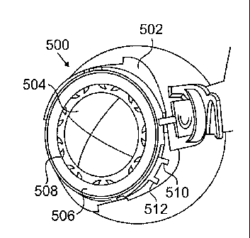
[0069] FIGURES 5A through 5C illustrate a fourth example
ocular fixation device 500 and an example use of the ocular
fixation device 500 in accordance with this disclosure.
The embodiment of the ocular fixation device 500 and its
use shown in FIGURES 5A through 5C are for illustration
only. Other embodiments of the ocular fixation device 500
and uses of the ocular fixation device 500 could be used
without departing from the scope of this disclosure.
[0070] As shown in FIGURE 5A, the ocular fixation device
500 is similar to the ocular fixation device 200 of FIGURES
2A through 2C. The ocular fixation device 500 includes a
base 502, a dome 504, and locking rings 506-508. In this
example, the base 502 is attached 'or secured to the dome
21843509.2
CA 02658168 2013-08-26
23
504 (or vice versa), and the locking rings 506-508 are
secured between the base 502 and the dome 504.
[0071] In this example embodiment, the locking rings
506-508 include tabs 510-512, respectively. The tabs 510-
512 extend outside of the base 502 and the dome 504. For
example, as shown in FIGURE 5B, one or more gaps 514 could
exist between the base 502 and the dome 504, and the tabs
510-512 may extend through one or more of the gaps 514.
The tabs 510-512 can be used to control the opening and
closing of the teeth on the locking rings 506-508. For
instance, the tabs 510-512 can be pulled apart to open the
teeth on the locking rings 506-508, and the tabs 510-512
can be pushed together to close the teeth on the locking
rings 506-508.
[0072] As shown here, the base 502 of the ocular
fixation device 500 includes portions 516 that project from
the main body of the ocular fixation device 500 and that
are arranged to lie generally on a patient's eye. The
portions 516 include straight edges or guides 518, and the
base 502 also includes notches 520. The guides 518 and the
notches 520 are used to align a surgical tool 522 during a
surgical procedure. For example,
the surgical tool 522
could include a projection 524, which can be inserted into
each of the notches 520 of the ocular fixation device 500.
Also, the surgical tool 522 can be positioned so that its
base is aligned with one of the straight guides 518 of the
ocular fixation device 500. The surgical tool 522 can then
be used to form an incision in the patient's eye, such as a
scleral tunnel for receiving a scleral prosthesis. In this
particular example, the ocular fixation device 500 includes
guides 518 and notches 520 in four locations, although any
other suitable number of locations could be supported.
21843509.2
CA 02658168 2013-08-26
24
[0073] In this way, the ocular fixation device 500
serves to secure the position of the patient's eye during a
surgical procedure. At the same time, the ocular fixation
device 500 facilitates the placement of the surgical tool
522 in the appropriate locations on the patient's eye.
[0074] Although FIGURES 5A through 5C illustrate a
fourth example of an ocular fixation device 500 and an
example use of the ocular fixation device 500, various
changes may be made to FIGURES 5A through 50. For example,
the relative sizes and dimensions of the features of the
ocular fixation device 500 are for illustration only and
can be altered in any suitable manner. Also, the guide
mechanisms described above (the straight guides 518 and the
notches 520) could be used with any other ocular fixation
device and any other surgical tool.
[0075] FIGURES 6A through 6C illustrate a fifth example
ocular fixation device 600 and an example use of the ocular
fixation device 600 in accordance with this disclosure.
The embodiment of the ocular fixation device 600 and its
use shown in FIGURES 6A through 60 are for illustration
only. Other embodiments of the ocular fixation device 600
and uses of the ocular fixation device 600 could be used
without departing from the scope of this disclosure.
[0076] As shown in FIGURE 6A, the ocular fixation device
600 is similar to other ocular fixation devices described
above. The ocular fixation device 600 includes a base 602,
a dome 604, and locking rings 606-608. In this example,
the base 602 is attached or secured to the dome 604 (or
vice versa), and the locking rings 606-608 are secured
between the base 602 and the dome 604. In this particular
example, the locking ring 606 includes multiple sets of
teeth (which could have different heights from the surface
21843509.2
CA 02658168 2013-08-26
of a patient's eye), and these teeth correspond to multiple
sets of teeth of the locking ring 608. As with the ocular
fixation device 500, the locking rings 606-608 also include
tabs 610-612, respectively, which extend outside of the
5 base 602 and the dome 604 and can be used to control the
opening and closing of the teeth on the locking rings 606-
608.
[0077] As shown here, the dome 604 of the ocular
fixation device 600 includes holes 614. The holes 614 in
10 this example are used to align a surgical tool 616 to one
or more locations of a patient's eye. The surgical tool
616 includes an alignment portion 618, which has two
extensions 620a-620b forming a partial circle around the
ocular fixation device 600. Each of the extensions 620a-
15 620b includes an end that can be inserted into one of the
holes 614 of the ocular fixation device 600. As shown in
FIGURES 6B and 60, the alignment portion 618 of the
surgical tool 616 also includes a stopper 622, which can be
depressed against the base 602 of the ocular fixation
20 device 600. Collectively, the ends of the extensions 620a-
620b and the stopper 622 represent three points that can be
used to ensure the proper positioning of the surgical tool
616 on the patient's eye.
[0078] In this example, the surgical tool 616 includes
25 two rotatable grasping clasps 624. As shown in FIGURE 6B,
the grasping clasps 624 could be opened before the surgical
tool 616 is pressed onto the patient's eye. As shown in
FIGURE 6C, when the surgical tool 616 is pressed onto the
patient's eye, the grasping clasps 624 rotate (either
inward or outward). This helps to secure the surgical tool
616 in place on the patient's eye.
[0079] In this example embodiment, the extensions 620a-
21843509.2
CA 02658168 2013-08-26
26
620b of the surgical tool 616 form a partial circle around
the ocular fixation device 600. This allows the surgical
tool 616 to be attached or mounted to the ocular fixation
device 600 while leaving a large portion of the dome 604
exposed. Among other things, this may allow the use of a
positioning tool 626, which can be used to place the ocular
fixation device 600 into one or more positions on the
patient's eye. Additional details regarding an example
positioning tool are provided below.
lo [0080] Although FIGURES 6A through 6C illustrate a fifth
example of an ocular fixation device 600 and an example use
of the ocular fixation device 600, various changes may be
made to FIGURES 6A through 6C. For example, the relative
sizes and dimensions of the features of the ocular fixation
device 600 are for illustration only and can be altered in
any suitable manner. Also, the guide mechanisms described
above (the holes 614 in the dome 604 and the alignment
portion 618 of the surgical tool 616) could be used with
any other ocular fixation device and any other surgical
tool.
[0081] FIGURE 7 illustrates an example positioning tool
700 for use with an ocular fixation device in accordance
with this disclosure. The embodiment of the positioning
tool 700 shown in FIGURE 7 is for illustration only. Other
embodiments of the positioning tool 700 could be used
without departing from the scope of this disclosure.
[0082] In this example embodiment, the positioning tool
700 represents a syringe structure having a body 702, a
plunger 704 inserted into the body 702, a spring 706, and a
suction cup 708. The spring 706 biases the plunger 704 in
the open position, meaning the spring 706 pushes the
plunger 704 away from the suction cup 708 at the end of the
21843509.2
CA 02658168 2013-08-26
27
body 702. The end of the plunger 704 can form an air-tight
seal within the body 702, and the suction cup 708 can form
an air-tight seal with an ocular fixation device.
[0083] To place an ocular fixation device on a patient's
eye, the plunger 704 can be depressed, such as by a surgeon
or other personnel. The suction cup 708 can be placed on
the ocular fixation device, such as on the dome of any of
the ocular fixation devices described above. The spring
706 is then allowed to push the plunger 704 away from the
suction cup 708. The air-tight
seals create a vacuum
within the body 702 of the positioning tool 700, causing
the suction cup 708 to attach to the ocular fixation
device. The ocular fixation device can therefore be picked
up, moved, and placed in the appropriate position on a
patient's eye using the positioning tool 700. Once in the
appropriate position (and possibly after the ocular
fixation device has been attached to the patient's eye),
the plunger 704 can be depressed. This
releases the
suction cup 708 from the ocular fixation device.
[0084] This type of positioning tool 700 represents only
one example of the types of tools that could be used to
position an ocular fixation device. Any other suitable
mechanism could be used to position an ocular fixation
device. For example,
an ocular fixation device could
include a handle, such as a flip-ring that can be used to
pick up the ocular fixation device and that can be rotated
to the side and laid on the dome or base of the ocular
fixation device. Any other
suitable handle or other
mechanism could be used to allow the ocular fixation device
to be handled and positioned.
[0085] Although FIGURE 7 illustrates one example of a
positioning tool 700 for use with an ocular fixation
218435092
CA 02658168 2013-08-26
28
device, various changes may be made to FIGURE 7. For
example, any other suitable device or technique could be
used to place an ocular fixation device on a patient's eye.
[0086] FIGURE 8 illustrates a sixth example ocular
fixation device 800 in accordance with this disclosure.
The embodiment of the ocular fixation device 800 shown in
FIGURE 8 is for illustration only. Other embodiments of
the ocular fixation device 800 could be used without
departing from the scope of this disclosure.
[0087] In this example, the ocular fixation device 800
includes a base 802, a fixation mechanism 804, and a handle
806. The base 802 may generally be pressed against a
patient's eye, such as by pressing the base 802 down on the
patient's cornea. The fixation mechanism 804 can then be
attached to the patient's sclera, fixing the tool 800 in
place and providing clear access to the patient's sclera.
The fixation mechanism 804 could use any suitable technique
to latch onto the patient's eye, such as prongs that can be
forced into the patient's scleral tissue. The handle 806
can be used to raise and lower the fixation mechanism 804
after the base 802 has been pressed onto the patient's eye.
[0088] Although FIGURE 8 illustrates a sixth example
ocular fixation device 800, various changes may be made to
FIGURE 8. For example, any suitable mechanisms could be
used to attach or otherwise associate the ocular fixation
device 800 to the patient's eye.
[0089] FIGURES 9A through 9C illustrate a seventh
example ocular fixation device 900 and an example use of
the ocular fixation device 900 in accordance with this
disclosure. The embodiment of the ocular fixation device
900 and its use shown in FIGURES 9A through 9C are for
illustration only. Other
embodiments of the ocular
21843509.2
CA 02658168 2013-08-26
29
fixation device 900 and uses of the ocular fixation device
900 could be used without departing from the scope of this
disclosure.
[0090] As shown in FIGURES 9A through 9C, the ocular
fixation device 900 includes a central portion 902, a
support 904 having a tool connection 906, and prongs 908.
In some embodiments, the central portion 902 of the ocular
fixation device 900 generally fits over the patient's
cornea or some other portion of the patient's eye. The
central portion 902 of the ocular fixation device 900 may
also be centered on the patient's eye. The support 904 may
be removably attached to the central portion 902, and the
tool connection 906 allows a surgical tool 910 to be
attached to the support 904.
[0091] The prongs 908 hold the central portion 902 of
the ocular fixation device 900 in place on the patient's
eye. For example, the prongs 908 could be extended out as
shown in FIGURE 9A prior to placement on the patient's eye.
The prongs 908 could then be pushed or rotated so that the
ends of the prongs 908 attach or secure to the patient's
eye.
[0092] In particular embodiments, the support 904 can be
attached in one orientation to the central portion 902, the
surgical tool 910 can be attached to the support 904, and a
scleral tunnel can be formed. This process could then be
repeated, with the support 904 being removed and attached
in a different orientation to the central portion 902 so
that the surgical tool 910 can form a scleral tunnel at
another location on the patient's eye.
[0093] Although FIGURES 9A through 9C illustrate a
seventh example ocular fixation device 900 and an example
use of the ocular fixation device 900, various changes may
21843509.2
CA 02658168 2013-08-26
be made to FIGURES 9A through 9C. For example, the ocular
fixation device 900 could include other mechanisms for
attachment to the patient's eye or to a surgical tool 910.
[0094] FIGURES 10A through 10D illustrate an eighth
5 example ocular fixation device 1000 in accordance with this
disclosure. The embodiment of the ocular fixation device
1000 shown in FIGURES 10A through 10D is for illustration
only. Other embodiments of the ocular fixation device 1000
could be used without departing from the scope of this
10 disclosure.
[0095] In this example, the ocular fixation device 1000
Includes an outer ring 1002, an inner ring 1004, and ring
connections 1006. The outer and inner rings 1002-1004
represent generally circular-shaped structures. As shown
15 in FIGURE 10A, the outer ring 1002 is generally in a
different plane than the smaller inner ring 1004. The ring
connections 1006 generally couple the outer and inner rings
1002-1004 together, forming an integrated structure. The
ring connections 1006 are shaped such that a portion of a
20 patient's eye can fit through the outer ring 1002 and
approach or contact the inner ring 1004.
[0096] As shown here, the ocular fixation device 1000
also includes multiple prongs 1008, which are rotatably
coupled to the outer ring 1002. As shown in FIGURE 10B,
25 the prongs 1008 can be opened prior to placement of the
ocular fixation device 1000 on the patient's eye. As shown
in FIGURES 10C and 10D, once placed on the patient's eye,
each of the prongs 1008 can be rotated such that the ends
of the prongs 1008 attach or secure to the patient's eye.
30 To release the ocular fixation device 1000, the prongs 1008
can be rotated again to remove the ends of the prongs 1008
from the patient's eye.
21843509.2
CA 02658168 2013-08-26
31
[0097] Although FIGURES 10A through 10D illustrate an
eighth example ocular fixation device 1000, various changes
may be made to FIGURES 10A through 10D. For example, the
rings 1002-1004 could have any suitable dimensions, and the
inner ring 1004 could have any suitable distance from the
outer ring 1002. Also, any suitable mechanisms could be
used to couple the rings 1002-1004 together and to attach
or otherwise associate the ocular fixation device 1000 to
the patient's eye.
[0098] FIGURES 11A and 11B illustrate a ninth example
ocular fixation device 1100 in accordance with this
disclosure. The embodiment of the ocular fixation device
1100 shown in FIGURES 11A and 11B is for illustration only.
Other embodiments of the ocular fixation device 1100 could
be used without departing from the scope of this
disclosure.
[0099] In this example, the ocular fixation device 1100
is formed from two rotatable segments 1102. Each segment
1102 includes prongs 1104 that can fix the segment 1102 to
a patient's eye, such as in the sclera of the eye. Each
segment 1102 also includes connection points 1104, which
represent areas where other components (such as a surgical
tool) can be attached to the ocular fixation device 1100.
In addition, the ocular fixation device 1100 can provide
reference markers identifying where scleral tunnels should
be formed in the patient's eye, such as at locations at or
between the prongs 1104. In some embodiments, one of the
segments 1102 can be attached to the patient's eye, and
then the other segment 1102 can be rotated out and attached
to the patient's eye.
[00100] Although FIGURES 11A and 11B illustrate a
ninth example ocular fixation device 1100, various changes
21843509.2
CA 02658168 2013-08-26
32
may be made to FIGURES 11A and 11B. For example, each
rotatable segment 1102 could include any suitable number of
prongs 1104.
[00101] FIGURES 12A
and 12B illustrate a tenth
example ocular fixation device 1200 in accordance with this
disclosure. The embodiment of the ocular fixation device
1200 shown in FIGURES 12A and 12B is for illustration only.
Other embodiments of the ocular fixation device 1200 could
be used without departing from the scope of this
disclosure.
[00102] In this
example, the ocular fixation device
1200 generally includes a ring 1202 with crossbars 1204.
The ring 1202 is generally sized and shaped to lie on a
patient's eye, such as by having a slanted inner edge that
generally lies on the sclera of the patient's eye. The
crossbars 1204 are generally sized and shaped to allow a
portion of the patient's eye to fit through the ring 1202
and approach or contact the crossbars 1204.
[00103] In this
example embodiment, twist picks 1206
are provided along the ring 1202. The twist picks 1206
represent screw-type structures that can attach to and
release the ocular tissue of the patient's eye. For
example, rotating the twist picks 1206 in one direction may
attach the twist picks 1206 to the ocular tissue of the
patient's eye. Rotating the
twist picks 1206 in the
opposite direction may release the ocular tissue. In this
way, the ring 1202 can be attached to the patient's eye
through simple rotation of the twist picks 1206.
[00104] As shown
here, the ocular fixation device
1200 also includes connection points 1208. The connection
points 1208 generally represent areas where, for example, a
surgical tool for forming scleral incisions can be mounted
21843509.2
CA 02658168 2013-08-26
33
on the ocular fixation device 1200. In this example, each
of the connection points 1208 includes an elevated area of
the ring 1202 adjacent to a notch in the ring 1202.
However, any other suitable mechanism could be used to
mount or otherwise couple any suitable surgical tool to the
ocular fixation device 1200.
[00105] Although
FIGURES 12A and 123 illustrate a
tenth example ocular fixation device 1200, various changes
may be made to FIGURES 12A and 123. For example, the ring
1202 and the crossbars 1204 could have any suitable shape
or dimensions, and the crossbars 1204 could join at any
suitable height above the ring 1202. Also, any suitable
mechanisms could be used to attach or otherwise associate
the ocular fixation device 1200 to the patient's eye.
[00106] FIGURES 13A through
13D illustrate an
eleventh example ocular fixation device 1300 in accordance
with this disclosure. The
embodiment of the ocular
fixation device 1300 shown in FIGURES 13A through 13D is
for illustration only. Other embodiments of the ocular
fixation device 1300 could be used without departing from
the scope of this disclosure.
[00107] In this
example, the ocular fixation device
1300 includes a ring 1302 having a lever 1304. The lever
1304 is used to control the movement of retractable pins
1306, which can be retracted into and extended out of the
ring 1302. For example, the lever 1304 could be placed in
the raised position as shown in FIGURE 13A to retract the
pins 1306 into the ring 1302. The ring 1302 could then be
placed on the patient's eye and positioned properly. After
that, the lever 1304 can be lowered as shown in FIGURE 13B,
causing the pins 1306 to extend from the ring 1302 and lock
onto the patient's ocular tissue. For instance, the pins
21M3g)92
CA 02658168 2013-08-26
34
1306 could penetrate the limbus of the patient's eye to a
depth of 200 microns. Any suitable mechanism could be used
to cause the pins 1306 to retract and extend under the
control of the lever 1304.
[00108] In this example
embodiment, the ocular
fixation device 1300 could also include vertical teeth
1308, which may or may not penetrate the surface of the
patient's eye. If the vertical teeth 1308 do not penetrate
the surface of the patient's eye, the vertical teeth 1308
could still grip the patient's eye and provide lateral
fixation, meaning the vertical teeth 1308 may help to
prevent sideways motion of the ocular fixation device 1300
on the patient's eye. In addition,
as with various
prostheses described above, the ocular fixation device 1300
can include one or more connection points 1310 and one or
more windows 1312.
[00109] Although
FIGURES 13A through 13D illustrate
an eleventh example ocular fixation device 1300, various
changes may be made to FIGURES 13A through 13D. For
example, the ring 1302, lever 1304, pins 1306, and other
elements could have any suitable shape or dimensions.
Also, any suitable mechanisms could be used to attach or
otherwise associate the ocular fixation device 1300 to the
patient's eye.
[00110] FIGURES 14A through 14C
illustrate a twelfth
example ocular fixation device 1400 in accordance with this
disclosure. The embodiment of the ocular fixation device
1400 shown in FIGURES 14A through 14C is for illustration
only. Other embodiments of the ocular fixation device 1400
could be used without departing from the scope of this
disclosure.
[00111] In this
example, the ocular fixation device
21843509.2
CA 02658168 2013-08-26
1400 includes a ring 1402 having a latch 1404 at one end
and a receptacle 1406 at its other end. The ring 1402 also
includes a hinge 1408, allowing two portions of the ring
1402 to open and close with respect to each other. In this
5 embodiment, the portions of the ring 1402 can be pushed
apart to open the ring 1402. The ring 1402 can be placed
on a patient's eye, and the latch 1404 can be pushed into
the receptacle 1406, forming a completed ring. As shown in
FIGURE 14B, the lower edge of the ring 1402 includes spikes
10 1410 that can dig into the ocular tissue of the patient's
eye, securing the ring 1402 in place on the patient's eye.
[00112] As shown
here, the ocular fixation device
1400 also includes connection points 1412, which generally
represent areas where, for example, a surgical tool for
15 forming scleral incisions can be mounted on the ocular
fixation device 1400. Any suitable
type of connection
points or other mechanisms could be used to mount or
otherwise couple any suitable surgical tool to the ocular
fixation device 1400.
20 [00113] The ocular
fixation device 1400 further
includes one or more windows 1414. The windows 1414 allow
a surgeon or other personnel to see through the ocular
fixation device 1400 so as to determine the position of the
ocular fixation device 1400 with respect to certain
25 features of the patient's eye. For example, the windows
1414 could allow a surgeon to ensure that the ocular
fixation device 1400 is attached to the area at or near the
limbus of the patient's eye. The windows 1414 could have
any suitable size, shape, and distribution in the ocular
30 fixation device 1400.
[00114] Although
FIGURES 14A through 14C illustrate
a twelfth example ocular fixation device 1400, various
21843509.2
CA 02658168 2013-08-26
36
changes may be made to FIGURES 14A through 14C. For
example, the ring 1402, latch 1404, and receptacle 1406
could have any suitable shape or dimensions. Also, any
suitable mechanisms could be used to attach or otherwise
associate the ocular fixation device 1400 to the patient's
eye.
[00115] For all of
the ocular fixation devices
described above, the various components or elements of the
ocular fixation devices could have any suitable shapes,
sizes, or dimensions. For example,
various ones of the
ocular fixation devices could have curved bottom surfaces,
allowing the ocular fixation devices to generally lie on
the surface of a patient's eye. Also, various elements or
features of one of the ocular fixation devices could be
used with others of the ocular fixation devices. Further,
while often described as being attached to or otherwise
associated with the patient's eye at the sclera, the ocular
fixation devices could be attached to or otherwise
associated with the patient's eye at other locations. In
addition, the ocular fixation devices are often described
as being used to support a surgical procedure involving the
implantation of scleral prostheses into scleral tunnels in
a patient's eye. However, any
other suitable surgical
procedure could be performed using the ocular fixation
devices.
[00116] In particular
embodiments, any of the ocular
fixation devices described above could be sized such that
the teeth, prongs, or other fixating means for associating
the ocular fixation device with an eye are secured to,
contact, are coupled to, or release tissue at or near the
limbus of the eye. This region of the eye may be well-
suited to this type of procedure as it heals rapidly.
21843509.2
CA 02658168 2013-08-26
37
However, each of the ocular fixation devices could have any
other suitable size or shape.
[00117] The use of various mechanisms have been
described above for securing or fixating ocular tissue,
such as rings or other devices having teeth, prongs, or
pins. However, ocular fixation devices could use any
suitable mechanism for securing or fixating ocular tissue.
In this document, the phrases "means for fixating" and
"fixating means" refer to any structure or portion thereof
that extends from, projects from, forms a part of, or is
otherwise associated with an ocular fixation device and
that is pressed against, contacts, or penetrates the
surface of a patient's eye. These "fixating means" include
one or more teeth, prongs, pins, outcroppings, or other
extensions or projections coupled to, attached to,
extending from, integrated with, or otherwise associated
with a ring or other structure placed proximate to the eye.
The "fixating means" also include other mechanical
structures such as one or more twist picks or sutures. In
some embodiments, "fixating means" such as teeth may be
planar or angled with respect to the structure with which
the means are associated. Moreover, in this document, an
ocular fixation device is said to be "associated with" an
eye when the ocular fixation device is secured or attached
to the eye.
[00118] FIGURE 15 illustrates an example method 1500
for ocular fixation in accordance with this disclosure.
The embodiment of the method 1500 shown in FIGURE 15 is for
illustration only. Other embodiments of the method 1500
could be used without departing from the scope of this
disclosure.
[00119] An ocular fixation device is placed on a
21843509.2
CA 02658168 2013-08-26
38
patient's eye at step 1502. This could
include, for
example, placing any of the ocular fixation devices
described above on the patient's eye. The ocular fixation
device could include a dome so that the central portion of
the patient's eye is covered and protected by the ocular
fixation device.
[00120] One or more
locking rings of the ocular
fixation device are rotated to secure the ocular fixation
device to the patient's eye at step 1504. This could
include, for example, using an external tool to move one or
more of the locking rings of the ocular fixation device.
This could also include rotating one or more portions of
the ocular fixation device to cause one or more of the
locking rings to rotate. This could
further include
rotating one or more tabs coupled to one or more of the
locking rings to cause one or more of the locking rings to
rotate. Any other suitable technique could be used here to
rotate one or more locking rings of the ocular fixation
device.
[00121] An ocular surgical
procedure occurs at step
1506. This could include, for example, forming one or more
scleral tunnels in sclera of the patient's eye.
Optionally, one or more scleral prostheses or other
implants are placed in the patient's eye at step 1507.
This could include, for example, inserting the scleral
prostheses as the tunnels are being formed (as shown in
FIGURES 4F through 41 above). This could
also include
inserting the scleral prostheses into the tunnels manually
or otherwise after a surgical tool has been mounted on the
ocular fixation device and used to form the scleral
tunnels.
[00122] One or more
locking rings of the ocular
21843509.2
CA 02658168 2013-08-26
39
fixation device are rotated to release the ocular fixation
device from the patient's eye at step 1508. This could
include, for example, using an external tool, one or more
portions of the ocular fixation device, or tabs coupled to
the locking rings to rotate one or more of the locking
rings. The ocular
fixation device is removed from the
patient's eye at step 1510. Optionally,
one or more
scleral prostheses or other implants are placed in the
patient's eye at step 1511. This could
include, for
example, inserting the scleral prostheses into the tunnels
manually or otherwise after the ocular fixation device has
been removed from the patient's eye. One or both of the
optional steps 1507 and 1511 show that the ocular fixation
device can be used in a variety of ways during a surgical
procedure.
[00123] Although
FIGURE 15 illustrates one example
of a method 1500 for ocular fixation, various changes may
be made to FIGURE 15. For example, any suitable surgical
procedure could involve the use of ocular fixation. Also,
the surgical procedure may, but need not, involve the
implantation of one or more scleral prostheses or other
implants or elements into the patient's eye. Further,
while described as rotating one or more locking rings to
secure and release the patient's eye, other techniques
(such as those associated with other embodiments of the
ocular fixation devices described above) could be used.
[00124] It may be
advantageous to set forth
definitions of certain words and phrases used throughout
this patent document. The terms "include" and "comprise,"
as well as derivatives thereof, mean inclusion without
limitation. The term "or" is inclusive, meaning and/or.
The term "ring" refers to a structure that is generally
21843509.2
CA 02658168 2013-08-26
circular or ovoidal in shape.
[00125] While this
disclosure has described certain
embodiments and generally associated methods, alterations
and permutations of these embodiments and methods will be
5 apparent to those skilled in the art. For example, while
shown as providing for the manual rotation or movement of
one or more rings or other structures in an ocular fixation
device, any suitable technology, such as a mechanical or
electrical mechanism, could be used to rotate or move one
10 or more rings or other structures in an ocular fixation
device. Accordingly,
the above description of example
embodiments does not define or constrain this disclosure.
Other changes, substitutions, and alterations are also
possible without departing from the spirit and scope of
15 this disclosure, as defined by the following claims.
21843509.2