Note : Les descriptions sont présentées dans la langue officielle dans laquelle elles ont été soumises.
CA 02658503 2009-01-20
WO 2008/013684 PCT/US2007/016005
MEMBRANE-BASED DOUBLE-LAYER TUBE FOR SAMPLE COLLECTIONS
BACKGROUND OF THE INVENTION
Field of the Invention
The present invention relates to a fluid sample collection device and, more
particularly, to a fluid sample collection device adapted to separate plasma
or serum from a
blood sample. More specifically, the present invention relates to an evacuated
fluid sample
collection device capable of separating plasma or serum from the cellular
material in a blood
sample through a porous filter also referred to as a membrane herein.
Description of Related Art
Plasma is the liquid portion of blood and is primarily comprised of water,
proteins,
glucose, amino acids, vitamins, inorganic salts, metabolites, and metabolic
waste products.
The generally solid portion of blood is comprised of a variety of cells
including red cells,
white cells, and platelets. Plasma is freely transferable with cells of the
body. As a whole,
plasma provides the medium to suspend white blood cells, red blood cells, and
other cellular
components for transport throughout a human or animal. If a plasma sample is
desired, its
separation from blood cells Must occur well before blood coagulation. An anti-
coagulation
reagent may be added to a blood collection device to prevent coagulation. If
blood is allowed
to coagulate, the remaining liquid portion of the collected blood sample is
called serum,'
which is devoid of some protein components of plasma. Separation of
plasma/serum from
blood cells is typically achieved by centrifugation.
Because plasma contains a rich source of components available for diagnostic
analysis, medical devices have been devised for separating plasma from a whole
blood
sample. Several known blood collection devices are provided as evacuated multi-
chamber
devices that incorporate a filter or membrane that is used to remove or
separate plasma from a
collected blood sample. In some devices, the devices include a detachable
chamber allowing
a user to access the separated plasma specimen. Typically, in these known
blood sample
collection and separating devices the separating filter or membrane has a
sufficiently small
pore size to prevent cellular components from passing through the filter or
membrane while
allowing the passage of liquid. However, such filters or membranes often
become clogged
during a blood collection and plasma separation procedure thereby rendering
typical vacuum
forces generated by the evacuated device inadequate to draw plasma from a
collected blood
1
CA 02658503 2015-10-09
WO 2008/013684 PCT/US2007/016005
sample. Several examples of known blood collection and separation devices are
discussed
hereinafter.
U.S. Patent No. 6,506,167 (Ishimito et al.) discloses a blood separating tube
that
includes an upstream tube separated by a filter from a downstream tube. The
tubes are
attachable to and detachable from each other and are initially provided in an
evacuated state.
During blood collection, blood is removed from a patient through intravenous
puncture and
transferred into the upstream tube through blood pressure and negative
pressure inside the
tube. In operation, a pressure differential is created between the upstream
tube and the
downstream tube as the blood contacts the filter between the two tubes.
Several filter types
are disclosed in this reference, including a membrane, glass fiber, filter
paper with large pores
and impregnated with anti-hemocyte antibodies, a filter impregnated with a
cationic
macromolecular substance to aggregate cells, and a laminated multi-layer
filter. One problem
associated with the device described in this patent is that blood cells often
clog the filter
during plasma separation resulting in inadequate vacuum force being present
between the
upstream tube and downstream tube during blood collection. A further problem
with the
device described in this patent is that the collected plasma in the downstream
tube may be
exposed to contaminants should the downstream tube be removed from the
upstream tube.
U.S. Patent No. 6,471,069 (Lin et al.) discloses a device adapted to separate
plasma/serum from blood cells and includes a flexible collapsible inner
container disposed
within a substantially rigid outer container. A closure seals the open top end
of the outer
container. A filter assembly is mounted to the open top end of the inner
container_ The filter
assembly includes a filter that permits lighter fractions of a collected fluid
sample to pass
therethrough, while blocking the heavier fractions. The filter assembly
further includes a
filter support including a slit valve that opens in response to fluid pressure
created by the
lighter fractions for permitting the lighter fractions to flow therethrough.
In use, a fluid
sample is delivered to the inner container and the device is subjected to
centrifugation which
causes the filter assembly to move toward the bottom end of the outer
container and allow the
lighter fraction of the fluid sample to flow through the slit valve and into
the space between
the inner and outer containers.
U.S. Patent No. 6,659,288 (Amano et al.) discloses a plasma/serum collection
device
which includes a filtering unit. The device is constructed with a space above
the filtering unit
to preserve the blood cells, and defines a space below -the filter into which
plasma/serum is
drawn under negative pressure. U.S. Patent 4,639,316 (Eldegheidy) discloses an
automatic
2
CA 02658503 2009-01-20
WO 2008/013684 PCT/US2007/016005
liquid component separator which utilizes a cross-flow filtration area
together with vacuum
force to cause separation of a cell-free fraction from a cell fraction in a
fluid sample. Other
prior art in which plasma separation through a filter within a container is
achieved through a
pressure differential is disclosed in U.S. Patent Nos.: 3,682,596; 3,687,296;
3,701,434;
3,814,079; 4,131,549; and 4,639,316.
The foregoing blood collection and separation devices each utilize a pressure
differential as the motive force to cause plasma separation from a whole blood
sample.
However, certain disadvantages are present in these devices, namely there is
often
insufficient differential pressure for complete plasma/serum separation, the
separation filters
easily become clogged with cellular material, and the separated plasma/serum
is easily
contaminated during removal from the device. Accordingly, there is a general
need for a
device and method that allow for rapid separation of plasma/serum from a blood
sample
ideally at the same location or a close proximity to the site of sample
collection.
SUMMARY OF THE INVENTION
The present invention overcomes many of the deficiencies present in the prior
art and
allows a medical practitioner to both collect a bodily fluid sample, typically
blood, and effect,
for example, plasma/serum separation from the sample at or near the site of
blood sample
collection. In one embodiment, a device is provided for collecting and
separating a fluid
sample and generally comprises an evacuated outer container and an inner
container. The
outer container has a first open end and a second closed end. A pierceable
closure closes the
first open end thereby defining a first interior chamber. The inner container
is contained
within the outer container and separates the first interior chamber into an
upper chamber
portion in fluid communication with a lower chamber. The inner container
defines a second
interior chamber separated from the lower chamber portion of the first
interior chamber
through a porous membrane. A port is provided for placing the second interior
chamber in
fluid communication with the first interior chamber.
The fluid sample to be collected may comprise blood. Plasma or serum of the
blood
drawn within the first interior chamber passes through the porous membrane and
into the
second interior chamber of the inner container based on a pressure
differential between the
first interior chamber and the second interior chamber established by the
blood contacting the
porous membrane proximate the lower chamber portion. The porous membrane
desirably
prevents transfer of blood cells therethrough.
3
CA 02658503 2009-01-20
WO 2008/013684 PCT/US2007/016005
In one embodiment, the porous membrane may comprise filter paper, for example,
one or more pieces of filter paper. The pore size of the porous membrane may
be of a size to
control the passing of desired molecules through the membrane. For example,
the porous
membrane may prevent molecules of 60,000 Daltons or higher from passing into
the inner
container. Additionally, the porous membrane may be capable or removing
albumin,
immunoglobulin, or other large molecules from the plasma or serum fraction of
a blood
sample. In another example, the porous membrane may prevent molecules of
10,000 Daltons
or higher from passing into the inner container. Further, the porous membrane
may have a
pore size to enable peptide extraction from a blood sample. Moreover, the
porous membrane
may prevent molecules 2,000 Daltons or higher from passing into the inner
container.
Furthermore, the porous membrane may have a pore size to enable separation of
metabolites
and other small molecules from a blood sample.
The pore size of the porous membrane may also be between 0.1 p.m to 2 um. For
example, the porous membrane may be a 0.22 um membrane which may be used to
remove
virus particles for bio-safety plasma or serum sample collection as examples.
In another
example, the porous membrane may be a 0.45-1.0 p.m membrane which may be used
for
platelet-free plasma or serum sample collection as examples. Further, the
porous membrane
may enable platelet-free plasma or serum sample collection.
In one variation, the inner container may be suspended within the outer
container. In
another variation, the inner container may be releasably connected with the
pierceable
closure. As a result, release of the inner container from the pierceable
closure may open the
port of the inner container and places the second interior chamber of the
inner container in
fluid communication with the first interior chamber. In a still further
variation, the inner
container may be movably supported within the first interior chamber of the
outer container.
As a result, movement of the inner container within the first interior chamber
may open the
port of the inner container and place the second interior chamber of the inner
container in
fluid communication with the first interior chamber. The outer container may
support the
inner container within the first interior chamber. Such support may occur
after movement of
the inner container within the first interior chamber.
The outer container may comprise an additive such agglutinating agents or
anticoagulants. The porous membrane may be made of high density polyethylene,
high
density polypropylene, ceramic, porous metal, porous glass, glass fibers,
polyvinyl polymers,
paper, natural fibers, and combinations of the foregoing.
4
CA 02658503 2009-01-20
WO 2008/013684 PCT/US2007/016005
In another embodiment, the device is provided for separating plasma or serum
from a
blood sample and generally comprises an evacuated collection assembly
comprising an outer
container and an inner container. The outer container comprises a pierceable
closure at one
end. The inner container is contained within the outer container and the
interior of the inner
container is separated from the outer container by a porous membrane on the
bottom of the
inner container. The inner container comprises a port in fluid communication
with the inner
container. The port is desirably releasably connected to the pierceable
closure_
The inner container may be entirely accommodated within the outer container.
The
port may be in fluid communication with the outer container, for example, when
disconnected from the pierceable closure. The release of the port from the
pierceable closure
may enable opening of the port to unrestricted fluid communication with the
outer container.
The inner surface of the outer container may maintain a position of the inner
container
relative to the outer container. In operation, a pressure differential
established between the
outer container and the inner container upon entry of the blood sample into
the outer
1 5 container may be used to facilitate transportation of plasma or serum
through the porous
membrane into the inner container, while preventing the transfer of blood
cells therethrough
In another aspect a method for separating plasma or serum from a blood sample
is
provided. The method may comprise a step of providing an evacuated collection
assembly
comprising an outer container having a pierceable closure at one end, an inner
container
contained within the outer container and defining an interior chamber therein,
and a porous
membrane separating the interior chamber of the inner container from the outer
container.
The method may further comprise a step of collecting a blood sample within the
outer
container of the assembly, thereby creating a pressure differential between
the outer container
and the inner container. The pressure differential generally causes plasma or
serum from the
blood sample to flow into the interior chamber of the inner container through
the porous
membrane. The plasma or serum flows into the inner container in a direction
generally
opposite to the direction of blood particle flow into the outer container
during the blood
sample collecting. Once collected, the plasma or serum may be removed from the
inner
container after the sample collection is complete. The inner container may
comprise a port,
and the method may further comprise a step of placing the port in fluid
communication with
the interior chamber of the outer container.
Further details and advantages of the invention will become clear upon reading
the
following detailed description in conjunction with the accompanying drawing
figures,
wherein like parts are designated with like reference numerals throughout.
5
CA 02658503 2009-01-20
WO 2008/013684 PCT/US2007/016005
BRIEF DESCRIPTION OF THE DRAWINGS
FIG. 1 is an exploded perspective view of a fluid sample collection device
pursuant to
one embodiment.
FIG. 2 is an exploded cross-sectional view of the device shown in FIG. 1.
FIG. 3 is a perspective view of a closure and inner container of the device
shown in
FIG. 1.
FIG. 4 is an assembled cross-sectional view of the device shown in FIG. 1.
FIG. 5 is an assembled cross-sectional view of the device shown in FIG. 1,
showing
the device in use during a fluid sample collection procedure.
FIG. 6 is an assembled cross-sectional view of the device shown in FIG. 1,
showing
initial fluid sample separation occurring within the device.
FIG. 7 is an assembled cross-sectional view of the device shown in FIG. 1,
showing
detachment of the inner container from the closure and resulting completion of
fluid sample
separation within the device.
FIG. 8 is an exploded perspective view of the fluid sample collection device
pursuant
to another embodiment.
FIG. 9 is a perspective view of the closure and inner container of the device
shown in
FIG. 8.
FIG. 10 is an assembled cross-sectional view of device shown in FIG. 8,
showing the
device accessed to accept a fluid sample for separation.
FIG. 11 is a top end view of the device shown in FIG. 8.
FIG. 12 is an assembled view of the fluid sample collection device pursuant to
further
embodiment, showing the device accessed to accept a fluid sample for
separation.
FIG. 13 is an assembled cross-sectional view of the device shown in FIG. 1
with an
alternative closure for the device.
FIG. 14 is an assembled cross-sectional view of the device shown in FIG. 1
with
another alternative closure for the device
DESCRIPTION OF PREFERRED EMBODIMENTS
For purposes of the description hereinafter, spatial orientation terms, if
used, shall
relate to the referenced embodiment as it is oriented in the accompanying
drawing figures or
otherwise described in the following detailed description. However, it is to
be understood
that the embodiments described hereinafter may assume many alternative
variations and
embodiments. It is also to be understood that the specific devices illustrated
in the
6
CA 02658503 2009-01-20
WO 2008/013684 PCT/US2007/016005
accompanying drawing figures and described herein are simply exemplary and
should not be
considered as limiting.
In one embodiment, a fluid sample collection device suitable for the
collection of a
blood sample and the separation of plasma, serum, or other fluid specimens
from the cellular
material (i.e., blood cells) of the blood sample is disclosed. However, the
device described
herein is generally applicable for separating solution (i.e., liquids) from
solids like a filtration
device. In particular, in one form, the device is adapted for collection of a
blood sample
through conventional sampling techniques and subsequent separation thereof by
use of an
assembly of components generally including an inner container, an outer
container, and a
closure member. The inner container generally draws plasma, serum, or other
liquid
specimens through a porous membrane, filter, or like separating member from
the outer
evacuated container to separate the plasma, serum, and/or other liquid
specimen from the
sample.
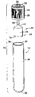
Referring initially to FIGS. 1-4, a device 10 for collecting and separating a
fluid
sample is generally shown. Device 10 is an assembly of components, namely a
first or outer
container or tube 12, a second or inner container or tube 14, and a closure 16
for sealing outer
and inner containers 12, 14. Outer and inner containers 12, 14 together form,
pursuant to one
embodiment, an evacuated collection assembly. Generally, outer container 12
encompasses
inner container 14, typically entirely accommodating the inner container 14
therein. Outer
container 12 may be any container or vessel capable of containing a fluid
sample, typically a
blood sample, therein, and is desirably in the form of a conventional blood
collection tube or
vessel that may be evacuated by conventional means. Outer container 12 may be
constructed
of any known material, such as glass or molded plastic material and, in one
particular
embodiment, is constructed of polyethylene terephthalate (PET). Closure 16 is
provided to
make an air-tight seal with the outer container 12 and enclose inner container
14 within outer
container 12. Closure 16 is also used to support inner container 14 within
outer container 12,
for example, in a suspended manner within the outer container 12.
Outer container 12 is a generally cylindrical-shaped structure comprising a
tubular
sidewall 18 defining a first open or top end 20 and further forming a second
closed or bottom
end 22 of the outer container 12. The closed end 22 may have a rounded or
arcuate form as a
conventional blood collection tube. Outer container 12 is sealed at open end
20 by closure 16
which is a pierceable component formed of rubber or molded plastic material
but may be
made of any pierceable clastomeric material. While closure 16 is generally
akin to rubber or
plastic tube stoppers known in the medical art, closure 16 possess several
novel features in its
7
CA 02658503 2009-01-20
WO 2008/013684 PCT/US2007/016005
own right as discussed herein. Closure 16 is surrounded, at least in part, by
a cap structure or
member 24 which is included for protecting the closure 16 when seated within
the open end
20 of outer container 12. Cap member 24 is formed with an annular end wall 26
and a
depending sidewall or skirt 28 which is configured to extend downward along
sidewall 18 of
outer container 12 when closure 16 is seated within the open end 20 of the
outer container 12.
As discussed herein, closure 16 includes an insertable portion which is seated
within the open
end 20 of outer container 12 and which is held therein by frictional
engagement with the
inner surface or side of sidewall 18 and/or with an adhesive. Sidewall 28 of
cap member 24
extends downward along the outer surface or side of sidewall 18 of outer
container 12 to
protect the exposed portion of closure 16 extending outward from the open end
20 of the
outer container 12. Annular end wall 26 defines a central aperture 30 to
expose a portion of
closure 16 to allow access to the interior of outer container 12, which is
typically accessed by
a piercing element, such as a needle cannula, which is inserted through the
pierceable closure
16 as described in greater detail herein.
Closure 16 is typically a unitary structure or body formed of rubber, plastic
or another
similar polymeric material, as described previously, and is generally an
elastomeric closure
element that is formed of suitable material capable of forming a substantially
gas and liquid-
tight seal with the open end 20 of outer container 12. Additionally, the body
of closure 16 is
desirably capable of being punctured with a puncturing device, such as a
needle cannula, as
described previously. Such a needle cannula may be part of a blood collection
device used to
transfer blood into outer container 12. Closure 16 is formed with a flanged
head or cap
portion 32 and a depending and integrally molded plug portion 34. Cap portion
32 is adapted
to seat or rest on a rim 36 defined by sidewall 18 of outer container 12 at
the open end 20 of
the outer container 12. Plug portion 34 is generally adapted to be inserted
into the open end
20 of outer container 12 and extend inward into the outer container 12 and
form a
substantially gas and liquid-tight seal with the inner surface or side of
sidewall 18. Thus,
with plug portion 34 of closure 16 seated within the open end 20 of outer
container 12, a first
interior chamber 38 is defined or formed within the outer container 12. First
interior chamber
38 may be placed under negative (i.e., vacuum) pressure with respect to
external atmospheric
pressure prior to sealing closure 16 in the open end 20 of outer container 12,
such that the
interior of outer container 12 is under negative (i.e., vacuum) pressure. For
example, after
assembly of device 10 wherein inner container 14 is inserted in outer
container 12, the outer
container 12 may be evacuated and subsequently sealed with closure 16 thereby
placing first
8
CA 02658503 2009-01-20
WO 2008/013684 PCT/US2007/016005
interior chamber 38 under negative (i.e., vacuum) pressure and simultaneously
placing the
interior of inner container 14 under negative (i.e., vacuum) pressure.
Another aspect of device 10 relates to inner container 14 being movable within
outer
container 12 to accomplish full separation of the collected fluid sample. As
shown in FIG. 2,
an internal limiting structure 40 is provided within first interior chamber 38
which is used to
limit internal movement of inner container 14 within outer container 12, as
discussed further
herein. In the embodiment illustrated, limiting structure 40 is in the form of
a circumferential
flange or tab that extends inward from sidewall 18 of outer container 12 and,
thus, is typically
formed integrally with the body of outer container 12. However, the specific
movement-
limiting structure illustrated in FIG. 2 as limiting structure 40 should not
be considered to
limit the possible range of variations for limiting structure 40. Such
variations may take
many forms, such as a circumferential restriction (i.e., narrowing) formed in
sidewall 18 of
outer container 12, a sleeve structure disposed within outer container 12 and
extending
upward from the closed end 22 thereof, a platform extending upward from the
closed end 22
of outer container 12, one or more posts or tabs extending radially inward
from sidewall 18 of
outer container 12, and like structures.
Cap portion 32 of closure 16 defines a top surface 42 which is typically
partially
enclosed by the annular end wall 26 of cap member 24. Top surface 42 is
exposed in the
open area defined by central aperture 30 in cap member 24, and this exposed
area of top
surface 42 is where a user of device 10 inserts a needle cannula or like
piercing element to
access the interior of outer container 12 and first interior chamber 38 in
particular.
Accordingly, to provide a blood sample to the first interior chamber 38, a
needle cannula or
like piercing element of a blood collection device is used to penetrate the
exposed portion of
the top surface 42 of cap portion 32 of closure 16 which places the first
interior chamber 38
in fluid communication with a needle inserted into a patient's vein for blood
collection
purposes. Since first interior chamber 38 is sealed and under negative (i.e.,
vacuum)
pressure, blood flows from the vein, through the blood collection device, and
into the first
interior chamber 38 via the needle cannula inserted through closure 16. If
desired, the top
surface 42 of cap portion 32 of closure may be recessed or otherwise shaped to
provide a
visual indication or cue of where to insert a needle cannula to appropriately
penetrate the
closure 16 and access the interior of outer container 12 without striking
inner container 14.
This recessed or shaped area is designated by reference numeral 44 in FIGS. 1-
7 and is
desirably part of the area of top surface 42 left exposed by central aperture
30 defined in the
annular end wall 26 of cap member 24. Further, plug portion 34 defines a bore
or tubular
9
CA 02658503 2009-01-20
WO 2008/013684 PCT/US2007/016005
shaped recess 46 which is provided to support inner container 14 within outer
container 12,
with inner container 14 depending or being suspended from plug portion 34 and
extending
into the first interior chamber 38 defined by outer container 12 and closure
16.
Second or inner container 14 is a generally tubular or cylindrical structure
in
analogous manner to outer container 12 but may take other forms. Inner
container 14 is
desirably contained fully within outer container 12 and is initially
associated with and
supported by closure 16 to extend into the outer container 12. In one
embodiment, inner
container 14 is a generally bell-shaped structure or unitary body which
includes a first or
distal end 50 and a second or proximal end 52. Inner container 14 is generally
comprised by
a bell-shaped containment portion 54 defining or forming the distal end 50 and
a tubular
structure or conduit 56 that extends upward from containment portion 54 and
defines or
forms the proximal end 52 of the inner container 14. Tubular conduit 56
forming the
proximal end 52 of inner container 14 is adapted to engage the bore 46 defined
in plug
portion 34 of closure 16 whereby the inner container 14 may be suspended
within outer
container 12. Containment portion 54 is hollow and defines a second interior
chamber 58
which is in fluid communication with the upward-extending tubular conduit 56.
In the embodiment illustrated in FIGS. 1-4, tubular conduit 56 is coaxially
aligned
and extends upward from containment portion 54 to engage bore 46 which is
further
desirably coaxially aligned with central aperture 30 in cap member 24.
However, the
diameter of tubular conduit 56 and, thus, bore 46 is desirably smaller than
central aperture 30
to allow a user to insert a needle cannula through closure 16 in an area
radially outward from
the proximal end 52 of inner container 14 and, hence, radially outward from
tubular conduit
56. As a result, the inserted needle cannula is inserted generally parallel to
tubular conduit
56, and is not inserted directly into tubular conduit 56. The proper insertion
of a needle
cannula through closure 16 is shown in FIG. 5 discussed herein. It will be
appreciated from
the foregoing that inner container 14 and outer container 12 are also
coaxially aligned by the
coaxial engagement of tubular conduit 56 in bore 46 in plug portion 34 of
closure 16. In
other embodiments discussed herein, inner container 14 and closure 16 may be
configured
such that inner container 14 is radially offset from a central axis L of outer
container 12, as
shown in FIGS. 8-12 discussed herein.
As described previously, in one embodiment, inner container 14 depends (i.e.,
is
suspended) from closure 16 and is supported to closure 16 by frictional and/or
adhesive
engagement of tubular conduit 56 in bore 46 defined in plug portion 34 of the
closure 16.
Thus, with the foregoing engagement, the proximal end 52 of inner container 14
is secured to
CA 02658503 2009-01-20
WO 2008/013684 PCT/US2007/016005
closure 16 with the distal end 50 projecting into the first interior chamber
38 when the closure
16 is inserted into and secured in the open end 20 of outer container 12. As
shown in FIG. 4,
for example, the distal end 50 of inner container 14 is spaced a distance "a"
from limiting
structure 40 which extends radially inward from the sidewall 18 of outer
container 12. The
positioning of inner container 14 within outer container 12 further separates
or segregates the
first interior chamber 38 into an upper chamber portion 60 and a lower chamber
portion 62.
Upper chamber portion 60 is generally defined by the area above bell-shaped
containment
portion 54 and the lower chamber portion 62 is generally defined by the area
below the
containment portion 54 (i.e., the area below distal end 50). Containment
portion 54 has an
outer diameter that is less than the inner diameter of outer container 12 to
allow fluid to flow
downward to lower chamber portion 62 from upper chamber portion 60 along the
inner
surface of the sidewall 18 of the outer container 12 once introduced into the
upper chamber
portion 60 via, for example, a needle cannula. Thus, annular spacing "S"
between the outer
diameter of containment portion 54 and the inner diameter of outer container
12 is sufficient
to allow the free flow of liquid, such as blood, from the upper chamber
portion 60 to the
lower chamber portion 62.
Tubular conduit 56 of inner container 14 further acts as a port which, during
use of
device 10, is adapted to selectively place the second interior chamber 58
defined by
containment portion 54 of inner container 14 in fluid communication the first
interior
chamber 38 defined by the confines defined by outer container 12 and closure
16. Such a
port is generally defined by an opening or port 64 at the end of tubular
conduit 56 and, hence,
at the proximal end 52 of inner container 14. To allow "outlet" port or
opening 64 to be in
fluid communication with the interior of outer container 12, tubular conduit
56 is desirably
releasably disposed in bore 46 in plug portion 34 of closure 16 and thereby
releasably
connected to closure 16. Thus, in order for outlet port or opening 64 to be in
fluid
communication with the fist interior chamber 38, tubular conduit 56 must first
be released of
engagement with closure 16. Once released of engagement, inner container 14
moves
downward within outer container 12 under the force of gravity and/or by force
exerted by a
user of device 10 as described herein. However, the length of downward
movement is
limited by limiting structure 40 disposed within outer container 12. In
particular, the
interference engagement between the distal end 50 of inner container 14 and
limiting
structure 40 limits downward movement of the inner container 14 within outer
container 12
to distance a. Distal end 50 of inner container 14 is desirably fully open so
that containment
portion 54 defines an end opening 66 for admittance of fluid into the
containment portion 54.
11
CA 02658503 2009-01-20
WO 2008/013684 PCT/US2007/016005
End opening 66 may be the diameter of containment portion 54 or have a smaller
diameter
than the containment portion 54.
The second interior chamber 58 defined by inner container 14 and, in
particular, by
containment portion 54 is separated from the first interior chamber 38 defined
by outer
container 12 and closure 16 by a porous member or filter element 70.
Typically, porous
membrane 70 is adapted to separate plasma or serum from a whole blood sample,
as will be
discussed in more detail herein. Porous membrane 70 is disposed in or over end
opening 66
in containment portion 54 and fully covers end opening 66 on an opposite side
of a top end or
side 72 of the containment portion 54. Additionally, porous membrane 70 may be
formed as
a disk-shaped structure with a filtering center area which is secured to the
distal end 50 of
inner container 14 and fully covers end opening 66 in containment portion 54,
thereby also
forming the distal end of containment portion 54. Porous membrane 70 may be
constructed
of any suitable material including pores which are large enough to draw plasma
or serum
therethrough under a normal negative (i.e., vacuum) pressure of a conventional
evacuated
blood collection tube, but small enough to prevent blood cell cells, including
red cells, white
blood cells, platelets, etc., and aggregates such as blood clots from passing
therethrough. As
examples, porous membrane 70 may be comprised of high density polyethylene,
high density
polypropylene, ceramic, porous metal, porous glass, glass fibers, polyvinyl
polymers, paper,
natural fibers, and combinations thereof. As used herein, the terms "porous
membrane" and
"filter" or "filter element" are used interchangeably and can relate further
to a column-like
filter, a filter paper (i.e., Whateman paper), two or more stacked filter
papers, a single
membrane, or multiple membranes. Variations of the structural shape or
supporting structure
of porous membrane 70 are therefore contemplated and are within the skill of
those skilled in
the art. In general, filter paper used for porous membrane 70 is suitable for
separating cells
from plasma/serum and a membrane 70 with a selected pore size according to the
molecular
weights of proteins may be used to separate proteins which are smaller than
the selected pore
size from a collected blood sample.
The pore size of porous membrane 70 may be varied according to the required
selectivity need by the user in separating a fluid sample. For example, the
pore size of porous
membrane 70 may be selected to achieve a selectivity according to the
molecular weight of
molecules desired to pass through the membrane. A pore size of 60,000 Daltons
is used to
prevent proteins or other macormolecules with 60,000 or higher molecular
weight from
passing to the second interior chamber 58. Alternatively, porous membrane 70
may be
adapted to remove albumin, immunoglobulin, and/or other large molecules from
the collected
12
CA 02658503 2009-01-20
WO 2008/013684 PCT/US2007/016005
plasma or serum. Further, porous membrane 70 may be a molecular weight cut-off
membrane of 10,000 Daltons or less for peptide extraction from the blood
sample, or a
molecular weight cut-off membrane of 2,000 Daltons or less to separate
metabolites and other
small molecules for biochemical analysis.
A porous membrane 70 having a pore size smaller than 50,000 Daltons allows
only
molecules smaller than 50,000 Daltons to pass through the porous membrane 70
so that, in
addition to cells and clots, albumin, antibodies, and other large molecules
remain in outer
container 12 and do not pass to inner container 14. This is important in the
context of
biomarker discovery, as albumin and many other large molecules in high
abundance in blood
often are not meaningful and can, thus, be easily removed. A porous membrane
70 of 3,000-
10,000 in pore size allows only peptides less than about 3,000-10,000 Daltons
to pass
through. These peptides are ready for proteomic and diagnostic analysis. For
general plasma
or serum collection, a regular filter paper or porous membrane with a 0.45-1.0
gm pore size
can be used for porous membrane 70. This porous membrane 70 can remove all
blood cells
including platelets and, therefore, the collected plasma or serum in inner
container 14 is a
platelet-free sample. As a further example, when a porous membrane 70 with a
pore size of
about 0.22 gm is used, bacteria cells and viral particles, such as HIV, in
addition to all blood
cells, will not pass to inner container 14 and will be retained in lower
chamber portion 62. As
a result, the plasma or serum collected in inner container 14 will be free of
infection,
providing bio-safety plasma or serum samples for downstream laboratory
analysis. A
desirable pore size range for the removal of bacteria cells and viral
particles is about 0.1 gm
to 2 p.m. Membranes with pore sizes of 3,000, 10,000, 30,000, 50,000, 100,000,
and 200,000
Daltons are commercially available.
It is contemplated that outer container 12 may include cell metabolism
regulators, an
agglutinating agent, and/or an anticoagulant therein. Agglutinating agents are
used to create
large aggregates of cells, which facilitates the filtering process. Suitable
agglutinating agents
include, but are not limited to, lectins, such as potato or wheat lectins.
Alternative
agglutinating agents may include antibodies with an affinity for blood cells
attached to
microbeads. The agglutinating agent may also be in the form of a solution,
pellet, pill, or
lyophilized specimen, such as granules, coated on a separate structure or
coated on an inner
surface of outer container 12, and/or both outer and inner surfaces of inner
container 14. An
anticoagulant such as heparin, EDTA, sodium citrate, or other known compound
for
preventing coagulation of blood can also be used. The term "agglutinating
agent" is used to
denote the use of an agglutinating agent alone to form cell aggregates, or the
use of an
13
CA 02658503 2009-01-20
WO 2008/013684 PCT/US2007/016005
agglutinating agent in combination with a structure that can impart desired
properties to the
cellular aggregates. For example, the structure may be a microbead of a
particular density,
coated with an agglutinating agent. In another example, the structure can have
a specific
geometry, such as a string or cylinder, to impart a desired shape to the
aggregates, such as a
shape that is less densely packed than cellular aggregates without the
structure, and which
permits plasma to pass through the aggregates. The foregoing examples are not
intended to
be limiting, and any structure having the desired properties may be used as
the starting
particles for forming the cellular aggregates. In all embodiments described
herein, the term
"agglutinating agent" will refer to the use of an agglutinating agent alone,
or in combination
with a structure as described hereinabove, which has been coated with an
agglutinating agent.
Inner container 14 may also optionally include an additive or additives
similar to
those in outer container 12 but which can interact only with the separated
liquid, typically
plasma or serum. Many additives have been found to cause hemolysis and other
damage to
blood cells. Accordingly, a benefit of the provided by the dual outer and
inner containers 12,
14 structure described in the foregoing description is the ability to place
distinct additives in
inner container 14 where they will not come into contact with blood cells
present in the whole
sample (i.e., in first interior chamber 38) thereby reducing any adverse
effects to the blood
cells. Examples of additives include anticoagulants, detergents,
preservatives, and enzymatic
inhibitors such as protease inhibitors such as 4-(2-Aminoethyl)-
benzenesulfonyl fluoride
hydrochloride (AEBSF).
The overall size of outer and inner containers 12, 14 are varied to provide
predetermined relative differences in volume between the outer and inner
containers 12, 14
and, correspondingly, predetermined relative differences between the upper and
lower
chamber portions 60, 62. These predetermined relative differences can be
chosen according
to known characteristics of the collected fluid sample, typically blood. For
example, the
volume of the lower chamber portion 62 may be designed to be about 5X ml of
fluid sample
(i.e., blood), while the volume of inner container 14 (including containment
portion 54 and
tubular conduit 56) is about 3X ml resulting in a ratio of volumes of about
5:3 which
corresponds to the volume ratio of cells-pellets to plasma in whole blood. "X"
in the
foregoing can be any whole number or fraction (i.e., 0.05-10) and can be
changed according
to the total volume of the first interior chamber 38 in outer container 12.
The total volume of
the upper chamber portion 60 is about 6X ml and the total sample volume
available in device
10 is about 8X ml in the foregoing example.
14
CA 02658503 2009-01-20
WO 2008/013684 PCT/US2007/016005
To assemble device 10, inner container 14 is affixed to closure 16 by
inserting tubular
conduit 56 into bore 46 in plug portion 34 of the closure 16 forming an
assembly structure
comprised of inner container 14 and closure 16, with inner container 14
suspended or
depending from closure 16. Outer container 12 and the assembly of inner
container 14 and
closure 16 are placed into an evacuator and, when a desired vacuum level is
reached, inner
container 14 and closure 16 are inserted into the open end 20 of outer
container 12. Once this
assembly is disposed in outer container 12, plug portion 34 of closure 16 is
inserted into the
open end 20 of outer container 12 which engages the inner surface of sidewall
18 of the outer
container 12 and forms a gas and liquid-tight seal with the inner surface of
the sidewall 18.
Cap portion 32 of closure 16 rests on the rim 36 of outer container 12.
Typically, cap
member 24 is preassembled to closure 16, with annular end wall 26 engaged with
the top
surface 42 of cap portion 32 of the closure 16 and the sidewall 28 of the cap
portion 32
extending around the circumference of closure 16. With closure 16 sealed in
the open end 20
of outer container 12, both the first interior chamber 38 defined by outer
container 12 and the
second interior chamber 58 defined by inner container 14 are at negative
(i.e., vacuum)
pressure. Device 10 is now ready for a fluid collection and separation
procedure.
Referring further to FIGS. 5-7 in addition to FIGS. 1-4, operational use of
device 10
in the collection and separation of a whole blood sample will now be
discussed. As indicated
immediately above, device 10 is initially provided in an evacuated state with
inner container
14 depending from closure 16 and extending into outer container 12 and both
containers 12,
14 in an evacuated state. The first interior chamber 38 is in fluid
communication with the
second interior chamber 58 through porous membrane 70 which is adapted to
separate plasma
or serum from the cellular components of a whole blood sample. A blood sample
B is
introduced into outer container 12 via a needle cannula N which is inserted
through closure
16 and into the first interior chamber 38 in outer container 12. Needle
cannula N may be
associated with a conventional blood collection device or set as described
previously. Needle
cannula N is inserted into the top surface 42 of closure 16 in the area left
exposed by central
aperture 30 in cap member 24, with the recessed area 44 in the top surface 42
providing a
visual indication or cue of where to insert the needle cannula N to
appropriately penetrate the
closure 16 and access the interior of outer container 12 without striking or
entering inner
container 14. Blood sample B is drawn into the first interior chamber 38 in
outer container
12 based on the negative (i.e., vacuum) pressure therein, and flows downward
from the upper
chamber portion 60 to the lower chamber portion 62 of the first interior
chamber 38 through
circumferential spacing or gap S between the inner container 14 and outer
container 12.
CA 02658503 2009-01-20
WO 2008/013684 PCT/US2007/016005
Blood sample B fills the lower chamber portion 62 to a level where it reaches
porous
membrane 70. When the blood sample B reaches porous membrane 70, the pressure
in inner
container 14 is approximately equal to that of outer container 12. As
additional blood sample
B fills outer container 12, it covers the outer or exposed surface of porous
membrane 70 until
the outer container 12 (i.e., into upper chamber portion 60) is filled with
the total volume of
the sample to be taken, based upon the vacuum pressure available within the
outer container
12. At this point, no further sample can be drawn as the negative (i.e.,
vacuum) pressure
within the first interior chamber 38 is exhausted or insufficient to continue
sample collection
and collection of blood sample B ceases. Additionally, the level of blood
sample B in outer
container 12 is above the inlet to inner container 14 (i.e., above porous
membrane 70) and a
pressure differential exists between the outer and inner containers 12, 14. A
residual negative
(i.e., vacuum) pressure is present within inner container 14 after blood
sample B collection
which adds to the pressure differential present between the outer and inner
containers 12, 14
due to the liquid height differential between the outer and inner containers
12, 14.
With the level of blood sample B in outer container 12 being above porous
membrane
70 and a residual vacuum being present within inner container 14, a pressure
differential
exists between the outer container 12 and inner container 14, with the first
interior chamber
38 in the outer chamber 12 being at a higher pressure than the second interior
chamber 58 in
inner container 14. This pressure differential forces the liquid portion of
the collected blood
sample B, which is plasma or serum (hereinafter "P/S"), through filtering
porous membrane
70. In particular, plasma or serum P/S passes through porous filter 70 in the
direction of
arrow A1 and enters the second interior chamber 58 defined by inner container
14 and
containment portion 54 of inner container 14 in particular, while the blood
sample B moves
in the opposite direction to arrow A1 (i.e., downward) in outer container 12.
Porous
membrane 70 prevents cellular material and platelets (hereinafter "C/P") from
entering the
second interior chamber 58 defined by inner chamber 14 and containment portion
54 in
particular. At this point, as illustrated in FIG. 6, only a portion of blood
sample B is filtered
with a partially recovered or separated portion of the plasma or serum P/S
present within the
second interior chamber 58 defined by inner chamber 14 and containment portion
54 thereof,
as the residual vacuum in inner container 14 is now substantially exhausted.
Additional
plasma or serum P/S is present in blood sample B but the remaining pressure
differential
present between the height level of blood sample B in the first interior
chamber 38 (i.e., in
upper chamber portion 60) in outer container 12 and the height level of plasma
or serum P/S
16
CA 02658503 2009-01-20
WO 2008/013684 PCT/US2007/016005
in the second interior chamber 58 in inner container 14 (i.e., in containment
portion 54) is
insufficient to cause further separation.
Referring now in particular to FIG. 7, additional separation of blood sample B
can be
effected by increasing the pressure differential between the level of blood
sample B in the
first interior chamber 38 in outer container 12 and the level of plasma or
serum P/S in the
second interior chamber 58 in inner container 14. This is accomplished by a
user of device
pressing downward on the top surface 42 of closure 16 in the open area defined
by annular
end wall 26 which has the effect of releasing inner container 14 from the
closure 16. In
particular, the user presses down on closure 16 in the direction of arrow A2
which causes
10 tubular conduit 56 to be released from bore 46 defined in the plug
portion 34 of the closure
16. Once released of engagement with the plug portion 34 of closure 16, port
64 in tubular
conduit 56 places the second interior chamber 58 in inner container 14 in
fluid
communication with the upper chamber portion 60 of the first interior chamber
38 in outer
container 12. Additionally, substantially simultaneously, inner container
14 moves
downward in outer container 12 under the force applied in the direction of
arrow A2 and/or by
the force of gravity. This downward movement is interrupted when the distal
end 50 of inner
container 14 comes into interference contact with limiting structure 40 in
outer container 12.
Thus, inner container 14 is movably supported within outer container 12.
With the disengagement of inner container 14 from closure 16 as just
described, an air
pressure equalization is now present between the upper chamber portion 60 of
the first
interior chamber 38 in outer container 12 and the second interior chamber 58
in inner
container 14. However, with the downward movement of inner container 14 within
outer
container 12, additional height differential exists between the level of blood
sample B in the
upper chamber portion 60 of the first interior chamber 38 and the level of
separated plasma or
serum P/S in the second interior chamber 58. This height differential provides
additional
pressure differential which "presses" additional plasma or serum through
porous membrane
70. Separation of plasma or serum P/S continues until the level of plasma or
serum P/S in
the second interior chamber 58 in inner container 14 substantially equalizes
with the level of
cellular material/platelets C/P in the first interior chamber 38 in outer
container 12, as
substantially shown in FIG. 7. At this point, the first interior chamber 38
and, primarily, the
lower chamber portion 62 thereof contains cellular material/platelets C/P
while the second
interior chamber 58 contains plasma or serum P/S. Separation can also be
accomplished by
disconnecting inner container 14 from outer container 12 in the manner just
described and
then placing device 10 in a centrifuge and spinning at a proper 0-force for 10-
30 minutes. It
17
CA 02658503 2009-01-20
WO 2008/013684 PCT/US2007/016005
will be appreciated that closure 16 is desirably made of an elastomeric
material with
sufficient resiliency to allow a user of device 10 to press down on the
closure 16 and cause
sufficient expansion of bore 46 in the plug portion 34 of the closure 16 with
finger pressure
alone to cause tubular conduit 56 to become disengaged from the bore 46.
Moreover, this
finger pressure alone may be sufficient to simply eject tubular conduit 56
from bore 46 in the
plug portion 34 of the closure 16.
As will be appreciated from the foregoing blood collection and separation
example,
closure 16 may be removed and inner container 14 removed from outer container
12. Plasma
or serum P/S present in the second interior chamber 58 in inner container 14
can then be
accessed for downstream tests. Additionally, the first interior chamber 38 in
outer container
12 contains primarily cellular material and platelets C/P which again can be
removed for
downstream testing.
Referring to FIGS. 8-11, another embodiment of device 10a is shown. Device 10a
is
similar in most respects to device 10 discussed previously but includes
certain modifications
to inner container 14a and closure 16a. In device 10a, tubular conduit 56a
extending from
containment portion 54a of inner container 14a is offset radially from a
central axis of the
containment portion 54a. As a result, the top end or side 72a of containment
portion 54a is
tapered or angled to form the transition to the tubular conduit 56a. As
tubular conduit 56a is
no longer coaxially aligned with containment portion 54a, inner container 14a
itself cannot
be mounted to closure 16a in the manner described previously. Closure 16a is
now formed to
accommodate the offset axis configuration of tubular conduit 56a of inner
container 14a. In
particular, bore 46a in the plug portion 34a of closure 16a is offset radially
from the central
axis of the closure 16a and, thus, from the central axis L of outer container
12a when the
closure 16a is seated in the open end 20a of the outer container 12a.
Accordingly, tubular
conduit 56a lies along an axis offset radially and generally parallel to the
central axis L of
outer container 12a when the tubular conduit 56a is joined to closure 16a and
the closure 16a
is seated in the open end 20a of the outer container 12a. As will be
appreciated from FIG.
10, containment portion 54a of inner container 14a lies generally coaxially
aligned with the
central axis L of outer container 12a, only tubular conduit 56a is offset
radially from the
central axis L.
The radially offset configuration of tubular conduit 56a provides additional
clearance
to one side of the tubular conduit 56a for insertion of needle cannula N into
outer container
12a, as shown in FIG. 10. This additional clearance provides a user of device
10a with
additional space for inserting needle cannula N into outer container 12a and
helps minimize
18
CA 02658503 2009-01-20
WO 2008/013684 PCT/US2007/016005
the possibility of inserting the needle cannula N directly into tubular
conduit 56a by mistake.
To further aid the user in inserting needle cannula N correctly into outer
container 12a,
closure 16a is slightly modified as shown in FIGS. 10 and 11 and, in
particular, slightly
modified over closure 16 discussed previously. Modified closure 16a includes a
generally
planar top surface 42a which features two markings. One or a first marking 74
denotes the
appropriate location for the user of device 10a to insert or pierce closure
16a with needle
cannula N while a second marking 76 denotes the location of the end of tubular
conduit 56a,
which is also the proximal end 52 of the inner container 14a. As a result, the
user is made
aware of the location of tubular conduit 56a and, further, the appropriate
location to pierce
closure 16a with needle cannula N. If desired, the central aperture 30a in the
annular end
wall 26a of cap member 24a may be made larger to provide a greater degree of
separation
between the first marking 74 denoting the location for insertion of needle
cannula N and the
second marking 76 denoting the location of tubular conduit 56a. Second marking
76 also
aids the user in locating his or her finger(s) to apply the force necessary to
dislodge tubular
conduit 56a from bore 46a during a fluid sample collection and separation
procedure. Other
than the foregoing differences, device 10a is similar in all respects to
device 10 and operates
in an analogous manner to device 10 as detailed previously.
FIG. 12 shows a further embodiment of device 10b which is similar in most
respects
to devices 10a just discussed and includes the same modifications to inner
container 14b and
closure 16b as found in inner container 14a and closure 16a. Device 10b
differs from device
10a in that limiting structure 40a found on the sidewall 18a of outer
container 12a of device
10a is not present in outer container 12b. In device 10b, closed end 22b of
outer container
12b forms the limiting structure for limiting downward movement of inner
container 14b in
outer container 12b during a fluid sample collection and separation procedure
involving
device 10b. As the closed end 22b forms the movement limiting structure for
inner container
14b, it will be apparent from FIG. 12 that tubular conduit 56b is elongated
over tubular
conduit 56a detailed previously. Other than the two foregoing differences,
device 10b is
similar in all respects to device 10a and operates in an analogous manner as
device 10b with
a few minor differences as detailed herein.
In use, device 10b collects a fluid sample in the manner described previously.
Such a
collection procedure begins with the insertion of needle cannula N through
closure 16b and
the depositing of a fluid sample in the first interior chamber 38b in outer
container 12b.
Separation of the fluid sample commences as described previously in connection
with device
10. As shown in FIG. 12, the distal end 50b of inner container 14b is
separated by a distance
19
CA 02658503 2009-01-20
WO 2008/013684 PCT/US2007/016005
"b" from the closed end 22b of outer container 12b. Distance b is
approximately the same
distance as distance or length a described previously in connection with
device 10. When it
is desired to "complete" the fluid sample separation, the user of device 10b
initiates the
detachment or disengagement of tubular conduit 56b from closure 16b in the
manner
described previously, but inner container 14b is limited in its downward
movement by
interference contact between the distal end 50b of the inner container 14b and
the closed end
22b of the outer container 12b. Final fluid sample separation occurs when the
distal end 50b
of inner container 14b abuts against the closed end 22b of outer container 12b
which forms
the limiting structure limiting movement of the inner container 14b within the
outer container
12b in this embodiment. This final separation procedure is similar to the
final fluid sample
separation which occurs when inner container 14 is released of engagement with
closure 16
and moves downward to contact limiting structure 40 within outer container 12
in device 10.
FIGS. 13-14 show two modifications to closure 16 which may be used in any of
the
embodiments of device 10, 10a, 10b described hereinabove. In FIG. 13, closure
16 includes
a depending portion 78 which depends from plug portion 34 and which is
intended to replace
bore 46 as the carrying structure for tubular conduit 56 of inner container
14. Accordingly,
depending portion 78 extends into the end opening 66 in tubular conduit 56 and
frictionally
engages the inner surface or side of the sidewall of tubular conduit 56 to
suspend inner
container 14 from closure 16. As further shown in FIG. 13, a needle guide slot
80 may be
defined in closure 16 and which extends through cap portion 32 and partially
through plug
portion 34 to help guide a user in locating a needle cannula (not shown) at
the proper location
to puncture or pierce the closure 16 to admit a fluid sample into the first
interior chamber 38
in outer container 12. Such a needle guide slot 80 is applicable to all the
closures 16, 16a,
16 b described previously. The central aperture 30 defined by annular end wall
26 of cap
member 24 may be sized (i.e., enlarged) in a similar manner to central
aperture 30a defined
by annular end wall 26a of cap member 24a so that additional radial clearance
may be
provided between the needle guide slot 80 the proximal end 52 of inner
container 14.
In FIG. 14, closure 16 includes a circumferential rim 82 which is formed as
part of
cap portion 32 and is configured to overlap and extend downward along the
sidewall 18 of
outer container 12. Rim 82 extends downward along the sidewall 18 of outer
container 12 in
a similar manner to sidewall 28 of cap member 24. Sidewall 28 of cap member 24
is now
generally coextensive with cap portion 32 and rim 82 of closure 16. Rim 82
provides
additional sealing on the outside of outer container 12 thereby providing more
robust sealing
between closure 16 and the open end 20 of outer container 12.
CA 02658503 2013-09-04
WO 2008/013684 PCT/US2007/016005
The scope of the claims should not be limited by the preferred embodiments set
forth in
the examples, but should be given the broadest interpretation consistent with
the description as a
whole.
21