Note : Les descriptions sont présentées dans la langue officielle dans laquelle elles ont été soumises.
CA 02659898 2016-06-09
Method and Device for determining and presenting
surface charge and dipole densities on cardiac walls
The invention discloses a method, a system, a computer program and a device
for deter-
mining the surface charge and/or dipole densities on heart walls in order to
locate the ori-
gin(s) of cardiac arrhythmias.
io For localizing the origin(s) of cardiac arrhythmias it is common
practice to measure the elec-
tric potentials located on the inner surface of the heart by
electrophysiological means within
the patient's heart. For example, for this purpose electrode catheters can be
inserted into
the heart and moved around while recording cardiac potentials during normal
heart rhythm
or cardiac arrhythmia. If the arrhythmia has a regular activation sequence,
the timing of the
electric activation measured in voltages at the site of the electrode can be
integrated when
moving the electrode around during the arrhythmia, to create a
threedimensional map of the
electric activation. By doing this, information on the localization of the
source of arrhyth-
mia(s) and mechanisms, ie. reentry circuits, can be diagnosed to initiate or
guide treatment
(radiofrequency ablation). This mapping procedure is often aided by computer
systems
generating three dimensional maps of catheter positions by localizing the
catheter with the
help of magnetic fields (the so called Carto System) or transthoracic
impedances (by Lo-
calisa and NavX). Because all the points of such maps are obtained by
electrode positions
in contact with the cardiac surface, this mapping system is called contact
mapping. It has
the inherent limitation that cardiac activation can only be assessed
simultaneously at the
points in contact with the myocardium. Hence, an instant map of the entire
cardiac activa-
tion is impossible because the entire heart chamber cannot be contacted
without compro-
mising blood circulation. An instant mapping of the simultaneous electric
activation of the
heart chamber, however, might be of advantage in unstable arrhythmias of short
duration,
rendering the mapping procedures (moving the electrode around during the
arrhythmia) too
long. In addition, an instant map of cardiac electric activation might be of
advantage during
irregular arrhythmias or arrhythmias With non-constant activation sequences
that render
integration of activation times from contact mapping impossible. Finally,
instant maps of
cardiac activation are probably also faster and easier obtained, than a
contact map gener-
1
CA 02659898 2009-02-03
WO 2008/014629 PCT/CH2007/000380
ated by time consuming catheters movements to different areas of the heart in
all sorts of
cardiac arrhythmias.
The disadvantage of contact mapping can be overcome by "non-contact mapping",
which
s allows for mapping cardiac activation of a heart chamber simultaneously
without contact to
the cardiac wall. For this purpose, for instance, a multi electrode array
mounted on an in-
flatable balloon can be inserted into the heart. The geometry of the heart
chamber is ob-
tained either (i) by reconstruction of a contact map, which is obtained from
integration of
movements with an electrode catheter within the heart chamber, or (ii) by
importing imaging
io data from computed tomography or MR1 (magnetic resonance imaging). Once
the geometry
of the cardiac chamber is outlined in a map the information of a simultaneous
recording of
cardiac farfield potentials (unipoles) by the multi electrode array can be
extrapolated to the
desired cardiac map using advanced mathematical methods. This non-contact
mapping has
the advantage that it provides the entire electric activation measured by
farfield unipolar
15 potentials either in sinus rhythm or during arrhythmia without the need
for moving an elec-
trode catheter around the cardiac chamber. This allows for a beat to beat
analysis of car-
diac activation and, therefore, unstable, irregular or multifocal arrhythmias
can be tracked
and treated. However, the disadvantage of non-contact mapping is that it
relies on farfield
potentials, which do not allow for the same precision in localization as
contact mapping (i.e.
20 measuring local electrograms (potentials) of cardiac activation by
touching the endocardium
at the site of interest with a mapping electrode). Furthermore, non-contact
mapping is more
prone to artifact generation and interference from potentials generated by
cardiac repolari-
zation and adjacent heart chambers (atria/ventricles). These drawbacks can be
overcome
to a certain extent with several filtering techniques. One the other side, in
many cases these
25 drawbacks also render the localization of cardiac arrhythmias a time-
consuming frustrating
intervention.
Therefore, the advantages of non-contact mapping, i.e. the instant cardiac
activation maps,
have to be balanced against the disadvantages, i.e. the decreased spatial
resolution due to
30 recording of far field signals, filtering of artifacts, etc.
Finally, another method for the non-invasive localization of cardiac
arrhythmias is body sur-
face mapping. In this technique multiple electrodes are attached to the entire
surface of the
thorax and the information of the cardiac electrograms (surface ECG) is
measured in volt-
2
CA 02659898 2009-02-03
WO 2008/014629
PCT/CH2007/000380
ages integrated to maps of cardiac activation. Complex mathematical methods
are required
in order to determine the electric activation in a heart model, for instance,
one obtained from
CT or MRI imaging giving information on cardiac size and orientation within
the thoracic
cavity.
The disadvantage of both mapping methods, i.e. contact and non-contact types,
is the rep-
resentation of the electric activity of the heart by means of potentials, that
are the result of a
summation of electric activities of many cardiac cells. The integration of all
these local elec-
tric ion charges generated by the cardiac cells provides for the potentials
that are measured
by current mapping systems.
Therefore, it is an object of the present invention to provide a method, a
system, a program
and a device for improving precision, accuracy and spatial resolution of
cardiac activation
mapping, when compared to prior art systems.
It was surprisingly found that the use of surface charge and/or dipole
densities and in par-
ticular their distribution in a heart chamber is a much better indicator of
cardiac arrythmias
than electric potentials in the heart.
In a first aspect, the present invention relates to a method for determining a
database
table of surface charge densities (p) of at least one given heart chamber, the
surface charge
density information comprising a table (data values) p(P, t), wherein:
i) the position P=(x,y,z ) of a point at the wall of the heart is defined in
x, y ,z-
coordinates,
ii) t is the time of measurement for said surface charge density, and
iii) p is the surface charge density at said time t and said position P
derived from a
measured electric potential from a given heart chamber,
comprising the following steps:
a) measuring and/or calculating one or more electric potential(s) Ve in one or
more
position(s) P at the cardiac wall at a given time t,
b) transforming Ve into said charge density p(P,t) by using an algorithm
suitable for
transforming an electric potential into surface charge density.
In an alternative aspect, the present invention relates to a method for
determining a data-
3
CA 02659898 2009-02-03
WO 2008/014629
PCT/CH2007/000380
base table of dipole densities v(P,t) of at least one given heart chamber, the
dipole density
information comprising a table (data values) v(P, t), wherein:
i) the position 10--=(x,y,z ) of a point at the wall of the heart is defined
in x, y ,z-
coordinates,
ii) t is the time of measurement for said dipole density, and
iii) v is the dipole density at said time t and said position P derived from a
measured
electric potential from a given heart chamber,
comprising the following steps:
a) measuring and/or calculating one or more electric potential(s) Ve in one or
more
position(s) P at the cardiac wall at a given time t,
b) transforming Ve into said dipole density v(P,t) by using an algorithm
suitable for
transforming an electric potential into dipole density.
Preferably, the electric potential(s) Ve is (are) determined by contact
mapping. Equally pre-
ferred the electric potential(s) Ve is (are) determined by non-contact
mapping.
In a preferred embodiment, the above mentioned algorithm method for
transforming said Ve
into surface charge density (p) or dipole densitiy (v) in step b) above
employs the boundary
element method (BEM).
It is preferred that the geometry of the probe electrode is ellipsoidal or
spherical.
In preferred embodiment, said measured potential(s) Ve is (are) transformed
into surface
charge densities p using the following equation:
(P) = - -1j P(P) dcy(P') (4)
s,I P¨P I
In an alternative preferred embodiment, said measured potential(s) Ve is (are)
transformed
into dipole densities V using the following equation:
1
V ae(P) = ¨ fv(P')
47/- s e anp, I P ¨ Pith:7(P') (5)
A further aspect of the present invention relates to a system for determining
a table of sur-
4
CA 02659898 2009-02-03
WO 2008/014629 PCT/CH2007/000380
face charge densities or dipole densities of a given heart chamber,
comprising:
a) one unit for measuring and recording at least one electric potential V, at
a given
position P on the surface of a given heart chamber,
b) one aid-converter for converting the measured electric potentials into
digital data,
c) one memory to save the measured and/or transformed data,
d) one processor unit for transforming the digital voltage data into digital
surface
charge density data.
Preferably, the unit for measuring and recording the electric potential V
comprises elec-
io trodes, which are in contact with at least one part of the heart
chamber.
Equally preferred is that the unit for measuring and recording the electric
potential V, com-
prises electrodes, which are not in contact with at least one part of the
heart chamber.
Preferably, the system of the invention comprises a unit for representing the
surface
charge densities p(P, t) and/or dipole densities v(P, t) as a 2-dimensional
picture or time-
dependent sequence of pictures (film).
It is also preferred the system of the invention comprises a unit for
representing the surface
charge densities p(P, t) and/or dipole densities v(P, t) as a 3-dimensional
picture or time-
dependent sequence of pictures (film).
In a preferred embodiment, the system of the invention is capable of
implementing the
above cited methods of the invention.
In a further aspect, the present invention is directed to a computer program
comprising in-
structions for implementing a method of the present invention.
Preferably, the computer program of the invention comprises instructions
implementing a
system of the invention.
It is also preferred that the computer program of the present invention
comprises a com-
puter readable programming-code, starting program after booting a computer
and/or a sys-
tem of the invention to use a method of the invention.
5
CA 02659898 2009-02-03
WO 2008/014629
PCT/CH2007/000380
A further aspect of the invention relates to a device for implementing a
method according to
the invention, comprising at least one an electrode for measuring the
electrode potential Ve
using the method of contact mapping and/or using the method of non-contact
mapping, at
least one processing unit for generating and transforming Ve into said surface
charge den-
sity p(P, t) and/or dipole density v(P, t) for presenting on a display.
Alternatively, the method of the present invention may be described as a
method for deter-
mining a database table of surface charge densities of at least one given
heart chamber,
the surface charge density information comprising at least one triple (data
values)
(W(P,t,L), wherein
i) P defines the position P(x,y,z) in x, y and z-coordinates of a given
surface
charge density of the at least one heart chamber,
ii) t is the time of measurement for said surface charge density, and
iii) L is the surface charge density at said time t and said position P
derived from a
measured electric potential of cardiac cells from a given heart chamber,
comprising the following steps:
a) measuring and/or calculating one or more electric potential(s) Ve of
cardiac cells
in one or more position(s) P(x,y,z) at the cardiac wall of at least one given
heart
chamber at a given time t,
b) generating at least one triple W(P,t, Ve) for each given time, position and
poten-
tial,
c) transforming at least one triple W(P,t, Ve) into said triple W(P,t,L) using
an algo-
rithm method suitable for transforming an electric potential into surface
charge
density.
Also, the method of the present invention may be described as a method for
determining a
database table of dipole densities of at least one given heart chamber, the
dipole density
information comprising at least one triple (data values) (W(P,t,D), wherein
i) P defines the position P(x,y,z) in x, y and z-coordinates of a given
surface
charge density of the at least one heart chamber,
ii) t is the time of measurement for said dipole density, and
iii) D is the dipole density at said time t and said position P derived from a
meas-
ured electric potential of cardiac cells from a given heart chamber,
6
CA 02659898 2009-06-16
11
comprising the following steps:
a) measuring and/or calculating one or more electric potential(s) Ve of
cardiac cells in one or more position(s) P(x,y,z) at the cardiac wall of at
least one given heart chamber at a given time t,
b) generating at least one triple W(P,t,Ve) for each given time, position and
potential,
C) transforming at least one triple W(P,t,Ve) into said triple
W(P,t,D) using an
algorithm method suitable for transforming an electric potential into dipole
density.
The other aspects and embodiments described above may also be applied
analogously the directly above mentioned alternatives.
In another aspect, the present invention provides a method for generating a
database
table of surface charge densities (p) of at least one given heart chamber, the
surface
charge density information comprising a table p(P, t) wherein
i) the position P=(x,y,z ) of a point at a cardiac wall of the heart is
defined
in x, y, z-coordinates,
ii) t is the time of measurement for said surface charge density, and
iii) p is the surface charge density at said time t and said position P
derived from a measured electric potential from a given heart chamber,
the method comprising the following steps:
a) determining one or more electric potential Ve at one or more position P
at the cardiac wall at a given time t, and
b) transforming the one or more electric potential Ve into said charge
density p(P,t).
In another aspect, the present invention provides a method for generating a
database
table of dipole densities v(P,t) of at least one given heart chamber, the
dipole density
information comprising a table v(P, t), wherein
i) the position P=(x,y,z ) of a point at a cardiac wall of the heart is
defined
in x, y, z-coordinates,
ii) t is the time of measurement for said dipole density, and
iii) v is the dipole density at said time t and said position P derived
from a
measured electric potential from a given heart chamber,
7
, -
CA 02659898 2009-06-16
the method comprising the following steps:
a) determining one or more electric potential V, at one or more position P
at the cardiac wall at a given time t, and
b) transforming the one or more electric potential V, into said dipole
density v(P,t).
In another aspect, the present invention provides a system that generates a
table of
surface charge densities or dipole densities of a given heart chamber,
comprising:
a) a measuring and recording unit that measures and records data used
to determine at least one electric potential V, at a given position P on
the surface of a given heart chamber,
b) an aid-converter that converts the at least one electric potentials V,
into
digital voltage data,
c) a processor that transforms the digital voltage data into digital
surface
charge density data,
d) a memory that stores one or more of the at least one electric potential
V, and the transformed data.
In another aspect, the present invention provides a computer program product
stored
in a memory and configured to, when executed by at least one processor,
perform a
method for generating a database table of surface charge densities (p) of at
least one
given heart chamber, the surface charge density information comprising a table
p(P,
t) wherein
i) the position P=(x,y,z ) of a point at the wall of the heart is defined
in x,
y, z-coordinates,
ii) t is the time of measurement for said surface charge density, and
iii) p is the surface charge density at said time t and said position P
derived from an electric potential from a given heart chamber,
the method comprising the following steps:
a) determining one or more electric potential V, in one or more position P
at the cardiac wall at a given time t, and
b) transforming the one or more electric potential V, into said
charge
density p(P,t).
7a
=
CA 02659898 2009-06-16
In another aspect, the present invention provides a computer program product
stored
in a memory and configured to, when executed by at least one processor,
perform a
method for generating a database table of dipole densities v(P,t) of at least
one given
heart chamber, the dipole density information comprising a table v(P, t),
wherein
i) the position P=(x,y,z ) of a point at the wall of the heart is defined
in x,
y, z-coordinates,
ii) t is the time of measurement for said dipole density, and
iii) v is the dipole density at said time t and said position P derived
from a
measured electric potential from a given heart chamber,
the method comprising the following steps:
a) determining one or more electric potential Ve in one or more position P
at the cardiac wall at a given time t, and
b) transforming the one or more electric potential Ve into said dipole
density v(P,t).
In a typical but non-limiting embodiment, the measured and/or calculated
potential Ve
will be recorded in a database in the form of a table. For generating the
triple W(P, t,
Ve) is the position P and the time of measurement t will be used. This triple
W(P,t,V.)
is the basis for generating a 2 or 3-dimensional map of the surface charge
density
and/or the dipol density. Therefore, the triple W(P,t,V,), comprising the
values and
data of measurement or
preliminary calculations is transformed into another triple comprising the
surface
charge and/or dipol charge. In a preferred embodiment, the triple W(P,t,Ve)
(e.g. after
storing) can be used to be transformed into a triple W(P,t,L) and/or a triple
W(P,t,D)
and/or a triple W(P,t,LD), wherein LD comprises the information of the surface
charge
and the dipole charge at position P at time t. The process and method for the
transformation is preferably based on an algorithm based on formula 4 and/or 5
and/or a BEM-algorithm for the discretisation of the wall of a heart chamber.
Research has indicated that the use of the surface charge densities (i.e.
their
distribution) or
dipole densities (i.e. their distribution) to generate distribution.map(s)
will lead to a
more detailed and precise information on electric ionic activity of local
cardiac cells
than potentials. Surface charge density or dipole densities represent a
precise and
7b
CA 02659898 2009-06-16
sharp information of the electric activity with a good spatial resolution,
whereas
potentials resulting from integration of charge densities provide only a
diffuse picture
of electric activiy. The electric nature of cardiac cell membranes comprising
ionic
charges of proteins and soluble ions can be precisely described by surface
charge
and dipole densities. The surface charge densities or dipole densities cannot
be
directly measured in the heart, but instead must be mathe-
7c
CA 02659898 2014-04-04
the method comprising the following steps:
a) determining one or more electric potential V, at one or more position P
at the cardiac wall at a given time t, and
b) transforming the one or more electric potential V, into said dipole
density v(P,t).
=
In another aspect, the present invention provides a system that generates a
table of
surface charge densities or dipole densities of a given heart chamber,
comprising:
a) a measuring and recording unit that measures and records data used
to determine at least one electric potential V, at a given position P on
the surface of a given heart chamber,
b) an a/d-converter that converts the at least one electric potentials Ve
into
digital voltage data,
c) a processor that transforms the digital voltage data into digital
surface
charge density data,
d) a memory that stores one or more of the at least one electric potential
V, and the transformed data.
In another aspect, the present invention provides a computer program product
stored
in a memory and configured to, when executed by at least one processor,
perform a
method for generating a database table of surface charge densities (p) of at
least one
given heart chamber, the surface charge density information 'comprising a
table p(P, .
t) wherein
i) the position P=(x,y,z ) of a point at the wall of the heart is defined
in x,
y, z-coordinates,
ii) t is the time of measurement for said surface charge density, and
iii) p is the surface charge density at said time t and said position P
derived from an electric potential from a given heart chamber,
the method comprising the following steps:
a) determining one or more electric potential V, in one or more position P
at the cardiac wall at a given time t, and
b) transforming the one or more electric potential V, into said
charge
density p(P,t).
8
CA 02659898 2014-04-04
In another aspect, the present invention provides a computer program product
stored in a
memory and configured to, when executed by at least one processor, perform a
method
for generating a database table of dipole densities v(P,t) of at least one
given heart
chamber, the dipole density information comprising a table v(P,t), wherein
1) the position P=(x, y, z) of a point at the wall of the heart is defined in
x, y, z-
coordinates,
ii) t is the time of measurement for said dipole density, and
iii) v is the dipole density at said time t and said position P derived from a
measured electric potential from a given heart chamber,
the method comprising the following steps:
a) determining one or more electric potential Ve in one or more position P at
the
cardiac wall at a given time t, and
b) transforming the one or more electric potential Ve into said dipole density
v(P,t).
In another aspect, the present invention provides a method for generating a
database table of surface charge densities (p) that embody an ionic nature of
cellular membranes across an endocardium of at least one given heart chamber,
the cellular membrane surface charge density information comprising a table
p(P',
t) wherein: i) a position P'=(x',y',z') of a point on the cellular membrane of
the
endocardial wall in a heart chamber is defined in x, y, z-coordinates, ii) t
is a time
of measurement for said cellular membrane surface charge density, and iii) p
is
the cellular membrane surface charge density at said time t and said position
P'
derived from a measured electric potential from the heart chamber, the method
comprising the following steps: a) determining electric potential data Ve at
locations P in the heart chamber at a given time t using a probe electrode of
a
mapping system, b) transforming the electric potential data Ve into said
cellular
membrane surface charge density p(P',t) at positions P' on the endocardial
wall
using a processor executing a set of conversion instructions stored in a
computer
memory, and c) storing each cellular membrane surface charge density in the
computer memory as a table of cellular membrane surface charge densities.
In yet another aspect, the present invention provides a method for generating
a
database table of dipole densities v(P',t) that embody an ionic nature of
cellular
membranes across an endocardium of at least one given heart chamber, the
9
CA 02659898 2014-04-04
dipole density information comprising a table v(P', t), wherein: i) a position
P'=(x',y',z') of a point on the cellular membrane of the endocardial wall of
the heart
chamber is defined in x, y, z-coordinates, ii) t is a time of measurement for
said
cellular membrane dipole density, and iii) v is the cellular membrane dipole
density at said time t and said position P derived from a measured electric
potential from the heart chamber, the method comprising the following steps:
a)
determining electric potential data Ve at locations P in the heart chamber at
a
given time t using a probe electrode of a mapping system, b) transforming the
electric potential data Ve into said cellular membrane dipole density v(P',t)
at
positions P' on the endocardial wall using a processor executing a set of
conversion instructions stored in a computer memory, and c) storing each
dipole
density in the computer memory as a table of cellular membrane dipole
densities.
In yet a further aspect, the present invention provides a system that
generates a table of surface charge densities p(P', t) that embody an ionic
nature
of cellular membranes across the endocardium of a given heart chamber,
comprising: a) a measuring and recording unit that measures and records
electric
potential data Ve at given positions P in the heart chamber, b) an a/d-
converter
that converts the electric potential data Ve into digital voltage data, c) a
processor
that transforms the digital voltage data into digital cellular membrane
surface
charge density data, and d) a memory that stores the electric potential data
Ve
and the transformed digital cellular membrane surface charge density data.
Brief Description of the Drawings
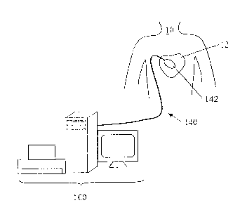
Fig. 1 is an exemplary embodiment of a mapping system, according to an aspect
of the present invention;
Fig. 2 is an exemplary embodiment of a computer architecture forming part of
the
mapping system of Fig. 1;
Fig. 3 is a flow chart outlining steps of a method of determining and storing
surface charge densities, in accordance with aspects of the present invention;
and
Fig. 4 is a flow chart outlining steps of a method of determining and storing
dipole
densities, in accordance with aspects of the present invention.
CA 02659898 2014-04-04
Detailed Description of the Preferred Embodiments
In a typical but non-limiting embodiment, the measured and/or calculated
potential Vewill
be recorded in a database in the form of a table. For generating the triple
W(P,t, Ve) is the
position P and the time of measurement t will be used. This triple W(P,t,Ve)
is the basis for
generating a 2 or 3-dimensional map of the surface charge density and/or the
dipol density.
Therefore, the triple W(P,t,Ve), comprising the values and data Of measurement
or
preliminary calculations is transformed into another triple comprising the
surface charge
and/or dipol charge. In a preferred embodiment, the triple W(P,t,Ve) (e.g.
after storing) can
be used to be transformed into a triple W(P,t,L) and/or a triple W(P,t,D)
and/or a triple
W(P,t,LD), wherein LD comprises the information of the surface charge and the
dipole
charge at position P at time t. The process and method for the transformation
is preferably
based on an algorithm based on formula 4 and/or 5 and/or a BEM-algorithm for
the
discretisation of the wall of a heart chamber.
Research has indicated that the use of the surface charge densities (i.e.
their distribution)
or dipole densities (i.e. their distribution) to generate distribution map(s)
will lead to a more
detailed and precise information on electric ionic activity of local cardiac
cells than
potentials. Surface charge density or dipole densities represent a precise and
sharp
information of the electric activity with a good spatial resolution, whereas
potentials
resulting from integration of charge densities provide only a diffuse picture
of electric
activiy. The electric nature of cardiac cell membranes comprising ionic
charges of proteins
and soluble ions can be precisely described by surface charge and dipole
densities. The
surface charge densities or dipole densities cannot be directly measured in
the heart, but
instead must be mathematically and accurately calculated starting from
measured
potentials. In other words, the information of voltage maps obtained by
current mapping
systems can be greatly refined when calculating surface charge densities or
dipole
densities from these.
The surface charge density means surface charge (Coulombs) per unit area
(cm2). A dipol
as such is a neutral element, wherein a part comprises a positive charge and
the other part
comprises the same but negative charge. A dipol might represent the electric
nature of
cellular membranes better, because in biological environment ion charges are
not
macroscopically separated.
11
CA 02659898 2014-04-04
In order to generate a map of surface charge densities (surface charge density
distribution)
according to the present invention, the geometry of the given heart chamber
must be
known. The 3D geometry of the cardiac chamber is typically assessed by
currently
available and common mapping systems (so-called locator systems) or,
alternatively, by
integrating anatomical data from CT/MRI scans. Fig. 1 shows an exemplary
embodiment
of a mapping system 100 that can be used to map a heart 12 of a human 10.
Mapping
system 100 can include a computer having known types of input devices and
output
devices, and a probe system 140. For the measurement of potentials the non-
contact
mapping method a probe electrode 142 will be used which forms part of probe
system 140.
,
12
CA 02659898 2014-04-04
The probe electrode 142 may be a multielectrode array with elliptic or
spherical shape.
The spherical shape has certain advantages for the subsequent data analysis.
For
example, when considering, for example, the ventricular cavity with the
endocardium and
take a probe electrode 142 with a surface Sp, which is located in the blood,
it is possible to
measure the potential V(x,y,z) at point x,y,z on the surface S. In order to
calculate the
potential at the endocardial surface Se the Laplace equation
a2 a2 a2
(1)
az' ay
needs to be solved, wherein V is the potential and x,y,z denote the three
dimensional
coordinates. The boundary conditions for this equation are V(x,y,z)=Vp(x,y,z)
on Sp,
wherein Vp is the potential on surface of the probe 142.
The solution is an integral that allows for calculating the potential
V(x'y'z') at any point x'y'z'
in the whole volume of the heart chamber that is filled with blood. For
calculating said
integral numerically a discretisation of the cardiac surface is necessary and
the so called
boundary element method (BEM) has to be used.
13
CA 02659898 2014-04-04
=
The boundary element method is a numerical computational method for solving
linear inte-
gral equations (i.e. in surface integral form). The method is applied in many
areas of engi-
neering and science including fluid mechanics, acoustics, electromagnetics,
and fracture
mechanics.
The boundary element method is often more efficient than other methods,
including the fi-
nite element method. Boundary element formulations typically give rise to
fully populated
matrices after discretisation. This means, that the storage requirements and
computational
time will tend to grow according to the square of the problem size. By
contrast, finite ele-
ment matrices are typically banded (elements are only locally connected) and
the storage
requirements for the system matrices typically grow quite linearly with the
problem size.
With the above in mind, all potentials VP (xi 'yl lz1') on the surface of the
probe 142 can be
measured . To calculate the potential Ve on the wall of the heart chamber, the
known ge-
ometry of the surface of the heart chamber must be divided in discret parts to
use the
boundary element method The endocardial potentials V, are then given by a
linear matrix
transformation T from the probe potentials Vp Ve T Vp .
zo After measuring and calculating one or more electric potential(s) V, of
cardiac cells in one
or more position(s) P(x,y,z) of the at least one given heart chamber at a
given time t . The
surface charge density and the dipol density is related to potential according
to the following
two Poisson equations:
(2)
AV = ¨(vc5s (P)) (3)
wherein p(P) is the surface charge density in position P=x,y,z, S, (F) is the
delta-
distribution concentrated on the surface of the heart chamber Se and u is the
dipol den-
sity.
There is a well known relationship between the potential V, on the surface of
the wall of the
heart chamber and the surface charge (4) or dipole densities (5).
14
CA 02659898 2014-04-04
Ve (P) ¨ P (-Pf )jd( õ,,
) (4)
I
(P) 1 ¨(P') 1
4jVo-- (p, ) (5)
IP' P Pil
(For a review see Jackson JD. Classical Electrodynamics, 2nd edition, Wiley,
New York
1975.)
The boundary element method again provides a code for transforming the
potential Ve in
formula 4 and 5 into the desired surface charge densities and dipole densities
, which can
be recorded in the database
In another embodiment of the method of the present invention the electric
potential(s) Ve is
(are) determined by contact mapping. In this case the steps for calculating
the electric
potential Ve are not necessary, because the direct contact of the electrode
142 to the wall
of the heart chamber already provides the electric potential Ve.
In a preferred embodiment of the method of the present invention the probe
electrode 142
comprises a shape that allows for calculating precisely the electric potential
Ve and, thus,
simplifies the calculations for transforming Ve into the desired charge or
dipole densities.
This preferred geometry of the electrode 142 is essentially ellipsoidal or
spherical.
In order to employ the method for determining a database table of surface
charge densities
of at least one given heart chamber in the context of the present invention,
it is preferred to
use a system comprising at least:
a) one unit for measuring and recording electric potentials V at a given
position
P(x,y,z) on the surface of a given heart chamber (Contact mapping) or a probe
electrode 142 positioned within the heart 12, but without direct wall contact
(non-
contact mapping)
b) one a/d-converter for converting the measured electric potentials into
digital data,
c) one memory to save the measured and/or transformed data,
d) one processor unit for transforming the digital data into digital surface
charge
density or dipole density data.
CA 02659898 2014-04-04
It is noted that numerous devices for localising and determining electric
potentials of
cardiac cells in a given heart chamber by invasive and non-invasive methods
are well
known in the art and have been employed by medical practitioners over many
years.
Hence, the method, system, and devices of the present invention do not require
any
particular new electrodes 142 for implementing the best mode for practicing
the present
invention. Instead, the invention provides a new and advantageous processing
of the
available data that will allow for an increase in precision, accuracy and
spatial resolution
of cardiac activation mapping when compared to prior art systems based on
electric
surface potentials in the heart 12 only. in the near future, the present
invention will allow
for providing superior diagnostic means for diagnosing cardiac arrhythmias and
electric
status of heart cells including metabolic and functional information.
Fig. 2 provides an exemplary embodiment of a computer architecture that can
form part of
mapping system 100. The mapping system 100 includes an ND converter for
converting
measured electric potentials from the probe system 140 into digital data; a
processor unit
for transforming the digital data into digital surface charge density or
dipole density data;
and a memory to save the measured and/or transformed data.
Fig. 3 and Fig.4 summarize methods for determining and storing surface charge
densities
and dipole densities, respectively, in accordance with aspects of the present
invention,
which have been described in detail above.
In method 300 of Fig. 3, in step 302, mapping system 100 is used to measure
and/or
calculate one or more electric potential(s) Ve in one or more position(s) P
within a heart
chamber at a given time t. In step 304, Ve is transferred into a surface
charge density
p(P',t). In step 306, the surface charge density p(P1,t) is stored in a
database table. The
method is repeated if there is another P, in step 308.
In method 400 of Fig. 4, in step 402, mapping system 100 is used to measure
and/or
calculate one or more electric potential(s) Ve in one or more positions(s) P
within a heart
chamber at a given time t. In step 404, Ve is transferred into a dipole
density v(P',t) by
using an algorithm suitable for transforming an electric potential into
surface charge
density. In step 406, the dipole density v(P',t) is stored in a database
table. The method
is repeated if there is another P, in step 408.
16