Note : Les descriptions sont présentées dans la langue officielle dans laquelle elles ont été soumises.
CA 02662692 2009-03-06
PCT/EP2007/007763 WO 2008/028653
Device and method for monitoring an access to a patient, in particular a
vascular
access in extracorporeal blood treatment
The invention relates to a device and a method for monitoring an access to a
patient, in which a fluid is withdrawn from the patient via a first tubular
conduit,
which comprises a first patient connector, and the fluid is returned to the
patient
via a second tubular conduit, which comprises a second patient connector, in
particular for monitoring a vascular access during an extracorporeal blood
treatment in which a patient's blood is withdrawn from the patient via an
arterial
tubular conduit, which comprises an arterial patient connector with an
arterial
puncture cannula, and is returned to the patient via a venous tubular conduit,
which
comprises a venous patient connector with a venous puncture cannula.
In the field of medical engineering, many devices are known with which fluids
can
be withdrawn from a patient or delivered to a patient via a tubular conduit.
The
access to the patient is usually made with a catheter for insertion into
organs of the
body, or a cannula for puncturing vessels. During the examination or
treatment, a
correct access to the patient has to be ensured. It is therefore necessary to
monitor
the patient access.
In blood purification methods such as haemodialysis, haemofiltration and
haemodiafiltration, blood is passed through an extracorporeal blood circuit.
If the
venous connection to the patient comes loose during the blood treatment,
bleeding
to death can be avoided only if the extracorporeal blood flow is stopped
within a
few seconds. Therefore, extracorporeal blood circuits are generally provided
with
protective systems which, in the event of an alarm, stop the blood pump, close
the
venous clamp and trigger an acoustic or optical warning signal.
DE 197 39 099 Cl describes a device for monitoring an access during an
extracorporeal blood treatment, in which an electric current is induced in the
CA 02662692 2009-03-06
2
connection of the extracorporeal blood circuit representing a closed conductor
loop, the current flowing in the conductor loop is measured, and a
characteristic
change in the current strength points to an incorrect vascular access. In
addition to
inductive injection and output, it is also known to perform capacitive
injection and
output of electric signals in the extracorporeal blood circuit.
US 6,932,786 B2 describes a monitoring device in which an AC voltage signal is
capacitively injected and output in the extracorporeal blood circuit. The
injection
and output of the AC voltage signal takes place by means of electrical contact
elements that enclose the tubular conduits. The electrical contact element in
this
case represents one "electrode" of a "capacitor", while the blood flowing in
the
tubular conduits represents the other "electrode" of the "capacitor". The
insulating
tubular conduit represents the dielectric of the capacitor lying between the
electrodes.
In the known monitoring device, the AC voltage signal generated by an AC
voltage signal generator is coupled to a venous contact element on the venous
blood conduit and to an arterial contact element on the arterial blood conduit
as
difference signal. In an alternative embodiment, one output of the frequency
generator is connected to a contact element enclosing the venous blood
conduit,
while the other output of the signal generator is at ground potential. Both
embodiments are based on the fact that the AC voltage signal is output as
difference signal with two contact elements that are arranged at different
locations
of the extracorporeal circuit, and the blood flowing in the extracorporeal
circuit is
at ground potential.
It has been found in tests that, in the method known from US 6,932,786 B2, the
output AC voltage signal can be superposed by relatively strong interference
signals. In practice, therefore, the known device can prove relatively
susceptible to
faults.
US 2003/0195454 Al deals with the problem of capacitive injection and output
of
measurement signals in the extracorporeal blood circuit and proposes injection
and
CA 02662692 2014-04-23
3
output of the measurement signals by means of electrical contact elements that
are
directly in contact with the blood flowing through the tubular conduits.
US 7,060,047 describes a device for monitoring a vascular access during a
dialysis
treatment, which device permits capacitive injection of an AC voltage signal,
wherein an electrical circuit is closed via a common earth fault. The device
in
principle permits connection of the patient to earth. However, the document
states
that such a coupling of the patient is not absolutely essential.
It is an object of the invention to make available a device that permits
monitoring
of an access to a patient with a high degree of reliability, even though the
measurement signal is injected and output capacitively. It is a further object
of the
invention to make available a blood treatment device that comprises a device
for
monitoring a patient access and permits monitoring of the patient access with
a
high degree of reliability. It is also an object of the invention to make
available a
method that permits monitoring of the access with a high degree of
reliability.
The monitoring device according to the invention and the monitoring method
according to the invention differ from the monitoring devices and monitoring
methods known from the prior art in that the AC voltage signal is injected and
output relative to a common ground potential. Moreover, a differential
measurement does not take place. It has been surprisingly found that, with
injection and output of the AC voltage signal relative to a common ground
potential, it is possible to reduce the interference signals that could
otherwise arise,
particularly during the unavoidable movements of the tubes.
In the device according to the invention and in the method according to the
invention, the AC voltage signal is injected only at one location of one of
the two
tubular conduits and is output only at one location of the other of the two
tubular
CA 02662692 2009-03-06
4
conduits. This also reduces the outlay for capacitive injection and output of
the
voltage signal.
The means for capacitive injection and output of the AC voltage signal are
preferably bodies of electrically conductive material, for example metal
sleeves,
that enclose the tubular conduits.
An incorrect vascular access, for example due to the venous or arterial
puncture
cannula slipping out of the venous or arterial blood conduit, results in a
change in
impedance, which in turn leads to a change in the amplitude of the output AC
voltage signal. Consequently, in the event of a characteristic change in the
amplitude of the measured AC voltage signal, preferably a reduction in said
amplitude, it can be concluded that the vascular access is not as it should
be.
During extracorporeal blood treatment, an incorrect vascular access may exist
not
only when the arterial and/or venous puncture cannula has slipped out of the
arterial or venous blood conduit, but also when the blood conduit is
interrupted.
The known arterial and venous blood conduits generally comprise tube couplers
that interconnect two tube portions upstream and downstream of the venous or
arterial puncture cannula. These tube couplers are generally Luer lock
couplers. If
the tube coupler were to come loose, there would no longer be an access to the
vessel. This situation too can be demonstrated by the device according to the
invention and the method according to the invention.
In the device according to the invention and the method according to the
invention,
the AC voltage signal is injected and output relative to ground potential at
any
desired location of the arterial or venous blood conduit, i.e. either upstream
or
downstream of the tube coupler, for example a Luer lock coupler, i.e. in the
tube
portion between the inlet to the dialyzer, or outlet from the dialyzer, and
the tube
coupler, or in the tube portion between tube coupler and puncture cannula. If
the
AC voltage signal is injected and output in the tubular conduit portions
between
tube coupler and puncture cannula, only a slipping out of the puncture cannula
can
be demonstrated, not a faulty tube coupler. Demonstration of a faulty tube
coupler
= CA 02662692 2009-03-06
=
requires that the injection or output takes place in a tubular conduit portion
between dialyzer and tube coupler.
Illustrative embodiments of the invention are explained in greater detail
below
5 with reference to the drawings, in which:
Figure 1 shows the main components of a blood treatment device, together with
a device for monitoring the patient access, in a greatly simplified
schematic representation,
Figure 2 shows the equivalent circuit diagram of the blood treatment device
from Figure 1,
Figure 3 shows the equivalent circuit diagram of an alternative embodiment of
the blood treatment device, in which the AC voltage signal is
injected downstream of an arterial tube coupler and the AC voltage
signal is output upstream of a venous tube coupler, the arterial and
venous tube couplers each connecting two tube portions of the
arterial or venous tubular conduit,
Figure 4 shows another embodiment of the blood treatment device in which the
AC voltage signal is injected downstream of the arterial tube
coupler and the AC voltage signal is output downstream of the
venous tube coupler, and
Figure 5 shows another embodiment of the blood treatment device in which the
AC voltage signal is injected upstream of the arterial tube coupler
and the AC voltage signal is output upstream of the venous tube
coupler.
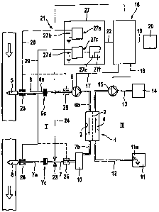
Figure 1 shows the main components of a blood treatment device, for example a
haemodialysis device, which comprises a device for monitoring the arterial and
venous vascular access. The haemodialysis device has a dialyzer 1 which is
CA 02662692 2009-03-06
6
divided by a semipeimeable membrane 2 into a blood chamber 3 and a dialysis
fluid chamber 4. An arterial tubular conduit 6 is connected to an artery of
the
patient by means of an arterial puncture cannula 5 and leads to the inlet of
the
blood chamber 3 of the dialyzer 1. Issuing from the outlet of the blood
chamber 3
of the dialyzer 1, there is a venous tubular conduit 7 which is connected to a
vein
of the patient by means of a venous puncture cannula 8. The arterial tubular
conduit 6 is routed into an occlusive blood pump 9 which conveys the blood in
the
extracorporeal blood circuit I. The venous tubular conduit 7 contains a bubble
trap
10, for example a drip chamber, which holds back air bubbles in the blood.
The dialysis fluid circuit II of the haemodialysis device comprises a means 11
which is used to prepare the dialysis fluid and to which a dialysis fluid
delivery
line 12 is attached that leads to the inlet of the dialysis fluid chamber 4 of
the
dialyzer 1. Issuing from the outlet of the dialysis fluid chamber 4 of the
dialyzer 1,
there is a dialysis fluid discharge line 13 that leads to a drain 14. A
dialysis fluid
pump 15 is coupled into the dialysis fluid discharge line 13.
The arterial and venous tubular conduits 6, 7 are part of a tube set, the
arterial and
venous tubular conduits each having two tubular conduit portions 6a, 6b and
7a,
7b, respectively. The conduit portions 6a, 6b of the arterial tubular conduit
6 and
the conduit portions 7a, 7b of the venous tubular conduit 7 are connected to
one
another by tube couplers 6c, 7c, for example Luer lock couplers, so that the
tube
portions towards the patient can be separated from the rest of the conduit
portions
of the tube set.
The dialysis device is controlled by a central control unit 16 which regulates
the
blood pump 9 and dialysis fluid pump 15 via control lines 17, 18,
respectively. The
central control unit 16 is connected by a data link 19 to an alarm unit 20,
which
emits an optical and/or acoustic alarm if a fault occurs.
A correct vascular access presupposes that both the arterial and venous
puncture
cannulas 5, 8 are located in the vessel. A correct vascular access also
presupposes
CA 02662692 2009-03-06
that the tube couplers 6c, 7c of the arterial and venous tubular conduits 6, 7
connect the two tubular conduit portions to one another.
For monitoring the vascular access, the dialysis device comprises a monitoring
device 21 which communicates with the control unit 16 via a data link 22. The
structure and the mode of operation of the monitoring device 21 will be
described
in detail below. The monitoring device 21 reports an incorrect vascular access
to
the control unit 16 via a data link 22, so that the control unit 16 activates
the alarm
unit 20, which emits an optical and/or acoustic alarm. Moreover, the control
unit
16 closes a venous tube clamp 23 which is arranged on the venous tubular
conduit
7 downstream of the blood chamber 3 of the dialyzer 1 and which is connected
to
the control unit 16 via a control link 24.
The monitoring device 21 has means 25 for capacitive injection of an AC
voltage
signal, and means 26 for capacitive output of an AC voltage signal, and also a
computing and evaluation unit 27. The means for capacitive injection and
output of
the AC voltage signal are metal sleeves enclosing the tubular conduits.
In the illustrative embodiment according to Figure 1, the arterial metal
sleeve 25
encloses the arterial tubular conduit portion between arterial puncture
cannula 5
and arterial tube coupler 6c, while the venous metal sleeve 26 encloses the
venous
tubular conduit portion between venous tube coupler 7c and venous puncture
cannula 8. It is also possible, however, to arrange the arterial metal sleeve
25 in the
arterial tubular conduit portion between the arterial tube coupler 6c and the
blood
chamber 3, preferably upstream of the blood pump 9, and to arrange the venous
metal sleeve 26 in the venous tubular conduit portion between the blood
chamber 3
and the venous tube coupler 7c, preferably upstream of the tube clamp 23. This
arrangement is shown in Figure 1 by broken lines.
The means 11 for preparation of the dialysis fluid ensures that the dialysis
fluid is
at ground potential, i.e. at the operational earth of the machine. For this
purpose,
the means 11 for preparing the dialysis fluid contains a symbolically
indicated
electrical contact element 11a, for example an earthing clip, which is in
contact
CA 02662692 2009-03-06
8
with the dialysis fluid. Since the dialysis fluid is in turn in contact with
the blood
via the membrane 2 of the dialyzer 1, the earthing of the dialysis fluid also
means
that the blood flowing through the tubular conduits 6, 7 is also connected to
ground
potential, i.e. connected to the operational earth of the machine.
The monitoring device 21 has means 27a for generating an AC voltage signal
with
a signal output 27b, one connector of the signal output being connected via an
electrical connection line 28 to the arterial metal sleeve 25, while the other
connector of the signal output is connected to ground potential, i.e. to the
operational earth of the dialysis machine. In addition, the monitoring device
has
means 27c for measuring an AC voltage signal with a signal input 27d. One
connector of the signal input 27d is connected via an electrical connection
line 29
to the venous metal sleeve 26, while the other connector of the signal input
is again
connected to ground potential, i.e. to the operational earth of the machine.
In addition, the monitoring device 21 has means 27e for evaluating the AC
voltage
signal measured by the means 27c. The means 27e for evaluating the AC voltage
signal in turn have means 27f for comparing the measured AC voltage signal to
a
predetermined limit value.
The monitoring device according to the invention operates as follows. An AC
voltage signal is generated which is capacitively injected into the
extracorporeal
blood circuit I on the arterial tubular conduit 6 and is capacitively output
from the
extracorporeal blood circuit I on the venous tubular conduit 7. The measured
current flowing into the patient via the blood tube upon application of the AC
voltage signal is negligible. The amplitude of the output AC voltage signal is
compared to a predetermined limit value. If the amplitude of the voltage
signal is
less than the predetermined limit value, the monitoring device 21 concludes
there
is an incorrect vascular access, so that an alarm is given and the
extracorporeal
blood circuit is interrupted.
CA 02662692 2009-03-06
9
Figures 2 to 4 show the electrical equivalent circuit diagrams of the dialysis
device
from Figure 1, for different arrangements of the means for injection and
output of
the AC voltage signal.
In Figures 2 to 4, the individual components of the dialysis device are
described by
their impedance Z, which with discrete components can be represented as a
series
connection of a resistor Rb and a parallel connection of a resistor Ra and of
a
capacitor Cx. For the impedances of the individual components, the following
abbreviations are used:
ZDD = Impedance: dialyzer, dialysate side
ZDB = Impedance: dialyzer, blood side
ZDDDB = Impedance: dialysate side ¨> blood side
ZBSS = Impedance: blood tube segment
ZBF = Impedance: bubble trap
ZKA = Impedance: capacitive output
ZL = Impedance: tube coupler (Luer lock)
ZKE = Impedance: capacitive injection
ZPSS = Impedance: pump tube segment
ZSP = Impedance: shunt patient
ZPKTV = Impedance: venous puncture
ZPKTA = Impedance: arterial puncture
Detachment of the arterial or venous puncture cannula 5, 8 signifies an
interruption
of the "electric circuit". Detachment of the arterial or venous tube coupler
also
signifies an interruption of the electric circuit. The interruption of the
electric
circuit results in an increase in the impedance, which is in turn reflected by
a
reduction in the amplitude of the measured AC voltage signal. The signal path
between the injection location and output location is indicated in Figures 2
to 5 by
a curved line.
Figure 2 shows the arrangement, illustrated by solid lines in Fig. 1, of the
metal
sleeves 25, 26 for injection and output of the AC voltage signal. With this
= CA 02662692 2009-03-06
arrangement, only a disconnection or at least dislocation of the arterial and
venous
cannulas 5, 8 can be reliably demonstrated on the basis of a significant
increase in
impedance or reduction in the amplitude of the AC voltage signal, but not the
detachment of the tube couplers.
5
Figure 3 shows the arrangement, illustrated by broken lines in Fig. 1, of the
arterial
and venous metal sleeves 25, 26. With this arrangement, only a disconnection
or at
least dislocation of the arterial and venous cannulas 5, 8 can be reliably
demonstrated on the basis of a significant increase in impedance or reduction
in the
10 amplitude of the AC voltage signal, but not the detachment of the
tube couplers.
By contrast, the detachment of a tube coupler leads only to a small signal
rise,
since there is no connection to ground through the impedance ZDDDB.
Figure 4 shows an illustrative embodiment in which the arterial metal sleeve
25 is
arranged in the tubular conduit portion 6a of the arterial tubular conduit 6
between
the blood chamber 3 and the arterial tube coupler 6c, while the venous metal
sleeve
26 is arranged in the tubular conduit portion 7a of the venous tubular conduit
7
between the venous puncture cannula 8 and the venous tube coupler 7c (Fig. 1).
Figure 5 shows the arrangement of the arterial metal sleeve 25 in the arterial
tubular conduit portion 6a between the arterial puncture cannula 5 and the
arterial
tube coupler 6c, while the venous metal sleeve 26 is arranged in the venous
tubular
conduit portion 7a between the blood chamber 3 and the venous tube coupler 7c.
In the illustrative embodiments according to Figures 4 and 5, detachment of
the
puncture cannulas 5, 8 and also of the tube couplers 6c, 7c is demonstrated by
an
increase in the impedance and a reduction in the amplitude of the measured AC
voltage signal.
In the monitoring device according to the invention, the amplitude of the
measured
AC voltage signal can be compared not only to one predetermined reference
value,
but to several reference values. Therefore, with an appropriate arrangement of
the
injection site and output site, it is in principle possible to differentiate
whether a
= CA 02662692 2009-03-06
11
puncture cannula or a tube coupler has come loose, since the respective fault
is
associated with a characteristic change in the impedance or signal amplitude.
The
extent of the significant change depends on the respective equivalent circuit
diagram. Characteristic values can be established by comparative measurements.