Note : Les descriptions sont présentées dans la langue officielle dans laquelle elles ont été soumises.
CA 02664590 2012-07-17
WO 2008/046192
PCT/CA2007/001793
ADAPTABLE DEVICE FOR DETECTING AND TREATING DENTAL
PATHOLOGIES
FIELD OF THE INVENTION
This invention relates to the field of dental devices, and more specifically
to
devices for detection of pathologies and dental treatment.
BACKGROUND OF THE INVENTION
Optical detection apparatuses have been described for use in detection of
dental
pathologies such as caries, tartar, fractures and the like. Such detection
systems have
also been described in combination with existing dental tools, such as tartar
removal
instruments. In commonly assigned US patent publication 2004/0106081, a tartar
removal instrument is provided with a modified handle and tip equipped with an
optical
light guide to allow a user to detect tartar at the working end of the tartar
removal
instrument. However, these tools lack flexibility and adaptability with regard
to their use
for multiple applications such as detection of pathologies and treatment of
the
pathologies. Accordingly there is a need for improved dental tools. In powered
scaling
or tartar removal instruments, the tip is normally part of an insert. The tip
is a
replaceable part that is subject to wear and replacement after such wear.
SUMMARY OF THE INVENTION
In a broad embodiment of the invention, there is provided a dental instrument
tip that
has a proximal portion made of a material able to withstand flexion and/or
vibration and
a distal portion made of a more brittle material having a better hardness at
the working
end.
In one aspect, a tartar removal tip has a metal proximal portion and a ceramic
distal
portion. In some embodiments, the tip is equipped with one or more optical
CA 02664590 2009-03-25
WO 2008/046192 PCT/CA2007/001793
2
fibers that extend along the proximal portion into the distal portion for the
purposes of emitting and/or collecting light as part of a tartar or other
dental
pathology detection system.
In a broad embodiment of the invention there is provided an insert comprising
optical means for detection of dental pathologies, said insert being adapted
to be
placed on a functional part of a dental instrument without requiring
modification of
the dental instrument to be equipped with the optical detection capability.
In one aspect of the invention, the insert has a suitable coupling for the
optical
detection system that is on the tip insert such that it does not change or
affect the
handle portion of the instrument. In another aspect of the invention, there is
provided a sheath for a dental instrument tip that fits over an existing
dental
instrument tip and includes optical components for an optical dental pathology
detection system.
In an aspect of the invention the insert is adapted to be placed at a tip of a
dental
cleaning instrument and the optical components can be protected by wear
resistant material.
In another aspect of the invention, a sheath for a tip of a dental instrument
BRIEF DESCRIPTION OF THE DRAWINGS
Further features and advantages of the present invention will become apparent
from the following detailed description, taken in combination with the
appended
drawings, in which:
SUBSTITUTE SHEET (RULE 26)
CA 02664590 2009-03-25
WO 2008/046192
PCT/CA2007/001793
3
Figure la illustrates a sectional side view of a tartar removal tip including
an
optical fiber and a translucent ceramic tip covering the end of the fiber;
Figure lb illustrates a sectional side view of a tartar removal tip similar to
Figure
la in which the ceramic tip is opaque and has an aperture for light
transmission;
Figure 2 illustrates a sectional side view of a tartar removal tip and sheath,
the
sheath being adapted to support an optical fiber end for optical detection of
tartar;
Figure 3 illustrates a removable, sterilizable, tartar removal tip insert
adapted to
provide a coupler for the optical detection system without having a handle
modification;
Figure 4 is a sectional side view of a self-contained, sterilizable tartar
detection
system that fits over and around a handle of a powered tartar removal
instrument
and is connected to a replaceable tip of the instrument, the tip having
optical
components for the detection of tartar;
Figure 5 is a sectional view of the instrument of Figure 4 about plane 5-5;
Figure 6 is a section view similar to that of Figure 5, in an embodiment
similar to
Figures 7 and 8;
Figure 7 is a perspective view of a tartar removal instrument having a fit
over
optical tartar detection system that form part of an enlarged handle of the
instrument;
SUBSTITUTE SHEET (RULE 26)
CA 02664590 2012-07-17
WO 2008/046192
PCT/CA20071001793
4
Figure 8 is a bottom view of the fit over system housing separate from the
instrument;
Figure 9 is a side sectional view of an embodiment in which a self-contained,
sterilizable tartar detection system is integrated as part of an insert tip by
providing an
extension of the insert shaft to receive a detection system housing on the
shaft.
DETAILED DESCRIPTION OF THE INVENTION
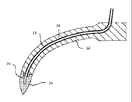
According to a first embodiment shown in Figures 1 a and 1 b, a tartar removal
tip, also
known as a scaling tip, has a hybrid construction with a first proximal part
made of
metal (metal descaling tip 12) and a second distal part made of ceramic
(ceramic
working end or ceramic tip 14). The metal provides the ability to flex under
the
conditions of being subjected to pressure by the user acting on the tooth, and
under
conditions of high intensity vibrations originating from the powered scaling
instrument.
The ceramic tip 14 has a hardness that allows it to be more wear resistance
than the
proximal part. A scaling tip made entirely of conventional ceramic would be
prone to
breaking under the mechanical stresses that the tip is subjected to.
In the embodiment of Figure 1 a, the ceramic working end 12 is transparent or
translucent and the optical fiber 16 terminates centrally inside the ceramic
portion. The
fiber 16 is made solid with the metal and ceramic portions using epoxy 18, so
that
vibrations do not damage the fiber or fibers. In the embodiment of Figure 1 b,
the
ceramic tip 14 has a channel 13 ending in an aperture for the fiber 16. While
the end of
the fiber 16 is recessed within the ceramic tip 14, and thus protected from
wear or
damage, light may pass through the aperture. The aperture may be filled with
another
transparent or translucent material to protect the end of the fiber 16 within
the channel
13 and the ceramic tip can be an opaque ceramic 15.
In the embodiment of Figure 2, the ceramic working end 14 is provide as a
sheath 22 to
press-fit over a conventional tartar removal tip 24. In this embodiment, the
optical cable
28 is fed along a side of the sheath 22 and terminates either to a side or
centrally within
the ceramic working tip or functional end 26.
CA 02664590 2012-07-17
WO 2008/046192
PCTICA2007/001793
In the embodiment of Figure 3, the tartar removal tip 24 is modified to
integrate an
optical fiber, either as show with a parallel optical fiber support 32 or by
using a central
channel in the tip to guide the fiber to the end of the tip. The tip is
somewhat longer
than a conventional replacement tip for the desired model of powered scaler,
and a
5 coupler is provided on an extension portion 34. The coupler may be an
optical cable-
based coupler 36 for coupling the one or more optical fibers to an optical
cable leading
to the optical tartar (and/or other dental pathology) detection system.
Alternatively, a
light source and light detector may be provided within the illustrated coupler
block, and
the coupler may be an electrical or tip-based coupler 38. The tartar removal
tip 24 is
mechanically attached within the scaler instrument and driven to have the
desired
scaling action at the tip or functional end 26.
There is provided an insert tip comprising optical means for detection of
dental
pathologies that can be inserted into an existing dental instrument such as a
powered
sonic or ultrasonic instrument or a rotary instrument for cleaning teeth. The
tip can be
connected to an optical analyzing means to detect the presence of dental
pathologies
such as calculus, caries, plaque, blood, dental fractures and the like. The
tip of the
present invention advantageously enables the user to adapt existing dental
cleaning
instruments for the detection of dental pathologies. In one embodiment the tip
is
adapted to perform the function of the instrument (such as cleaning tartar)
when placed
on the instrument. In one embodiment the tip is advantageously designed to be
removably attached ("retrofit' tip) to the device allowing repeated use of the
same tip,
easy replacement of the tip with another tip or for sterilization purposes.
The tip may
also be disposable.
Instruments on which the tip can be inserted comprise but are not limited to:
Sonic
Instruments such as ultrasonic Instruments (piezoelectric e.g. EMS or ACTEON ,
magnetostricitive e.g. Dentsply Inc.) and hand instruments such as rotary
instruments.
The optical components are at least in part comprised within the tip and are
adapted to
resist wear during normal use of the instrument. In particular, the optical
components
CA 02664590 2012-07-17
WO 2008/046192
PCT/CA2007/001793
6
should be protected from friction and water. Various designs can be used to
achieve
these goals.
In one embodiment, the tip can be designed such that the light injection port
and the
detection port are in direct contact with the teeth when in use. These optical
components may be surrounded by hard and wear resistant materials. The optical
means inside the tip can be uncovered and terminate at the extremity of the
probe tip.
Having hard materials around these optical means protects the optical means
even if
the optical means are in direct contact with dental structures. Material such
as
sapphire, ceramics, tungsten carbide, zirconia, ruby or other very hard
material can be
used to increase resistance. High hardness epoxy can also be used to attach
the
optical means to the probe tip.
The optical detection means could be covered and protected from stress with a
disposable sleeve. The material used could be plastic, epoxy, Teflon e PTFE or
other
translucent materials.
In another embodiment, the tip can consist of a wear resistant shell
completely
surrounding the optical components. Material translucent or transparent to the
wavelength(s) used for detection of the pathologies can be used to cover
optical
components. Examples of such materials include but are not limited to Teflon
coated
tip, Epoxy coating, plastic.
It will be appreciated that when the optical components are covered with
translucent or
transparent material, reflection inside this material should be taken into
account to
adjust detection parameters.
The tip may be partially or completely made with wear resistant materials. For
example,
the insert tip can be made solely in ceramic or partially made in ceramic.
However,
because ceramic can be susceptible to fracture, the proportion of ceramic can
be
adjusted while still being compatible with the function of the instrument. The
junction
between the insert and the instrument (typically metal) can be realized by
laser fusion
CA 02664590 2012-07-17
WO 2008/046192
PCT/CA2007/001793
7
fixation, pressfit fixation, Epoxy fixation, ceramic coating and other such
processes as
would be known to one skilled in the art.
Because the tip can be made with wear resistant material it can be shaped with
a
special morphology to enhance its functionality such as calculus removal
efficiency. For
example hemispheric shaped tips can be designed for this purpose. The tip of
the
insert can include domes or grooves to enhance scaling performance. The shape
of the
insert tip can increase the number of stressing point in contact with the
calculus and
therefore increase the speed of calculus removal and decrease the damage to
tooth
surface. This can be more easily made with ceramic because of its high
resistance to
wear. It is also possible to include material such as diamond that will
enhance removal
of hard tissue.
Fiber assembly in the insert:
Plastic fiber: The fibers in the detection optical means can be plastic fiber
optics. Plastic
fibers cost less (can be made disposable), are less susceptible to fractures
when
subjected to vibrations, have thinner core to maximize examination area.
Removable fibers: The fiber can be inserted in the tip such as to be
removable.
Retractable fibers : The insert tip can be the cap of the optical detection
that protects
the fibers which can be retracted when not in use. In the case of instruments
using a
bur the optic fibers can be incorporated inside the bur and connections made
at the tip
of the bur.
In another embodiment, optical detection means (ODM) can be protected by using
a
reduced power strategy. Thus to reduce wear impact on ODM is to
instruct the operator to reduce power of the powered instrument. This will
enable the tip
to wear slowly and therefore keep the tip and ODM working for a longer period.
Stainless steel, like actual marketed tips, could be used with this strategy.
CA 02664590 2012-07-17
WO 2008/046192
PCT/CA20071001793
8
Optical components comprise an optical analyzing means (OAM) which can be made
to be removable and be coupled with another unit or insert. The OAM can be
positioned on the powered scaler unit and use fiber optics to go along the
cable and
then connect to the insert. The OAM can be adapted to be fitted on the handle
or at the
cable end of the powered ultrasonic or sonic instrument and connect with or
without
fiber optics to the insert. The OAM can be inside the insert's handle
(specially for
magnetostrictive inserts) the OAM can be made to be sterilizable but
preferably be
removable from the insert handle before sterilization.
It is possible to think to optically connect the insert to the cable optical
connector or
directly to optical analyzing means with optical path only (without physical
contact). For
example focusing lenses could be used to focus light onto fiber optics on the
insert.
This could reduce the impact of vibrations on physical components.
The optical components may comprise components that are well known in the art
such
as optic fibers, mirrors, reflectors and the like. The optical assembly is
designed such
as to resist wear during normal use and to be resistant to multiple
sterilizations. In this
respect it is desirable to provide friction protection of fiber optics using
Teflon @ tubing
for example. For water protection Silicone or the like can be used.
The optic fibers can be cleaved and otherwise processed as needed before
insertion
into the tip insert. In one embodiment the fibers can be fixed inside the
insert with
Epoxy having a high hardness and heat resistance.
To protect the fibers from friction stress inside the insert a buffer (made of
rubber,
plastic, gel, silicone, Teflon, or polished metal
) can be inserted between the fibers
and the insert rigid structure. The surface in contact with fiber optics can
be treated to
reduce friction (sand polishing, chemically treated (acids), metal deposit
using
electricity ... )
To verify the smoothness of the insert tip, to determine if the probe tip is
not altered and
dangerous for the tooth structure a hard material can be used to scrub on it
the insert
CA 02664590 2012-07-17
WO 2008/046192
PCT/CA2007/001793
9
tip and determine if the tip can be used. If deep scratches (or resistance is
noted during
or after scratching) it may mean that the probe tip has been broken and
therefore
should not be used to prevent damage of tooth surface.
The various parts of the device are preferably sterilizable. The optical cable
or a part of
the cable (more particularly the portion of the optical cable next to the
insert) can be
made to be detachable from OAM. Such as to enable sterilization.
Tip connection detection can be provided to ensure proper connections.
The insert tip can also include a pocket measurement means to prevent changing
instrument in order to proceed with periodontal pocket measurement.
The detection technology can be used for caries detection. The sonic or
ultrasonic
powered instruments can be used for caries removal or preparation of the tooth
structure. The detection can also be adapted to be integrated inside a rotary
drilling
insert for high speed or slow speed. For caries detection the device can be
separated
from the working tip and be attached only on the handle.
In one embodiment the detection and the "working" tip are separated where more
particularly the detection tip can be retractable by using for example a
flexible material
containing the optical component. The operator can accordingly deploy or
retract the
detection tip during the session where the working tip is used. It is then
more
convenient than having to switch to an independent detection instrument.
In the embodiment of Figures 4 and 5, the optical tartar detection system has
a self-
contained fit-over housing 52 that conforms to the shape of the tartar removal
instrument 54. While the handle of the instrument is enlarged by the addition
of the
detection system which can comprise electronic components 56 and an audio
transducer 58, the combination can still be hand-held and manipulated for use.
The
optical fiber connection between the tip and the detection system housing can
be
CA 02664590 2012-07-17
WO 2008/046192
PCT/CA2007/001793
permanent and sealed, but is preferably detachable. The optical detection
system thus
includes the tartar removal tip and can be sterilized.
In the embodiment of Figures 6, 7 and 8, the optical tartar detection system
has a
housing that is adapted to slide over or clip to the tartar removal instrument
54. The
5 housing may extend over the whole length of the instrument handle while a
narrow
elongated circuit board 66 provides the electronic circuitry, optoelectronics
and power
source for the detection system. The resilient biased side members 68 are
resiliently
biased against the sides of the instrument. This embodiment allows for good
gripping
(using finger grip 61) of the combination of the instrument and detection
housing. The
10 housing can be sterilized as needed. The optical fiber connection
between the tip and
the detection housing can be permanent, or preferably by way of an optical
coupler so
that the tip can be replaced separately without replacing the detection unit.
In the embodiment of Figure 9, the optical tartar detection unit comprises a
tartar
removal instrument 54 with a handle 91 that is self-contained and provided on
a
somewhat larger extension portion to a tartar removal tip 24 as in the
embodiment of
Figure 3. The usual optoelectronic components 92, electronic components, such
as a
DSP 94, and battery 96 can be contained in a small annular housing such as a
tartar
detection unit 98 on a shaft of the tip. As in the embodiment of Figure 3, the
optical light
guide can be fed through a channel in the tip. The tip may also incorporate
the
structure of the embodiments of Figures 1 a or 1 b.
The technology can be used to perform the detection in reflectance,
fluorescence,
interferometry, Raman spectroscopy and the like.
The concept described herein could be used with another detection principle
(having a
connection means connecting to an analyzing mean) such as ultrasonic, sonic,
acoustic and the like. The insert could also be designed to fit onto a laser.
The device of the invention may also comprise 3D global positioning, voice
recognition
and similar features that make its use more efficient.
CA 02664590 2012-07-17
WO 2008/046192
PCT/CA2007/001793
11
While the invention has been described in connection with specific embodiments
thereof, it will be understood that it is capable of further modifications and
this
application is intended to cover any variations, uses, or adaptations of the
invention
following, in general, the principles of the invention and including such
departures from
the present disclosures as come within known or customary practice within the
art to
which the invention pertains and as may be applied to the essential features
herein
before set forth, and as follows in the scope of the appended claims.