Note : Les descriptions sont présentées dans la langue officielle dans laquelle elles ont été soumises.
CA 02670932 2014-12-18
1
DETECTION OF THE DETACHMENT OF IMMOBILIZED CONTRAST AGENT IN
MEDICAL IMAGING APPLICATIONS
Technical field
The present invention relates to the medical imaging field. More specifically,
the
present invention relates to contrast agent imaging applications.
Background
Medical imaging is a well-established technique (in the field of equipments
for
medical applications), which allows analyzing a body-part of a patient in a
substantially
non-invasive manner. A specific medical imaging technique is based on the
recording of
an echo signal that results from the application of ultrasound waves to the
body-part.
This technique can advantageously be implemented with the administration of an
ultrasound contrast agent (UCA) to the patient (for example, consisting of a
suspension
of phospholipid-stabilized gas-filled microbubbles); as the contrast agent
acts as an
efficient ultrasound reflector, it enhances the visualization of the vascular
system within
the body-part where it is present.
Target-specific contrast agents, adapted to reach a specific (biological)
target and
then remain immobilized thereon, have also been proposed in the last years for
facilitating the detection of specific pathologies. Particularly, a target-
specific contrast
agent is capable of attaching to the corresponding target ¨ such as particular
tissues or
receptors - by means of a specific interaction therewith; for example, the
desired
behavior can be achieved by incorporating a target-specific ligand in the
formulation of
the contrast agent (such as capable of interacting with inflammatory or
tumoral tissues).
Once the target-specific contrast agent has reached the target remaining
immobilized
thereon, its detection may allow distinguishing pathologies that would be
otherwise
CA 02670932 2014-12-18
2
difficult to identify.
A problem associated with the target-specific contrast agents is that only a
relatively small fraction of the total amount of the administered target-
specific contrast
agent actually reaches the target and remains immobilized thereon. Most of the
target-
specific contrast agent continues to circulate, for example, until it is
filtered out by the
lungs and/or in the liver of the patient. The echo signal that is measured is
then the result
of different contributions, which are due to the immobilized (target-specific)
contrast
agent, to the circulating or free-flowing (target-specific) contrast agent and
to surrounding
tissue. Therefore, it is quite difficult to distinguish the echo signal
generated by the
immobilized contrast agent from the one generated by the circulating contrast
agent and
tissue; particularly, it is almost impossible to differentiate the low
concentration of the
immobilized contrast agent (often consisting of single particles thereof that
reach the
target individually) from the far higher concentration of the circulating
contrast agent.
In the current practice, it is necessary to wait until the circulating
contrast agent
has completely disappeared (i.e., filtered out) before the immobilized
contrast agent can
be identified. However, this may require a relatively long time (up to tens of
minutes).
A solution for facilitating the detection of the immobilized contrast agent is
disclosed in the International patent application No.PCT/EP2006/068305 filed
on 9
November 2006. The proposed solution exploits the difference in flow dynamics
between the immobilized contrast agent and the circulating contrast agent.
Particularly,
the echo signal is filtered so as to remove (possibly high-) intensity peaks
of short
durations caused by the (fast) passage of the circulating contrast agent; the
durations of
the intensity peaks are shorter than a predefined filtering window. The
desired result is
achieved by applying a modified version of the Minimum Intensity Projection
(Min IP)
algorithm. This allows detecting the immobilized contrast agent with an
acceptable
degree of accuracy at an early instant after the administration of the target-
specific
contrast agent to the patient (for example, in the first 2-5 minutes).
CA 02670932 2014-12-18
3
Nevertheless, the detection of the target-specific contrast agent that is
actually
immobilized (i.e., it remains attached to the desired target substantially
permanently) is
hindered by several disturbing factors.
For example, a problem may be caused by a non-specific interaction of the
target-
specific contrast agent with a passive target. In this case, the target-
specific contrast agent
detaches after having been immobilized temporarily, because the non-specific
interaction is
weaker than the specific-interaction with the intended (active) target;
typically, this happens
when the passive target includes a receptor similar to the one of the active
target, or when
the target-specific contrast agent has lost its specificity in the patient
(such as under the
action of his/her immune system). Anyway, this temporarily-immobilized
contrast agent -
while it is attached to the passive target - is completely indistinguishable
from the
permanently-immobilized contrast agent. Therefore, if the body-part is
analyzed at an early
instant after the administration of the target-specific contrast agent to the
patient, any
temporarily-immobilized contrast agent leads to an incorrect identification
and localization
of the desired target (false positives).
Moreover, the above-described solution is unable to discriminate the
permanently-
immobilized contrast agent from the circulating contrast agent that moves very
slowly
(such as at the micro-vascular level). Particularly, when the slowly-moving
contrast agent
remains around the same locations for a period of time longer than the
filtering window of
the modified Min_IP algorithm, it appears as immobilized at these instants;
indeed, the
intensity peaks of the echo signal caused by this apparently-immobilized
contrast agent are
too broad to be removed by the modified Min IP algorithm.
All of the above may adversely affect the spatial delineation and the
quantification
of the permanently-immobilized contrast agent, thereby hindering the correct
detection of
the pathologies of interest.
Summary
CA 02670932 2014-12-18
4
In its general terms, the present invention is based on the idea of detecting
the
contrast agent that detaches after being substantially stationary (for
example, because it is
temporality-immobilized or apparently-immobilized).
Particularly, the present invention provides a solution as set out in the
independent
claims. Advantageous embodiments of the invention are described in the
dependent claims.
More specifically, an aspect of the invention proposes a method for imaging a
body-part that is perfused with a contrast agent. The method includes the step
of
providing a sequence of input images (for example, acquired with an ultrasound
scanner); the input images offer a digital representation over time of the
body-part. Each
input image includes a plurality of input values (i.e., pixel or voxel
values); each input
value is indicative of a response to an interrogation signal (such as an echo
signal for
ultrasound waves) of a corresponding location of the body-part, which possibly
includes
the contrast agent. The method further includes the step of generating at
least one
filtered image from a plurality of selected ones of the input images (such as
all of them
or a subset thereof). Each filtered image includes a filtered value for each
of a plurality
of selected ones of the locations (for example, in a region of interest, or
ROI). The
filtered value is obtained by reducing, where present, a contribution of the
contrast agent
that is substantially stationary at the selected location along the selected
input images for
a period of time equal to or shorter than a first non-zero threshold.
According to an
embodiment of the invention, this operation of reducing the contribution of
any
circulating contrast agent may be omitted when this contribution is not
included in the
input images (e.g., because the contribution has been previously removed or
because the
input images were acquired after disappearance of the circulating contrast
agent). In
addition to the above, the filtered value is obtained by also reducing a
contribution of the
contrast agent that is substantially stationary at the selected location along
the selected
input images for a period of time equal to or longer than a second threshold
higher than
the first threshold. The filtered value of each selected location is thus
indicative of the
contrast agent that leaves the selected location after being substantially
stationary at the
CA 02670932 2014-12-18
selected location for a period of time, which is comprised between the first
threshold and
the second threshold (e.g., because the contrast agent detaches from the
location after
having been immobilized thereon for a certain period).
In a preferred embodiment, a target-specific contrast agent is used.
5 In a specific implementation, the selected input images are pre-
processed to
reduce a contribution of the circulating contrast agent; each filtered value
is then
calculated by cumulating a variation value, which is indicative of the
variation of a
comparison value (based on a corresponding set of input values from a
comparison set
of the selected input images) with respect to a reference value (consisting of
the
preceding input value).
In a different implementation, the same result may be achieved by combining
the
two operations in a single step - with each variation value that is now
indicative of the
variation of the comparison value with respect to a reference value, being
based on the
lowest response (i.e., the minimum) in a set of preceding input values.
Typically, a further sequence of filtered images is generated (with each
filtered
value that is obtained by cumulating the variation value with the preceding
filtered
value).
The proposed solution is particularly advantageous when each filtered image is
displayed in substantial synchrony with an acquisition instant of a specific
selected input
image corresponding thereto (i.e., with a short delay but without waiting for
the
completion of the acquisition process), thus providing a real-time display of
the filtered
images.
In an embodiment of the invention, the comparison set of selected input images
consists of the specific selected input image only (with the comparison value
that is set
to the corresponding input value directly).
Alternatively, the comparison set of selected input images consists of the
specific
selected input image and one or more selected input images preceding the
specific
CA 02670932 2014-12-18
6
selected input image in the sequence.
In the latter case, it is also possible to temporally sub-sample the
comparison set
of selected input images (for example, when a frame rate is extremely high).
Typically, the comparison value is set to the input value of the comparison
set
that is indicative of the highest response (i.e., the echo signal) at the
selected location.
For example, when the input values increase with the responses at the
corresponding locations, this result is achieved by setting the comparison
value to the
maximum input value of the comparison set.
In a proposed implementation, the variation value is set to the absolute value
of
the difference between the comparison value and the reference value.
In a preferred embodiment, the variation value is set to a delta value, which
is
indicative of the subtraction of the comparison value from the reference value
at the
selected location (when the response - i.e., the echo signal - decreases from
the reference
value to the comparison value), or to a null value otherwise.
For example, when the input values increase with the responses at the
corresponding locations, the variation value is set to the reference value
minus the
comparison value (when the comparison value is lower than the reference
value), or to
zero otherwise.
Typically, a contribution of tissue in the selected input images has been
substantially removed, or at least reduced (for example, by acquiring them
with a
contrast-specific imaging mode). =
A way to further improve the solution is to subtract a background image (for
example, taken before the arrival of the contrast agent in the body-part) from
the
selected input images.
In a preferred implementation, the selected input images are spatially sub-
sampled according to an estimated resolution thereof (for example, based on
the size of
speckle grains that typically occur in ultrasound imaging).
CA 02670932 2014-12-18
7
As a further improvement, it is possible to compensate a relative motion of
each
selected input image (with respect to a reference image).
Moreover, the selected input images may also be linearized (so as to make
their
input values substantially proportional to a concentration of the contrast
agent at the
corresponding locations).
Preferably, the filtered images are overlaid onto the input images - for
example,
by overriding the representation of the permanently-immobilized contrast agent
with the
representation of the temporarily/apparently-immobilized contrast agent
(preferably over
a background representing the body-part under analysis).
In this case, it is advised to use different visual coding for the filtered
values and
the input values (such as with the temporarily/apparently-immobilized contrast
agent
represented in color and the other information represented in gray).
Another aspect of the present invention proposes a computer program for
performing the method.
A further aspect of the present invention proposes a corresponding system for
implementing the method illustrated above.
Brief description of the drawings
The invention itself, as well as further features and the advantages thereof,
will
be best understood with reference to the following detailed description, given
purely by
way of a non-restrictive indication, to be read in conjunction with the
accompanying
drawings, in which:
Figure 1 is a pictorial representation of an ultrasound scanner in which the
solution
according to an embodiment of the invention is applicable;
Figures 2a-2b are a schematic representation of an exemplary application of
the
solution according to an embodiment of the invention;
CA 02670932 2014-12-18
8
Figures 3a-3c are a schematic representation of an exemplary application of
the
solution according to a different embodiment of the invention;
Figures 4a-4c are a schematic representation of an exemplary application of
the
solution according to a further embodiment of the invention;
Figures 5a-5b are a schematic representation of another exemplary application
of
the solution according to an embodiment of the invention;
Figures 6a-6c are a schematic representation of a further exemplary
application of
the solution according to an embodiment of the invention;
Figure 7 shows an exemplary application of the solution according to an
embodiment of the invention in a simulated situation;
Figures 8a-8d show an example of in-vivo application of the solution according
to
an embodiment of the invention; and
Figure 9 depicts the main software and hardware components that can be used
for
practicing the solution according to an embodiment of the invention.
Detailed description
With reference in particular to Figure 1, a medical imaging system consisting
of
an ultrasound scanner 100 is illustrated. The ultrasound scanner 100 includes
a central
unit 105 and a hand-held transmit-receive imaging probe 110 (for example, of
the array
type). The imaging probe 110 transmits ultrasound waves consisting of a
sequence of
pulses (for example, having a center frequency between 1 and 50 MHz), and
receives a
(raw) radio-frequency (RF) echo signal resulting from the reflection of the
ultrasound
pulses; for this purpose, the imaging probe 110 is provided with a
transmit/receive
multiplexer, which allows using the imaging probe 110 in the above-mentioned
pulse-
echo mode.
The central unit 105 houses a motherboard 115, on which the electronic
circuits
controlling operation of the ultrasound scanner 100 (such as a microprocessor,
a working
CA 02670932 2014-12-18
9
memory and a hard-disk drive) are mounted. Moreover, one or more daughter
boards
(denoted as a whole with 120) are plugged on the motherboard 115; the daughter
boards
120 provide the electronic circuits for driving the imaging probe 110 and for
processing
the received echo signal. The ultrasound scanner 100 can also be equipped with
a drive
125 for reading removable disks 130 (such as floppy-disks). A monitor 135
displays
images relating to the analysis in progress. Operation of the ultrasound
scanner 100 is
controlled by means of a keyboard 140, which is connected to the central unit
105 in a
conventional manner; preferably, the keyboard 140 is provided with a trackball
145 that
is used to manipulate the position of a pointer (not shown in the figure) on a
screen of
the monitor 135.
The above-described ultrasound scanner 100 is used to analyze a body-part 155
of a patient 160. For this purpose, a contrast agent (capable of enhancing
ultrasound
images) is administered to the patient.
The contrast agent can be administered orally (for example, for imaging the
gastro-intestinal tract), via a nebulizer into the airways (for imaging the
lungs), or by
injection. Administration by injection includes, for instance, intravenous,
intra-arterial,
intralymphatic, subcutaneous, intramuscular, intradermal, intraperitoneal,
interstitial,
intrathecal or intratumoral administration. Preferably, the contrast agent is
administered
intravenously, either as a continuous infusion (typically by means of a pump)
or as a
bolus (typically by hand with a syringe). The contrast agent circulates within
the patient,
so as to be received by the body-part 155; for example, the contrast agent can
move
along the gastrointestinal tract (in case of oral administration), or within
the vascular
system (in case of intravenous administration, wherein the body-part 155 is
perfused
with said contrast agent). The contrast agent may be administered to the
patient before
and/or during the imaging of the body-part 155.
Suitable contrast agents for ultrasound imaging include suspensions of gas
bubbles in a liquid carrier; typically, the gas bubbles have diameters on the
order of 0.1-
5 [tm, so as to allow them to pass through the capillaries of the patient. The
gas bubbles
are generally stabilized by entraining or encapsulating the gas or a precursor
thereof into
CA 02670932 2014-12-18
a variety of systems, including emulsifiers, oils, thickeners, sugars,
proteins or polymers;
stabilized gas bubbles are referred to as gas-filled microvesicles. The
microvesicles
include gas bubbles dispersed in an aqueous medium and bound at the gas/liquid
interface by a very thin envelope involving a surfactant, i.e., an amphiphilic
material
5 (also
known as microbubbles). Alternatively, the microvesicles include suspensions
in
which the gas bubbles are surrounded by a solid material envelope formed of
lipids or of
natural or synthetic polymers (also known as microballoons or microcapsules).
Another
kind of contrast agent includes suspensions of porous microparticles of
polymers or
other solids, which carry gas bubbles entrapped within the pores of the
microparticles.
10 Examples
of suitable aqueous suspensions of microvesicles, in particular microbubbles
and microballoons, and of the preparation thereof are described in EP-A-
0458745, WO-
A-91/15244, EP-A-0554213, WO-A-94/09829 and WO-A-95/16467. An example of a
commercial ultrasound contrast agent comprising gas-filled microvesicles is
SonoVue
by Bracco International By.
Preferably, the contrast agent is a target-specific contrast agent. The target-
specific contrast agent is substantially free to circulate within the patient;
however, the
target-specific contrast agent is also capable of being immobilized on a
selected
(biological) target, so as to remain in a substantially fixed position for the
whole
duration of an analysis process (or at least a large portion thereof).
For this purpose, the target-specific contrast agent is formulated in such a
way as
to bind selectively to the desired target by means of a specific interaction
therewith. For
example, this behavior can be achieved by incorporating a target-specific
ligand capable
of selectively binding (such as through biochemical affinity and/or
electrostatic
interaction) to a desired tissue or receptor. Examples of target-specific
ligands (which
may be inserted into a membrane of the microbubbles) are monoclonal
antibodies,
peptides, or polysaccharides. The term tissue includes (within its meaning)
individual
cells as well as aggregates of cells, such as membranes or organs. The term
refers to
either normal (healthy) or abnormal (pathological) cells or aggregates of
cells. Examples
of tissue are myocardial tissue (including myocardial cells and
cardiomyocytes),
CA 02670932 2014-12-18
11
membranous tissue (such as endothelium and epithelium), and connective tissue;
examples of pathological tissue are infarcted heart tissue, blood clots,
atherosclerotic
plaques, inflammatory tissue and tumoral tissue. The receptors include any
molecular
structure located on the tissue (for example, within the cells or on their
surfaces), which
is capable to selectively bind to a specific substance. Exemplary receptors
are
glycoprotein GPlIbIlIa or fibrin (for example, located in blood clots or
thrombi), P-
Selectin (for example, located on activated endothelium of inflamed tissue) or
KDR (for
example, located in tumoral tissue). Examples of suitable target-specific
contrast agents
and of target-specific ligands are described in "G.M. Lanza and S.A. Wickline,
Targeted
Ultrasonic Contrast Agents for Molecular Imaging and Therapy, Progress in
Cardiovascular Diseases, 44(1), 2001, 13-31", and in WO-A-2006018433.
During the analysis process, the imaging probe 110 is typically placed in
contact
with the skin of the patient 160 in the area of the body-part 155. A series of
ultrasound
pulses with low acoustic energy (such as with a mechanical index MI=0.01-0.1)
is
applied to the body-part 155, so as to involve a negligible destruction of the
contrast
agent (such as less than 10%, and preferably less than 5% of its local
concentration
between successive ultrasound pulses). The echo signal that is recorded in
response to
the ultrasound pulses over time provides a representation of the evolution of
the body-
part 155 during the analysis process (either while the patient 160 undergoes
the
administration of the contrast agent or later on). The echo signal is then
converted into a
sequence of digital images (or frames) in standard Brightness mode (B-mode),
which
images represent the body-part 155 at corresponding successive acquisition
instants (for
example, with a sampling rate of 10-30 images per second). Each image is
defined by a
bitmap consisting of a matrix (for example, with M=512 rows and N=512 columns)
of
values for respective visualizing elements, i.e., basic picture elements
(pixels) or basic
volume elements (voxels); each pixel (or voxel) corresponds to a location,
which is
formed by a basic portion of the body-part 155. Typically, the pixel value
consists of a
gray-scale level (for example, coded on 8 bits) defining the brightness of the
pixel; the
pixel value increases from 0 (black) to 255 (white) as a function of the
intensity of the
CA 02670932 2014-12-18
12
corresponding echo signal.
The echo signal and then the corresponding images generally result from the
superimposition of different contributions, which are generated by the target-
specific
contrast agent that is still circulating, by the target-specific contrast
agent that is
immobilized on the target, and by surrounding tissue.
Preferably, the ultrasound scanner 100 operates in a contrast-specific imaging
mode so as to substantially remove, or at least reduce, the dominant (linear)
contribution
of tissue in the echo signal, with respect to the (non-linear) contribution of
the
(circulating and immobilized) target-specific contrast agent; examples of
contrast-
specific imaging modes include harmonic imaging (HI), pulse inversion (PI),
power
modulation (PM) and contrast pulse sequencing (CPS) techniques, as described,
for
example, in "Rafter et al., Imaging technologies and techniques, Cardiology
Clinics 22
(2004), pp. 181-197".
Moreover, the images are preferably acquired at a time point substantially
delayed with respect to the administration of the target-specific contrast
agent (for
example, 10 minutes after its injection); in this way, the circulating
contrast agent has
disappeared (i.e., filtered out by the lungs and/or in the liver of the
patient), so that it
does not appear in the images any longer. More preferably, the images are pre-
processed
to substantially remove, or at least reduce, the contribution of the
circulating contrast
agent; for example, this result may be achieved by applying the modified
Min_IP
algorithm described in the above-mentioned International patent application
No.PCT/EP2006/068305. In this case, the analysis process can start before or
immediately after the administration of the target-specific contrast agent
(without the
need to wait for the complete disappearance of the circulating contrast
agent).
Briefly, for this purpose each pixel value is updated by replacing it with the
minimum in a filtering set including the pixel value itself and the
corresponding pixel
value in one or more preceding images. More specifically, the updated pixel
value is
obtained by applying the following formula:
IP(x, y, k) = MIN [VP(x, y,k)...VP(x, y,k ¨ n)] with n>I ,
CA 02670932 2014-12-18
13
where VP(x,y,k-i) is the (original) pixel value identified by the spatial
coordinates x,y
(row and column number, respectively) in the image taken at the instant k and
in the
preceding images taken from the instant k-1 back to the instant k-n, MINI] is
a function
determining the minimum between its arguments, and IP(x,y,k) is the (updated)
pixel
value at the same instant k. The number n specifies a filtering length
indicating the
number of pixel values in the filtering set (i.e., the number of images) that
are taken into
account for calculating the desired minimum. The filtering length n
corresponds to a time
window (given by the product of the filtering length n by the inverse of the
imaging frame
rate), which defines the degree of temporal low-pass filtering applied by the
modified
Min IP algorithm. Indeed, the modified Min IP algorithm is able to remove any
peak of
the pixel values over time having a width smaller than the extent of the
filtering window.
In this way, the target-specific contrast agent is considered immobilized only
when it
remains at the same location for a period of time longer than the filtering
window.
The solution according to an embodiment of the present invention, as described
in detail in the following, is based on the idea of detecting the contrast
agent that leaves
any location after having been substantially stationary in it (such as for a
period of time
longer than the filtering window of the modified Min_IP algorithm).
A possible application of the proposed solution consists of the detection of
the
target-specific contrast agent that detaches after being immobilized
temporarily.
Particularly, this is due to a non-specific interaction of the target-specific
contrast
agent with a passive target consisting of any other biological elements (i.e.,
tissues or
receptors) different from the actual (active) target of the target-specific
contrast agent. For
example, the target-specific contrast agent may attach to a receptor that is
similar to the
active target (such as including a component interacting which the target-
specific contrast
agent). As another example, the target-specific contrast agent may be modified
by the
patient's immune system (such as when it is recognized as a non-self component
of the
blood, thus being opsonized by blood proteins and then phagocytosed by
monocytes or
macrophage); in this case, the modified target-specific contrast agent may
loose its
specificity or it may acquire a weaker specificity with other biological
elements different
CA 02670932 2014-12-18
14
from the active target. In any case, it is possible to remove the contribution
of the
temporarily-immobilized contrast agent from the result of the detection of the
immobilized contrast agent (so as to leave the contribution of the permanently-
immobilized contrast agent only). This allows avoiding any false positives
caused by the
incorrect identification and localization of the target (due to the
temporarily-immobilized
contrast agent).
Moreover, the target-specific contrast agent may also be temporarily
immobilized at
locations having a reduced specific interaction with the target-specific
contrast agent. For
example, this may be due to a low concentration of the receptors for the
target-specific
contrast agent. In this way, the detection of the detached contrast agent (and
then of the
temporarily-immobilized contrast agent) allows distinguishing pathologies at
their early
stage of development; moreover, the same information may be used to monitor
the
evolutions of pathologies already diagnosed (for example, to verify the
response of the
patient to a corresponding treatment).
The same solution also allows detecting the target-specific contrast agent
that
moves very slowly - as soon as it leaves any location where it was stationary
for a period of
time long enough to have it appear as immobilized (such as longer than the
filtering
window of the modified Min IP algorithm). As above, the contribution of the
apparently-
immobilized contrast agent can be removed from the result of the detection of
the
immobilized contrast agent (so as to leave the contribution of the permanently-
immobilized contrast agent only). This facilitates the identification and
localization of the
desired target, especially at the micro-vascular level.
In an embodiment of the present invention, the desired result is achieved by
exploiting the persistence of the temporarily-immobilized contrast agent and
the
apparently-immobilized contrast agent, which is different compared to the
persistence of
the permanently-immobilized contrast agent. Indeed, the attachment of the
permanently-
immobilized contrast agent to the corresponding active target is highly
persistent (the
target-specific contrast agent being expressly designed for this purpose).
Conversely, the
persistence of the temporarily-immobilized contrast agent and the apparently-
CA 02670932 2014-12-18
immobilized contrast agent is substantially lower. Particularly, the
persistence of the
temporarily-immobilized contrast agent depends on the strength of the non-
specific
interaction between the target-specific contrast agent and the relevant
passive target (as
described in "S.C. Kuo et al., Relationship between Receptor/Ligand Binding
Affinity
5 and Adhesion Strength, Biophysical Journal, 65, 1993, pp. 2191-2200", or
on the
available concentration of the receptors for the target-specific contrast
agent at the
location. On the other hand, the persistence of the apparently-immobilized
contrast agent
depends on the flow velocity of the slowly-moving contrast agent.
Therefore, the echo signal originating from the permanently-immobilized
10 contrast agent is represented in the sequence of images by corresponding
pixel values
(for the same location) that exhibit a high stability (from one instant to the
other) - i.e.,
the pixel values remain substantially constant over time; conversely, the echo
signal
originating from the temporarily-immobilized contrast agent and the apparently-
immobilized contrast agent is represented by corresponding pixel values that
exhibit a
15 low stability - i.e., the pixel values substantially decrease over time.
The images are then
processed so as to substantially suppress (or at least attenuate) the pixel
values showing
a high level of persistence (at the same time preserving the pixel values
showing a low
level of persistence).
For this purpose, in an embodiment of the present invention, the difference
between each pixel value and the corresponding pixel value in the preceding
image is
calculated and accumulated. More formally, this result is achieved by applying
the
following proposed cumulative difference algorithm:
OP(x, y, k) = OP(x, y,k ¨1) + ABS [IP(x, y,k ¨1)¨ IP(x, y, k)],
where IP (x,y,k) and IP (x,y,k-1) are the (input) values of the pixel in the
image taken at
the instant k and in the preceding image taken at the instant k-1,
respectively, ABS[] is a
function determining the absolute value of its argument, and OP (x,y,k) and
OP(x,y,k-1)
are the (output) value of the pixel at the same instant k and at the preceding
instant k-1,
respectively. In other words, the cumulative difference algorithm makes
persistent any
variation in consecutive pixel values (i.e., in the concentration of the
contrast agent at
CA 02670932 2014-12-18
16
the corresponding location over time).
An example of application of this cumulative difference algorithm is
represented
schematically in Figures 2a-2b. Particularly, Figure 2a shows a portion
(consisting of 5
pixels P1-P5) of exemplary images taken at consecutive instants (Ti -T8) -
with the
contribution of tissue and the contribution of the circulating contrast agent
that have been
completely suppressed. For the sake of simplicity, each pixel P1-135 is
represented as
completely black in the absence of any (permanently, temporarily or
apparently)
immobilized contrast agent and completely white when the immobilized contrast
agent is
detected.
As shown in the figure, at the beginning (instant Ti) the pixel P1 and the
pixel P3
are white to indicate the presence of an immobilized particle of contrast
agent (such as a
microbubble) at each corresponding location, while the other pixels (P2, 134,
P5) are black
(since no immobilized contrast agent is present). During the instants T2-T4,
the
immobilized particles of contrast agent remain at the pixels P1 and P3. At the
instant T5,
the particle of contrast agent at the pixel Pi suddenly detaches and then
disappears (since
it is filtered out by the application of the modified Min IP algorithm that
removes the
contribution of the circulating contrast agent), as shown by the pixel P1 that
becomes
black; on the contrary, the other immobilized particle of contrast agent
remains at the pixel
Ps for the next instants (T5-T8).
The application of the proposed cumulative difference algorithm to the example
described above generates a corresponding image that is shown in Figure 2b.
Particularly,
every pixel P1-P5 that does not change between consecutive images will remain
black;
conversely, when a pixel P1-P5 changes (from white to black or vice-versa)
between
consecutive images it becomes white and then maintains this value. As a
result, the
immobilized particle of contrast agents at the pixel P1 (instants T1-T4) and
at the pixel P3
(instants T1-T8) disappear; on the contrary, the detachment of the particle of
contrast agent
at the pixel P1 at the instant T5 is detected and preserved (instants T5-T8).
In this way, the detachment of the immobilized contrast agent can be detected
in
real-time (while the images are acquired). Particularly, the detachment is
revealed as soon
CA 02670932 2014-12-18
17
as the target-specific contrast agent leaves its target. Therefore, the
results of the analysis
may be available at an early time point after the administration of the target-
specific
contrast agent (without the need of waiting for the completion of its wash-out
phase).
Although quite effective in detecting the detached contrast agent, the above-
described cumulative difference algorithm might suffer some problems when the
analysis process starts before some particles of the target-specific contrast
agent are
immobilized; typically, this happens when the target-specific contrast agent
is
immobilized substantially late ¨ such as 5-10 minutes after its administration
(during
late phase opacification).
For example, the application of the cumulative difference algorithm to such an
exemplary condition is represented schematically in Figures 3a-3c.
Particularly, in Figure 3a the same scenario described in Figure 2a is
repeated for
the pixels Pi -P4. However, a further particle of contrast agent is
immobilized at the pixel
P5 at the instant T4, and it remains there for the next instants (T5-T8). The
application of
the cumulative difference algorithm to this example generates a corresponding
image that
is shown in Figure 3b. As in the preceding case, the immobilized particle of
contrast
agents at the pixel P1 (instants T1-T4) and at the pixel P3 (instants T1-T8)
disappear, while
the detached particle of contrast agent at the pixel P1 is detected and
preserved (instants
T5-T8). However, when the pixel P5 changes (from black to white) at the
instant 14 in
Figure 3a, it becomes white and then maintains this value; therefore, the
attachment of the
particle of contrast agent at the pixel P5 at the instant T4 is likewise
detected and remains
visible during the next instants T5-T8.
In other words, the cumulative difference algorithm in the form provided above
confuses the detached contrast agent with the contrast agent that immobilizes
later on
(even if it remains so); this is due to the fact that the cumulative
difference algorithm
makes persistent any variation of consecutive pixel values in both directions
(i.e., when
they either decrease or increase).
However, the above-mentioned problem can be solved by modifying the
cumulative difference algorithm according to the following formula:
CA 02670932 2014-12-18
18
OP(x, y,k) = OP(x, y, k ¨1) + MAX [0, IP(x, y, k ¨1)¨ IP(x, y, k)1,
where MAX[] is a function determining the maximum between its arguments. In
other
words, the modified cumulative difference algorithm now makes persistent the
decrease
in consecutive pixel values only (indicative of a reduction of the
concentration of the
contrast agent at the corresponding location over time).
The application of the modified cumulative difference algorithm to the same
scenario of Figure 3a generates a corresponding image that is shown in Figure
3c. As can
be seen, the pixels P2-P5 remain always black (instants T1-T8), since their
values are
stationary or increase; as above, the pixel Pi becomes white at the instant T5
and then
remains so (since its value decreases). Therefore, the contribution of the
immobilized
contrast agent completely disappears (even if it is immobilized late).
A further problem may be caused by the immobilized contrast agent that
transiently disappears in the images (even if it disappears for a very short
time, down to a
single image); typically, this is due to noise in the images or to their
misalignment.
For example, the application of the (modified) cumulative difference algorithm
to
such an exemplary condition is represented schematically in Figures 4a-4c.
Particularly,
in Figure 4a the same scenario of Figure 2a is again repeated; however, the
immobilized
particle of contrast agent at the pixel P3 now transiently disappears at the
instants T3-T4
(with the pixel P3 turning black and then returning white at the instant T5).
This may
happen when a pixel (before applying the modified Min IP algorithm for
detecting the
immobilized contrast agent) becomes black even for a single instant (since it
is replaced
by the minimum in the filtering set including this pixel value); therefore,
after applying
the modified Min _IP algorithm, the same pixel becomes black for a number of
instants
equal to the filtering length n of the modified Min_IP algorithm (2 in the
example at
issue).
In this condition, the application of the cumulative difference algorithm
generates a
corresponding image that is shown in Figure 4b. As in the preceding case, the
immobilized
particle of contrast agents at the pixel P1 (instants T1-T4) and at the pixel
P3 (instants Ti -
T2) disappear, while the detached particle of contrast agent at the pixel P1
is detected and
CA 02670932 2014-12-18
19
preserved (instants T5-T8). However, when the pixel P3 turns black at the
instant T3 in
Figure 4a, it becomes white after applying the cumulative difference algorithm
and then
maintains this value; therefore, the transient disappearance of the
immobilized particle of
contrast agent at the pixel P3 at the instant T3 is interpreted as its
detachment, even if the
immobilized particle of contrast agent reappears immediately afterwards
(instants T5-T8).
However, the above-mentioned problem can be solved by exploiting a
comparison set of pixel values for calculating the variation (i.e., the
decrease) of each
pixel value (with respect now to the pixel value preceding this comparison
set). The
comparison set consists of the pixel value itself and the corresponding pixel
values in
one or more preceding images. More specifically, the cumulative difference
algorithm is
further modified according to the following formula (similar considerations
apply if this
modification is applied to the original cumulative difference algorithm):
OP(x, y,k) = OP(x, y,k ¨1) + MAX [0, 1P(x, y,k ¨ m)¨CP(x, y, k)]
CP(x, y, k) = MAX [1P(x, y,k)...IP(x, y,k ¨ m + I)] with m>/,
where CP (x,y,k) is a comparison value used to determine the decrease of the
pixel
values; particularly, the comparison value CP (x,y,k) is defined as the
maximum among
the pixel values of the comparison set, which consists of the values of the
same pixel in
the image taken at the instant k and possibly in the preceding images taken
from the
instant k-1 back to the instant k-m+ 1 . The number m specifies a comparison
length
indicating the number of pixel values in the comparison set (i.e., the number
of images)
that are taken into account for calculating the comparison value (down to a
single pixel
value as above when m=/). The comparison length m corresponds to a time window
(given by the product of the comparison length m by the inverse of the imaging
frame
rate), which defines the degree of temporal low-pass filtering applied by the
modified
cumulative difference algorithm. Indeed, the modified cumulative difference
algorithm
now disregards any short decrease of the pixel values over time having a width
smaller
than the extent of the comparison window. In this way, the immobilized
contrast agent
will be considered detached only when it disappears from the relevant location
for a time
longer than the comparison window. Preferably, the value of the comparison
length m
CA 02670932 2014-12-18
(and then of the comparison window) is selected according to the quality of
the available
images (for example, ranging from 2 to 4-6). Particularly, higher values of
the
comparison length in allow removing the effects of noise and/or misalignment
in images
of very poor quality; however, this delays the instant at which the detached
contrast
5 agent is
detected (since the corresponding pixel becomes white only after the pixel
remained black for a period of time longer than the comparison window).
The application of the modified cumulative difference algorithm (with a
comparison length m=3) to the same scenario of Figure 4a generates a
corresponding
image that is shown in Figure 4c. As can be seen, the pixels P2. P4-P5 remain
always
10 black
(instants T1-T8), since their values are stationary; the pixel P1 becomes
white at the
instant T7 - with a delay of m-/ instants - and then remains so (since its
value decreases).
However, in this case the pixel P3 is always black, since it does not decrease
in two (or
more) consecutive images. Therefore, any transient disappearing of the
immobilized
contrast agent (for a period of time at most equal to the comparison window)
is filtered
15 out.
This improves the robustness of the method; therefore, the detached contrast
agent can be detected with a higher accuracy (thereby increasing the
reliability of the
obtained results).
Naturally, in a real application each pixel can be represented by any gray-
scale
20 level
(instead of just black or white). Particularly, the pixel values are a
function of the
concentration of the immobilized contrast agent at the corresponding location;
the pixels
may be quite dark in the presence of a low concentration of the immobilized
contrast agent
(such as when a few particles thereof are immobilized) while they can be very
bright in the
presence of a high concentration of the immobilized contrast agent.
For example, the application of the (modified) cumulative difference algorithm
to
such an exemplary condition is represented schematically in Figures 5a-5b.
Particularly,
as shown in Figure 5a, during instants T1-T4 many particles of contrast agent
are
immobilized at the pixel P1 (white). Some of the immobilized particles of
contrast agent at
the pixel P1 detach at the instant T5, as shown by the pixel P1 that darkens
slightly
CA 02670932 2014-12-18
21
(becoming light gray); the detachment of the immobilized particles of contrast
agent at the
pixel P1 continues at the next instants T6 (dark gray) and T7 (far dark gray),
until the
instant T8 when all the immobilized particles of contrast agent have left the
pixel Pi
(completely black). At the same time, a few particles of contrast agent are
immobilized at
the pixel P3 (gray) at the instants T1-T8.
The application of the cumulative difference algorithm (with a comparison
length
m=/) to this example generates the image shown in Figure 5b. As in the
preceding case,
the immobilized particles of contrast agents at the pixel P3 (instants T1-T8)
disappear. As
far as the immobilized particles of contrast agent detach from the pixel P1,
this pixel
becomes dark gray (instant T5), less dark gray (instant T6), light gray
(instant T7), and
finally white (instant T8) - then maintaining this value.
Therefore, the cumulative difference algorithm also detects the gradual
detachment
of the target-specific contrast agent (when the pixel values become
increasingly darker).
At the same time, this provides additional information about the dynamic of
the process
(i.e., the rate of the detachment). Moreover, the pixel values so obtained
allow quantifying
the amount of the target-specific contrast agent that detaches (since the
pixel values are
proportional to the target-specific contrast agent concentration).
Similar considerations apply to the slowly-moving contrast agent. For example,
the application of the cumulative difference algorithm to such an exemplary
condition is
2 0 represented schematically in Figures 6a-6c.
Particularly, Figure 6a shows a sequence of images as original acquired (i.e.,
before applying the modified Min JP algorithm to remove the contribution of
the
circulating contrast agent). In the example at issue, a slowly-moving particle
of contrast
agent reaches the pixel P1 at the instants T1; the slowly-moving particle of
contrast agent
remains at the same location at the next instant T2. The slowly-moving
particle of contrast
agent then moves to the pixel P2 (instants T3-14), to the pixel P3 (instants
T5-T6), to the
pixel P4 (instants T7-T8), and to the pixel P5 (instants T9-T10) ¨ then
exiting from the
portion of the images shown in the figure (instant T11).
The application of the modified Min IP algorithm (with a filtering length n=2)
to
CA 02670932 2014-12-18
22
the example described above generates a corresponding image that is shown in
Figure 6b.
As can be seen, every pixel P1-135 is white only when it maintains this value
for at least
two consecutive instants; in the example at issue, this happens for the pixel
Pi at the
instant T2, the pixel P2 at the instant T4, the pixel P3 at the instant T6,
the pixel P4 at the
instant Tg, and the pixel P5 at the instant T10 In this way, the slowly-moving
particle of
contrast agent is detected as soon as it remains stationary for a time at most
equal to the
duration of the filtering window of the modified Min_IP algorithm.
The application of the cumulative difference algorithm (with a comparison
length
m=/) to this example generates the image shown in Figure 6c. As above, as soon
each
pixel P1-P5 turns from white to black between consecutive images it becomes
white and
then maintains this value; in the example at issue, this happens for the pixel
P1 (instants
T3-T11), the pixel P2 (instants T5-T11), the pixel P3 (instants T7-T11), the
pixel P4 (instants
T9-T11), and the pixel P5 (instant T11). As a result, whenever the slowly-
moving particle
of contrast agent leaves a pixel (where it was stationary for a period of time
longer than
the filtering window), the event is detected and preserved.
In a different embodiment of the present invention, it is possible to combine
the
modified Min_IP algorithm and the cumulative difference algorithm into a
single formula.
More formally, each pixel value is set to:
OP(x, y, k) = OP(x, y, k -1) + MAX [0, RP(x, y, k) - CP(x, y, k)1
CP(x, y,k) MAX [IP(x, y,k)...IP(x, y, k - m + 1)1 with in?],
RP(x, y, k) = MIN [IP(x, y, k - m)...IP(x, y, k - in - n)] with n>1,
where RP (x,y,k) is a reference value used to determine the decrease of the
pixel values;
the reference value RP (x,y,k) is defined as above as the minimum among the
pixel
values of a reference set, which consists of the pixel values in the images
preceding the
comparison set from the instant k-m back to the instant k-m-n (and then with a
number of
pixel values equal to the filtering length n).
In this way, the desired result may be achieved with a single processing step
(without having to remove the contribution of the circulating contrast agent
beforehand).
CA 02670932 2014-12-18
23
The simulation of an exemplary application of the cumulative difference
algorithm
described above is illustrated in Figure 7. Particularly, the figure shows the
results that are
obtained on a synthetic dataset simulating different instants of the passage
of a volume
of target-specific contrast agent (consisting of gas-filled microvesicles)
over a target
region (located in the center of the images). The leftmost column illustrates
the situation
before the target-specific contrast agent has reached the target region (wash-
in phase),
the middle column illustrates the situation when the target-specific contrast
agent has
just passed the target region (wash-out phase), and the rightmost column
illustrates the
situation at a late instant after the administration of the target-specific
contrast agent
(late phase) when all the circulating contrast agent has completely
disappeared (e.g., due
to lung filtration).
Row (A) represents the original sequence of (video) images. As can be seen,
the
immobilized contrast agent (in the target region) can be differentiated from
the
circulating contrast agent (and then detected) only after all the circulating
contrast agent
has left the target region. In the case the target-specific contrast agent is
administered
through a bolus injection, this can take several minutes; for the target-
specific contrast
agent administered through an infusion, the wash-out phase starts only after
the infusion
has been stopped (typically, after 10 minutes).
Row (B) represents the result obtained after the application of the modified
Min IP algorithm (using a filtering length n=9). As can be seen, the
contribution of the
circulating contrast agent is completely suppressed during the wash-in and
wash-out
phases; therefore, it is possible to detect the immobilized contrast agent as
soon as it
remains immobilized in the target region. However, the contrast agent that
detaches
from the target region disappears.
Row (C) represents the result obtained with the cumulative difference
algorithm
as described above (using a comparison length m=1). As can be seen, the
contribution of
the detached contrast agent is now detected; therefore, it is possible to
locate the
temporarily-immobilized contrast agent and/or the apparently-immobilized
contrast
agent as soon as it leaves the target region.
CA 02670932 2014-12-18
24
Figures 8a-8d show an example of in-vivo application of the above described
algorithms. For this purpose, a region of inflamed tissue was induced by a 30
ml
injection of TNF-a in the hind limb of a recombinant OF1 mouse, 6-10 weeks of
age
(TNF-a is a physiological proinflammatory cytokines secreted by macrophages
and
other cell-types and induces gene expression of P-Selectin). Gas-filled
microvesicles
were functionalized with a rat anti mouse P-Selectin antibody (CD62P, RB40.34,
BD
Pharmingen). The hind limb was analyzed by means of an imaging probe of the
linear
array type (15L8) connected to a Sequoia ultrasound scanner (Siemens Medical
Solution, Erlangen, Germany). The ultrasound scanner was operated in CPS mode.
The
transmit frequency and mechanical index used were 14 MHz and 0.20,
respectively.
Randomized boluses of 7x10⁷ bubbles of either target-specific contrast agent
(i.e. the
functionalized gas-filled microvesicles) or control contrast agent (i.e. the
same gas-filled
microvesicles, without the antibody) were administered in the jugular vein by
a bolus
injection at an initial instant t=0 minutes (in the same animal during
successive
experiments). The inflammation region was scanned for a period of 10 minutes,
including the wash-in and wash-out phases of the contrast agent. The images so
obtained
were recorded on tape using a digital video recorder and processed off-line in
a region of
interest, which included part of the hind limb containing the inflamed tissue;
the results
obtained by applying the above-described algorithms were superimposed on the
original
images.
The images were first pre-processed to reduce the contribution of the
circulating
contrast agent (by means of the modified Min_IP algorithm with a filtering
length n=12
applied in the region of interest); the corresponding images are shown in
Figure 8a and
in Figure 8b for the control contrast agent and the target-specific contrast
agent,
respectively. The images on the left-hand side relate to the instant
immediately after the
injection of the contrast agent. As can be seen, the region of interest in the
images is
completely black, since the contrast agent has not reached yet the
corresponding region
of the body-part. The images in the middle depict the situation 2 minutes
after the
injection of the contrast agent (at the beginning of the wash-out phase). The
depiction of
CA 02670932 2014-12-18
the control contrast agent shown in Figure 8a is due to the relatively high
concentration
of the contrast agent (the MM_IP algorithm is unable to suppress the high
concentration
of circulating contrast agent with a window length of n=12 in the bigger
arteries). It is
clear from Figure 8b that a high fraction of particles of the target-specific
contrast agent
5 are depicted to be attached to the inflamed tissue (target tissue) by the
MM_IP algorithm.
A homogeneous opacification of the whole region of inflamed tissue is shown.
This is
even more evident in the images shown on the right-hand side, which depict the
situation
6 minutes after the injection of the contrast agent. The control contrast
agent is hardly
visible (Figure 8a), whereas the target-specific contrast agent (Figure 8b)
nicely
10 delineates the region of inflamed tissue.
Figures 8c-8d show the situation after the application of the cumulative
difference algorithm (with a comparison length m=2), for the control contrast
agent and
the target-specific contrast agent, respectively. The images shown on the left-
hand side,
in the middle and on the right-hand side in these figures, correspond to the
same instants
15 as mentioned above (i.e., immediately after injection, 2 minutes after
injection and 6
minutes after injection of the contrast agent, respectively). It is clear from
the images on
the right-hand side of Figure 8c, that most of the particles of the control
contrast agent
are detached 6 minutes after injection, indicating the high non-specificity of
the control
contrast agent. However, at the same instant, only a few particles of the
target-specific
20 contrast agent are detached (right-hand side of Figure 8d), showing its
high specificity.
Moreover, the locations of the detached particles are nicely spatially
depicted, possibly
indicating regions of lower binding strength (due to a lower receptor
density), or regions
of non-specific binding (due to the absence of the specific receptors).
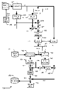
Moving now to Figure 9, the main software and hardware components that can be
25 used for practicing the solution according to an embodiment of the
invention are denoted
as a whole with the reference 900. The information (programs and data) is
typically stored
on the hard disk and loaded (at least partially) into the working memory when
the
programs are running, together with an operating system and other application
programs
(not shown in the figure). The programs are initially installed onto the hard
disk, for
CA 02670932 2014-12-18
26
example, from CD-ROM.
Particularly, a driver 903 controls the imaging probe (not shown in the
figure); for
example, the imaging probe driver 903 includes a transmit beam former and
pulsers for
generating the ultrasound pulses to be applied to the body-part under
analysis. The
corresponding (analog RF) echo signal that is received from said body-part is
supplied to a
receive processor 906. Typically, the receive processor 906 pre-amplifies the
analog RF
echo signal and applies a preliminary time-gain compensation (TGC); the analog
RF echo
signal is then converted into digital values by an Analog-to-Digital Converter
(ADC), and
combined into a focused beam signal through a receive beam former. The digital
signal so
obtained is preferably processed through further digital algorithms and other
linear or non-
linear signal conditioners (such as a post-beam-forming TGC). Particularly,
the receive
processor 906 applies a contrast-specific algorithm to suppress the
contribution of tissue
(such as based on the above-mentioned HI, PI, PM or CPS techniques). The
digital
signal is then demodulated, log-compressed, and scan-converted into a video
format. This
process results in the recording of a sequence of (video) input images Ii
(each one
including MxN pixel values). More specifically, each pixel value of the input
images Ii is
determined by the intensity of the acoustical response at the location in the
body-part
corresponding to said pixel.
Optionally, the receive processor 906 includes a motion compensation module,
carrying out a method for reducing the misalignment of the input images Ii
with respect to
a reference image (for example, due to motion resulting from patient breathing
or from
involuntary movement of the imaging probe); an example of motion compensation
method that is well suited for this purpose is described in WO-A-2006/015971.
The input images Ii are supplied to a pre-processor 907. The pre-processor 907
removes the contribution of the circulating contrast agent in the input images
Ii.
Preferably, the pre-processor 907 applies the method described in the above-
mentioned
International patent application No.PCT/EP2006/068305. In this way, each input
image
Ii is converted into a corresponding pre-processed image Ip; the pre-processed
image Ip
provides a representation of the (permanently-, temporarily- and apparently-)
CA 02670932 2014-12-18
27
immobilized contrast agent in a region of interest, which information is
preferably
overlaid on the original input images Ii.
A drawing module 909 is used to predefine a region-of-interest for the
analysis of
the pre-processed images Ip (typically the same as the one used by the pre-
processor 907).
The operation generates a limitation mask Ml, which consists of a matrix of
binary values
with the same size as the pre-processed images Ip (i.e., Mx/V); all binary
values inside
the region of interest are assigned the logic value 1, whereas the binary
values outside
the region of interest are assigned the logic value 0.
A linearizer 912 is optionally used to linearize the pre-processed images Ip,
so as
to make each pixel value thereof directly proportional to the corresponding
local
concentration of the immobilized contrast agent; for example, this result can
be achieved
by applying an inverse log-compression and then squaring the value so obtained
(for
example, as described in WO-A-2004/110279).
A selector 915 is used to select and latch one of the (video or linearized)
pre-
processed images Ip to be used as a background image (denoted with Ib); for
example, the
background image lb is selected among the pre-processed images Ip taken before
the
contrast agent has reached the body-part under analysis.
A multiplier operatOr 921 receives the background image lb (from the selector
915) and the limitation mask Ml (from the drawing module 909). The operator
921
multiplies the background image lb by the limitation mask MI pixel-by-pixel,
so as to
generate a corresponding limited background image Llb (this operation needs to
be done
only once, but it can be repeated any time during the analysis process).
Another
multiplier operator 924 receives the pre-processed images Ip in succession
(from the
linearizer 912) and the limitation mask Ml (from the drawing module 909). The
operator
924 multiplies each pre-processed image Ip by the limitation mask Ml pixel-by-
pixel, so
as to generate a corresponding sequence of limited pre-processed images Lip.
As a
result, the limited background image Llb and the limited pre-processed images
Llp only
include the pixel values of the background image lb and of the pre-processed
images Ip,
respectively, that are inside the region of interest (defined by the
limitation mask M1),
CA 02670932 2014-12-18
28
while the other pixel values are reset to 0.
A difference operator 927 receives the limited background image Mb (from the
multiplier 921) and the limited pre-processed images Lip (from the multiplier
924). The
operator 927 subtracts the limited background image LIb from each limited pre-
processed images Lip pixel-by-pixel, so as to remove any residual clutter (for
example,
due the contribution of tissue that has not been completely removed by the
contrast-
specific algorithm applied by the receive processor 906). The operation
generates a
corresponding sequence of corrected images k, which is provided to a spatial
sub-
sampler 933.
The module 933 sub-samples the corrected images k according to a factor
determined by the spatial resolution of the corrected images k along each
dimension (for
example, 2 to 6 pixels). Preferably, the spatial sub-sampling comprises low-
pass filtering
followed by sub-sampling according to a sub-sampling factor. The low-pass
filtering has a
cutoff frequency, which can be chosen as the highest frequency component
containing
significant energy in a selected one of the corrected images k (for example,
determined
by Fourier analysis). The sub-sampling is performed according to a factor that
can be
determined, for example, as a value resulting in a spatial sub-sampling
frequency equal to
twice the cutoff frequency. In this way, each corrected image k is transformed
into a
corresponding (spatially) sub-sampled image /s; each value of the sub-sampled
image Is
thus represents a cell corresponding to a group of adjacent pixels in the
correct image k
(which cell has a size defined according to the above-mentioned spatial
resolution).
The sub-sampled images /s so obtained are stored in succession into a stack
936,
which acts as a buffer memory for further processing according to the above-
described
cumulative difference algorithm. The stack 936 provides storage for q sub-
sampled
images Is. The value of q is determined by the choice of the comparison depth
/71 of the
cumulative difference algorithm and a temporal sub-sampling parameter p
(ranging from 0
to m-2), according to the relation q---(m+1)(p+1). The required set of m+1 sub-
sampled
images Ns among the ones available in the stack 936 (for the reference set of
m sub-
sampled image /s and the preceding sub-sampled image Is) is thus created and
made
CA 02670932 2014-12-18
29
available for further processing. In most practical situations, the sub-
sampling parameter
p is set to 0 so that q=m+I. The set of sub-sampled images Ms then consists of
the last
m+1 sub-sampled images Is stored in the stack 936 (so that every sub-sampled
image Is
is considered). Conversely, when the sub-sampling parameter p is higher than
0, q sub-
sampled images Is (q>m+1) must be stored in the stack 936, in order to make
n,+1 sub-
sampled images Is available for the application of the cumulative difference
algorithm.
This temporal sub-sampling may be advantageously exploited when the ultrasound
scanner works at ultra-high frame rates (for example, 100-500 images per
second), in
which case an analysis of every available sub-sampled image Is does not
provide any
useful benefit.
At the same time, the (original) pre-processed images Ip provided by the pre-
processor 907 are latched into another stack 937, which consists of a first-in-
first-out
(FIFO) shift register, with a size equal to q (so as to store the last q pre-
processed images
/P).
A filter 939 receives the set of (m+1) sub-sampled images Sis from the stack
936.
The filter 939 calculates a filtered image Ifs by applying the above-described
cumulative
difference algorithm on this set of sub-sampled images SR
The filtered image Ifs so obtained is then passed to a mask generator 942,
which
also receives a predefined threshold value TH for the cell values (for
example, ranging
from 0 to 5% of their maximum allowable value). The mask generator 942 creates
a
corresponding overlay mask Mo; the overlay mask Mo is obtained from the
filtered
image Ifs by assigning (to each cell) the logic value 1 if its value is
strictly higher than
the threshold value TH or the logic value 0 otherwise.
The overlay mask Mo is subsequently provided to a spatial-interpolator 945.
The
spatial-interpolator 945 restores the full-size of the overlay mask Mo
corresponding to the
size of the input images Ii (i.e., MxN binary values); for this purpose, the
value of each cell
in the overlay mask Mo is replicated for the corresponding group of pixels.
The operation
generates a corresponding interpolated mask RMo.
At the same time, the filtered image Ifs is also provided to a post-processor
948.
CA 02670932 2014-12-18
The post-processor 948 optionally converts the cell values of the filtered
image Ifs into
corresponding discrete values (for example, consisting of 64 or 128 levels
that are
uniformly distributed between the lowest value and the highest value of all
the cells), by
possibly applying a gain factor. Optionally, when the input images Ii are
linearized by the
5 module
912, the post-processor 948 may also apply a non-linear processing (such as a
log-
compression) so as to produce images with well-balanced contrast. The post-
processor
948 also accesses a color lookup table 951. The color lookup table 951
associates all the
possible levels with the representation of corresponding colors (that are
preferably brighter
as the levels increase); for example, each color is defined by an index for
accessing a
10 location
within a palette containing its actual specification. In this way, each cell
in the
filtered image Ifs is assigned the corresponding color representation.
The filtered image Ifs (either post-processed or as originally built) is
provided to
another spatial-interpolator 954. The spatial-interpolator 954 restores the
full-size of the
filtered image If corresponding to the size of the input images Ii (i.e., MxN
pixel values)
15 by means
of interpolation techniques (such as based on the nearest neighbor, bilinear,
or
bicubic technique). For this purpose, the value of each cell in the filtered
image Ifs is
replicated for the corresponding group of pixels (nearest neighbor
interpolation method)
and optionally filtered spatially (such as using a low-pass 2D or 3D spatial
filter). The
operation generates a corresponding interpolated image RI.
20 A
multiplier operator 957 receives the interpolated image RI (from the spatial
interpolator 954) and the interpolated mask RMo (from the spatial interpolator
945). The
operator 957 multiplies the interpolated image RI with the interpolated mask
Idlo pixel-
by-pixel, so as to obtain a masked image MI; as a result, the masked image MI
only
includes the pixel values of the corresponding interpolated image RI that are
higher than
25 the
threshold value TH (while the other pixel values are reset to 0). The
threshold value
TH allows tuning the level of masking of the interpolated image RI, down to
none when
TH=0; indeed, in this case every pixel of the overlay mask Mo and of the
interpolated
overlay mask RA/10 is at the logic value 1, so that the masked image MI will
be exactly
the same as the interpolated image RI. The masked image MI is then latched
into a
CA 02670932 2014-12-18
31
single-image buffer 958 (replacing its previous content). In this way, the
masked image
MI in the buffer 958 is updated whenever the filter 939 outputs a new filtered
image Ifs,
while it remains unchanged otherwise (so as to maintain the masked image MI
that was
obtained from the filtered image Ifs last calculated).
The interpolated mask RMo is also supplied from the spatial interpolator 945
to
an inverter 960, which generates a corresponding inverted interpolated mask
RMo (by
exchanging the logic values 0 and 1). The interpolated mask RMo is likewise
latched into
another single-image buffer 961 (replacing its previous content), so as to be
always
synchronized with the masked image MI in the buffer 958. The inverted
interpolated
mask RMo latched in the buffer 961 is then passed to a multiplier operator
963. The
multiplier operator 963 also receives a delayed image Id from the stack 937.
Every time
the inverted interpolated mask Nilo is latched into the buffer 961, the
corresponding
delayed image Id exits from the stack 937, thus allowing the operator 963 to
multiply the
delayed image Id and the inverted interpolated mask RMo pixel-by-pixel, so as
to obtain
a masked delayed image MId; as a result, the masked delayed image MId only
includes
the pixel values of the delayed image Id that are not included in the
corresponding
masked image MI (while the other pixel values are reset to 0).
An adder operator 969 receives the masked delayed image MId (from the
multiplier 963) and the masked image MI (latched in the buffer 958). The
operator 969
adds the masked image MI and the masked delayed image MId pixel-by-pixel
(correctly
synchronized) so as to obtain an overlay image Jo. In this way, each pixel
value of the
delayed image Id is overridden by the corresponding pixel value of the masked
image
MI if and only if the latter has a significant value (i.e., higher than the
threshold value
TN).
The overlay image Jo is passed to a monitor driver 972, which controls its
visualization. At the same time, the overlay image Jo may also be added to a
repository
975. The same operations described above are reiterated for each new input
image Ii that
is recorded. Particularly, the corresponding pre-processed image Ip is pushed
into the
stack 937; this causes the shifting of the preceding pre-processed images Ip
in the stack
CA 02670932 2014-12-18
32
937, and the output of the oldest one. At the same time (after optional
linearization,
limitation to the desired region of interest, subtraction of the background
image lb, and
spatial sub-sampling) the corresponding sub-sampled image Is is added to the
stack 936.
As a result, the overlay images lo are displayed in succession on the monitor
of the
ultrasound scanner; it should be noted that each overlay image Jo is available
with a
delay (with respect to the acquisition time of the corresponding input image
Ii), which is
defined by the time required to apply the modified Min_IP algorithm (in the
pre-
processor 907) plus the time required to apply the cumulative difference
algorithm (i.e.,
to cross the whole stack 937). Moreover, the sequence of overlay images lo so
obtained
is also available in the repository 975 for further analysis.
In this way, the detached contrast agent is easily recognized (by its color
coding)
with respect to the permanently-immobilized contrast agent and the possible
background
(in the original gray-scale pixel levels) ¨ with the degree of overlay that
can be updated
according to contingent requirements (by means of the threshold value TH), so
as to tune
the impact of the operation on the pre-processed images Ip. The reading of the
overlay
images Jo is further facilitated when each different color (representing the
concentration
of the immobilized contrast agent that has detached) bears a quantitative
meaning of its
own; for example, this value can be read out from a color bar, which is
displayed on the
monitor close to the sequence of overlay images Jo. Moreover, when the pre-
processed
images Ip show the immobilized contrast agent over the original input images
Ii, the
overlay images Jo provide an enhanced visual perception of the detached
contrast agent,
which is contextualized on the actual representation of the body-part under
analysis.
In any case, the proposed solution facilitates the spatial delineation and the
quantification of the permanently-immobilized contrast agent, the temporarily-
immobilized contrast agent, and/or the apparently-immobilized contrast agent,
thereby
allowing the correct detection of the pathologies of interest. Therefore, the
accuracy of any
analysis of the obtained results is strongly increased.
CA 02670932 2014-12-18
33
Modifications
Naturally, in order to satisfy local and specific requirements, a person
skilled in
the art may apply to the solution described above many modifications and
alterations.
More specifically, although the present invention has been described with a
certain
degree of particularity with reference to preferred embodiment(s) thereof, it
should be
understood that various omissions, substitutions and changes in the form and
details as
well as other embodiments are possible. Particularly, the proposed solution
may even be
practiced without the specific details (such as the numerical examples) set
forth in the
preceding description to provide a more thorough understanding thereof;
conversely,
well-known features may have been omitted or simplified in order not to
obscure the
description with unnecessary particulars. Moreover, it is expressly intended
that specific
elements and/or method steps described in connection with any disclosed
embodiment of
the invention may be incorporated in any other embodiment as a matter of
general
design choice.
For example, the proposed solution lends itself to be implemented with an
equivalent method (by using similar steps, removing some steps being non-
essential, or
adding further optional steps); moreover, the steps may be performed in a
different
order, concurrently or in an interleaved way (at least in part).
According to an alternative embodiment, the pixel values outside the selected
region of interest may be reset to 0 (so that the portion of the overlay image
outside the
region of interest is black); however, the application of the proposed
solution to the
whole content of the images is contemplated (without selecting any region of
interest).
It is emphasized that the above-described applications of the proposed
solution
are merely illustrative, and they must not be interpreted in a 'imitative
manner. For
example, it is possible to administer a target-specific contrast agent in its
basic
formulation without the relevant ligand; the detection of any detached
contrast agent
allows identifying undesired interactions of the target-specific contrast
agent with other
passive targets.
Likewise, the solution of the invention lends itself to be put into practice
with
CA 02670932 2014-12-18
34
equivalent target-specific contrast agents for whatever (biological) target;
for example,
the contrast agent can be specific for enhancing Magnetic Resonance imaging or
X-ray
Computed Tomography imaging. However, the application of the same solution to
a non
target-specific contrast agent - for example, for detecting the slowly-moving
contrast
agent only - or even to a mixture of target-specific contrast agent and non
target-specific
contrast agent, is not excluded.
As described in detail above, the solution according to an embodiment of the
present invention is preferably put into practice by starting from images
wherein the
contribution of the circulating contrast agent has already been removed (or at
least
substantially reduced); this result may be achieved by applying the modified
Min-IP
algorithm. However, it is also possible to wait until the circulating contrast
agent has
disappeared, or to apply any other algorithm providing equivalent outcomes.
Alternatively, the same result may also be achieved by acting directly on the
original
images (i.e., removing the circulating contrast agent and the
temporarily/apparently-
immobilized contrast agent at the same time). Anyway, the solution according
to the
present invention also lends itself to be implemented with whatever algorithm,
which is
capable of detecting the contrast agent leaving any location after being
substantially
immobilized thereon (for a period of time longer that a predefined duration).
For
example, in a very simplified implementation it is possible to compare the
images taken
during the analysis process (with the permanently-, temporarily- and
apparently-
immobilized contrast agent) with an image taken at the end of the analysis
process
(wherein the temporarily/apparently-immobilized contrast agent disappeared to
leave the
permanently-immobilized contrast agent only).
Although the proposed technique finds its preferred application for monitoring
the evolution of the body-part over time during the analysis process, nothing
prevents
providing a single image at a specific instant (for example, at the end of the
wash-in
phase). In any case, each filtered value may be calculated by accumulating the
preceding
variation values in a different way; for example, it is possible to avoid
processing the
CA 02670932 2014-12-18
pixels once they have become white, or to calculate the filtered values
directly according
to the history of the corresponding pixel values.
Although the present invention has been specifically designed for use in real-
time, the analysis of the obtained results off-line is within the scope of the
invention.
5 Moreover,
the proposed cumulative difference algorithm may be implemented by
setting the comparison value to either the current pixel value or a comparison
value
based on a comparison set of pixel values at multiple instants; in the latter
case, the set
may have any other size, even defined dynamically according to an estimated
quality of
the available images.
10
Alternatively, the temporal sub-sampling of the input images may be performed
according to any other criteria (or it may be omitted altogether).
The use of different formulas for setting the comparison value (based on the
corresponding set of pixel values) is contemplated.
The solution described above assumes a direct relation between the amplitude
of
15 the echo
signal and the corresponding pixel value (i.e., a larger amplitude of the echo
signal results in a brighter pixel). Conversely, in a system based on negative
images
(wherein the pixel values decrease with the amplitude of the echo signal) all
the
equations given above would need to be modified to reflect the reverse logic.
Likewise, any other formula may be used to calculate the variation value to be
20
accumulated (so as to indicate the variation of the comparison value with
respect to the
reference value, consisting of either the preceding pixel value or a value
based on a
reference set of multiple preceding pixel values). More generally, the
filtered images
may be generated by any other algorithm capable of detecting the different
persistence
of the contrast agent at each location (as defined by the time pattern of the
corresponding
25 pixel values).
Moreover, any other technique for acquiring the input images is within the
scope
of the present invention (for example, using Doppler-based algorithms).
Alternatively,
the original images used as background in the overlay may also be based on non
CA 02670932 2014-12-18
36
contrast-specific images (such as fundamental B-mode images being obtained
from the
echo signals of the imaging probe driver). Naturally, the proposed method
would still be
applied in a preferred way on images provided by a contrast-specific imaging
modality
for generating the filtered images.
It should be appreciated that the feature relating to the subtraction of the
background image is not strictly necessary (and it may be omitted in some
implementations of the invention).
Similar considerations apply if the images are spatially sub-sampled with a
different procedure (for example, according to a predefined sub-sampling
factor), or if
the spatial sub-sampling is performed beforehand or afterward; in any case,
the
application of the proposed solution at the pixel level (instead of at the
level of groups of
pixels defined by the above-mentioned spatial sub-sampling) is not excluded.
Likewise, it is also possible to omit compensating the motion of the input
images
(for example, when this motion is far slower than the flow of the circulating
contrast
agent).
Alternatively, the images may be linearized in a different way; for example,
the
linearized images might be already available for other purposes (such as when
parametric analysis techniques are implemented); in this case, it is possible
to exploit the
available information without any additional linearization operation. Anyway,
nothing
prevents the application of the proposed solution to the log-compressed images
directly.
It is also possible to leave the choice of overlaying the filtered images on
the pre-
processed images to the preference of a user; for example, the pixel values of
the
(original) pre-processed images within the region of interest may be set to
zero in order
to display the filtered images against a black background, thus improving
contrast. More
generally, the obtained information may be used in any other way. For example,
it is
possible to display the filtered images alone, to overlay the filtered images
on the input
images (without the pre-processing to remove the contribution of the
circulation contrast
agent), to overlay the filtered images on the pre-processed images within the
region of
interest and on the input images outside the region of interest, and in any
other
CA 02670932 2014-12-18
37
combination thereof; moreover, it is possible to subtract the filtered images
from the pre-
processed images to remove the contribution of the temporarily/apparently-
immobilized
contrast agent.
Alternatively, any other different visual coding may be used to differentiate
the
detached contrast agent from the permanently-immobilized contrast agent in the
overlay
images; for example, it is possible to use shades of a first color (such as
yellow) for the
detached contrast agent, and shades of a second color (such as red) for the
permanently-
immobilized contrast agent (over a background in gray scale).
Similar considerations apply if the program (which may be used to implement
each embodiment of the invention) is structured in a different way, or if
additional
modules or functions are provided; likewise, the memory structures may be of
other
types, or may be replaced with equivalent entities (not necessarily consisting
of physical
storage media). In any case, the program may take any form suitable to be used
by any
data processing system or in connection therewith (for example, within a
virtual
machine); particularly, the program may be in the form of external or resident
software,
firmware, or microcode (either in object code or in source code ¨ for example,
to be
compiled or interpreted). Moreover, it is possible to provide the program on
any
computer-usable medium; the medium can be any element suitable to contain,
store,
communicate, propagate, or transfer the program. For example, the medium may
be of
the electronic, magnetic, optical, electromagnetic, infrared, or semiconductor
type;
examples of such medium are fixed disks (where the program can be pre-loaded),
removable disks, tapes, cards, wires, fibers, wireless connections, networks,
broadcast
waves, and the like. In any case, the solution according to the present
invention lends
itself to be implemented with a hardware structure (for example, integrated in
a chip of
semiconductor material), or with a combination of software and hardware.
Similar considerations apply if the ultrasound scanner has a different
structure or
includes other units (such as with an imaging probe of the linear-, convex-,
phased-, or
matrix- array type). Alternatively, the proposed solution is applied in a
medical imaging
system that consists of an ultrasound scanner and a distinct computer (or any
equivalent
CA 02670932 2014-12-18
38
data processing system); in this case, the measured data is transferred from
the
ultrasound scanner to the computer for its processing (for example, through a
removable
disk, a memory key, or a network connection). In any case, the application to
any other
medical imaging system, such as based on Magnetic Resonance Imaging (MRI) or X-
ray
Computed Tomography (CT), is within the scope of the invention.