Note : Les descriptions sont présentées dans la langue officielle dans laquelle elles ont été soumises.
CA 02673877 2009-06-25
WO 2008/100675
PCT/US2008/051289
SPECIFICATION
TITLE
"ENHANCED SIGNAL DETECTION FOR ACCESS DISCONNECTION
SYSTEMS"
PRIORITY CLAIM
[001] This application claims priority to and the benefit as a continuation-in-
part application of U.S. Patent Application "Conductive Polymer Materials And
Applications Thereof Including Monitoring And Providing Effective Therapy",
Serial
No. 10/760,849, filed January 19, 2004, which is a continuation-in-part
application of
U.S. Patent Application "Access Disconnection Systems And Methods", Serial No.
10/121,006, filed April 10, 2002.
BACKGROUND
[0021 ¨The ____ pieseardisclosure -relates-generally -to -patient access
disconnection
systems and methods for medical treatments. More specifically, the present
disclosure
relates to the detection of patient access disconnection, such as dislodgment
of a
patient access device during medical treatments or therapies including
dialysis therapy.
[003] A variety of different medical treatments relate to the delivery of
fluid
to and/or from a patient, such as the delivery of blood between a patient and
an
extracorporeal system connected to the patient via a needle or needles or any
suitable
access device inserted within the patient. For example, hemodialysis,
hemofiltration
and hemodiafiltration are all treatments that remove waste, toxins and excess
water
directly from the patient's blood. During these treatments, the patient is
connected to
an extracoporeal machine, and the patient's blood is pumped through the
machine.
Waste, toxins and excess water are removed from the patient's blood, and the
blood is
infused back into the patient. Needles or other suitable access devices are
inserted into
the patient's vascular access in order to transfer the patient's blood to and
from the
extracoporeal machine. Traditional hemodialysis, hemofiltration and
hemodiafiltration
treatments can last several hours and are generally performed in a treatment
center
about three to four times per week.
[004) During any of these blood treatments, dislodgment of the access device
can occur, such as dislodgment of a needle inserted into the patient's
vascular access
including an arterio-venous graft or fistula. If not detected immediately,
this can
1
CA 02673877 2009-06-25
WO 2008/100675 PCT/US2008/051289
produce a significant amount of blood loss to the patient. The risks
associated with a
needle dislodgment are considerable. Important criteria for monitoring blood
loss
include, for example, the sensitivity, specificity and response time with
respect to the
detection of needle dislodgment. With increased levels of sensitivity,
specificity, and
response time, the detection of needle dislodgment can be enhanced, and blood
loss
due to dislodgment can be minimized.
[005] Typically, patients undergoing medical treatment, such as hemodialysis,
hemofiltration or hemodiafiltration, are visually monitored in order to detect
needle
dislodgment. However, the needle may not be in plain view of the patient or
medical
staff (i.e., it may be covered by a blanket) such that it could delay
detection and, thus,
responsive actions to be taken in view of dislodgment, such as stopping the
blood
pump of the extracorporeal machine to minimize blood loss to the patient.
[006] Moreover, in view of the increased quality of life, observed reductions
in both morbidity and mortality and lower costs than in-center treatments, a
renewed
interest has arisen for self care and home hemodialysis therapies. Such home
hemodialysis therapies (whether hemodialysis, hemofiltration or
hemodiafiltration)
allow for both nocturnal as well as daily treatments. During these self care
and home
hemodialysis sessions, especially during a nocturnal home hemodialysis
session, when
the patient is asleep, dislodgment risks are more significant because nurses
or other
attendants are not present to detect the dislodgment.
[007] A need exists to make an access disconnection ("ADS") system operate
as quickly as possible to minimize blood loss.
[008] A need also exists to make the ADS system operate to without false
triggers, which needlessly disrupt therapy and the patient.
[009] A further need exists to provide such an ADS system readily and
relatively inexpensively to machines already in use which may not have an ADS
system or one that operate as well as the systems described herein.
SUMMARY
[010] The present disclosure provides improved devices, apparatuses,
systems, and methods for detecting dislodgment or disconnection of an access
device,
such as dislodgment of a needle inserted in a patient during dialysis therapy.
The
devices, apparatuses, systems and methods of the present disclosure utilize an
electrical circuit with a number of electrical contacts which are in contact
with the
2
CA 02673877 2009-06-25
WO 2008/100675 PCT/US2008/051289
fluid circuit such that an electrical signal can be injected into at least a
segment
including, for example, a loop defined along at least a portion of the
conducting fluid
circuit. A direct-contact measurement can be used to provide immediate
detection of a
change in an electrical value in response to a change in access conditions,
such as a
change in impedance due to dislodgment of a needle or other access device from
the
patient during medical therapy including, for example, dialysis therapy and
medication
delivery.
[011] The devices, apparatuses, systems and methods of the present
disclosure in one embodiment are provided in a stand-alone, retrofit package
that can
be mounted to and made operable with an existing machine not having an ADS
system
or one that operates as well as the systems described herein. The stand-alone
ADS
system includes a detector module mounted to and operable with the blood
tubing set
and a protector module mounted to and made operable with the blood treatment,
e.g.,
hemodialysis machine. The detector module and protector module communicate
wirelessly, e.g., via radio frequency, in one embodiment. The detector module
detects
an access disconnection and sends a corresponding output to the protector
module,
which is configured then to clamp one or both the venous and arterial tubing.
The
clamping of the tubing will cause an increase in pressure, for example in the
venous
line, which is detected by the hemodialysis machine and causes the blood pump
to shut
down.
[0121 Described below are multiple systems for enhancing impedance signal
output and reducing disposable cost. One problem with the impedance sensing
systems is the effect of patient grounding. The dialysis system is connected
to earth
ground through the dialyzer and dialysate path for safety reasons. The patient
can
become electrically at the same potential as earth ground despite attempts to
shield the
patient. When this happens a ground current path can exist between the patient
and the
system's isolated ground even upon a needle dislodgment, making the system
ineffective.
[013] One apparatus and method for combating the effects of patient
grounding is to use a two outputs of a signal source to create two points of
equal
potential on either side of a section of the dialysate or ground path. This
causes a
virtual open circuit to exist between the points, breaking the current path
from earth
ground to the system's isolated ground.
3
CA 02673877 2009-06-25
WO 2008/100675 PCT/US2008/051289
[014] This technique is used alternatively or additionally to stop a portion
of
the signal current from flowing through the blood pump. In the systems below,
current
is induced into the blood using a voltage source and high resistance resistor.
The
current is normally split into two paths, one going through patient access,
the other
through the blood pump. It is desirable from a sensing standpoint for all of
the current
to go through the patient access path and none through the blood pump path. If
too
much current flows through the blood pump path, the increase in impedance due
to a
needle dislodgment may not be great enough to overcome a threshold. The above-
described open circuit creating circuitry is accordingly placed in the blood
pump path.
This virtually precludes current from flowing through blood in the blood pump
path,
forcing virtually all the current through the patient access pathway.
[015] Another apparatus and method for combating the effects of patient
grounding is to move the blood circuit contacts as close to the patient as
possible. It
has been determined that reducing the impedance from the blood circuit
contacts to the
patient access is beneficial at least in part because it minimizes the effect
of parallel
current paths. This can be done by moving the blood circuit contacts as close
to the
patient as possible, minimizing the tubing lengths from the contacts to the
patient
access and thus the impedance in such tubing lengths.
[016] A further apparatus and method for combating the effects of patient
grounding is to place a second sensing circuitry in the ground current path,
e.g., in the
dialysate path. Here, a pair of contacts is added to the dialysate tubing.
These contacts
are not disposable and therefore do not add to disposable cost. The impedance
in the
ground path increases upon a needle dislodgement. A patient to earth ground
path
alone can be used to detect a patient access. If the patient is not grounded,
the circuitry
detects this condition and causes the system to use the blood circuit sensing
circuitry
instead for access disconnection detection. In a further alternative
implementation, the
system combines the signals from the blood circuit circuitry and the ground
loop
circuitry to detect and access disconnection.
[017] Further described below is a system and method for detecting an access
disconnection using the dialysate path. Here, multiple sets of contacts are
placed in
the to- and from- dialyzer dialysate tubing. The above-described open circuit
creating
circuitry is connected to two of the contact pairs, creating electrical
opening circuits
between the contacts. This forces all induced current induced into the
dialysate circuit
4
CA 02673877 2009-06-25
=
WO 2008/100675 PCT/US2008/051289
to flow through the dialyzer, the blood circuit and patient access to ground.
The
patient is grounded so that the current returns through earth ground to the
system's
isolated ground. The system provides a ground strap connected to the patient,
e.g.,
through a blood pressure cuff. The ground strap can be connected to earth or
system
ground. This system is advantageous because it places the fluid contacts in
the
dialysate path where they do not have to be discarded after each therapy.
[018] An advantage of the present disclosure is to provide an improved
device, apparatus, system and/or method for detecting access disconnection.
[019] A further advantage of the present disclosure is to provide an improved
device, apparatus, system and/or method for detecting dislodgment of an access
device
from a patient during medical therapy including dialysis therapy.
[020] Another advantage of the present disclosure is to provide an improved
device, apparatus, method and/or system for detecting needle drop-out during
dialysis
therapy.
[0211 Yet another advantage of the present disclosure is to provide a
sensitive, specific and responsive apparatus and/or device for detecting
access
disconnection during selfcare and home hemodialysis treatments.
[022] Moreover, an advantage of the present disclosure is to provide a viable
device or apparatus for allowing a patient or other non-medical personnel in a
non-
medical facility to administer a dialysis therapy that uses a portion of the
patient's
circulatory system.
[023] Still further, an advantage of the present disclosure is to provide an
improved apparatus for detecting access disconnection that uses a direct
conductivity
measurement.
[024] Yet still further, an advantage of the present disclosure is to provide
an
access disconnection detection device, method and/or system that employs an
electrical circuit in fluid and electrical contact with blood flowing through
a blood
circuit allowing direct conductivity measurements to be made.
[025] Furthermore, an advantage of the present disclosure is to provide an
improved device, system and method for monitoring and/or controlling blood
loss
from a patient.
[026] Another advantage of the present disclosure is an improved method for
dialysis that employs an apparatus, device, and/or system capable of detecting
access
CA 02673877 2009-06-25
=
WO 2008/100675
PCT/US2008/051289
disconnection, such as dislodgment of a needle inserted into a patient through
which
blood flows during dialysis therapy, and minimizing any resulting blood loss.
[027] Yet another advantage of the present disclosure is an improved device
for connecting an electrical contact to a fluid circuit allowing fluid and
electrical
communication between the electrical contact and fluid flowing through the
fluid
circuit.
[028] Still another advantage of the present disclosure is an improved
apparatus, device, system and/or method for detecting access disconnection,
such as
needle drop-out during dialysis therapy, with enhanced sensitivity, accuracy
and
responsiveness.
[029] Yet still another advantage of the present disclosure are improved
apparatuses, devices, systems and/or methods for the detection of fluid loss
due to, for
example, dislodgment of a single access device during medical therapies, for
example,
medication delivery and single needle hemodialysis therapies.
[030] Yet still a further advantage of the present disclosure are improved
apparatuses, devices, systems and/or methods for the detection of fluid loss
due to, for
example, dislodgment of a single access device during medical therapies, which
can be
retrofitted to an existing machine not having an ADS system or one that
operates as
well as the systems described herein.
[031] A further still advantage of the present disclosure is to provide an
access disconnection system that combats the effects of patient grounding and
enhances the sensed impedance by creating an open circuit in the ground
current path.
[032] Another advantage of the present disclosure is to provide an access
disconnection system that combats the effects of patient grounding by moving
the
blood contacts close to the patient.
[033] A further advantage of the present disclosure is to provide an access
disconnection system that combats the effects of patient grounding by placing
a second
sensing circuitry in the ground current loop.
[034] Yet another advantage of the present disclosure is to provide an access
disconnection system that places the fluid contacts in the dialysate circuit,
such that the
contacts are not disposable.
6
CA 02673877 2015-05-22
[034a] Yet another advantage of the present disclosure is to provide an access
disconnection system comprising: an extracorporeal circuit including a blood
pump and a
dialyzer; arterial and venous contacts provided in the extracorporeal circuit;
arterial and
venous patient access apparatuses; a signal source communicating with at least
one of the
arterial and venous contacts and configured to generate a signal within fluid
flowing
through the extracorporeal circuit; and a sensing apparatus configured to
sense a first
portion of the signal indicative of a first impedance produced by fluid
flowing through a
first portion of the extracorporeal circuit, the first impedance including at
least one of an
impedance produced by fluid flowing through a portion of the extracorporeal
circuit
between the venous contact and the venous patient access apparatus or an
impedance
produced by fluid flowing through a portion of the extracorporeal circuit
between the
arterial contact and the arterial patient access apparatus, wherein at least
one of the
arterial and venous contacts is placed close enough to a respective arterial
and venous
patient access apparatus such that a second portion of the signal indicative
of a second
impedance produced by fluid flowing through a second portion of the
extracorporeal
circuit increases relative to the first portion of the signal to an extent
that the effects of a
parallel ground loop can be ignored, the second impedance including at least
one of an
impedance produced by fluid flowing through a portion of the extracorporeal
circuit
between the venous contact and the dialyzer or an impedance produced by fluid
flowing
through a portion of the extracorporeal circuit between the arterial contact
and the blood
pump.
[034b] Yet another advantage of the present disclosure is to provide an access
disconnection system comprising: an extracorporeal circuit including a blood
pump and a
dialyzer; arterial and venous contacts provided in the extracorporeal circuit;
arterial and
venous patient access apparatuses; a signal source communicating with at least
one of the
arterial and venous contacts and configured to generate a signal within fluid
flowing
through the extracorporeal circuit; and a sensing apparatus configured to
sense a first
portion of the signal indicative of a first impedance produced by fluid
flowing through a
first portion of the extracorporeal circuit, the first portion of the
extracorporeal circuit
including the arterial and venous patient access apparatuses, the first
impedance including
at least one of an impedance produced by fluid flowing through a portion of
the
extracorporeal circuit between the venous contact and the venous patient
access apparatus
or an impedance produced by fluid flowing through a portion of the
extracorporeal circuit
between the arterial contact and the arterial patient access apparatus,
wherein at least one
of the arterial and venous contacts is placed close enough to a respective
arterial and
6a
CA 02673877 2015-05-22
venous patient access apparatus such that a second portion of the signal
indicative of a
second impedance produced by fluid flowing through a second portion of the
extracorporeal circuit is increased relative to the first portion of the
signal to an extent that
the second portion of the signal can be effectively ignored, the second
impedance
including at least one of an impedance produced by fluid flowing through a
portion of the
extracorporeal circuit between the venous contact and the dialyzer or an
impedance
produced by fluid flowing through a portion of the extracorporeal circuit
between the
arterial contact and the blood pump.
[034c] Yet another
advantage of the present disclosure is to provide a use of a
signal for detecting access disconnection of at least one of an arterial or
venous patient
access apparatus of an extracorporeal circuit, the signal being capable of
being
generated, within fluid flowing through the extracorporeal circuit, via at
least one of an
arterial or a venous contact provided in the extracorporeal circuit, a first
portion of the
signal being sensible, the first portion of the signal being indicative of a
first impedance
produced by fluid flowing through a first portion of the extracorporeal
circuit, the at least
one of the arterial or venous contacts being located at a first location in
the
extracorporeal circuit, and if at least one of the arterial or venous patient
access
apparatuses becomes disconnected: (a) the first impedance increases above a
threshold
amount if a parallel ground loop does not exist, and (b) the first impedance
remains
below the threshold amount if the parallel ground loop exists, and at least
one of the
arterial or venous contacts being located at a second location in the
extracorporeal circuit
between the first location and the respective arterial and venous patient
access apparatus,
such that a second portion of the signal indicative of a second impedance
produced by
fluid flowing through a second portion of the extracorporeal circuit is
increased relative
to the first portion of the signal indicative of the first impedance, and such
that if at least
one of the arterial or venous patient access apparatuses becomes disconnected,
the first
impedance increases above the threshold amount regardless of whether or not
the parallel
ground loop exists.
6b
CA 02673877 2009-06-25
WO 2008/100675 PCT/US2008/051289
[035] Additional features and advantages of the present disclosure are
described in, and will be apparent from, the following Detailed Description
and the
figures.
BRIEF DESCRIPTION OF THE FIGURES
[036] Fig. 1A illustrates a schematic view of an embodiment of the present
disclosure showing two needles insertable within a patient through which blood
flows
to and from an extracorporeal system.
[037] Fig. 1B illustrates a schematic view of an embodiment of the present
disclosure capable of detecting needle dislodgment during dialysis therapy.
[038] Fig. IC illustrates a perspective view of an embodiment of the present
disclosure showing access disconnection detection capabilities during medical
therapies administered via a single needle.
[039] Fig. 2A illustrates an exploded view of an electrical contact coupling
device in an embodiment of the present disclosure.
[040] Fig. 2B illustrates a side sectional view of the coupling device of
Fig. 2A in an embodiment of the present disclosure.
[041] Fig. 2C illustrates another embodiment of the coupling device of the
present disclosure.
[042] Fig. 2D illustrates another embodiment of the coupling device of the
present disclosure showing a threaded engagement between the components of
same.
[043] Fig. 2E illustrates a sectional view of Fig. 2D.
[044] Fig. 3 schematically illustrates an embodiment of the present disclosure
relating to processing of a measurable voltage signal to correct for changes
in baseline
impedance during treatment.
[045] Fig. 4A schematically illustrates a hemodialysis machine in an
embodiment of the present disclosure.
[046] Fig. 4B schematically illustrates a hemodialysis machine coupled to a
patient's access via a tubing set in an embodiment of the present disclosure.
[047] Figs. 5A and 5B illustrate a coupler according to an embodiment of the
present disclosure.
[048] Fig. 6 illustrates a dialyzer according to an embodiment of the present
disclosure.
7
CA 02673877 2009-06-25
WO 2008/100675 PCT/US2008/051289
[049] Figs. 7A and 7B illustrate a sensor assembly according to an
embodiment of the present disclosure.
[050] Figs. 8A and 8B illustrate a single-piece sensor according to an
embodiment of the present disclosure.
[051] Fig. 9 illustrates a multi-chamber bag according to an embodiment of
the present disclosure.
[052] Fig. 10 illustrates a multi-chamber bag with a peelable seal according
to
an embodiment of the present disclosure.
[053] Fig. 11 illustrates an automated peritoneal dialysis system according to
an embodiment of the present disclosure.
[054] Fig. 12 is an isometric view of one embodiment of an access
disconnection system employing the access disconnection methods herein, which
can
be retrofitted readily to an existing blood treatment machine.
[055] Fig. 13 schematically illustrates a detector module of the access
disconnection system of Fig. 12.
[056] Fig. 14 schematically illustrates a protector module of the access
disconnection system of Fig. 12.
[057] Fig. 15 is a schematic electrical diagram modeling a blood circuit and a
parallel earth ground path.
[058] Fig. 16 is a schematic electrical diagram modeling a blood circuit and
sensing circuitry without a parallel earth ground path showing both needles
lodged.
[059] Fig. 17 is a schematic electrical diagram modeling a blood circuit and
sensing circuitry without a parallel earth ground path showing at least one
needle
dislodged.
[060] Fig. 18 is a schematic electrical diagram modeling a blood circuit and
sensing circuitry with a parallel earth ground path showing at least one
needle
dislodged and a parallel current path to isolated ground through earth ground.
[061] Fig. 19 is a plot of digitized sensed patient grounded lodged versus
patient grounded dislodged impedance signals versus kOhms for the system of
Fig. 18.
[062] Fig. 20 is a schematic electrical diagram showing one system for
combating the effects of patient grounding and for increasing the impedance
through
the blood tubing operating with the peristaltic pump.
8
CA 02673877 2009-06-25
WO 2008/100675 PCT/US2008/051289
[063] Fig. 21 is a plot of digitized sensed patient grounded lodged versus
patient grounded dislodged impedance signals versus kOhms for the system of
Fig. 20.
[064] Fig. 22 is a plot for another system for combating the effects of
patient
grounding showing digitized sensed impedance signals for high and low
conductivity
blood for different blood contact spacings from patient access.
[065] Fig. 23 is a schematic electrical diagram showing a further system for
combating the effects of patient grounding using dual sensing circuitry, a
first circuitry
sensing impedance in the blood path, the second circuitry sensing impedance in
the
ground path.
[066] Figs. 24 to 27 are various plots of digitized sensed lodged versus
dislodged impedance signals versus kOhms for both sensing circuitries of the
system
of Fig. 23.
[067] Fig. 28 is a schematic electrical diagram showing a further system for
combating the effects of patient grounding and using the dialyste path for
sensing
impedance with both needles lodged.
[068] Fig. 29 is a schematic electrical diagram for the system of Fig. 28 with
the venous needle dislodged.
[069] Figs. 30 to 32 are various plots of output voltage or digitized
impedance
signal versus time for the dialysate sensing system of Figs. 28 and 29.
[070] Fig. 33 shows the hardware associated with the dialysate sensing
system of Figs. 28 and 29.
DETAILED DESCRIPTION
[071] The present disclosure provides medical devices, apparatuses, systems
and methods for detecting access disconnection. More specifically, the present
disclosure provides medical devices, apparatuses, systems, and methods that
employ,
in part, an electrical circuit with electrical contacts in fluid contact and
electrical
communication with a fluid circuit allowing a direct conductivity measurement
to be
used such that dislodgment of a needle or other access device through which
fluid
flows between a patient and the fluid circuit can be immediately detected.
Fluid loss
(e.g., blood loss) due to, for example, dislodgment of a needle from a patient
undergoing medical treatment, such as dialysis therapy, medication delivery or
the
like, can be controllably minimized.
9
CA 02673877 2009-06-25
WO 2008/100675
PCT/US2008/051289
[072] It should be appreciated that the present disclosure is not limited to
the
detection of needle dislodgment but can be utilized to detect the dislodgment
or
disconnection of any suitable access device. As used herein, the term "access
disconnection" or other like term can mean any suitable condition or event
which can
cause a loss or leak of an electrically conductive fluid flowing along a fluid
circuit
connected to the patient provided that a change in the electrical continuity
between
electrical contacts coupled to the fluid circuit can be detected. It should be
appreciated
that a change in the electrical continuity as measured by an electrical value,
such as
impedance, may be detected even in the absence of dislodgment of an access
device
from the patient. The term "access device" as used herein or other like term
can mean
a suitable device that can be inserted within a patient such that fluid,
including blood,
can pass to, through and/or from the patient via the access device. The access
device
can include a variety of different and suitable shapes, sizes and material
make-up.
Examples of an access device include needles, catheters and cannulas. The
access
device can be made of any suitable material including, for example, stainless
steel,
plastic or like biocompatible materials.
[073] Although in the embodiment set forth below the apparatus and/or
device is designed for use in a dialysis therapy, such as hemodialysis,
hemofiltration or
hemodiafiltration, it should be noted that the present disclosure can be used
in a
number of different medical therapies that employ a variety of different and
suitable
fluid systems, such as extracorporeal blood systems. For example, the systems
of the
present disclosure can be used during intravenous infusion that can employ the
use of a
single needle insertable within the patient for delivering a medical solution
or drug,
blood, blood products, processed blood or the like between the patient and the
fluid
system. In addition, the systems of the present disclosure can be used in
plasma
exchange therapies, in which a membrane is used to separate whole blood into
plasma
and cellular components.
[074] With respect to dialysis therapy, the systems of the present disclosure
can be used in a variety of different therapies to treat kidney failure.
Dialysis therapy
as the term or like terms are used throughout the text is meant to include and
encompass any and all forms of therapies that utilize the patient's blood to
remove
waste, toxins and excess water from the patient. Dialysis
therapies include
hemodialysis, hemofiltration, hemodiafiltration, and continuous renal
replacement
CA 02673877 2009-06-25
WO 2008/100675 PCT/US2008/051289
therapy ("CRRT"). CRRT includes slow continuous ultrafiltration ("SCUF"),
continuous veno-venous hemofiltration ("CVVH"), continuous veno-hemodialysis
("CVVHD") and continuous veno-venous hemodiafiltration ("CVVHDF"). Dialysis
therapy can also include peritoneal dialysis, such a continuous ambulatory
peritoneal
dialysis, automated peritoneal dialysis and continuous flow peritoneal
dialysis.
Further, although the present disclosure, in an embodiment, can be utilized in
methods
providing a dialysis therapy for patients having chronic kidney failure or
disease, it
should be appreciated that the present disclosure can be used for acute
dialysis needs,
for example, in an emergency room setting. Last, the intermittent forms of
therapy
(i.e., hemofiltration, hemodialysis and hemodiafiltration) may be used in in-
center,
self/limited care as well and home settings.
[075] In an embodiment, the systems of the present disclosure include an
electrical circuit with a number of electrical contacts, such as a pair of
electrical
contacts, in fluid contact and electrical communication with the fluid
circuit. The
electrical contacts can include any suitable device through which electrical
connection
can be made with the fluid circuit to define a conductive pathway or conductor
loop
therein. Changes in an electrical value or any suitable parameter associated
with the
conductor loop can then be monitored in response to changes in access
conditions as
described below. In an embodiment, the electrical contact includes an
electrode which
can be coupled to the fluid circuit such that an electrical connection can be
made in
fluid contact with fluid flowing through the fluid circuit as discussed below.
[076] For example, a constant current or other suitable electrical signal can
be
injected into the fluid circuit via an electrode pair in contact with the
fluid flowing in
between the electrodes to define a loop along at least a portion of the
conducting fluid
circuit. A change in an electrical value, e.g., impedance, can then be
measured in
response to access disconnection. This can provide a direct conductivity
measurement
capable of detecting a change in impedance or other suitable electrical
parameter of the
fluid, such as an electrically conductive fluid including blood, medical
solutions or the
like, as it flows between a patient and a fluid system (i.e., an
extracorporeal blood
system) via a needle, needles or other access device(s) inserted within the
patient.
[077] The systems of the present disclosure can effectively detect
dislodgment of a needle (e.g., a venous needle and/or an arterial needle) or
other
access device through which blood or other suitable fluid can flow, for
example, to,
11
CA 02673877 2009-06-25
WO 2008/100675 PCT/US2008/051289
through, and from the patient, such as a blood circuit used during dialysis
therapy.
The detection capability of the present disclosure is believed to be immediate
based on
the measurable change in, for example, impedance of the electrically
conductive fluid
or fluids due to fluid loss resulting from disconnection of the access device
from the
patient.
[078] The immediate detection capabilities of the present disclosure are
important, particularly as applied to dialysis therapy where a significant
amount of
blood loss can occur within a relatively short period of time if delays in
detection and
responsive actions to stop the blood loss occur. Under typical dialysis
conditions, if
twenty seconds or more time elapses before blood loss due to dislodgment is
detected
and stopped, over one-hundred milliliters of blood can be lost based on
typical blood
flow rates of four-hundred milliliters/minute.
[079] Applicants have discovered that the present disclosure can detect access
disconnection, particularly in response to venous needle dislodgment during
dialysis
therapy, with a high degree of sensitivity and specificity in addition to its
immediate
detection capabilities. The direct-contact measurement of the present
disclosure is
capable of detecting a change of an electrical value, e.g., impedance, due to
needle
dislodgment or the like as the blood flows through the blood circuit during
dialysis
therapy. As used herein, the term "electrical value" or other like terms means
any
suitable electrical parameter such as, impedance, resistance, voltage,
current, rates of
change thereof and combinations thereof. The detection of a change in
impedance or
the like is an indication that the needle has become dislodged or other like
condition
has occurred. It is noted that the detection capabilities of the present
disclosure can
also effectively detect blood loss during medical therapy resulting from a
disconnection in the fluid circuit, even if the needle or needles have not
become
dislodged. The systems of the present disclosure can controllably minimize
blood loss
from the patient based on the ability of the present disclosure to immediately
measure
a change in impedance or the like due to blood loss with a high degree of
sensitivity
and specificity.
[080] The devices and apparatuses of the present disclosure can include a
variety of different components and configurations depending on the applied
medical
therapy such that fluid loss, particularly blood loss due to needle
dislodgment or the
like, can be effectively monitored.
12
CA 02673877 2009-06-25
WO 2008/100675 PCT/US2008/051289
MULTIPLE ACCESS DISCONNECTION
[081] Referring now to Fig. 1A, an embodiment of the apparatus 10 of the
present disclosure includes a pair of electrical contacts 12 in fluid contact
with a blood
tubing set 14 of a blood circuit 16. The blood circuit 16 connects a patient
18 to an
extracorporeal blood system 20 as applied to, for example, dialysis therapy
including
hemodialysis, hemofiltration, hemodiafiltration, continuous renal replacement
or the
like or plasma therapies. The pair of electrical contacts 12 includes a first
electrical
contact 22 and a second electrical contact 24 which are attached to a
respective first
tube member 26 and second tube member 28 of the blood circuit 16. The first
tube
member 26 is connected to a venous needle or other suitable access device
inserted
into a vascular access region (not shown) of the patient. The second tube
member 28
is connected to an arterial needle or the like also inserted into a vascular
access region
(not shown) of the patient. During dialysis therapy, for example, blood flows
from the
patient 18 through the arterial needle to the extracorporeal blood system 20
(e.g., a
dialysis machine) via the second tube member 28 where the blood is treated and
delivered to the patient 18 through the venous needle via the first tube
member 26.
[082] As the blood flows through the blood circuit during dialysis therapy, a
controller 29 and associated electronics generates a constant electric current
or the like,
which is injected or passed into the flowing blood via the electrical contact
pair 12,
e.g., an electrode pair as described below. The electrode pair 12 connected to
the
controller 29 or other suitable electronic device can then be used to measure
a voltage
change across an unknown fluid (e.g., blood) impedance or other like
electrical value
to detect a change in impedance or the like across the vascular access region.
In an
embodiment, one electrode can be used to inject the electrical signal into the
fluid
circuit, while the other electrode of the pair can be used to sense a change
in the
electrical value and pass an electrical signal indicative of the same to the
controller for
processing and detection purposes. Upon dislodgment of at least one of the
venous
needle and arterial needle from the blood circuit or other suitable condition,
an
immediate and detectable increase in impedance or the like can be measured as
= compared to the impedance or other suitable parameter measured under
normal
operating conditions.
[0831 It should be appreciated that the present disclosure as embodied in Fig.
1A can be modified in a variety of suitable ways depending on the medical
therapy as
13
CA 02673877 2009-06-25
WO 2008/100675 PCTfUS2008/051289
applied. For example, the venous and arterial needles can be inserted into the
vascular
access of the patient at any suitable part of the patient's body, such as the
upper arm,
lower arm, upper thigh area or the like during dialysis therapy. As previously
discussed, the present disclosure can be applied to a variety of different
medical
therapies including intravenous infusions, plasma exchanges, medication
delivery,
drug delivery, blood delivery and dialysis therapies (i.e., hemofiltration,
hemodialysis,
hemodiafiltration and continuous renal replacement).
[084] As illustrated in Fig. 1B, an embodiment of a system 30, such as a
dialysis system, of the present disclosure is shown as applied during dialysis
therapy.
In an embodiment, the present disclosure includes a venous needle 32 and
arterial
needle 34 inserted within a patient access 36. The venous needle 32 and
arterial
needle 34 are connected to blood circuit 35 via venous line 26 and arterial
line 28,
respectively. Other tubes 38 connect various components of blood circuit 35
including, for example, a venous drip chamber 40, a dialyzer 42, an arterial
drip
chamber 44 and a blood pump 46. It should be appreciated that one or more of
the
components of the dialysis system can be provided within a dialysis machine
coupled
to the blood circuit.
[085] As shown in Fig. 1B, a first electrical contact coupling device 48 and a
second electrical contact coupling device 50 are positioned in blood circuit
35 between
the venous needle 32/ arterial needle 34 and the tubes 38 connecting venous
drip
chamber 40, dialyzer 42, arterial drip chamber 44 and a blood pump 46. As used
herein, the term "electrical contact coupling device," "coupling device" or
other like
term can mean any suitable device that can be used to connect an electrical
contact to
the fluid circuit. In an embodiment, the electrical contact coupling device
can be used
to contact the electric contact to the fluid circuit allowing fluid contact
and electrical
connection with the fluid flowing through the fluid circuit as described
below.
[086] In an embodiment, the electrical contact pair is connected to a
controller 52 or other suitable electronic device. The controller can be used
to inject
an electric signal via the electrode pair and into the blood and/or other
fluid as it flows
through the blood circuit. This provides a conductor loop along which changes
in
electrical parameters or values can be measured. Controller 52, which is
coupled to
the electrode pair, can also be used to measure this change. It should be
appreciated
that controller 52 can include a single electronic device or any suitable
number of
14
CA 02673877 2009-06-25
WO 2008/100675 PCT/US2008/051289
devices in electrical connection with the electrical contacts to input an
electrical signal
into the blood circuit to define a conductor loop, to measure a change in an
electrical
parameter or value associated with the conductor loop and/or perform any other
suitable task, such as processing the detectable signal as discussed below.
[087] The electrical signal is generated in one embodiment from a constant
current that is supplied to the electrodes until dislodgment occurs. The
voltage across
an unknown impedance of the fluid (e.g., blood) circulating through the blood
circuit
can then be measured to detect a change in impedance due to changes in access
conditions. However, it should be appreciated that any suitable electrical
parameter
and changes thereof can be monitored to detect needle drop-out or the like as
previously discussed.
[088] As demonstrated below, the detection capabilities of the present
disclosure are highly sensitive, specific and virtually immediate in response
to access
disconnection, such as needle dislodgment. Further, the electronic circuit of
the
present disclosure is relatively simple in design, in which only one electrode
pair is
necessary to conduct direct conductivity measurement. This can reduce costs
and
effort as compared to known vascular access monitoring techniques that only
employ
non-invasive detection techniques, such as, capacitive couplers and induction
coils as
previously discussed.
[089] Applicants have discovered that the total impedance measured ("Z")
can be modeled as two lumped impedances in parallel with one impedance ("ZD")
being produced by the pump segment, the dialyzer, the drip chambers and/or
other
suitable components of the dialysis system and/or the like. The other
impedance
component ("Zp") is formed by the patient's vascular access and associated
tubing,
which carries blood to and from the vascular access and/or the like. The total
impedance measured can be characterized as a function of both ZD and Zp as
follows:
[090] Z = (1/ ZD 1/ ZP )-1
[091] Despite this parallel impedance, Applicants have discovered that the
electrical contacts in connection with the controller can be used to measure a
change in
impedance along the conductor loop as blood flows through the blood circuit in
response to access disconnection, such as needle dislodgment. If needle
dislodgment
occurs, the conductor loop along at least a portion of the fluid circuit
changes from a
closed circuit to an open circuit and thus Z = ZD where Zp approaches
infinity. The
CA 02673877 2009-06-25
WO 2008/100675 PCT/US2008/051289
direct conductive measurement capabilities of the present disclosure can be
effectively
used to detect access disconnection.
[092] Applicants note that the ZD component can produce a level of electrical
interference associated with the time-varying high impedance of the components
of a
medical system coupled to the fluid circuit, such as a dialysis system and its
components including, for example, a blood pump, a drip chamber and/or the
like.
Applicants have discovered that the interference due to the ZD component can
be
effectively eliminated, or at least reduced, if necessary. In an embodiment,
the signal
associated with the detection of Z or the like can be further processed as
discussed
below. Alternatively, in an embodiment, the electrical circuit of the present
disclosure
can be designed to block or bypass one or more components of the dialysis
system
from the conductor loop or pathway defined along the blood circuit as
described
below. The accuracy, sensitivity and responsiveness with respect to the
detection of
access disconnection can be enhanced.
[093] In an embodiment, a third electrical contact point 53 can be utilized to
minimize or effectively eliminate the interferences with respect to the high
impedance
components coupled to the blood circuit, such as the blood pump and the like.
The
additional contact point can be made in any suitable way. For example, the
third
contact point can be an electrode or other suitable device through which
electrical
continuity can be established between it and one of the electrodes of the
coupling
devices. In an embodiment, the third electrical contact can be attached to a
fluid
circuit in fluid and electrical communication with fluid flowing through same.
[094] The third contact point 53 can be positioned at any suitable position
along the blood circuit. Third contact point 53 in the illustrated embodiment
is
positioned at any suitable location between the blood pump 46 and arterial
coupling
device 50 as shown in Fig. 1B. An equalization potential can then be applied
between
the third contact point 53 and the electrode of the coupling device 50. The
potential is
applied at a voltage that is equal to the potential applied between the
electrodes of the
first coupling device 48 and the second coupling device 50.
[095] This effectively causes the electric current or the like, once injected
into
the blood circuit, to bypass one or more of the components of the dialysis
system. In
an embodiment, the third contact point 53 can be positioned such that the
electric
current or the like would effectively bypass all of the components of the
dialysis
16
CA 02673877 2009-06-25
WO 2008/100675 PCT/US2008/051289
system as shown in Fig. 1B. That is, the same voltage applied at contacts 50
and 53
creates a virtual open circuit between contacts 50 and 53, such that a current
injected
into tubes 38 by controller and associated electronics 52 at either coupler 48
or 50
virtually completely follows the path of least resistance though venous tube
26, arterial
tube 28 and patient access 36, rather than splitting through those
tubes/patient access
36 and the remainder of the extracorporeal circuit including venous drip
chamber 40,
dialyzer 42, arterial drip chamber 44 and a blood pump 46.
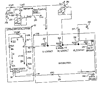
SINGLE ACCESS DISCONNECTION
[096] The electrical contacts of the present disclosure can be positioned in
any suitable location relative to the needle, needles or suitable access
device inserted
within the patient. As illustrated in Fig. 1C, an embodiment of the present
disclosure
as applied with respect to the detection of access detection, such as the
dislodgment of
a single access device inserted within the patient is shown. This type of
application is
applicable to a variety of different and suitable medical therapies
administered via a
single access device, such as a single needle, including intravenous infusion
and
dialysis therapy including hemodialysis, hemofiltration, hemodiafiltration and
continuous renal replacement.
[097] As applied, an electrically conductive fluid, such as blood, a blood
product, a medical fluid or the like flows between the patient and a fluid
system via a
single access device. Dislodgment detection of a single access device can
include, for
example, the detection of needle dislodgment during the delivery of any
suitable and
electrically conductive fluid or fluids including, for example, blood or
medical drug or
solution (e.g., a medication contained in an electrically conductive fluid,
such as
saline), processed blood, blood products, intravenous solutions, the like or
combinations thereof. The fluid delivery can be made between a suitable
container,
such as blood bags or like fluid delivery devices, and a patient. The systems
of the
present disclosure monitor and control the needle access so as to provide
immediate
and responsive detection of access disconnection of a blood or medical fluid
access,
such as a medication or drug, during medical therapy administered via a single
needle.
[098] As shown in Fig. 1C, an embodiment of the apparatus or device 54 of
the present disclosure includes an access device 56; such as a needle,
inserted into a
blood vessel 58 within a needle insertion site 60 of the patient 62. The
needle 56 is
connected to the fluid system 63, such as a fluid infusion system, via a tube
member
17
CA 02673877 2009-06-25
WO 2008/100675 PCT/US2008/051289
64. The infusion system includes, for example, an infusion pump 66 for
transferring
the blood or the like from a container 68 (e.g., blood bag) to the patient. A
first
electrical contact 70 is spaced apart from the needle 56 along the tube member
64 and
a second electrical contact 72 is attached to the patient near the insertion
site 60. The
first electrical contact 70 is in fluid contact with the fluid as it flows
from the delivery
container 68 to the patient.
[099] In this configuration, the first and second electrical contacts, e.g.,
electrodes, can be used to monitor changes in an electrical value, e.g.,
impedance,
within a conductor loop formed by at least a portion of the fluid circuit as
an electric
signal passes therein. The electrical contact points can be coupled to an
electronic
device 74, which is capable of processing a detectable signal transmitted
through the
electrodes in response to a change in impedance or the like due to dislodgment
of the
single access device as described in detail below. The electrical signal in
one
embodiment is generated by a constant current supplied to the electrodes such
that a
direct conductivity measurement can be conducted to detect a change in
impedance or
the like in response to changes in vascular access conditions, such as
dislodgment of
the access needle.
[0100] It is believed that the measured impedance, in the single needle
application, is a function of both the impedance of the fluid (i.e., blood)
and the
impedance as measured across the insertion site. The electronic device 74 can
be
adjusted to detect the impedance at the level equivalent to the combined
impedance of
all items of the electrical path (i.e., the conductive fluid in the tube,
needle, blood
stream of venous vessel, body tissue, impedance across the skin with respect
to the
sensing electrode 72 and the like).
ELECTRICAL CONTACTS
[0101] As previously discussed, the electrical contacts of the present
disclosure
are in fluid contact with the fluid as it flows through the fluid circuit. The
electrical
contacts allow for a direct conductivity measurement which is capable of
immediately
detecting, with high sensitivity and specificity, a change (e.g., an increase)
in
impedance or the like due to access disconnection, such as dislodgment of a
venous
needle (arterial needle or both) from the blood circuit during dialysis
therapy.
[0102] The electrical contacts can be composed of any suitable conductive and
biocompatible material, such as, any suitable electrode material including
stainless
18
CA 02673877 2009-06-25
WO 2008/100675 PCT/US2008/051289
steel, other suitable conductive materials or combinations thereof. It is
essential that
the electrode material is biocompatible.
[0103] It should be appreciated that the electrical contacts can be
constructed
in a variety of different shapes and sizes, illustrative examples of which are
described
below. For example, the electrical contacts can be configured or designed as a
plaster
electrode which includes an agent capable of expanding when in contact with
moisture. The agent can include a variety of suitable materials including gels
that are
known to expand more than ten times in volume upon contact with moisture.
[0104] In an embodiment, the plaster electrode can be utilized to detect fluid
(i.e., blood leakage) at an insertion site of an access device insertable
within a patient
during the administration of medical therapy via a single access device as
previously
discussed. Upon contact with the fluid, the plaster electrode would
necessarily expand
to such an extent that the electrode contact is broken, thus causing a
detectable
increase in impedance of the fluid as it flows from the fluid system to the
patient via
the needle.
[0105] In an embodiment, one or more electrodes (not shown), such as one or
more plaster electrodes as previously discussed, can be used in combination
with the
electrical contact pair as shown, for example, in Figs. 1A and 1B. For
example, a
plaster electrode can be attached to the patient near the insertion site of
either or both
of the arterial and venous needles. The plaster electrode(s) can be utilized
to detect
leakage of fluid, such as blood, from the insertion site of the access
device(s).
[0106] In an embodiment, an electrode pair is coupled to the blood circuit in
an
invasive manner (illustrated in Figs. 2A to 2C as discussed below) such that
the
electrodes contact the blood as previously discussed. An excitation source
that
includes a constant current source or the like can be applied to the
electrodes to inject
an electric signal into the blood circuit thereby defining a conductor loop
along which
direct conductivity measurements can be performed.
[0107] To ensure patient safety, the excitation source is typically isolated
from
the instrument power. The excitation source can produce a constant electrical
current
that passes through the blood via the electrodes. Any suitable amount of
current can
be generated for detection purposes. In an embodiment, the electrical current
as it
passes through the blood is maintained at a level of about ten microamperes or
lessõ
e.g., about five microamperes or less. It should be appreciated that the
present
19
CA 02673877 2009-06-25
WO 2008/100675 PCT/US2008/051289
disclosure can be operated at low levels of current (e.g., ten microamperes or
less)
such that the level of current has negligible, if any, effect on the health
and safety of
the patient.
[0108] It should be appreciated that the impedance or other suitable parameter
can be measured and calculated in a variety of different and suitable ways.
For
example, the amplitude, phase and/or frequency of the constant current
excitation
source can be measured and varied during the detection of a change in
impedance.
Impedance levels can then be detected by measuring the voltage across the
electrodes.
The amplitude, frequency and/or phase of the voltage can then be measured and
utilized in combination with the measured amplitude, frequency and/or phase of
the
excitation source to calculate blood impedance levels based on derivations or
equations which are typically used to calculate impedance.
[0109] The electrical contacts can be connected to the blood circuit in a
variety
of different and suitable ways. For example, the electrical contacts can be an
integral
component of the extracorporeal system, a disposable component that can be
connected and released from the tubing members of the blood circuit, a
reusable
component that can be autoclaved between uses, or the like.
ELECTRICAL CONTACT COUPLING DEVICE
[0110] In an embodiment, the apparatus of the present disclosure includes an
electrical contact coupling device that can be utilized to secure the
electrical contacts
to the blood circuit such that the electrodes effectively contact the blood
and, thus, can
be used to effectively monitor changes in access conditions as previously
discussed.
The coupling device of the present disclosure can also be designed to
facilitate the
protection of the user against contact with potential electrical sources. In
an
embodiment, the device can include a conductive element connected to a tube,
through
which a medical fluid can flow wherein the conductive element has a first
portion
exposed to the medical fluid, such as blood, and a second portion external to
the tube.
[0111] The coupling device of the present disclosure can include a variety of
different and suitable configurations, components, material make-up or the
like. In an
embodiment, the present disclosure can include a device for connecting an
electrical
contact to a fluid conduit providing fluid and electrical communication
between the
electrical contact and fluid flowing through the fluid conduit. The device can
include a
first member including an annular portion capable of accommodating the
electrical
CA 02673877 2009-06-25
WO 2008/100675 PCT/US2008/051289
contact and a first stem portion connected to the annular member wherein the
stem
portion has an opening extending therethrough to the annular portion; a second
member including a base portion with a groove region and a second stem portion
with
an opening extending therethrough to the groove region allowing the first
member to
be inserted and secured to the second member; and a contact member adapted to
fit the
first and second stem portions allowing the contact member to abut against at
least a
portion of the electrical contact member allowing an electrical connection to
be made
between the electrical contact and the contact member. Illustrative examples
of the
electrical contact coupling device of the present disclosure are described
below.
101121 As illustrated in Figs. 2A and 2B, the electrical contact coupling
device
80 includes a probe member 82 that has a cylindrical shape with an opening 84
extending therethrough. An electrical contact, such as an electrode 86 having
a
cylindrical shape can be inserted into the opening 84 such that the electrode
86 is
secure within the probe member 82. In an embodiment, the probe member 82 has a
channel 85 extending along at least a portion of the opening 84 within which
the
electrode 86 can be inserted into the probe member 82. A tube member, for
example,
from a blood tubing set, connector tube member of a dialysis machine or the
like, can
be inserted into both ends of the opening 84 of the probe member 82 in contact
with an
outer portion of the channel 85 allowing blood or other suitable fluid to make
fluid
contact with the electrode 86 in any suitable manner. The electrode 86 has an
opening
88 that extends therethrough within which blood (not shown) or other suitable
fluid
from the fluid circuit can flow. In an embodiment, the diameter of the opening
88 of
the electrode 86 is sized to allow blood flow through the electrode 86 such
that blood
flow levels under typical operating conditions, such as during dialysis
therapy, can be
suitably maintained. The coupling device of the present disclosure can be
readily and
effectively attached to a fluid circuit, including a blood circuit or the
like, for use
during medical therapy including, for example, dialysis therapy. It should be
appreciated that the coupling device 80 of the present disclosure can be
attached to the
fluid circuit in any suitable way such that electrical and fluid connection
can be made
with the fluid flowing through the fluid circuit.
[0113] The probe member 82 also includes a stem portion 90 that extends from
a surface 92 of its cylindrical-shaped body. The stem portion 90 has an
opening 93
that extends therethrough. In an embodiment, the stem portion 90 is positioned
such
21
CA 02673877 2009-06-25
WO 2008/100675 PCT/US2008/051289
that at least a portion of the electrode 86 is in contact with the opening 93
of the stem
portion 90.
[0114] To secure the electrode 86 to the blood circuit, the coupling device 80
includes a socket member 94 that includes a body portion 96 with an opening 98
for
accepting the probe member 82 and for accepting a blood tube member (not
shown) of
the blood circuit such that blood directly contacts the electrode as it
circulates through
the blood circuit during dialysis therapy. In an embodiment, the socket member
94
includes a stem portion 100 extending from the body member 96 wherein the stem
portion 100 includes an opening 102 extending therethrough. As the probe
member 82
is inserted through the opening 98 of the body member 96, the stem portion 90
of the
probe member 82 can be inserted into the opening 102 of the stem portion 100
of the
body 96 of the socket member 94.
[0115] In an embodiment, the socket member 94 includes a groove region 104
extending along at least a portion of the body 96 of the socket member 94. The
probe
member 82 can be inserted through the opening 98 and then moved or positioned
into
the groove region 104 to secure the probe member 82 within the body 96 of the
socket
member 94.
[0116] In an embodiment, the coupling device 80 includes an electrical contact
member 106 that is inserted within the opening 102 of the stem portion 100 of
the
body 96 of the socket member 94 such that the electrical contact member 106
extends
through the opening 93 of the stem portion 90 of the probe member 82 to
contact at
least a portion of a surface 108 of the electrode 86.
[0117] The electrical contact member 106 is utilized to connect the
electronics
(not shown) of, for example, the excitation source, a signal processing
device, other
like electronic devices suitable for use in monitoring and/or controlling
changes in
access conditions, such as needle dislodgment. The electrical contact member
106 can
be made of any suitable material, such as any suitable conductive material
including,
stainless steel, other like conductive materials or combinations thereof. To
secure the
electrical contact member 106 in place, a contact retainer member 110 is
inserted
within the opening 102 of the stem portion 100 at an end region 112 thereof.
[0118] In an embodiment, the coupling device is mounted to a dialysis
machine, device or system in any suitable manner. For example, the coupling
device
can be mounted as an integral component of the dialysis machine. The coupling
22
CA 02673877 2009-06-25
WO 2008/100675
PCT/US2008/051289
device can also be mounted as a separate ancUor stand alone component which
can
interface with any of the components of the apparatus and system of the
present
disclosure. In an embodiment, the coupling device 80 can be insertably mounted
via
the stem portion 100 of the socket member 94 to a dialysis machine or other
suitable
components.
[0119] It should be appreciated that the electrical contact coupling device
can
include a variety of different and suitable shapes, sizes and material
components. For
example, another embodiment of the coupling device is illustrated in Fig. 2C.
The
coupling device 114 in Fig. 2C is similar in construction to the coupling
device as
shown in Figs. 2A and 2B. The coupling device 114 of Fig. 2C can include, for
example, a cylindrical-shaped electrode or other suitable electrical contact,
a probe
member for accepting the electrode and securing it in place within a socket
member of
the sensing device. The probe member includes a stem portion that is
insertable within
a stem portion of the socket member. An electrical contact member is
insertable
within the stem portion such that it can contact the electrode. The coupling
device of
Fig. 2C can also include a contact retainer member to hold the electrical
contact
member in place similar to the coupling device as shown in Figs. 2A and 2B.
[0120] As shown in Fig. 2C, the probe member 116 of the electrical contact
coupling device 114 includes a handle 118 which can facilitate securing the
probe
member 116 within the socket member 120. The handle 118, as shown, has a solid
shape which can facilitate the use and manufacture of the coupling device 114.
In
addition, the stem portion (not shown) of the probe member 116 is larger in
diameter
than the stem portion of the probe member as illustrated in Fig. 2A. By
increasing the
stem size, the probe member can be more easily and readily inserted within the
socket
member. Further, the probe member is greater in length as compared to the
probe
member as shown in Figs. 2A and 2B such that the end regions 122 of the probe
member 116 extend beyond a groove region 124 of the socket member 120. This
can
facilitate securing the probe member within the groove region 124 of the
socket
member 120.
[0121] In an embodiment, an opening 126 of the socket member 120 can
include an additional opening portion 128 to accommodate the insertion of the
stem
portion of the probe member 116, having an increased size, therethrough. This
can
ensure proper alignment of the probe member with respect to the socket member
23
CA 02673877 2009-06-25
WO 2008/100675 PCT/US2008/051289
before insertion of the probe member into the socket member thus facilitating
the
insertion process.
01221 It should be appreciated that the probe member, socket member and
contact retainer member can be composed of a variety of different and suitable
materials including, for example, plastics, molded plastics, like materials or
combinations thereof. The various components of the coupling device, such as
the
probe member, socket member and contact retainer member, can be fitted in any
suitable way. For example, the components can be fitted in smooth engagement
(as
shown in Figs. 2A and 2B), in threaded engagement (as shown in Figs. 2D and
2E)
and/or any suitable fitting engagement or arrangement to one another.
[0123] As shown in Figs. 2D and 2E, the coupling device 130 of the present
disclosure can be made of threaded parts, which are removably connected to one
another to form the coupling device. The threaded parts can facilitate
securing the
electrode to the blood circuit as well as general use of same as described
below.
[0124] In an embodiment, the stem portion 132 of the body 134 of the coupling
device 130 has a threaded region 136, which can be insertably attached to a
dialysis
machine or other suitable mounting device in threaded engagement. This can
facilitate
the ease in which the coupling device is attached and detached from the
mounting
device.
[0125] As shown in Fig. 2E, the stem portion 132 is threaded on both sides
allowing it to be in threaded engagement with an annular member 138. The
annular
member 138 provides direction and support allowing the electrical contact
member
140 to abut against the electrode 142 housed in the probe member 144 as
previously
discussed.
[0126] In an embodiment, a plate member 146 made of any suitably
conductive material can be depressed against a spring 148 as the probe member
144 is
secured to the body 134. At the same time, another spring 150 can be displaced
against the electrical contact member 140 in contact with the retainer 152,
which is
inserted within an annular region of the annular member 138 to secure the
electrical
contact member 140 to the body 134.
[0127] The spring mechanism in an embodiment of the present disclosure
allows the parts of the coupling device 130 to remain in secure engagement
during use.
24
CA 02673877 2009-06-25
WO 2008/100675 PCT/US2008/051289
It can also facilitate use during detachment of the parts for cleaning,
maintenance or
other suitable purpose.
[0128] As previously discussed, the present disclosure can be effectively
utilized to detect dislodgment of an access device, such as a needle, inserted
within a
patient through which fluid can pass between the patient and a fluid delivery
and/or
treatment system. The present disclosure can be applied in a number of
different
applications, such as medical therapies or treatments, particularly dialysis
therapies. In
dialysis therapies, access devices, such as needles, are inserted into a
patient's arteries
and veins to connect blood flow to and from the dialysis machine.
[0129] Under these circumstances, if the needle becomes dislodged or
separated from the blood circuit, particularly the venous needle, the amount
of blood
loss from the patient can be significant and immediate. The systems of the
present
disclosure can control and effectively minimize blood loss from a patient due
to
dislodgment of the access device, such as during dialysis therapy including
hemodialysis, hemofiltration, hemodiafiltration and continuous renal
replacement.
SIGNAL DETECTION AND PROCESSING
[01301 As previously discussed, the electrical contacts in connection with the
controller can be used to detect a change in impedance or the like in response
to needle
drop-out or other like changes in access conditions. In an embodiment, the
present
disclosure can be adapted to correct for any variations in the baseline
impedance over
time. This can increase the level of sensitivity with respect to the detection
capabilities
of the present disclosure. If changes in the baseline impedance are too great
and not
adequately corrected for, changes in impedance due to needle dislodgment may
not be
as readily, if at all, detectable above baseline values.
[0131] From a practical standpoint, there are a number of different process
conditions that may influence a change in the baseline impedance over time.
For
example, a gradual drift or change in the baseline can occur due to a change
in the
characteristics, such as the hematocrit, plasma protein, blood/water
conductivity and/or
the like, of the blood or other suitable fluid during treatment. This can
arise due to
changes in the level of electrolytes or other components during dialysis
therapy.
[0132] As illustrated in Fig. 3, the present disclosure can process a
measurable
voltage signal to correct for changes in baseline impedance over time. This
can
enhance the detection capabilities of the present disclosure as previously
discussed. In
CA 02673877 2009-06-25
WO 2008/100675 PCT/US2008/051289
an embodiment, a current source 160 or the like generates an electric current
to pass
through the blood as it circulates into, through and out of the patient along
the
extracorporeal blood circuit 162, which connects the patient via venous and
arterial
needles to the dialysis system including a variety of process components. The
electric
current is injected into the blood circuit via a first electrical contact 163a
to define a
conductor loop or pathway along the blood circuits. The current is maintained
at a
constant level until dislodgment occurs in one embodiment.
[0133] A second electrode 163b is used to sense voltage or the like along the
conductor loop and then pass a signal indicative of same and/or changes
thereof to an
electronic device for detection and processing as previously discussed. The
voltage
signal can be measured and processed in any suitable manner.
[0134] In an embodiment, the signal is passed through a series of components
including a filter or filters 164 which can act to filter noise from the
signal, particularly
noise derived from the rotation from the pump in order to minimize a false
negative
and/or positive detection of needle dislodgment, a rectifier 166, a peak
detector 168
and an analog to digital converter ("ADC") 170 to digitize the signal. the
digital signal
can then be stored in a computer device (not shown) for further processing.
The
voltage signal is continually measured and processed over time. With each
measurement, the digitized signals are compared to evaluate changes due to
baseline
changes associated with variations in process conditions over time, such as a
change in
the characteristics of blood as previously discussed. If a baseline change is
determined, the digitized signal can be further processed to correct for the
change in
baseline.
[0135] The voltage data is continually sent to a control unit 172 coupled to
the
ADC. The control unit continually performs a calculation to determine whether
a
change in impedance or the like in response to needle dislodgment has
occurred. In an
embodiment, dislodgment of an access device is detected when [V(t) V(t-T)] >
Cl,
where t is time, where T is the period of blood pump revolution, where Cl is a
constant and where V(t) =I * Z, where Io is current and where Z is the
impedance of
the bloodline which is a function of the impedance associated with patient's
vascular
access and the impedance associated with various components of the dialysis
system,
such as the dialyzer, as previously discussed.
26
CA 02673877 2009-06-25
WO 2008/100675 PCMJS2008/051289
[0136] If disconnection of the patient from the blood circuit is detected, the
control unit 172 can be utilized to process the signal in order to minimize
blood loss
from the patient. In an embodiment, the controller is in communication with a
dialysis
system as applied to administer dialysis therapy including, for example,
hemodialysis,
hemofiltration, hemodiafiltration and continuous renal replacement. This
communication can be either hard-wired (i.e., electrical communication cable),
a
wireless communication (i.e., wireless RF interface), a pneumatic interface or
the like.
The controller can process the signal to communicate with the dialysis system
or
device to shut off or stop the blood pump 174 associated with the hemodialysis
machine and thus effectively minimize the amount of blood loss from the
patient due
to needle dislodgment during hemodialysis.
[0137] The controller can communicate with the dialysis system in a variety of
other ways. For example, the controller and hemodialysis machine can
communicate
to activate a venous line clamp 176 for preventing further blood flow via the
venous
needle thus minimizing blood loss to the patient. In an embodiment, the venous
line
clamp is activated by the controller and attached to or positioned relative to
the venous
needle such that it can clamp off the venous line in close proximity to the
needle.
Once clamped, the dialysis system is capable of sensing an increase in
pressure and
can be programmed to shut-off the blood pump upon sensing pressure within the
blood
flow line which is above a predetermined level. Alternatively, the venous line
clamp
can be controllably attached to the dialysis system.
[0138] In an embodiment, an alarm can be activated upon detection of blood
loss due to, for example, needle dislodgment during dialysis therapy. Once
activated,
the alarm (i.e., audio and/or visual or the like) is capable of alerting the
patient, a
medical care provider (i.e., doctor, registered nurse or the like) and/or a
non-medical
care provider (i.e., family member, friend or the like) of the blood loss due
to, for
example, needle dislodgment. The alarm function is particularly desirable
during
dialysis therapy in a non-medical facility, such as in a home setting or self
care setting
where dialysis therapy is typically administered by the patient and/or a non-
medical
care provider in a non-medical setting or environment excluding a hospital or
other
like medical facility.
[0139] The alarm activation, for example, prompts the patient to check that
the
blood pump has been automatically shut off, so that blood is minimized. Thus,
the
27
CA 02673877 2014-09-04
patient has the ability to act without the assistance of a third party (i.e.,
to act on his or
her own) to ensure that responsive measures are taken to minimize blood loss.
The
alarm can thus function to ensure the patient's safety during the
administration of
dialysis therapy, particularly as applied to home hemodialysis treatments in
which at
least a portion of the dialysis therapy can be administered while the patient
is sleeping.
DIALYSIS MACHINE
[01401 As previously discussed, the present disclosure can be adapted for use
with any suitable fluid delivery system, treatment system or the like. In an
embodiment, the present disclosure is adapted for use with a dialysis machine
to detect
access disconnection as blood flows between the patient and the dialysis
machine
along a blood circuit during treatment, including, for example hernodialysis,
hernofiltration and hemodiafiltration.
[01411 The present disclosure can include any suitable dialysis machine for
such purposes. An example, of a h,emodialysis machine of the present
disclosure is
disclosed in U.S. Patent No. 6,143,181. In an embodiment, the dialysis machine
190
comprises a mobile chassis 192 and it has at the front side 194 thereof with a
common mechanism 196 for connecting tubing or the like by which a patient can
be
connected to the dialysis machine as shown in Fig. 48. A flat touch screen
197,
which can show several operational parameters and is provided with symbols and
fields for adjustment of the dialysis machine. Touch screen 197 can be
adjusted
vertically and can be universally pivoted relative to chassis 192 of dialysis
machine
190 and can be fixed in the desired adjusted position.
[01421 In an embodiment, dialysis machine 190 includes a chassis 192 having
one or more connectors for connecting a patient to the dialysis machine via a
blood
circuit allowing blood to flow between the patient and the dialysis machine
during
dialysis therapy, wherein one or more electrical contact is connected to the
blood
circuit in fluid communication with the blood allowing detection of a change
in an
electrical value in response to access disconnection as the blood flows
through the
blood circuit having an electrical signal passing therein.
[0143] In an embodiment, dialysis machine 190 can be designed to
accommodate one or more of the electrical contact coupling devices, such as a
pair of
coupling device, used to detect access disconnection as shown in Fig. 4B. For
example, one or more coupling devices 198 can be attached to the front panel
194 of
28
CA 02673877 2009-06-25
WO 2008/100675 PCT/US2008/051289
the dialysis machine 190. This can be done in any suitable way. In an
embodiment, a
stem portion of the coupling device is insertably mounted via a threaded fit,
frictional
fit or the like, as previously discussed. This connects the patient to the
dialysis
machine 190 via a blood tubing set 202. The blood tubing set includes a first
blood
line 204 and a second blood line 206. In an embodiment, the first blood line
204 is
connected to the patient via an arterial needle 208 or the like through which
blood can
flow from the patient 200 to the dialysis machine 190. The second blood line
206 is
then connected to the patient 200 via a venous needle 210 or the like through
which
fluid flows from the dialysis machine to the patient to define a blood
circuit.
[0144] Alternatively, the first blood line and the second blood line can be
coupled to the venous needle and the arterial needle, respectively. The blood
lines are
made from any suitable medical grade material. Access disconnection, such as
dislodgment of an arterial needle and/or a venous needle can be detected as
previously
discussed. Alternatively, the coupling device can be attached to the blood
tubing set
which is then attached to the dialysis machine in any suitable way.
DIALYSIS TREATMENT CENTERS
[0145] As previously discussed, the present disclosure can be used during
dialysis therapy conducted at home and in dialysis treatment centers. The
dialysis
treatment centers can provide dialysis therapy to a number of patients. The
treatment
centers include a number of dialysis machines to accommodate patient demands.
The
therapy sessions at dialysis treatment centers can be performed twenty-four
hours a
day, seven days a week depending on the locale and the patient demand for use.
[0146] In an embodiment, the dialysis treatment centers are provided with the
capability to detect access disconnection during dialysis therapy pursuant to
an
embodiment of the present disclosure. For example, one or more of the dialysis
machines can be adapted for use with an electrical contact coupling device
along with
the necessary other components to detect access disconnection as previously
discussed.
[0147] In an embodiment, the electrical contact coupling device can be
directly
attached to one or more of the dialysis machines of the dialysis treatment
center. It
should be appreciated that the apparatuses, devices, methods and/or systems
pursuant
to an embodiment of the present disclosure can be applied for use during
dialysis
therapy administered to one or more patients in the dialysis treatment center
in any
suitable way. In an embodiment, the treatment center can have one or more
patient
29
)
, CA 02673877 2009-06-25
WO 2008/100675
PCT/US2008/051289
stations at which dialysis therapy can be performed on one or more patients
each
coupled to a respective dialysis machine. Any suitable in-center therapy can
be
performed including, for example, hemodialysis, hemofiltration and
hemodiafiltration
and combinations thereof. As used herein, the term "patient station" or other
like
terms mean any suitably defined area of the dialysis treatment center
dedicated for use
during dialysis therapy. The patient station can include any number and type
of
suitable equipment necessary to administer dialysis therapy.
[0148] In an embodiment, the dialysis treatment center includes a number of
patient stations each at which dialysis therapy can be administered to one or
more
patients; and one or more dialysis machines located at a respective patient
station. One
or more of the dialysis machines can include a chassis having one or more
connectors
for connecting a patient to the dialysis machine via a blood circuit allowing
blood to
flow between the patient and the dialysis machine during dialysis therapy
wherein a
pair of electrical contacts are connected to the blood circuit in fluid
communication
with the blood allowing detection of a change in an electrical value in
response to
access disconnection as the blood flows through the blood circuit having an
electrical
signal passing therein.
[0149] As previously discussed, the access disconnection detection
capabilities
of the present disclosure can be utilized to monitor and control a safe and
effective
dialysis therapy. Upon dislodgment of an access device, such as a needle, from
the
patient, the direct conductive measurement capabilities of the present
disclosure can be
used to provide a signal indicative of dislodgment that can be further
processed for
control and/or monitoring purposes. In an embodiment, the signal can be
further
processed to automatically terminate dialysis therapy to minimize blood loss
due to
dislodgment as previously discussed. Further, the signal can be processed to
activate
an alarm which can alert the patient and/or medical personnel to the
dislodgment
condition to ensure that responsive measures are taken. It should be
appreciated that
the present disclosure can be modified in a variety of suitable ways to
facilitate the
safe and effective administration of medical therapy, including dialysis
therapy.
[0150] Applicants have found that the direct conductive measurement
capabilities of the apparatus of the present disclosure can immediately detect
blood
loss or the like due to access disconnection, such as needle dislodgment, with
high
sensitivity and selectivity such that responsive measures can be taken to
minimize
CA 02673877 2009-06-25
WO 2008/100675 PCT/US2008/051289
blood loss due to same. The ability to act responsively and quickly to
minimize blood
loss upon detection thereof is particularly important with respect to needle
dislodgment
during hemodialysis. If not detected and responded to immediately, the amount
of
blood loss can be significant. In an embodiment, the present disclosure is
capable of
taking active or responsive measures, to minimize blood loss (i.e., shut-off
blood
pump, activate venous line clamp or the like) within about three seconds or
less, e.g.,
within about two to about three seconds, upon immediate detection of needle
dislodgment.
[0151] In addition, the controller can be utilized to monitor and/or control
one
or more treatment parameters during hemodialysis. These parameters can
include, for
example, the detection of blood due to blood loss upon needle dislodgment, the
change
in blood flow, the detection of air bubbles in the arterial line, detection of
movement of
the sensor during treatment, detection and/or monitoring of electrical
continuity of the
sensor or other like treatment parameters. In an embodiment, the controller
includes a
display (not shown) for monitoring one or more of the parameters.
[0152] As used herein "medical care provider" or other like terms including,
for example, "medical care personnel", means an individual or individuals who
are
medically licensed, trained, experienced and/or otherwise qualified to
practice and/or
administer medical procedures including, for example, dialysis therapy, to a
patient.
Examples of a medical care provider include a doctor, a physician, a
registered nurse
or other like medical care personnel.
[0153] As used herein "non-medical care provider" or other like terms
including, for example, "non-medical care personnel" means an individual or
individuals who are not generally recognized as typical medical care
providers, such
as doctors, physicians, registered nurses or the like. Examples of non-medical
care
providers include patients, family members, friends or other like individuals.
[0154] As used herein "medical facility" or other like terms including, for
example, "medical setting" means a facility or center where medical procedures
or
therapies, including dialysis therapies, are typically performed under the
care of
medical care personnel. Examples of medical facilities include hospitals,
medical
treatment facilities, such as dialysis treatment facilities, dialysis
treatment centers,
hemodialysis centers or the like.
31
CA 02673877 2009-06-25
WO 2008/100675 PCT/US2008/051289
[0155] As used herein "non-medical facility" or other like terms including,
for
example, "non-medical setting" means a facility, center, setting and/or
environment
that is not recognized as a typical medical facility, such as a hospital or
the like.
Examples of non-medical settings include a home, a residence or the like.
[0156] It should be appreciated that the electrode output signal can be
combined with other less sensitive blood loss detection methods, such as
venous
pressure measurements, systemic blood pressure, the like or combinations
thereof, to
improve specificity to needle dislodgment.
CONDUCTIVE POLYMER
[0157] The present disclosure provides conductive polymer materials and
devices, apparatuses, systems and methods that employ same. The conductive
polymer material can be utilized in a number of different applications, such
as to
monitor patient therapy. For example, the conductive polymer materials can be
utilized to monitor patient access conditions as discussed above and as
further detailed
below. Other types of monitoring applications include, for example, monitoring
solution mixing or compounding as described in greater detail below. The
present
disclosure contemplates monitoring one or a combination of condition changes
associated with patient therapy, such as monitoring patient access conditions
and
solution mixing conditions, alone or in combination.
[01581 In an embodiment, the conductive polymer material includes a polymer
matrix and a conductive component that is incorporated in the polymer matrix.
Alternatively, the conductive polymer material, in an embodiment, includes a
conductive polymer component without a separate conductive component, such as
stainless steel. It should be appreciated that the conductive polymer material
can be
made from any suitable types and amounts of materials and in any suitable way.
[0159] As discussed above, the conductive polymer can include a polymer
matrix and a conductive component incorporated in the matrix. The polymer
matrix
can include a variety of different polymer-based materials that are suitable
for use in a
variety of applications, particularly including medical applications, such as
dialysis
therapy. In an embodiment, the polymer matrix includes polyvinyl chloride,
acrylonitrile butadiene styrene, polycarbonate, acrylic, a cyclo olefin
copolymer, a
cyclo olefin copolymer blend, a metallocene-based polyethylene, like polymeric
materials and suitable combinations thereof.
32
CA 02673877 2009-06-25
WO 2008/100675 PCT/US2008/051289
[0160] The conductive component can include any suitable material or
combination of materials that have conductive properties applicable for a
number of
different applications including, for example, detecting patient access
disconnection
during medical therapy as previously discussed, monitoring the mixing or
compounding of solution components to form a mixed solution, and/or other like
applications. Preferably, the conductive component includes stainless steel,
fillers,
carbon black, fibers thereof and/or the like.
[0161] The conductive component can be sized and shaped in any suitable way
such that it can be readily incorporated in the polymer matrix. For example,
the
conductive component can include conductive fibers made from any suitable
material,
such as stainless steel, a carbonaceous material and/or the like. The fibers,
in an
embodiment, have an aspect ratio that ranges from about 2:1 to about 30:1.
[01621 The conductive polymer material can include any suitable amount of
the conductive polymer matrix and the conductive component. This can vary
depending on the application of the conductive polymer material. In an
embodiment,
the conductive component includes greater than about 10% by weight of the
conductive polymer material. Preferably, the conductive component ranges from
about 10% to about 50% by weight of the conductive polymer material. It should
be
appreciated that more than about 50% by weight of the conductive component can
be
utilized but may provide minimal, if any, increase in performance of the
conductive
polymer material depending on the application. Preferably, the conductive
component
is uniformly dispersed throughout the polymer matrix.
[0163] As previously discussed, the conductive polymer material, in an
embodiment, is composed of a conductive polymer component. This type of
component has sufficient electrical conductivity properties such that an
additional
conductive component, such as stainless steel, is not required. Examples of
conductive
polymer material components include polyaniline, polypyrrole, polythiophenes,
polyethylenedioxythiophene, poly(p-phenylene vinylene), the like and mixtures
thereof.
[0164] As previously discussed, the conductive polymer material can be made
in any suitable way. In general, the conductive component is mixed with the
polymer
component under suitable processing conditions including temperature and
pressure,
for example, to form a polymer matrix with the conductive component
incorporated
33
CA 02673877 2009-06-25
WO 2008/100675 PCT/US2008/051289
therein. The mixing should take place over a suitable period of time and with
a
sufficient amount of force such that the conductive component is uniformly
distributed
throughout the polymer matrix.
[0165] The polymer matrix incorporated with a conductive component is then
shaped and formed into a final product in any suitable way. For example, the
polymer
matrix incorporated with the conductive component can be formed into a single
piece
part via an injection molding process, extrusion process or the like under
suitable
processing conditions. Thus, the conductive polymer material can be readily
made
with manufacturing techniques, such as injection molding and extrusion. This
can
effectively provide a cost savings to the manufacturing process that can be
inevitably
passed along to the consumer.
[0166] The conductive polymer material can be formed into any suitable shape
and size depending on the application. In an embodiment, the conductive
polymer
material is formed into an electrode or other like electrical contact that can
be utilized
for a number of different applications, including, for example, monitoring
patient
access conditions and/or monitoring solution mixing or compounding as
discussed
above and described below in greater detail. The conductive polymer electrode
can
have a variety of different and suitable configurations depending on the
application.
For example, the conductive polymer electrode can be made into a coupler that
can be
used to join tubing to form a tubing joint as described below.
[0167] As shown in Figs. 5A and 5B, the conductive polymer coupler has a
generally cylindrical shape. With this configuration, the conductive polymer
can be
readily attached to a tube through which fluid flows, thus forming a tubing
joint.
[0168] As shown in Fig. 5A, the coupler 220 has a member 222 that extends
from an inner surface 224 of the coupler electrode 220. The member 222 acts as
a stop
for the tube 226 that is attached to the electrode such that a desired length
of the tubing
joint 228 can be preset. The coupler 220 as shown in Fig. 5A, in an
embodiment, is
made via an injection molding process.
[0169] As shown in Fig. 5A, a first tube member 230 is attached to a first end
232 of the coupler 220 and positioned or stopped by a first end 234 of the
member
222. A second tube member 236 is attached to a second end 238 of the coupler
220
and stopped or positioned by a second end 240 of the member 222. This forms a
34
CA 02673877 2014-09-04
tubing joint 228, such as a tubing joint that is integrated within a blood
circuit and
utilized during dialysis therapy as described in the present application.
[01701 As shown in Fig. 5B, the coupler 242 is formed without a member that
allows the length of the tubing joint to be preset as discussed above. In this
regard, the
tubing joint length can be adjusted accordingly depending on the application.
Further,
the coupler 242 as shown in Fig. 5B can be made via an extrusion process
instead of
an injection molding process. This can provide a further cost savings with
respect to
manufacturing of the coupler as compared to an injection molding process as
discussed
above. As shown in Fig. 58, a first tube member 244 is attached to a first end
246 of
the coupler 242 and a second tube member 248 is attached to the second end 250
of the
coupler 242, thus forming the tubing joint 252.
[01711 The tube member can be attached to the conductive polymer coupler in
any suitable way. For example, the conductive polymer material can be solvent
bonded, heat sealed, laser welded, radio frequency seated, or the like to the
tubing.
[0172] The tubing can be made of any suitable material depending on the
application. For example, the tubing can be made from polyvinyl chloride
("PVC").
Preferably, the PVC tubing is attached to a conductive polymer material that
is made
with a polymer matrix composed of acrylonitrile butadiene styrene ("ABS")
where the
ABS-based conductive material is solvent bonded to the PVC tubing.
101731 However, the tubing can be made of a variety of different materials
depending on the application. In an embodiment, the tubing includes a non-PVC
material, such as metallocene-based polyethylene polymers, cyclo olefin
copolymers,
c-ycto olefin copolymer blends and the like. The non-PVC materials can include
any
suitable type and amount of constituents. Metallocene-based polyethylene
polymers
and the like illustrative of the present disclosure can be found, for example,
in U.S.
Patent No. 6,372,848. These types of non-PVC polymers can include a polymer
blend that has a first ethylene and a-olefiri copolymer obtained using a
single site
catalyst present in an amount by weight of from about 0% to about 99% by
weight of
the blend and having a melt flow index from fractional, such as about 0.1 g/10
min to
about 5 g/10 inM, a second ethylene and a-olefin copolymer obtained using a
single
site catalyst and being present in an amount by weight of the blend from about
0% to
about 99% and having a melt flow index from higher than about 5 g/10 min to
about
20 g/10 min; and a third ethylene and a-olefin copolymer obtained using a
single-site
:35
CA 02673877 2014-09-04
catalyst and being present in an amount by weight of the blend from about 0%
to
about 99% and having a melt flow index greater than about 20 g/10 min. In an
embodiment, the a-olefin copolymer has a molecular weight distribution of less
than
about 3.
[0174] Cycio olefin copolymers and blends thereof illustrative of the present
disclosure can be found, for example, in U.S. Patent 6,255,396. These types of
non-
PVC polymers can include as a component homopolymers or copolymers of cyclic
olefins or bridged polycyc,lic hydrocarbons. For example, the polymer
composition
includes a first component obtained by copolymerizing a norbornene monomer and
an ethylene monomer wherein the first component is in an amount from about 1-
99%
weight of the composition; and a second component of an ethylene and a-olefin
copolymer that has six carbons wherein the second component is in an amount
from
about 99% to about 1% by weight of the composition. In an embodiment, the
polymer composition can include an additional component, such as a second
homopolymer or copolymer of a cyclic olefin or a bridged polycyclic
hydrocarbon.
[0175J The non-PVC based tubing and the non-PVC based conductive coupler
can be joined in any suitable way to form a tubing joint In an embodiment, the
non-
PVC based tubing and coupler are joined via solvent bonding, such as disclosed
in
U.S. Patent Nos. 6,255,396 and 6,372,848. As used herein the term solvent
bonding or
other like terms means that the tubing can be exposed to a solvent to melt,
dissolve or
swell the tubing and then be attached to another polymeric component to form a
permanent bond. Suitable solvents typically include those having a solubility
parameter of less than about 20 (Mpa)I/2. Suitable solvents can also have a
molecular
weight less than about 200 Wrriole. The solvent can include, for example,
aliphatic
hydrocarbons, aromatic hydrocarbons, and mixtures thereof. As used herein, the
terms
aliphatic hydrocarbon and aromatic hydrocarbon are compounds containing only
carbon and hydrogen atoms.
[0176] Suitable aliphatic hydrocarbons can include substituted and
unsubstituted hexane, heptane, cyclohexane, cycloheptane, decalin and the
like.
Suitable aromatic hydrocarbons can include substituted and unsubstitutecl
aromatic
hydrocarbon solvents, such as xyIene, tetralin, toluene, cumene and the like.
Suitable
hydrocarbon substituents can include aliphatic substiments that have from 1-12
36
CA 02673877 2014-09-04
carbons and include propyl, ethyl, butyl, hexyl, tertiary butyl, isobutyl, the
like and
combinations thereof.
[0177] As previously discussed, the conductive polymer of the present
disclosure can be constructed and arranged into a variety of different
configurations,
such as a conductive polymer coupler as shown in Figs. SA and 5B and discussed
above. Another example includes a dialyzer header that is made from the
conductive
polymer according to an embodiment. Referring to Fig. 6, a dialyzer 253 is
generally
illustrated. The dialyzer 253 includes a body member 254 that generally
includes a
casing 256. The casing 256 includes a core section as well as two bell members
260
located at each end of the dialyzer. Located within the core is a fiber bundle
258. The
dialyzer also includes a dialysate inlet 262 and a diaiysate outlet 264.
[0178] Located at a first end 266 of the dialyzer 253 is a fluid inlet 268 and
at a
second end 270 is a fluid outlet 272 defined by a fluid inlet header 274 and a
fluid
outlet header 276, respectively. The dialyzer 253 is connected to a dialysis
blood
circuit in any suitable manner. In an embodiment, the inlet 274 and outlet 276
headers
are made from the conductive polymer material of the present disclosure as
discussed
above. The inlet 274 and outlet 276 headers can be connected to a controller
278 such
that the conductive polymer headers can be utilized to monitor patient access
conditions as previously discussed.
[0179] A variety of different header and dialyzer designs can be utilized. For
example, U.S. Patent No. 6,623,638 and U.S. Patent Publication No.
2003/0075498
provide a number of different examples illustrative of the present disclosure.
[0180] The conductive polymer of the present disclosure can be utilized in any
suitable way and in a variety of different devices, apparatuses, systems and
applications thereof. For example, the conductive polymer can he utilized to
detect a
change in impedance in response to dislodgement of an access device, to detect
a
change in conductivity in response to a change in solution composition and/or
other
suitable applications.
[0181] In an embodiment, the conductive polymer is part of a sensor assembly
or apparatus that can be utilized, for example, for monitoring dialysis
applications as
discussed in the present application. The sensor apparatus of th.e present
disclosure
37
CA 02673877 2009-06-25
WO 2008/100675 PCT/US2008/051289
can include a number of different configurations and designs. Two examples of
such
designs illustrative of the present disclosure are described below in Figs. 7A
and 7B.
[01821 In Fig. 7A, the tubing joint 284 that includes the conductive polymer
electrode 286 attached to a tube member 288 as described above, for example,
is
positioned in place by a holding device 290 or holder for purposes of
detection
capabilities associated with the sensor apparatus 291 in an embodiment. In
general,
the holder 290 as shown in Fig. 7A has a hub design. More specifically, the
holder
290 includes a base member 292 onto which the tubing joint 284 can be placed.
The
base member 292 includes a first portion 294 that is made from a plastic or
other
suitable material. The first portion 294 defines an outer surface 296 of the
base
member 292. Along the outer surface 296, the first portion 294 includes two
openings
that are spaced apart as shown in Fig. 7A. The first opening 298 is located on
a first
edge 300 of the first portion of the base and the second opening 302 is
located on a
second edge 304 of the first portion of the base. The openings can be
configured in
any suitable way and be utilized for mounting purposes.
[0183] The second portion of the base member includes a conductive portion
306 as shown in Fig. 7A. In an embodiment, the conductive portion 306 includes
a
single piece part 308 that is made from any suitable conductive material, such
as
stainless steel and/or the like. As shown in Fig. 7A, the conductive polymer
of the
tubing joint is placed against a curved edge 310 of the second portion that
substantially
forms to an outer surface 312 of the conductive polymer electrode 286. The
electrode
is substantially cylindrical in shape.
[0184] The holder 290 further includes an arm member 314 that is pivotally
attached to the base member 292 as shown in Fig. 7A. The arm member 314
includes
a generally curved region such that the arm 314 can be positioned over the
tubing joint
284 allowing it to substantially conform to the generally cylindrical surface
of the tube
joint and thus further securing the tubing joint 284 in place.
[0185] Another configuration of a hub design illustrative of the sensor
apparatus of the present disclosure is shown in Fig. 7B. In general, this
design
provides a box-like holder 316 that encloses the tubing joint 318 wherein the
tubing
joint includes the conductive polymer electrode 320 in the form of a coupler
that is
attached to the tube member 322 as discussed above. The holder 316 includes a
base
member 324. The base member 324 includes side portions 326, a bottom portion
328
38
CA 02673877 2009-06-25
WO 2008/100675 PCT/US2008/051289
and an opening 330 at a top portion 332. As shown in Fig. 7B, the sensor
apparatus
334 includes a conductive member 336 that is contained in the base member 324.
The
conductive member 336 can be made of any suitable material as described above.
The
conductive member 336 includes an annular-shaped surface 338 against which the
tubing joint 318 can be placed. The sensor apparatus 334 further includes a
lid 340
that is pivotally attached to the base member 324. The lid 340 has a member
342 that
abuts against a portion of the tubing joint in a closed position. This secures
the tubing
joint in place for use.
[0186] As previously discussed, the sensor apparatus of the present
application
can be used in a number of suitable applications. For example, the sensor
apparatus
can be suitably coupled to a blood circuit and used for purposes of detecting
disconnection of an access device as described in the present application.
Another
application includes the monitoring of solution compounding as described in
greater
detail below. In this regard, the sensor apparatus as shown in Figs. 7A and 7B
can be
used in combination with or in place of the electrical coupling devices as
illustrated in
Figs. 2A-2E and further described above. Thus, the present disclosure can be
utilized
to monitor one or a combination of conditions, such as patient access and
solution
mixing, during use. As applied, the sensor apparatus can be connected to a
controller
or other like device for detection purposes. The controller can include one or
a
number of different devices that are in electrical contact with the sensor
apparatus in
any suitable way.
[0187] In another embodiment, the sensor apparatus can include a single piece
part that is made from the conductive polymer material. The single piece part
can be
made in any suitable way such as through injection molding as described above.
A
number of different and suitable shapes and sizes can be formed. One such
example
illustrative of the present disclosure of a single piece part conductive
electrode 344 is
shown in Figs. 8A and 8B.
[0188] In general, the conductive polymer electrode 344 is configured as a
coupler that can join tubing to form a tubing joint through which fluid can
flow as
shown in Figs. 8A and 8B. The conductive polymer electrode 344 includes a base
member 346 that has an annular opening 348 extending therethrough. A tube
member
350 can be attached to the ends 351 of the annular opening 348 such that the
tubing
joint can be formed as described above. The base member 346 includes a stem
portion
39
CA 02673877 2009-06-25
WO 2008/100675 PCT/US2008/051289
352 that extends from a portion of a surface 354 of the base member 346. This
can be
used to mount or attach the conductive polymer electrode 344 to a control
panel or
other suitable component, such as a hemodialysis machine as described above.
The
stem portion 352 defines an annular-shaped channel 356 that ends from the
surface
354 of the base member 346. Within the annular-shaped channels, an inner
annular-
shaped channel 360 is also provided that extends from the surface 354 of the
base 346,
as shown in Fig. 8B. The stem portion can be utilized to provide a pathway
through
which the electrode can be in electrical contact with one or more other
devices, such as
a controller, in any suitable way. The base member further includes a top
member 362
that extends from a portion of the surface 354 of the base member 346. The top
member 362 can be used to secure the stem portion 352 of the base member 346
in
place for use and/or to remove the electrode after use.
[0189] As shown in Figs. 8A and 8B, the conductive polymer electrode 344
includes a member 366 that extends from an inner surface of the annular
opening 348.
The member 366 acts as a stop against which the tubing can be placed to form
the
tubing joint. The member 366 has a generally-cylindrical shape with an opening
368
through which fluid can flow.
[0190] As previously discussed, the conductive polymer material of the present
disclosure can be utilized in a number of different applications. In an
embodiment, the
conductive polymer material can be utilized to monitor patient access
conditions, such
as to detect disconnection of an access device that is inserted in a patient
through
which fluid flows during medical therapy. Preferably, the disconnection
detection
application is applied during dialysis therapy, such as during hemodialysis
therapy.
[0191] As applied to dialysis applications, the conductive polymer can be
formed into an electrode and attached to a dialysis blood circuit in any
suitable
manner. As shown in Figs. IA and 2A and further described above, at least one
of the
sensors can include an electrode made with the conductive polymer material of
the
present disclosure. The sensors 22 and 24 are in electrical contact with a
controller 29
and thus the conductive polymer electrode can be utilized for detection,
monitoring
and control purposes related to dialysis therapy as described above. As shown
in Figs.
4A and 4B and described in the present application, the sensors 198 can be
attached
directly to the hemodialysis machine wherein at least one of the sensors
includes a
conductive polymer electrode according to an embodiment of the present
disclosure.
CA 02673877 2009-06-25
WO 2008/100675
PCT/US2008/051289
The conductive polymer sensor can be configured in any suitable way, such as
the
coupler and hub design (See, Figs. SA, 5B, 7A and 7B), the single-piece part
design
(See, Figs. 8A and 8B) and dialyzer header design (See, Figs. 8A and 8B) as
described
above. It should be appreciated that the present disclosure contemplates the
use of one
or a combination of different sensors to monitor medical therapy, such as
patient
access and solution mixing conditions.
[0192] In an embodiment, the conductive polymer material of the present
disclosure can be utilized for monitoring the mixing of solutions to form a
mixed
solution, such as a mixed solution used during medical therapy. One type of
application illustrative of the present disclosure for such monitoring
purposes is during
dialysis therapy, particularly during peritoneal dialysis. In general,
the conductive
polymer material can be formed into an electrode or other sensing device that
can
effectively detect changes in conductivity associated with a dialysis solution
that is
administered to the patient during peritoneal dialysis.
[0193] The dialysis solution can be formed from a number of solution
components that are mixed to form a mixed dialysis solution prior to
administration.
The dialysis solution components can have varying pH levels, such as ranging
from
about 1.8 to about 9.2. Once mixed, the pH of the mixed dialysis solution
should be at
a physiologically acceptable level, such as ranging from about 6.8 to about
7.5, prior to
use. The pH level can be monitored in relation to changes in the conductivity
level of
the dialysis solution. In this regard, the conductive polymer of the present
disclosure
can be utilized to detect changes in conductivity level and thus can be
utilized to
determine whether the solution components are properly mixed to form the mixed
dialysis solution at an acceptable pH level prior to use. A general
description of
peritoneal dialysis is provided below and is illustrative of the present
disclosure.
[0194] Peritoneal dialysis utilizes a sterile dialysis solution, which is
infused
into a patient's peritoneal cavity and into contact with the patient's
peritoneal
membrane. Waste, toxins and excess water pass from the patient's bloodstream
through the peritoneal membrane and into the dialysis solution. The transfer
of waste,
toxins, and excess water from the bloodstream into the dialysis solution
occurs due to
diffusion and osmosis during a dwell period as an osmotic agent in the
dialysis
solution creates an osmotic gradient across the membrane. The spent solution
is later
41
CA 02673877 2009-06-25
WO 2008/100675 PCT/US2008/051289
drained from the patient's peritoneal cavity to remove the waste, toxins and
excess
water from the patient.
[0195] There are various types of peritoneal dialysis therapies, including
continuous ambulatory peritoneal dialysis ("CAPD") and automated peritoneal
dialysis. CAPD is a manual dialysis treatment, in which the patient connects
the
catheter to a bag of fresh dialysis solution and manually infuses fresh
dialysis solution
through the catheter or other suitable access device and into the patient's
peritoneal
cavity. The patient disconnects the catheter from the fresh dialysis solution
bag and
allows the solution to dwell within the cavity to transfer waste, toxins and
excess water
from the patient's bloodstream to the dialysis solution. After a dwell period,
the
patient drains the spent dialysis solution and then repeats the manual
dialysis
procedure. Tubing sets with "Y" connectors for the solution and drain bags are
available that can reduce the number of connections the patient must make. The
tubing sets can include pre-attached bags including, for example, an empty bag
and a
bag filled with dialysis solution.
[0196] In CAPD, the patient performs several drain, fill, and dwell cycles
during the day, for example, about four times per day. Each treatment cycle,
which
includes a drain, fill and dwell, takes about four hours.
[01971 Automated peritoneal dialysis is similar to continuous ambulatory
peritoneal dialysis in that the dialysis treatment includes a drain, fill, and
dwell cycle.
However, a dialysis machine automatically performs three or more cycles of
peritoneal
dialysis treatment, typically overnight while the patient sleeps.
[01981 With automated peritoneal dialysis, an automated dialysis machine
fluidly connects to an implanted catheter. The automated dialysis machine also
fluidly
connects to a source or bag of fresh dialysis solution and to a fluid drain.
The dialysis
machine pumps spent dialysis solution from the peritoneal cavity, through the
catheter,
to the drain. The dialysis machine then pumps fresh dialysis solution from the
source,
through the catheter, and into the patient's peritoneal cavity. The automated
machine
allows the dialysis solution to dwell within the cavity so that the transfer
of waste,
toxins and excess water from the patient's bloodstream to the dialysis
solution can take
place. A computer controls the automated dialysis machine so that the dialysis
treatment occurs automatically when the patient is connected to the dialysis
machine,
for example, when the patient sleeps. That is, the dialysis system
automatically and
42
CA 02673877 2014-09-04
sequentially pumps fluid into the peritoneal cavity, allows for dwell, pumps
fluid out
of the peritoneal cavity, and repeats the procedure.
[01991 Several drain, fill, and dwell cycles will occur during the treatment.
Also, a final volume "last fill" is typically used at the end of the automated
dialysis
treatment, which remains in the peritoneal cavity of the patient when the
patient
disconnects from the dialysis machine for the day. Automated peritoneal
dialysis frees
the patient from having to manually perform the drain, dwell, and fill steps
during the
day.
[02001 In general, the dialysis solution includes an osmotic agent, such as
dextrose or other suitable constituent in any suitable amount, such as from
about 1.5%
to about 4.25% by weight. The dialysis solution further includes one or more
electrolytes, such as sodium, calcium, potassium, magnesium chloride and/or
the like
in any suitable amount. The dialysis solution may also include other
constituents, such
as buffers including lactate and bicarbonate, or the like, and other
constituents, such as
stabilizers. The dialysis solution can be made from multiple solution
components that
can vary in the amounts and types of constituents thereof and have varying pH
levels.
[02011 A variety of different and suitable types of multi-part dialysis
solutions
can be utilized. For example, a multi-part bicarbonate-based solution can be
found in
U.S. Patent Application 09/955,248, entitled "Biochemically Balanced
Peritoneal
Dialysis Solutions", filed on September 17, 2001. An example of a multi-part
lactate-based solution can be found in U.S. Patent Application No. 10/628,065,
entitled "Dialysis Solutions With Reduced Levels Of Glucose Degradation
Products",
filed on July 25, 2003.
[0202} Another example of a bicarbonate-based solution can be found in U.S.
Patent Application No. 10/044,234, entitled "Bicarbonate-Based Solutions For
Dialysis Therapies", filed on January 11, 2002 and as further disclosed in
U.S. Patent
No. 6,309,673. The bicarbonate-based solution can be made from solution
components that have varying pH conditions, such as under moderate and extreme
pH conditions. In an embodiment, the solution components can vary in pH from
between about 1.0 to about 10Ø Once mixed, the desired pH of the mixed
solution
is a physiological acceptable level, such as between about 6.5 to about 7.6
(i.e., close
to the pH of blood),
43
CA 02673877 2009-06-25
WO 2008/100675 PCT/US2008/051289
[0203] For example, under moderate pH conditions, the bicarbonate-based
solution can be formulated by the mixing of a bicarbonate concentrate with a
pH that
ranges from about 7.2 to about 7.9, preferably from about 7.4 to about 7.6,
and an
electrolyte concentrate with a pH that ranges from about 3.0 to about 5Ø
Under
extreme pH conditions, for example, the bicarbonate concentrate has a pH that
can
range from about 8.6 to about 9.5 and is mixed with an electrolyte concentrate
that has
a pH from about 1,7 to about 2.2. A variety of different and suitable acidic
and/or
basic agents can be utilized to adjust the pH of the bicarbonate and/or
electrolyte
concentrates. For example, a variety of inorganic acids and bases can be
utilized, such
as hydrochloric acid, sulfuric acid, nitric acid, hydrogen bromide, hydrogen
iodide,
sodium hydroxide, the like or combinations thereof.
[0204) The solution components, such as the electrolyte concentrate and the
dextrose concentrate, can then be mixed in the solution bag and then
administered as a
mixed solution to the patient during peritoneal dialysis. An illustrated
example of a
multi-chamber container that separately contains solution components of a
dialysis
solution according to embodiment of the present disclosure is shown in Fig. 9.
[0205] It should be appreciated that the components of the dialysis solutions
of
the present disclosure can be housed or contained in any suitable manner such
that the
dialysis solutions can be effectively prepared and administered. In an
embodiment, the
present disclosure includes a multi-part dialysis solution in which two or
more parts
are formulated and stored separately, and then mixed just prior to use. A
variety of
containers can be used to house the various parts of the dialysis solution,
such as
separate containers (i.e., flasks or bags) that are connected by a suitable
fluid
communication mechanism.
[0206] In an embodiment, a multi-chamber container or bag can be used to
house the separate components of the solution including, for example, a
dextrose
concentrate and a buffer concentrate. In an embodiment, the separate
components are
mixed within the multi-chamber bag prior to use, such as applied during
peritoneal
dialysis.
[02071 Fig. 9 illustrates a suitable container for storing, formulating,
mixing
and administering a dialysis solution, such as during continuous ambulatory
peritoneal
dialysis, according to an embodiment of the present disclosure. The multi-
chamber
bag 380 has a first chamber 382 and a second chamber 384. The interior of the
44
CA 02673877 2014-09-04
container is divided by a heat seal 386 into the two chambers. It should be
appreciated
that the container can be divided into separate chambers by any suitable seal.
[02083 In an embodiment, the container can be divided into separate
chambers, such as two or more chambers, by a peel seal. With the use of a peel
seal,
a frangible connector or other suitable type of connector would not be
required to
mix the solution components within the multi-chamber bag. An example of a
multi-
chamber solution bag that includes a peel seal is disclosed in U.S. Patent No.
6,319,243. As shown in Fig. 10, a container 388 includes at least three
chambers
390, 392 and 394. The chambers 390, 392 and 394 are designed for the separate
storage of liquids and/or solutions, that can be mixed within the container to
form a
mixed solution ready-for-use. It should be appreciated that more or less than
three
chambers can be utilized.
[02093 The peelable seals 396 and 398 are provided between the chambers
390, 392 and 394, respectively. Examples of peelable seals can be found in
U.S.
Patent Application No. 08/033,233 filed on March 16, 1993 entitled "Peelable
Seal
And Container Having Same". The peelable seals allow for the selective opening
of
the chambers to allow for the selective mixing of the liquids contained
therein,
[02103 The container 388 can also include tubular ports, such as tubular ports
400, 402 and 404 as shown in Fig. 10. The tubular ports are mounted to the
container
so as to allow fluid communication with the container and specifically with
chambers
390, 392 and 394. To this end, the tubular ports 400, 402 and. 404 can include
a
membrane that is pierced, for example, by a earmula or a spike or an
administration, set
for delivery of the contents of the container to the patient. It should be
appreciated that
more or less than three ports can be utilized.
[02113 As shown in Fig. 9, the multi-chamber container 380 has a frangible
connector 406 to sealingly couple the first chamber 382 to the second chamber
384
instead of a pee/able seal. To mix the solution within the multi-chamber bag
380, the
frangible connector 406 is broken.
[02123 The first container or chamber 382 includes two port tubes 408 of
suitable sizes and lengths.. It should be appreciated that more or less than
two port
tubes may be used. One of the port tubes, for example, can be utilized to add
other
constituents to the first chamber 382 during formulation of the solution of
the present
CA 02673877 2014-09-04
disclosure, if necessary. The remaining port tube, for example, can be
utilized to
adaptedly couple the first chamber 382 to the patient via a patient's
administration line
(not shown), be used to add additional other constituents or the like. The
second
container or chamber 384 has a single port tube 410 extending there from. In
an
embodiment, the port tube 410 is connected to a patient's administration line
through
which a solution can flow to the patient once the solution is mixed as
described below,
[0213] In an embodiment, the transfer of product within the multi-chamber bag
380 can be initiated from the first chamber 382 to the second chamber 384 such
that
the components of each chamber can be properly mixed to form the dialysis
solution
of the present disclosure. In an embodiment, a dextrose concentrate 412 is
contained
in the first chamber 382 and a buffer concentrate 414 is contained in the
second
chamber 384. It should be appreciated that any suitabl.e type or number of
solution
components can be separated with a multi-chamber bag and then mixed to form a
mixed solution prior to administration to the patient. Illustrative examples
of
peritoneal dialysis solutions include those described in U.S. Patent
Application Nos,
09/955,298 and 10/628,065 and U.S. Patent No. 6,309,673 as described above.
[02141 The first chamber 382 is. smaller in volume than the second chamber
384 such that the components of each chamber can be properly mixed once the
transfer from the first chamber to the second chamber has occurred. Thus, the
multi-
chamber bag 380 can house at least two solution component parts that after
mixture
will result in a ready-to-use dialysis solution. An example of the multi-
chamber
container is set forth in U.S. Patent No, 5,431,496. The multi-chamber bag can
be
made from a gas permeable material, such as polypropylene, polyvinyl chloride
or
the like.
[02151 It should be appreciated that the multi-chamber bag can be
manufactured from a variety of different and suitable materials and configured
in a
number of suitable ways such that the dialysis solutions of the present
disclosure can
be effectively formulated and administered to the patient during medical
therapy in
any suitable manner. For example, the first chamber can be larger in volume
than the
second chamber and further adapted such that the dialysis solution of the
present
disclosure can be readily and effectively made and administered to the
patient.
[02161 In an embodiment, the dialysis solution is contained and administered
from a multi-chamber solution bag during peritoneal dialysis, such as during
C.APD.
46
CA 02673877 2009-06-25
WO 2008/100675
PCT/US2008/051289
The solution bag can include multiple chambers that each contain separate
components
of the dialysis solution prior to mixing as discussed above. This may be
necessary to
maintain separation of the non-compatible solution components prior to mixing
for
purposes of stability, sterility, effectiveness or the like associated with
the dialysis
solution prior to use.
[0217] In another embodiment, the solution components can be prepared and
stored in separate containers and then mixed via an admix device prior to use,
such as
applied during automated peritoneal dialysis. As shown in Fig. 11, a first
solution
component, such as a dextrose concentrate 416 and a second solution component,
such
as a buffer concentrate 420 are stored in the respective separate containers
422 and 424
or bags which are fluidly connected to an admix device 426 suitable for use
during
automated peritoneal dialysis. In addition to the first and second components,
a first
bag 428 and last bag 430 filled with a suitable solution can also be used
during dialysis
therapy as generally known.
[0218] In an embodiment, an effective amount of the first solution component
416 and the second solution component 420 are drawn from each respective
container
and into a heater bag 432 where the solution components (e.g., dextrose and
buffer
concentrates) can be mixed and heated prior to infusion into a patient 434
during
dialysis therapy. As further shown in Fig. 11, a drain line 436 is coupled to
the admix
device 426 from which waste fluids can be removed from the patient during
therapy.
[0219] According to an embodiment of the present disclosure, the conductive
polymer material can be used as a sensor to monitor solution compounding, such
as
during peritoneal dialysis. For example, the conductive polymer sensor 438 can
be
attached to a tube 440 through which the mixed dialysis solution flows to the
patient
from the multi-chamber solution bag 380 as shown in Fig. 9. The conductive
polymer
sensor 438 is in electric contact with a controller 442 or other like device
such that a
change in conductivity of the mixed dialysis solution that is fed to the
patient can be
monitored. Based on the conductivity level, one can monitor the pH level of
the mixed
solution to determine whether the solution components (e.g., dextrose
concentrate and
buffer concentrate) have been properly and sufficiently mixed to form the
dialysis
solution prior to use. If the dialysis solution is not properly mixed, the
conductivity
level will exist above or below a baseline conductivity level that is
generally
associated with a desired pH level of a dialysis solution that is ready-for-
use. As
47
CA 02673877 2009-06-25
WO 2008/100675 PCT/US2008/051289
previously discussed, the desired pH of the mixed dialysis solution is
maintained at a
physiological acceptable level, such as between about 6.5 to about 7.6 prior
to use.
Based on this information, adjustments can be made to the process such that
the
solution chemistry of the dialysis solution is modified for proper use. This
can
facilitate the safe and effective use of the solution during use, such as
during dialysis
therapy.
1102201 As shown in Fig. 11, the conductive polymer sensor 444 of the present
disclosure can be applied during automated peritoneal dialysis. More
specifically, the
conductive polymer sensor 444 of the present disclosure can be attached to the
tube
member 446 through which a dialysis solution flows to the patient. The
dialysis
solution is a product of the mixing of solution components that are stored in
separate
solution bags as previously discussed. The conductive polymer sensor 444 can
be
attached to a controller 448 or other like device in electrical contact such
that the
conductivity level and thus the pH level of the solution that is administered
to the
patient can be monitored as previously discussed. Optionally, at least one
additional
conductive polymer sensor 450 in an embodiment can also be utilized as shown
in Fig.
11. In this regard, the additional sensor(s) can be utilized to monitor the
conductivity
level of the solution components prior to mixing. This can be utilized to
evaluate
whether the solution components are maintained at desired pH levels based on a
conductivity measurement as discussed above.
STAND ALONE DISCONNECTION SYSTEM AND METHOD
[02211 Referring now to Fig. 12, an embodiment of a stand-alone access
disconnection system 220 is illustrated. System 220 includes a dialysis
machine 190,
such as the one described above in connection with Figs. 4A and 4B. For
example,
machine 190 includes a chassis 192 and a touch screen 197. Machine 190
includes a
blood circuit 35 having a venous line 26 and an arterial line 28 connecting to
venous
needle 32 and arterial needle 34, respectively, forming patient access 36.
Arterial line
28 extends from patient access 36 to a detector module 225, which is shown in
more
detail below in connection with Fig. 13. In general, detector module 225
includes
electrodes that communicate electrically with contacts 22 and 24 (Fig. 1A)
provided
with venous and arterial lines 26 and 28, respectively. Alternatively,
detector module
225 includes venous and arterial contact producing coupling devices 48 and 50,
respectively, referenced above in connection with Fig. 1B, which contact blood
48
CA 02673877 2009-06-25
WO 2008/100675 PCT/US2008/051289
directly, obviating the need for contacts 22 and 24 on the blood set. Detector
module
225 also includes electronics capable of detecting an access disconnection and
sending
a remote or wireless signal to a protector module 230 described in more detail
below in
connection with Fig. 14.
[0222] A first tubing segment 38a of blood circuit 35 extends from detector
module 225 to blood pump 46. A second tube segment 38b extends from segment
38a
at blood pump 46 to dialyzer 42. A third tube segment 38c extends from
dialyzer 42 to
detector module 225. Venous line 26 extends from detector module 225 to
patient
access 36. As illustrated, detector module 225 is positioned to clamp one or
more of
tube segment 38a or 38c upon a sensing of a disconnection at patient access 36
by
detector module 225. Venous drip chamber 40 is shown operating with tube
segment
38c. Although not illustrated, an arterial drip chamber 44 can be placed
additionally in
tube segment 38a. Venous drip chamber 40 is shown operating with a pressure
sensor
214.
[0223] It is expressly contemplated to provide system 220 including detector
module 225 and protector module 230 as either an integrated part of machine
190, an
option in ordering machine 190, or as a retrofit kit to an existing dialysis
machine (or
any other type of blood treatment or medical delivery machine described
herein).
Thus, in one embodiment, the electronics associated with detector module 225
and
protector module 230 are independent from (except perhaps input power) the
electronics of machine 190.
[0224] Referring additionally to Fig. 13, detector module 225 in one
embodiment monitors the electrical impedance of blood in the extracorporeal
circuit,
as described herein, and generates an alarm causing protector module 230 to
clamp at
least one of arterial tube segment 38a and venous tube segment 38c of blood
circuit 35
should a venous or arterial dislodgement at patient access 36 occur.
[0225] Besides detector module 225 and protector module 230, system 220
also includes a disposable portion in one embodiment as shown in Fig. 13. The
disposable portion operates with detector module 225 and includes two contacts
22
and 24 shown in Fig. 1A, which make electrical contact with flowing blood, or
clamp
over the blood-set tubing. In a retrofit embodiment, prior to treatment,
disposable
electrodes 22 and 24 are inserted into blood circuit 35. As shown, arterial
tube 28 and
tubing 38a are fitted sealingly over contact 24, while venous tube 26 and
tubing 38c
49
CA 02673877 2009-06-25
WO 2008/100675 PCT/US2008/051289
are fit sealingly over contact 22. Or, blood circuit 35 can be provided with
contacts 22
and 24 preinstalled. In any case, contacts 22 and 24 can be metallic
components or be
made of a conductive polymer. When contacts 22 and 24 are provided with blood
circuit 35, detector module 225 is provided with two electrodes 222 and 224,
which
make electrical contact with contacts 22 and 24 of blood circuit 35,
respectively.
Electrodes 222 and 224 in the illustrated embodiment are provided as spring
clips that
hold contacts 22 and 24 and associated tubes 26, 28, 38a and 38b of blood
circuit 35 in
place physically.
[02261 In an alternative embodiment, blood circuit 35 is not provided with
contacts 22 and 24 (not illustrated) and instead detector module 225 is
provided with a
pair of one of the coupling devices 80, 114 and 130 described above in
connection
with Figs. 2A to 2E. Coupling devices 80, 114 and 130 establish electrical
connection
with the blood, precluding the need for contacts 22 and 24 provided with blood
circuit
35.
[0227] Detector module 225 senses a needle dislodgement by measuring the
impedance between the electrodes as is described herein. To do so, detector
module
225 injects an electrical current into the flowing blood. Current injection is
performed
either invasively (direct blood contact, e.g., from source 160, through
electrode 222 or
224 to contacts 22 and 24) or non-invasively (with no contact as discussed
below). In
one embodiment, detector module 225 induces and measures impedance directly
and
invasively. Invasive measurement requires that disposable contacts 22 and 24
be
placed in physical contact with the flowing blood in blood circuit 35.
[0228] Alternative to invasive measurement, detector module 225 can be
configured to measure impedance non-invasively, for example by capacitive
coupling,
or via magnetic induction of current. To achieve capacitive current coupling,
detector
module 225 places capacitive electrodes (not illustrated) over blood circuit
35.
Detector module 225 then applies an alternating voltage to the outer
electrodes to
induce an ionic current that travels from one electrode to the other. In an
inductive
embodiment, detector module 225 includes a magnetic coil (not illustrated),
which is
wrapped around the blood tubing. Detector module 225 induces an ionic current
using
the magnetic coil. The alternating current applied to the coil changes the
magnetic
flux in the coil and induces an ionic current in the blood.
CA 02673877 2009-06-25
WO 2008/100675 PCT/US2008/051289
[0229] Whether the current is introduced to the blood directly, capacitively,
or
inductively, detector module 225 includes electronics configured to measure a
change
in electrical impedance. Detector module 225 in an embodiment is a small,
light-
weight, battery-operated device that connects to disposable ADS electrodes 22
and 24
(Fig. 1A). Detector module 225 includes an excitation voltage source 160 (Fig.
3),
which is converted to an electrical current that is induced into the blood.
The returning
current (which is indicative of an impedance of blood circuit 35) through one
of the
contacts 22 and 24 is converted to a voltage, measured and processed.
Alternatively,
detector module 225 measures a voltage across contacts 22 and 24, which is
also
indicative of an impedance of blood circuit 35.
[0230] As illustrated, detector module 225 includes any one or more of voltage
source 160 (connected electrically to contacts 22 and 24 through electrodes
222 and
224 of module 25), filter(s) 164, rectifier 166, peak detector 168, analog to
digital
converter ("ADC") 170, control unit 172 (Fig. 3) and a wireless emitter 226,
which is
set to communicate with protector module 230. The electronics of detector
module
225 in one embodiment are provided on a printed circuit board ("PCB") 216 and
connected electrically to each other via traces 218.
[02311 Control unit 172 can include a memory, such as a random access
memory ("RAM"), and a processor, such as a digital signal processor ("DSP").
RAM
stores software and buffers the digital signals from ADC 170. Processor
processes the
buffered signals using the software. Upon an access disconnection, impedance
of
blood circuit 35 changes dramatically. Processor of control unit 170 senses
this
change and sends an output though emitter 226 to receiver 228 of protector
module
230. Emitter 226 and receiver 228 in one embodiment operate via radio
frequency
("RF'), but alternatively operate via microwave or other suitable frequency.
Further
alternatively, detector module 225 is hardwired to protector module 230.
[0232] Protector module 230 is shown in more detail in Fig. 14. Protector
module 230 includes wireless receiver 228, which is set to look only for the
particular
transmission from its corresponding emitter 226 and detector module 225. In
this
manner, multiple machines 190 employing system 220 can be set side-by-side. In
an
embodiment, the signal received by receiver 228 is digitized or is otherwise
conditioned by receiver 228 to be in a form suitable for a processor 232, such
as a
digital signal processor ("DSP") to accept. In an embodiment processor 232 is
set to
51
CA 02673877 2009-06-25
WO 2008/100675 PCMS2008/051289
look for a signal from receiver 228 upon which processor 232 causes clamps or
occluders 234 and 236 to close one or both of arterial tubing 38a and venous
tubing
38c fully or partially as described in more detail below. Clamps or occluders
234 and
236 in one embodiment are solenoid valves powered off of machine 190.
Processor
232 operates with, e.g., solid state switches 238 and 240, which close when
directed to
allow operating power to reach solenoids 234 and 236, respectively.
[0233] Upon receiving an access disconnection signal, protector module 230 is
configured to clamp at least venous tubing 38c and in one embodiment both
venous
tubing 38c and arterial tubing 38a. Clamping both venous tubing 38c and
arterial
tubing 38a, however, could cause a pressure spike to occur more quickly in
venous
tubing 38c, which in turn is sensed more quickly by pressure transducer 214
coupled to
venous drip chamber 40. A pressure spike sensed by pressure transducer 214
causes
circuitry, e.g., within machine 190, to shut down blood pump 46, in one
embodiment,
before the pressure can increase enough to damage venous tubing 38c. This
circuitry
can already be present within machine 190, so that it would not have to be
added to
system 220. Alternatively, protector module 230 can include the necessary
circuitry.
[0234] In an alternative embodiment, if pressure transducer 214 is not
provided
or if the transducer is simply for reading out pressure rather than for
control, both
venous tubing 38c and arterial tubing 38a bloodlines are occluded. Arterial
bloodline
38a is occluded completely to prevent any further blood from being lost (other
than
what is already in the extracorporeal circuit). Venous bloodline 38c is
occluded
partially to slow down loss of blood already in the extracorporeal circuit,
while
preventing damage to bloodline 38c due to an excess pressure.
[0235] Detector module 225 and protector module 230 are relatively simple,
inexpensive devices. It is contemplated to mount protector module 230 to the
front
194 of machine 190, however, protector module 230 can be mounted alternatively
to
any part of machine 190 to which venous tubing 38c and arterial tubing 38a can
reach
and still reach patient access 36. Protector module 230 in one embodiment is
powered
from machine 190. Detector module 225 as mentioned above can be battery
powered.
To that end, battery 244 of detector module 225 in one embodiment is
rechargeable
and protector module 230 in one embodiment includes an electrical socket to
receive a
power recharging connector of detector module 225 for recharging the battery
244 of
and storing detector module 225 between uses.
52
CA 02673877 2009-06-25
WO 2008/100675 PCT/US2008/051289
[0236] Detector module 225 in one embodiment includes a low power alarm
242, which alerts a patient or caregiver when detector module 225 needs to
have its
battery 244 recharged or replaced. Battery 244 powers voltage source 160 and
any one
or more of filter(s) 164, rectifier 166, peak detector 168, ADC 170, control
unit 172,
alarm 242 and wireless emitter 226 needing power. Although not illustrated,
alarm
242 can interface though emitter 226 to cause protector module 230 to clamp
the blood
lines 38a and 38c and potentially stop blood pump 46 until battery 244 of
detector
module 225 is recharged. To that end, detector module 225 is configured to
accept AC
power (not illustrated) in one embodiment, so that therapy can be resumed
without
having to recharge or replace a battery immediately.
[0237] Either one of detector module 225 and protector module 230 can
include a small monitor and/or data port (not illustrated) to download stored
information in real time or later for diagnostic purposes. In one embodiment
such
apparatus is provided on detector module 225, so that retrieved data does not
have to
be sent to protector module 230. Alternatively, e.g., for power or space
reasons,
monitor and/or data port are provided with protector module 230. In such case,
necessary software and processing capability are added to protector module
230.
[0238] The data retrieved can include any one of peak impedance (e.g., low
blood flowrate), low impedance (e.g., high blood flowrate), average impedance
(e.g.,
average blood flowrate), frequency of impedance spikes, etc. For example, it
is
contemplated that the blood pump's cyclical occlusion of blood circuit 35 will
create
impedance spikes having a signature frequency. If the frequency changes it
could be a
sign of blood pump wear or improper functioning or signal that the patient is
causing
impedance spikes, e.g., by kinking a line. This data retrieval and analysis
can be
performed by any of the systems described herein.
[02391 The electronics of detector module 225 and protector module 230 in
one embodiment are stand-alone and do not need to interface with those of
machine
190, except perhaps for powering protector module 230 as discussed above. It
is
expressly contemplated however to incorporate detector module 225 and
protector
module 230 in a newly sold machine 190, for example as an option, which may or
may
not be sold with the new machine 190.
53
CA 02673877 2009-06-25
WO 2008/100675 PCT/US2008/051289
MAXIMIZING UPPER BRANCH IMPEDANCE AND MINIMIZING PATIENT
GROUNDING
[0240] It has been found that the above-described access disconnection
("ADS") systems using electrical impedance measurement to detect the
dislodgement
of an arterial or venous needle during hemodialysis to prevent undetected
blood loss is
effected by patient "grounding". Patient grounding is caused by the connection
of the
patient either directly, or through other devices, to earth ground. Patient
grounding
reduces the detection sensitivity of the ADS systems, at least in part,
because the
dialyzer, which is normally connected to earth ground for patient safety,
provides an
alternate impedance path in parallel with the measured impedance between the
electrodes as shown below.
[0241] In the following figures, the method and apparatus of the present
disclosure for minimizing the effect of patient grounding on the ADS system is
illustrated as an electrical lumped model for blood in blood circuit 35 in
combination
with a model 250 for the parallel path of earth ground 242 as shown in Fig.
15.
Sections of tubing containing either blood, dialysate or both are modeled as
impedances that, at low frequencies, are considered to be close to
resistances. The
impedances for blood circuit 35 are as follows:
[0242] RUA is the impedance of the blood in tubing section 38a from arterial
contact 24 to the outlet of blood pump 46;
[0243] RPUMP is the impedance of the peristaltic pump (which varies during
each revolution);
[0244] RPD is the impedance of blood in tubing section 38b connecting blood
pump 46 to dialyzer 42;
102451 RUV is the impedance of blood in the tubing 38c from dialyzer 42 to
venous contact 22;
[0246] RDV is the impedance of blood in the venous tubing 26 from venous
contact 22 to patient access 36; and
[0247] RDA is the impedance of blood in the arterial tubing 28 from patient
access 36 to arterial contact 24.
[0248] As discussed above, arterial contact 24 and venous contact 22 are two
disposable items that connect the ADS system electrically to the arterial and
venous
branches of the extracorporeal blood circuit, respectively. The electrical
circuit at
54
CA 02673877 2009-06-25
WO 2008/100675
PCT/US2008/051289
arterial contact 24 node and venous contact 22 node divides two branches in
parallel,
an upper branch 244 and a disconnection branch 246, each branch 244 and 246
including different sets of lumped impedances.
[0249] Disconnection branch 246 includes impedances RDA and RDV in
series, while upper branch 244 includes the combination of RUA, RPUMP, RPD and
RUV (assuming the impedance of patient access 36 and dialyzer 42 is negligible
compared to the rest of the impedances). Disconnection branch 246 includes
impedances RDA and RDV and has a lower overall impedance than does upper
branch
244, having RUA, RPUMP, RPD and RUV due to (i) the fundamentally different
lengths of tubing in different sections of the extracorporeal system and (ii)
tubing
occlusion of peristaltic pump 46.
[02501 Fig. 16 shows one electrical diagram for the systems described herein,
which measure the impedance of blood in blood circuit 35 using a voltage
source 160
and a high resistance resister 248 (e.g., one mOhms or more) to convert the
voltage to
a known current, injecting the known current between the arterial contact 24
and the
venous contact 22 and monitoring the resulting voltage on the arterial contact
24. In
the illustrated embodiment, a capacitor 252 is positioned to remove any direct
current
("DC") component from the current signal. As illustrated, controller 172
(illustrated
above) measures impedance by measuring a corresponding voltage measured at
SENSE 1 of operational amplifier 254. The signal at SENSE 1 is conditioned,
e.g., via
an amplifier and an analog to digital converter.
[0251] Needle dislodgement increases impedance and reduces the current
signal for the constant voltage applied by source 160 according to the
principles of
Ohm's Law. If the current signal changes enough, so that the corresponding
impedance increases above a certain threshold, a corresponding controller 172
triggers
an alarm. Fig. 16 illustrates normal operation in which both arterial and
venous
needles are lodged in patient access 36. Current induced by source 160 is
split
between upper current I,, flowing through upper branch 244 and lower path
current Id
flowing though disconnection branch 246. Plot 256 in Fig. 16 shows that the
outputted
voltage signal SENSE 1 indicative of an impedance of blood circuit 35 is below
an
alarm threshold voltage.
[0252] Fig. 17 illustrates the same ADS system as shown in Fig. 16 during a
dislodgement of the arterial or venous needle of patient access 36. Here,
impedance of
= CA 02673877 2009-06-25
WO 2008/100675
PCT/US2008/051289
disconnection branch 246 increase, causing current Id flowing though
disconnection
branch 246 to go to zero. This causes the amplitude of SENSE 1 as depicted on
plot
256 to increase over the threshold impedance level. The change in SENSE 1 is a
function of the impedance of the upper branch 244 and disconnection branch 246
of
blood circuit 35. Impedance of the upper branch 244 is not constant. The
rotation of
blood pump rotor 46 on the tubing creates a modulation in impedance because it
exerts
different degrees of occlusion as it rotates. It should be appreciated that
maximizing
the impedance of the upper branch 244 under normal operation, thereby
decreasing
upper branch 244 current L and maximizing disconnection branch 246 current Id
would magnify the effect of increased amplitude at SENSE 1.
[0253] The electrical system of Figs. 16 and 17 does not completely describe
the electrical situation when dialyzer 42 is connected to earth ground 242 (as
is
generally required) as shown and described in Fig. 15. Fig. 18 illustrates the
disconnection state of Fig. 10 when dialyzer 42 is connected to earth ground
242 of
parallel ground path 250. Here, the system as shown includes a potential
connection
of the patient to earth ground 242. When the patient is connected to earth
ground 242,
another return path exists for the injected current Id, as seen in bold in
Fig. 18. In
Fig. 18:
[0254] RPG represents the patient's body impedance to ground; and
[0255] RDGI and RDG2 represent dialysate impedances.
[0256] The problem of patient grounding exists despite electrical isolation
(e.g., through an isolation transformer) of the excitation voltage from earth
ground
242. The effect of patient grounding is to significantly reduce the measured
change in
impedance due to dislodgement as depicted in plot 256 of Fig. 18. Here, an
alternative
current path is made allowing disconnection current Id upon an access
disconnection to
flow from earth ground 242 to a system ground 258, which is connected to
venous
contact 22. Instead of stopping upon an access disconnection, disconnection
branch
246 current Id continues to flow. As a result, the impedance of the parallel
ground
return path 250 reduces the change in impedance measured at SENSE 1 to such a
degree that it does not rise above the threshold. Here, an alarm will not be
sounded
nor a blood pump stopped, etc.
[0257] Fig. 19 shows a simulation of the change in measured voltage due to
dislodgement as a function of the patient impedance to ground from a resistive
model
56
CA 02673877 2009-06-25
WO 2008/100675 PCT/US2008/051289
based on blood measurements for the system of Fig. 18. The y-axis is a linear
scale
proportional to measured voltage (at SENSE 1, in which output voltage is fed
through
an analog to digital converter, which scales the voltage into counts per time,
e.g., zero
counts corresponding to no voltage and 256 counts corresponding to a maximum
voltage). The x-axis is a logarithmic scale showing changes in patient
impedance to
earth ground (RPG). The data show voltages for the conditions of needle lodged
(diamonds) and dislodged (squares). As can be seen by comparing the two plots,
if the
impedance change threshold (in plot 256) is set at forty counts, the system
will not
detect a dislodgement when patient impedance to ground is less than about 100
kOhm.
Since in practice the patient can be connected to ground through a varying
impedance,
this can cause large variations in measured impedance compared to the
detection
threshold. The effect of patient grounding should therefore be minimized for
effective
detection of needle dislodgement using impedance.
Maximizing Upper Branch Impedance
[0258] Fig. 20 illustrates an apparatus and method for maximizing the
impedance of the upper branch 244 under normal operation, thereby decreasing
upper
branch 244 current It, and maximizing disconnection branch 246 current Id
under
normal operation. In Fig. 20, a signal producing source 260, such as an
operational
amplifier, is placed across a portion of upper branch 244. Here, voltage
source 160 in
combination with resistor 248 induces a current from node 262 into operational
amplifier 260, which amplifies the current by one and outputs a voltage which
is at the
same potential as node 262 at G CONTACT 262. In this manner a same potential
exists at node 262 and G CONTACT 262, at opposite ends of a tubing segment
having
an impedance RCG1, which simulates an open circuit, driving upper branch 244
current L effectively to zero as seen in Fig. 20 and forcing the current
though lower
path 246, which enhances the signal at SENSE 1.
Ground Current Reduction Circuitry
[0259] Fig. 20 also illustrates an apparatus and method for minimizing the
effect of patient grounding using the same approach described above for
maximizing
the impedance of the upper branch 244. Again, the method is based on the
principle of
counteracting current flow by creating a virtual open circuit, which is
achieved using
electronic feedback to dynamically create two contact points 266 and 268 of
equal
57
CA 02673877 2009-06-25
WO 2008/100675 PCT/US2008/051289
potential across a section of tubing, here a dialysate path 264 leading to or
from
dialyzer 42. It should be appreciated that in contrast to blood side contacts
22 and 24,
contacts 266 and 268 are reusable and do not add to disposable cost.
[0260] A signal producing source 270, such as an operational amplifier, is
placed across a portion of dialysate path 264. The voltage potential at
contact 266 is
fed to operational amplifer 270, which amplifies the signal by one and outputs
a
voltage which is at the same potential as contact 266 at second contact 268.
The
electronics operating with parallel ground path 250 monitor the electrical
potential on
earth ground 242 with respect to isolated system ground 258, and generate the
same
potential at contact 268 between RDGI and RDG2. In this manner a same
potential
exists at opposite ends of a tubing segment of dialysate path 264 having an
impedance
RDGI, which simulates an open circuit, driving ground current Ig effectively
to zero
as seen in Fig. 20.
[0261] Experimental results obtained on a laboratory implementation of the
ADS system of Fig. 20 are plotted on Fig 21. Fig. 21 shows the same type of
simulation as Fig. 19. Here, a measured voltage due to dislodgement as a
function of
the patient impedance to ground for the system of Fig. 20 is illustrated. The
y-axis is a
linear scale proportional to measured voltage (at SENSE 1, in which output
voltage is
fed through analog to digital converter, which scales the voltage into counts
per time
as discussed above). The x-axis is again a logarithmic scale showing changes
in
patient impedance to earth ground (RPG). The data show voltages for the
conditions
of needle lodged (diamonds) and dislodged (squares).
[0262] As can be seen by comparing the two plots, the effect of the ground
current reduction circuitry is very noticeable when this graph is compared to
the one in
Fig. 19. Indeed it is believed that even without circuitry 260 used to drive
upper path
244 current It, to zero, circuitry 270 used to drive ground current Ig to zero
alone is
adequate to provide an accurate and repeatable ADS system.
Movement Of Measurement Electrodes To Combat Patient Grounding
[02631 A mathematical analysis of the ADS system of Fig. 18 shows that the
ratio between the measured impedance when the patient is dislodged to that
measured
when the patient is lodged, is especially sensitive to the ratio between RUV,
the
impedance between dialyzer 42 and venous contact 22, and RDV, the impedance
58
CA 02673877 2009-06-25
WO 2008/100675 PCT/US2008/051289
between venous access contact 22 and patient access 36. As the ratio of RUV to
RDV
increases, so increases the difference between dislodged and lodged
measurements of
impedance. Accordingly, the ADS system of Fig. 18 can be made more immune to
the
effect of patient grounding by increasing impedance RUV with respect to
impedance
RDV.
[0264] Mathematical analysis also shows that lower values of impedance
RDA, the impedance between patient access 36 and arterial contact 24, also
increase
the difference between dislodged and lodged measurements. Reducing impedance
RDA and impedance RDV while simultaneously increasing impedance RUV is
desirable. Placing venous contact 22 and arterial contact 24 as close to the
patient as
possible reduces impedances RDA and RDV while increasing impedance RUV.
[0265] The effect of moving venous contact 22 and arterial contact 24 closer
to
the patient is shown in Fig. 22. The y-axis is the digitized measured voltage
(dependent on the impedance and the same as in Figs. 19 and 21) and the
abscissa is
the distance of venous contact 22 and arterial contact 24 from the patient's
fistula set
connector at patient access 36. For both low conductivity blood (diamonds) and
high
conductivity blood (squares), impedance for a given input signal and blood
circuit 35
is higher at SENSE 1 when venous contact 22 and arterial contact 24 are moved
closer
to the patient.
[0266] Moving venous contact 22 and arterial contact 24 closer to the patient
may be used alone or in combination with one or more of the other methods for
reducing the effects or patient grounding and increasing the impedance
measured at
SENSE 1.
Dual Sense Circuitry To Combat Patient Grounding
[0267] Referring now to Fig. 23, another apparatus and method for combating
the effects of patient grounding relies on the fact that connection between
the patient
and earth ground 242 forces a portion of the excitation current Ig to flow in
the ground
return path and reduce the signal Id on SENSE 1 to levels below the detection
threshold. In the system of Fig. 23, a second sensing circuitry 274 is placed
in
dialysate path 264 at contacts 266 and 268. As with sensing circuitry 254,
controller
172 (illustrated above) measures a ground impedance by measuring a
corresponding
59
CA 02673877 2009-06-25
WO 2008/100675 PCT/US2008/051289
voltage measured at SENSE 2 of operational amplifier 274. The signal at SENSE
2 is
conditioned, e.g., via an amplifier and an analog to digital converter.
[0268] Monitoring the excitation current Ig in the parallel ground path 264
not
only indicates whether the patient is grounded or not, it is also used to
enhance the
detection of a dislodgement from patient access 36 when the patient is
grounded. In
such a case, the current Ig flowing in the ground return path 264 would
increase
dramatically upon a dislodgement.
[0269] The ADS system of Fig. 23 accordingly uses both signals SENSE 1 and
SENSE 2. The combination of SENSE 1 and SENSE 2 covers the entire range of
impedances between the patient and earth ground 242. That is, if the patient
is isolated
from earth ground 242, SENSE 1 is adequate to detect dislodgement. When the
patient's impedance to earth ground 242 is very low, e.g., the patient is at
earth
ground, controller 172 is caused to use SENSE 2 as the monitoring source. At
intermediate stages of patient connection to earth ground, a combination of
both
signals is used for reliable dislodgement detection.
[0270] In one embodiment, controller 172 is configured to choose between one
of the three signal scenarios: (i) SENSE 1 only, (ii) SENSE 2 only, and (iii)
SENSE 1
plus SENSE 2 to determine (i) when SENSE 1 is high enough to trigger an alarm,
(ii)
when SENSE 2 is high enough to trigger an alarm, and (iii) pick when SENSE 1
or
SENSE 2 must both be used to trigger an alarm.
[0271] Alternatively, controller 172 is configured to always use the third
scenario SENSE 1 plus SENSE 2. Here, controller 172 can also look to SENSE 2
to
indicate whether the patient is grounded or not.
[0272] Experimental Results obtained on a laboratory implementation of the
resistive high-conductivity blood model of the system of Fig. 23 are plotted
in Fig. 24.
The equivalent graph for low-conductivity blood is plotted in Fig. 25.
Laboratory
measurements performed on bovine blood are plotted on Fig. 26, for high
conductivity
blood, and on Fig. 27, for low conductivity blood. In each case, using SENSE 2
appears to be an effective way to detect an access disconnection up to at
least about
3000 to 5000 kOhms of impedance above ground.
CA 02673877 2009-06-25
WO 2008/100675
PCT/US2008/051289
USING THE DIALYSATE PATH TO ELIMINATE DISPOSABLE CONTACTS IN
ADS DEVICES
[0273] Referring to 28, a dialysate sensing system is illustrated in which
dialyzer 42 provides an electrical path from the blood in the extracorporeal
circuit,
through the dialysate fluid in path 264, to earth ground 242. Noticeably,
blood circuit
35 does not include contacts 22 and 24. The contacts are provided instead in
the
dialysate path 264 as shown in more detail below. This is advantageous because
the
contacts are not part of the disposable of the system, saving cost.
[0274] A signal injected in the dialysate fluid will circulate through the
blood
in the extracorporeal circuit and the patient's body as long as a return path,
from the
patient to either earth ground or the signal's isolated ground 258, is
provided. In one
embodiment, a return path to earth ground 242 or isolated ground 258 is
facilitated by
a grounding strap 280 fixed to the cuff of the blood pressure monitor that is
attached to
the patient's arm (Fig. 33) during blood treatment, e.g., hemodialysis
treatment.
[0275] In the dialysate sensing system of Fig. 28, the method proposed is
illustrated by an electrical lumped model for blood in the blood circuit 35 in
conjunction with a model for the parallel earth ground path 250. In Fig. 28, S
CONTACT 282, GR CONTACT 284 and 10 CONTACT 286 are three non-disposable
contacts that connect the dialysate sensing system to dialysate path 264
inside the, e.g.,
hemodialysis instrument. These contacts divide:
[0276] RDG1 (between IG CONTACT 286 and earth ground 242), the first
section of the impedance between earth ground 242 and dialyzer 42;
[0277] RDG2 (between GR CONTACT 284 and IG CONTACT 286), the
second section of the impedance between earth ground 242 and dialyzer 42;
[02781 RDG3 (between S CONTACT 282 and GR CONTACT 284), the third
section of the impedance between earth ground 242 and dialyzer 42; and
[02791 RDG4 (between dialyzer 42 and S CONTACT 282), the forth section of
the impedance between earth ground 242 and dialyzer 42.
[0280] In Fig. 28, a signal producing source 290, such as an operational
amplifier, is placed across portion RDG3 of dialysate path 264. Here, voltage
source
160 in combination with resistor 248 induces a current into operational
amplifier 290,
which amplifies the current by one and outputs a potential at GR CONTACT 284,
which is at the same potential as a potential that it outputs at S CONTACT
282. In
61
CA 02673877 2009-06-25
WO 2008/100675 PCT/US2008/051289
this manner a same potential exists at S CONTACT 282 and GR CONTACT 284, at
opposite ends of a tubing segment having an impedance RDG3, which simulates an
open circuit, driving ground current current Ig effectively to zero as seen in
Fig. 28.
The guard signal injected at GR CONTACT 284 prevents current from circulating
through RDG3.
[0281] Voltage source 160 and resister 248 also injects a current at S
CONTACT 282, which the open circuit forces through RDG4 and then to split into
first branch 294 current I, and second branch 296 current 12, which are
electrically in
parallel. First branch 294 includes impedance RUV in series with impedance
RDV,
while second branch 296 includes series impedances RPD, RPUMP, RUA and RDA.
First branch 294 (RUV and RDV) has a much lower impedance than does second
branch 296 (RPD, RPUMP, RUA and RDA) due to the fundamentally different
lengths of tubing in different sections of the extracorporeal system and
tubing
occlusion by peristaltic pump 46. Accordingly, current It though branch 294 is
much
greater than current b though branch 296. The currents circulate through
patient
access 36 into earth ground 242 and, finally, through RDG1 to IG CONTACT 286
connected to isolated ground 258 through the patient's connection 288 to earth
ground
242 and isolation return path 292. In an alternative embodiment, the patient
ground
path 288 is connected directly to isolation ground 258.
[0282] The dialysate sensing system of Fig. 28 measures impedance at SENSE
using operational amplifier 254 as described above. The signal at SENSE is
conditioned, e.g., via an amplifier and an analog to digital converter. The
dialysate
sensing system measures the impedance between S CONTACT 282 and IG
CONTACT 286 by monitoring the resulting voltage on the S CONTACT 282. Venous
needle dislodgement increases the measured impedance measured at SENSE. The
dialysate sensing system triggers an alarm if the impedance change exceeds a
threshold level.
[0283] In Fig. 28, the dialysate sensing system is depicted in normal
operation
with both needles lodged at patient access 36. As seen in plot 256, the
measured
impedance at SENSE is normally below the threshold amount.
[0284] In Fig. 29, the dialysate sensing system is shown during a needle
dislodgment, e.g., a venous needle dislodgement at patient access 36. Here,
current II,
through path 294 goes to zero and all current I2 is forced through high
impedance path
62
CA 02673877 2009-06-25
WO 2008/100675 PCT/US2008/051289
296. The effect of increased amplitude of SENSE is depicted at plot 256. The
change
in impedance is due to the fact that only high impedance (RPD, RPUMP, RUA and
RDA) branch 294 of blood circuit 35 is connected. Detecting a venous needle
(return
path) dislodgement is more important because damage from an arterial path
dislodgement (pre-pump) is inherently mitigated.
[0285] Fig. 30 shows a simulation of the SENSE voltage signal on a model of
a patient, here, with low hematocrit. The y-axis shows voltage. The x-axis
shows
time. The section between zero and fifty milliseconds is the SENSE signal with
both
needles lodged at patient access 36, while the section between fifty and one-
hundred
milliseconds is the signal during venous dislodgement. The voltage output at
SENSE
more than doubles upon a needle dislodgment. These results are similar for a
patient
with high hematocrit.
[0286] Figs. 31 and 32 show the SENSE signal digitized into counts (less
counts corresponding to lower impedance, more counts corresponding to higher
impedance) over time for low and high hematocrit bovine blood, respectively.
Figs.
31 and 32 each include a lodged, dislodged and back to lodged sequence.
Impedance
at SENSE increases measurably in both cases. When patient access 36 is re-
lodged,
impedance at SENSE returns to the initial level, showing that the system is
repeatable.
[0287] Fig. 33 shows hardware associated with the dialysate sensing system.
Dialysate path 264 is split into to-dialyzer path 264a and from-dialyzer path
264b to
produce counter-current flow. S CONTACT 282, GR CONTACT 284 and IG
CONTACT 286 are in electrical communication with the dialysate in both to-
dialyzer
path 264a and from-dialyzer path 264b. Circuitry 160, 248, 252, 254 and 280
are
shown as described above. Blood pump 46 pumps blood from patient access 36,
through arterial line 28, arterial drip chamber 44, dialyzer 42, venous drip
chamber 40,
venous line 26, back to patient access 36.
[0288] In Fig. 33, the dialysate sensing system is shown in two
configurations.
Both configurations connect return path 288 to the patient via a grounding
strap 280
fixed to the blood pressure cuff attached for example to the patient's arm. In
one
implementation, return path 288 is made to isolated ground 242. In a second
implementation, return path 288 is made to earth ground 242. It is believed
that both
grounding implementations provide an effective and repeatable impedance
measuring
63
CA 02673877 2014-09-04
access disconnection system. Again, the dialysate sensing system of Figs. 21
to 26 is
advantageous because it does not include disposable metal contacts.
[02893 It should be understood that various changes and modifications to the
presently preferred embodiments described herein will be apparent to those
skilled in
the art. Such changes and modifications can be made without departing from the
scope of the present disclosure and without diminishing its intended
advantages. It is
therefore intended that such changes and modifications be covered by the
appended
claims.
64