Note : Les descriptions sont présentées dans la langue officielle dans laquelle elles ont été soumises.
CA 02680567 2009-09-16
INTRAVASCULAR PRESSURE SENSOR
FIELD OF THE INVENTION
The present invention relates generally to medical
devices, and specifically to implantable pressure
sensors.
BACKGROUND OF THE INVENTION
To properly monitor and control conditions such as
hypertension, it is desirable to measure intra-arterial
pressure continuously. Existing intra-arterial pressure
monitors, however, are for the most part not suitable for
ambulatory use.
As one possible solution, U.S. Patent 6,053,873,
whose disclosure is incorporated herein by reference,
describes a pressure-sensing stent. A flow parameter
sensor is fixed to the stent and measures a parameter
relating to a rate of blood flow through the stent. A
transmitter transmits signals responsive to the measured
parameter to a receiver outside the body.
As another example, U.S. Patent 6,939,299, whose
disclosure is incorporated herein by reference, describes
an implantable miniaturized pressure sensor, which
integrates a capacitor and an inductor in one small chip,
forming a resonant LC circuit. The sensor is
hermetically sealed and has a membrane that is deflected
relative to the upper capacitor plate by an external
fluid, gas, or mechanical pressure. The resonant
frequency of the sensor can be remotely monitored and
continuously measured with an external detector pick up
coil disposed proximate the sensor. The pressure sensor
may be used to measure intraocular pressure,
CA 02680567 2009-09-16
intravascular pressure, intracranial pressure, pulmonary
pressure, biliary-duct pressure, blood pressure, pressure
in joints, and pressure in any body tissue or fluid.
Other types of implantable sensors that can be used
for pressure measurements are described in U.S. Patent
6,802,811 and in U.S. Patent Application Publication
2002/0045921, whose disclosures are incorporated herein
by reference.
SUNMARY OF THE INVENTION
Embodiments of the present invention that are
described hereinbelow provide a percutaneously-
implantable intravascular pressure sensing device. The
device comprises a pressure sensor die, which is inserted
percutaneously through the wall of a blood vessel, and an
electronics package, which is connected to the pressure
sensor die by a wire and can be implanted immediately
below the skin over the blood vessel. The electronics
package may be powered and interrogated by a control unit
outside the body, which is placed next to the skin above
the package.
This sensing device enables continuous, accurate,
ambulatory blood pressure monitoring, while minimizing
leakage from the vessel and interference with normal
blood flow. It can be introduced using a minimally-
invasive procedure, which minimizes trauma to the blood
vessel wall and surrounding tissues. The split design
permits the pressure sensor to be made very small, while
still providing a sophisticated electronics package,
which can be accessed easily from outside the body.
2
CA 02680567 2009-09-16
Unlike methods of ambulatory blood pressure monitoring
that are known in the art, which are generally non-
invasive, the minimally-invasive approach of embodiments
of the present invention provide accurate, objective,
continuous readings. This feature is of particular
importance in monitoring patients with labile
hypertension.
Although the embodiments that are described
hereinbelow relate specifically to intra-arterial
pressure measurement, the principles of the present
invention may similarly be applied in measuring pressure
inside other anatomical structures, such as veins and
other body organs and passages.
There is therefore provided, in accordance with an
embodiment of the present invention, pressure-sensing
apparatus, including:
a sensor die, which is configured for percutaneous
insertion through a wall of a blood vessel of a patient
so as to generate an electrical signal that is responsive
to a pressure in the blood vessel;
a wire having a first end connected to the sensor
die and having a second end;
an electronics package, which is configured for
subcutaneous implantation and is connected to the second
end of the wire so as to receive and process the
electrical signal that is generated by the sensor die in
order to provide an output that is indicative of the
pressure.
3
CA 02680567 2009-09-16
Typically, the sensor die has a transverse outer
dimension that is no greater than 1 mm.
In some embodiments, the apparatus includes an
anchor, which is connected to the wire and is configured
to open beneath skin of the patient following
implantation of the sensing device in order to prevent
accidental removal of the sensor die from the blood
vessel.
Additionally or alternatively, the apparatus
includes a control unit, which is configured to receive
the output from the electronics package via a wireless
link and to process the output so as to provide a reading
of the pressure. In one embodiment, the device is
configured to provide the reading of the pressure
continuously and to automatically apply a therapy to the
patient responsively to the pressure. The reading may be
provided and the therapy applied by a control unit that
is strapped to a limb of the patient.
In a disclosed embodiment, the apparatus includes a
trocar for insertion through skin of the patient into
proximity with the blood vessel; a puncture tool, which
is configured to be inserted through the trocar and to
make a hole through a wall of the blood vessel; and an
inserter, which is configured to be inserted through the
trocar after withdrawal of the puncture tool so as to
insert the sensor die via the trocar through the hole
into the blood vessel. The trocar may include a shaft
having a longitudinal slot, wherein the wire passes
through the slot while the sensor die is in the trocar so
4
CA 02680567 2009-09-16
as to connect the sensor die to the electronics package
outside the trocar.
There is also provided, in accordance with an
embodiment of the present invention, a method for sensing
pressure, including:
implanting a sensor die percutaneously through a
wall of an anatomical structure in a body of a patient;
subcutaneously implanting an electronics package,
which is connected to the sensor die by a wire;
processing in the electronics package an electrical
signal received via the wire from the sensor die in order
to provide an output that is indicative a pressure in the
anatomical structure.
In some embodiments, implanting the sensor die
includes inserting a trocar through skin of the patient
into proximity with the anatomical structure; passing a
puncture tool through the trocar so as to make a hole
through a wall of the anatomical structure; and inserting
the sensor die via the trocar through the hole into the
anatomical structure.
In a disclosed embodiment, implanting the sensor die
includes inserting the sensor die through the wall of an
artery, such as a brachial artery. The artery may be
visualized using ultrasound imaging prior to inserting
the sensor die.
The present invention will be more fully understood
from the following detailed description of the
5
CA 02680567 2009-09-16
embodiments thereof, taken together with the drawings in
which:
BRIEF DESCRIPTION OF THE DRAWINGS
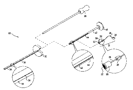
Fig. 1 is a schematic, pictorial illustration of a
kit for implantation of an intravascular pressure sensing
device, in accordance with an embodiment of the present
invention;
Fig. 2 is a schematic, pictorial illustration
showing a method for implantation of a pressure sensing
device in the arm of a patient, in accordance with an
embodiment of the present invention;
Figs. 3-5 are schematic, pictorial illustrations
showing details of successive stages in a method for
implantation of an intravascular pressure sensing device,
in accordance with an embodiment of the present
invention;
Fig. 6 is a schematic, pictorial illustration
showing deployment of an intravascular pressure sensing
device after implantation, in accordance with an
embodiment of the present invention; and
Fig. 7 is a schematic pictorial illustration showing
a control unit for intravascular pressure measurement, in
accordance with an embodiment of the present invention.
DETAILED DESCRIPTION OF EMBODIMENTS
Fig. 1 is a schematic, pictorial illustration
showing a kit 20 for use in implantation of an
intravascular pressure sensing device 21, in accordance
with an embodiment of the present invention.
6
CA 02680567 2009-09-16
Device 21 itself comprises a sensor die 22, which is
configured for percutaneous insertion through a wall of a
blood vessel, as shown in the figures that follow.
Sensor die 22 is connected by a wire 26 to an electronics
package 24, which processes the electrical signal that is
generated by the sensor die in order to provide an output
that is indicative of the pressure in the blood vessel.
To facilitate insertion through the vessel wall, the
transverse outer dimension of the sensor die is typically
less than 1 mm, although larger and smaller dies sizes
may also be used depending on technological constraints
and application requirements. The electronics package is
made of a biocompatible material, suitable for
implantation under the skin, and may be made in the form
of a rectangle about 3-4 mm on a side and about 0.5-0.8
mm in height. The electronics package may be powered and
interrogated by a control unit outside the body, which is
placed next to the skin above the package, as shown in
Fig. 7.
Sensor die 22 may comprise any suitable type of
pressure-sensitive element. For example, the sensor die
may comprise a capacitor, which deforms and thus varies
its capacitance under pressure. The capacitor may be
connected as part of a resonant circuit as described in
the above-mentioned U.S. Patent 6,939,299, wherein the
inductor and other elements of the resonant circuit may
also reside in die 22 and/or in electronics package 24.
The capacitance of the sensor die may then be measured by
detecting the resonance peak in the circuit response.
Alternatively, die 22 may comprise a piezoelectric
element, which outputs a voltage signal to electronics
7
CA 02680567 2009-09-16
package 24 in proportion to the pressure encountered by
the die. Alternatively, die 22 may comprise any other
suitable type of pressure-sensing element that is known
in the art.
In the embodiment shown in Fig. 1, wire 26 includes
an anchor 30 for tension relief after deployment of the
sensing device in the patient's body. In addition, the
wire may be connected to sensor die 22 by a safety
release mechanism 28 to prevent the sensor die from being
accidentally pulled out of the vessel wall after
implantation.
Sensing device 21 is implanted percutaneously in a
blood vessel using the components of kit 20 that are
shown in Fig. 1, which include:
= A trocar 32, having a shaft 34 with a longitudinal
slot 36 (for accommodating wire 26, as shown
below).
= A puncture tool 38.
= An inserter 40, which also comprises a shaft 42
with a slot 44 for wire 26.
Fig. 2 is a schematic, pictorial illustration
showing a medical practitioner 48 using elements of kit
20 to implant sensing device 21 in an arm 50 of a patient
52, in accordance with an embodiment of the present
invention. In the pictured example, practitioner 48 is
inserting the sensor die in the patient's brachial
artery. This procedure may be performed under local
anesthetic, possibly with the use of ultrasound imaging
or other means to visualize the artery and avoid
8
CA 02680567 2009-09-16
accidentally damaging nerves and other nearby structures,
since in this percutaneous procedure, the practitioner is
generally not able to see the target artery directly.
The brachial artery is convenient because it is easily
accessible by percutaneous approach. Alternatively, the
devices and methods described herein may be applied to
other arteries, as well as to veins and to measurement of
pressure in other organs and body passages that are
amenable to this sort of approach.
Figs. 3-5 are schematic, pictorial illustrations
showing details of successive stages in implantation of
sensing device 21 in a blood vessel 56, such as the
brachial artery, in accordance with an embodiment of the
present invention. First, as shown in Fig. 3,
practitioner 48 passes the distal end of trocar 32
through skin 54 of patient 52 and through the underlying
tissue until it reaches vessel 56. The practitioner then
inserts puncture tool 38 through the shaft of the trocar
and makes a small hole in the vessel wall using a distal
point 58 at the end of the puncture tool.
Next, as shown in Fig. 4, the practitioner withdraws
the puncture tool and uses inserter 40 to push the sensor
die (which is not seen in this figure) through trocar 32
into vessel 56. The proximal end of wire 26 extends out
to electronics package 24 through slots 44 and 36 (since
the electronics package is too large to fit into the
trocar shaft) At full extension, as shown in Fig. 5,
sensor die 22 protrudes slightly into vessel 56, while
electronics package 24 remains just above the surface of
skin 54. The small size of the sensor die and the
9
CA 02680567 2009-09-16
percutaneous approach to implantation that is illustrated
here minimize trauma to the blood vessel and leakage of
blood and promote rapid healing after implantation. The
sensor die may be anchored in place within the blood
vessel using an anchoring mechanism (not shown in the
figures) that extends to the sides inside the vessel
wall, such as a mechanism similar to that described in
U.S. Patent 6,783,499, whose disclosure is incorporated
herein by reference.
Fig. 6 is a schematic, pictorial illustration
showing deployment of sensing device 21 after
implantation, in accordance with an embodiment of the
present invention. After sensor die 22 has been inserted
through the wall of the blood vessel, as shown in Fig. 5,
practitioner 48 withdraws inserter 40 and trocar 32
through skin 54. Withdrawal of the trocar causes anchor
30 on wire 26 near the sensor die to open outward, thus
preventing the sensor from being accidentally pulled out
of vessel 56. The practitioner may also implant
electronics package 24 just under the surface of skin 54
through a suitable incision.
Fig. 7 is a schematic pictorial illustration showing
a control unit 60 for intravascular pressure measurement
in conjunction with sensor die 22 and electronics package
24, in accordance with an embodiment of the present
invention. The control unit may, for example, be
strapped around a limb of the body, such as arm 50, as
shown in the figure, and may thus receive pressure
readings from the sensor die (via the electronics
package) continuously. Alternatively, the control unit
CA 02680567 2009-09-16
may be placed next to the arm only intermittently, as
needed. In either case, the control unit may transfer
electrical power to electronics package 24 and receive
output signals from the electronics package by wireless
link, using induction, for example.
In the configuration shown in Fig. 7, a
microcontroller 62 in control unit 60 interrogates
electronics package 24 via the wireless link and
processes the output of the electronics package to derive
a calibrated pressure reading. This reading may be
presented on a display 64. Alternatively or
additionally, the reading may be stored in memory in the
control unit and/or conveyed to a monitoring station (not
shown) by a wireless or wired connection.
As another option, the continuous pressure
monitoring provided by control unit 60 can be used to
regulate closed-loop drug administration or other therapy
for controlling hypertension. For example, in the
embodiment shown in Fig. 7, control unit 60 comprises a
drug reservoir and pump (not shown), which dispense
medication through a tube 66 into the patient's body,
with dosage based on the measured pressure.
It will be appreciated that the embodiments
described above are cited by way of example, and that the
present invention is not limited to what has been
particularly shown and described hereinabove. Rather,
the scope of the present invention includes both
combinations and subcombinations of the various features
described hereinabove, as well as variations and
11
CA 02680567 2009-09-16
modifications thereof which would occur to persons
skilled in the art upon reading the foregoing description
and which are not disclosed in the prior art.
12
