Note : Les descriptions sont présentées dans la langue officielle dans laquelle elles ont été soumises.
CA 02681815 2009-09-24
1
Description
Method and device for visually assisting a catheter
application
The invention relates to a method and to a device for visually
assisting a catheter application on the heart of a patient
using at least one image of the patient obtained by means of a
C-arm X-ray device and using electroanatomical mapping data
relating to the patient, obtained by means of an
electromagnetic position-detection and mapping system.
In various fields where medical technology finds application,
it is customary nowadays for a medical instrument, a punction
needle or a catheter, for example, to be introduced into a
patient in a targeted manner with the aid of image information
made available by imaging devices, in order to examine or
treat the patient or a tissue or organ of the patient with the
instrument.
Thus, for example, cardiac dysrhythmias in a patient are
treated using what is known as an ablation, in which an
ablation catheter is introduced through veins or arteries into
one of the chambers of the patient's heart, under X-ray
monitoring, based on X-ray images, for example, and the tissue
responsible for the dysrhythmias is destroyed by high
frequency current. The prerequisite for carrying out a
successful catheter ablation is the precise locating of the
cause of the dysrhythmias in the chamber of the heart. This is
achieved by means of an electrophysiological investigation in
which electrical potentials are detected in a locally resolved
manner using a mapping catheter introduced into the chamber of
the heart. From this electrophysiological investigation, known
as electroanatomical mapping, 3D mapping data, for example,
CA 02681815 2009-09-24
2
which can be visualized on a monitor, are thus obtained. The
mapping function and the ablation function are frequently
combined in one catheter so that the mapping catheter is also
an ablation catheter at the same time.
A known electroanatomical 3D mapping method, such as is
possible, for example, using the CARTO-System from the company
Biosense Webster Inc., USA, is based on electromagnetic
principles. Three different low-intensity electromagnetic
alternating fields are generally established using
transmitters arranged under a patient supporting device. By
means of electromagnetic sensors incorporated in the catheter
tip of the mapping catheter, it is then possible to measure
the changes in voltage within the electromagnetic alternating
fields induced by catheter movements and to calculate the
position of the mapping catheter at any time with the aid of
mathematical algorithms. Through point-by-point mapping of the
endocardial contour of a chamber of the heart with the mapping
catheter and simultaneous detection of the electrical signals
from the sensors, 3D mapping data is obtained or an
electroanatomical three-dimensional map is produced, in which
the electrical signals are reproduced in a color-coded manner.
The guidance of the ablation catheter can thus be achieved not
only with the aid of the aforementioned X-ray images, but also
using the electroanatomical mapping data, which can be
generated in real time using for example, the aforementioned
CARTO- System from the company Biosense Webster Inc., USA and
displayed on a monitor. The fact is that the X-ray images do
not in fact show in detail the anatomy of the patient or, in
particular, the anatomy of the patient's heart. A 3D view of
anatomical details of the heart could increase the precision
relative to the morphology of the heart tissue during an
ablation procedure, speed up the ablation procedure and lead
CA 02681815 2009-09-24
3
to a reduction in the X-ray dose applied to a patient during
an ablation.
Electrophysiologists welcome the opportunity, particularly in
complex cases, of being able to carry out the ablation using a
combination of electrophysiological and morphological
criteria. It would therefore be helpful for
electrophysiologists to have at their disposal a combined
visualization of 3D image data obtained using an image-
generating device and electroanatomical 3D mapping data.
DE 103 40 455 Al and DE 103 40 546 Al disclose, for example,
methods for visually assisting a catheter application, in
which 3D image data of an area of the patient that is to be
treated is acquired by a method of tomographic 3D image
generation before the catheter application is carried out, a
3D surface contour of objects in the area to be treated being
extracted from the 3D image data by segmentation and
electroanatomical 3D mapping data that are subsequently
provided and 3D image data forming the 3D surface contour
being assigned to each other in the correct position and
dimensions and being visualized during the catheter
application procedure, for example, such that they are
superimposed on one another. This superimposition of 3D image
data relating to the patient obtained before the catheter
application, by means of computer tomography or magnetic
resonance tomography, for example, and electroanatomical 3D
mapping data relating to the patient sometimes requires a
time-consuming and error-prone marker-based or surface-based
registration of the data with one another. Here, errors in
registration can have a negative effect on the quality and
reliability of images that comprise merged data. In this
method, the precision of registration therefore also depends
on the number of surface points that are obtained using the
mapping system.
= CA 02681815 2009-09-24
4
The object underlying the invention is therefore to provide a
method and a device of the type mentioned in the introduction,
such that the combined use of image data and mapping data is
simplified.
This object is achieved according to the invention by a method
and a device for visually assisting a catheter application on
the heart of a patient using at least one image of the patient
obtained by means of a C-arm X-ray device and using
electroanatomical mapping data relating to the patient,
obtained by means of an electromagnetic position-detection and
mapping system. The C-arm X-ray device and the electromagnetic
position-detection and mapping system are calibrated in
relation to each other, by determining a coordinate
transformation between a coordinate system assigned to the C-
arm X-ray device and/or a coordinate system that is assigned
to at least one image generated by the C-arm X-ray device and
a coordinate system assigned to the electromagnetic position-
detection and mapping system. The position of the patient is
determined during the acquisition of the image and/or during
the acquisition of the electroanatomical mapping data and is
at least indirectly assigned to the image and/or to the
electroanatomical mapping-data.
By calibrating the C-arm X-ray device and the electromagnetic
position-detection and mapping system in relation to each
other, registration of image data relating to an image
generated by the C-arm X-ray device and mapping data generated
by the electromagnetic position-detection and mapping system
during a catheter application is no longer required, since the
transformation relationship for the data from the two systems
or devices is now known. Consequently, assuming that the
patient has not changed position between and after the
acquisition of data using the two systems or devices, it is
possible to merge together or superimpose on one another image
= CA 02681815 2009-09-24
data from images of the patient obtained before or during a
catheter application using the C-arm X-ray device, whether
comprising 2D images or 3D images, and mapping data obtained
before or during the catheter application, whether it is 2D or
3D mapping data , without having to carry out a time-
consuming or error-prone registration of the data. Moreover,
as a result of the fact that the position of the patient is
determined during the acquisition of the image and/or the
acquisition of the electroanatomical mapping data and is
assigned at least indirectly to the image and/or to the
electroanatomical mapping data, it is additionally possible
accordingly to take into account, in the image processing,
changes in the position of the patient after the acquisition
of the image using the C-arm X-ray device and the acquisition
of the electroanatomical mapping data.
On the C-arm X-ray device, which is provided for angiographic
applications, for example, an X-ray source and X-ray receiver
are arranged opposite each other on a C-arm, which can be
adjusted to record 2D projections from different projection
directions around the patient. A volume data set can be
reconstructed from a series of 2D projections recorded with
the C-arm X-ray device at projection directions that differ
from each other. On the basis of the known dimensions of the
C-arm X-ray device and the known projection geometries of the
2D projections, the transformation relationship between a
coordinate system assigned to the C-arm X-ray device and a
coordinate system assigned to the volume data set or to a 3D
image generated from the volume data set is also known.
According to a variant of the invention, a patient supporting
device is assigned to the C-arm X-ray device in a defined
manner, that is, the C-arm X-ray device and patient supporting
device are arranged relative to each other in a known manner.
= CA 02681815 2009-09-24
6
According to a further variant of the invention, the
electromagnetic position-detection and mapping system
comprises at least one transmitter to generate an
electromagnetic alternating field and a catheter with at least
one sensor. Usually the electromagnetic position-detection and
mapping system comprises a plurality of transmitters, for
example, three transmitters, to generate three different
alternating fields and the catheter comprises three sensors,
such that the position of the catheter can be determined in a
coordinate system assigned to the electromagnetic position-
detection and mapping system with the aid of the three sensors
incorporated in the catheter.
To determine the coordinate transformation between a
coordinate system assigned to the C-arm X-ray device and/or a
coordinate system assigned to at least one image generated
with the C-arm X-ray device and a coordinate system assigned
to the electromagnetic position-detection and mapping system,
according to a variant of the invention the transmitter is
arranged in a defined manner on the C-arm X-ray device or on
the patient supporting device. According to an embodiment of
the invention, the transmitter is arranged in a defined manner
on the C-arm of the C-arm X-ray device. In this way, a firm
relationship can be established between the C-arm X-ray device
and the electromagnetic position-detection and mapping system.
According to an embodiment of the invention, the transmitter
is detachable from the C-arm X-ray device or from the patient
supporting device. Therefore, if the transmitter interferes,
for example, with the taking of images of the patient using
the C-arm X-ray device, then it can be detached from the C-arm
X-ray device or the patient supporting device while images are
being taken by the C-arm X-ray device and then be arranged
again in the defined position on the C-arm X-ray device or on
the patient supporting device.
CA 02681815 2009-09-24
7
According to a different variant of the invention, at least
one positioning and orientation sensor of the electromagnetic
position-detection and mapping system is arranged on or in the
patient supporting device in a defined manner. In this case,
on the basis of the known relationship between the C-arm X-ray
device and the patient supporting device and on the basis of
the known design arrangement of the at least one positioning
and orientation sensor on or in the patient supporting device,
the transformation relationship between the coordinate system
assigned to the C-arm X-ray device and thus also between the
coordinate system assigned to an image recorded using the C-
arm X-ray device and the coordinate system assigned to the
electromagnetic position-detection and mapping system can be
determined by detecting the positioning and orientation sensor
with the electromagnetic position-detection and mapping
system.
According to an embodiment of the invention, the at least one
positioning and orientation sensor of the electromagnetic
position-detection and mapping system is detachable from the
patient supporting device. Preferably, the positioning and
orientation sensor or the positioning and orientation sensors
is/are arranged in a module that is detachable from the
patient supporting device or in a plurality of modules that
are detachable from the patient supporting device, said
modules being arranged relative to each other on the patient
supporting device in a defined manner.
According to a variant of the invention, the determination of
the coordinate transformation between a coordinate system
assigned to the C-arm X-ray device and/or a coordinate system
assigned to at least one image generated with the C-arm X-ray
device and a coordinate system assigned to the electromagnetic
position-detection and mapping system is achieved by means of
at least one marker, which is depicted in an image that is
CA 02681815 2009-09-24
8
generated and is detectable using the electromagnetic
position-detection and mapping system. Usually a plurality of
markers, preferably at least three markers, are used to
determine the coordinate transformation, the coordinates of
each of the individual markers being determined in the
respective coordinate systems and the coordinate
transformation between the coordinate systems being
ascertained using the coordinates that have been determined
for the respective markers in the coordinate systems.
The marker or markers are X-ray positive markers, for example
small metal balls, which are depicted in at least two X-ray
projections taken at projection angles that differ from each
other or in a 3D image. According to embodiments of the
invention, the images of the markers can be located in the
projection images or the 3D image either manually through a
graphic user interface or automatically using a method of
pattern recognition, such that the marker coordinates required
for the determination of the coordinate transformation can be
determined in the coordinate system assigned to the projection
images or the 3D image by back projection.
In order to determine the coordinates of the markers in the
coordinate system assigned to the electromagnetic position-
detection and mapping system, a position sensor of the
electromagnetic position-detection and mapping system is used,
with which the respective markers are touched.
According to a variant of the invention, at least one marker
is an X-ray positive position sensor of the electromagnetic
position-detection and mapping system. Since the marker itself
is a position sensor of the electromagnetic position-detection
and mapping system, said marker does not specifically have to
be touched by another position sensor in order for the
coordinates thereof in the coordinate system assigned to the
CA 02681815 2009-09-24
9
electromagnetic position-detection and mapping system to be
detected and determined, but can be detected directly and
automatically by the electromagnetic position-detection and
mapping system. Here, the marker or the X-ray positive
position sensor can be a catheter tip of a catheter from the
electromagnetic position-detection and mapping system.
According to an embodiment of the invention, a phantom
comprising at least one marker is provided for the
determination of the coordinate transformation, said phantom
being arranged in an appropriate manner on the patient
supporting device relative to the C-arm X-ray device and the
electromagnetic position-detection and mapping system in order
to determine the coordinate transformation.
According to a variant of the invention, at least one marker
of said phantom may be a position sensor of the
electromagnetic position-detection and mapping system such
that, as already mentioned, the marker does not specifically
have to be touched by a position sensor, but can be detected
directly and automatically by the electromagnetic position-
detection and mapping system.
According to an embodiment of the invention, all the markers
of the phantom are position sensors of the electromagnetic
position-detection and mapping system such that the markers of
the phantom can be detected directly and automatically by the
electromagnetic position-detection and mapping system and the
coordinates thereof can be determined in the coordinate system
assigned to the electromagnetic position-detection and mapping
system.
According to a further variant of the invention, the phantom
comprises, at least one, preferably a plurality of further
markers that are not position sensors, in addition to the
CA 02681815 2009-09-24
position sensor of the electromagnetic position-detection and
mapping system, the positions or coordinates of the further
markers of the phantom being known relative to the at least
one position sensor of the phantom. In this case it is
sufficient to detect the position sensor automatically, using
the electromagnetic position-detection and mapping system and
to determine the coordinates thereof. The coordinates of the
further markers in the coordinate system assigned to the
electromagnetic position-detection and mapping system are then
obtained from the known coordinates of the further markers
relative to the position sensor.
According to an embodiment of the invention, the phantom that
comprises markers and the electromagnetic position-detection
and mapping system are arranged in a defined manner relative
to each other, such that the coordinate transformation between
a coordinate system assigned to the phantom and a coordinate
system assigned to the electromagnetic position-detection and
mapping system is known. In this case, therefore, the
coordinates of the markers of the phantom do not themselves
have to be determined in the coordinate system assigned to the
electromagnetic position-detection and mapping system. In
fact, in order to calculate the coordinate transformation
between the coordinate system assigned to the C-arm X-ray
device or the coordinate system assigned to an image obtained
using the C-arm X-ray device and the coordinate system
assigned to the electromagnetic position-detection and mapping
system, it is only the coordinates of the markers of the
phantom that have to be determined in the coordinate system
assigned to the image obtained using the C-arm X-ray device,
in order to be able to calculate the coordinate
transformation.
The methods described hitherto for the determination of the
coordinate transformation between the coordinate system
CA 02681815 2009-09-24
11
assigned to the C-arm X-ray device or the coordinate system
assigned to the image acquired using the C-arm X-ray device
and the coordinate system assigned to the electromagnetic
position-detection and mapping system are what are known as
offline methods, in which a patient who needs to be examined
is not present.
The determination of the coordinate transformation may also be
achieved online, however, that is, in the presence of the
patient. According to this variant of the invention, the
patient or a part or an organ of the patient is provided with
markers, preferably with X-ray positive markers, the images
whereof are located in an image obtained using the C-arm X-ray
device and which can be detected using the electromagnetic
position-detection and mapping system. Next, the coordinates
of the markers in the coordinate systems assigned to the image
and the coordinates of the markers in the coordinate system
assigned to the electromagnetic position-detection and mapping
system are again determined and on this basis, the coordinate
transformation between the coordinate system assigned to the
C-arm X-ray device or to the image generated using the C-arm
X-ray device and the coordinate system assigned to the
electromagnetic position-detection and mapping system is
determined.
According to a variant of the invention, the position of the
patient is determined continuously or intermittently during
the catheter application. In this way, the position of the
patient is constantly known, so that changes in the position
of the patient since the acquisition of the image using the C-
arm X-ray device and/or the acquisition of the
electroanatomical mapping data is recorded and can be taken
into account accordingly in the image processing and use of
the image data.
CA 02681815 2009-09-24
12
According to an embodiment of the invention, the image data
for the image obtained using the C-arm X-ray device and the
electroanatomical mapping data is merged together or
superimposed during the acquisition of the image and of the
electroanatomical mapping data, or superimposed on one
another, taking into account the position of the patient (P).
Therefore, if for example, the patient changes position
between the acquisition of the image data for the image using
the C-arm X-ray device and the acquisition of the
electroanatomical mapping data, then on the basis of the
change in the position of the patient that has been recorded
and determined, the previously determined coordinate
transformation may be adapted accordingly, such that the
electroanatomical mapping data and the image data for the
image that was generated previously can continue to be merged
together or superimposed with positional and locational
precision.
According to a further variant of the invention, at least part
of a catheter that is detectable using the electromagnetic
position-detection and mapping system or of an instrument that
is detectable using the electromagnetic position-detection and
mapping system is blended into the image of the patient and/or
the electroanatomical mapping data, taking into account the
position of the patient. In this case, too, an adjustment of
the coordinate transformation is made, taking into account the
change in the position of the patient, such that an image of
the catheter or of another instrument can be blended with
positional and locational precision into an image that has
been taken of the patient that can also comprise
electroanatomical mapping data that has been acquired
beforehand or at the same time during the navigation of the
catheter or of the instrument.
CA 02681815 2009-09-24
13
According to an embodiment of the invention, the patient is
provided with at least one reference sensor of the
electromagnetic position-detection and mapping system for
detecting the position of the patient, with the result that
the position of the patient can be determined in the
coordinate system assigned to the electromagnetic position-
detection and mapping system during the determination of an
image using the C-arm X-ray device, during the determination
of the electroanatomical mapping data and during the catheter
application using the electromagnetic position-detection and
mapping system.
According to a variant of the invention, the positions or
coordinates determined using the electromagnetic position-
detection and mapping system are determined relative to the
reference sensor of the patient. In this case there is a
direct registration between the coordinate system assigned to
an image and a coordinate system assigned to the patient
through a reference sensor.
According to an embodiment of the invention, a time
synchronization is achieved between the acquisition of an
image of the patient using the C-arm X-ray device and the
position of the patient, the time of acquisition being
assigned to an image that has been taken and the position of
the patient being determined over time, such that the position
of the patient at the time of acquisition of the image can be
determined by comparing the times and the image acquired can
be assigned to this position of the patient.
According to a different embodiment of the invention, the C-
arm X-ray device assigns an identifier to an image that has
been taken of the patient at the time of acquisition of the
image and transmits the identifier to the electromagnetic
position-detection and mapping system at the time of
CA 02681815 2009-09-24
14
acquisition of the image. The electromagnetic position-
detection and mapping system then stores the position of the
patient, including the identifier, at the time of acquisition
of the image.
According to a further variant of the invention, the C-arm X-
ray device retrieves the position of the patient from the
electromagnetic position-detection and mapping system at the
time an image of the patient is acquired and assigns it to the
image that has been acquired.
These variants of the invention make it possible in each case
to register a change in the position of the patient relative
to a position the patient assumed earlier, in which an image
was generated, and to take into account the change of position
in the superimposition or merging of image data and mapping
data or when blending in a catheter or an instrument into the
image data recorded beforehand.
An embodiment of the invention is shown in the attached
schematic drawings. The drawings show:
FIG 1 a device comprising an electromagnetic position-
detection and mapping system, a C-arm X-ray device
and computing devices, together with a patient
supporting device and
FIG 2 the device from FIG 1 comprising a phantom that is
arranged on the patient supporting device.
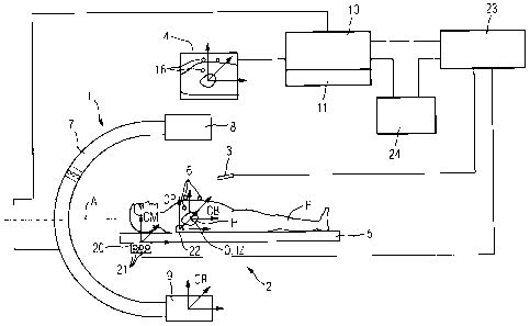
FIG 1 shows a device comprising a C-arm X-ray device 1 and an
electromagnetic position-detection and mapping system 2, which
device is used for visually assisting a catheter application
on a living being, for example, a catheter ablation on the
heart H of the patient P. In the case of the present
embodiment, a catheter 3 of the electromagnetic position-
CA 02681815 2009-09-24
detection and mapping system 2, which in the present case is
both a mapping catheter and an ablation catheter, is to be
guided through veins or arteries into the chamber of the heart
H of the patient P, supported by at least one image of the
heart H of the patient P obtained using the C-arm X-ray device
1 and supported by mapping data of the heart H of the patient
P obtained using the electromagnetic position-detection and
mapping system 2, in order to make it possible to carry out an
ablation procedure there to treat cardiac dysrhythmias. For
this purpose, image data for an image obtained using the C-arm
X-ray device 1 and electroanatomical mapping data obtained
using the electromagnetic position-detection and mapping
system 2 are to be merged together or superimposed on one
another in order to simplify the intervention on the patient
P. Moreover, the position of the catheter 3 that has been
introduced into the patient P is to be blended into the image
data and mapping data that has been merged together or
superimposed on one another and is displayed on a monitor 4.
In order to be able to merge together or superimpose on one
another the image data for the image obtained using the C-arm
X-ray device 1 and the electroanatomical mapping data obtained
using the electromagnetic position-detection and mapping
system, or to be able to blend an image of the catheter 3 into
the merged or superimposed data with locational and positional
precision, the C-arm X-ray device 1 and the electromagnetic
position-detection and mapping system 2 are calibrated
relative to each other.
For this purpose, the patient P is supported on a patient
supporting device 5 assigned to the C-arm X-ray device 1 in a
defined manner. Assignment of the C-arm X-ray device 1 and the
patient supporting device 5 in a defined manner is understood
to mean that the spatial arrangement of the two devices is
known even when the devices or parts of the devices are moved
CA 02681815 2009-09-24
16
relative to each other. The patient P is preferably provided
with at least 3 X-ray positive markers 6 in the region of
their heart H. The markers 6 are arranged on the patient P
such that said markers are depicted in X-ray images of the
heart H of the patient P that can be taken using the C-arm X-
ray device 1 without obscuring details of the heart H, and
such that these details can be detected by the electromagnetic
position-detection and mapping system 2. In this way, a
coordinate transformation between a coordinate system CR
assigned to the C-arm X-ray device 1 or between a coordinate
system CB that is assigned to an image generated by the C-arm
X-ray device 1 and a coordinate system CM assigned to the
electromagnetic position-detection and mapping system 2 can be
determined by means of the markers 6.
In the case of the present embodiment, a volume data set or a
3D image of the heart H of the patient P is acquired using the
C-arm X-ray device 1. This involves rotating the C-arm 7 of
the C-arm X-ray device 1, which arm is provided with an X-ray
source 8 and an X-ray receiver 9, around its orbital axis 0 or
its angulation axis A within an angle range of about 1900, a
plurality of 2D projections of the heart H of the patient P
being taken from different projection directions. An image
calculator 10 reconstructs a volume data set or 3D-image of
the heart H of the patient P from said 2D X-ray projections.
As the heart H is an active organ, the reconstruction of the
volume data set or of the 3D-image of the heart H is achieved
in the case of the present embodiment by using ECG signals
from the patient P recorded during the acquisition of the 2D
X-ray projections. The ECG equipment is not shown in FIG 1
since it is designed in the known manner. Such an ECG-
triggered reconstruction method is described, for example, in
DE 10 2005 016 472 Al.
CA 02681815 2009-09-24
17
The reconstructed 3D-image of the heart H of the patient P
also shows the markers 6. In preparation for the calculation
of the aforementioned coordinate transformation, the images 16
of the markers 6 are located manually or even automatically in
the 3D image using a method of pattern recognition and the
coordinates of the markers 6 in the coordinate system CB
assigned to the 3D image or to the coordinate system CR
assigned to the C-arm X-ray device 1 are determined. This is
possible because the dimensions and geometries of the C-arm X-
ray device 1 and the projection geometries for the acquisition
of the 2D X-ray projections are known. Here the coordinate
origin of the coordinate system CB can be located, for
example, in the isocenter IZ of the C-arm 7. As stated in the
aforementioned, the markers 6 are arranged on the patient P
such that they do not conceal any interesting structures of
the heart H. Finally, the volume data set or 3D-image of the
heart H of the patient P is stored in an intermediate image
memory 11 so that the 3D image data can be used for merging or
superimposition with other data or for a navigation of the
catheter 3 or of another instrument.
In the case of the present embodiment, the electromagnetic
position-detection and mapping system 2 comprises three
transmitters 21 combined in one unit 20, each of which
generates an electromagnetic alternating field, said
alternating fields differing from each other. In the case of
the present embodiment, the unit 20 that comprises the three
transmitters 21 is detachably arranged in a defined position
on the patient supporting device S. If the unit 20 for
example, were to cause interference during the recording of
the 2D X-ray projections, it can then be removed from the
patient supporting device 5 and be arranged on the patient
supporting device 5 once again after the X-ray projections
have been taken. The unit 20 with the three transmitters 21
CA 02681815 2009-09-24
18
could also be detachably arranged in a different location on
the patient supporting device 5 or on the C-arm X-ray device 1
that is arranged in a defined manner relative to the patient
supporting device 5, as indicated, for example, with dotted
lines in FIG 1 on the C-arm 7 of the C-arm X-ray device 1.
The catheter 3 of the electromagnetic position-detection and
mapping system 2 is provided at the catheter tip with three
sensors that are not further shown in FIG 1. If the catheter 3
is moved within the alternating fields of the transmitters 21,
the position of the catheter 3 can be determined in the
coordinate system CM assigned to the electromagnetic position-
detection and mapping system 2 using a computing device 23 of
the electromagnetic position-detection and mapping system 2.
In the case of the present embodiment, the patient P has a
reference sensor 22 of the electromagnetic position-detection
and mapping system 2 fitted to their back, such that movements
of the patient P can also be determined using the
electromagnetic position-detection and mapping system. A
patient coordinate system CP is assigned to the reference
sensor 22 or to the patient P.
For the determination of the coordinate transformation between
the coordinate system CR assigned to the C-arm X-ray device 1
or the coordinate system CB assigned to the image acquired
using the C-arm X-ray device 1 and the coordinate system CM
assigned to the electromagnetic position-detection and mapping
system or to the patient coordinate system CP, the coordinates
of the markers 6 in the coordinate system CM assigned to the
electromagnetic position-detection and mapping system 2 or to
the patient coordinate system CP are determined by the markers
being touched by the catheter 3. Preferably, but not
necessarily, the coordinates of the markers 6 are shown in the
patient coordinate system CP.
CA 02681815 2009-09-24
19
If the coordinates of the markers 6 are available in the image
calculator 10 in the coordinate system CB assigned to the
volume data set or to the 3D image, and likewise if the
coordinates of the markers 6 are available in the computing
device 23 of the electromagnetic position-detection and
mapping system 2 in the patient coordinate system CP or in the
coordinate system CM assigned to the electromagnetic position-
detection and mapping system 2, the respective coordinate
transformations can be determined using the image computer 10,
the computing device 23 or a further computing device 24.
Furthermore, before or during a catheter application on the
heart H of the patient P, electroanatomical mapping data,
preferably 3D mapping data, can be acquired using the catheter
3. By means of electromagnetic sensors incorporated into the
tip of the catheter 3, it is possible to measure the changes
in voltage induced by catheter movements of the catheter 3
within the alternating fields of the transmitters and to
measure the position of the catheter 3 at any time with the
aid of mathematical algorithms. Through point-by-point mapping
of areas of a chamber of the heart with the catheter 3 and
simultaneous detection of the electrical signals from the
sensors, an electroanatomical three-dimensional map or 3D
mapping data is or are thus produced, it being possible for
the electrical signals to be reproduced in a color-coded
manner, for example. Since the coordinate transformation
between the coordinate system CB assigned to the 3D image
produced by the C-arm X-ray device 1 and the patient
coordinate system CP or the coordinate system CM that is
assigned to the electromagnetic position-detection and mapping
system 2 is known, the image data for the 3D image and the 3D
mapping data can be merged together or superimposed on one
another and displayed on the monitor 4 in order to visually
assist a catheter application on the heart H of the patient P.
CA 02681815 2009-09-24
In addition, the catheter 3, for example, can be navigated
relative to the heart H of the patient P, on the basis, for
example, of the merged data, by means of an image of the
catheter 3, based on the known coordinate transformation,
being accordingly merged into the data that has been merged
together or superimposed on one another.
It therefore becomes clear that, by means of the procedure
according to the invention, it is possible for image data that
has been recorded before a catheter application, using image
data generated using an imaging device, to be merged or
superimposed with 3D mapping data likewise generated before
the catheter application or during the catheter application
and a catheter 3 can be navigated using the merged or
superimposed data relating to a patient P.
In order to avoid having to determine the coordinate
transformation anew following a change in the position of the
patient P, after the acquisition of the 3D image, the patient
P, as already stated, is provided with a reference sensor 22
of the electromagnetic position-detection and mapping system
2, such that changes in the position of the patient P are
determined by detection of the reference sensor 22 and the
coordinate transformation can be modified according to the
movement of the patient P, for example.
In order to correctly take into account a change in the
position of the patient P, one method of operation allows for
time synchronization to be achieved between the acquisition of
the 3D image using the C-arm X-ray device 1 and the position
of the patient P, by assigning the time of acquisition tl of
the 3D image to the 3D image that has been recorded, and
determining the position of the patient over time, every
second, for example, so that the position of the patient P at
the time ti of acquisition of the 3D image can be determined
CA 02681815 2009-09-24
21
by a manual or automatic comparison of the times. In this way,
it is therefore possible for a change in the position of the
patient P after the acquisition of the 3D image to be detected
and for this to be taken into account in transformation
calculations, in particular in the merging or superimposition
of image data relating to the 3D image and currently recorded
mapping data or in the current navigation of the catheter 3 on
the basis of the 3D image. For the time synchronization,
preferably both the image calculator 10 and the computing
device 23 have a clock, said clocks being synchronized with
each other, via the computer 24, for example.
The position of the patient P during the acquisition of the 3D
image can also be captured such that the C-arm X-ray device 1
or the image computer 10 assigns an identifier to the 3D image
at the time of acquisition of the 3D image and transmits the
identifier in real time at the time of acquisition of the 3D
image to the electromagnetic position-detection and mapping
system 2 or to the computing device 23 of the electromagnetic
position-detection and mapping system 2. Said computing device
23 of the electromagnetic position-detection and mapping
system 2 stores the position of the patient (P), including the
identifier at the time of acquisition of the 3D image.
Furthermore, the C-arm X-ray device 1 or the image calculator
can retrieve the position of the patient P from the
computing device 23 of the electromagnetic position-detection
and mapping system 2 at the time of acquisition of the 3D
image of the patient P and assign it to the 3D image that has
been acquired.
In all these cases, during the acquisition of the 3D image,
the position of the patient P is assigned to the 3D image of
the patient P that has been acquired, so that even in the
event of a change in the position of the patient P relative to
CA 02681815 2009-09-24
22
the position that the patient P assumed during the acquisition
of the 3D image, the transformation relationship, once
determined, does not have to be determined again but can be
adjusted accordingly according to the change in the patient's
position. The merger or superimposition of currently acquired
mapping data with image data for the 3D image acquired before
the change in position or the blending in of current images of
the catheter 3 into the image data for the 3D image acquired
before the change in position is then achieved on the basis of
the transformation relationship that has been modified
according to the new position of the patient P.
Should the transmitter unit 20 have to be removed from the
patient supporting device 5 during the acquisition of the
volume data set or of the 3D image using the C-arm X-ray
device, due to a lack of space, for example, then the position
of the patient P can be determined before the removal and
after the re-connection of the transmitter unit 20. If the
patient P has moved during the acquisition of the volume data
set or of the 3D image, then it is for the user to decide,
depending on the position values acquired, whether the
position value recorded before the removal of the transmitter
unit 20, the position value recorded after the re-connection
of the transmitter unit 20 or a mean value is assigned to the
volume data set or to the 3D image as a position value. In
this procedure, too, the position of the patient P is
determined during the acquisition of the volume data set or of
the 3D image.
The determination of the coordinate transformation between the
coordinate system CR assigned to the C-arm X-ray device 1 or
the coordinate system (CB) that is assigned to the 3D image
generated by the C-arm X-ray device 1 and the coordinate
system CM assigned to the electromagnetic position-detection
and mapping system 2 or to the patient coordinate system CP,
CA 02681815 2009-09-24
23
wherein markers 6 are arranged on the patient CP, has been
described with the aid of FIG 1. The determination of the
coordinate transformation can also be carried out in what is
known as an offline procedure, that is, without the patient P.
This can be achieved as shown in FIG 2, for example, such that
preferably three X-ray positive sensors 25 of the
electromagnetic position-detection and mapping system 2, for
example, three catheters 3, are arranged on the patient
supporting device 5 and the coordinates of the position
sensors 25 in the coordinate system CM assigned to the
electromagnetic position-detection and mapping system 2 are
determined. Moreover, at least two projection images from
different projection directions or an X-ray image of the
position sensors 25 are acquired by the C-arm X-ray device 1,
the images of the position sensors 25 are located in the
images or in the 3D image either manually or using a method of
pattern recognition, and the coordinates of the position
sensors are determined in the coordinate system CR assigned to
the C-arm X-ray device 1 or in the coordinate system CB
assigned to the projection images or to the 3D image. If the
coordinate transformation is determined on the basis of
coordinates that have been determined in the coordinate
systems, the C-arm X-ray device 1 and the electromagnetic
position-detection and mapping system 2 are calibrated
relative to each other.
Alternatively, a phantom 30 provided with X-ray positive
markers 31 can be used. If a 3D image of the phantom 30, for
example, in which the X-ray positive markers 31 are depicted
is generated by the C-arm X-ray device 1, then the images of
the markers 31 can be located in the 3D image again either
manually by clicking on them by hand or automatically using a
method of pattern recognition. On this basis, based on the
known design of the C-arm X-ray device and the known
CA 02681815 2009-09-24
24
projection geometries of the 2D projections that form the
basis of the 3D image, the coordinates of the markers 31 in
the coordinate system CR assigned to the C-arm X-ray device 1
or in the coordinate system CB assigned to the 3D image can be
determined. The determination of the coordinates of the
markers 31 in the coordinate system CM assigned to the
electromagnetic position-detection and mapping system 2 is
achieved, for example, by touching the markers 31 with the
catheter 3. The coordinate transformation can then be
calculated again using the coordinates determined in the
coordinate systems.
Alternatively, all the markers 31 of the phantom 30 can be
position sensors in the electromagnetic position-detection and
mapping system 2, such that to determine the coordinates of
the markers 31 in the coordinate system CM of the
electromagnetic position-detection and mapping system 2, the
markers 31 do not have to be expressly touched by the catheter
3.
Alternatively, only one marker 31 of the phantom 30 can be a
position sensor of the electromagnetic position-detection and
mapping system 2. In this case, if the positions of the
remaining markers 31 of the phantom 30 relative to the at
least one position sensor of the phantom 30 are known, then
only the coordinates of this one position sensor in the
coordinate system CM of the electromagnetic position-detection
and mapping system 2 have to be determined, while the others
can be determined from their known positions in relation to
the position sensor of the phantom 30 that has been detected.
A further possible way of establishing a relationship between
the coordinate system CR assigned to the C-arm X-ray device 1
and the coordinate system CM assigned to the electromagnetic
position-detection and mapping system 2 consists in arranging,
CA 02681815 2009-09-24
for example, three positioning and orientation sensors 35 of
the electromagnetic position-detection and mapping system 2 in
a defined manner on or in the patient supporting device 5. In
this case, on the basis of the known design relationship
between the C-arm X-ray device 1 and the patient supporting
device 5 and the known design arrangement of the three
positioning and orientation sensors 35 on or in the patient
supporting device 5, the transformation relationship between
the coordinate system CR assigned to the C-arm X-ray device 1
and thus also between the coordinate system CB assigned to an
image recorded using the C-arm X-ray device 1 and the
coordinate system CM assigned to the electromagnetic position-
detection and mapping system 2 can be determined by means of
the detection of the three positioning and orientation sensors
detectable with the electromagnetic position-detection and
mapping system 2. In this case, the three positioning and
orientation sensors 35 of the electromagnetic position-
detection and mapping system 2 can be designed to be
detachable from the patient supporting device by, for example,
arranging said sensors in one or a plurality of detachable
modules.
The C-arm X-ray device 1 and the electromagnetic position-
detection and mapping system 2 can thus be calibrated relative
to each other offline. Consequently, patient images can then
be taken using the C-arm X-ray device 1 and the
electromagnetic position-detection and mapping system 2 and
image data acquired using the C-arm X-ray device 1 and mapping
data from the electromagnetic position-detection and mapping
system 2 can be merged together or superimposed on one
another. Furthermore, images of navigation instruments such as
the catheter 3 that are detectable using the electromagnetic
position-detection and mapping system 2 can be blended into
the images acquired using the C-arm X-ray device 1 and/or into
CA 02681815 2009-09-24
26
the mapping data and/or into data that have been merged
together or superimposed on one another. This allows changes
in the position of the patient P to be registered, determined,
and taken into account accordingly through the detection of
the reference sensor 22, as already disclosed.
Moreover, the markers arranged on the patient can also be
positioning and orientation sensors in the electromagnetic
position-detection and mapping system.
Furthermore, a plurality of reference sensors can be arranged
on the patient P.
As already disclosed, apart from the catheter 3, other
instruments provided with a positioning and orientation sensor
can also be used in the device disclosed. If, for example,
instead of or in addition to the catheter 3, a catheter is
used with which image data from inside the body can be
acquired, for example, the ultrasound catheter AcuNav from
Siemens Medical Solutions, then the image data acquired using
this catheter can be combined with image data for the 3D image
acquired using the image generation device. Catheter
monitoring systems such as the Niobe System from Stereotaxis
can also be used in the device.