Note : Les descriptions sont présentées dans la langue officielle dans laquelle elles ont été soumises.
CA 02682441 2009-09-29
WO 2008/121826 PCT/US2008/058668
METHOD FOR THE TREATMENT OF FABRY DISEASE USING
PHARMACOLOGICAL CHAPERONES
CROSS-REFERENCE TO RELATED APPLICATIONS
This application claims priority to U.S. Provisional Application Serial
No. 60/909,185, filed March 30, 2007, which is hereby incorporated by
reference in
its entirety herein.
FIELD OF THE INVENTION
The present invention provides a method for treating Fabry disease by
restoring a-galactosidase A activity by at least 3-fold, and more particularly
by
restoring its activity to levels in the normal range. In addition, the present
invention
provides a method for monitoring the treatment of an individual having Fabry
disease
with a specific pharmacological chaperone by evaluating changes in the
presence
and/or quantity of specific surrogate markers. The present invention also
provides a
method for monitoring the treatment of an individual having Fabry disease with
a
specific pharmacological chaperone by evaluating the effects of treatment at
the sub-
cellular level.
BACKGROUND
Fabry disease is a glycosphingolipid (GSL) storage disease caused by an X-
linked inherited deficiency of lysosomal a-galactosidase A (a-Gal A), an
enzyme
responsible for the hydrolysis of terminal a-galactosyl residues from
glycosphingolipids. A deficiency in the enzyme activity results in a
progressive
deposition of neutral glycosphingolipids, predominantly globotriaosylceramide
(ceramide trihexoside, CTH, GL-3), in vascular endothelial cells causing renal
failure
along with premature myocardial infarction and strokes in patients with this
condition.
This disorder is classified by clinical manifestations into two groups: a
classic form
with generalized vasculopathy, and an atypical variant form, with clinical
manifestations limited to cardiac tissue.
The frequency of the disease is estimated to be about 1:40,000 in males, and
is
reported throughout the world within different ethnic groups. In classically
affected
males, the clinical manifestations include angiokeratoma (small, raised
reddish-purple
CA 02682441 2009-09-29
WO 2008/121826 PCT/US2008/058668
blemishes on the skin), acroparesthesias (burning in hands and feet),
hypohidrosis
(decreased ability to sweat), and characteristic corneal and lenticular
opacities (The
Metabolic and Molecular Bases of Inherited Disease, 8th Edition 2001, Scriver
et al.,
ed., pp. 3733-3774, McGraw-Hill, New York). Lipid storage may lead to impaired
arterial circulation and increased risk of heart attack or stroke. The heart
may also
become enlarged and the kidneys may become progressively involved. Other
symptoms include fever and gastrointestinal difficulties, particularly after
eating.
Some female carriers may also exhibit symptoms.
The affected male's life expectancy is reduced, and death usually occurs in
the
fourth or fifth decade as a result of vascular disease of the heart, brain,
and/or kidneys.
In contrast, patients with the milder "cardiac variant" normally have 5-15% of
normal
a-Gal A activity, and present with left ventricular hypertrophy or a
cardiomyopathy.
These cardiac variant patients remain essentially asymptomatic when their
classically
affected counterparts are severely compromised. Recently, cardiac variants
were
found in 11 % of adult male patients with unexplained left ventricular
hypertrophic
cardiomyopathy, suggesting that Fabry disease may be more frequent than
previously
estimated (Nakao et al., N. Engl. J. Med. 1995; 333: 288-293).
The a-Gal A gene has been mapped to Xq22, (Bishop et al., Am. J. Hum.
Genet. 1985; 37: A144), and the full-length cDNA and entire 12-kb genomic
sequences encoding a-Gal A have been reported (Calhoun et al., Proc. Natl.
Acad.
Sci. USA. 1985; 82: 7364-7368; Bishop et al., Proc. Natl. Acad. Sci. USA.
1986; 83:
4859-4863; Tsuji et al., Eur. J. Biochem. 1987; 165: 275-280; and Kornreich et
al.,
Nucleic Acids Res. 1989; 17: 3301-3302). There is a marked genetic
heterogeneity of
mutations that cause Fabry disease (The Metabolic and Molecular Bases of
Inherited
Disease, 8th Edition 2001, Scriver et al., ed., pp. 3733-3774, McGraw-Hill,
New
York.; Eng et al., Am. J. Hum. Genet. 1993; 53: 1186-1197; Eng et al., Mol.
Med.
1997; 3: 174-182; and Davies et al., Eur. J. Hum. Genet. 1996; 4: 219-224). To
date,
a variety of missense, nonsense, and splicing mutations, in addition to small
deletions
and insertions, and larger gene rearrangements have been reported.
Treatment
Treatment of Fabry disease is predominantly by enzyme replacement therapy
(ERT) with recombinant a-Gal A, e.g., with the product marketed as Fabrazyme
(Genzyme, Inc.) and Replagal (TKT, Inc.). ERT typically involves intravenous,
subcutaneous or intramuscular infusion of a purified form of the corresponding
wild-
2
CA 02682441 2009-09-29
WO 2008/121826 PCT/US2008/058668
type protein, or implantation of the protein in a bio-erodable solid form for
extended-
release. One of the main complications with ERT is attainment and maintenance
of
therapeutically effective amounts of protein due to rapid degradation of the
infused
protein. As a result, ERT requires numerous, high-dose infusions and as a
result, is
costly and time consuming.
ERT has several additional drawbacks, such as difficulties with large-scale
generation, purification and storage of properly folded protein, obtaining
glycosylated
native protein, generation of an anti-protein immune response in some
patients, and
failure of protein to cross the blood-brain barrier in sufficient quantities
to affect
diseases having significant central nervous system involvement.
Gene therapy using recombinant vectors containing nucleic acid sequences
that encode a functional protein, or genetically modified human cells that
express a
functional protein, is also being used to treat protein deficiencies and other
disorders
that benefit from protein replacement. Although promising, this approach is
also
limited by technical difficulties such as the inability of vectors to infect
or transduce
dividing cells, low expression of the target gene, and regulation of
expression once
the gene is delivered (e.g., many viral vectors require cells to be dividing
for
efficacy).
A third, relatively recent approach to treating protein deficiencies involves
the
use of small molecule inhibitors to inhibit synthesis the natural substrate of
the
deficient enzyme protein, thereby ameliorating the pathology. This "substrate
deprivation" approach has been specifically described for a class of about 40
related
enzyme disorders called lysosomal storage disorders or glycosphingolipid
storage
disorders. These heritable disorders are characterized by deficiencies in
lysosomal
enzymes that catalyze the breakdown of glycolipids in cells, resulting in an
abnormal
accumulation of lipids, which disrupts cellular function. The small molecule
inhibitors proposed for use as therapy are specific for inhibiting the enzymes
involved
in synthesis of glycolipids, reducing the amount of cellular glycolipid that
needs to be
broken down by the deficient enzyme. This approach is also limited in that
glycolipids are necessary for biological function, and excess deprivation may
cause
adverse effects. Specifically, glycolipids are used by the brain to send
signals from
one neuron to another. If there are too few or too many glycolipids, the
ability of the
neuron to send signals is impeded.
3
CA 02682441 2009-09-29
WO 2008/121826 PCT/US2008/058668
In addition, treatment with one substrate inhibitor, NB-DNJ, for lysosomal
storage disease Gaucher disease is associated with numerous adverse events in
humans, including peripheral neuropathy and severe gastrointestinal effects.
These
effects present even at low doses of 150 mg/day, which is not a
therapeutically
effective dose. In view of the foregoing, administration of NB-DNJ for the
treatment
of Gaucher in the U.S. and Europe is very limited to adults with mild-to-
moderate
type I Gaucher patients where ERT is not an option, and is not approved for
therapy
in Canada (Weinreb et al., Am J. Hematology. 2005. 80: 223-29).
A fourth approach, a specific chaperone strategy, rescues mutated proteins
from degradation presumably in the endoplasmic reticulum (ER) or in other
cellular
protein degradation/disposal systems. Previous patents and publications
describe a
therapeutic strategy for rescuing endogenous enzyme proteins, including
misfolded
lysosomal enzymes, from degradation by the ER quality control machinery. In
particular embodiments, this strategy employs small molecule reversible
inhibitors
which specifically bind to a defective lysosomal enzyme associated with a
particular
lysosomal disorder. In the absence of therapy, the mutated enzyme protein
folds
improperly in the ER (Ishii et al., Biochem. Biophys. Res. Comm. 1996; 220:
812-
815), is retarded in its maturation to a final product, and is subsequently
degraded in
the ER. The chaperone strategy involves the use of a compound that facilitates
the
correct folding of a mutated protein, to prevent undue or abnormal degradation
from
the ER quality control system, or accumulation of misfolded protein in the
cell. These
specific chaperones are designated pharmacological chaperones (or active site-
specific chaperones).
The chaperone strategy has been described and exemplified for enzymes
involved in lysosomal storage disorders as in U.S. Patent Nos. 6,274,597,
6,583,158,
6,589,964, 6,599,919, and 6,916,829 to Fan et al., which are incorporated
herein by
reference in their entirety. For example, a small molecule derivative of
galactose, 1-
deoxygalactonojirimycin (DGJ), a potent competitive inhibitor of the mutant
Fabry
enzyme a-galactosidase A (a-Gal A), effectively increased in vitro stability
of the
human mutant a-Gal A(R301Q) at neutral pH, and it enhanced the mutant enzyme
activity in lymphoblasts established from Fabry patients with R301Q or Q279E
mutations by 45% or more. Furthermore, oral administration of DGJ to
transgenic
mice overexpressing a mutant (R301Q) a-Gal A substantially elevated the enzyme
activity in major organs (Fan et al., Nature Med. 1999; 5: 112-115). Similar
rescue of
4
CA 02682441 2009-09-29
WO 2008/121826 PCT/US2008/058668
acid (3-glucosidase (glucocerebrosidase; Gba) from Gaucher patient cells in
tissue
culture has been described using another iminosugar, isofagomine (IFG), and
its
derivatives, described in pending U.S. Patent No. 6,583,158 to Fan et al., and
using
other compounds specific for Gba (described in pending U.S. Patent Application
Serial Nos. 10/988,428, and 10/988,427, both filed November 12, 2004).
Successful rescue of a misfolded protein by a chaperone that is an inhibitor
depends on achieving a concentration of the specific inhibitor in vivo that is
lower
than necessary to completely inhibit the enzyme, in contrast to the substrate
deprivation approach in which enzyme inhibitory concentrations are required.
The
low dose of chaperone that will be effective also reduces the amount and
severity of
any adverse side effects that plagues the use of substrate inhibitors.
However, to date
the clinical and preclinical studies on these approaches to treating Fabry
disease
suggest that the improvement, though beneficial, cannot bring the patient to
normal
levels of a-Gal A activity. Furthermore, the effects of clinical treatment of
Fabry
disease using a pharmacological chaperone on surrogate markers or at the
subcellular
level remain unknown.
BRIEF DESCRIPTION OF THE DRAWINGS
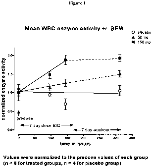
Figure 1. Figure 1 depicts a-Gal A enzymatic activity data over time for
placebo, 50 mg b.i.d. and 150 mg b.i.d. dosages in healthy volunteers.
Figure 2. Figure 2 depicts the average increases in white blood cell activity
in
Fabry patients as a result of DGJ administration over 48 weeks.
Figure 3. Figure 3 depicts GL-3 levels in two Fabry patients following
treatment with DGJ for 12 weeks.
SUMMARY OF THE INVENTION
The present invention provides a method of treating and method of monitoring
the response to treatment of Fabry patients with specific pharmacological
chaperones
such as DGJ by evaluating changes in the presence and/or levels of surrogate
markers
associated with Fabry disease.
In one embodiment, the method of treatment involves determining whether
there is an improvement of a surrogate marker that is associated with Fabry
disease
following administration of a specific pharmacological chaperone of a-
galactosidase
A.
5
CA 02682441 2009-09-29
WO 2008/121826 PCT/US2008/058668
In a specific embodiment, an improvement indicates that the patient is a
responder.
In one embodiment, the surrogate marker is a systemic surrogate marker. In
particular embodiments, the marker is lysosomal a-galactosidase A activity in
cells
and tissue or GL-3 accumulation. In another embodiment, the surrogate marker
is a
sub-cellular surrogate marker selected from aberrant trafficking of a-
galactosidase A
in cells from Fabry patients from the ER to the lysosome; aberrant trafficking
of
cellular lipids though the endosomal pathway; the presence of increased
amounts
misfolded a-galactosidase A in the ER or cytosol; the presence of cellular
stress
resulting from toxic accumulation of a-galactosidase A (as determined by gene
and/or
protein expression of stress-related markers); aberrant endosomal pH levels;
aberrant
cell morphology; suppression of the ubiquitin/proteasome pathway; and an
increase in
the amount of ubiquitinated proteins.
In a specific embodiment, the pharmacological chaperone is an inhibitor of a-
galactosidase A. In another embodiment, the inhibitor is a reversible
competitive
inhibitor, such as 1-deoxygalactonojirimycin.
The present invention also provides for a method for monitoring a therapeutic
response of a patient with Fabry disease following administration of a
specific
pharmacological chaperone of a-galactosidase A. This method includes
evaluating
the effect on the cytoplasmic staining pattern of a cell from the patient,
wherein
detection of a staining pattern in the cell that is similar to the staining
pattern in a cell
from a healthy individual indicates that the individual with Fabry disease is
a
responder.
In one embodiment, cytoplasmic staining is lysosomal staining. In other
embodiments, the lysosomal staining is detection of the presence of a-
galactosidase
A, LAMP-1 expression, or polyubiquitinated proteins.
In a specific embodiment, the pharmacological chaperone is an inhibitor of a-
galactosidase A. In another embodiment, the inhibitor is a reversible
competitive
inhibitor, such as 1-deoxygalactonojirimycin.
The present invention also provides a method for increasing the activity of a-
galactosidase A protein in an individual in need thereof. This method includes
administering to the individual an effective amount of a specific
pharmacological
chaperone that binds to the protein in an amount effective to increase
activity of the
protein in the individual by at least about 50%.
6
CA 02682441 2009-09-29
WO 2008/121826 PCT/US2008/058668
In a particular embodiment, the a-galactosidase A protein is a wild type
protein. In another embodiment, a-galactosidase A protein is an enzyme. In one
embodiment, the enzyme is a lysosomal enzyme. In yet another embodiment, the
lysosomal enzyme is a wild type a-galactosidase A protein.
In one embodiment of this invention, the pharmacological chaperone is an
inhibitor of a-galactosidase A. In another embodiment, the inhibitor is a
reversible
competitive inhibitor, such as 1-deoxygalactonojirimycin.
In one embodiment, the 1-deoxygalactonojirimycin is administered in an
amount effective to increase the activity of the protein by at least about
50%,
preferably by at least 2-fold (about 100%), more preferably at least about 3-
fold to 5-
fold, more preferably at least about 10-fold, and even more preferably at
least about
15-fold.
In one embodiment of the invention, the individual is homozygous for the wild
type protein. In another embodiment, the individual is heterozygous for the
wild type
protein and has a mutant genotype for the other allele encoding the protein.
In
paritcular embodiments, the individual suffers from Fabry disease.
In another embodiment of the invention, the a-galactosidase A activity
increases by at least 2-fold, preferably at least 3-fold to 5-fold, more
preferably at
least 10-fold. In a specific embodiment, the a-galactosidase A activity
increases to
within a normal range.
The present invention also provides for a method of treating Fabry disease in
a
patient in need thereof, wherein the method includes administering to the
individual
an effective amount of 1-deoxygalactonojirimycn, wherein the 1-
deoxygalactonojirimycn binds to alpha-galactosidase A in an amount effective
to
increase activity of the alpha-galactosidase A in the patient by at least
about 2-fold,
preferably at least 3-fold to 5-fold, more preferably at least 10-fold. In a
specific
embodiment, the a-galactosidase A activity increases to within a normal range.
DETAILED DESCRIPTION
The present invention provides for titrating a dose of a specific
pharmacological chaperone to achieve at least about a 3-fold increase in the
level of
enzyme activity, or increase the level of enzyme activity to unprecedented,
normal,
levels. In specific embodiments, the specific pharmacological chaperone is a
reversible competitive inhibitor of a-Gal A, preferably DGJ.
7
CA 02682441 2009-09-29
WO 2008/121826 PCT/US2008/058668
In addition, the present invention provides method for monitoring a patient's
response to treatment of Fabry disease with a specific pharmacological
chaperone that
is a reversible competitive inhibitor of a-Gal A, such as 1-DGJ. In
particular,
treatment is monitored by assaying biological samples for markers associated
with
Fabry disease, including but not limited to cellular deposits of
globotriaosylceramide
(GL-3) in cells, including cells within blood vessel walls, and kidney, heart,
and skin
cells; urinary GL-3 levels; a-Gal A activity in leukocytes, plasma, skin,
kidney, and
heart cells; and urinary proteinuria. Additional markers include the presence
of
angiokeratomas, decreased sweating, painful tingling or burning sensations in
the
extremities, gastrointestinal disturbances, cataracts or other visual
impairment, and
hearing loss.
In addition, evaluation of sub-cellular markers of disease, such as
trafficking
of mutant protein and alleviation of cell stress due to toxic accumulation of
mutant
proteins is also contemplated.
The invention is based, in part, on results obtained from Phase I and II tests
of
DGJ for treatment of Fabry disease. Among the discoveries that were made were
identification of changes to surrogate markers of Fabry disease indicative of
a
therapeutic effect of the therapy; identification of a dosage range and dosage
regimen
that increase a-Gal A activity; and, surprisingly, that DGJ treatment could be
titrated
to achieve at least a 2-fold, and more preferably a 3-fold to 5-fold, increase
in a-Gal A
activity, and even up to 10-fold and 15-fold in some instances, e.g., as
measured in
white blood cells. In addition, administration of DGJ could be titrated to
restore a-Gal
A activity levels, as measured in white blood cells, to the normal range, an
unprecedented therapeutic effect.
Definitions
The terms used in this specification generally have their ordinary meanings in
the art, within the context of this invention and in the specific context
where each
term is used. Certain terms are discussed below, or elsewhere in the
specification, to
provide additional guidance to the practitioner in describing the compositions
and
methods of the invention and how to make and use them.
The term "Fabry disease" refers to an X-linked inborn error of
glycosphingolipid catabolism due to deficient lysosomal a-galactosidase
activity.
This defect causes accumulation of globotriosylceramide and related
8
CA 02682441 2009-09-29
WO 2008/121826 PCT/US2008/058668
glycosphingolipids in vascular endothelial lysosomes of the heart, kidneys,
skin, and
other tissues.
The term "atypical Fabry disease" refers to patients with primarily cardiac
manifestations of the a-Gal A deficiency, namely progressive
globotriaosylceramide
accumulation in myocardial cells that leads to significant enlargement of the
heart,
particularly the left ventricle.
A "Fabry disease patient" refers to an individual who has been diagnosed with
Fabry disease due to a mutated a-galactosidase A as defined further below.
Human a-galactosidase A refers to an enzyme encoded by the human Gla
gene which has an amino sequence as set forth in SEQ ID NO: 2. The human a-Gal
A enzyme consists of 429 amino acids and is in GenBank Accession No.U78027.
As used herein the term "mutant a-Gal A" refers to an a-Gal A which has a
mutation which results in the inability of the enzyme to achieve its native
conformation under the conditions normally present in the ER. The failure to
achieve
this conformation results in the enzyme being degraded, rather than being
transported
through their normal pathway in the protein transport system to the lysosome.
This
term also encompasses an other a-Gal A mutations that result in decreased
enzyme
activity or a more rapid turnover.
Exemplary a-Gal A mutations associated with Fabry disease include R301 Q,
L166V, A156V, G272S, and M2961.
As used herein, the term "specific pharmacological chaperone" ("SPC") or
"pharmacological chaperone" refers to any molecule including a small molecule,
protein, peptide, nucleic acid, carbohydrate, etc. that specifically binds to
a protein
and has one or more of the following effects: (i) enhancing the formation of a
stable
molecular conformation of the protein; (ii) inducing trafficking of the
protein from
the ER to another cellular location, preferably a native cellular location,
i.e.,
preventing ER-associated degradation of the protein; (iii) preventing
aggregation of
misfolded proteins; and/or (iv) restoring or enhancing at least partial wild-
type
function and/or activity to the protein. A compound that specifically binds to
e.g., a-
Gal A, means that it binds to and exerts a chaperone effect on a-Gal A and not
a
generic group of related or unrelated enzymes. In the present invention, the
SPC may
be a reversible competitive inhibitor. Following is a description of some
specific
pharmacological chaperones contemplated by this invention:
9
CA 02682441 2009-09-29
WO 2008/121826 PCT/US2008/058668
1-deoxygalactonojirimycin refers to a compound having the following
structures:
CH2O H
HO NH
H\ OH2 OH
OH
6CH2OH 4 OH OH
3 or
5 This term includes both the free base and any salt forms. The hydrochloride
salt of DGJ is known as migalastat hydrochloride (Miglustat).
Still other SPCs for a-Gal A are described in U.S. Patents 6,274,597,
6,774,135, and 6,599,919 to Fan et al., and include a-3,4-di-epi-
homonojirimycin, 4-
epi-fagomine, and a-allo-homonojirimycin, N-methyl-deoxygalactonojirimycin, 0-
1-
C-butyl-deoxygalactonojirimycin, and a-galacto-homonojirimycin, calystegine
A3,
calystegine B2, N-methyl-calystegine A3, and N-methyl-calystegine B2.
A "surrogate marker" or "surrogate clinical marker" of Fabry disease refers to
the abnormal presence of, increased levels of, abnormal absence of, or
decreased
levels of a biomarker or symptom that is associated with Fabry disease (but is
not
associated with a healthy individual), and which is a reliable indicator of
Fabry
disease either alone or in combination with other abnormal markers or symptoms
of
Fabry disease.
As non-limiting examples, surrogate markers of Fabry disease include
decreased lysosomal a-Gal A activity in cells (e.g., fibroblasts) and tissue;
cellular
deposition of GL-3; increased plasma concentrations of homocysteine and
vascular
cell adhesion molecule-1 (VCAM-1); GL-3 accumulation within myocardial cells
and
valvular fibrocytes, leading to cardiac hypertrophy (especially of the left
ventricle),
valvular insufficiency, and arrhythmias; proteinuria; increased urinary
concentrations
of lipids such as CTH, lactosylceramide, ceramide, and decreased urinary
concentrations of glucosylceramide and sphingomyelin (Fuller et al., Clinical
Chemistry. 2005; 51: 688-694); the presence of laminated inclusion bodies
(Zebra
bodies) in glomerular epithelial cells; renal failure; hypohidrosis (which
causes heat
intolerance); the presence of angiokeratomas; and hearing abnormalities such
as high
frequency sensorineural hearing loss progressive hearing loss, sudden
deafness, or
tinnitus. Neurological symptoms include transient ischemic attack (TIA) or
stroke;
CA 02682441 2009-09-29
WO 2008/121826 PCT/US2008/058668
and neuropathic pain manifesting itself as acroparaesthesia (burning or
tingling in
extremities).
Characteristic markers of Fabry disease can occur in male hemizygotes and
female heterozygotes (carriers) with the same prevalence, although females
typically
are less severely affected.
Other surrogate markers are present at the sub-cellular level ("sub-cellular
surrogate markers") and include aberrant trafficking of a-Gal A in cells from
Fabry
patients from the ER to the lysosome; aberrant trafficking of lipids though
the
endosomal pathway; the presence of increased amounts misfolded a-Gal A in the
ER
or cytosol; the presence of cellular stress resulting from toxic accumulation
of a-Gal
A (as determined by gene and/or protein expression of stress-related markers);
aberrant endosomal pH levels; aberrant cell morphology; suppression of the
ubiquitin/proteasome pathway; or an increase in the amount of ubiquitinated
proteins.
An "an improvement in a surrogate marker" refers to an effect, following
treatment with an SPC of the amelioration, reduction, or increase in of one or
more
clinical surrogate markers which are abnormally present, abnormally absent, or
present in increased or decreased quantities in Fabry disease relative to a
healthy
individual who does not have Fabry disease and who does not have an other
disease
that accounts for the abnormal presence, absence, or altered quantities of
that
surrogate marker.
A "responder" is an individual diagnosed with a disease associated with Fabry
disease and an associated a a-Gal A mutation and treated and monitored
according to
the presently claimed method, who exhibits an improvement in one or more
surrogate
markers, and/or amelioration of, or reversal of, disease progression.
In addition, a determination whether an individual is a responder can be made
at the sub-cellular level by evaluating, e.g., intracellular trafficking of
the mutant a-
Gal A protein in response to treatment with an SPC. Restoration of trafficking
from
the ER to the lysosome is indicative of a response. Other sub-cellular
evaluations that
can be assessed to determine if an individual is a responder include
improvements in
the above-referenced sub-cellular surrogate markers.
In a specific embodiment, the invention provides for a method of treatment
with DGJ to achieve about a 3-fold increase in a-Gal A activity, e.g., as
determined
in white blood cells from the patient. In a further embodiment, the invention
provides
a method of treatment with DGJ to achieve at least a 5-fold increase in a-Gal
A
11
CA 02682441 2009-09-29
WO 2008/121826 PCT/US2008/058668
activity, or a 10-fold increase in a-Gal A activity, and in specific
embodiments, a 15-
fold increase in a-Gal A activity, e.g., as measured in white blood cells. The
invention
minimally provides for at least a 2-fold increase in a-Gal A activity.
Similarly, the invention provides for achieving an increase in a-Gal A
activity
in white blood cells from Fabry patients to within the normal range, a result
that has
not been achieved with other therapeutic strategies, and which the preclinical
data
could not have predicted would be the case. In a specific embodiment, the
"normal
range" is the range of activity as described in data from The Metabolic &
Molecular
Bases of Inherited Disease, 8th Edition McGraw Hill, 2001).
The terms "therapeutically effective dose" and "effective amount" refer to the
amount of the specific pharmacological chaperone that is sufficient to result
in a
therapeutic response. A therapeutic response may be any response that a user
(e.g., a
clinician) will recognize as an effective response to the therapy, including
improvements in any of the foregoing symptoms and surrogate clinical markers.
A
therapeutic response may be any response that a user (e.g., a clinician) will
recognize
as an effective response to the therapy, including improvements in the
foregoing
symptoms and surrogate clinical markers. Thus, a therapeutic response will
generally
be an amelioration of one or more symptoms or markers of a disease or
disorder, such
as those described above.
The phrase "pharmaceutically acceptable" refers to molecular entities and
compositions that are physiologically tolerable and do not typically produce
untoward
reactions when administered to a human. Preferably, as used herein, the term
"pharmaceutically acceptable" means approved by a regulatory agency of the
Federal
or a state government or listed in the U.S. Pharmacopoeia or other generally
recognized pharmacopoeia for use in animals, and more particularly in humans.
The
term "carrier" refers to a diluent, adjuvant, excipient, or vehicle with which
the
compound is administered. Such pharmaceutical carriers can be sterile liquids,
such
as water and oils. Water or aqueous solution saline solutions and aqueous
dextrose
and glycerol solutions are preferably employed as carriers, particularly for
injectable
solutions. Suitable pharmaceutical carriers are described in "Remington's
Pharmaceutical Sciences" by E.W. Martin, 18th Edition.
The terms "about" and "approximately" shall generally mean an acceptable
degree of error for the quantity measured given the nature or precision of the
measurements. Typical, exemplary degrees of error are within 20 percent (%),
12
CA 02682441 2009-09-29
WO 2008/121826 PCT/US2008/058668
preferably within 10%, and more preferably within 5% of a given value or range
of
values. Numerical quantities given herein are approximate unless stated
otherwise,
meaning that the term "about" or "approximately" can be inferred when not
expressly
stated.
Formulations and Administration
Any appropriate pharmaceutical formulation may be used to deliver the SPC
systemically. In certain embodiments, the SPC is a reversible competitive
inhibitor.
In one embodiment, the inhibitor is an imino sugar, e.g., DGJ, which is
administered
as monotherapy, preferably in an oral dosage form. In this embodiment, it is
contemplated that the dosing regimen should be one that provides a constant,
steady
state level of compound in the plasma of the individual being treated. This
can be
obtained either by daily administration in divided doses, or controlled-
release
formulations, or by less frequent administration of sustained-release dosage
forms.
Formulations
The SPC, can be administered in a form suitable for any route of
administration, including e.g., orally in the form tablets, capsules, or
liquid, or in
sterile aqueous solution for injection. In certain embodiments, the SPC is an
inhibitor, preferably a reversible competitive inhibitor such as a DGJ sugar.
When the
compound is formulated for oral administration, the tablets or capsules can be
prepared by conventional means with pharmaceutically acceptable excipients
such as
binding agents (e.g., pregelatinized maize starch, polyvinylpyrrolidone or
hydroxypropyl methylcellulose); fillers (e.g., lactose, microcrystalline
cellulose or
calcium hydrogen phosphate); lubricants (e.g., magnesium stearate, talc or
silica);
disintegrants (e.g., potato starch or sodium starch glycolate); or wetting
agents (e.g.,
sodium lauryl sulphate). The tablets may be coated by methods well known in
the art.
In a specific embodiment, DGJ-HC1 is administered as a powder-filled, hard,
gelatin capsule with magnesium stearate (vegetable) and starch 1500 (25 mg
DGJ/capsule).
Liquid preparations for oral administration may take the form of, for example,
solutions, syrups or suspensions, or they may be presented as a dry product
for
constitution with water or another suitable vehicle before use. Such liquid
preparations may be prepared by conventional means with pharmaceutically
acceptable additives such as suspending agents (e.g., water, sorbitol syrup,
cellulose
13
CA 02682441 2009-09-29
WO 2008/121826 PCT/US2008/058668
derivatives or hydrogenated edible fats); emulsifying agents (e.g., lecithin
or acacia);
non-aqueous vehicles (e.g., almond oil, oily esters, ethyl alcohol or
fractionated
vegetable oils); and preservatives (e.g., methyl or propyl-p-hydroxybenzoates
or
sorbic acid). The preparations may also contain buffer salts, flavoring,
coloring and
sweetening agents as appropriate. Preparations for oral administration may be
suitably formulated to give controlled or sustained release of the ceramide-
specific
glucosyltransferase inhibitor.
In another specific embodiment, 100 mg of DGJ is administered in an oral
solution in water (about 240 ml).
The pharmaceutical formulations of the SPC suitable for parenteral/injectable
use generally include sterile aqueous solutions, or dispersions and sterile
powders for
the extemporaneous preparation of sterile injectable solutions or dispersion.
In all
cases, the form must be sterile and must be fluid to the extent that easy
syringability
exists. It must be stable under the conditions of manufacture and storage and
must be
preserved against the contaminating action of microorganisms such as bacteria
and
fungi. The carrier can be a solvent or dispersion medium containing, for
example,
water, ethanol, polyol (for example, glycerol, propylene glycol, and
polyethylene
glycol, and the like), suitable mixtures thereof, and vegetable oils. The
proper fluidity
can be maintained, for example, by the use of a coating such as lecithin, by
the
maintenance of the required particle size in the case of dispersion and by the
use of
surfactants. Prevention of the action of microorganisms can be brought about
by
various antibacterial and antifungal agents, for example, parabens,
chlorobutanol,
phenol, benzyl alcohol, sorbic acid, and the like. In many cases, it will be
reasonable
to include isotonic agents, for example, sugars or sodium chloride. Prolonged
absorption of the injectable compositions can be brought about by the use in
the
compositions of agents delaying absorption, for example, aluminum monosterate
and
gelatin.
Sterile injectable solutions are prepared by incorporating the SPC in the
required amount in the appropriate solvent with various of the other
ingredients
enumerated above, as required, followed by filter or terminal sterilization.
Generally,
dispersions are prepared by incorporating the various sterilized active
ingredients into
a sterile vehicle which contains the basic dispersion medium and the required
other
ingredients from those enumerated above. In the case of sterile powders for
the
preparation of sterile injectable solutions, the preferred methods of
preparation are
14
CA 02682441 2009-09-29
WO 2008/121826 PCT/US2008/058668
vacuum drying and the freeze-drying technique which yield a powder of the
active
ingredient plus any additional desired ingredient from previously sterile-
filtered
solution thereof.
The above formulations can contain an excipient or excipients.
Pharmaceutically acceptable excipients which may be included in the
formulation are
buffers such as citrate buffer, phosphate buffer, acetate buffer, and
bicarbonate buffer,
amino acids, urea, alcohols, ascorbic acid, phospholipids, proteins, such as
serum
albumin, collagen, and gelatin; salts such as EDTA or EGTA, and sodium
chloride;
liposomes; polyvinylpyrollidone; sugars such as dextran, mannitol, sorbitol,
and
glycerol; propylene glycol and polyethylene glycol (e.g., PEG-4000, PEG-6000);
glycerol, glycine or other amino acids and lipids. Buffer systems for use with
the
formulations include citrate, acetate, bicarbonate, and phosphate buffers.
Phosphate
buffer is a preferred embodiment.
The formulations can also contain a non-ionic detergent. Preferred non-ionic
detergents include Polysorbate 20, Polysorbate 80, Triton X-100, Triton X-114,
Nonidet P-40, Octyl a-glucoside, Octyl (3-glucoside, Brij 35, Pluronic, and
Tween 20.
Administration
The route of administration of the SPC may be oral (preferably) or parenteral,
including intravenous, subcutaneous, intra-arterial, intraperitoneal,
ophthalmic,
intramuscular, buccal, rectal, vaginal, intraorbital, intracerebral,
intradermal,
intracranial, intraspinal, intraventricular, intrathecal, intracisternal,
intracapsular,
intrapulmonary, intranasal, transmucosal, transdermal, or via inhalation.
Administration of the above-described parenteral formulations of the SPC may
be by periodic injections of a bolus of the preparation, or may be
administered by
intravenous or intraperitoneal administration from a reservoir which is
external (e.g.,
an i.v. bag) or internal (e.g., a bioerodable implant). See, e.g., U.S. Pat.
Nos.
4,407,957 and 5,798,113, each incorporated herein by reference. Intrapulmonary
delivery methods and apparatus are described, for example, in U.S. Pat. Nos.
5,654,007, 5,780,014, and 5,814,607, each incorporated herein by reference.
Other
useful parenteral delivery systems include ethylene-vinyl acetate copolymer
particles,
osmotic pumps, implantable infusion systems, pump delivery, encapsulated cell
delivery, liposomal delivery, needle-delivered injection, needle-less
injection,
nebulizer, aeorosolizer, electroporation, and transdermal patch. Needle-less
injector
devices are described in U.S. Pat. Nos. 5,879,327; 5,520,639; 5,846,233 and
CA 02682441 2009-09-29
WO 2008/121826 PCT/US2008/058668
5,704,911, the specifications of which are herein incorporated by reference.
Any of
the formulations described above can be administered using these methods.
Furthermore, a variety of devices designed for patient convenience, such as
refillable injection pens and needle-less injection devices, may be used with
the
formulations of the present invention as discussed herein.
Dosage
Persons skilled in the art will understand that an effective amount of the
compounds used in the methods of the invention can be determined by routine
experimentation, but is expected to be an amount resulting in serum levels
between
0.01 and 100 M, preferably between 0.01 and 10 M, most preferably between
0.05
and 1 M. Clinical studies, and preclinical studies in mice, suggest that the
pharmacological effect (enzyme activity enhancement) of DGJ is seen at a
plasma
concentration of approximately 0.4 M.
The effective dose of the SPC is expected to be between 0.5 and 1000 mg/kg
body weight per day. In specific embodiments where the SPC is DGJ, the dose is
between about 10-600 mg/day, more specifically 25-300 mg/day, more
specifically,
50-150 mg/day.
In particular embodiments, DGJ is administered at 25 mg b.i.d., 100 mg b.i.d.
or 250 b.i.d. Data from the multiple-dose phase 1 study indicate that a trough
level of
0.4 M is obtained with dosing of 50 mg b.i.d. Data from a phase II study on
Fabry
patients demonstrated increased enzyme activity at the lowest dose evaluated
(25 mg
b.i.d.).
Fabry Disease Treatment Monitoring Using Surrogate Markers
The present invention also provides a method for monitoring the treatment of
Fabry patients with specific pharmacological chaperones. Specifically, various
assays
are employed to evaluate the progress of the disease and its response to
treatment with
DGJ. In particular, various systemic and sub-cellular markers can be assayed.
The
monitoring aspect of the present invention encompasses both invasive and non-
invasive measurement of various cellular substances.
Globotriaosylceramide accumulation. A method for measuring
globotriaosylceramide (GB3, or GL-3) levels in plasma and urine of humans
affected
by Fabry disease is described in, e.g., Boscaro et al., Rapid Commun Mass
Spectrom.
2002;16(16):1507-14. In this reference, the analyses are performed using flow
16
CA 02682441 2009-09-29
WO 2008/121826 PCT/US2008/058668
injection analysis-electrospray ionization-tandem mass spectrometry (FIA-ESI-
MS/MS).
Immunoelectron-microscopic detection of GL-3 accumulated in the skin of
patients with Fabry disease has been recently described in Kanekura et al., Br
J
Dermatol. 2005;153(3):544-8. This method is sensitive enough to detect
lysosomal
accumulation of GL-3. Skin biopsies can be obtained by using a "punch" device,
which removes a sample layer of skin.
Renal biopsies are performed using ultrasound, x-ray or CT scan guidance.
Under some circumstances, the biopsy is be performed by running the biopsy
catheter
through one of the neck veins-this is called a trans-jugular biopsy. GL-3
accumulation in kidney, specifically in all renal cell types, including
vascular
endothelial cells, vascular smooth muscle cells, mesangial cells and
interstitial cells,
podocytes and distal tubular epithelial cells, had been described in Thurburg
et al.,
Kidney Int. 2002;62(6):1933-46. Ultrastructural study (electron microscopy) of
kidney biopsies can reveal typical inclusion bodies in the cytoplasm of all
types of
renal cells (Sessa et al., J Inherit Metab Dis. 2001; 24 Suppl 2:66-70). The
cells are
characterized by concentric lamellation of clear and dark layers ("zebra" or
"onion-
skin" appearance) with a periodicity of 35-50 A.
Kidney function can be assessed by determining glomerular filtration rate
(mi/min) and by assessing serum creatine levels according to well-established
methods. Other renal assessments include 24-hour protein excretion, urine
protein
electrophoresis, total protein, microalbumin, urine beta-2 microglobulin
titers.
Reduction in GL-3 sediment and proteinuria is a direct measurement of renal
health.
Recently, atmospheric pressure photoionization mass spectrometry (APPI-
MS) was shown to be an efficient method for the analysis of GL-3 molecular
species,
both in direct injection and by coupling with liquid chromatography (LC). This
technique allowed the detection of a great number of species from biological
samples
isolated from Fabry patients (Delobel et al., J Mass Spectrom. 2005 Nov 14;
[Epub
ahead of print]).
a-Galactosidase activity. As indicated above, non-invasive assessment of a-
Gal A activity can be measured in blood leukocytes or in cultured fibroblasts
from
skin biopsies. Such assays typically involve extraction of blood leukocytes
from the
patient, lysing of the cells, and determining the activity in the lysate upon
addition of
17
CA 02682441 2009-09-29
WO 2008/121826 PCT/US2008/058668
an enzyme substrate such as 4-methyl umbelliferal alpha-D-galactoside an/or N-
acetylgalactosamine (see U.S. 6.274,597).
Two sensitive immunoassays for the measurement of a-Gal A activity and
protein to determine the concentrations of alpha-galactosidase in blood and
plasma
are described in Fuller et al., Clin Chem. 2004;50(11):1979-85.
According to the present invention, it is possible to titrate dosage to
achieve at
least a 2-fold increase in a-Gal A activity, and in some cases at least a 3-
fold increase
in a-Gal A activity, at least 5-fold, up to 10-fold, and in some embodiments,
a 15-fold
increase. These results can be observed with white blood cells.
In another embodiment, the invention provides for titrating a dosage of DGJ to
achieve normal levels of a-Gal A activity in a Fabry patient.
Cardiac evaluation. Increases in alpha-Gal A activity may play a role
monitoring or detecting heart disease or in at least a subset of heart disease
patients.
Evaluation of GL-3 in cardiac cells can be achieved through endomyocardial
biopsies.
This is an invasive procedure that involves using a bioptome (a small catheter
with a
grasping device on the end) to obtain a small piece of heart muscle tissue.
GL-3 present in perinuclear vacuoles will stain positive with an acid stain.
In
addition, histological examination of the biopsies can be done using
transmission
electron microscopy to ascertain thickening of endocardium to measure
ventricular
mass, or to determine the presence of hypertrophic myocardial fibers.
In addition, common carotid and radial artery diameter, intima-media
thickness (IMT) and distensibility have been assessed using high-definition
echotracking systems and aplanation tonometry. (Boutouyrie et al., Acta
Paediatr
Suppl. 2002;91(439):62-6. Cardiac myocytes will also be examined for
accumulation
of GL-3. Macroscopic cardiac morphology can be assessed using MRI or Doppler
echocardiography. Cardiac function can be assessed by, e.g., determining left
ventricular ejection fraction and using electrocardiograms.
Neuropathic pain/peripheral neuropathy. Pain in the extremities can be
assessed using subjective tests given to the patient. In addition, to evaluate
neuropathy, Quantitative Sensory Testing (CASE study) can be used. A CASE
study
is a biophysical technique in which a patient is asked to push a button as
soon as he
feels either a sensation of cold, warmth, or vibration. These stimulations are
delivered
by an electrode that is put on the skin of the hand or foot.
18
CA 02682441 2009-09-29
WO 2008/121826 PCT/US2008/058668
Cerebrovascular. In addition to stroke and hypertension, other Fabry-related
cerebrovascular signs and symptoms associated can include hemiparesis,
vertigo,
double vision; seizures; basilar artery ischema and aneurism; labyrinthine
disorders or
cerebral hemorrhage.
Neurological. In male and female patients with Fabry disease, significant
age-related cerebral white matter lesions (WMLs) can be found. Evaluation of
neurological effects can be assessed using, e.g., Quantitative Sudomotor Axon
Reflex
Test (QSART). QSART is a routine test of autonomic function and a sensitive
test in
distal small-fiber neuropathy such as observed in Fabry disease.
Hypohydrosis/anhidrosis. Impaired sweating and heat intolerance in Fabry
patients has been attributed to selective peripheral nerve damage or to
intracytoplasmic lipid deposits in the small blood vessels surrounding sweat
glands.
Hilz et al. (Acta Paediatr Suppl. 2002;91(439):38-42) have described the
methods to assess impairment of temperature perception, vibratory perception,
sudomotor and eccrine sweat gland function, and limb and superficial skin
blood flow
and vasoreactivity in Fabry patients. These methods include thermal
provocation
tests, quantitative sudomotor axon reflex testing (QSART) and venous occlusion
plethsmography. QSART has three parts and measures resting skin temperature,
resting sweat output, and stimulated sweat output. Measurements are typically
taken
on arms, legs or both. A small plastic cup is placed on the skin and the
temperature
and amounts of sweat under the skin are measured. To stimulate sweat a
chemical is
delivered electrically through the skin to a sweat gland, but the patient will
only feel
warmth. A computer is used to analyze the data to determine how well the
nerves and
sweat glands are functioning.
In addition, a reduction of tears and saliva is also observed in about 40% of
Fabry disease patients.
Temperature intolerance. In addition to heat intolerance, cold and heat
sensitivity often results from lipid deposition in small vessel walls,
perineural cells,
and unmyelinated or myelinated nerve cells resulting in small fiber
neuropathy.
Ophthalmologic opacities. Fabry disease patients almost universally exhibit
whorled corneal opacities, lenticular opacities, and vascular lesions of the
conjunctivae and retina. Corneal opacities can be seen using slit larnp
microscopy.
Two types of lens opacities have been noted in Fabry patients: cream-colored
anterior
19
CA 02682441 2009-09-29
WO 2008/121826 PCT/US2008/058668
capsular deposits in the lens (sometimes distributed like a propeller), and
whitish,
granular spoke-like deposits on the posterior lens (referred to as Fabry
cataracts).
Hearing loss. Non-invasive methods to evaluate cochlear functions using
conventional audiometry, tympanometry, ABR audiometry, and otoacoustic
emissions
is described in Germain et al., BMC Med Genet. 2002;3(l):10).
Gastrointestinal disturbance. Gastrointestinal symptoms may result from
deposition of glycosphingolipids in mesenteric blood vessels and autonomic
ganglia.
Symptoms include postprandial bloating; abdominal cramping and pain; early
satiety;
diarrhea; constipation; nausea; vomiting.
Other surrogate markers. Other markers of Fabry disease include
Lymphoedema (swelling of the extremities) due to accumulation of GL-3. In
addition, it was recently discovered that there was a signififcant decrease in
diastolic
blood pressure in patients with Fabry disease, which may account for exercise
tolerance (Bierer et al., Respiration. 2005;72(5):504-11).
It is to be understood that these markers can be used to monitor treatment
only
if they are identified to be abnormal prior to treatment. In addition, it is
understood
that the abnormal elevation of the markers be correlated with the presence of
Fabry
disease, and not attributed to other causes or concomitant diseases such as
kidney
disease or other cerebrovascular disease.
Molecular Biology Monitoring Assays to Detect Sub-Cellular Markers
Monitoring of treatment of Fabry disease with specific pharmacological
chaperones can be done at the subcellular level in addition to the systemic or
macroscopic level, described above. For example, disturbances in endosomal-
lysosomal membrane trafficking of lipids to the Golgi complex are a
characteristic of
lysosomal storage disease (Sillence et al., J Lipid Res. 2002;43(11):1837-45).
Accordingly, one way of monitoring treatment of Fabry would be to contact
cells
from patients with labeled lipid (BODIPY-cholesterol) and monitor its
trafficking in
endosomal structures. Pathological accumulation in endosomal structures, for
example, would be indicative that the patient is not responding well to
treatment.
As one example, pH-sensitive fluorescent probes that are endocytosed by the
cells can be used to measure pH ranges in the lysosomes and endosomes (i.e.
fluorescein is red at pH 5, blue to green at 5.5 to 6.5). Lysosome morphology
and pH
will be compared in wild type and chaperone treated and untreated patient
cells. This
CA 02682441 2009-09-29
WO 2008/121826 PCT/US2008/058668
assay can be run in parallel with the plate reader assay to determine the pH-
sensitivity. For example, BODIPY-LacCer is trafficked to the Golgi in normal
cells,
but accumulates in the lysosomes of cells with lipid storage disorders. BODIPY-
LacCer fluoresces green or red depending on the concentration in the membrane,
and
the green/red color ratio in the lysosome can be used to measure changes in
concentration. Living healthy cells and patient cells, treated and untreated
with
compounds, will be incubated with BODIPY-LacCer and the red/green color ratio
can
be measured by the FACS and/or confocal microscope and the staining pattern
(lysosome vs. Golgi) can be determined using a confocal microscope.
Trafficking occurs in cells along pH gradients (i.e. ER pH about 7, Golgi pH
about 6.2-7.0, trans-Golgi network pH about 6.0, early and late endosomes pH
about
6.5, lysosomes pH about 4.5) and luminal and endosomal pH is disrupted in
cells with
trafficking defects such as Fabry cells. Accordingly, an assay to determine pH
sensitivity in wild type, SPC-treated and untreated patient cells, if
correlated to
positive effects of pH on trafficking, can be used to monitor restoration of
trafficking
in Fabry patients. If patient cells are more sensitive to changes in pH, than
it would
be possible to create a screening assay for SPCs that reduce the cells pH
sensitivity,
restores lysosome morphology or function, or more generally restores normal
trafficking.
In addition, mitigation of the trafficking defect can be assessed at the
molecular level by determining co-localization of the deficient enzyme (a-Gal
A) with
a lysosomal marker such as Lyso-Tracker . Localization of a-Gal A in the
lysosome
is evidence that trafficking from the ER to the lysosome is restored by
treatment with
the specific pharmacological chaperone. In brief, normal and patient cells,
treated and
untreated with SPCs, are fixed and stained with primary antibodies to the
enzyme and
endosome/lysosome markers (e.g., Rab7, Rab9, LAMP-1, LAMP-2, dystrophin-
associated protein PAD) and fluorescently tagged secondary antibodies. The
FACS
and/or confocal microscope is used to quantify the amount of fluorescence due
to the
concentration of enzyme and other endocytic pathway markers, and the confocal
microscope can be used to determine changes in staining patterns. In addition,
traditional biochemical methods, such as pulse-chase metabolic labeling
combined
with Endoglycosidase H treatment. Endo H only cleaves proteins which have
acquired ER glycosylation (high mannose N-linked), i.e., which are localized
to the
ER, but will not cleave proteins that have made it out of the ER to the Golgi
and have
21
CA 02682441 2009-09-29
WO 2008/121826 PCT/US2008/058668
acquired additional glycosylation in the Golgi. Accordingly, the greater the
level of
Endo H sensitive a-Gal A, the more accumulation of the protein in the ER. If
the a-
Gal A has made it into the Golgi, the glycosidase PNGase F can be used to.
confirm
whether the protein has exited the Golgi since it cleaves all N-linked sugars.
ER Stress. The toxic accumulation of misfolded proteins in the ER cells, such
as the misfolded a-Gal A in Fabry patients, often results in ER stress. This
leads to
induction of the cell stress response which attempts to resolve the disruption
in cell
homeostasis. Accordingly, measuring markers of ER stress in patients following
treatment with the specific pharmacological chaperone provides another way to
monitor the effects of treatment. Such markers include genes and proteins
proteins
associated with the Unfolded Protein Response, which include BiP, IRE1,
PERK/ATF4, ATF6, XBP1 (X-box binding factor 1) and JNK (c-Jun N-terminal
kinase). One method to assess ER stress is to compare expression levels
between
wild type and Fabry patient cells, and also between SPC-treated and untreated
cells.
ER stress inducers (e.g., tunicamycin for the inhibition of N-glycosylation
and
accumulation of unfolded proteins in the ER, lacatcystin or HZO2) and stress
relievers
(e.g., cyclohexamide to inhibit protein synthesis) can be used as controls.
Another method contemplated for monitoring the ER stress response is via
gene chip analysis. For example, a gene chip with a variety of stress genes
can be
used to measure expression levels and type of ER stress response (early, late,
apoptosis etc.). As one example, the HG-U95A array can be used. (Affymetrix,
Inc.).
Lastly, since prolonged ER stress can result in apoptosis and cell death,
depending on the level of unfolded proteins in the ER, and the resulting
stress level,
cells will be more or less sensitive to ER stress inducers such as tunicamycin
or
proteasome inhibitors. The more sensitive the cells are to the stress
inducers, the
higher the number of apoptotic or dead cells is observed. Apoptosis can be
measured
using fluorescent substrates analogs for caspase 3 (an early indicator of
apoptosis).
FACS, confocal microscopy, and/or using a fluorescence plate reader (96 well
format
for high through put assays) to determine the percentage of cells positive for
apoptosis
or cell death (FACS and/or confocal microscopy), or fluorescence intensity can
be
measured relative to protein concentration in a 96 well format with a
fluorescence
plate reader.
Another response to cell stress resulting from toxic protein accumulation in
the ER is suppression of the ubiquitin/proteasome pathway. This leads to a
general
22
CA 02682441 2009-09-29
WO 2008/121826 PCT/US2008/058668
disruption of the endocytic pathway (Rocca et al., Molecular Biology of the
Cell.
2001; 12: 1293-1301). Misfolded protein accumulation is sometimes correlated
with
increased amounts of polyubiqutin (Lowe et al., Neuropathol Appl Neurobiol.
1990;
16: 281-91).
Proteosome function and ubiquitination can be assessed using routine assays.
For example, evaluation of 26S proteasome function in living animals by
imaging has
been achieved ubiquitin-luciferase reporter for bioluminescence imaging (Luker
et al.,
Nature Medicine. 2003. 9, 969 - 973). Kits for proteasome isolation are
commercially available from, for example, Calbiochem (Cat. No. 539176).
Ubiquitination can be examined by morphological studies using
immunohistochemistry or immunofluorescence. For example, healthy cells and
patient cells, treated and untreated with SPCs, can be fixed and stained with
primary
antibodies to ubiquitinated proteins and fluorescence detection of secondary
antibodies by FACS and/or confocal microscopy will be used to determine
changes in
ubiquitinated protein levels.
Another assay to detect ubiquitinated proteins is AlphaScreenTM (Perkin-
Elmer). In this model, the GST moiety of a GST-UbcH5a fusion protein is
ubiquitinated using biotin-Ubiquitin (bio-Ub). Following ubiquitin activation
by El,
in the presence ofATP, bio-Ub is transferred to UbcH5a. In this reaction,
UbcH5a acts
as the carrier to transfer the bio-Ub to its tagged GST moiety. The protein
which
becomes biotinylated and ubiquitinated is then captured by anti-GST Acceptor
and
streptavidin. Donor beads resulting in signal generation. No signal will be
generated
in the absence of ubiquitination.
Lastly, an ELISA sandwich assay can be used to capture ubiquitinated mutant
a-Gal A. The primary antibody to the a-Gal A (e.g., rabbit) would be absorbed
to the
surface, enzyme would be captured during an incubation with cell lysate or
serum,
then an antibody (e.g., mouse or rat) to ubiquitinated protein, with secondary
enzyme-
linked detection, would be used to detect and quantify the amount of
ubiquitinated
enzyme. Alternatively, the assay could be used to quantify the total amount of
multi-
ubiquitinated proteins in cell extract or serum.
Female Carriers
Some female carriers remain asymptomatic and have normal concentrations of
a-Gal A, where some experience mild to severe clinical symptoms of disease,
23
CA 02682441 2009-09-29
WO 2008/121826 PCT/US2008/058668
especially with increasing age. This variability is thought to be partly the
result of
inactivation of one X chromosome in some or all cells of the female embryo. In
one
study of 11 female carriers, proteinuria, acroparaesthesia and angiokeratoma
were
found to be the most common symptom of the disease (Guffon, Journal of Medical
Genetics. 2003; 40: e38). Some of the patients experienced symptoms including
vertigo, abdominal pain, depression, exercise/heat intolerance, chilliness or
fever and
sweating, weakness, and depression. Cardiovascular abnormalities, including
valvulopathy and severe hypertrophic myocardiopathy occurred in the fourth,
fifth, or
sixth decades, and two of the patients experienced terminal renal
insufficiency
requiring transplantation. One woman had a capsulothalamic stroke, and
pulmonary
complications (chronic obstructive bronchopneumopathy in one and pulmonary
embolism) were also recorded in two patients.
In view of the foreging, the methods of the present invention also can be
employed where treatment of female carriers is initiated.
Combination Therapy
The therapeutic monitoring of the present invention is also applicable
following treatment of patients with a combination of DGJ and ERT or gene
therapy.
Such combination therapy is described in commonly-owned, U.S. patent
application
publication numbers 2004/0180419 (serial number 10/771,236, and 2004/0219132
(serial number 10/781,356). Both applications are herein incorporated by
reference in
their entirety.
EXAMPLES
The present invention is further described by means of the examples,
presented below. The use of such examples is illustrative only and in no way
limits
the scope and meaning of the invention or of any exemplified term. Likewise,
the
invention is not limited to any particular preferred embodiments described
herein.
Indeed, many modifications and variations of the invention will be apparent to
those
skilled in the art upon reading this specification. The invention is therefore
to be
limited only by the terms of the appended claims along with the full scope of
equivalents to which the claims are entitled.
24
CA 02682441 2009-09-29
WO 2008/121826 PCT/US2008/058668
EXAMPLE 1: Administration of DGJ to a Transgenic Mouse Expressing
Human Mutant a-Gal A in an Endogenous Enzyme
Deficient Background
Transgenic mice that exclusively express human mutant a-Gal A (R3 01 Q) in
an a-Gal A knock-out background (TgM/KO mice) were established and evaluated.
This Example serves as a biochemical model to study and evaluate
pharmacological
chaperone therapy for Fabry disease, which is specific for those missense
mutations
that cause misfolding of a-Gal A.
Methods
Mice. Fabry R301Q Tg/KO mice were a gift from Dr. Robert Desnick. Male
C57BL/6 mice were purchased from Taconic Farms, Germantown, NY and housed in
wire cages at 4 mice per cage. All studies were conducted at 8 weeks of age
and
conducted under strict adherence to IACUC guidelines.
Drug Administration. For the a-Gal A assay, four groups of 10 male C57BL/6
mice were dosed with 0, 1, 10 or 100 mg/kg/day IFG HCl in drinking water for
28
days. For the GL-3 assay, two groups of 6-7 male R301Q Tg/KO mice were dosed
daily with 0 or 30 mg/kg DGJ HCl per os for 28 days. After dosing, indicated
tissues
were harvested and treated as described below.
Enzyme activity assay. After dosing, indicated tissues were harvested and
frozen. Tissue lysates were prepared by homogenizing about 50 mg tissue in
assay
buffer. 2.5 L lysate was combined with 17.5 L of the assay buffer and 50 L
of the
respective substrate solution (same as detailed in sections above). Reaction
mixtures
were then incubated at 37 C for 1 hr. Afterward, 70 L stop solution (0.4 M
glycine,
pH 10.8) was added, and fluorescence was read on a Victor plate reader at 355
nm
excitation and 460 nm emission. Enzyme activity in the lysates was background
subtracted, and normalized for protein concentration using the MicroBCA
Protein
Assay Kit (Pierce). A 4-MU standard curve was run (same as detailed in
sections
above) for conversion of fluorescence data to absolute enzyme activity
expressed as
nmoles / mg protein / hr or further normalized to % of untreated activity.
GL-3 analysis from mouse tissues. Tissue samples were washed free of blood,
weighed and homogenized with a solvent system in a FastPrep system.
Homogenate
was then extracted using Solid Phase Extraction on a C 18 cartridge. The
eluent was
evaporated and reconstituted prior to injection onto a LC/MS system. Nine GL3
CA 02682441 2009-09-29
WO 2008/121826 PCT/US2008/058668
isoforms were measured using positive ESI-MS/MS. LC separation was achieved on
a
Zorbax C 18 column.
Results
Enzyme Activity. Daily oral gavage administration of DGJ HCl (30 mg/kg; 4
weeks) to male R301 Q Tg/KO mice increases mutant GLA activity significantly
(*p<0.05, t-test, vs. untreated) in heart, kidney, skin, liver, and spleen
tissues (11
0.1, 5.5 0.3, 6.2 0.7, 3 0.02, and 5 0.1 -fold respectively; n=6-7
mice for each
tissue). The data is representative of two separate measurements. Two similar
experiments were carried out in male and female R301 Q Tg/KO mice orally
administered DGJ HCl (30 mg/kg/day in drinking water; 4 weeks) and comparable
increases in mutant GLA activity were observed in all treated tissues.
Tissue GL-3. Daily oral gavage administration of DGJ HCl (30 mg/kg; 4
weeks) to male R301Q Tg/KO mice reduces GL-3 substrate levels (measured by LC-
MS/MS) significantly in skin and heart. DGJ HCI significantly reduced GL-3
levels
2.2-fold after daily per os dosing in skin tissue of treated R301 Q Tg/KO mice
(*p<0.05; n=6-7 mice per group). DGJ HCl significantly reduced GL-3 levels 1.6-
fold after daily per os dosing in heart tissue of treated R301Q Tg/KO mice
(*p<0.01;
n=6-7 mice per group). GL-3 levels were also analyzed in kidney tissue and
showed a
trend towards reduction, but did not reach statistical significance.
Discussion
The foregoing findings are significant since they represent the first evidence
of
substrate clearance in vivo in key tissues affected by Fabry disease in
response to
treatment with a pharmacological chaperone.
As indicated infta, the only approved treatment for Fabry disease is enzyme
replacement therapy. Recently, it has been demonstrated that GL-3 persists in
heart,
kidney, brain (parahippocampus), intestines, adrenal gland, aorta, skin,
liver, and
spleen from a deceased Fabry patient who had been on long-term ERT therapy
with
Fabrazyme (7 years) (Askari et al., manuscript submitted). While this patient
did
not have GL-3 lysosomal inclusions in vascular endothelial cells, GL-3
immunoreactivity was observed in cell membranes and cytoplasm of cells from
all
organs. In vascular endothelial cells and fibroblasts of the kidney, GL-3 co-
localized
with lysosomal, ER, and nuclear markers, and presence in these compartments as
well
26
CA 02682441 2009-09-29
WO 2008/121826 PCT/US2008/058668
as the cell membrane was confirmed with immunogold electron microscopy.
Cultured skin fibroblasts from another Fabry patient on ERT for 7 years showed
similar findings. Tissues from three unaffected controls was uniformly
negative for
GL-3 by IHC and EM. These data suggest that pharmacological chaperone therapy
will be as good or better at clearing substrate in key organs in patients with
Fabry
disease.
In conclusion, a substantial amount GL-3 immunoreactivity remains in cells
and tissues even after years of ERT in Fabry disease. For the first time we
demonstrate the presence of accumulated globotriaosylceramide in
extralysosomal
cellular regions. These findings are crucial for the understanding of disease
mechanism and suggest the use of immunostaining for globotriaosylceramide to
assess response to novel specific therapies.
EXAMPLE 2: Administration of Single Dose DGJ to Evaluate Safety,
Tolerability
and Pharmacokinetics
This example describes a randomized, double blind, placebo controlled Phase
I study of ascending single oral dose of DGJ to evaluate the safety,
tolerability and
pharmacokinetics of DGJ in healthy volunteers.
Study Design and Duration. This study was first-in-man, single-center, Phase
I, randomized, double-blind, single-dose, placebo controlled, ascending dose
study to
evaluate the safety, tolerability and pharmacokinetics of DGJ following oral
administration. The study tested 4 groups of 8 subjects (6 active and 2
placebo) who
received a single dose of 25, 75, 225 and 675 mg of DGJ or placebo
administered
orally, in a dose-escalating regimen, with a minimal 1-week safety evaluation
period
between successive cohorts. Dose escalation to the next dose level (i.e., next
group)
proceeded following review of safety and tolerability of the previous
group(s).
Subjects were housed in the treatment facility from 14 hours prior to dosing
until 24
hours after dosing. Meals were controlled by schedule and subjects remained
abulatory for 4 hours post drug administration.
Study Population. Subjects were healthy, non-institutionalized, non-smoking
male volunteers between 19 and 50 years of age (inclusive) consisting of
members of
the community at large.
27
CA 02682441 2009-09-29
WO 2008/121826 PCT/US2008/058668
Safety and Tolerability Assessments. Safety was determined by evaluating
vital signs, laboratory parameters (serum chemistry, hematology, and
urinalysis),
ECGs, physical examination and by recording adverse events during the
Treatment
Period.
Pharmacokinetic Sampling. Blood samples (10 mL each) were collected in
blood collection tubes containing EDTA before dosing and at the following
times
thereafter: 0.25, 0.5, 0.75, 1, 1.5, 2, 3, 4, 5, 6, 8, 10, 12, 16 and 24
hours. Blood
samples were cooled in an ice bath and centrifuged under refrigeration as soon
as
possible. Plasma samples were divided into two aliquots an dstored at 20 10
C
pending assay. At the end of the study, all samples were transferred to MDS
Pharma
Services Analytical Laboratories (Lincoln) for analysis. The complete urine
output
was collected from each subject for analysis of DGJ for the following
intervals:
= -4-0 hours
= 0-4 hours
= 4-8 hours
= 8-12 hours
= 12-24 hours
Statistical Analysis. Safety data including laboratory evaluations, physical
exams, adverse events, ECG monitoring and vital signs assessments were
summarized
by treatment group and point of time of collection. Descriptive statistics
(arithmetic
mean, standard deviation, median, minimum and maximum) were calculated for
quantitative safety data as well as for the difference to baseline. Frequency
counts
were compiled for classification of qualitative safety data. In addition, a
shift table
describing out of normal range shifts was provided for clinical laboratory
results. A
normal-abnormal shift table was also be presented for physical exam results
and
ECGs.
Adverse events were coded using the MedDRA version 7.0 dictionary and
summarized by treatment for the number of subjects reporting the adverse event
and
the number of adverse events reported. A by-subject adverse event data listing
including verbatim term, coded term, treatment group, severity, and
relationship to
treatment was provided. Concomitant medications and medical history were
listed by
treatment.
Pharmacokinetic parameters were summarised by treatment group using
descriptive statistics (arithmetic means, standard deviations, coefficients of
variation,
28
CA 02682441 2009-09-29
WO 2008/121826 PCT/US2008/058668
sample size, minimum, maximum and median). In addition, geometric means were
calculated for AUCO-t, AUCinf and Cmax.
Results.
Five subjects (16%) reported 10 treatment-emergent AEs during this study.
Two subjects had AEs in Cohort B (75 mg), and three subjects had AEs in Cohort
D
(675 mg). No subjects presented with AEs in Cohorts A (25 mg) or C (225 mg).
No
trends were observed with respect to increasing dose levels. No AE was
reported by
at least 10% of subjects.
AEs occurred in five body systems: the gastrointestinal system (stools
watery),
musculoskeletal and connective tissues (musculoskeletal stiffness), nervous
system
(dizziness, headache), respiratory, thoracic and mediastinal system
(pharyngolaryngeal pain), and skin and subcutaneous system (rash).
Laboratory deviations from normal ranges occurred after dosing, but none was
judged clinically significant. There were no clinically relevant mean data
shifts in any
parameter investigated throughout the course of the study. No clinically
relevant
abnormality occurred in any vital sign, ECG, or physical examination
parameter.
DGJ appeared to be safe and well tolerated by this group of healthy male
subjects as doses increased from 25 mg through 675 mg inclusive.
Pharmacokinetic Evaluation. The following table summarizes the
pharmacokinetic data obtained during study.
Table 1.
AUCo_ta AUCinf a
Dose (ng*h/m (ng*h/m Cmax a tmax t1i2 CLR Ae b
(mg) L) L) (ng/mL) (hours) (hours) (L/h) (%)
1092 1129 201 3.00 3.04 5.90 (1.5) 8.78
(34.2) (33.6) (35.5) (21.1) (15.7) (173.8)
75 4661 4730 685 2.92 4.05 5.73 23.4
(9.0) (8.6) (16.7) (41.2) (16.4) (29.9) (85.8)
225 11177 11353 1997 3.17 4.62 7.54 40.3
(59.8) (59.2) (56.1) (23.8) (15.4) (17.4) (37.2)
675 35275 35675 6492 2.67 4.19 (7.3) 7.66 39.6
(22.3) (22.4) (24.4) (30.6) (20.9) (14.9)
a Geometric mean was used to present Cma,, and AUC;,,f pharmacokinetic
parameters.
29
CA 02682441 2009-09-29
WO 2008/121826 PCT/US2008/058668
b Cumulative percentage of DGJ excreted over the 12-hour postdose period.
The pharmacokinetics of DGJ were well characterized in all subjects and at all
dose
levels. On average, peak concentrations occurred at approximately 3 hours for
all
dose levels. The mean half-life across all dose levels ranged from
approximately 3.0
to 4.6 hours. AUC and C,,,,,_, of DGJ seemed to increase in a dose-
proportional manner
when doses were increased from 25 mg to 225 mg.
Mean renal clearance values ranged from 5.90 to 7.66 L/h as doses increased
from 25 mg to 675 mg. The mean cumulative percentage of DGJ excreted in urine
generally increased as doses increased from 25 mg to 75 mg and were comparable
at
dose levels of 225 mg and 675 mg.
EXAMPLE 3: Administration of Single Dose DGJ to Evaluate Safety,
Tolerability and Pharmacokinetics, and Affect on a-
Galatosidase A Enzymatic Activity
This example describes a randomized, double blind, placebo controlled Phase
Ib study of twice daily oral doses of DGJ to evaluate the affects of DGJ on
safety,
tolerability, pharmacokinetics, and a-Galatosidase A (a-Gal A) enzymantic
activity in
healthy volunteers.
Study Design and Duration. This study was first-in-man, single-center, Phase
Ib, randomized, double-blind, twice daily-dose, placebo controlled study to
evaluate
the safety, tolerability, pharmacokinetics, and a-Gal A enzymantic activity
affects of
DGJ in healthy volunteers following oral administration. The study tested two
groups
of of 8 subjects (6 active and 2 placebo) who received a twice daily-dose of
50 or 150
mg b.i.d. of DGJ or placebo administered orally for seven consecutive days,
accompanied by a seven day follow up visit. Subjects were housed in the
treatment
facility from 14 hours prior to dosing until 24 hours after dosing. Meals were
controlled by schedule and subjects remained abulatory for 4 hours post drug
administration
Pharmacokinetic parameters were calculated for DGJ in plasma on Day 1 and
Day 7. In addition, the cumulative percentage of DGJ excreted (12 hours post
dose)
in urine was calculated. a-Gal A activity was calculated in white blood cells
(WBC)
before dosing began, and again at 100 hours, 150 hours, and 336 hours into the
trial.
CA 02682441 2009-09-29
WO 2008/121826 PCT/US2008/058668
Study Population. Subjects were healthy, non-institutionalized, non-smoking
male volunteers between 19 and 50 years of age (inclusive) consisting of
members of
the community at large.
Safety and Tolerability Assessments. Safety was determined by evaluating
vital signs, laboratory parameters (serum chemistry, hematology, and
urinalysis),
ECGs, physical examination and by recording adverse events during the
Treatment
Period.
Pharmacokinetic Sampling. Blood samples (10 mL each) were collected in
blood collection tubes containing EDTA before dosing and at the following
times
thereafter: 0.25, 0.5, 0.75, 1, 1.5, 2, 3, 4, 5, 6, 7, 8, 9, 10, 11 and 12
hours. Blood
samples were cooled in an ice bath and centrifuged under refrigeration as soon
as
possible. Plasma samples were divided into two aliquots and stored at 20 10
C
pending assay. At the end of the study, all samples were transferred to MDS
Pharma
Services Analytical Laboratories (Lincoln) for analysis. The complete urine
output
was collected from each subject for analysis of DGJ to determine renal
clearance for
the first 12 hours after adiministration of DGJ on days 1 and 7.
WBC a-GAL A Enzymatic Activity Sampling. Blood samples (10 mL each)
were collected in blood collection tubes containing EDTA and WBC extracted
before
dosing and at the following times thereafter: 100 hours, 150 hours, and 336
hours.
Samples were treated as described above, and WBC a-Gal A enzymatic activity
levels
were determined as described in Desnick, R.J. (ed) Enzyme therapy in genetic
diseases. Vo12. Alan R Liss, New York, pp 17-32. Statistical Analysis. Safety
data
including laboratory evaluations, physical exams, adverse events, ECG
monitoring
and vital signs assessments were summarized by treatment group and point of
time of
collection. Descriptive statistics (arithmetic mean, standard deviation,
median,
minimum and maximum) were calculated for quantitative safety data as well as
for
the difference to baseline. Frequency counts were compiled for classification
of
qualitative safety data. In addition, a shift table describing out of normal
range shifts
was provided for clinical laboratory results. A normal-abnormal shift table
was also
presented for physical exam results and ECGs.
Adverse events were coded using the MedDRA version 7.0 dictionary and
summarized by treatment for the number of subjects reporting the adverse event
and
the number of adverse events reported. A by-subject adverse event data listing
including verbatim term, coded term, treatment group, severity, and
relationship to
31
CA 02682441 2009-09-29
WO 2008/121826 PCT/US2008/058668
treatment was provided. Concomitant medications and medical history were
listed by
treatment.
Pharmacokinetic parameters were summarised by treatment group using
descriptive statistics (arithmetic means, standard deviations, coefficients of
variation,
sample size, minimum, maximum and median).
Results. No placebo-treated subjects had AEs and no subject presented with
AEs after receiving 50 mg b.i.d. or 150 mg b.i.d. DGJ. DGJ appeared to be safe
and
well tolerated by this group of healthy male subjects as doses were
administered at 50
mg b.i.d. and 150 mg b.i.d.
Laboratory deviations from normal ranges occurred after dosing, but none was
judged clinically significant. There were no clinically relevant mean data
shifts in any
parameter investigated throughout the course of the study. No clinically
relevant
abnormality occurred in any vital sign, ECG, or physical examination
parameter.
Pharmacokinetic Evaluation. The following table summarizes the
pharmacokinetic data obtained during study.
Table 2.
50 mg bid dose 150 mg bid dose
Day 1 Day 7 Day 1 Day 7
Cmax( M) 2.3 0.3 3.9 0.5 11.3~ 1.5 10.8f 1.4
tmax (h) 2.9 0.4 2.5 0.4 3.1 ~ 0.4 2.9 ~ 0.4
t%2(h) 2.5 0.1 2.4~0.05
Cmin ( M) 0.4 0.03 1.2 ~ 0.1
12h cumulative 16 6 48 7 42 7 60 ~ 5
renal excretion
(%)a
a Cumulative percentage of DGJ excreted over the 12-hour post dose period.
The pharmacokinetics of DGJ were well characterized in all subjects and at all
dose levels. On average, peak concentrations occurred at approximately 3 hours
for
all dose levels. Cma,, of DGJ increased in a dose-proportional manner when
doses
were increased from 50 mg to 150 mg.
The mean elimination half-lives (tii2) were comparable at dose levels of 50
and
150 mg on Day 1 (2.5 vs. 2.4 hours).
32
CA 02682441 2009-09-29
WO 2008/121826 PCT/US2008/058668
The mean percentage of DGJ excreted over the 12-hour post dose period was
16% and 42% at dose levels of 50 and 150 mg, respectively, on Day 1,
increasing to
48% and 60%, respectively, on Day 7.
a-Galactosidase A (a-Gal A) Enzymatic Activity. The a-Gal A enzymatic
activity data obtained during the study is shown in Figure 1. DGJ did not
inhibit
WBC a-Gal A enzymatic activity in subjects at dosages of 50 mg b.i.d. or 150
mg
b.i.d. Furthermore, DGJ produced a dose-dependent trend of increased WBC a-Gal
A
activity in healthy volunteers. a-Gal A enzymatic levels were measured in WBC
of
subjects administered placebo, 50 mg b.i.d. DGJ, and 150 mg b.i.d. DGJ.
Placebo had
no affect on WBC a-Gal A enzymatic levels. Variations in enzymatic levels in
response to placebo were not clinically significant. Both 50 mg b.i.d. and 150
mg
b.i.d. DGJ increased normalized WBC a-Gal A enzymatic levels. In response to
50
mg b.i.d. DGJ, WBC a-Gal A enzymatic activity increased to 120%, 130%, and
145%
pre-dose levels at 100 hours, 150 hours, and 336 hours post-dose,
respectively. In
response to 150 mg b.i.d. DGJ, WBC a-Gal A enzymatic activity increased to
150%,
185%, and 185% pre-dose levels at 100 hours, 150 hours, and 336 hours post-
dose,
respectively.
EXAMPLE 4: Administration of DGJ to Fabry Patients and Monitoring
the Therapeutic Response
This example describes a Randomized, Blinded Phase II study of DGJ in
Fabry patients with a-Gal A folding mutants and proposed monitoring.
Patient enrollment. Fabry patients with known missense mutations in a-Gal A
(verified by genotype); patients currently receiving ERT (Fabrazyme ) who are
willing to stop ERT for up to 6 months; or newly diagnosed patients who have
never
been treated with ERT.
Study Design. A two-arm design was planned. In one arm, eligible patients
were given 150 mg of DGJ-HCl every other day for 12 weeks, with an extension
up to
48 weeks. In the second arm, escalating doses of 25, 100, and then 250 mg
b.i.d. over
6 weeks, followed by 50 mg/day for the remainder of the study.
Monitoring
GL-3 deposits. Skin, kidney and heart biopsies will be performed at baseline,
3 months and six months and evaluated for GL-3 deposits in skin fibroblasts,
cardiac
33
CA 02682441 2009-09-29
WO 2008/121826 PCT/US2008/058668
myocytes, and various renal cells. It is anticipated that clearance of GL-3
will be
observed in all cells. Clearance in cardiac myocytes or renal podocytes or
skin tissue
has not previously been shown upon treatment with ERT (although changes in
urinary
sediment at 6 and 18 months of ERT suggested that accumulations of
glycosphingolipids in renal tissues were cleared by enzyme replacement; Clin
Chim
Acta. 2005;360(1-2):103-7).
a-Gal A activity. In addition, a-Gal A activity will be assessed in
fractionated
tissue obtained from biopsies and in blood leukocytes and plasma (from blood
collected at baseline and every month). It is anticipated that DGJ treatment
will
increase a-Gal A activity from about 2-fold to about 10-fold above baseline in
leukocytes, fibroblasts and plasma. It is also anticipated that increases in a-
Gal A
activity will be observed in tissue, which has not been demostrated with ERT
treatment.
Urinalysis. Urine and urinary sediment will be analysed at baseline and
monthly for a-Gal A and GL-3. In addition, the abnormal presence of other
lipids,
such as CTH, lactosylceramide, ceramide, and abnormal decrease or absence of
glucosylceramide and sphingomyelin will also be evaluated
Urine will also be analyzed for the presence of protein including albumin
(proteinuria) and creatine to monitor the status of renal disease. It is
anticipated that
DGJ treatment will reduce proteinurea and reduce GL-3 sedimient.
Cardiac anaylsis. In addition to the biopsies described above, MRIs and
echocardiogram with strain rate evaluations will be performed at baseline, 3
and 6
months to assess cardiac morphology (e.g., left ventricular hyertrophy) and
cardiac
function (e.g., congestive heart failure, ischemia, infarction, arrhythmia).
Direct
reduction in left ventricular hypertrophy, or increase in left ventricular
ejection
fraction, has never been demonstrated by other treatments. Hypertension will
also be
ealuated since hypertension (associated with renal dysfunction) can increase
the risk
for hemorrhagic stroke.
Electrocardiograms will be performed at baseline and at every visit for
analysis of improvement in any conduction abnormalities, arrhythmias, bundle
branch
blocks, or tachy or bradycardia. Previous treatments have not shown
improvements in
patients presenting with these symptoms.
Renal analysis. Renal podocytes will be evaluated using light and electron
microscopy for clearance of GL-3.
34
CA 02682441 2009-09-29
WO 2008/121826 PCT/US2008/058668
Brain analysis. MRI and MRA will be performed at baseline and at the end
of the study to assess for a reduction in ischemic arease, which can cause
ischemic
strokes. The reduction in GL-3 buildup by DGJ is anticipated to reduce the
incidence
of strokes. Since replacement enzyme cannot cross the blood brain-barrier,
improvements in brain ischemia has never been achieved with ERT.
Opthamology. Opthalmologic exams will be performed to assess reduction in
corneal and lens opacities such as cataracts.
Neuropathic pain. Subjective patient questionaires will be administered to
patients at baseline and at each monthly visit to evaluate reduction in
acroparaesthesia. This may be evidece of clearance of GL-3 in the
microvasculature
of peripheral nerve cells.
Neuropathy. Quantitative Sensory Testing (CASE study) will be used to
evaluate peripheral neuropathy.
Hypohidrosis. Sweat glands will be evaluated using quantitative sudomotor
axon reflex test (QSART), which assesses the small nerve fiblers that are
linked to the
eccrine sweat glands. Improvements in the sweat glands should correlate with
an
increase in sweating, and may also be evidence of clearance of GL-3 in the
microvasculature of peripheral nerve cells. This analysis will be performed at
baseline
and at 3 and 6 months.
Results
a-Gal A Activity. The available data from the first eleven patients treated
with DGJ for at least 12 weeks suggest that treatment with DGJ leads to an
increase in
the activity of the enzyme deficient in Fabry disease in 10 of the 11 patients
(Figure
2) Three patients in the study received 150 mg of DGJ every other day
throughout the
entire study (represented on Figure 2 by open squares, triangles, and
diamonds). Eight
patients in the study received an acending dose of 25, 100, and then 250 mg
b.i.d.
over 6 weeks, followed by 50 mg/day for the remainder of the study
(represented on
Figure 2 by closed circles). For purposes of calculating the percentage of
normal in
the table, the level of a-GAL that is normal was derived by using the average
ofthe
levels of a-GAL in white blood cells of 15 healthy volunteers from the
multiple-dose
Phase I study. The 11 patients represented 10 different genetic mutations and
had
baseline levels of a-Gal A that ranged from zero to 30% of normal.
CA 02682441 2009-09-29
WO 2008/121826 PCT/US2008/058668
GL-3 levels. Kidney GL-3 levels were assessed by an independent expert
using electron microscopy (Figure 3) Data available for two patients to date
showed
an observed decrease in GL-3 in multiple cell types of the kidney of one
patient after
12 weeks of treatment (mesangial cells and cells of the glomerular endothelium
and
distal tubules). A second patient showed a decrease of GL-3 levels in the same
kidney
cell types after 24 weeks of treatment, but these decreases were not
independently
conclusive because of the patient's lower levels of GL-3 at baseline. Both
patients
showed a decrease of GL-3 levels in other kidney cell types including cells of
the
interstitial capillaries, but the decreases were not significant.
Urine and plasma GL-3 levels at baseline and after treatment as assessed by
liquid chromatography mass spectrometry were available for 10 patients. Most
patients had GL-3 levels in urine and plasma that were normal or near normal
both
before and after treatment. For the few patients that had elevated levels of
GL-3 in
urine or plasma at baseline, the results were difficult to interpret due to
high intra-
patient variability.
Skin GL-3 levels at baseline and after treatment as assessed by light and
electron microscopy are available for 10 patients. Seven patients had skin GL-
3 levels
that were normal or near normal both before and after treatment. Results for
the three
other patients were difficult to interpret because they showed evidence of a
decrease
in GL-3 in some skin cell types and an increase in GL-3 in other skin cell
types, with
variability over time.
Most patients in these studies had normal or near normal cardiac, renal and
central nervous system function before treatment, and no clinically meaningful
changes have been observed after 12 to 48 weeks of treatment.
It is anticipated that more pronounced GL-3 reductions will be evident over
longer treatment periods and/or in patients who are more severe and present
with
higher GL-3 accumulation prior to treatment.
Discussion
These results represent proof-of-principle for the efficacy of chaperone
therapy. DGJ was shown to increase the activity of a-Gal A to levels
significantly
higher in afflicted individuals that what would have been expected from
previous in
vitro and transgenic animal studies. Unexpectedly, enzyme activity remains
elevated
36
CA 02682441 2009-09-29
WO 2008/121826 PCT/US2008/058668
even when the dose was reduced after 6 weeks to the lowest dose (50 mg/day),
which
demonstrates the potency of the chaperone therapy.
Importantly, since chaperones are small molecules, they easily cross
endothelial barriers, such as the blood-brain barrier and those barriers
surrounding
organs such as the kidney, and they penetrate connective tissue. This
represents a
significant improvement over ERT, since exogenously adminsitered enzyme does
not
penetrate these barriers, and therefore has no effect on GL-3 levels in
cardiomyocytes,
pericytes, and vascular smooth muscle in the heart, renal podocytes,
pericytes,
perineurium, or the muscular layer of arterioles, histiocytes and fibrocytes
(Eng et al.,
Am J Hum Genet. 2001; 68(3): 711-722). Moreover, as indicated above, it has
recently been demonstrated that GL-3 persists in heart, kidney, brain
(parahippocampus), intestines, adrenal gland, aorta, skin, liver, and spleen
from a
deceased Fabry patient who had been on long-term ERT therapy with Fabrazyme
(7
years) (Askari et al., manuscript submitted). Thus,
* * *
The present invention is not to be limited in scope by the specific
embodiments described herein. Indeed, various modifications of the invention
in
addition to those described herein will become apparent to those skilled in
the art
from the foregoing description and the accompanying figures. Such
modifications are
intended to fall within the scope of the appended claims.
Patents, patent applications, publications, product descriptions, and
protocols
are cited throughout this application, the disclosures of which are
incorporated herein
by reference in their entireties for all purposes.
37