Note : Les descriptions sont présentées dans la langue officielle dans laquelle elles ont été soumises.
CA 02682955 2009-10-02
WO 2008/124566
PCT/US2008/059365
System and Method for Pain Detection and
Computation of a Pain Quantification Index
Inventor(s): Erwin Roy JOHN, Leslie S. PR1CHEP and Emile HIESIGER
Background
[1] Pain measurement has generally been substantially subjective. That is,
while
diagnostic procedures (e.g., MRI, x-ray, ultrasound, etc.) provide data
allowing for
accurate determinations of the physiological condition, pain is almost always
measured
by asking for feedback from the patient. For example, in situations where
opioids
and/or other analgesics are being administered, and particularly, in Patient
Controlled
Analgesia (PCA), it would be desirable to measure objectively the pain
experienced by
the patient to surveil, and possibly over-ride, unwarranted and excessive
administration
of analgesia. Undertreatment of acute and especially chronic pain is
widespread and
often occurs because physicians have no objective way to assess patients' pain
reports,
especially in cases where the source of pain is not identifiable on routine
anatomically
based radiological studies or routine diagnostic electrophysiological studies
of the
peripheral nervous system. Even in emergency departments, when evaluating or
treating a patient with a broken leg or dislocated shoulder, the staff may
undertreat or
overtreat with analgesia, selecting the level based on little more than past
experience
on the assumption that all patients respond similarly to similar doseages.
[2] The lack of such objective assessment also impairs the control of Patient
Controlled Analgesia (RCA) ¨ narcotics delivered intravenously via a pump
controlling
both a basal rate of the dose infused hourly as well as a dose and frequency
of
additional narcotic a patient may self administer per hour. If a patient is in
pain despite
the basal and additional bolus narcotic administration, the pump parameters
must be
CA 02682955 2016-06-10
adjusted upward. This process which requires the attention of trained nursing
staff and the orders
of a physician may cause unfortunate delay and unnecessary pain before the P
CA parameters are
properly adjusted. Furthermore, relying heavily on patient reports of pain
provides opportunities for
patients to manipulate the physician to obtain more pain medication than is
necessary.
[3] As animals are unable to provide subjective assessments of their pain
levels and the extent
of discomfort can only be guessed at by noting behavi oral changes (e.g.,
limping), an objective
measurement of pain would be particularly valuable.
[4] In addition to medical applications, quantitative reference data may be
important in any
situation where it may be necessary to objectively assess a level of pain
(e.g., lawsuits, insurance
and disability claims, etc.).
[5] Brain imaging methods such as Positron Emission Tomography (PET),
Single Photon
Emission Computed Tomography (SPECT) and Functional Magnetic Resonance Imaging
(fMRI)
are sometimes used to help understand pain processing mechanisms in those with
acute,
experimentally created, and le
ss commonly, chronic pain. Low resolution qEEG brain imaging methods such as
LORETA
(Pascual-Marqui et al 1999) may be used to provide physiological information
about the brain
regions involved in processing various types of acute and/or chronic pain, as
well as the effect of
treatment on the physiological activity of these regions but have provided no
objective measures of
pain. Although documents such as Patent Publication US 2004/079372 (ERWIN),
Patent US 5
010 891 (CHAMOUN) and Patent Publication WO 2006/071891 (BECERRA et al.)
suggest the use
of EEG to detect pain experienced by a patient, there is still a need for an
improved technology to
objectively determine the presence and level of pain experienced by a patient.
Summary of the Invention
[6] The present invention is directed to a system and method for detecting
pain. The method
comprises the steps of generating brain wave data based on brain wave activity
of a subject,
comparing the brain wave data to reference data to generate result data, and
determining a
presence of pain experienced by the subject as a function of the
2
8485001.1
CA 02682955 2009-10-02
WO 2008/124566
PCT/US2008/059365
result data. The reference data corresponds to at least one of (i) population
normative
data indicative of brain wave activity of a first plurality of individuals in
an absence of
pain, (ii) population reference data indicative of brain wave activity of a
second plurality
of individuals generated in response to a pain event inflicted on the
individuals, (iii) self
normative data indicative of brain wave activity of the subject in an absence
of pain and
(iv) subjective population reference data indicative of brain wave activity of
a third
plurality of individuals reporting a sensation of pain. As would be understood
by those
skilled in the art, the various pluralities of individuals from whom data is
obtained may
be entirely separate from one another or may overlap partially or entirely. In
addition,
those skilled in the art will understand that any data based on reports of
pain by
individuals or patients may be either in response to prompts for this
information or
spontaneously offered.
Brief Description of the Drawings
[7] Fig. 1 shows an exemplary embodiment of a system for detecting pain
according
to the present invention;
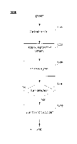
[8] Fig. 2 shows a flow chart for an exemplary embodiment of a pain assessment
algorithm according to the present invention.
[9] Fig. 3 shows a flow chart for an exemplary embodiment of a cluster
analysis
procedure used in developing a cluster analysis module utilized in the pain
assessment
algorithm.
[10] Fig. 4 shows a flow chart for an exemplary embodiment of a discriminant
analysis
procedure for developing a discriminant analysis module utilized in one
embodiment of
the pain assessment algorithm.and
[11] Fig. 5 shows an exemplary embodiment of a method for detecting pain
according
3
CA 02682955 2009-10-02
WO 2008/124566
PCT/US2008/059365
to the present invention.
Detailed Description
[12] The present invention may further understood with reference to the
following
description and the appended drawings, wherein like elements are provided with
the
same reference numerals. The present invention describes a system and method
for
detecting pain by analyzing brain wave data collected from an
electroencephalogram
(EEG). Those skilled in the art will understand that other types of data
relating to brain
activity may be manipulated in a manner similar to that described herein to
achieve
similar results. Thus, the description of EEG and the specific descriptions of
EEG
features are illustrative of exemplary embodiments of the invention and should
not be
construed to limit the scope of this invention. For example, activity of any
body system
sensitive to pain (e.g., autonomic nervous system, sweating or galvanic skin
response
(GSR), tearing of the eyes, contraction of muscles on the forehead ¨
frontalis,
orbicularis, skin around the eyes, etc.) may be measured and quantified to
determine
whether a subject is experiencing pain. Additionally, other analysis
modalities which are
sensitive to the amount of neuronal and/or metabolic activity in any brain
region(s) may
be utilized, e.g., electromyogram (EMG), magneto-encephalogram (MEG),
functional
magnetic resonance imaging (fMRI), near infrared spectroscopy (NIRS) or other
optical
tomographic methods (e.g., SPECT), etc.
[13] Although the invention is described in detail with regard to human
subjects, those
skilled in the art will understand that substantially similar methods may be
used to
obtain the same results for animals. Specifically, after making adjustments
for
anatomical differences mammals such as horses, cats, dogs, etc. may be
subjected to
analyses similar to that described for humans. Initially, this animal analysis
may more
commonly apply self norms by comparing brain activity for an individual before
and after
the onset of pain (e.g., before and after a surgical procedure) than by
comparing brain
4
CA 02682955 2009-10-02
WO 2008/124566
PCT/US2008/059365
activity of a subject to population data. However, those skilled in the art
will understand
that population data for animals may be compiled in the same manner described
below
for human subjects.
[14] As would be understood by those skilled in the art, an
electroencephalogram
(EEG) detects neurophysiological activity by measuring an intensity and
pattern of
electrical signals generated by the brain. Spontaneous oscillations in the
electrical
signals are typically referred to as brain waves or EEG. The EEG is a record
derived
from the spontaneously oscillating electrical signals and other electrical
activity (e.g.,
"noise" or electrical activity of a non-cerebral origin, transient potentials
elicited by
sensory stimuli, e.g., event-related potentials (ERPs), etc.). The EEG is
typically used
to assist in the diagnosis, in children and adults, of epilepsy, space
occupying lesions,
neurological and psychiatric disorders and other abnormalities of brain
function.
115] In the exemplary embodiment, data corresponding to brain activity (e.g.,
EEG
data) is utilized to detect and quantify pain experienced by a subject.
Differences
between brain waves produced in the presence and absence of pain, as well as
differences indicative of varying levels of pain, are assessed statistically
by a
comparison of the subject's EEG data to data in one or more databases. For
example,
the subject's EEG data may be compared to normative data indicative of normal
brain
wave activity for a control population comprised of individuals substantially
similar to the
Subject (e.g., age, gender, etc.). The subject's EEG data may further be
compared to
caled or "calibrated" reference data indicative of brain wave activity of the
control
population in the presence of varying of degrees of pain introduced, for
example, by
incremental quantified stimulation. The database may further include self-
normative
data indicative of the subject's brain wave activity in the presence and
absence of pain,
and/or self-reported reference data collected from the control population
indicating brain
wave activity when individuals within the population subjectively report the
presence of
pain (e.g., acute, chronic). The database may further include medical
histories and
physical and/or neurological examination results for individuals in the
population and/or
CA 02682955 2009-10-02
WO 2008/124566
PCT/US2008/059365
the subject. Those skilled in the art will understand that the term database
is not being
used to connote a specific data structure. Rather, it is to be understood
broadly to
include any searchable collection of data records residing in any type of
memory.
[16] The present invention relates to a system and method for detecting,
quantifying
and imaging pain, the Pain Quantification Index/Image. The method comprises
the
steps of extracting quantitative electroencephalographic (qEEG) features from
brain
electrical activity recorded from varying numbers of electrodes located at
standardized
positions on the scalp and forehead of a subject, comparing the brain wave
data to age-
appropriate normative data to generate standard or Z-score result data, and
determining the presence and/or chronicity and/or intensity of pain
experienced by the
subject as a function of the result data. The database corresponds to at least
one of (i)
population normative data indicative of brain wave activity of a first
plurality of
individuals in an absence of pain, (ii) population reference data indicative
of brain wave
activity of a second plurality of normal individuals generated in response to
a series of
calibrated pain events inflicted on the individuals, (iii) subjective
population reference
data (chronic and acute) indicative of brain wave activity of a third
plurality of individuals
reporting a graded or scaled sensation of pain; and (iv) population of
reference data
(chronic and acute cases) reflective of brain wave activity of a fourth
plurality of
individuals following an intervention which has changed the state of their
subjective
report of pain.
[17] The result data can be used for source localization of the most probable
neuroanatomical generators within the brain of the abnormal qEEG activity that
was
detected on the scalp by the PDI. Low Resolution Electromagnetic Tomographic
Analysis (Pascual-Marqui et al 1999), Variable Resolution Electromagnetic
Tomographic Analysis (Bosch-Bayard et al 2001) or similar methods are
exemplary of
the inverse solution techniques that may be used to visualize these sources.
The
computed sources may be depicted upon transaxial, sagittal or coronal slices
from the
Probabilistic MRI Atlas (Evans et al 1993), color coding each voxel in each
brain region
6
CA 02682955 2009-10-02
WO 2008/124566
PCT/US2008/059365
using a palette to represent ther statistical significance of the detected
source(s). These
LORETA images can be computed by an instrument that uses an appropriate array
of
electrodes placed upon the scalp, in accordance with the International 10/20
Electrode
Placement System (Jasper 1958) and implements all computations from a set of
digital
amplifiers controlled and analyzed by a personal desktop or laptop computer.
[18] Fig. 1 shows an exemplary embodiment of a system 1 for pain detection
according to the present invention. The system 1 includes a pain detecting
instrument
(PD)) 16 which provides objective corroboration, and optionally
quantification, of the
pain experienced by a subject 20 at a given time. In the exemplary embodiment,
the
PDI 16 is implemented as a portable, handheld device for use in clinical and
non-clinical
settings. In an example of the latter case, an EMT or other medical personnel
in the
field may use the PDI 16 to detect and/or quantify pain experienced by a
subject to, for
example, determine an appropriate pain management strategy or, in conjunction
with
palpation, etc. assist in making a preliminary diagnosis. The pain quantified
by, for
example, a comparison of the subject's EEG data to a pain quantification index
may be
recorded for future reference in patient records, employer records of work-
related
injuries, etc. Those of skill in the art will understand that the PDI 16 may
be utilized in
various other situations including, but not limited to, management of acute
and/or
chronic pain, rehabilitation treatment, treatment prescription, evaluation of
treatment
efficacy, monitoring or regulating delivery of analgesic to patients,
including patient
controlled analgesic (PGA) delivery, differentiating between levels of pain,
reducing pain
through appropriate neurofeedback paradigms, etc.
[19] The PDI 16 is coupled to electrode(s) 8 to receive electrical data
corresponding
to brain activity of the subject 20. Similar electrode placements may be used
to record
the SEP or EEG. Electrode(s) may be placed in a standard array such as the
International 10/20 Electrode Placement System (Jasper 1958) or in selected
advantageous positions on the head, forehead or cheeks. The electrical data is
quantitatively analyzed to generate digital quantitative EEG (qEEG) data
and/or
7
CA 02682955 2009-10-02
WO 2008/124566
PCT/US2008/059365
quantitative somatosensory evoked potential (qSEP) data which is then compared
with
reference data in a database 6. As would be understood by those skilled in the
art, the
database 6 may be stored in a memory within the P0116 or may be accessed from
a
remote storage via, for example a wireless or wired connection. Alternatively,
portions
of the database 6 may be stored locally while others are accessed from one or
more
remote locations. As will be described in more detail below, the reference
data in the
database 6 corresponds to brain wave data detected from: (i) individuals
experiencing
various levels of pain from no pain to extreme levels of pain (calibrated pain
norms), as
determined experimentally and explained further below; (ii) individuals with
acute or
chronic pain (the subject 20) seen in the absence of pain (self-referenced
norm); and/or
(iii) individuals self-reporting pain (acute or chronic). A comparison of the
subject's
brain wave data with the database 6 indicates a presence/absence of pain and a
quantification of any pain experienced. Changes in the EEG data and/or the SEP
data
may correlate to conditions in the database 6. In a pain treatment
protocol/procedure,
for example, comparing the subject's brain wave data before and after the
treatment to
the reference data may be used to quantify neurobiological effectiveness of
the
treatment or procedure and/or guide further treatment.
[20] In constructing the database 6, an EEG and/or SEP may be generated for
each
individual in the population, and the EEG and the SEP are analyzed to generate
the
reference data. For example, a resting EEG and/or SEP may be analyzed to
extract
features correlated with subjective reports of pain (and corresponding
intensity and/or
chronicity) by the individuals. As understood by those of skill in the art,
peaks in an
EEG power spectra reflect spontaneous activation of different neuronal
ensembles in
the brain regions sampled by the electrodes 8. The successive peaks in the
waveshapes of SEPs correspond to processing of signals related to stimuli
administered to the subject 20 as the signals pass through sequential
anatomical
regions, e.g., the medial lemniscal pathway structures of the nervous system.
The
shape of the EEG power spectrum and/or timing of peaks in the SEP (e.g.,
latency
periods) provide information regarding functioning of the nervous system and
8
CA 02682955 2009-10-02
WO 2008/124566
PCT/US2008/059365
processing of the signals through neuronal transmission pathways.
[21] In particular, distinctive changes in power may occur at particular
frequencies in
the EEG power spectrum when recorded from particular portions of the scalp,
e.g., in a
preferred exemplary embodiment, from a dorsolateral, prefrontal lead, a
periorbital
prefrontal lead, a mesial frontal lead over the anterior cingu late gyrus, a
midline or
lateralized lead in central, parietal or other regions over a cortical areas
sensitive to
spontaneous activity arising from acute or chronic painful sensory inputs
reaching
cortical regions which correspond to the sensation of pain. Such distinctive
reactions to
pain are extracted from the database(s) 6 and are used to define a "qEEG pain
signature" which constitutes a critical set of features for a "qEEG pain
discriminant
function, P1."
[22] The EEG data may be recorded in analog or digital format. If the data is
recorded
in analog format, this data is digitized and may then be subject to
artifacting or any other
quality assurance procedure as would be understood by those skilled in the
art. After
this, a selected set of features is extracted from the digital data of
acceptable quality. In
short, a selected set of features from the qEEG data which are particularly
relevant to
assessment of pain experienced are extracted from the overall data set
obtained and
compared to reference data including at least one of (i) population normative
data
indicative of brain wave activity of individuals in an absence of pain, (ii)
population
reference data indicative of brain wave activity of individuals generated in
response to
pain events inflicted thereon, (iii) subjective population reference data
indicative of brain
wave activity of individuals reporting a sensation of pain, and (iv)
population of
reference data indicative of brain wave activity of individuals following an
intervention
which has changed a subjective report of pain. Thereafter, in steps 130 and
135,
respectively, univariate or multivariate (e.g., Mahalanobis distances) data
features are
computed for the extracted features and these features may be transformed (for
example, log transformed) where appropriate to obtain a normal Gaussian
distribution.
As those skilled in the art would understand, the actual value of change or
difference
9
CA 02682955 2009-10-02
WO 2008/124566
PCT/US2008/059365
scores may alternately be used as criteria without these transforms or
obtaining
standard scores.
[23] For example, in a first step of a procedure to extract the selected set
of features,
ANOVAS and other statistical methods are used to search out features "A"
within the
external qEEG database (or other criterion values) which are significantly
different
between two or more groups of interest (such as individual experiencing pain
compared
to individuals in an absence of pain). Then, results from the first step, the
set of
features "A" are input to a classifier function such as a multiple stepwise
discriminant
function, the results of which will be used (a) as a classifier function
itself to later be
considered as part of a pain assessment strategy and/or (b) to reduce the
number of
features to a more sensitive set of features "B" to be entered into the next
step which
may include, for example, cluster analysis. The set of features "B' is input
to a cluster
analysis the results of which will be used (a) as a classifier function itself
for later
consideration as part of the pain assessment strategy and/or (b) to reduce the
number
of features to a more sensitive set of features "C" which may be used alone or
in
combination with the feature sets "A" and/or "B" as inputs to logistic
regression.
Similarly, a neural network may be used which receives as input a large
unselected set
of features extracted from the qEEG data and which outputs a reduced set of
features
"D" which can be used as a classifier function itself to later be considered
as part of the
pain assessment strategy and/or to reduce the number of features to a more
sensitive
set of features combined with the set of features "A" and/or the set of
features "B"
and/or the set of features "C" to be entered in the logistic regression.
Alternatively, as
would be understood by those skilled in the art, a single classifier function
may be used
to estimate a probability that pain is being sensed and the results from this
single
classifier function may be compared to data from the database to make
determinations
regarding the presence/absence of pain and the intensity and/or type thereof.
[24] Thereafter, as will be described in more detail below, the data is
entered into the
one or more classifier function(s) through which it is statistically
associated with a
CA 02682955 2009-10-02
WO 2008/124566
PCT/US2008/059365
subgroup representing individuals in similar or related states of pain within
the
database. Those skilled in the art will understand that, in place of the
database, a look-
up table or other structure representing similar data or other predefined
criteria may be
employed. By applying a decision making CLASSIFIER FUNCTION such as regression
to the individual's data the statistical likelihood that the individual is
properly classes
within a group with a specified degree and/or type of pain will be determined.
The
Regression module, the preferred embodiment being Logistic Regression, fits a
common slope cumulative model, which is a parallel lines regression model,
based on
the cumulative probabilities of the response categories rather than on their
individual
probabilities. Taking into account k predictive variables for n individuals,
the model is:
Log [p,/1- pi] = a + 131xi1 + fl2X12 fikXik
[25] This equation which has been previously trained on the database including
individuals and subjects enduring varying amounts/types of pain is applied to
the
individual and a pain assessment is made. Those skilled in the art will
understand that,
in other embodiments any or a combination of discriminant functions, cluster
algorithms,
neural networks and/or other classifier functions will be applied to the data
for the
purpose of assessing pain.
[26] Those skilled in the art will understand that, although the exemplary
embodiment
of the pain assessment algorithm 180 described below specifically discloses
steps for
utilizing a Cluster Analysis module 400, a Discriminant Analysis module 500
and a
Logistic Regression module, in that order, the pain assessment algorithm of
the present
invention may include some or all of these modules in any particular order.
Furthermore, rules obtained by various other classifier modules, such as by
using a
neural network, may also be incorporated into the pain assessment algorithm
180 to
generate a prediction of the expected cognitive decline of the patient. In
addition, as
those skilled in the art would understand, all of the modules utilized in the
pain
assessment algorithm 180 may be re-derived and/or modified following any or
all
11
CA 02682955 2009-10-02
WO 2008/124566
PCT/US2008/059365
changes to the database and improvements, refinements or future iterations of
the
classifier algorithms. However, in a preferred embodiment logistic regression
is the final
step in the procedure. For example, as shown in Fig. 2, in Step 1 (200) the
selected
features are input to a classification procedure such as, for example, a
discriminant
function to determine the relative probability of two or more levels of pain.
The set of
features identified in this analysis may also be input to other classifier
functions such as
cluster analysis and/or logistic regression.
[27] For example, in Step 2 (210) the selected features are input to a
classification
procedure such as for example a cluster analysis to determine the relative
probability
that a subject is experiencing pain corresponding to that of two or more
clusters
reflecting different levels and/or types of pain. The set of features
identified in this
analysis can be used as selected input to other classifier functions such as
discriminant
analysis and or logistic regression.
128] Then, in step N (220), selected features are input to, for example, a
classification
procedure such as a regression and/or logistic regression to determine the
relative
probability of two or more states of pain. The set of features identified in
this analysis
may also be used as selected input to other classifier functions such as
cluster analysis
and/or discriminant function.
1291 Thereafter, in step 230, the probability score computed by the
classification
function of Step N is converted to a confidence level using Receiver Operating
Characteristic (ROC) Curves as would be understood by those skilled in the
art. Using
plots of sensitivity versus specificity, the probability corresponding to
various P levels
(such as 0.10, 0.05, 0.01, etc.) may be specified. Where the results of
multiple classifier
functions are used to make the pain assessment, they may be combined using a
"voting
strategy" in Step 240 as would be understood by those skilled in the art. In
addition, the
pain assessment algorithm may utilize additional data (e.g., data concerning
pre-
existing conditions, clinical history/symptoms, neurobiological Or genetic
information) to
12
CA 02682955 2009-10-02
WO 2008/124566
PCT/US2008/059365
further refine pain assessments.
[30] In a similar manner, a "QSEP pain signature" will be derived from
recordings from
brain regions mediating painful sensations reflected as changes in the
amplitude, peak
latencies or intervals between peaks, area under the SEP between selected
latency
points, selected morphology descriptors or a neuroanatonnical distribution of
the
patient's SEPs elicited by calibrated stimulation with different, calibrated
intensities of
constant current electrical or infrared laser pulses. These stimuli are
delivered to
particular places on the body surface (e.g., the median nerve on the wrist) or
may be
delivered directly to the dermatome or body surface region nearest to that
reported as
most severely painful. In such implementation, the SEP waveshapes may be
compared
quantitatively to a calibrated normative database or, if the pain is
lateralized or
otherwise localized, by comparison to SEPs elicited by stimulation of the
afflicted versus
the homologous or homotopic non-afflicted counterpart or control region.
Quantitative
SEP analyses (qSEP) may be performed using a variety of mathematical
techniques,
such as independent component analysis (ICA), principal component analysis
(PCA) or
t-tests between regions or between stimulation conditions or intensities to
decompose
the SEP into quantitative descriptors and compare the subject SEP descriptor
values to
normative descriptor values obtained from a normative or pain reference
database
analogous to those described above for the qEEG. The elements of the pain
signature
are used as variables in a "qSEP pain discriminant function. P2."
[31] The qEEG and/or the qSEP are used to detect pain-related activity in the
subject's brain waves as indicated by one or more parameters. In the qEEG, the
parameters may include, but are not limited to, power in a particular wide
band
frequency domain (e.g., delta, theta, alpha, beta, gamma), or in some
particular
frequency quantified using very narrow band (VNB) spectral analysis in a
preselected
portion of the brain, coherence and/or asymmetry of power in any wide or VNB
frequency band between the preselected portions of the brain, etc. In the
qSEP, the
parameters may include the latency periods, amplitude and/or area under any
peak in
13
CA 02682955 2009-10-02
WO 2008/124566
PCT/US2008/059365
preselected portions of the brain, coherence and/or asymmetry of total SEPs
and/or
individual peaks from any pair of homologous portions of the brain (inter-
hemispheric),
power ratio or asynchronization between any pair of electrodes on the same
hemisphere (intra-hemispheric). In analysis of the qEEG and/or the qSEP, three-
dimensional images of the current sources within the brain may be generated,
and the
parameters may include power and/or current flow and/or Z-score at any
frequency in
selected voxels in the brain or voltages which are related to particular peak
latencies or
latency intervals in the analysis epoch of the the SEP waveshape.
[32] As shown in Fig. 1, the PDI 16 is coupled to one or more EEG electrodes 8
which
are applied to the scalp of a subject 20 being analyzed for the presence of
pain in any
chosen configuration (e.g., 10/20 system). When constructing the database 6,
the
electrodes 8 are coupled to the scalps of the individuals in the population.
Those of skill
in the art will understand that any conventional EEG biosensor electrodes may
be used
in conjunction with the present invention and that the electrodes 8 may be
reusable or
disposable. For example, the electrodes 8 may be pre-gelled, self-adhesive
disposable
electrodes. Alternatively, the electrodes 8 may have multiple small barbs, a
needle
electrode or a conductive disc temporarily attached to the scalp. The
electrodes 8 may
also utilize conductive gel to provide rapid and secure attachment to the
scalp while
limiting noise in the electrical signals returned by the electrodes 8. In
other exemplary
embodiments (e.g., a portable system), the electrodes 8 may be coupled to a
cap
placed on the head of the subject 20 and oriented to place the electrodes 8 in
any
chosen configuration relative to the scalp. The capjmay facilitate placement
of the
electrodes 8 in a non-clinical setting and reduce problems associated with
attachment of
the electrodes 8 to the scalp. In another exemplary embodiment, the electrodes
8 may
be contained in a self-adhering strip of material in an array. Thus, the PDI
16 may be
configured to receive data from any number and/or type of biosensor electrodes
and
may be configured to separate data from groups of electrodes 8 allowing, for
example,
simultaneous use with multiple patients, e.g., in hospital beds, a
neurotraumatology
ICU, multi-hospital trauma network, etc. In this embodiment, the database 6
may be
14
CA 02682955 2009-10-02
WO 2008/124566
PCT/US2008/059365
populated by several entities simultaneously. The electrodes 8 may be coupled
to the
PDI 16 via a wired or wireless connection. Using wired electrodes, leads may
transfer
signals from the electrodes 8 to the PDI 16, whereas, with wireless
electrodes, radio
frequency signals may transfer the signals to the PDI 16 using radio frequency
transmitters. The PDI 16 includes a receiving arrangement (e.g., cable
connector, radio
frequency receiver) to receive signals from the electrodes 8. Because the PDI
16 may
be configured to receive signals from electrodes on multiple subjects, the
system 1 may
be implemented over multiple hospital beds, a neurotraumatology ICU, a multi-
hospital
trauma network, etc.
[33] A stimulator 13 may be coupled to (via a connector) or integral with the
PDI 16
when, for example, monitoring the SEP and/or constructing the database 6. The
stimulator 13 may inflict one or more pain events on the subject 20 and/or
individuals in
the population at some selected repetition rate, usually in the range of 5-11
pulses/second, or at random intervals. The stimulator 13 may include or be
controlled
by software for varying a type, intensity and/or duration of pain events to
generate the
SEP and brain activity simulating various types of pain, e.g., neuropathic,
musculoskeletal or visceral, unilateral or bilateral, localized or
generalized, acute or
chronic, emotional or psychic pain, etc.
[34] To compare regional sensitivities and to avoid habituation, stimulation
sites
and/or intensity may be controlled by the PD1 16. The pain events may vary in
type
and/or duration from simulating instantaneous pain to chronic pain. The
stimulator 13
may use electrical, mechanical, chemical, optical and/or thermal mechanisms
and/or
auditory sounds or visual scenes to simulate the pain events. For example, the
stimulator 13 may apply electrical shocks, laser stimulation, compressive
force and/or
temperature variations to the individual to elicit "physical pain." In one
exemplary
embodiment, the stimulator 13 applies a variable electric current or laser
energy to a
sensory nerve (e.g., the median nerve at the wrist, the posterior tibialis
nerve at the
ankle, the skin surface, etc.) so that an amount of pain inflicted may be
accurately
CA 02682955 2009-10-02
WO 2008/124566
PCT/US2008/059365
controlled while the PD1 16 measures brain activity of the individual(s) in
conjunction
with an onset of the pain event(s) to capture the SEP data, as well as other
data
corresponding to the pain experienced by the individual, e.g., qEEG, Auditory
Evoked
Potentials, Visual Evoked Potentials, EKG, etc.
[35] Multimodal stimuli may be devised to distinguish between psychic pain
(e.g.,
emotional or mental) and physical pain, and may include repeated presentations
of
graphic visual images or sounds, especially in evaluation of patients with
post-traumatic
stress disorder (PTSD),
[363 The electrodes 8 may be placed over selected locations, or in the
traditional
10/20 system, to harvest brain waves and generate the EEG and SEP data
therefrom,
These brain waves may also be utilized to construct a three-dimensional image
of the
brain using signal source localization algorithms, e.g., low resolution
electromagnetic
tomographic analysis (LORETA), variable resolution electromagnetic tomographic
analysis (VARETA), etc. Using these methods regions of the brain may be
evaluated
and which may reflect awareness, quality and/or intensity of pain include, but
are not
limited to, the dorsolateral, mesial, midline and periorbital regions of the
prefrontal
cortex, the insula, the anterior, middle and posterior regions of the
cingulate gyrus, the
somatosensory regions of the central and parietal cortex, the amygdala, the
putamen
and the thalamus. The electrodes 8 may be placed over selected locations, or
in the
traditional 10/20 system to harvest brain waves and generate the EEG and SEP
data
therefrom.
[37] The electrical signals monitored by the electrodes 8 are transferred to a
high-
gain, low-noise amplifier 17 in the PDI 16 and then filtered by a filtering
arrangement 19
to detect and reject artifact contamination of the signals. Detection of the
artifacts in the
electrical signals may be accomplished by, for example, (1) detecting non-
stationarities
relative to statistical parameters derived from a sliding window of multiple
successive
2.5 second long segments of the EEG, (2) applying a set of rules defining
16
CA 02682955 2009-10-02
WO 2008/124566
PCT/US2008/059365
characteristics of common artifacts such as EMG (body), EKG (heart beats)
and/or
electro-oculogram (eye movements) and/or (3) using independent component
analysis
(ICA) to identify artifacts by multivariate statistical procedures, and/or
fractal
dimensional analysis. As understood by those of skill in the art, the
filtering
arrangement 19 minimizes the impact of noise and/or other artifacts resulting
from, for
example, subject movement, loosely applied electrodes, interference, etc. to
generate
data representative of and restricted to the brain activity of the subject 20.
Electrodes may be protected by driven shields to reduce artifacts.
[38] After artifact removal, the EEG may be subjected to spectral analysis
using a
signal processing technique such as, Fast Fourier Transform (FFT), wavelet
analysis or
fractal dimensional analysis performed on samples of the EEG approximately 2.5
seconds long. The EEG power spectrum may be divided into wide bands (e.g., low
delta (0.5-1.5Hz), high delta (1.5-3.5Hz), theta (3.5-7.5Hz), low alpha (7.15-
10Hz), high
alpha (10.0-12.5Hz), beta (12.5-25Hz), low gamma (25-35Hz), high gamma (35-
50Hz)
and ultra high (50-200Hz). The variance-covariance matrix of all frequencies
in all leads
versus all frequencies in all leads can be computed to evaluate the spectrum
and the
bispectrum. That is, phase relationships and coherence between and among
different
frequencies within and among all leads will be examined.
[39] Alternatively, the spectrum may be divided into very narrow bands (VNB)
of
approximately 0.39-0.50Hz in width. Absolute power and relative power,
monopolar
and bipolar derivations, bilateral symmetry, coherence and bicoherence between
homologous leads, coherence and bicoherence and power gradients between all
pairs
of leads and multivariate compressions (e.g., Mahalanobis Distances) of
various
combinations of the above-listed features may be computed and expressed as
scores
(e.g., Z-scores) relative to normative distributions. Similarly, the fractal
dimension can
vary between one and two, a fractal database can be constructed, and fractal Z-
scores
may be computed for the EEG from different electrodes in the presence or
absence of
pain.
17
CA 02682955 2009-10-02
WO 2008/124566 PC
T/US2008/059365
[40] The amplifier 17 amplifies the electrical signals monitored by the
electrode 8
using, for example, an amplification gain at approximately 100,000dB, a high
input
impedance (¨ 1 Megohm), a bandwidth of approximately 0.5Hz - 1500kHz and/or a
noise level of less than approximately 11.N. The amplifier 17 may include an
input
isolation circuit to protect against current leakage, such as a photo-diode
light-emitting
diode isolation coupler, and may be protected from electrical interference by
a radio-
frequency filter and/or a 60-cycle notch filter as is known in the art. The
amplified
electrical signals from each electrode 8 are output by the amplifier 17 and
converted to
digital signals by an analog-to-digital converter (ADC) 18 which, in the
exemplary
embodiment, operates at a minimum conversion rate of approximately 8,000
samples
per second and a resolution of 24 bits. Note that this may be accomplished by
using a
24 bit digital amplifier for each electrode.
[41] The digital signals are transmitted to a digital signal processor (DSP)
21 which
may be included in or electrically coupled to a central processing unit (CPU)
25. The
DSP 21 utilizes a digital signal processing technique to filter the digital
signal as known
in the art, and the CPU 25 compares the signals output by the DSP 21 to the
reference
data stored in the database 6, which may be stored locally in a memory in the
PDI 16
(e.g., a nonvolatile memory) or remotely accessed via a network (e.g., the
Internet, an
intranet). Those of skill in the art will understand that the PDI 16 may be
coupled to the
network via a wired and/or wireless connection. As a result of the comparison,
the CPU
25 outputs data indicating whether pain was detected and, if so, a magnitude
of the pain
(i.e., corresponding to the pain index). Analysis of the digital signal will
be described
further below.
[42] The PDI 16 may include or be coupled to one or more input/output (I/O)
arrangements 24. In the exemplary embodiment, the PDI 16 is coupled to a
display for
displaying brain wave data (i.e., EEG), the reference data and/or the pain
index, etc, of
the subject 20. The PDI 16 may include a keypad for configuring the
18
CA 02682955 2009-10-02
WO 2008/124566
PCT/US2008/059365
components/settings of the PDI 16 and manipulating the EEG, a printer for
printing the
EEG and/or other subject-related data including the pain index and/or any
other
peripheral component integral with or coupleable to the PDI 16. To communicate
with
these and any other peripheral components, the PDI 16 preferably includes
suitable
hardware ports and software drivers for communicating with the components.
031 The qEEG analysis may reveal values of quantitative variables that
exceed
normative thresholds as identified in the database 6, indicative of the
presence of pain.
Alternatively, selected samples from the qEEG and/or SEP measurements may be
input
into one or more classifier functions, such as, for example, discriminant
functions and/or
regression equations to determine the existence/intensity of pain. A first
discriminant
function A estimates a probability that pain is being experienced (A=presence
of pain)
and a second discriminant function B estimates a probability that pain is not
being
experienced (B=absence of pain). Note that A may be the qEEG pain discriminant
function, P1, the qSEP pain discriminant function, P2, or the sum, P1 +f,
multimodal
qEEG plus qSEP pain discriminant function. Then, eA+eB=1, and eA/(1 - eB)
equals a
probability P that pain is present (from 0.00 to 1.00). A pain quantification
index (PC21)
may be calibrated to correspond to clinical scales of pain ranging from
absent,
sensation of discomfort, mild pain, moderate pain, severe pain and intolerable
pain
using this or any other suitable method. Those skilled in the art will
understand that
similar results may be obtained by training a neural network to arrive at the
same
conclusions. The system may be configured to determine that pain is being
experienced by a subject whenever the value of the PQI is at least a threshold
level t
whenever eA/(1 - eB) > t. Depending on the consequences of false positives,
this
threshold level may be varied. For example, the system may determine the
subject is
sensing pain whenever the value of t is > 0.95. For cases in which the
consequences of
a false positive are more severe the system may determine the subject is
sensing pain
whenever the value of t is > 0.99. Furthermore, those skilled in the art will
understand
that actual data may give results of eA+e8 which, while approaching 1 may not
equal 1.
19
CA 02682955 2009-10-02
WO 2008/124566
PCT/US2008/059365
[44] When constructing the database 6, the CPU 25 instructs the stimulator 13
to
administer pain events to the individual. For example, when performing an SEP
assessment of sensitivity to pain in an individual, the CPU 25 instructs the
stimulator 13
to administer trains of constant current electrical pulses at some repetition
rate (e.g.,
approximately 9.7Hz) to a pair of electrodes located over the median nerve at
the wrist.
Approximately 250 pulses, each having a square wave duration of approximately
2004s, are administered in steps at intensities of about 5mA to about 25mA (or
to a
maximum intensity considered so painful by the individual as to be barely
tolerable).
The EEG data generated during administration of the pulses is subjected to
spectral
analysis (e.g., Fast Fourier Transform) at each intensity during an interval
from pulse-to-
pulse to detect an amplitude and phase of each very narrow band (VNB)
frequency at
intervals (e.g., approximately 0.5Hz) across the bandwidth of the amplifier
17.
Alternatively, successive samples of the EEG may be averaged with each sample
beginning at an onset of each stimulus to a sample point just prior to an
onset of a next
stimulus in each pulse train (e.g., SEP latency epoch). Accordingly, a time
series of
voltages which are time-locked to pulse delivery times is constructed to
represent the
waveshape of the single somatosensory evoked potential elicited by each
stimulus.
Therefore, the brain activity data may either be used to construct the power
spectrum or
averaged to provide an updateable average SEP (ASEP) which is a sum of N
samples
(e.g., 250 SEP latency epochs time-locked to the onset of the stimuli divided
by the
number of samples) collected separately at each intensity. When computed, the
power
spectra and/or ASEP values may be stored in, for example, the memory of the
PDI 16,
a removable storage medium and/or transmitted to a remote storage device.
[45] As understood by those skilled in the art, the brain electrical activity
recordings
data may be contaminated by voltage associated with body movements (e.g., eye
movements), abnormal physiological events or ambient environmental electrical
noise,
etc. These voltages are typically greater than those created by brain
activity, and the
present invention utilizes algorithms to minimize any impact of such
contaminating
events. For example, as brain activity is detected through the EEG amplifiers,
an
CA 02682955 2009-10-02
WO 2008/124566
PCT/US2008/059365
updateable voltage threshold may be computed continuously for the EEG channel
(or
separately for each channel in the case of more than one EEG channel) by
calculating a
root mean square (rms)-voltage for a sliding 20-second window and multiplying
the rms-
voltage by a constant selected so that the rms-voltage is approximately 0.2
standard
deviations of the amplitude of the electrical signals. The EEG is filtered to
discard
segments of the electrical signals containing voltages larger than the
threshold. In the
exemplary embodiment, the threshold is a multiple of the rms-voltage
approximately
equal to six (6) times the standard deviation of the amplitude. In other
exemplary
embodiments, the threshold may be a static value expected to be generated by
brain
activity (i.e., a value above which all voltages are considered to result from
artifacts).
After non-brain derived electrical activity has been removed from the
recordings by
these or similar denoising methods, the residual electrical signals may be
assumed to
be substantially artifact- free and are compiled to form a continuous,
artifact-free sample
of brain electrical activity to be used for further qEEG or qSEP processing.
Ongoing
brain electrical activity related to the EEG, as well as remaining non-brain
derived
electrical noise may be removed by denoising the SEP by the filtering
arrangement 19
as described in U.S. Patent No. 6,566,861, entitled "Fetal Brain Monitor," the
entire
disclosure of which is hereby expressly incorporated by reference.
[46] It is important that means be provided enabling the PDI to adaptively
monitor the
quality of the data being acquired to ensure acceptable test-retest
reliability and to
ensure that adequate information is acquired while avoiding excessive and/or
redundant
data. This is particularly important since the data collection process may
involve patient
or subject discomfort. Although it may be desirable to maintain an option
allowing the
operator to collect a predetermined amount of data, there are numerous methods
by
which adaptive quality control may be more efficiently be accomplished. One
exemplary means for achieving such self-evaluation of reliability is to
compute a
"coefficient of variation [Standard Deviation divided by Mean Value], or CV,
for all
variables of interest such as for example the VNB spectral power at each
frequency or
the SEP amplitude in each lead, progressively as data are acquired. Data are
21
CA 02682955 2009-10-02
WO 2008/124566
PCT/US2008/059365
considered acceptably reliable, i.e., "replicable" when the CV converges to a
preselected threshold defining acceptability. Alternatively, odd and even
split halves
may be constructed by assigning intervals alternately to two interlocked, but
independent samples, each containing, for example, 10 light averages which are
derived from approximately 25 stimuli to comprise a total of 250 "odd" and 250
"even"
samples. The standard deviation within each of the split halves may be
computed at
each sample point of each light average as:
_10
02 Ir2/ N _
[(VIN)2]
,=1
where V = voltage at each time point
V2/N = mean square of the voltage; and
(V/N)2 = squared mean of the voltage.
[47] When the PDI 16 reveals that the individual is experiencing pain, the
measures
revealing it should preferably be reliably replicable. Replicability may be
tested using
the t-test at each time point t as follows:
t =
lovi2 OV2211/2
[48] In an alternative embodiment, a cross-correlation may be utilized across
an entire
wave with a threshold for minimum correlation which would be acceptable to
indicate
replicability. Once normative data is available for the means and the voltages
at each
time point, the present method may be used to detect and quantify the pain
experienced
by the subject 20. If pain is detected with a significance of P>0.05, for
example,
replication yields p2<0.0025. Results from the two split-halves may be
combined (i.e.,
averaged) for display, with replicated significant results highlighted.
22
CA 02682955 2009-10-02
WO 2008/124566
PCT/US2008/059365
[49] In another exemplary embodiment, a digital comb filter is used to reduce
the
adverse effects of noise in the electrical signals. For example, the system 1
may
employ a digital comb filter as described in U.S, Pat. No. 4,705,049 entitled
"Interoperative Monitoring or EP Evaluation System Utilizing an Automatic
Adaptive
Self-optimizing Digital Comb Filter," the entire disclosure of which is
expressly
incorporated herein by reference. As noted above, the PDI 16 may utilize a
digital comb
filter to improve a signal-to-noise ratio (SNR) of the electrical signals
gathered by the
electrode(s) 8. In the digital comb filter, band pass frequencies are selected
to form
teeth of the comb using a phase variance parameter timed to correspond to the
presence and absence of the pain events. As understood by those of skill in
the art,
optimal digital filtering may be an alternative to signal averaging for
improving the SNR.
For example, where the stimulator 13 produces approximately 250 somatosensory
events with about 9.7 events per second for signal averaging, the SEP is
synced to the
pain events while the noise is random. When the SEP is calculated, the noise
is
reduced proportional to the square root of the number of stimuli, e.g., pain
events.
However, the signal averaging is relatively slow compared to the digital comb
filtering.
Since EEG and ambient noise can be approximately 100-250 V, the residual non-
SEP
"noise" after simple signal averaging may remain as high as 150/. The peak to
peak
SEP amplitude is on the order of 2.51.V. Thus, the signal to noise ratio may
be as poor
as 1:6. Further denoising is possible by using optimal digital filtering.
[50] The ASEP data and/or the qEEG data is evaluated using a quantitative
assessment of an expected normality (e.g., absence of pain) of the signals
such as
"Neurometrics" (the computerized quantitative analysis of brain electrical
activity). In
Neurometric analysis, features are extracted from the qEEG and the SEPs,
transformed
to obtain Gaussianity, compared to expected normative values (population/self
norms
as described above) and expressed in standard deviation units from the normal
population (i.e., pain index). The results can be displayed as color-coded
topographic
probability maps of brain function, or color-coded segments of a curve in the
time
domain (voltage vs. latency). Utilizing these methods greatly enhances the
sensitivity,
23
CA 02682955 2009-10-02
WO 2008/124566
PCT/US2008/059365
specificity and clinical utility of such data. Profiles of the qEEG and SEP
may be
mathematically described in the presence of pain and compared to normative
profiles in
the absence of pain.
[51] In order to evaluate the filtered SEP, an algorithm is applied to the SEP
which
automatically detects each of several peaks in a curve time-locked to the
stimuli to
identify the latency of each peak. For example, after 3-point smoothing of the
filtered
average, a resulting SEP is amenable to automatic peak detection by a
procedure
which hunts for zero-values of the second derivative and which outputs a
series of time
points corresponding to the peaks. The computed peaks are marked on the
optimally
filtered qSEP and superimposed on a normal template and, as described above,
the
latencies are compared to normative data collected from pain experiments on
the
control population and/or the subject 20 when not experiencing pain.
[52] An exemplary embodiment of a method 1200 for detecting pain in the
subject 20
according to the present invention is shown in Fig. 5. In step 1202, the
system 1 is
initialized and calibrated. The PDI 16 and the I/O arrangement(s) 24 may be
powered
and configured for pain detection in accordance with the methodology described
herein.
The system 1 may be configured based on subject data, e.g., height, weight,
age,
medical history, etc. The subject data may be utilized when determining the
existence
and level of pain. For example, the database 6 may be queried based on the
subject
data and the subject's brain wave data.
[53] In step 1204, the PDI 16 receives signals corresponding to the brain
activity of
the subject 20 (e.g., electrical signals from electrodes 8 attached to the
scalp of the
subject 20). In step 1206, the signals are processed by the P0116 in the
manner
described above. That is, the qEEG and/or the qSEP data of the subject 20 are
used to
generate data corresponding to the brain activity which corresponds to pain
experienced by the subject 20. As described above, the signals are filtered
and
smoothed to reduce the effects of ambient noise and artifacts.
24
CA 02682955 2009-10-02
WO 2008/124566
PCT/US2008/059365
[54] In step 1208, the brain activity data is compared to the reference data
to
determine whether pain exists. The brain wave data is compared to the
reference data
in the database 6, and a value corresponding to the pain index (i.e.,
corresponding to a
level of pain experienced by the subject 20) is returned and output by the I/0
arrangement 24. When the brain wave data is indicative of the presence of
pain, the
method may proceed to optional step 1210 where the pain is quantified a PQI.
[55] The present invention allows objective corroboration of pain in a
subject. The
determination of the presence of pain is advantageous in clinical and non-
clinical
settings. For example, in a lawsuit, damages are often awarded based on pain
suffered
and whether or not the pain is treatable or chronic. Objective corroboration
of pain
allows the damages to be calculate more accurately.
[56] It will be apparent to those skilled in the art that various
modifications and
variations can be made in the structure and the methodology of the present
invention,
without departing from the spirit or scope of the invention. Thus, it is
intended that the
present invention cover the modifications and variations of this invention
provided they
come within the scope of the appended claims and their equivalents.