Note : Les descriptions sont présentées dans la langue officielle dans laquelle elles ont été soumises.
CA 02683713 2009-10-13
WO 2008/128106 PCT/US2008/060116
TREATMENT OF GAUCHER DISEASE WITH SPECIFIC
PHARMACOLOGICAL CHAPERONES AND MONITORING TREATMENT
USING SURROGATE MARKERS
CROSS-REFERENCE TO RELATED APPLICATIONS
This application claims priority to U.S. Provisional Application Serial
No. 60/911,699, filed April 13, 2007; and to U.S. Provisional Application
Serial No.
61/028,123, filed February 12, 2008, both of which are hereby incorporated by
reference in their entireties herein.
FIELD OF THE INVENTION
The present invention provides a method for monitoring the treatment of an
individual having Gaucher disease with a specific pharmacological chaperone by
determining the presence and levels of specific surrogate markers such as
glucocerebrosidase, glucosylceramide, chitotriosidase, inflammatory cytokines
and
chemokines, glucosylceramide-containing macrophages, makers of bone
metabolism,
and a-synuclein. The present invention also provides a method for monitoring
the
treatment of an individual having Gaucher disease with a specific
pharmacological
chaperone by evaluating the effects of treatment at the cellular level.
BACKGROUND
Gaucher Disease
Gaucher disease is a lysosomal storage disorder that is associated with the
accumulation of glycosphingolipids (GSL) in cells, particularly monocytes and
macrophages, of afflicted individuals. This aberrant build up of GSL results
from a
genetic deficiency (mutation) in the lysosomal enzyme acid 0-glucosidase
(GCase;
glucocerebrosidase), the lysosomal hydrolase that breaks down the GSL
glucosylceramide (GluCer). The majority of Gba mutations cause GCase protein
to
misfold in the endoplasmic reticulum (ER). Misfolded GCase is recognized by
the
ER quality control system and subsequently degraded instead of being processed
and
trafficking to the lysosome (Street et al., Proc Natl Acad Sci U S A 2006;
vol. 103;
no. 37: 13813-18).
Gaucher disease is pan-ethnic, with an overall disease frequency of about 1 in
50,000-100,000 births. Certain populations have a higher prevalence of Gaucher
1
CA 02683713 2009-10-13
WO 2008/128106 PCT/US2008/060116
disease. In the Ashkenazi Jewish population, for example, about 1 in 15 people
are
carriers for a Gba mutation (Aharon-Peretz et al., New Eng. J. Med. 2004; 351:
1972-
77). According to the National Gaucher Foundation, about 2,500 Americans
suffer
from Gaucher disease.
Gaucher disease is an autosomal recessive disorder and is the most common
lysosomal storage disease. The disease has been classified into three clinical
types,
depending on neurological involvement and disease severity (Cox et al., Q J
Med.
2001; 94: 399-402). Type 1 is the most common and is characterized by an
absence
of neurological involvement. Type 1 patients exhibit a broad spectrum of
severity,
and some can remain asymptomatic throughout life. Most Type 1 patients exhibit
enlargement of the spleen and liver, skeletal abnormalities and bone lesions,
and
sustained inflammatory reactions. Hepatic glucocerebroside levels are elevated
from
23-fold to 389-fold above normal levels in Type I Gaucher patients.
Type 2 Gaucher disease is the rarest, most severe form, and is associated with
early onset of acute neurologic disease. The characteristic feature of
neuronopathic
Gaucher disease is an abnormality of horizontal gaze. Afflicted patients
develop
progressive encephalopathy and extrapyrimidal symptoms such as rigidity and
Parkinson's-like movement (parkinsonism). Most Type 2 Gaucher patients die in
early childhood from apnea or aspiration due to neurological deterioration.
Type 3 Gaucher disease also has neurological involvement, although to a
lesser extent than Type 2. Type 3 patients also have the hepatosplenomegaly
and
skeletal defects characteristic of Type 1, and central nervous system symptoms
that
include poor coordination of movements (ataxia), seizures, paralysis of the
eye
muscles, epilepsy, and dementia. People with Type 3 Gaucher disease can live
into
adulthood, but may have a shortened life span. Three sub-classifications of
Type 3
have been reported: Type 3a, which is associated with prominent
hepatosplenomegaly
and bone marrow disease; Type 3b, which is associated with limited systemic
symptoms; and Type 3c, which is associated with hepatosplenomegaly, corneal
opacities, progressive ataxia and dementia, and cardiac valve and aortic root
calcification.
Over 200 Gba mutations have been identified in affected Gaucher patients.
Most Gaucher patients exhibit some residual GCase activity. However, a poor
correlation of genotype with phenotype has plagued efforts to elucidate the
molecular
basis for phenotypic variation (Sidransky, Mol. Genetics and Metab. 2004; 83:
6-15).
2
CA 02683713 2009-10-13
WO 2008/128106 PCT/US2008/060116
There is a lack of phenotypic consistency even among identical twins harboring
the
same genetic mutations. Despite this, different mutations are associated with
the three
disease types. The presence of point mutation N370S on at least one allele
(heterozygotes) is almost universally associated with type 1 Gaucher disease
(Cox,
supra).
Treatment
Treatment of clinically manifested Types 1 and 3 disease is predominantly by
enzyme replacement therapy (ERT) of recombinant GCase (Ceredase(K and
Cerezyme , Genzyme Inc.). Bone marrow transplants (BMT) also have been
employed as treatment for Gaucher disease (Types 1 and 3). Because macrophages
are derived from bone marrow stem cells, allogeneic bone marrow
transplantation
(BMT) has been applied successfully in a small number of Gaucher patients.
However, BMT can be associated with severe morbidity and mortality, and only a
small fraction of patients have appropriate histocompatible donors.
A third, relatively recent approach to treating protein deficiencies involves
the
use of small molecule inhibitors to inhibit synthesis the natural substrate of
the
deficient enzyme protein, thereby ameliorating the pathology. This "substrate
reduction" approach (SRT) has been specifically described for a class of about
40
related enzyme disorders called lysosomal storage disorders or
glycosphingolipid
storage disorders including Gaucher disease.
A fourth approach, a specific chaperone strategy, rescues mutated proteins
from degradation presumably in the endoplasmic reticulum (ER) or in other
cellular
protein degradation/disposal systems. In particular embodiments, this strategy
employs small molecule reversible inhibitors which specifically bind to a
defective
lysosomal enzyme associated with a particular lysosomal disorder. In the
absence of
therapy, the mutated enzyme folds improperly in the ER (Ishii et al., Biochem.
Biophys. Res. Comm. 1996; 220: 812-815), is retarded in its maturation to a
final
product, and is subsequently degraded via ER associated degradation pathways.
The
chaperone strategy involves the use of a compound that facilitates the correct
folding
of a mutated protein, to prevent undue or abnormal degradation from the ER
quality
control system, or accumulation of misfolded protein in the cell. These
specific
chaperones are designated specific pharmacological chaperones (or active site-
specific chaperones).
3
CA 02683713 2009-10-13
WO 2008/128106 PCT/US2008/060116
The chaperone strategy has been described and exemplified for enzymes
involved in lysosomal storage disorders as in U.S. Patent Nos. 6,274,597,
6,583,158,
6,589,964, 6,599,919, and 7,141,582, to Fan et al., which are incorporated
herein by
reference in their entirety. Rescue of GCase from Gaucher patient cells has
been
described using the imino sugar, isofagomine (IFG), and its derivatives., and
using
other compounds specific for GCase (described in pending U.S. Patent
Application
Serial Nos. 10/988,428, and 10/988,427, both filed November 12, 2004). Such
compounds include glucoimidazole ((5R,6R,7S,8S)-5-hydroxymethyl-5,6,7,8-
tetrahydroimidazo [ 1,2a]pyridine-6,7, 8-triol).
Surrogate Markers
Despite the phenotypic inconsistency, Gaucher patients exhibit several
consistent surrogate markers of the disease that are used to evaluate clinical
response
to treatment. The present invention relates to a method of monitoring
treatment of a
Gaucher patient following treatment with a specific pharmacological chaperone,
by
evaluating changes in at least one, and preferably multiple, surrogate markers
of
Gaucher disease.
SUMMARY OF THE INVENTION
The present invention provides a method for monitoring treatment of a
Gaucher disease patient with a specific pharmacological chaperone for acid 0-
glucosidase (GCase), by evaluating changes in the presence and/or level of a
surrogate marker that is associated with Gaucher disease, where an improvement
indicates that the individual is responding to the chaperone therapy.
In one embodiment, the surrogate marker is a systemic surrogate marker.
Systemic surrogate markers include at least one of the following: decreased
lysosomal GCase activity in cells and urine; the presence of lipid-laden
macrophages
("Gaucher macrophages"); hepatosplenomagaly; increased levels of
chitotriosidase;
increased levels of liver enzymes; increased levels of lysosomal proteins
including
LAMP-1 and saposin C, increased levels of pulmonary chemokine PARC/CCL18;
increased levels of plasma a-synuclein; increased levels of angiotensin
converting
enzyme (ACE) and total acid phosphatase; immunological defects such as anemia,
thrombocytopenia, leukopenia, hypergammaglobulinemia, decreased amount of T-
4
CA 02683713 2009-10-13
WO 2008/128106 PCT/US2008/060116
lymphocytes in the spleen, systemic B cell hyperproliferation, plasmacytosis,
increased levels of inflammatory cytokines (TNF-a, IL-10, IL-la, IL-6) and
chemokines including those associated with bone metabolism and multiple
myeloma
(TNF-a, IL-8, IL-17, MIP-la, MIP-10, VEGF, and TRACP 5b, BAP), the presence of
inflammatory foci in tissues or organs comprising macrophages, lymphocytes,
and
neutrophils, and impaired neutrophil chemotaxis; skeletal defects such as
infiltration
of Gaucher cells in the bone marrow, lytic lesions, osteosclerosis,
osteoporosis, bone
crises and bone pain, fractures, vertebral collapse, and reduced levels of
triglycerides;
decreased levels of bone-specific alkaline phosphatase, neurological symptoms
such
as neuronal loss, neurodegeneration, horizontal gaze abnormalities, myoclonic
movements, corneal opacity, ataxia, dementia, spasticity; seizures, auditory
impairment; cognitive impairment; and pulmonary infiltration of Gaucher
macrophages and pulmonary hypertension.
In a specific embodiment, the combination of markers expected following
treatment of Gaucher disease with a pharmacological chaperone are as follows:
increased (3-glucocerebrosidase (GCase) levels in white blood cells, skin,
cerebrospinal fluid (CSF) and urine; decreased glucocerebroside (G1cCer)
levels in
white blood cells, plasma, serum, urine, CSF and skin; decreased a-synuclein
levels in
plasma and CSF; increased bone-specific alkaline phosphatase (BAP) activity in
plasma; decreased tartrate-resistant acid phosphatase 5b (TRACP 5b) activity
in
plasma, decreased chitotriosidase activity in plasma; decreased pulmonary and
activation regulated chemokine (PARC) in plasma and urine, and decreased
interleukin 8, interleukin 17, VEGF MIP-10 and MIP-la level in plasma as well
as
LAMP-1 and cathepsin D. Additional markers evaluated include decrease in liver
and
spleen volume from baseline; increase in hemoglobin level from baseline;
change in
hematocrit level from baseline; change in platelet count from baseline;
improvement
in bone mineral density from baseline; improvement in radiographic findings
from
baseline; decreased GM3 levels in plasma, urine, white blood cells (WBC) and
CSF;
decreased chitotriosidase activity in plasma and CSF, in particular IL-8, IL-
6,
membrane markers in CSF.
In another embodiment, the surrogate marker is a sub-cellular surrogate
marker.
Sub-cellular surrogate markers include at least one of the following: aberrant
trafficking of GCase in cells from Gaucher patients from the ER to the
lysosome;
5
CA 02683713 2009-10-13
WO 2008/128106 PCT/US2008/060116
aberrant trafficking of cellular lipids though the endosomal pathway; the
presence of
increased amounts misfolded GCase in the ER or cytosol; the presence of ER
and/or
stress resulting from toxic accumulation of GCase (as determined by gene
and/or
protein expression of stress-related markers); aberrant endosomal pH levels;
the
presence of increased plasma membrane expression of MHCII and/or CD1d on
monocytes; aberrant cell morphology; suppression of the ubiquitin/proteasome
pathway; and an increase in the amount of ubiquitinated proteins.
In a specific embodiment, the individual has Type 3 Gaucher disease with
cardiac involvement and the surrogate marker is calcification of the aortic
and/or
mitral valves.
In a further embodiment, the specific pharmacological chaperone used in the
therapy is an inhibitor of acid (3-glucosidase, such as a reversible
competitive
inhibitor.
In specific embodiments, the inhibitor is isofagomine, C-benzyl-isofagomine
or compounds disclosed in U.S. Patent Nos. 6,583,158; 6,744,135; 6,599,919;
6,589,964, 6,916,829; 7,141,582; 5,844,102; 5,863,903; 6,046,214; 5,854,272;
6,541,836; 6,316,489; 6,239,163; 6,590,118 and PCT Application No. WO
04/037233
all of which are incorporated by reference.
The present invention also provides a method for treating Gaucher disease
with effective amount of a specific chemical chaperone that binds to acid 0-
glucosidase, and monitoring its effect on cytoplasmic staining of cells, where
restoration of an abnormal indicates that the individual with Gaucher disease
is
responding to chaperone treatment. In one embodiment, the cytoplasmic staining
is
lysosomal staining, in particular, detection of acid (3-glucosidase or LAMP-1
expression in the lysosome.
In another embodiment, the cytoplasmic staining is detection of
polyubiquitinated proteins.
In a particular embodiment, the specific pharmacological chaperone is an
inhibitor of acid 0-glucosidase, such as a reversible competitive inhibitor.
In specific embodiment, the inhibitor is isofagomine, C-benzyl-isofagomine,
or glucoimidazole.
BRIEF DESCRIPTION OF THE DRAWINGS
6
CA 02683713 2009-10-13
WO 2008/128106 PCT/US2008/060116
This patent application contains at least one drawing executed in color.
Copies
of this patent or patent application publication with color drawing(s) will be
provided
by the Office upon request and payment of the necessary fee.
Figure 1. Figure 1 depicts GCase enhancement results from a Phase 1
multiple-ascending dose study of isofagomine tartrate in healthy volunteers.
Figure 2A-D. Figure 2 depicts changes in GCase activity in liver (2A), spleen
(2B), lung (2C), and brain (2D), following treatment with isofagomine (IFG).
Figure 3A-B. Figure 3 depicts the effects of treatment with isofagomine on
body tissue (spleen and liver, 3A-B, respectively) weights at over 2-24 weeks.
Figure 4. Figure 4 depicts changes in levels of chitotriosidase in a mouse
model of Gaucher disease following treatment with IFG.
Figure 5A-D. Figure 5 shows serum parameters for cholesterol (5A), liver
enzymes ALT (5B) and AST (5C) and IgG (5D) following treatment with IFG for 2,
4
and 12 weeks.
Figure 6. Figure 6 depicts a comparison of plasma a-synuclein levels from
healthy volunteers and patients with Gaucher disease.
Figures 7A-N. Figure 7 depicts fluorescent staining of lysosomes using
LysoTracker Red in cells from Gaucher fibroblasts (7A) and normal fibroblasts
(7B).
Staining for lysosomal protein LAMP-1 was also performed on normal fibroblasts
(7C) and Gaucher fibroblasts (7D). Figures 7E-F show an overlay of dual GCase
and
LAMP-1 staining in Gaucher fibroblasts. Also depicted is a dual overlay (LAMP-
1
and GCase) of Gaucher cells treated with the specific pharmacological
chaperone
isofagomine (7G-H), or C-benzyl-isofagomine (7I-J). Lastly, Figures 7K-N show
staining of Gaucher cells for GCase only. Control Gaucher cells were stained
with
secondary antibody only (7K), or were not treated (7L), or were treated with
isofagomine (7M), or C-benzyl-isofagomine (7N).
Figure 8. Figure 8 is comparison of Gcase activity in WBCs, GlcCer
concentration in WBC, chitotriosidase activity in plasma and a-synuclein
levels in
plasma in Gaucher Patients as compared to controls.
Figure 9. Figure 9 is a comparison of TRACP 5b Activity In Plasma
(Females), TRACP 5b Activity In Plasma (Males), BAP Activity in Plasma
(Females)
and BAP Activity in Plasma (Males) in Gaucher Patients as compared to
controls.
7
CA 02683713 2009-10-13
WO 2008/128106 PCT/US2008/060116
Figure 10. Figure 10 is a comparison of PARC, IL-8, MIP-la, IL-17,
VEGF, and IL-17 vs. VEGF Activity In Plasma (Femalesin Gaucher Patients as
compared to controls.
DETAILED DESCRIPTION
The present invention demonstrates a response to treatment with SPCs in a
Gaucher disease model as evidenced by evaluation of specific surrogate markers
of
Gaucher disease following treatment. Accordingly, the present invention
provides
standards of care for evaluating response to SPC treatment in Gaucher patients
by
evaluating the patient for changes, i. e. , improvements, in specific
surrogate markers.
Definitions
The terms used in this specification generally have their ordinary meanings in
the art, within the context of this invention and in the specific context
where each
term is used. Certain terms are discussed below, or elsewhere in the
specification, to
provide additional guidance to the practitioner in describing the compositions
and
methods of the invention and how to make and use them.
The term "Gaucher disease" includes Type 1, Type 2 and Type 3 (including
3a, 3b and 3c), and intermediates and subgroups thereof based on phenotypic
manifestations.
A Gaucher disease patient refers to an individual who has been diagnosed with
Gaucher disease due to a mutated acid 0-glucosidase as defined further below.
A "mutated GCase" refers to a GCase protein that contains a mutation which
affects folding and processing of the GCase protein in the ER. Accordingly,
upon
folding of the mutant into a proper conformation using a specific
pharmacological
chaperone, the mutated GCase protein will be able to progress or traffic from
the ER
through the Golgi to the lysosome. Mutations which impair folding, and hence,
trafficking of GCase, can be determined by routine assays well known in the
art, such
as pulse-chase metabolic labeling with and without glycosidase treatment to
determine whether the protein enters the Golgi apparatus, or fluorescent
immunostaining for GCase localization within the cell. Specific embodiments of
GCase folding mutants associated with neuronopathic diseases include but are
not
limited to: N370S, L444P, K198T, D409H, R496H, V394L, 84GG, and R329C.
"MIP" as used herein means macrophage inflammatory protein.
8
CA 02683713 2009-10-13
WO 2008/128106 PCT/US2008/060116
"TNF" means tumor necrosis factor.
"IL" means Interleukin.
"GM3" means ST3 beta-galactoside alpha-2,3-sialyltransferase 5, which is
also known as, ST3GAL5 or ganglioside GM3.
As used herein, the term "specific pharmacological chaperone" ("SPC") refers
to any molecule including a small molecule, protein, peptide, nucleic acid,
carbohydrate, etc. that specifically binds to a protein and has one or more of
the
following effects: (i) enhancing the formation of a stable molecular
conformation of
the protein; (ii) inducing trafficking of the protein from the ER to another
cellular
location, preferably a native cellular location, i.e., preventing ER-
associated
degradation of the protein; (iii) preventing aggregation of misfolded
proteins; and/or
(iv) restoring or enhancing at least partial wild-type function and/or
activity to the
protein. A compound that specifically binds to e.g, GCase, means that it binds
to and
exerts a chaperone effect on GCase and not a generic group of related or
unrelated
enzymes. Following is a description of some specific pharmacological
chaperones
contemplated by this invention:
Isofagomine (IFG; (3R,4R,5R)-5-(hydroxymethyl)-3,4-piperidinediol) refers
to a compound having the following structure:
OH
HO
HO NH
IFG has a molecular formula of C6H13NO3 and a molecular weight of 147.17. This
compound is further described in U.S. Patents 5,844,102 to Sierks et al., and
5,863,903, to Lundgren et al. N-alkyl IFG derivatives are described in U.S.
patent
6,046,214.
C-benzyl-IFG, refers to a compound having the following structure:
OH
HO CH2Ph
HO NH
Other SPCs for GCase include hydroxypiperidine derivatives, which are
described in pending PCT publications WO 2005/046611 and WO 2005/046612, and
in U.S. Patent Application Serial No. 10/988,428, filed November 12, 2004.
Also,
chaperones for GCase include glucoimidazole and polyhydroxycyclohexenyl amine
9
CA 02683713 2009-10-13
WO 2008/128106 PCT/US2008/060116
derivatives which are described in U.S. Patent Application Serial No.
10/988,427 filed
on November 12, 2004.
As one example, glucoimidazole refers to a compound having the following
structure:
OH
N
HO
HO N
H
Still other SPCs for GCase are described in U.S. Patent 6,599,919 to Fan et
al.,
and include calystegine A3, calystegine A5, calystegine B1, calystegine B2,
calystegine
B3, calystegine B4, calystegine C1, N-methyl-calystegine B2, DMDP, DAB,
castanospermine, 1-deoxynojirimycin, N-butyl-deoxynojirimycin, 1-
deoxynojirimycin
bisulfite, N-butyl-isofagomine, N-(3-cyclohexylpropyl)-isofagomine, N-(3-
phenylpropyl)-isofagomine, and N-[(2E,6Z,1 OZ)-3,7,11-trimethyldodecatrienyl]-
isofagomine.
A "surrogate marker" or "surrogate clinical marker" of Gaucher disease refers
to the abnormal presence of, increased levels of, abnormal absence of, or
decreased
levels of a biomarker that is associated with Gaucher disease and that is a
reliable
indicator of Gaucher disease (but is not associated width a healthy
individual) either
alone or in combination with other abnormal markers or symptoms of Gaucher
disease.
As non-limiting examples, surrogate markers of Gaucher disease, include
decreased lysosomal GCase activity; the presence of lipid-laden macrophages
("Gaucher macrophages"); hepatosplenomagaly; increased chitotriosidase;
increased
pulmonary chemokine PARC/CCL18; increased levels of angiotensin converting
enzyme (ACE) and total acid phosphatase; hematologic or immune abnormalities
including anemia, thrombocytopenia, leukopenia, and hypergammaglobulinemia, T-
lymphocyte deficiency in the spleen, systemic B cell hyperproliferation,
plasmacytosis, the presence of inflammatory foci in tissue or organ comprising
macrophages, lymphocytes, and neutrophils, elevated inflammatory cytokines
(e.g.,
TNF-a, IL-1(3, IL-6, IL-17, MIP-la, VEGF), impaired neutrophil chemotaxis;
imbalances in T cell and monocyte subsets; over-expression of cell membrane
expression MHCII and Cdld on monocytes; skeletal defects, including
infiltration of
CA 02683713 2009-10-13
WO 2008/128106 PCT/US2008/060116
Gaucher cells in the bone marrow, bone-specific alkaline phosphatase activity
in
plasma (BAP), lytic lesions, osteosclerosis, bone pain, fractures, vertebral
collapse, or
reduced triglyceride presence; neurological symptoms such as neuronal loss,
neurodegeneration, horizontal gaze abnormalities, myoclonic movements, corneal
opacity, ataxia, dementia, and spasticity; and pulmonary infiltration of
Gaucher
macrophages, possibly leading to pulmonary hypertension, pulmonary and
activation
regulated chemokine (PARC) activity in plasma, and tartrate-resistant acid
phosphatase 5b (TRACP 5b) activity in plasma.
Other surrogate markers are present at the sub-cellular level ("sub-cellular
surrogate markers") and include aberrant trafficking of GCase in cells from
Gaucher
patients from the ER to the lysosome; aberrant trafficking of lipids though
the
endosomal pathway; the presence of increased amounts misfolded GCase in the ER
or
cytosol; the presence of ER and/or cell stress resulting from toxic
accumulation of
GCase (as determined by gene and/or protein expression of stress-related
markers);
aberrant endosomal pH levels; aberrant cell morphology; suppression of the
ubiquitin/proteasome pathway; or an increase in the amount of ubiquitinated
proteins.
An "an improvement in a surrogate marker" refers to an effect, following
treatment with an SPC, of the amelioration or reduction of one or more
clinical
surrogate markers which are abnormally present or abnormally elevated in
Gaucher
disease, or the presence or increase of one or more clinical surrogate markers
which
are abnormally decreased or absent in Gaucher disease, relative to a healthy
individual who does not have Gaucher disease, and who does not have an other
disease that accounts for the abnormal presence, absence, or altered levels of
that
surrogate marker.
A "responder" is an individual diagnosed with a disease associated with a Gba
mutation which causes misfolding of the GCase protein, such as Gaucher
disease, and
treated according to the presently claimed method who exhibits an improvement
in,
amelioration of, or prevention of, one or more clinical symptoms, or
improvement in
one or more surrogate markers referenced above.
In addition, a determination whether an individual is a responder can be made
at the sub-cellular level by evaluating improvements in the sub-cellular
surrogate
markers, e.g., intracellular trafficking of the mutant GCase protein in
response to
treatment with an SPC. Restoration of trafficking from the ER is indicative of
a
response. Other sub-cellular evaluations that can be assessed to determine if
an
11
CA 02683713 2009-10-13
WO 2008/128106 PCT/US2008/060116
individual is a responder include improvements in the above-referenced sub-
cellular
surrogate markers.
The terms "therapeutically effective dose" and "effective amount" refer to the
amount of the specific pharmacological chaperone that is sufficient to result
in a
therapeutic response. A therapeutic response may be any response that a user
(e.g., a
clinician) will recognize as an effective response to the therapy, including
improvements in the foregoing symptoms and surrogate clinical markers. Thus, a
therapeutic response will generally be an amelioration of one or more symptoms
of a
disease or disorder, such as those described above.
The phrase "pharmaceutically acceptable" refers to molecular entities and
compositions that are physiologically tolerable and do not typically produce
untoward
reactions when administered to a human. Preferably, as used herein, the term
"pharmaceutically acceptable" means approved by a regulatory agency of the
Federal
or a state government or listed in the U.S. Pharmacopeia or other generally
recognized
pharmacopeia for use in animals, and more particularly in humans. The term
"carrier"
refers to a diluent, adjuvant, excipient, or vehicle with which the compound
is
administered. Such pharmaceutical carriers can be sterile liquids, such as
water and
oils. Water or aqueous solution saline solutions and aqueous dextrose and
glycerol
solutions are preferably employed as carriers, particularly for injectable
solutions.
Suitable pharmaceutical carriers are described in "Remington's Pharmaceutical
Sciences" by E.W. Martin, 18th Edition.
The terms "about" and "approximately" shall generally mean an acceptable
degree of error for the quantity measured given the nature or precision of the
measurements. Typical, exemplary degrees of error are within 20 percent (%),
preferably within 10%, and more preferably within 5% of a given value or range
of
values. Alternatively, and particularly in biological systems, the terms
"about" and
"approximately" may mean values that are within an order of magnitude,
preferably
within 10- or 5-fold, and more preferably within 2-fold of a given value.
Numerical
quantities given herein are approximate unless stated otherwise, meaning that
the term
"about" or "approximately" can be inferred when not expressly stated.
Formulations, Dosage, and Administration
IFG and derivatives can be administered in a form suitable for any route of
administration, including e.g., orally in the form tablets, capsules, or
liquid, or in
12
CA 02683713 2009-10-13
WO 2008/128106 PCT/US2008/060116
sterile aqueous solution for injection. In a specific embodiment, the IFG
tartrate is
administered as a powder-filled capsule. IFG tartrate is described in pending
provisional patent applications 60/808,020 and 60/890,719, herein incorporated
by
reference. When the compound is formulated for oral administration, the
tablets or
capsules can be prepared by conventional means with pharmaceutically
acceptable
excipients such as binding agents (e.g., pregelatinized maize starch,
polyvinylpyrrolidone or hydroxypropyl methylcellulose); fillers (e.g.,
lactose,
microcrystalline cellulose or calcium hydrogen phosphate); lubricants (e.g.,
magnesium stearate, talc or silica); disintegrants (e.g., potato starch or
sodium starch
glycolate); or wetting agents (e.g., sodium lauryl sulphate). The tablets may
be coated
by methods well known in the art.
Liquid preparations for oral administration may take the form of, for example,
solutions, syrups or suspensions, or they may be presented as a dry product
for
constitution with water or another suitable vehicle before use. Such liquid
preparations may be prepared by conventional means with pharmaceutically
acceptable additives such as suspending agents (e.g., water, sorbitol syrup,
cellulose
derivatives or hydrogenated edible fats); emulsifying agents (e.g., lecithin
or acacia);
non-aqueous vehicles (e.g., almond oil, oily esters, ethyl alcohol or
fractionated
vegetable oils); and preservatives (e.g., methyl or propyl-p-hydroxybenzoates
or
sorbic acid). The preparations may also contain buffer salts, flavoring,
coloring and
sweetening agents as appropriate. Preparations for oral administration may be
suitably formulated to give controlled or sustained release of the ceramide-
specific
glucosyltransferase inhibitor.
The pharmaceutical formulations of IFG or derivatives suitable for
parenteral/injectable use generally include sterile aqueous solutions, or
dispersions
and sterile powders for the extemporaneous preparation of sterile injectable
solutions
or dispersion. In all cases, the form must be sterile and must be fluid to the
extent that
easy syringability exists. It must be stable under the conditions of
manufacture and
storage and must be preserved against the contaminating action of
microorganisms
such as bacteria and fungi. The carrier can be a solvent or dispersion medium
containing, for example, water, ethanol, polyol (for example, glycerol,
propylene
glycol, and polyethylene glycol, and the like), suitable mixtures thereof, and
vegetable
oils. The proper fluidity can be maintained, for example, by the use of a
coating such
as lecithin, by the maintenance of the required particle size in the case of
dispersion
13
CA 02683713 2009-10-13
WO 2008/128106 PCT/US2008/060116
and by the use of surfactants. Prevention of the action of microorganisms can
be
brought about by various antibacterial and antifungal agents, for example,
parabens,
chlorobutanol, phenol, benzyl alcohol, sorbic acid, and the like. In many
cases, it will
be reasonable to include isotonic agents, for example, sugars or sodium
chloride.
Prolonged absorption of the injectable compositions can be brought about by
the use
in the compositions of agents delaying absorption, for example, aluminum
monosterate and gelatin.
Sterile injectable solutions are prepared by incorporating IFG or derivatives
in
the required amount in the appropriate solvent with various of the other
ingredients
enumerated above, as required, followed by filter or terminal sterilization.
Generally,
dispersions are prepared by incorporating the various sterilized active
ingredients into
a sterile vehicle which contains the basic dispersion medium and the required
other
ingredients from those enumerated above. In the case of sterile powders for
the
preparation of sterile injectable solutions, the preferred methods of
preparation are
vacuum drying and the freeze-drying technique which yield a powder of the
active
ingredient plus any additional desired ingredient from previously sterile-
filtered
solution thereof.
The above formulations can contain an excipient or excipients.
Pharmaceutically acceptable excipients which may be included in the
formulation are
buffers such as citrate buffer, phosphate buffer, acetate buffer, and
bicarbonate buffer,
amino acids, urea, alcohols, ascorbic acid, phospholipids, proteins, such as
serum
albumin, collagen, and gelatin; salts such as EDTA or EGTA, and sodium
chloride;
liposomes; polyvinylpyrollidone; sugars such as dextran, mannitol, sorbitol,
and
glycerol; propylene glycol and polyethylene glycol (e.g., PEG-4000, PEG-6000);
glycerol, glycine or other amino acids and lipids. Buffer systems for use with
the
formulations include citrate, acetate, bicarbonate, and phosphate buffers. .
Phosphate
buffer is a preferred embodiment.
The formulations can also contain a non-ionic detergent. Preferred non-ionic
detergents include Polysorbate 20, Polysorbate 80, Triton X-100, Triton X-114,
Nonidet P-40, Octyl a-glucoside, Octyl 0-glucoside, Brij 35, Pluronic, and
Tween 20.
Administration
The route of administration of IFG or derivatives may be oral (preferably) or
parenteral, including intravenous, subcutaneous, intra-arterial,
intraperitoneal,
14
CA 02683713 2009-10-13
WO 2008/128106 PCT/US2008/060116
ophthalmic, intramuscular, buccal, rectal, vaginal, intraorbital,
intracerebral,
intradermal, intracranial, intraspinal, intraventricular, intrathecal,
intracisternal,
intracapsular, intrapulmonary, intranasal, transmucosal, transdermal, or via
inhalation.
Administration of the above-described parenteral formulations of IFG or
derivatives may be by periodic injections of a bolus of the preparation, or
may be
administered by intravenous or intraperitoneal administration from a reservoir
which
is external (e.g., an i.v. bag) or internal (e.g., a bioerodable implant).
See, e.g., U.S.
Pat. Nos. 4,407,957 and 5,798,113, each incorporated herein by reference.
Intrapulmonary delivery methods and apparatus are described, for example, in
U.S.
Pat. Nos. 5,654,007, 5,780,014, and 5,814,607, each incorporated herein by
reference.
Other useful parenteral delivery systems include ethylene-vinyl acetate
copolymer
particles, osmotic pumps, implantable infusion systems, pump delivery,
encapsulated
cell delivery, liposomal delivery, needle-delivered injection, needle-less
injection,
nebulizer, aeorosolizer, electroporation, and transdermal patch. Needle-less
injector
devices are described in U.S. Pat. Nos. 5,879,327; 5,520,639; 5,846,233 and
5,704,911, the specifications of which are herein incorporated by reference.
Any of
the formulations described above can be administered using these methods.
Furthermore, a variety of devices designed for patient convenience, such as
refillable injection pens and needle-less injection devices, may be used with
the
formulations of the present invention as discussed herein.
Dosage
Persons skilled in the art will understand that an effective amount of the IFG
or derivatives used in the methods of the invention can be determined by
routine
experimentation, but is expected to be an amount resulting in serum levels
between
0.01 and 100 M, preferably between 0.01 and 10 M, most preferably between
0.05
and 1 M. The effective dose of the compounds is expected to be between 0.5
and
1000 mg/kg body weight per day, preferably between 0.5 and 100, most
preferably
between 1 and 50 mg/kg body weight per day. In a specific embodiment, the dose
is
between about 10-600 mg/day, more specifically 25-300 mg/day, more
specifically,
50-150 mg/day, or at appropriate intervals as determined. For example, two
dosing
regimens contemplated include treatment with 150 mg/day IFG tartrate for about
7
days, followed by interval dosing of about every 4 or every 7 days thereafter.
CA 02683713 2009-10-13
WO 2008/128106 PCT/US2008/060116
Gaucher Disease Treatment Monitoring using Surrogate Markers
The present invention provides a method for monitoring the treatment of
Gaucher patients with specific pharmacological chaperones. Specifically,
various
assays are employed to evaluate the progress of the disease and its response
to
treatment with IFG. In particular, various systemic and sub-cellular markers
can be
assayed. The monitoring aspect of the present invention encompasses both
invasive
and non-invasive measurement of various cellular substances.
Glucosylceramide (GluCer) accumulation. GluCer is glycolipid that
pathologically accumulates in Gaucher patients, primarily in Type 1 and Type
III
patients. Levels can be measured in urine and in plasma and tissues using a
variety of
accepted methods. In addition, one prevalent Gaucher surrogate marker is the
presence of the "Gaucher macrophage." The Gaucher macrophage is an enlarged,
lipid-laden macrophage that has a distinct morphology indicative of an
activated
macrophage.
Notably accumulation GluCer only presents only in the macrophages of
individuals with Type I Gaucher disease. The presence of Gaucher macrophages
is
easily assessed morphologically by e.g., hematoxylin and eosin staining and
microscopy.
Acid /1-glucosidase activity. Decreased GCase is associated with all three
types of Gaucher disease. As indicated above, non-invasive assessment of GCase
activity can be evaluated of peripherally lymphocytes and polymorphonuclear
cells
(PMNs) derived from Gaucher patients. Cultured fibroblasts from skin biopsies
can
also be used. Such assays typically involve extraction of blood leukocytes
from the
patient, lysing the cells, and determining the activity upon addition of a
substrate such
as 4-methyl umbelliferyl beta-D-glucoside, or 4-heptyl-umbelliferyl-beta-D-
glucoside
(see e.g., Forsyth et al., Clin Chim Acta. 1993; 216(1-2):11-21; Beautler et
al., JLab
Clin Med. 1970; 76:747-755. Another assay employs the use of short-acyl chain
substrate, N-(1-hexanoyl)-D-erythro-glucosylsphingosine (hexanoyl-G1cCer). A
strict
correlation was observed between levels of hexanoyl-G1cCer hydrolysis and
Gaucher
type in human skin fibroblasts (Meivar-Levy et al., Biochem J. 1994;303 ( Pt
2):377-
82).
Flow cytometry can also be used to evaluate GCase activity in patient cells
(Lorincz et al., Blood. 1997; 189: 3412-20; and Chan et al., Anal Biochem.
2004;334(2):227-33). This method employed the fluorogenic GCase substrate
16
CA 02683713 2009-10-13
WO 2008/128106 PCT/US2008/060116
CMFDG1u which was loaded into cells by pinocytosis. The cells were then
evaluated
using conventional fluorescein emission optics. Levels of fluorescence
correlate with
the amount of GCase activity.
Cell morphology. Ultrastructural analysis of blood leukocytes and PMNs has
been described (Laslo et al., Acta Paediatr. Hung. 1987; 28: 163-73). Briefly,
electron
microscopy revealed pathology in vacuole formations in patients with Gaucher
disease. This method can also be used to determine the presence of Gaucher
macrophages.
Chitotriosidase. Type 1 Gaucher patients have elevated activity of the
enzyme chitotriosidase (chitinase 1) in plasma (Hollak et al., J. Clin.
Invest. 1994; 93:
1288-92). Chitotriosidase is a 39 kDa human chitin hydrolase (chitinase). The
function of this enzyme in Gaucher disease in unclear since its substrate,
chitin, a
component found in bacterial cell walls, fungi, nematodes and other pathogens.
In the
plasma of almost all symptomatic Gaucher patients, but not pre-symptomatic
individuals, chitotriosidase (chitinase) activity is at least 100-fold (and up
to 600-fold)
increased above normal values. In asymptomatic individuals, chitotriosidase
activity
is also elevated, and is intermediate between normal individuals and
symptomatic
Gaucher patients. The chitotriosidase is secreted by the Gaucher macrophages
and
PMNs, and is reduced upon supplementation with wild-type GCase in ERT.
It has been suggested that chitotriosidase activity above 15,000 nmol ml-I h-I
indicates necessity for treatment for Gaucher disease (Aerts et al., Phil.
Trans. R. Soc.
Lond. B 2003; 358: 905-14). Numerous assays can be used to detected elevated
chitotriosidase, including but not limited to detection of enzyme activity in
cells
isolated from patients by addition of a substrate for the enzyme. One such
substrate is
substrate molecule, 4-methylumbelliferyl-(4-deoxy)chitobiose. An assay
employing
this substrate for chitotriosidase activity is described in Aguilara et al., J
Biol Chem.
2003; 278(42):40911-6.
Hyperlipidemia. Gaucher patients show decreased plasma total cholesterol,
low-density lipoprotein cholesterol (LDL) and high-density lipoprotein
cholesterol
(HDL) levels, as well as decreased apolipoprotein (apo) A-I and B. Conversely,
concentrations of plasma apo E are elevated. Analysis of cholesterol levels
can be
achieved by routine cholesterol testing.
Bone marrow analysis. As indicated above, Gaucher patients exhibit
infiltration of Gaucher cells in the bone marrow. In addition to bone marrow
biopsies
17
CA 02683713 2009-10-13
WO 2008/128106 PCT/US2008/060116
(aspiration) to detect the Gaucher macrophages, magnetic resonance (MR)
imaging of
bone marrow has recently been described (Poll et al., Skleletal Radiol. 2001;
30: 496-
502). This study evaluated Gaucher patients following ERT and used MR to
evaluate
changes in the appearance of yellow marrow. Increased signal intensity
demonstrated
partial reconversion of fatty marrow following treatment, in contrast with non-
homogenous, patchy signal intensity in patients with Gaucher having bone
infarcts.
In addition, quantitative chemical shift imaging has been applied to study the
triglyceride content of lumbar bone marrow (Hollak and Aerts, J. Inherit.
Metab. Dis.
2001; 24: 97-105). Triglyceride content is lower due to displacement of
triglyceride
adipocytes by the Gaucher macrophages. Thus, a correction in bone marrow fat
content following therapy is predictive for the occurrence of bone
complications.
Bone analysis. Skeletal manifestations of Gaucher disease range from
asymptomatic Erlenmeyer flask deformity of the distal femora to pathologic
fractures,
vertebral collapse, lytic lesions, and acute bone crises which result from
episodes of
bone infarction, leading to osteosclerosis. Osteopenia, osteonecrosis,
avascular
necrosis also present. Bone pain is associated with skeletal involvement.
Skeletal
manifestations of Gaucher disease can be detected and evaluated using skeletal
radiography, and dual-energy x-ray absorptiometry (DEXA) scanning has been
used
to assess osteopenia.
In one embodiment, DKK1 levels are measured, in which lower levels of
DKK1 is indicative of Gaucher Diseae.
Biochemical indices of bone involvement can be measured using markers of
bone metabolism and lumbar BMD such as serum concentrations of calcium,
phosphorus, bone-specific alkaline phosphatase, carboxyterminal propeptide of
type I
procollagen (PICP), carboxyterminal telopeptide of type I collagen (ICTP),
osteocalcin, intact parathyroid hormone), and urinary calcium, phosphorus,
hydroxyproline and free deoxypyridinoline (Ciana et al., J Inherit Metab Dis.
2005;28(5):723-32).
Hematologic manifestations. Hematologic manifestations of Gaucher disease
include cytopenia and acquired coagulopathy caused by deficiency of factor XI.
When cytopenia occurs following splenectomy, there presents marrow
infiltration by
Gaucher cells. Thrombocytopenia, anemia and leucopenia are especially
prevalent.
Impaired immunologic abnormalities in Gaucher disease, include
hypergammaglobulinemia, T-lymphocyte deficiency in the spleen, and impaired
18
CA 02683713 2009-10-13
WO 2008/128106 PCT/US2008/060116
neutrophil chemotaxis. Other immune abnormalities include systemic B cell
hyperproliferation, plasmacytosis, the presence of inflammatory foci in tissue
or organ
comprising macrophages, lymptocytes, and neutrophils, and elevated
inflammatory
cytokines (e.g., TNF-a, IL-1(3, IL-6, IL-8, IL-17, MIP-la and VEGF).
Evaluation of
the foregoing can be achieved using routine biochemical tests, such as CBC to
determine cytopenia.
In one embodiment, where patients with Gaucher disease have been or are
currently being treated with Enzyme Replacement Therapy (ERT) and/or Substrate
Reduction Therapy (SRT) IL-la, IL-1p, IL-6 and IL-7 are excluded as surrogate
markers for Gaucher Disease whereas for ERT and/or SRT naive patients or
patients
that have been off ERT and/or SRT long enough for IL-la, IL-10, IL-6 and IL-7
levels to return to pre-ERT and/or pre-SRT levels, IL-la, IL-10, IL-6 and IL-7
are
included as surrogate markers for Gaucher Disease.
In addition, increased cell membrane expression of MHCII antigens and the
lipid-binding molecule CD1d have been observed on monocytes from Type 1
Gaucher patients, suggesting an impairment in endosomal trafficking of lipids
(Balreira et al., Br. J. Haematology. 2005; 129: 667-76). Treatment with ERT
alleviated the MHCII overexpression, and restored the balance of T cell
subsets in
those patients. As such, MHCII and CDId are biomarkers of Gaucher disease,
whose
overexpression can be monitored on monocytes from patients treated with
chaperone
therapy using, e.g., FACS analysis and/or reverse transcriptase PCR.
Pulmonary biomarkers. Type 1 Gaucher patients often exhibit pulmonary
hypertension, especially following splenectomy. This correlates with increased
severity of the disease. Diagnosis of PH can be achieved by assessing
ventricular
systolic pressure (RVSP) using Doppler echocardiography. Echocardiography is
routinely performed to assess tricuspid incompetence (TI) gradient, as an
indirect
measure of pulmonary artery pressure. Other markers of pulmonary function
abnormalities include airways obstruction, reduced expiratory flows, reduction
in lung
volumes, and alveolar-capillary diffusion abnormality. These parameters can be
assessed by observing e.g., reduced functional residual capacity, and
reduction of total
lung capacity and signs of airtrapping. Functional residual capacity (FRC) can
be
measured by the classic open-circuit, nitrogen wash-out technique and standard
spirometry. Airtrapping is evidence by elevated residual volume or residual
volume/total lung capacity). Chest x-rays also can be used to assess the
extent of
19
CA 02683713 2009-10-13
WO 2008/128106 PCT/US2008/060116
pulmonary manifestations. Lastly, high-resolution CT (HRCT) can be used to
assess
for adverse changes in the vertebrae which can also contribute to pulmonary
abnormalities.
In Gaucher disease, a pulmonary chemokine designated PARC/CCL18, has
been identified as a biomarker for clinical development that reflects disease
severity
and response to treatment (Cox et al., Acta Paediatr Suppl. 2005; 94(447):39-
42).
Elevated levels of PARC/CCL18 (10-50-fold) in Gaucher patients, were shown to
be
a reliable indicator of increased splenic and liver volume, and decreased
platelet
count.
Organomegaly. Physical examination in all Types of Gaucher disease usually
reveals the presence of hepatosplenomegaly. Splenomegaly can have a range from
a
5-fold to more than 80-fold increase in size when adjusted for body weight
Nodules
on the surface of the spleen may represent regions of extramedullary
hematopoiesis,
collections of Gaucher cells, or resolving infarcts. Subcapsular splenic
infarcts =can
present as localized abdominal pain. Short stature and wasting occasionally
are found
in patients with massive organomegaly.
Hepatomegaly occurs in more than 50% of patients with type I Gaucher
disease, and in most patients with Types 2 and 3 disease. Liver volumes range
from
normal to about 8.7-fold over normal. Hepatic glucocerebroside levels are
elevated
from 25 fold to 400-fold. Minor elevations of liver enzymes such as AST and
ALT
are common, even in patients who are affected mildly with Gaucher disease, but
the
presence of jaundice or impaired hepatocellular synthetic function is a poor
prognostic indicator. On liver biopsy, glycolipid-laden Gaucher cells are
evident in
the sinusoids.
Ultrasonography of the abdomen or MR imaging can determine extent of
organomegaly in Gaucher patients.
Neurological and ocular symptoms. Types 2 and 3 Gaucher disease are
associated with neuronopathic symptoms due to accumulation of GluCer and its
metabolite in the brains of patients. Such symptoms include neuronal loss,
neurodegeneration, horizontal gaze abnormalities, myoclonic movements, corneal
opacity, ataxia, dementia, spasticity, auditory abnormalities, abnormal EEG/
seizures,
cognitive impairment, and progressive bulbar palsy. Particular eye movement
abnormalities include horizontal Saccade Initiation Failure (hSIF) (also known
as
ocular motor apraxia), Horizontal Saccade Slowing , Vertical Saccade
Initiation
CA 02683713 2009-10-13
WO 2008/128106 PCT/US2008/060116
Failure (vSIF) (especially downward) , Vertical Saccade slowing (especially
downward), and 6th nerve paresis.
In addition, accumulation of lipid in vitreous bodies from Gaucher disease
patients with vitreous opacities was detected with the extraction matrix-
assisted laser
desorption ionization time-of-flight mass spectrometry (DE MALDI-TOF-MS)
method (Fujiwaki et al., J Chromatogr B Analyt Technol Biomed Life Sci. 2004;
806(1):47-51).
Cardiovascular. A type 3 Gaucher phenotype with calcification of the aortic
and mitral valves has also been identified (George et al., Clin Genet.
2001;59(5):360-
3).
Other surrogate markers. Angiotensin converting enzyme (ACE) and as is
total acid phosphatase also are elevated in Gaucher patients.
It is to be understood that these markers can be used to monitor treatment
only
if they are identified to be abnormal prior to treatment. For example, about 5-
6% of
the population is unable to express chitotriosidase due to a gene mutation. It
is
axiomatic that chitotriosidase would not be elevated in Gaucher patients
having this
gene defect. As such, chitotriosidase would not be an appropriate surrogate
marker
with which to assess treatment. In addition, it is preferable that the
abnormal
elevation of the markers be correlated with the presence of the disease, and
not
attributed to other causes or concomitant diseases such as liver disease,
avascular
necrosis, osteoporosis, or gammopathy.
Molecular Biology Monitoring Assays to Detect Sub-Cellular Markers
Monitoring of treatment of Gaucher disease with specific pharmacological
chaperones can be done at the sub-cellular level in addition to the systemic
or
macroscopic level, described above. For example, disturbances in endosomal-
lysosomal membrane trafficking of lipids to the Golgi complex are
characteristic of
lysosomal storage disease (Sillence et al., J Lipid Res. 2002;43(11):1837-45).
Accordingly, one way of monitoring treatment of Gaucher would be to contact
cells
from patients with labeled lipid (BODIPY-LacCer) and monitor its trafficking
in
endosomal structures. Pathological accumulation in endosomal structures, for
example, would be indicative that the patient is not responding well to
treatment.
21
CA 02683713 2009-10-13
WO 2008/128106 PCT/US2008/060116
As one example, pH-sensitive fluorescent probes that are endocytosed by the
cells can be used to measure pH ranges in the lysosomes and endosomes (i.e.
fluorescein is red at pH 5, blue to green at 5.5 to 6.5). Lysosome morphology
and pH
will be compared in wild type and chaperone treated and untreated patient
cells. This
assay can be run in parallel with the plate reader assay to determine the pH-
sensitivity. For example, BODIPY-LacCer is trafficked to the Golgi in normal
cells,
but accumulates in the lysosomes of cells with lipid storage disorders. BODIPY-
LacCer fluoresces green or red depending on the concentration in the membrane,
and
the green/red color ratio in the lysosome can be used to measure changes in
concentration.
Living healthy cells and patient cells, treated and untreated with compounds,
will be
incubated with BODIPY-LacCer and the red/green color ratio can be measured by
the
FACS and/or confocal microscope and the staining pattern (lysosome vs. Golgi)
can
be determined using a confocal microscope.
Trafficking occurs in cells along pH gradients (i.e. ER pH about 7, Golgi pH
about 6.2-7.0, trans-Golgi network pH about 6.0, early and late endosomes pH
about
6.5, lysosomes pH about 4.5) and luminal and endosomal pH is disrupted in
cells with
trafficking defects such as Gaucher cells. Accordingly, an assay to determine
pH
sensitivity in wild type, SPC-treated and untreated patient cells, if
correlated to
positive effects of pH on trafficking, can be used to monitor restoration of
trafficking
in Gaucher patients. If patient cells are more sensitive to changes-in pH,
than it would
be possible to create a screening assay for SPCs that reduce the cells pH
sensitivity,
restores lysosome morphology or function, or more generally restores normal
trafficking.
In addition, mitigation of the trafficking defect can be assessed at the
molecular level by determining co-localization of the deficient enzyme (GCase)
with
a lysosomal marker such as Lyso-Tracker . Localization of GCase in the
lysosome
is evidence that trafficking from the ER to the lysosome is restored by
treatment with
the specific pharmacological chaperone. Such an assay is described below in
Example 3. In brief, normal and patient cells, treated and untreated with
SPCs, are
fixed and stained with primary antibodies to the enzyme and endosome/lysosome
markers (e.g., Rab7, Rab9, LAMP-1, LAMP-2, dystrophin-associated protein PAD)
and fluorescently tagged secondary antibodies. The FACS and/or confocal
microscope is used to quantify the amount of fluorescence due to the
concentration of
22
CA 02683713 2009-10-13
WO 2008/128106 PCT/US2008/060116
enzyme and other endocytic pathway markers, and the confocal microscope can be
used to determine changes in staining patterns.
In addition, traditional biochemical methods, such as pulse-chase metabolic
labeling combined with Endoglycosidase H treatment. Endo H only cleaves
proteins
which have acquired ER glycosylation (high mannose N-linked), i.e., which are
localized ER, but will not cleave proteins that have made it out of the ER to
the Golgi
and have acquired additional glycosylation in the Golgi. Accordingly, the
greater the
level of Endo H sensitive GCase, the more accumulation of the protein in the
ER. If
the GCase has made it into the Golgi, the glycosidase PNGase F can be used to.
confirm whether the protein has exited the Golgi since it cleaves all N-linked
sugars.
ER Stress. The toxic accumulation of misfolded proteins in the ER of cells,
such as the misfolded GCase in Gaucher patients, often results in ER stress.
This leads
to induction of the cell stress response which attempts to resolve the
disruption in cell
homeostasis. Accordingly, measuring markers of ER stress in patients following
treatment with the specific pharmacological chaperone provides another way to
monitor the effects of treatment. Such markers include genes and proteins
associated
with the Unfolded Protein Response, which include BiP, IREl, PERK/ATF4, ATF6,
XBP1 (X-box binding factor 1) and JNK (c-Jun N-terminal kinase). One method to
assess ER stress is to compare expression levels between wild type and Gaucher
patient cells, and also between SPC-treated and untreated cells. ER stress
inducers
(e.g., tunicamycin for the inhibition of N-glycosylation and accumulation of
unfolded
proteins in the ER, lacatcystin or H202) and stress relievers (e.g.,
cyclohexamide to
inhibit protein synthesis) can be used as controls.
Another method contemplated for monitoring the ER stress response is via
gene chip analysis. For example, a gene chip with a variety of stress genes
can be
used to measure expression levels and type of ER stress response (early, late,
apoptosis etc.). As one example, the HG-U95A array can be used. (Affymetrix,
Inc.).
Lastly, since prolonged ER stress can result in apoptosis and cell death,
depending on the level of unfolded proteins in the ER, and the resulting
stress level,
cells will be more or less sensitive to ER stress inducers such as tunicamycin
or
proteasome inhibitors. The more sensitive the cells are to the stress
inducers, the
higher the number of apoptotic or dead cells is observed. Apoptosis can be
measured
using fluorescent substrates analogs for caspase 3 (an early indicator of
apoptosis).
FACS, confocal microscopy, and/or using a fluorescence plate reader (96 well
format
23
CA 02683713 2009-10-13
WO 2008/128106 PCT/US2008/060116
for high through put assays) to determine the percentage of cells positive for
apoptosis
or cell death (FACS and/or confocal microscopy), or fluorescence intensity can
be
measured relative to protein concentration in a 96 well format with a
fluorescence
plate reader.
Another response to ER stress resulting from toxic protein accumulation in the
ER is suppression of the ubiquitin/proteasome pathway. This leads to a general
disruption of the endocytic pathway (Rocca et al., Molecular Biology of the
Cell.
2001; 12: 1293-1301). Misfolded protein accumulation is sometimes correlated
with
increased amounts of polyubiquitin (Lowe et al., Neuropathol Appl Neurobiol.
1990;
16: 281-91).
Proteasome function and ubiquitination can be assessed using routine assays.
For example, evaluation of 26S proteasome function in living animals by
imaging has
been achieved ubiquitin-luciferase reporter for bioluminescence imaging (Luker
et al.,
Nature Medicine. 2003. 9, 969 - 973). Kits for proteasome isolation are
commercially available from, for example, Calbiochem (Cat. No. 539176).
Ubiquitination can be examined by morphological studies using
immunohistochemistry or immunofluorescence. For example, healthy cells and
patient cells, treated and untreated with SPCs, can be fixed and stained with
primary
antibodies to ubiquitinated proteins and fluorescence detection of secondary
antibodies by FACS and/or confocal microscopy will be used to determine
changes in
ubiquitinated protein levels.
Another assay to detect ubiquitinated proteins is A1phaScreenTM (Perkin-
Elmer). In this model, the GST moiety of a GST-UbcH5a fusion protein is
ubiquitinated using biotin-Ubiquitin (bio-Ub). Following ubiquitin activation
by El,
in the presence of ATP, bio-Ub is transferred to UbcH5a. In this reaction,
UbcH5a
acts as the carrier to transfer the bio-Ub to its tagged GST moiety. The
protein which
becomes biotinylated and ubiquitinated is then captured by anti-GST Acceptor
and
streptavidin. Donor beads resulting in signal generation. No signal will be
generated
in the absence of ubiquitination.
Lastly, an ELISA sandwich assay can be used to capture ubiquitinated mutant
GCase. The primary antibody to the GCase (e.g., rabbit) would be absorbed to
the
surface, enzyme would be captured during an incubation with cell lysate or
serum,
then an antibody (e.g., mouse or rat) to ubiquitinated protein, with secondary
enzyme-
linked detection, would be used to detect and quantify the amount of
ubiquitinated
24
CA 02683713 2009-10-13
WO 2008/128106 PCT/US2008/060116
enzyme. Alternatively, the assay could be used to quantify the total amount of
multi-
ubiquitinated proteins in cell extract or serum.
Combination Therapy
The therapeutic monitoring of the present invention is also applicable
following treatment of patients with a combination of IFG and derivatives and
ERT or
gene therapy. Such combination therapy is described in commonly-owned, U.S.
patent application publication number 2004/0180419 (serial number 10/771,236),
and
in U.S. patent publication 2004/0219132 (serial number 10/781,356). Both
applications are herein incorporated by reference in their entirety.
EXAMPLES
The present invention is further described by means of the examples,
presented below. The use of such examples is illustrative only and in no way
limits
the scope and meaning of the invention or of any exemplified term. Likewise,
the
invention is not limited to any particular preferred embodiments described
herein.
Indeed, many modifications and variations of the invention will be apparent to
those
skilled in the art upon reading this specification. The invention is therefore
to be
limited only by the terms of the appended claims along with the full scope of
equivalents to which the claims are entitled.
EXAMPLE 1: Phase I Studies of the Safety, Pharmacokinetics and
Pharmacodynamics of IFG Tartrate for the Treatment of
Gaucher Disease.
Using cell-based and animal models it has been shown that isofagomine
increases cellular levels of glucocerebrosidase (GCase), the enzyme deficient
in
Gaucher disease. Randomized double-blind Phase I clinical studies were
performed
in 72 healthy volunteers, (39 male, 33 female). Isofagomine tartrate was
orally
administered as an aqueous solution. In a first-in-human single ascending dose
study, doses of 8, 25, 75, 150 (two cohorts), and 300 mg were administered (6
active,
2 placebo in each cohort). In a multiple ascending dose study, doses of 25,
75, and
225 mg were administered daily for seven days (6 active, 2 placebo in each
cohort).
CA 02683713 2009-10-13
WO 2008/128106 PCT/US2008/060116
In both studies, isofagomine tartrate was generally well tolerated at all
doses and
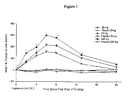
treatment-emergent adverse events in both studies were mostly mild. No serious
adverse events occurred.
Isofagomine tartrate showed good systemic exposure via the oral route. In the
single-dose study, plasma AUC and Cmax values were linearly correlated with
administered dose. Mean plasma levels peaked at 3.4 hr. (SEM: 0.6 hr.) and the
plasma elimination half-life was 14 hr. (SEM: 2 hr.). In the multiple-dose
study, after
7 days of oral administration, the pharmacokinetic behavior was found to be
linear
with dose, with no unexpected accumulation of isofagomine tartrate. Tmax and
half-
life values were similar to those observed in the single-dose study.
In the multiple-dose study, GCase activity in isolated white blood cells was
measured at days 1, 3, 5 and 7 during administration of isofagomine tartrate,
and at
days 9, 14 and 21 during the post-treatment washout period. In all subjects
receiving
isofagomine tartrate there was a marked increase in GCase levels during the
drug
treatment period, followed by a decrease upon removal of the drug and a return
to
near baseline levels by day 21 (Fig. 1). The increase in enzyme level was dose-
related, reaching approximately 3.5-fold above baseline levels. These results
for the
safety, pharmacokinetics and preliminary pharmacodynamic effects in healthy
volunteers support the further evaluation of isofagomine tartrate for the
treatment of
Gaucher disease.
EXAMPLE 2: Determination of Surrogate Markers of Gaucher Disease in
L444P Transgenic Mice Treated with Specific
Pharmacological Chaperones
L444P transgenic mice (homozygous for human L444P mutated Gba on a
glucosylceramide synthase null background) exhibit multi-system inflammation;
B
cell hyperproliferation; deficiency in GCase activity in the brain, liver,
spleen, and
lung; increased liver and spleen weights; elevated plasma levels of
chitotriosidase at 3
months; and elevated plasma levels of IgG (Mizukami et al., J. Clin. Inves.
2002; 109:
1215-21). However, due to the disruption in the glucosylceramide synthase
gene,
these mice do not exhibit accumulation of GluCer in e.g., macrophages.
Concomitant
glucosylceramide synthase disruption is necessary since previously made L444P
transgenic mice died within 3 days of birth due to impaired permeability
barrier
function in the epidermis.
26
CA 02683713 2009-10-13
WO 2008/128106 PCT/US2008/060116
In this experiment, the L444P transgenic mice were treated with isofagomine
or C-benzyl-isofagomine and surrogate markers were measured at 1, 3, 6 and 12
months to determine efficacy of the chaperones. In addition, mice in a
"washout"
period of 2 weeks of non-chaperone treatment following 4 weeks of treatment
were
also evaluated for reversion of surrogate markers back to levels seen in
untreated
controls.
Methods
Isofagomine treatment. Mice were administered isofagomine tartrate in their
drinking water, ad libitum, at a concentration of 20 mg/kg.
Surrogate marker measurement. At the end of 4, 12, or 24 weeks, mice were
sacrificed and evaluated for (i) enhancement of GCase enzyme activity in
liver,
spleen, lung and brain; (ii) chitotriosidase activity; (ii) body, spleen, and
liver weight;
and (iv) serum IgG, cholesterol, and liver enzyme levels. In addition,
chaperone
concentration in plasma and in the foregoing tissues will also be determined.
a. GCase activity assays in tissue: Liver, brain, spleen, and lung tissue
is freshly harvested (blood washed away with PBS), or thawed from frozen
stock.
Tissue is minced tissue and homogenized on ice in 200-500 l Mcllvaine (MI)
buffer
(0.25% sodium taurocholate, 0.1% Triton x-100 in 0.1M citrate and 0.2M
phosphate
buffer, pH 5.2), and centrifuged at 10,000 x g. The supernatant is collected
and may
be frozen at this step.
About 1-10 l of supernatant from the tissue homogenates is added to a clear
96-well plate for the Micro BCA Protein Assay (Pierce, cat# 23235) to
quantitate the
amount of total protein according to the manufacturer's protocol. As a
negative
control, another 10 l is added to a black plate, mixed with 10 1 of 2.5 mM
CBE
(2.7mg Conduritol B Epoxide in 6.7 ml buffer), an inhibitor of GCase activity,
and
left at room temperature (RT) for 30 minutes. 50 l of 3 mM 4-methal
Umbelliferyl
beta-D-glucoside (4-MU-beta-D-glucoside; made fresh, powder is dissolved in
0.2 ml
of DMSO, then q.s. to proper volume with MI buffer), a GCase substrate, is
then
added, and the black plate is further incubated at 37 C for 1 hr. After
incubation, 10
1 of supematant is added to a second black plate, mixed with 10 l of MI
buffer and
50 l 6 mM of GCase substrate 4-MU-beta-D-glucoside, and incubated at 37 C for
1
27
CA 02683713 2009-10-13
WO 2008/128106 PCT/US2008/060116
hr. The reaction is then stopped by adding 70 l 0.2 M glycine, pH 10.8. The
plate is
read in a plate-reader (Victor2 1420 multilabel counter; Wallac) at F460.
Relative beta-glucose activity is determined by the following equation:
F460 without CBE - F460 with CBE) /(Ass0 sample - A550 buffer)
F460 reading is converted into nmole 4-MU based on 4-MU standard curve and
A550 is converted into mg of protein based on the protein standard curve. One
unit of
GCase activity is defined as nmole of 4-MU released in one hour.
b. Body and tissue weight measurements: Animals were weighed prior
to sacrifice after 4, 3, 6 and 12 months. Following sacrifice, spleen and
liver were
removed and weighed.
c. Chitotriosidase activity: Plasma is collected for the assay in 5 l
aliquots (in duplicate), and the remaining is stored at -80 C. 5 l of
plasma/EDTA is
mixed with 100 122 M 4-MU-b-D-N,N'N"-triacetylchitotriose in citrate
phosphate
buffer (0.1M citrate and 0.2M phosphate buffer, pH5.2; made by mixing 185 ml
0.1
M citric acid and 200 ml 0.2 M sodium phosphate) in a 96 well black plate. 5
l of
EDTA/PBS (no plasma) is used as a negative control. A standard curve with
standard
serum is prepared by serial dilution in one row of the plate. The plate is
then
incubated for 15 minutes at 37 C (floating in a hot water bath), and the
reaction
stopped by adding 150 l 1M glycine, pH 10.8. The plate is read at F355/F460
in a
Victor2 1420 multilabel counter (Wallac).
d. IgG measurement: The mouse IgG ELISA quantitation kit (Bethyl
Laboratories, Cat # E90-131) was used for determination of IgG concentration
in
plasma. 96-well plates were coated with 100 l of the coating buffer (made by
dissolving 1 capsule of coating antigen in 100 ml of double deionized water)
and
incubated for 1 hr at room temperature. The wells were then washed 3 times
with 150
l of wash buffer (50 mM Tris HCl (pH 8.0); 0.14 M NaCI; 0.05% Tween 20)
followed by aspiration after each wash). Following washing, 200 l of blocking
solution was added (50 mM Tris HCl (pH 8.0); 0.14 M NaCI; 1% BSA), and the
plates were incubated either at RT for 1 hour or at 4 C overnight. Following
incubation, the wells were washed 3x again with wash buffer, and 95 l of
sample
diluent buffer (50 mM Tris HCI, pH 8.0; 0.14 M NaCI; 0.05% Tween 20; 1% BSA).
and 5 l of test plasma were added to the wells and incubated for an hour at
RT.
28
CA 02683713 2009-10-13
WO 2008/128106 PCT/US2008/060116
As a standard, 100 gl of the serial diluted standard mouse IgG antibody of
known concentration was added to one row of wells (diluted in diluent buffer
at
concentrations of 5000 ng/ml to 7.8 ng/ml).
Following incubation, wells were washed 5 times with wash buffer to remove
the unbound sample 100 gl of secondary antibody (1: 20000 in diluent buffer)
was
added, followed by incubation again for 1 hour at RT. Following washing (5x)
to
remove the unbound sample, 100 l of developer (equal proportions of reagent A
and
B) were added to each well and incubated for 20 minutes at RT. The reaction
was
stopped by adding 100 l of 1 M phosphoric acid, and the color intensity was
measured at 450 nm in the plate reader.
e. Cholesterol and liver enzyme measurement. These were measured
according to ordinary techniques.
Washout study. To determine if and in what time frame the effects of
drinking water dosed isofagomine on L444P mice regress after cessation of the
treatment, a washout study was performed. Nine male 3 month old L444P mice
were
dosed at about 10 mg/kg/day for 4 weeks with an equal number of mice untreated
as a
control. Four treated and four untreated mice were sacrificed at the end of 4
weeks,
and the remaining animals were not further treated with isofagomine, i.e.,
they were
given normal drinking water, for another two weeks prior to sacrifice and
evaluation
of the above-described surrogate markers.
Results
GCase Activity in Tissue. Significant increase in GCase activity was observed
after as little as two weeks of treatment with isofagomine in liver, spleen
lung and
brain (Fig. 2A-D), which persisted through 4-12 weeks. Notably, in brain,
isofagomine treatment resulted in an increase from about 1 U/mg in untreated
mice, to
about 4.5 U/mg after 2 and 4 weeks of treatment, and further increased to
about 6
U/mg after 12 weeks (p < 0.001) (Fig. 2B). It is expected that increased GCase
activity will persist at 3, 6 and 12 months and for as long as the chaperones
are
administered.
Similarly, after two weeks, the C-benzyl-isofagomine-treated mice also
exhibited significant increased GCase activity in the spleen, and a trend
toward
29
CA 02683713 2009-10-13
WO 2008/128106 PCT/US2008/060116
increased activity in the lung and brain (data not shown). It is expected that
increases
in GCase activity will be observed in other organs upon further treatment.
Body and Tissue Weight. After 12 weeks of isofagomine treatment, treated
mice exhibited a body weight of about 33 g, intermediate between wild-type
mice
(about 40 g) and untreated mice (about 29 g) (data not shown). By contrast,
spleen
weight (Fig. 3A) was significantly decreased by 12 weeks of treatment in
treated mice
(0.09 mg) compared with untreated mice (0.11 mg). Wild-type spleens were about
.08 mg. This persisted (reaching significance) after 12 weeks of treatment,
where
spleen weight was 0.12 mg in the treated mice compared with 0.15 mg in
untreated
mice and 0.10 mg in normal mice. Normalization of spleen weight is expected to
continue for the duration of treatment.
Liver weight did not significantly change among treated, untreated and control
wild-type mice after 12 weeks. (Fig. 3B), but achieved a significant reduction
after 24
weeks (data not shown).
Chitotriosidase Activity. Although no difference in chitotriosidase levels
were
observed after 4 weeks of isofagomine treatment, levels were decreased after
12
weeks (about 17,000 g of protein) compared with levels seen in untreated mice
(>20,000 F460 g of protein) (p = 0.1) (Fig. 4). However, levels were still
elevated
compared to wild-type mice (which had about 7500 g of protein). Again,
continued
decrease in chitotriosidase levels is expected with continued treatment.
IgG, cholesterol, and liver enzymes. There were no significant differences in
cholesterol, or liver aminotransferases (aspartate aminotransferase (AST) or
alanine
aminotransferase (ALT)) in isofagomine-treated versus untreated mice at 4 or
12
weeks (Fig. 5B-D, respectively)). By contrast, serum IgG levels showed
significant
decrease by 2 weeks of treatment, which persisted to 4 and 12 weeks of
treatment,
compared with untreated mice (Fig. 5A). This is significant improvement over
treatment with recombinant GCase during enzyme replacement therapy. According
to
the manufacturer, 15% of patients develop IgG against the recombinant enzyme,
46%
of whom also develop hypersensitivity reactions as a result.
Washout. Similar to above, after 4 weeks of treatment at 10
mg/kg/day, GCase activity was significantly elevated in liver, spleen, lung
and brain
in the L444P transgenic mice. Similarly, IgG was significantly decreased.
CA 02683713 2009-10-13
WO 2008/128106 PCT/US2008/060116
EXAMPLE 3: IFG Increases Levels of Glucocerebrosidase, Inflammatory
Cytokines, and Bone Metabolism in Gaucher Patient-
Derived Cells
To evaluate the effects of IFG on mutant GCase levels, an ex vivo response
study with macrophages and EBV-transformed lymphoblasts derived from
peripheral
leukocytes of 60 patients was conducted. Plasma was also screened for
potential
biomarkers associated with inflammation, bone metabolism, and multiple
myeloma.
The study was conducted at eight sites in the United States.
Results
The study included 21 males with type I Gaucher disease, 1 male with type III
Gaucher disease, and 18 females with type I Gaucher disease. Patients ranged
in age
from 7 to 83 years, and 38 of 40 patients were on enzyme replacement therapy
(ERT).
Macrophages were successfully derived from 34 of 40 patients, of which 32
demonstrated a dose-dependent increase in GCase levels (average = 2.8-fold)
when
treated with IFG tartrate (5 days). Similar results were observed for 5
additional
patient-derived lymphoblast cell lines. IFG significantly increased GCase
levels in
cells from patients with different genotypes including N370S/N370S (11),
N370S/L444P (8), N370S/84insG (11), N370S/R163X, N370S/Y212H, L444P/del
136T, L444P/F216Y, L444P/L174F, G202R/R463C, and K79N/complex B exon 9/10
(type III GD). Maximum enhancement of GCase in macrophages was achieved at
about 30 M of IFG.
TRACP 5b, a marker of osteoclast activity, was elevated in the plasma of
Gaucher subjects, especially in males, even those patients who were on ERT.
Concurrently, the activity of bone-specific alkaline phosphatase (BAP) was
reduced
in the plasma of Gaucher subjects (especially females). This suggests that
bone
resorption may be favored over bone deposition in Gaucher patients, possibly
contributing to the bone disease.
Other proinflammatory cytokines and chemokines also were elevated in some
Gaucher subjects. These included IL-8, IL-17, VEGF, PARC, and MIP-la. This
combination of cytokines also has been associated with bone resorption and
multiple
myeloma (especially IL-17, VEGF, and MIP-la). This is interesting in view of
the
fact that Gaucher patients, untreated or treated with ERT, have an elevated
risk for
31
CA 02683713 2009-10-13
WO 2008/128106 PCT/US2008/060116
developing multiple myeloma. Plasma levels of anti-inflammatory cytokines (IL-
lra,
IL-2, IL-4, IL-5 IL- 10, IL- 13) were not elevated in Gaucher subjects.
EXAMPLE 4: Identification of a Surrogate Marker in the Plasma of
Gaucher
Patients
Recently, a link between mutations in lysosomal enzymes and neurological
disorders other than LSDs has been established. As one example, there is a
well-
established link between mutations in the Gba gene and parkinsonism and
Parkinson's
disease. In one study, a group of 17 patients with rare, early onset,
treatment-resistant
parkinsonism were found to have at least one allele with a Gba missense
mutation,
including homozygous and heterozygous individuals for N370S, a mutation
typically
associated with type 1, non-neuronopathic disease (Tayebi et al., Mol. Genet.
Metab.
2003; 79; 104-109). In another study, a population of 99 Ashkenazi Jews with
idiopathic Parkinson's disease were evaluated for six Gba mutations (N370S,
L444P,
84GG, V394L, and R496H). Thirty-one Parkinson's patients had one or two mutant
Gba alleles: 23 were heterozygous for N370S; 3 were homozygous for N370S; 4
were heterozygous for 84GG; and 1 was heterozygous for R496H (Aharon-Peretz et
al., New Eng. J. Med. 2004; 351: 1972-77). The frequency of a mutant N370S
allele
was 5 times that among 1573 normal subjects, and that of 84GG was 21 times
that of
normal subjects. Among patients with Parkinson's disease, patients carrying a
Gba
mutation also were younger than those who were not carriers. This study
suggests
that heterozygosity for a Gba mutation may predispose Ashkenazi Jews to
Parkinson's disease.
Parkinson's and Gaucher diseases also share some pathological features,
including neuronal loss, astrogliosis, and the presence of cytotoxic Lewy-body-
like a-
synuclein inclusions in hippocampal neurons (the CA2-4 region). A recent
publication described the extent of neurological pathology in all three forms
of
Gaucher disease (Wong et al., Mol. Genet. Metabol. 2004; 38: 192-207).
Abnormalities in cerebral cortical layers 3 and 5, hippocampal CA2-4, and
layer 4b
were found in Gaucher patients having all three types. Neuronal loss was
evident
only in patients with types 2 and 3, whereas type 1 patients presented with
astrogliosis
(Wong et al., supra). Two patients with type 1 Gaucher and
parkinsonism/dementia
exhibited a-synuclein positive inclusions in hippocampal CA2-4 neurons, one
patient
32
CA 02683713 2009-10-13
WO 2008/128106 PCT/US2008/060116
had brainstem-type and cortical-type Lewy bodies, and one had marked neuronal
loss
of substantia nigra neurons (Wong et al., supra). In summary, all 4 patients
with
parkinsonism and dementia had hippocampal CA2-4 gliosis, and neuronal
depletion,
gliosis, and brainstem-type Lewy bodies in the substantia nigra.
Plasma levels of a-synuclein, when measure by ELISA, are elevated in
Parkinson's patients compared to helathy controls (Lee et al. J Neural Transm.
2006;113(10):1435-9).(). To determine whether a-synuclein was elevated in
plasma
from patients with Gaucher disease, for use as a biomarker to monitor the
progress of
treatment with chaperone therapy, ERT, SRT or other treatments, plasma a-
synuclein
levels were measured in 40 patients with Gaucher disease and compared with
levels in
plasma from 12 healthy volunteers.
Methods
Patient samples. Patient plasma samples were obtained as described above in
Example 3.
ELISA. a-synuclein levels were determined. using a commercially available
ELISA kit (BioSource International, Camarillo, CA) according to the
manufactures
instructions. Briefly, the ELISA plate was coated by overnight incubation with
1
g/mL of nonbiotinylated mAb 211 (100 L/well; Santa Cruz Biotechnology, Santa
Cruz, CA), in 200 mM NaHCO3 (Sigma, St. Louis, MO, USA), pH 9.6, containing
0.02% (w/v) sodium azide at 4 C, washed 4 times with PBST (PBS containing
0.05%
Tween 20), and incubated with 200 L/well of blocking buffer (PBS containing
2.5%
gelatin and 0.05% Tween 20) for 2 h at 37 C. The plate was washed 4 times with
PBST, and 100 L of the samples to be tested were added to each well (neat).
The
plate was incubated at 37 C for 2 h. After washing 4 times with PBST, 100 L
of
biotinylated mAb 211 diluted to 1 g/mL in blocking buffer was added, and
incubated
at 37 C for 2 h. The wells were washed 4 times with PBST and incubated with
100
L/well of ExtrAvidin-Alkaline phosphatase (Sigma) diluted 3:5000 in blocking
buffer and incubated for 1 h at 37 C. The wells were then washed 4 times with
PBST,
before adding the enzyme substrate Yellow "pNPP" (Sigma) (100 L/well) and
leaving the color to develop for 30 min at room temperature. Absorbance values
at
405 nm were determined and results were compared using an unpaired two tailed
t-
test.
33
CA 02683713 2009-10-13
WO 2008/128106 PCT/US2008/060116
Results
Compared to the healthy volunteer controls, a-synuclein levels were
significantly elevated in plasma from the Gaucher patients (p =.019; Fig. 6).
The
variances between the populations were not significantly different.
Accordingly, plasma a-synuclein can be used as a prognostic marker to
evaluate treatment for Gaucher disease.
EXAMPLE 5: Restoration of Disrupted Lysosomal Trafficking in Gaucher
Fibroblasts
Although N370S Gaucher fibroblasts (from a human patient) do not
demonstrate an accumulation of substrate (i.e. , GluCer) in the cytoplasm,
these
fibroblasts exhibit abnormal lysosomal protein and GCase staining compared
with
wild-type fibroblasts. Treatment of N370S fibroblasts with SPC isofagomine
increased the amount of GCase seen in the lysosome and restored a normal
lysosomal
staining pattern to the cells.
Methods
Cell culture. N370S fibroblasts (DMN89.15) were cultured in DMEM with
10% FBS and 1% penn/strep at 37C with 5% CO2. Wild-type fibroblast cell line
CRL-2097 form a healthy individual was used as a control. Cells were sub-
cultured
from 10 cm plates into 12-well plates with cover slips. Cells from one
confluent 10
cm plate were diluted in 38 ml of culture medium. Isofagomine or C-benzyl-
isofagomine was added from a 10 mM stock solution (5% DMSO) to each well of a
12-well plate at the following concentrations:
C-benzyl-isofagomine-controi (secondary antibody only); untreated; 0.03
M; 0.1 M; 0.3 M; 1.0 M; 3.0 M; and 10.0 M.
Isofagomine-control (secondary antibody only); untreated; 10 M; 30 M;
100 M; 1 nM; 3 nM; and 10 nM.
Cells were cultured for a total of about 6 days.
Fixing and Staining. Cells were washed for 5 minutes in PBS, fixed for 15
minutes in 3.7% paraformaldahyde (in PBS), washed again for 5 minutes in PBS,
and
penneabilized with 0.5% saponin for 5 minutes. Cells were then washed with PBS
containing 0.1% saponin, treated for 5 minutes with fresh 0.1% sodium
34
CA 02683713 2009-10-13
WO 2008/128106 PCT/US2008/060116
borohydride/0.01% saponin, and washed 3 times with PBS with 0.1% saponin/ 1%
BSA for 5 minutes each.
Cells were incubated for 1 h with 500 l of primary anti-GCase (1:200) or
anti-LAMP-1 (1:200; BD Pharmingen, Cat. No. 555798) antibody solution in PBS
with 1% BSA. Lysosomal staining using LysoTracker Red (Cambrex, East
Rutherford, NJ) was performed according to the manufacturer's instructions.
Following incubation, cells were washed 3 times in 1% BSA containing 0.1 %
saponin
in PBS, followed by incubation with the secondary antibody solution (1:500;
anti-
rabbit AlexaFluor588 for anti-GCase and anti-mouse IgG AlexaFluor594 for anti-
LAMP-1). Cells were mounted onto coverslips, sealed, and immediately viewed.
Confocal Microscopy. Cells were visualized using a confocal microscope.
The red and green channel gains were set to 6 and the laser power was
optimized
using the intensity window, and were not adjusted for the rest of the
experiment. All
slides were analyzed at the same sitting and all images were gathered without
any
zoom using the 20x and 60x lens, the small pinhole, optimal pixel size, an
average of
2 scans, and red and green channels were acquired simultaneously as in all
previous
experiments.
All images were displayed at the same intensity and red + green channel
intensity graphs were generated for each image by placing the cursor over the
maximum number of cells.
Future measurements can be made by calculating a ratio for overlapping red
(LAMP-1) and green (GCASE) pixels.
Results
Gaucher N370S fibroblasts that have been confluent for more than 5 days
exhibit a granular lysosomal staining pattern using LysoTracker Red (Fig. 7A)
compared with a normal fibroblast, which has a punctuate staining pattern
(Fig. 7B).
Similar results were shown for L444P fibroblasts (data not shown). Staining
for
lysosomal LAMP-1 is shown in both N370S and normal fibroblasts (Figs. 7C-D,
respectively). More LAMP-1 is shown in Gaucher fibroblasts.
Treatment with 30.0 M isofagomine (Fig. 7G-H) and 3.0 l C-benzyl-
isofagomine (Fig. 7I-J) increased the amount of GCase in the lysosomes and re-
established a normal lysosome punctuate staining pattern for GCase and LAMP-1
CA 02683713 2009-10-13
WO 2008/128106 PCT/US2008/060116
compared with an untreated control (Fig. 7E-F), as indicated by dual staining.
Figures 7K-N shows changes in GCase lysosomal staining in N370S Gaucher
fibroblasts as follows: (K)-control (secondary antibody only); (L)-untreated
N370S
fibroblasts; (M)- 30 M isofagomine; and (N) 3 M C-benzyl-isofagomine. GCase
staining is shown to localize to lysosomes in chaperone-treated versus
untreated
controls. Similar results were obtained for L444P Gaucher fibroblasts (data
not
shown).
These results establish that treatment with pharmacological chaperones
increases GCase levels in the lysosome. In addition to increased trafficking
of the
lysosome, pulse-chase experiments demonstrated that IFG increases N370S GCase
in
the ER (data not shown).
This improvement in normal cell morphology with chaperone treatment is due
to a decrease in the amount or accumulation of mutant GCase, possibly in the
form of
aggregates, in the ER and/or cytosol. Since it has been demonstrated that the
SPCs
evaluated cross the blood-brain barrier, this strategy could relieve CNS
symptoms
Gaucher patients with neuronopathic Gaucher disease (Types 2 and 3).
EXAMPLE 6: Increase of Polyubiquinated Proteins with Chaperone
Treatment in Gaucher Fibroblasts; Restoration of the
Proteasome Degradation Pathway
Anti-polyubiquitinated protein (PUP) and anti-GCase labeling of healthy
human fibroblast was compared with that in fibroblasts from a Gaucher patient
having
the L444P Gba mutation, and Gaucher patient fibroblasts having the N370S Gba
mutation.
Methods
Cell culture. L444P Gaucher fibroblasts (cell line GM10915); N370S
Gaucher fibroblasts (cell line DMN89.15); and fibroblasts from a healthy
individual
(CRL-2097) were cultured in DMEM with 10% FBS and 1%PS at 37C with 5% C02.
Cells were sub-cultured from 10 cm plates into 12-well plates with sterile
cover slips.
N370S cells from one confluent T-75 flask were diluted 1:6 and cultured for
another 4
days.
36
CA 02683713 2009-10-13
WO 2008/128106 PCT/US2008/060116
Chaperones isofagomine or C-benzyl-isofagomine was added from a 10 mM
stock solution (5% DMSO) to each row of a 12-well plate at the following
concentrations:
C-benzyl-isofagomine: untreated; control (secondary antibody only); 0.03
M; 0.1 M; 0.3 M; 1.0 M; 3.0 M; and 10.0 M.
Isofagomine: untreated; control (secondary antibody only); 10 .M; 30 M;
100 M; 1 nM; 3 nM; and 10 nM.
Fixing and staining. Cells were washed once in PBS for 5 minutes, followed
by fixation for 15 minutes in fresh 3.7% paraformaldehyde. Cells were then
washed
once in PBS for 5 minutes, followed by permeabilization for 5 minutes in 0.2%
Triton
X-100. Cells were then washed again in PBS for 5 minutes and treated for 5-10
minutes with fresh 0.1% sodium borohydride. Cells were washed three times in
PBS
with 1% BSA (5 min each) prior to staining.
Cells were next incubated for 1 hour with 500 l of the following primary
antibodies (diluted 1:200 in PBS with 1% BSA):
1. Mouse monoclonal antibody to ubiquitinated proteins clone FK1
(AFFINITI Research Products Cat. No. PW 8805)
2. Rabbit anti-GCase antibody
Cells were then washed three times with PBS with 1% BSA, followed by
incubation
for 1 hour with a 1:500 dilution of the following secondary antibodies:
1. Goat Anti-Mouse IgM ( chain) AlexaFluor568 (Molecular Probes Cat.
No. A21043);
2. Goat Anti-Rabbit IgG (H+L) highly cross absorbed AlexaFluor488
(Molecular Probes Cat. No. A11034)
Cells were washed three times in PBS with BSA, mounted, and stored at 4 C
prior to
visualization.
Results
Initial experiments indicated that the concentration of polyubiquitinated
proteins (PUP) in cells is greater (very intense) in healthy cells than in
Gaucher
N370S and L444P fibroblasts (much less intense). In addition, treatment of
Gaucher
fibroblasts with specific chemical chaperones increases the PUP staining in
the
Gaucher cells. As stated above, this is likely because protein aggregation is
known to
37
CA 02683713 2009-10-13
WO 2008/128106 PCT/US2008/060116
inhibit the ubiquitin/proteasome pathway. Accordingly, decreasing aggregation
using
chaperones may re-start the proteasome-mediated degradation pathway.
EXAMPLE 7: Analysis of Surrogate Markers in Human Gaucher Patients
and Human Controls
This study included 26 males with type I GD, 6 males with type III GD, 26
females with type I GD and 5 females with type III GD representing 19
different
genotypes. Patients ranged in age from 4 to 83 years; 59 of 63 patients were
receiving
enzyme replacement therapy and blood was drawn immediately prior to enzyme
infusion. Analysis of untreated WBCs yielded reduced GCase activity compared
to controls, normal G1cCer levels (most patients were receiving ERT), and
elevated
chitotriosidase activity (Fig. 8). Since multiple studies have identified
mutations in
Gba, the gene that encodes for GCase, as a potential risk factor for
synucleinopathies,
we screened for plasma levels of a-synuclein. Surprisingly, GD patients showed
elevated levels of total a-synuclein compared to controls (Fig. 8).
Interestingly, we
have also found that a-synuclein accumulation correlates with the accumulation
of
glucosylceramide of mouse models with significantly reduced GCase activity.
Markers of osteoclast (TRACP 5b) and osteoblast (BAP) activities were abnormal
for
most patients. In general, TRACP 5b activity was elevated in many patients
while
BAP levels were lower than normal (Fig. 9). These results suggest that bone
metabolism is altered in most patients, favoring osteoclast activity and bone
resorption. Interestingly, proinflammatory cytokines and chemokines PARC
(CCL18), IL-8, IL-17, VEGF and MIP-la were elevated in some patients
compared to controls, and a significant correlation (p < 0.0001) was observed
for IL-
17 and VEGF levels (Fig. 10). IL-17 is produced exclusively by CD4+ memory T-
Cells and can induce the production of VEGF by other cells. These cytokines
can
promote osteoclastogenesis and osteoclast survival and have also been
implicated in
the pathogenesis of multiple myeloma, which may be relevant to GD since it
has been reported that Gaucher patients have an increased risk for developing
multiple
myeloma.
We also screened patients for DKKI and found them to be lower in most
Gaucher patients relative to age-matched controls (data no shown). Plasma
levels of
other proinflammatory (IL-1 [a, 0], IL-6, IL-7, IL-12p40, IL-12p70, IL-15, IL-
17,
fractalkine, EGF, TGFa, sCD40L, GM-CSF, eotaxin, sCD14, IP-10, IFN-v, G-CSF,
38
CA 02683713 2009-10-13
WO 2008/128106 PCT/US2008/060116
MIP-10, TNFa, HSP60, HSP70), anti-inflammatory (IL-lra, IL-2, IL-4, IL-5, IL-
10,
IL-13) and cardiovascular (CRP, SAA, SAP) markers were unremarkable for most
GD patients when compared to controls (data not shown).
39
CA 02683713 2009-10-13
WO 2008/128106 PCT/US2008/060116
* * ~
The present invention is not to be limited in scope by the specific
embodiments described herein. Indeed, various modifications of the invention
in
addition to those described herein will become apparent to those skilled in
the art
from the foregoing description and the accompanying figures. Such
modifications are
intended to fall within the scope of the appended claims.
It is further to be understood that all values are approximate, and are
provided
for description.
Patents, patent applications, publications, product descriptions, and
protocols
are cited throughout this application, the disclosures of which are
incorporated herein
by reference in their entireties for all purposes.