Note : Les descriptions sont présentées dans la langue officielle dans laquelle elles ont été soumises.
CA 02687409 2009-11-16
WO 2008/139137 PCT/GB2008/001480
Testing Vision
The present invention relates to a system and method for testing vision, for
example
visual fields. The invention is particularly suited to testing visual fields
in children.
Background of the Invention
The detection of visual field defects is crucial in the management of children
with cerebral
visual impairment, cerebral tumour and raised intracranial pressure. There is
also call for
a reliable and sensitive method to monitor visual field changes in children
who are taking
Vigabatrin for epilepsy. Unfortunately, most existing methods of perimetry are
not
suitable for children. Children have different needs and requirements to
adults when it
comes to perimetry, because of their reduced ability to learn tasks and to
provide
appropriate responses during conventional testing. Children are also less
inclined to
cooperate due to a lack of understanding of the test methods and their short
attention
span.
In adults, the reliability of most standard visual field testing methods is
dependent upon
the ability of the subject to learn the task asked of them during the test.
The two main
tasks are to fixate on a central target and to indicate by, for example,
pressing a button
when they see a light stimulus in their field of vision. In order to keep a
continuous
fixation on a central target when a stimulus is presented in their visual
field, the subject
needs conscious effort to inhibit their natural response to fixate on the
stimulus, thereby
preventing the loss of fixation on the central target. It is much more
difficult for children,
especially children below the age of five to inhibit the natural saccadic
reaction that is
triggered by light stimuli in the visual field.
Recognising such difficulties, various groups have looked for better methods
of perimetry
in children. Methods of perimetry can be loosely divided into kinetic and
static perimetry.
Among the more popular methods used with children is kinetic and double-arc
perimetry,
as described by Quinn GE, Fea AM, Minguini N. "Visual fields in 4- to 10-year-
old
children using Goldmann and double-arc perimeters."
J.Pediatr.Ophthalmol.Strabismus
1991 Nov-Dec; 28(6): 314-319. Kinetic perimetry gives some control and freedom
to the
examiner, thus conferring the advantage of adaptability to the child's age and
maturity.
However, a disadvantage is the need for the child's cooperation in maintaining
a
continuous fixation on a central target during the test. Another disadvantage
is that
results of the test are dependent upon the examiner's skills and knowledge,
which means
that they cannot be used to provide quantitative data for serial comparison
studies.
1
CA 02687409 2009-11-16
WO 2008/139137 PCT/GB2008/001480
Automated static perimetry (ASP) using perimeters such as the Humphrey Visual
Field
Analyzer, on the other hand, allows for measurement of quantitative data that
can be
used for serial comparison. However, a continuous central target fixation is
still required
of the child.
Two groups have modified the ASP test using the Octopus 2000R perimeter in
attempts
to make it more suitable for children. As described in the article
"Feasibility of automated
visual field examination in children between five and eight years of age".
Br.J.Ophthalmol. 1996 Jun; 80(6): 515-518, Safran et al used a specially
designed
programme conducted for progressive familiarisation and a custom "two-level"
strategy.
The mean test duration was thirteen minutes per eye for five year-olds and
seven
minutes for eight year-olds. It was found that such strategy was suitable for
children age
eight and above. For children age seven and below, a preliminary
familiarisation phase is
mandatory. "Automated visual field examination in children aged five to eight
years. Part
I: Experimental validation of a testing procedure." Vision Res. 1998 Jul;
38(14): 2203-
2210 describes a meticulous four or five phase procedure with test trials to
be used with
customised software strategies and a perimeter using a "little bear" as the
central fixation
target. It was found that such a method is suitable for children age six and
above. Whilst
both of these methods have the advantage of catch trials for detection of
false-positive
and false-negative responses, the lengthy and meticulous training and
familiarisation
phases used may not be practical for usage in a busy clinical setting.
In recent years, new methods of perimetry that use a personal computer with
LCD
screens have been used in children: for example, Rarebit perimetry and High
Pass
Resolution (HPR) Perimetry. The Rarebit perimetry uses a dynamic and moving
fixation
target, which enhances fixation. Subjects were asked to respond by single or
double
mouse-clicks depending upon the number of perceived dots in the moving
fixation target.
This is a dynamic test as the pace can be adapted to the reaction time of the
subject,
thus allowing them to feel more in control of the whole process. The test
duration can be
varied according to the type of information needed and ranges from less than 1
minute to
5 minutes. The Rarebit perimetry is popular amongst the children as it mimics
a
computer game. Another advantage is that it does not require the use of a
headrest or a
bowl, which is appealing to children. One of the disadvantages of Rarebit
perimetry is the
high false positive response rate, which reduces its reliability. Moreover,
subjects need to
possess fairly sophisticated motor skills to respond appropriately.
2
CA 02687409 2009-11-16
WO 2008/139137 PCT/GB2008/001480
High Pass Resolution (HPR) perimetry uses a personal computer, a monitor and
ring-
shaped high pass spatial frequency filtered targets of different sizes. Like
Rarebit
perimetry, this has been popular as it appears like a pleasant game to the
children. The
test duration is five minutes and was found to be suitable for children age
five and above.
HPR perimetry, like Rarebit perimetry, does not use a headrest and adapts the
pace of
the test to the current reaction time of the subject. However, a disadvantage
of HPR is
the need for a continuous fixation on a central target during the test.
All the methods of perimetry described above depend upon subjective patient
responses.
An objective method of measuring visual field is the multifocal-Visual Evoked
Potential
(m-VEP). This involves placing electrodes on the scalp and recording
electrical activity
from the occipital cortex following presentation of light or pattern stimulus
to the retina.
This needs minimum cooperation from the subject, and thus is highly suitable
for use
with children. The test duration for one eye is four minutes for m-VEP.
However, M-VEP
has inherent disadvantages. There is inter-individual anatomical variation of
occipital
cortex folding making cortical mapping of visual field responses almost
impossible to
calibrate. The subject also needs to maintain a continuous fixation on a
central target
during the test. More importantly for children, the subjects' movements are
restrained
during the test with bipolar occipital inion straddle electrodes. Not all
children can tolerate
such devices.
ASP is the visual field assessment method of choice in adults. However, it is
rarely
reliable in children under nine years of age. Research efforts to perform ASP
in children
have concentrated on studying the feasibility of using current adult perimetry
techniques
with children. Following the development of algorithms designed to provide
faster testing
time such as SITA Fast and TOP, investigations into using these techniques
with children
were made. However, the youngest age able to produce reliable results is still
only in the
region of seven to eight years. Training and familiarisation strategies for
particular
techniques have been looked at as a route to improving reliability, which has
shown
some success. However, this still does not address the fundamental problems
inherent
in performing ASP on children.
US 5,459,536 describes an automated perimetry system, in which a patient's
visual field
can be tested. This requires that the patient's head be restrained in a very
restricted
position. US 4,059,348 describes another visual field testing system, but
again the
patient's head has to be held in a fixed position. In both cases, the
requirement for the
3
CA 02687409 2016-07-27
patient's head movement to be severely restricted makes these systems
unsuitable for
use with young children.
Nunokawa et al have proposed a perimetry method that uses saccadic eye
movement as
an index, see "Development of an Objective Automatic Perimetry Using Saccadic
Eye
Movement", International Congress Series 1282 (2005) 585-589, and "The
Influence of
Fixation on New Visual Field Measurement Using Saccadic Eye Movement",
International Congress Series 1282 (2005) 674-687. This method involves
presenting a
visual stimulus to a user and tracking the movement of a patient's eye in
response to that
stimulus. The eye tracking is done using an Eyetracker Toolbox provided by
Cambridge
Research Systems. This has a video eye tracker module that is mounted on a
rigid
EyeLockTM headrest. In use, a patient has to place their head on the headrest
so that
the eye tracker can track eye movement. Whilst using the movement of a
patient's eye
to measure perimetry has significant advantages, the requirement for
restricted head
movement can be problematic when children or people with learning disabilities
are
involved.
GB 2,096,791 describes a system for determining visual field in which the
patient has to
wear a head-mounted helmet that has an integral screen. Various shapes are
shown at
different positions on the screen, which is a fixed distance from the
patient's eyes, so that
the patient's visual field can be tested. WO 99/22638 describes a system for
testing a
patient's visual field by tracking eye movements using goggles that are
connected to an
eye tracking system. Whilst in these systems, the patient is allowed some
degree of head
movement the requirement for a patient to wear some form of measurement
equipment
on their head can be problematic when children or people with learning
disabilities are
involved.
Summary of the Invention
According to one aspect of the present invention, there is provided a system
for testing
visual fields comprising:
a display for presenting a visual stimulus;
an eye tracker for tracking movement of at least one of a patient's eyes to
determine its position in three dimensions, wherein the patient is free of
tracking
equipment;
4
CA 02687409 2016-07-27
means for changing the position of the stimulus on the display by an amount
that
is a function of the tracked three dimensional position of the patients eye
and a visual field
position that is to be tested, and
means for using the change to the position of the stimulus and any tracked eye
movement to assess the patients visual field.
4a
CA 02687409 2009-11-16
WO 2008/139137 PCT/GB2008/001480
Using an eye tracker arrangement that can track eye movement over a wide range
of
head positions without requiring a patient to wear eye-tracking equipment
allows a
patient freedom to move. At the same time, by tracking the 3D position of a
patient's
eye, test accuracy can be ensured. Hence, patient comfort and so compliance
with the
test can be significantly improved, whilst ensuring a high level of test
reliability. This is
essential for testing visual field for young children. Testing visual fields
is very useful as
it can provide an early indication of various conditions, for example
glaucoma.
The system may further comprise means for varying the size of the stimulus
presented,
as a function of three-dimensional position of the patient's eye and a visual
field position
that is to be tested, so that stimuli are presented at a constant angular
size.
The system may further comprise means for varying the shape of the stimulus
presented,
as a function of three-dimensional position of the patient's eye relative to
the display
screen and a visual field position that is to be tested, so that stimuli are
presented at a
constant angular shape.
Preferably, the eye tracker has a minimum head position tolerance of 10 cm x
10 cm x
10cm, for example 20cm x 10cm x 20cm (Horizontal x Vertical x Distance from
the
tracker).
Preferably, the eye tracker provides data for the calculation of the 3-
dimesional position
in space of each eye relative to the eye tracker.
Preferably, the eye tracker includes or consists of the Integration Eye
Tracking
Component provided by Tobii Technology.
The system may be adapted to present a first stimulus; capture gaze data for
that first
stimulus; present a second stimulus, and test whether the patient has seen
that second
stimulus.
The test of determining whether a patient has seen the second stimulus may
involve
comparing the direction in which the patient's eye(s) moved with the vector
direction
associated with a line between the first stimulus and the second stimulus. In
the event
that the direction is substantially the same or the same within pre-determined
limits, it is
assumed that the patient could be looking at the second stimulus. In this
case, the test
5
CA 02687409 2009-11-16
WO 2008/139137 PCT/GB2008/001480
may further involve comparing the angular distance between the first fixation
point and
the second fixation point and the angular distance between the first and
second stimuli.
In the event that these distances are the same or the same within pre-
determined limits,
it is assumed that the patient is looking at the second stimuli.
The system may be operable to measure at least one of visual acuity, colour
vision,
contrast sensitivity and colour-contrast sensitivity. To this end, means may
be provided
for varying the separation of two stimuli for testing visual acuity and/or the
colour of the
stimulus presented for testing colour and/or contrast of the stimulus for
testing contrast
sensitivity.
According to yet another aspect of the invention, there is provided a computer
program
or computer program product preferably on a data carrier or computer readable
medium
or a processor comprising code or instructions for presenting a visual
stimulus on a
display; receiving data from an eye tracker on the three dimensional position
of a
patient's eye(s); changing the position of the stimulus on the display by an
amount that is
a function of the tracked three dimensional position of the patient's eye and
a visual field
position that is to be tested, displayed at a size which is a function of the
tracked three
dimensional position and predetermined angular size, and using the change to
the
position of the stimulus and any tracked eye movement in response to the
change of the
position to assess the patient's visual field.
The program/product/processor may be adapted to relocate the position of the
stimulus
on the screen and use eye-tracking information to determine whether the
patient is
looking at the stimulus at its new position.
The program/product/processor may be adapted to present a first stimulus;
capture gaze
data for that first stimulus; present a second stimulus, and test whether the
patient has
seen that second stimulus.
Determining whether a patient has seen the second stimulus may involve using
eye
tracking information to compare the direction in which the patient's eye(s)
moved with the
vector direction associated with a line between the first stimulus and the
second stimulus.
In the event that the direction is substantially the same or the same within
pre-
determined limits, the test further involves comparing the distance between
the first
fixation point and the second fixation point and the distance between the
first and second
6
CA 02687409 2016-07-27
stimuli. In the event that these distances are the same or the same within pre-
determined limits, it is determined that the patient is looking at the second
stimuli.
The program/product/processor may be arranged to determine a time between
showing
a first stimulus, deleting that stimulus and displaying a second stimulus and
the time for a
patient to respond to the change in stimulus. This time information can then
be used to
identify, at least in part, whether the patient is responding to the stimulus.
According to another aspect of the invention, there is provided a system for
testing visual
fields comprising: a display for presenting a visual stimulus; an eye tracker
for tracking
movement of at least one of a patient's eyes to determine its position in
three dimensions
without requiring the patient to wear any tracking equipment; means for
changing the
position of the stimulus on the display; means for determining the visual
field position
associated with the new position of the stimulus using its position on the
display and the
three dimensional position of the patient's eye(s), and means for using the
change to the
position of the stimulus, the determined visual field position and any tracked
eye
movement in response to the change of the position to assess the patient's
visual field.
According to still another aspect of the present invention, there is provided
a system for
testing vision, the system comprising a display for presenting a visual
stimulus; means
for causing a change to the stimulus; an eye tracker for tracking movement of
a patient's
eye and means for monitoring the change to the stimulus and any corresponding
tracked
eye movement to assess the patient's vision, wherein the means for causing a
change to
the visual stimulus are operable to vary separately or simultaneously two or
more of the
position of the stimulus; the colour of the stimulus; the contrast of the
stimulus. In this
way, there is provided a simple and effective system for conducting multiple
different
types of eye tests.
Two or more stimuli may be presented and the means for causing a change may be
operable to vary the separation of the stimuli, thereby to allow visual acuity
to be
measured.
According to a further aspect of the present invention, there is provided a
method for testing
vision comprising:
presenting a visual stimulus on a screen;
tracking movement of at least one of a patient's eyes using an eye tracker to
determine a gaze point and its position in three dimensions, wherein the
patient is free of
tracking equipment;
7
CA 02687409 2016-07-27
changing the position of the stimulus on the display by an amount that is a
function
of the tracked three dimensional position of the patient's eye and a visual
field position that
is to be tested, and
using the change to the position of the stimulus and any tracked eye movement
in
response to the change of the position to assess the patient's visual field.
According to a further aspect of the present invention, there is provided a
non-transitory
computer readable medium having stored thereon instructions for execution by a
computer
to carry out the following steps: presenting a visual stimulus on a display;
receiving data
from an eye tracker on a three dimensional position of a patient's eye or
eyes; changing
the position of the stimulus on the display by an amount that is a function of
the tracked
three dimensional position of the patient's eye and a visual field position
that is to be tested;
and using the change to the position of the stimulus and any tracked eye
movement in
response to the change of the position to assess the patient's visual field.
According to a further aspect of the present invention, there is provided a
system for testing
visual fields comprising:
a display for presenting a visual stimulus;
an eye tracker for tracking movement of at least one of a patient's eyes to
determine its position in three dimensions, wherein the patient is free of
tracking
equipment;
means for changing the position of the stimulus on the display;
means for determining the visual field position associated with the new
position of
the stimulus using its position on the display and the three dimensional
position of the
patient's eye or eyes, and
means for using the change to the position of the stimulus, and any tracked
eye
movement in response to the change of the position to assess the patient's
visual field.
According to a further aspect of the present invention, there is provided a
system for testing
vision comprising a display for presenting a visual stimulus; means for
causing a change
to the stimulus; an eye tracker for tracking movement of a patient's eye and
means for
using the change to the stimulus and any corresponding tracked eye movement to
assess
the patient's vision, wherein the means for causing a change to the visual
stimulus are
operable to vary separately or simultaneously two or more of the position of
the stimulus;
the colour of the stimulus; the contrast of the stimulus.
7a
CA 02687409 2016-07-27
According to a further aspect of the present invention, there is provided a
non-transitory
computer readable medium having stored thereon instructions for execution by a
computer
to carry out the following steps: presenting a visual stimulus on a screen;
causing a change
to the stimulus; and using the change to the stimulus and any corresponding
tracked eye
movement to assess a patient's vision, wherein the code or instructions for
causing a
change to the visual stimulus are adapted to vary separately or simultaneously
two or more
of the position of the stimulus; the colour of the stimulus; the contrast of
the stimulus relative
either to a background or another stimulus.
Brief Description of the Drawings
Various aspects of the invention will now be described by way of example only
and with
reference to the accompanying drawings, of which:
Figure 1 is a schematic diagram of a system for measuring visual fields;
7b
CA 02687409 2009-11-16
WO 2008/139137 PCT/GB2008/001480
Figure 2 illustrates the effect of distance from a test screen on the location
of the
stimulus that is presented;
Figure 3 illustrates the effect of distance from a test screen on the size of
the
stimulus that is presented;
Figure 4 (a) is a plot of direction bias limits as a function of visual field
angle;
Figure 4 (b) is a plot of angular distance bias limits as a function of visual
field
angle;
Figure 5 is a flow diagram of a process for testing perimetry, and
Figure 6 is flow diagram of a sub-routine for a fixation change properties
test.
Specific Description of the Drawings
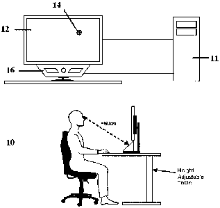
Figure 1 shows a system 10 for measuring peripheral vision. This has a
personal
computer that has a processor 11 with a screen 12 for presenting a visual
stimulus 14,
typically a circular stimulus, and an eye tracking system 16 for tracking
saccadic eye
movement when the stimulus 14 is presented in the periphery of a patient's
visual field.
Included in the processor is software for controlling the presentation of the
stimulus on
the screen and using the known position of this and information from the eye
tracking
module to determine whether a patient is looking at a stimulus or not. The
only task
required of the patient, typically a child, is to follow his natural reaction
to fixate on the
stimulus 14 of interest when he sees it. By monitoring saccadic eye movements,
the
system is able to detect changes in eye gaze position. The vector of the
saccadic eye
gaze movements indicates whether the patient perceives the stimulus 14 or not.
The
time interval between presentation of the stimulus 14 and the child's response
can also
be used to confirm that the eye saccadic movements are a reaction to the
peripheral
stimulus 14 presented.
Any suitable eye tracking system could be used, provided it is able to track
eye
movement over a wide range of head positions, ideally with a range of
20x10x20cm, and
able to provide data for calculation of the 3-dimensional position in space of
each eye
relative to the eye tracker, whilst not requiring the patient to wear any form
of head
mounted equipment or to have a fixed head position. In a preferred example,
the eye
tracking system provided by Tobii is used. This has a head position tolerance
of
30x15x20cm and is able to detect the distance of a child's eyes from the eye
tracker and
the position of the eyes in the camera field of view, thereby allowing for the
calculation of
the three dimensional position of the eyes from a current fixation point. This
is essential
for calculating where stimuli are to be displayed for assessing any particular
visual field
angle. The device does not need positional calibration, so the child is free
to move
8
CA 02687409 2009-11-16
WO 2008/139137 PCT/GB2008/001480
around within the field of detection. Also, it provides good gaze data
accuracy and the
ability to detect the very fast movements of the eye in real time and does not
require the
subject to wear any equipment on their head. This allows freedom of head
movements,
which is a clear benefit for children. Also, the eye tracker has no moving
parts and as
such is highly unobtrusive, which is of importance as children can be easily
distracted.
In order to allow the calculation of the appropriate screen position for any
particular visual
field point being assessed, the position of each eye in 3-dimensional space
relative to the
display screen must be known. The Tobii eye tracker provides real time data
giving the
distance of each eye from the eye tracker camera and the position of each eye
in the
camera field of view. This allows the real time calculation of the 3D position
of each eye
relative to any point on the display screen and so provides a way of
determining the
correct size and position of the stimuli to be presented on the screen at any
moment and
for any particular visual field point being assessed.
Figure 2 demonstrates how the screen position for a visual field point being
assessed is
dependant upon the 3D position of the subject's eyes relative to the display.
This shows
two examples (a) and (b) that allow testing of the patient's visual field at
angle 0. In each
case, 0 remains unchanged, but the position of the second stimulus (S2)
changes as a
function of the position of the patient's eyes relative to the screen. Hence,
to ensure that
there is an accurate measure of the patient's vision at a particular angle,
the 3D position
in space of each eye has to be carefully monitored, so that the stimulus can
be
positioned and correctly associated with the particular visual field point
being assessed.
As well as varying the position of the stimuli as a function of the position
of the patient's
eye, the actual size of the stimuli being displayed on the screen also has to
be varied
dependant upon the distance the subject's eyes are away from the screen. This
is
because the angular stimulus size must be kept constant for all visual field
points during
a test. Hence, the size is also varied depending on the location of the
patent's eyes, as
shown in Figure 3.
Optionally, the system may also be adapted to take into account the fact that
the stimuli
displayed to test visual field points may not be directly in front of the
patient and so to this
end, the system may vary the shape of the stimulus presented, as a function of
three-
dimensional position of the patient's eye relative to the display screen and
the visual field
position that is to be tested, so that stimuli are presented at a constant
angular shape.
9
CA 02687409 2009-11-16
WO 2008/139137 PCT/GB2008/001480
For example, where the test stimulus is circular and the display screen is
flat, in the event
that the stimulus is not directly in front of the patient, the shape presented
on screen may
in fact be elliptical, so that when viewed from an angle, is it perceived as
being circular.
The response of a subject's eye gaze is monitored when stimuli 14 are shown in
different
positions on the display screen 12 corresponding to different points in a
subject's visual
field. An algorithm is used to decide whether the subject was able to see the
new
stimulus based on the direction and length of movement of eye gaze and the
timing of
that movement. If a subject sees something in their peripheral vision the
natural
response is to gaze at it. If the point is not seen there could be no change
in eye gaze
position or a searching eye gaze movement. The algorithm automatically
distinguishes
these responses in real time based on the direction and angular length of any
fixation
change made immediately following the presentation of a new visual field point
stimulus,
and the timing of any such fixation change. The vector change in fixation
point is
compared directly with the vector change in the screen position of the stimuli
to decide if
the subject was able to see the stimuli or not.
In practice, there is a natural variation in the difference between the vector
change in
fixation point and the vector change in the screen position of the stimuli,
even when a
fixation change does relate to the stimulus displayed. Hence, it is important
to know how
much variation is acceptable before a stimulus is classed as being 'unseen'.
To deal with
this, parameters used within the algorithm designed to make this decision are
based on
data collected from many subjects and are dependant upon the size of visual
field angle
being assessed. Examples of the limits for the difference in direction and
angular length
of the two vectors being compared are shown in Figure 4.
Figure 5 shows the steps in a method for testing visual fields. In practice,
this method is
implemented using a computer program running on the processor/personal
computer.
Typically, each of the patient's eyes is measured separately and the results
averaged,
although this is not essential. At the start of the test, a stimulus 14 is
displayed in the
centre of the screen 12 to cause the patient to fixate on a central point.
When the
stimulus 14 is displayed the eye tracker 16 captures eye gaze data in order to
verify
whether the patient is indeed gazing at the stimulus 14. In the event that the
gaze data
suggests that the patient is not fixating on the correct point, further gaze
data is captured.
This is repeated until the system is confident that the patient is looking at
the central
stimulus 14. Animations to attract attention can be used if required.
CA 02687409 2009-11-16
WO 2008/139137 PCT/GB2008/001480
After the system determines that the patient is fixating on the first, central
stimulus, the
position and size of the next stimulus are calculated. The position of the
next stimulus is
calculated using the distance from the current fixation point (in this case
the central
stimulus) to the subject's eyes, as measured in real time using the eye
tracker, and the
points in the subject's visual field that are to be assessed. As illustrated
in Figure 2, the
on-screen position can vary depending on the measured distance of the patient
from the
fixation point. For a particular examination there are a number of different
visual field
points to be assessed. Generally, the order in which the selected points are
assessed is
random. Once the next stimulus position is determined, the current stimulus is
erased
and the next stimulus displayed in the new position. A timer is started when
the stimulus
is displayed. The timer runs in the background and acts as a countdown. If the
displayed
stimulus is not detected as 'Seen' within this time then the stimulus is
categorised as 'Not
Seen'. Typically, this time is set to a pre-determined limit, for example 1
second.
Next, gaze data is captured. For the Tobii tracker this is done every 20ms or
so. The
captured data is used to determine whether there has been a change in the
fixation point.
If there has, the following gaze data point is used to determine whether there
has been a
further change in the fixation point, this continues until no change is found.
In this manner
the movement of the user's eye towards an end, fixation point can be found. If
there is
no change in fixation with a gaze data point and the prior gaze data point did
indicate a
change in fixation then this indicates the end of the subject's fixation
change. Once a
change is detected the fixation change properties are tested to identify
whether the
current fixation change corresponds to the location of the new stimulus.
Figure 6 illustrates steps for testing the fixation change properties.
Firstly, the fixation
change properties are used to identify whether the direction of the change of
the fixation
is correct when compared with a vector from the first stimulus to the new
stimulus. If not,
then the fixation is determined not to relate to the new stimulus and the
system returns to
the capture gaze data step of Figure 5. If yes, then the fixation data and
three-
dimensional eye position data is used to determine the angular distance
between the first
fixation point and the second fixation point. If this distance differs from
the known
angular separation between the first stimulus and the second stimulus, then
the fixation
is determined not to relate to the new stimulus and the system returns to the
capture
gaze data step of Figure 5. In contrast, if the angular distance does
correspond to the
separation of the first stimulus and the second stimulus then it is concluded
that the
fixation does relate to the displayed stimulus.
11
CA 02687409 2009-11-16
WO 2008/139137 PCT/GB2008/001480
Returning to Figure 5, in the event that the next stimulus is identified as
having been
seen, this event is recorded. The stimulus timer is then stopped and reset. If
not all
points have been tested then the test is run again with a new visual field
point.
Otherwise, the test is completed.
Using data captured in accordance with the invention, it is possible to
perform ASP
without needing the patient to be in a fixed position.
For children, this is a very
significant technical advantage. The system could also be used to assess a
person's
reaction to peripheral stimuli within a recreated "real-world" environment
such as street or
home scene or driving situation. This can be helpful as their reactions can be
affected by
different disease states.
In the method described above the visual field position to be tested is
selected and the
position of the stimulus on the display calculated as a function of the
patient's three-
dimensional eye position. In an alternative embodiment, the stimulus may be
positioned
at one of a plurality of selected positions on the display and then the visual
field position
associated with this position calculated using the three dimensional position
of the
patient's eye(s).
The peripheral stimulus presented will generally be of the same type for all
subjects, for
example a circle or disc. To keep the test interesting for children, however,
the visual
field stimulus can be changed to interesting audio-visual animations
appropriate to their
developmental age once the original peripheral stimulus has been detected as
'seen'.
For instance, the human face is a potent visual fixation stimulus for an
infant and cartoon
figures can be used for young children. This will motivate the children to
submit to the
test and increase compliance rate without changing the properties of the
required visual
field stimulus. The test can also be interrupted anytime by sounds or video to
draw the
attention of a distracted child back to the screen.
The method in which the invention is embodied does not require any cooperation
or
understanding from a child. Also, the child is not required to learn any task
or to give any
subjective response. This removes the need for vigilance, and endurance or
understanding of the test method on the part of the child. Moreover, the
absence of
subjective responses from the child also eliminates false-positive and false-
negative
errors, which increases the reliability of the test. Also, the child's head
movement is not
restricted in any way and the system does not require any form of physical
contact with
12
CA 02687409 2009-11-16
WO 2008/139137 PCT/GB2008/001480
the child, which makes it more comfortable. A further advantage is that a
continuous
fixation on a stationary central fixation target is not needed.
As well as measuring visual fields, the system of Figure 1 can be used for
numerous
other eye tests, for, example visual acuity, colour vision, contrast
sensitivity and colour-
contrast sensitivity, in both children and adults. Visual acuity is the
ability to discriminate
two stimuli separated in space whilst colour vision is the ability to
differentiate two
different colours. Contrast sensitivity is the ability to discriminate the
difference between
shades of grey or hues of colour.
Conventional methods used for children depend upon a skilled orthoptist
observing the
child's direction of gaze while presenting the child with picture cards. The
technique used
is called 'preferential looking'. The picture cards are designed so that a
picture is located
either in the top half or bottom half of the card. The picture design is that
of the vanishing
optotype because the pictures seem to disappear from the perspective of the
child, when
at their resolution limit. If the child's visual acuity is good enough the
picture is visible and
they will orientate their gaze in the direction of the picture, otherwise the
picture is
merged with the background and seems invisible to the child, in this situation
the picture
cannot be seen and so the child cannot look at the picture. A problem with
conventional
techniques is that they rely on a skilled orthoptist observing the child's
direction of gaze.
This can make it difficult to accurately detect the change of direction.
Eye tracking has the potential to more accurately and reliably detect change
of direction
of gaze towards a test target. Using the arrangement of Figure 1, this could
be done by
presenting vanishing optotype targets and using the eye tracker to detect if
the subject
looks at a picture. Alternatively stationary pictures with varying levels of
visual acuity and
contrast sensitivity could be presented on the screen. Likewise, different
colour images
could be presented, so as to test colour vision deficiency. In any case, the
pictures are
presented somewhere away from the subject's gaze point and eye movement is
tracked.
If the subject looks at the picture then it can be concluded that they were
able to see it,
and if they did not look at the picture then they were not able to see it. The
test proceeds
based on the subject's responses, i.e. if a picture is seen then the next
picture that is
shown is at a more difficult level of acuity or contrast sensitivity or of a
different colour
vision deficiency test. This is repeated until the subject's visual acuity or
contrast
sensitivity level is reached, or specific colour vision deficiency tests have
been carried
out. In this case, the software for controlling the test is similar to that
used for visual field
assessment, although it is simpler, as it is only required to recognise that
the subject
13
CA 02687409 2009-11-16
WO 2008/139137 PCT/GB2008/001480
looks at the picture rather than assessing fixation change properties from one
point to
another.
A skilled person will appreciate that variations of the disclosed arrangements
are
possible without departing from the invention. For example, although the
invention has
been described for use in testing visual fields in children, it will be
appreciated that it
could also be used for those with learning difficulties and the elderly. In
addition,
although the invention is described primarily with reference to a conventional
flat screen
monitor, other screens, for example curved screens could be used. This could
extend
the vision test range. Accordingly the above description of the specific
embodiment is
made by way of example only and not for the purposes of limitation. It will be
clear to the
skilled person that minor modifications may be made without significant
changes to the
operation described.
14