Note : Les descriptions sont présentées dans la langue officielle dans laquelle elles ont été soumises.
CA 02689675 2009-08-06
WO 2008/095307 PCT/CA2008/000248
1
BIOCERAMIC IMPLANTS HAVING BIOACTIVE SUBSTANCE
BACKGROUND OF THE INVENTION
1. The Field of the Invention
The present invention relates to bioceramic implants that include at least one
bioactive substance. Additionally, the present invention relates to systems
and
processes for use in co-printing a bioceramic substrate and bioactive
substances so
as to produce bioceramic implants that include the bioactive substances.
Furthermore, the present invention relates to systems and processes that
employ
inkjet printing technologies to produce bioceramic implants that can induce
tissue
1o repair, cell migration, cell proliferation, cell or tissue differentiation,
wound healing,
tissue growth, vascularization within and locally surrounding the bioceramic
implant.
2. The Related Technology
Biocompatible materials are commonly used in the healthcare industry to
provide various products for use in specific settings. Usually, the
biocompatible
material is a synthetic or natural material used to replace part of a living
organism or
to function in intimate contact with living tissue. While some biocompatible
materials are configured. to be used transiently within a living organism,
other
biocompatible materials are configured to be used as permanent implants. Also,
some biocompatible materials that are used as implants are configured to
operate as
a substitute or replacement for an anatomical feature that is damaged,
diseased, or
nonfunctional, or within a healing compromised patient. However, even when the
biocompatible implant is configured as a substitute or replacement for an
anatomical
feature, the implant material is usually different from the natural material
produced
by the living organism. For example, a biocompatible implant material, such as
hydroxyapatite, configured to be a bone substitute may not have the features
of a
natural bone even though hydroxyapatite has a composition and properties
similar to
natural bone.
Bioceramics, such as hydroxyapatite, have been used in orthopedic surgical
settings as implants. Usually, the bioceramic is used as a biocompatible
coating on
another material, as a body of an implant, or as an endoprosthesis. Bioceramic
coatings usually do not have adverse interactions with the tissue surrounding
them
CA 02689675 2009-08-06
WO 2008/095307 PCT/CA2008/000248
2
and can protect the living organism from an underlying material. Bioactive
osteoconductive calcium-phosphate coatings ensure the growth of bone tissue
over
its surface, and osteoconductive hydroxyapatite compositions ensure the
formation
of new bone on its surface. Additionally, a bioceramic endoprosthesis, such as
a
bone graft substitute, can be used for building up of bone and filling hollows
of
missing, diseased, non-functional, or damaged bone. However, it is preferred
that
bioceramic endoprostheses have the necessary porosity to provide the
intergrowth of
bone tissue into the artificial implant pores, and strength to withstand the
implantation procedure and use before bone growth is complete.
It has been found that pores are important in a bioceramic endoprosthesis
because they are conduits for blood supply and hence tissue growth. Pores can
also
provide a way for living bone to attach itself permanently to a bioceramic
endoprosthesis. Also, pore geometry of a bioceramic endoprosthesis has been
found
to be an important factor in bone healingP'2] Typically, it is preferred for
the pore
to be larger than 200 microns or even larger then 300 microns in diameter.
Bioceramic endoprostheses can be prepared into a variety of shapes and sizes
using well-established processes for manufacturing ceramics. Recently, direct
rapid
prototyping processes have been used to prepare bioceramics in order to
control the
geometry and composition of a bioceramic endoprosthesis.[3-41 However, current
direct rapid prototyping processes include a high temperature sintering
step.[5-71
Such sintering (e.g., high temperature) can limit the types of materials that
can be
included in the bioceramic endoprosthesis. For example, the sintering step can
preclude the ability to include organic compounds and bioactive substances
within
the bioceramic endoprosthesis, and the endoprosthesis itself cannot be made
from
thermally unstable ceramic compounds such as hydrated calcium phosphates.
On the other hand, current low temperature rapid prototyping methods are
indirect, whereby slurries of calcium phosphate cement are impregnated into a
negative pattern, such as in a wax material. After the cement sets, the
negative wax
pattern is dissolved at room temperature or melted to leave the desired pore
geometry. Such low temperature rapid prototyping processing can allow for some
bioactive substances to be included within the bioceramic endoprosthesis.
However,
these low temperature rapid prototyping processes are indirect and are not as
efficient as direct rapid prototyping processes.
CA 02689675 2009-08-06
WO 2008/095307 PCT/CA2008/000248
3
Accordingly, it would be advantageous to have a bioceramic endoprosthesis
that includes a bioactive substance that can stimulate tissue repair, cell
migration,
cell proliferation, cell or tissue differentiation, would healing, tissue
growth, induce
vascularization within and locally surrounding the endoprosthesis.
Additionally, it
would be advantageous to have a direct rapid prototyping system and process to
manufacture the bioceramic endoprosthesis. Furthermore, it would be
advantageous
to have a system and process that employs three-dimensional printing
technologies
in the direct rapid prototyping system and process to co-print the ceramic and
bioactive substance to form the bioceramic endoprosthesis.
SUMMARY OF THE INVENTION
In one embodiment, the present invention is a direct printing method for
preparing a bioceramic endoprosthesis having a bioactive substance. Such a
method
can include the following: applying a ceramic powder to a substrate; spraying
a
binder solution onto the ceramic powder so as to form a bound ceramic;
depositing
at least one bioactive substance solution onto the bound ceramic so as to
incorporate
the bioactive substance with bound ceramic at a temperature that does not
degrade
the bioactive substance; and repeating.
In one embodiment, the present invention is a method for preparing a
bioceramic endoprosthesis 'having a bioactive substance. Such a method can
include
the following: providing a ceramic powder; forming a ceramic endoprosthesis;
and
depositing at least one bioactive substance to the ceramic endoprosthesis so
as to
incorporate the bioactive substance with the ceramic endoprosthesis at a
temperature
that does not degrade the bioactive substance.
In one embodiment, the present invention is a direct printing method for
preparing a bioceramic endoprosthesis. Such a method can include the
following:
applying a ceramic powder to a substrate; spraying a binder solution onto the
ceramic powder so as to form a bound ceramic; depositing at least one hydrogel
or
polymer onto the bound ceramic so as to incorporate the hydrogel or polymer
with
bound ceramic; and repeating.
In one embodiment, the present invention is a method for preparing a
bioceramic endoprosthesis. Such a method can include the following: providing
a
ceramic powder; forming a ceramic endoprosthesis; and depositing at least one
CA 02689675 2009-08-06
WO 2008/095307 PCT/CA2008/000248
4
hydrogel or polymer to the ceramic endoprosthesis so as to incorporate the
hydrogel
or polymer with the ceramic endoprosthesis.
In one embodiment, the method for forming the ceramic endoprosthesis
includes at least one of rapid prototyping, molding, machining, or compacting.
In on embodiment, the method further includes applying a hardening
solution to the ceramic, and hardening the ceramic into a hardened ceramic
having
the bioactive substance.
In one embodiment, the method further includes applying an aqueous
solution to the hardened ceramic, and maintaining a hydrothermal-conversion
temperature of the hardened ceramic while in contact with the aqueous solution
so as
to further harden the hardened ceramic, wherein the hydrothermal-conversion
temperature is higher than the low temperature.
In one embodiment, the bioactive substance is not homogeneously
distributed in the endoprosthesis.
In one embodiment, the bioactive substance is deposited in discrete locations
in the endoprosthesis.
In one embodiment, the bioactive substance is selected from the group
consisting of extracellular matrix component, synthetic extracellular matrix
component, proteins, peptides, polypeptides, drugs, cytokines, DNA, RNA,
cells,
.20 bone-inducing factors, bone morphogenic proteins (BMPs), growth factors,
extra
cellular matrix proteins (ECM), epidermal growth factor-growth factor family
(EGF), transforming growth factor alpha or beta (TGF alpha, TGF beta),
hepatocyte
growth factor (HGF/SF), heparin-binding epidermal growth factor (EGF), basic
fibroblast growth factor (bFGF), acidic fibroblast growth factor (aFGF), other
fibroblast growth factors (FGF), keratinocyte growth. factor (KGF),
transforming
growth factors (TGF) (e.g., beta-1, beta-2, and beta-3), platelet derived
growth factor
(PDGF), vascular endothelial growth factors (VEGF), tumor necrosis factor
(TNF),
interleukin-1 (IL-1), interleukin-6 (IL-6), other interleukin/cytokine family
members, insulin-like growth factor 1 (IGF-1), colony-stimulating factor 1
(CSF-1),
and granulocyte macrophage colony stimulating factor (GM-CSF), copper, copper
salt, copper amino acid chelate, copper sulfate, selenium, selenium salt,
selenium
amino acid chelate, cobalt, cobalt salt, cobalt amino acid chelate, mammalian
cell, a
CA 02689675 2009-08-06
WO 2008/095307 PCT/CA2008/000248
transformed mammalian cell configured to produce a bioactive substance, and
combinations thereof.
In one embodiment, the mammalian cell or transformed mammalian cell is
characterized by at least one of the following: being combined with a hydrogel
5 carrier; part of a heterogenous population of cells types; includes platelet
rich
plasma (PRP); are autologous cells; are allogeneic cells; have been lethally
irradiated; or have been treated exogenously with a growth factor.
In one embodiment, the ceramic powder is selected from the group
consisting of bioinert ceramic, alumina, surface-bioactive ceramics, silicon
carbide,
zirconia, hydroxyapatite (HA), bioglasses, resorbable bioactive ceramics,
alpha
and/or beta tricalcium phosphates (TCP), tetracalcium phosphate (TTCP),
octacalcium phosphate, calcium sulfate, dicalcium phosphate dihydrate (DCPD),
hydrated calcium phosphates, calcium hydrogen phosphate, dicalcium phosphate
anhydrous (DCPA), low-crystallinity HA, calcium pyrophosphates (anhydrous or
hydrated), calcium polyphosphates (n>3), calcium polyphosphate, calcium
silicates,
calcium carbonate, amorphous calcium salts, whitlockite, zeolite, artificial
apatite,
brushite, calcite, gypsum, phosphate calcium ore, iron oxides, calcium
sulphate,
magnesium phosphate, calcium deficient apatites, amorphous calcium phosphates,
combinations thereof, crystalline forms thereof, amorphous forms thereof,
2o. anhydrous forms thereof, or hydrated forms thereof.
In one embodiment, the methods of the present invention can further include
the following: fabricating the bioceramic endoprosthesis so as to have at
least one
pore having a diameter greater than about 200 microns; and localizing a
portion of
the bioactive substance within a ceramic matrix adjacent to or on a surface of
the
pore.
In one embodiment, the methods of the present invention can further include
fabricating the bioceramic endoprosthesis so as to have at least one
longitudinal
channel, pore, wedge, groove, slot, corrugation, or spoke extending through
the
endoprosthesis so as to direct tissue growth therethrough.
In one embodiment, the methods of the present invention can further include
combining a pharmacologic excipient with the endoprosthesis. Such excipient
can
be suitable for orthopedics. Examples of excipients can be those well known in
the
art as well as fibrin, fibrin sheets, and cell stabilizing composites.
CA 02689675 2009-08-06
WO 2008/095307 PCT/CA2008/000248
6
In one embodiment, the present invention is a bioceramic endoprosthesis
prepared by a method of the invention as described herein. Such a bioceramic
endoprosthesis can include a biocompatible ceramic matrix having a body
defining a
external surface of the endoprosthesis, and at least one of a bioactive
substance,
hydrogel, or polymer. The bioactive substance, hydrogel, or polymer can be
spatially localized within the endoprosthesis.
BRIEF DESCRIPTION OF THE DRAWINGS
To further clarify the above and other advantages and features of the present
invention, a more particular description of the invention will be rendered by
1o reference to specific embodiments thereof which are illustrated in the
appended
drawings. It is appreciated that these drawings depict only typical
embodiments of
the invention and are therefore not to be considered limiting of its scope.
The
invention will be described and explained with additional specificity and
detail
through the use of the accompanying drawings in which:
Figure 1A is a schematic diagram illustrating an embodiment of the system
and process for direct rapid prototyping 3D printing of ceramic and bioactive
substance to form a bioceramic having reservoirs of bioactive substance.
Figure 1 B is a schematic diagram illustrating an embodiment of the system
and process for direct rapid prototyping 3D printing of HA and DCPD at low
temperature to form the bioceramic, and shows the phase composition,
compressive
strength (CS), relative porosity, and specific surface area (SSA) during the
printing,
hardening, and hydrolysis stages.
Figure 1 C includes scanning electron micrographs showed the set cement
microstructures to consist of 10-20 m angular particles of unreacted cement
components in a matrix of 5-10 m.tabular crystals of DCPD or 2-5 m plate-
like
crystals of HA.
Figure 2A is a schematic diagram illustrating an embodiment of mating
halves of a DCPD implant (8 x 8 x 3 mm) with Y shaped macropore in the x-y
plane
before implantation. The main pore opening is marked 1. The ranched pore
opening is marked 2. The closed pore end is marked 3.
Figure 2B is a schematic diagram illustrating an embodiment of the
assembled implant illustrating the orientation of the image in Figure 2C.
CA 02689675 2009-08-06
WO 2008/095307 PCT/CA2008/000248
7
Figure 2C is a schematic diagram illustrating an embodiment of a
superimposed reconstruction of three Y shaped pores (different shades) showing
dimensional tolerance achieved ( 110 m) from CT data. Dots around the pores
are micropores in the cement matrix.
Figure 2D is a schematic diagram illustrating an embodiment of the
superimposed CT images of Figure 2C at a slice 100 m below the centerline
through three assembled cuboids. Sharp corners are less well reproduced than
curved and straight features.
Figure 3A are photographs of DCPD implants with (i) one untreated and (ii)
one treated with VEGF at the blind end of the closed pore. Tissue in-growth in
control implants (untreated) extended about 2 mm into open pores. In contrast
well
formed vascular tissue extended from the main pore opening to closed pore end
and,
to a limited extent, along the open branch pore in factor treated implants.
Microscopic observation (far right column), shows microvessel formation in the
tissue only in the closed pore ends of the VEGF treated implants.
Figure 3B are photographs of light microscopy of HPS stained paraffin
sections of tissue found in or near the closed pore ends of the implants. In
contrast to
untreated implants (i), organized connective tissue (arrowheads) was observed
in the
angiogenic factor loaded implants (ii). Field width is 960 gm.
Figure 3C is a bar graph depicting the mean distance (+ s.d.) covered by
microvessels relative to total distance (dashed line) from main open pore to
closed
pore end.
Figure 4A is a schematic representation of a photograph of barium chloride
loaded DCPD cylinder (35 x 20 mm). A curved central pore runs through the
center.
Figure 4B is a schematic representation of a rendered CT reconstruction of
Figure 4A showing a barium chloride loaded DCPD cylinder (35 x 20 mm). A
curved central pore is colored white and runs through the center. Radiopaque
barium chloride treated regions are light grey. The DCPD cement is colored
darker
grey and surrounds the lighter grey radiopague barium chloride regions.
Figure 6A provides examples of complex 3D shapes made in DCPD, which
shows a disc with 32 x 1.5 mm diameter holes, and human skulls made by
reducing
the scale of CT data by a factor of 4, and one skull is sectioned to show
internal
detail.
CA 02689675 2009-08-06
WO 2008/095307 PCT/CA2008/000248
8
Figure 6B is a schematic diagram of a 2D branched structure hand drawn
using a computer aided design (CAD) package reproduced in a 25 x 25 x 5 mm HA
cuboid.
Figure 6C is an X-ray photograph of the 2D branched structure of Figure 6B.
The X-ray photograph confirms the 2D branched structure of the computer aided
design (CAD) package reproduced in a 25 x 25 x 5 mm HA cuboid.
Figure 7 depicts adsorption of concentration gradients of serum proteins and
saline on DCPD that shows the implants were stable (i.e., gradients were
stable) in
vitro for up to 3 weeks. The photographs show the plan view and cross section.
1o Field widths: plan view 6 mm, cross section 5 mm.
Figures 8A-8E are schematic representations illustrating embodiments of
bioceramic endoprosthesis in accordance with the present invention.
DETAILED DESCRIPTION OF THE PREFERRED EMBODIMENTS
Generally, embodiments of the present invention include a bioceramic
endoprosthesis impregnated with a bioactive substance, such as angiogenic
growth
factor, mammalian cell, transformed mammalian cell, bacteria cell, or
transformed
bacteria cell, or the like, which can induce tissue repair, cell migration,
cell
proliferation, cell or tissue differentiation, would healing, tissue growth,
induce
vascularization within and locally surrounding the endoprosthesis. It should
be
understood that an endoprosthesis is an object that can be implanted into any
part of
a patient's body. For example, an endoprosthesis can be an implant that is
implanted into an arm, leg, head, mouth, jaw, torso, and the like. As such,
the terms
endoprosthesis and implant are substantially synonymous and can be used
interchangeably.
An endoprosthesis or implant in accordance with the present invention can
also be a ceramic that is configured for oral administration. Accordingly, the
endoprosthesis can be configured similarly to a pill. It can be beneficial for
such
an orally administered endoprosthesis to retain the bioactive agent while
passing
through the stomach so that it can be released in the intestines. Similarly,
the
endoprosthesis or implant in accordance with the present invention can also be
a
ceramic that is configured as a suppository.
The endoprosthesis can include any biocompatible ceramic that can be
fabricated into an endoprosthesis for implantation into a living organism
using the
CA 02689675 2009-08-06
WO 2008/095307 PCT/CA2008/000248
9
system an d processes described herein. Additionally, the endoprosthesis can
include
any bioactive substance that is biologically active, such as those that can
facilitate
vascularization of the endoprosthesis or other biological function.
Furthermore,
embodiments of the present invention can include a system and process to
prepare
the bioceramic implant. Such a system and process can include a direct rapid
prototyping printing system and process to manufacture the bioceramic implant.
Also, the direct rapid prototyping system and process can use inkjet printing
technologies to co-print the ceramic and bioactive substance to form the
bioceramic
implant. Moreover, the system and process can operate at low temperatures,
such as
room temperature, so that temperature-sensitive bioactive substance, such as
angiogenic proteins or cells, can be integrated into the bioceramic.
1. Bioceramic
The bioceramic endoprosthesis can be configured to include features that can
promote vascularization, tissue morphogenesis, and/or other biological
function. As
such, the bioceramic endoprosthesis can be configured to increase
concentrations of
cell signalling molecules in the vicinity of the endoprosthesis and within the
pores of
the endoprosthesis. In order to increase the concentrations of cell-signalling
molecules and enhance signals associated with tissue growth and repair, the
bioceramic can provide controlled release of a bioactive substance, such as
bioactive
ions and molecules, in three dimensions. Such release of bioactive substances
can
be useful for stimulating and guiding tissue regeneration.
The bioceramic endoprosthesis can be configured into various forms. As
such, the endoprosthesis can be a bone substitute, bone grafts, substitute for
autograft, implant, medical device, stent, coating, or the like. The
bioceramic
material can include the bioactive substance at distinct locations or
distributions
throughout the bioceramic matrix.
The bioceramic endoprosthesis can be configured to be a macroporous
osteoconductive bioceramic that can be used for bone grafting. The pores can
allow
bone to grow into the endoprosthesis. As such, the endoprosthesis can have
varying
porosity in order to allow vasculature to form within the pores. Also, the
pores can
be large enough to allow vessels, which may have the same or different sizes,
to
form within the pores. For example, the endoprosthesis can include a relative
porosity from about 40% to about 95% and include pores that are from about 200
CA 02689675 2009-08-06
WO 2008/095307 PCT/CA2008/000248
microns to about 4000 microns. However, the endoprosthesis can be configured
to
have a higher or lower porosity depending on the use and the loads that may be
applied after implantation. Further, the pores can be larger than 300 microns
in
order to allow formation of more vasculature or bone after implantation. The
5 features of the bioceramic endoprosthesis are described in more detail
below.
Also, the bioceramic endoprosthesis can include at least one longitudinal
channel, pore, wedge, groove, slot, corrugation, or spoke extending through
the
endoprosthesis so as to direct tissue growth therethrough. When the bioceramic
includes such features, it may or may not have pores. Also, when such features
are
10 present as primary channels or the like, the endoprosthesis can be devoid
of a
secondary network of channels or the like. Additionally, when the
endoprosthesis
includes a plurality of such features, the features may or may not be
interconnected.
The bioactive substance can be fabricated into the bioceramic by being
included in within the ceramic matrix, such as being impregnated within the
matrix,
disposed within lattice, disposed within the ceramic, disposed on a ceramic
surface,
disposed in spatial locations, disposed within depositions, disposed within
solid or
liquid reservoirs, or by being deposited at discrete locations. This can
include the
depositions or reservoirs being disposed within discrete locations within the
bioceramic matrix, such as near an external surface, adjacent to a pore, at
the end of
2o a closed pore, circumferentially around a pore, or on a pore surface. Also,
the
depositions or reservoirs can be macro, micro, or nano reservoirs, which can
include
depositions or reservoirs consisting of one or more bioactive substances,
wherein the
reservoirs can be solid, liquid, gel, paste, or the like. In some instances,
the
bioactive substance can be deposited within the endoprosthesis so as to form a
pore
surface. The bioactive substance can be included regions that entrap the
bioactive
substance, incorporate the bioactive substance, absorb the bioactive
substance, such
as macro, micro, or nano pore surfaces. Also, a pore can be configured to be a
bioactive substance depot or reservoir that forms into a pore as the bioactive
substance diffuses or dissolves into the body of the living organism. In
instances
where the bioceramic endoprosthesis includes other anatomically similar
features,
the bioactive substance can be properly positioned with respect to such
features in
order for the bioceramic matrix to release the bioactive substance with
respect to the
features. Moreover, the bioactive substance can be homogeneously distributed
CA 02689675 2009-08-06
WO 2008/095307 PCT/CA2008/000248
11
throughout the bioceramic, or it can be distributed at varying concentrations
or
concentration gradients in two or three dimensions. Alternatively, the
bioactive
substance can be printed onto or within an internal or external surface of a
bioceramic in accordance with the processes described herein.
Multiple bioactive substances may be disposed within the endoprosthesis so
as to be capable of being released simultaneously and/or sequentially. The
release
of multiple bioactive substances can provide multiple tissue growth or repair
signals.
This can be performed by printing different bioactive substances at different
depths
from a pore or outer surface, or by co-printing the bioactive substance with
polymers
to control release of the bioactive substance.
The bioceramic can be comprised of ceramic materials that are hardened into
a ceramic matrix. For example, the cement powder can be configured as a
cementitous material that hydrates or cures into a hardened bioceramic.
Examples
of bioceramic materials can include bioinert ceramic, alumina, surface-
bioactive
ceramics, silicon carbide, zirconia, hydroxyapatite (HA), bioglasses,
resorbable
bioactive ceramics, alpha and/or beta tricalcium phosphates (TCP),
tetracalcium
phosphate (TTCP), octacalcium phosphate, calcium sulfate, dicalcium phosphate
dihydrate (DCPD), hydrated calcium phosphates, calcium hydrogen phosphate,
dicalcium phosphate anhydrous (DCPA), low-crystallinity HA, calcium
pyrophosphates (anhydrous or hydrated), calcium polyphosphates (n?3), calcium
polyphosphate, calcium silicates, calcium carbonate, amorphous calcium salts,
whitlockite, zeolites, artificial apatite, brushite, calcite, gypsum,
phosphate calcium
ore, iron oxides, calcium sulphate, magnesium phosphate, calcium deficient
apatites,
amorphous calcium phosphates, and combinations thereof. Various ceramics can
be
crystalline, amorphous, glassy, anhydrous, or hydrated. Ceramics generally
contain
one or more of titanium, zinc, aluminium, zirconium, magnesium, potassium,
calcium, iron, ammonium and sodium ions or atoms in addition to one or more of
an
oxide, a phosphate (ortho, pyro, tri, tetra, penta, meta, poly etc), a
silicate, a
carbonate, a nitride, a carbide, a sulphate, ions thereof, or the like. Also,
other
materials with similar properties that can be fabricated into a ceramic as
described
herein can be included in the present invention.
The bioactive substance can be any biological or synthetic compound,
element, or substance that can provide a biological function. For example, the
CA 02689675 2009-08-06
WO 2008/095307 PCT/CA2008/000248
12
bioactive substance can induce tissue repair, cell migration, cell
proliferation, cell or
tissue differentiation, would healing, tissue growth, induce vascularization
within
and locally surrounding the endoprosthesis. As such, the bioactive substance
can be
an extracellular matrix component, synthetic extracellular matrix component,
an
angiogenic factor, growth/cytokine factor, or a combination of angiogenic
factor and
growth/cytokine factor, drug, peptide, polypeptide, active peptide sequences,
DNA,
RNA, cells, bone-inducing factors (e.g., bone morphogenic proteins (BMPs)),
growth factors, and the like. Examples of suitable bioactive substances
include extra
cellular matrix proteins (ECM), epidermal growth factor-growth factor family
(EGF), transforming growth factor alpha or beta (TGF alpha, TGF beta),
hepatocyte
growth factor (HGF/SF), heparin-binding epidermal growth factor (EGF), basic
fibroblast growth factor (bFGF), acidic fibroblast growth factor (aFGF), other
fibroblast growth factors (FGF), keratinocyte growth factor (KGF),
transforming
growth factors (TGF) (e.g., beta-1, beta-2, and beta-3), platelet derived
growth factor
(PDGF), vascular endothelial growth factors (VEGF), tumor necrosis factor
(TNF),
interleukins, interleukin-1 (IL-1), interleukin-6 (IL-6), other
interleukin/cytokine
family members, insulin-like growth factor 1 (IGF-1), colony-stimulating
factor 1
(CSF-1), and granulocyte macrophage colony stimulating factor (GM-CSF).
Additionally, the bioactive substance can be an ion, such as copper, or ion
complex,
such as a copper amino acid chelate or copper sulfate, that can enhance
vascularization. Also, the bioactive substance can be selenium, selenium salt,
selenium amino acid chelate, cobalt, cobalt salt, cobalt amino acid chelate,
platelet
rich plasma (PRP), mammalian cell, a transformed mammalian cell configured to
produce a bioactive substance, or the like. The present invention is not
limited to the
listed angiogenic agents, and additional bioactive substances that are
determined to
promote vascularization can be included in the bioceramic endoprosthesis.
Accordingly, proteins, peptides, polypeptides, drugs, cytokines, ECM
components, ECM mimicking components, and nucleic acids encoding for a
bioactive polypeptide, such as an angiogenic factor, may be configured for
inkjet
printing into the bioceramic. Also, the bioactive substance can be suitable to
accelerate healing, induce blood vessel formation, determine tissue type
formed,
prevent scar/fibrous tissue formation, improve cell attachment, pattern or
direct cell
migration, prevent infection, transfect surrounding tissue, and the like.
Also, cells
CA 02689675 2009-08-06
WO 2008/095307 PCT/CA2008/000248
13
that can provide or promote a biological function can be bioactive substances
and
can be included in the bioceramic as described herein. Examples of cells are
those
that can induce tissue repair, cell migration, cell proliferation, cell or
tissue
differentiation, would healing, tissue growth, induce vascularization within
and
locally surrounding the endoprosthesis, and the like.
In the instance the bioactive substance is mammalian cell or transformed
mammalian cell, the cells can be characterized by at least one of the
following:
being combined with a hydrogel carrier; being a cell in a heterogenous
population of
cells types combined with the endoprosthesis; having platelet rich plasma
(PRP);
being a cell in a population of autologous cells combined with the
endoprosthesis;
being a cell in a population of allogeneic cells combined with the
endoprosthesis;
have been lethally irradiated; or have been treated exogenously with a growth
factor.
Additionally, some of the foregoing bioactive substances are temperature
sensitive and susceptible to degradation or denaturation under common
processing
techniques. As such, a-"temperature sensitive bioactive substance" is meant to
refer
to a bioactive substance that can degrade or denature at elevated
temperatures.
While any substance or compound may be capable of degrading at elevated
temperatures, "temperature sensitive bioactive substance" is specifically
intended to
refer to bioactive substances that are susceptible to degradation or
denaturation at
elevated temperatures, such as temperatures that degrade or denature proteins
or
polypeptides. However, a "temperature sensitive bioactive substance" is
intended to
cover any bioactive substance and should not be construed to be limited to
proteins
or polypeptides. In any event, the process of preparing the bioceramic can be
performed at a low temperature such that the temperature sensitive bioactive
substance does not degrade or denature. This can include a low temperature
that is
lower than a temperature that renders the bioactive substance biologically
inactive.
Partial degradation may be allowable as long as at least a portion of the
bioactive
substance is biologically functional. Further, the printing, spraying, or
deposition
steps that occur with a bioactive substance or after the bioactive substance
has been
incorporated into the bioceramic can be performed at the low temperature.
Those skilled in the art will recognize that additional bioactive molecules or
substance can also be used in the methods and compositions of the invention.
CA 02689675 2009-08-06
WO 2008/095307 PCT/CA2008/000248
14
Examples of other types of bioactive substance can include drugs that promote
healing, inhibit infection, reduce pain, and the like.
Also, a ceramic or cement powder may contain polymeric powders, granules,
microspheres, and the like that can be incorporated into the bioceramic.
Alternatively, the polymer can be formulated into a solution so as to be
capable of
being inkjet printed into the bioceramic. The polymer can be biodegradable or
biostable. A biodegradable polymer can be useful for incorporation into the
bioceramics so that a pore or cavity forms when the polymer degrades. Also,
biodegradable polymers can be useful for controlling release of a bioactive
substance by bioerosion. A biostable polymer can be useful for incorporation
into
the ceramic as to form non-erodible or stable features, and may also be useful
for
controlling release of the bioactive substance by controlling diffusion
through the
biostable polymer. In some instances, the polymer can function as a binder for
the
ceramic powder, or function to modulate the physical and/or mechanical
properties
of the bioceramic. In other instances, the polymer can be a carrier for the
bioactive
substance, such as in a microsphere or by being co-printed with the bioactive
substance. Also, the polymer can be configured to-be directly inkjet printed
into the
bioceramic as is commonly performed in rapid prototyping inkjet systems and
methods.
Polymers that do not substantially inhibit the cement setting reaction may be
included in the ceramic powder or printed. into the bioceramic in order to
change
diffusion characteristics of the cement matrix for the bioactive substance.
For
example, gelatin, collagen, or chitosan can be added in regions where plasmid
DNA
(e.g., encoding for a polypeptide bioactive substance), DNA/carrier, or
DNA/carrier/microsphere is added. Also, polymers that are binders or adhesives
can
be used to bind the ceramic powder, which can include (polyacrylates,
polysiloxanes, polyisobutylenes) and the like.
In one embodiment, the bioceramic can be impregnated with a polymer in
order to control release of the bioactive substance from spatially localized
depots
within the endoprosthesis. Examples of such polymers include hydrogels,
alginates,
polysaccharides, hyaluronic acids, or the like. Also, the polymers can be
configured
such that they form a hydrogel with calcium during the fabrication process.
Also,
such polymers, such as hydrogels, can be incorporated into or onto the
CA 02689675 2009-08-06
WO 2008/095307 PCT/CA2008/000248
endoprosthesis as described with respect to the bioactive substance. That is,
the
polymer, such as a hydrogel, can be present in discrete locations,
homogeneously
distributed, or heterogeneously distributed.
Additionally, some, of the bioactive substance and/or polymer can be
5 incorporated into the bioceramic post printing. This can include depositing,
absorbing, or otherwise impregnating the bioactive substance and/or polymer
into
the bioceramic matrix. For example, a second bioactive substance can be
absorbed
into the ceramic matrix after printing the first bioactive substance and/or
hydrating
the cementious composition.
10 In one embodiment, the bioceramic endoprosthesis includes a biocompatible
ceramic matrix having a body defining the external surface of the
endoprosthesis,
and a bioactive substance being spatially localized within the endoprosthesis.
The
bioactive substance can be spatially localized within the endoprosthesis by at
least
one of the following: spatially localized within the ceramic matrix; disposed
on a
15 surface of the ceramic matrix, which can be a surface of a pore or external
surface;
disposed in the ceramic matrix; disposed within a depot; spatially localized
in three-
dimensions within the endoprosthesis; spatially localized in a two-dimensional
pattern within the endoprosthesis; spatially localized in at least one ring or
layer;
spatially localized in at least two concentric rings or layers; and the like.
As
described, the bioactive substance disposed in a ring or layer that is three-
dimensional, a layer of bioactive substance within the endoprosthesis;
disposed at a
closed end of at least one pore; or the like.
In one embodiment, the bioceramic includes a diffusion matrix containing a
depot of bioactive substance. Optionally, the diffusion matrix is comprised of
a
polymer. In another option, the diffusion matrix is comprised of at least one
pore.
In yet another option, a portion of the endoprosthesis can be biodegradable.
In one embodiment, the bioceramic endoprosthesis includes at least one pore.
Usually, the pore has an opening in the external surface. Also, the pore can
be a part
of a network of pores, wherein a portion of the network of pores can be
interconnected. As such, the bioactive substance is capable of diffusing into
the
pore. Additionally, the bioactive substance is capable of diffusing out of the
endoprosthesis.
CA 02689675 2009-08-06
WO 2008/095307 PCT/CA2008/000248
16
II. Manufacturing Bioceramic Endoprosthesis
Additionally, embodiments of the present invention can include a system and
process to prepare the bioceramic having the bioactive substance, such as an
angiogenic growth factor, in order to form the bioceramic endoprosthesis. As
such,
the present invention can include a direct rapid prototyping system and
process to
manufacture the bioceramic endoprosthesis so as to include the bioactive
substance.
This can include a system and process that employs inkjet printing
technologies in
the direct rapid prototyping system and process to co-print the ceramic and
bioactive
substance to form the bioceramic endoprosthesis. The system and process can be
configured such that a temperature sensitive bioactive substance, such as
VEGF, can
be incorporated into the bioceramic matrix without substantial degradation or
denaturation. Such systems and processes are described in more detail below.
In one embodiment, the system and process for preparing the bioceramic
endoprosthesis can include direct three-dimensional (3D) printing. Direct 3D
powder printing can be used for rapid prototyping or for large-scale
manufacturing.
As such, the bioceramic can be custom made or prepared in an assembly line
manner. Rapid prototyping commoxily refers to a class of technologies that can
automatically construct physical models in 3D from Computer-Aided Design (CAD)
files. Rapid prototyping machines can be considered to be three dimensional
printers
that allow for prototypes or functional products to be quickly created and
manufactured. In addition to prototypes, rapid prototyping systems and
processes
can also be used to make production-quality objects and is sometimes referred
to as
rapid manufacturing. For small production runs and complicated objects, rapid
prototyping can be advantageous over other manufacturing processes. This is
especially true given that the systems and processes can be modulated to
account for
various temperatures, pressures, or other processing iimitations that may be
imposed
by a particular product or reagent (e.g:, temperature sensitive bioactive
substance).
While the process is relatively fast, some bioceramics may require from three
to
seventy-two hours to build, depending on the size and complexity of the
endoprosthesis.
In order to design a bioceramic endoprosthesis, a software package virtually-
slices a CAD model into a number of thin (about 100 microns) layers so that
the
direct inkjet printing component can then built up one layer atop another in
order to
CA 02689675 2009-08-06
WO 2008/095307 PCT/CA2008/000248
17
form the endoprosthesis. As such, direct inkjet printing is an additive
process that
combines successive layers of ceramic and/or bioactive substance to create a
solid
endoprosthesis. Generally, direct inkjet printing can include the following
steps:
create a CAD model of the design using a computing system; convert the CAD
model to STL format or other appropriate format using the computing system;
slice
the STL file into virtual thin cross-sectional layers using the computing
system;
physically construct the model one layer atop another layer by sequentially
inkjet
printing each layer in successive steps; and clean and finish the bioceralnic
endoprosthesis.
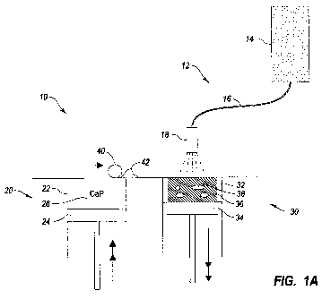
Figures 1A-1B illustrate embodiments of a direct rapid prototyping inkjet
printing system and process for using inkjet technologies in order to prepare
a
bioceramic having a bioactive siibstance. Generally, direct rapid prototyping
inkjet
systems and methods that are well known in the art can be configured to
operate
under the present invention. Briefly, such direct rapid prototyping systems
can be
configured to operate at room temperature or under minimal heat so that
proteins or
polypeptides can be included within the printing cartridges, reservoirs,
and/or
ceramic matrix without undergoing degradation or denaturation. As such, direct
rapid prototyping systems and processes can be configured to eliminate a
sintering
step or other step that causes excessive heat and/or pressure. As used herein,
direct
inkjet printing refers to an entire class of machines that employ inkjet
technology to
sequentially build a bioceramic endoprosthesis layer-by-layer. An example of
such a
direct inkjet printer capable of operating under the present invention is a
ZCorp 3D
printer, produced by Z Corporation of Burlington, MA.
Figure 1A depicts an embodiment of a direct inkjet printing system 10 and
process in accordance with the present invention. The direct inkjet printing
system
10 includes an inkjet printer 12, a powder delivery system 20, a roller 40,
and a
fabrication system 30.
The inkjet printer 12 has at least one inkjet cartridge 14 that can include
any
composition capable of being inkjet printed. Additionally, the inkjet printer
12
includes an inkj et line 16 that routes the inkj et composition from the inkj
et reservoir
14 to an inkjet printer head 18. Also, the inkjet printer 12 can be configured
to
include any number of cartridges 14, lines 16, or printer heads 18. Usually,
the
CA 02689675 2009-08-06
WO 2008/095307 PCT/CA2008/000248
18
inkjet printer 12 includes at least one binder cartridge and at least one
bioactive
substance cartridge.
The powder delivery system 20 has at least one powder delivery chamber 22
that provides a chamber for a powder delivery piston 24. In combination, the
powder delivery chamber 22 and powder delivery piston 24 cooperate to contain
the
ceramic powder 26. The powder delivery piston 24 is configured to move upward
as
shown by the arrows after each layer of powder is used in the direct inkjet
printing
process.
The roller 40 is depicted to be a conventional rolling object, such as one
rolling part of a calender, which can roll a layer 42 of the ceramic powder 26
from
the powder delivery system 20 to the fabrication system 30. However, a
squeegee or
other similar mechanical instrument can be used to scrape or move a top layer
of
ceramic powder from the powder delivery system 20 to the fabrication system
30.
The fabrication system 30 has at least one fabrication chamber 32 that
provides a chamber for a fabrication piston 34. In combination, the
fabrication
chamber 32 and fabrication piston 34 cooperate to contain the bioceramic
endoprosthesis 36 as it is being fabricated. The fabrication piston 24 is
configured
to move downward as shown by the arrows after each layer of powder is
deposited
onto the endoprosthesis 36 and fixed by a binder solution contained in an
inkjet
cartridge 14.
As shown, the bioceramic endoprosthesis 36 is built in the fabrication
chamber 32 on a substrate or platform situated on or integral with the
fabrication
piston 34. As such, the powder delivery piston 24 rises so that a top layer 42
of the
ceramic powder 26 in the powder delivery chamber 22 is rolled by the roller 40
into
the fabrication chamber 32. After the lop layer 42 of the ceramic powder 26 is
deposited onto the fabrication piston 34, the inkjet printing head 18
selectively
deposits or inkjet prints a binder fluid to cure or otherwise fuse the ceramic
powder
26 together in the desired areas. Unbound powder can remain to support the
part or
bound layer of the bioceramic endoprosthesis that has been hardened. After
hardening the bound layer, the fabrication piston 34 is lowered, more ceramic
powder 26 is added from the powder delivery chamber 22 to the fabrication
chamber
34 and leveled, and the process is repeated. Typical layer thicknesses are on
the
order of 100 microns. When finished, the bioceramic endoprosthesis is
considered
CA 02689675 2009-08-06
WO 2008/095307 PCT/CA2008/000248
19
to be a green body that is then removed from the unbound ceramic powder, and
excess unbound powder is blown off or washed away.
At some point in the process, which can be before, during, or after the
ceramic is hardened, the bioactive substance can be inkjet printed or
otherwise
deposited onto the bound powder. The inkjet printing of the bioactive
substance can
be performed so as to form a bioactive substance reservoir 38 within the
bioceramic
endoprosthesis 36. While a bioactive substance reservoir 38 is described
herein,
such a reservoir can be a single molecule, substance, or cell or a depot
having a
plurality of molecules, substances, or cells. In any event, the green body is
prepared
so as to have the bioactive substance. This can be used to provide discretely
located
bioactive substances, to provide bioactive substance gradients, or to
homogeneously
distribute the bioactive substance throughout the bioceramic matrix.
Alternatively,
the bioactive substance can be contained in microspheres that are mixed into
the
ceramic powder 26 and applied therewith.
The printed body having the bioactive substance can then be cured or
otherwise finished into a bioceramic endoprosthesis. While the printed body
can be
partially cured or hardened during printing, an additional curing step can be
advantageous to finish the product. Such curing or finishing can be performed
at
low temperatures by immersing the printed body into a curing solution or
hardening
solution that causes the ceramic powder to react and harden to its fully
hardened
state. However, other low temperature curing techniques can be employed that
retain the functionality and integrity of the bioactive substance.
In one embodiment, the direct inkjet printing process starts by depositing a
layer of ceramic powder 26 at the top of a fabrication chamber 32. To
accomplish
this, a measured quantity of ceramic powder 26 is first dispensed from a
similar
supply chamber 22 by moving a piston 24 upward incrementally. The roller 40
then
distributes the ceramic powder 42, and compresses the powder at the top of the
fabrication chamber 32. A multi-channel inkjet printing head 18 subsequently
deposits a liquid binder in a two dimensional pattern onto the layer of the
powder
which becomes bonded in the areas where the binder is deposited to form a
layer of
the bioceramic endoprosthesis (bound powder). The multi-channel inkjet
printing
head 18 subsequently deposits a liquid composition having a bioactive
substance in
a two dimensional pattern onto the layer of the bound powder to form a
bioactive
CA 02689675 2009-08-06
WO 2008/095307 PCT/CA2008/000248
substance reservoir 38. The bioactive substance composition can be configured
to
be retained on the bound powder by also comprising various additives, binders,
gels,
pastes, or adhesives. Also, the bioactive substance composition can be
configured to
dry on the bound powder, absorbed or incorporated during the binding stage or
5 binding reaction, or configured to be retained as a liquid after a
subsequent powder
layer is applied and bound on top of the bioactive substance. Once a layer is
completed, the fabrication piston 34 moves down by the thickness of a layer.
In one embodiment, the bioactive substance is co-printed with the binder so
that the bioactive substance is incorporated within the bioceramic matrix.
This can
10 include the bioactive substance and binder being intermittently inkjet
printed in one
pass of the printing head, or the bioactive substance and binder can be inkjet
printed
in different passes of the printing head, or deposited in any manner during 3D
printing of the bioceramic. However, the binder and bioactive substance can be
printed with different printing heads or even different printing devices.
15 In one embodiment, the process of depositing powder and inkjet printing
binder is repeated until a substantial portion of the endoprosthesis is formed
within
the powder bed so as to have an open chamber of unbound powder. The
endoprosthesis is then elevated and the unbound powder within the open chamber
is
then blown or washed away to leave an open chamber. Any system and/or process
20 that can be used to remove unbound powder can be included within the
present
invention. The bioactive substance can then be inkjet printed into the open
chamber,
and subsequent layers of powder can be deposited and bound over the chamber so
as
to form a bioactive substance reservoir 38. The open chamber can be as small
as
one layer or can be as large as multiple layers of bound powder.
In one ernbodiment, a spraying process or other deposition process can be
used to impregnate the bioceramic with the bioactive substance. As suoh, the
inkjet
system and process can include any fluid deposition device to deposit the
bioactive
substance within the bioceramic matrix.
Additionally, the methods of the present invention can be performed with a
spraying device other than an inkjet printer. As such, the recitation herein
of direct
inkjet printing can also include direct spraying. Accordingly, systems and
methods
that can be used for spraying compositions can be adapted to for use in the
system
and methods described herein.
CA 02689675 2009-08-06
WO 2008/095307 PCT/CA2008/000248
21
Furthermore, the process of preparing the bioceramic can be performed at a
low temperature such that the temperature sensitive bioactive substance does
not
degrade or denature. This can include a low temperature that is lower than a
temperature that degrades or denatures the bioactive substance. Also, this can
include temperatures lower than sintering temperatures, which are temperatures
required to sinter the ceramics of the present invention. Such temperatures
can be
less than about 1000 C, less than about 800 C, less than about 600 C, less
than
about 400 C, less than about 200 C, less than about 100 C, less than about 50
C, or
less than about 37 C, depending on the bioactive substance. Further, the
printing or
spraying steps that occur with a bioactive substance or after some of the
bioactive
substance has been incorporated into the bioceramic can be performed at the
low
temperature.
Additionally, multiple materials can be incorporated into the direct printing
process described herein, as well as other methods of making the bioceramic
endoprosthesis. This can include using two materials simultaneously or nearly
simultaneously in preparing two regions. For example, this can allow for
preparing
the endoprosthesis -to include a hydrogel within the ceramic matrix.
Figure 1B illustrates a more specific embodiment of the direct inkjet printing
system and process described in connection with Figure 1A. As shown, the
direct
inkjet printing process can be configured for preparing brushite or
hydroxyapatite
ceramics. The inkjet printing system is configured such that the inkjet
cartridge
includes a phosphoric acid solution that can be used to bind the ceramic
powder.
However, other compositions that can bind the ceramic powder may be used.
The process for preparing the brushite ceramic includes the direct inkjet
printing process described herein. As shown, the ceramic powder includes about
70% TCP, 17% brushite, and about 13% monetite, and the binder solution is a
20%
phosphoric acid solution that can cause the ceramic to harden so as to be
capable of
being handled without substantial deformation or breakage. The printed ceramic
was tested to have a compressive strength (CS) of about 5.3 MPa and to have a
porosity of about 45%. In order to increase the mechanical properties, the
printed
ceramic is then processed in a post-hardening process by immersing or soaking
the
printed ceramic into a hardening solution. As shown, the hardening solution is
a
20% phosphoric acid solution, and the ceramic is hardened by being immersed in
the
CA 02689675 2009-08-06
WO 2008/095307 PCT/CA2008/000248
22
hardening solution 3 times for 60 seconds. The hardening solution should be
maintained at a low temperature as described herein. After hardening, the
ceramic
was characterized as having about 27% TCP, 52% brushite, and about 21%
monetite, and having a CS of about 22.3 MPa, porosity of about 29%, and
specific
surface area (SSA) of about 1.4 m2/g.
The process for preparing the hydroxyapatite ceramic includes the direct
inkjet printing process described herein. As shown, the ceramic powder
includes
about 81% TTCP, 5% brushite, about 5% hydroxyapatite, and about 9% monetite.
The binder solution is a 10% phosphoric acid and 1 Mol NaH2PO4 solution that
can
1o cause the ceramic to harden so as to be capable of being handled without
substantial
deformation or breakage. The printed ceramic was tested to have a CS of about
1.9
MPa and to have a porosity of about 60%. In order to increase the mechanical
properties, the printed ceramic is then processed in a post-hardening process
by
immersing or soalcing the printed ceramic into a hardening solution. As shown,
the
hardening solution is a 10% phosphoric acid solution, and the ceramic is
hardened
by being immersed in the hardening solution for 30 seconds. After hardening,
the
ceramic was characterized as having about 49% TTCP, 26% brushite, 10%
hydroxyapatite, and about 15% monetite. The ceramic also had a CS of about 5.1
MPa and porosity of about 55%. The hardened ceramic is then processed in a
hydrothermal-conversion process by being immersed or soaked into an aqueous
solution for 7 days at 37 C. While hydrothermal-conversion was conducted at 37
C,
other low temperatures, such as those described herein, can be used. As shown,
the
aqueous solution can include 2.5% NaH2PO4. After hydrothermal-conversion, the
ceramic was characterized has having about 27% TTCP, 8% brushite, 57%
hydroxyapatite, and about 8% monetite, and having a CS of about 5.8 MPa,
porosity
of about 59%, and SSA of about 12.1 m2/g.
However, the post-hardening process and/or hydrothermal-conversion
process can be avoided in some instances. As such, the binder solution
composition
or amount inkjet printed into the ceramic powder can be modified in order to
provide for hardening or hydro-conversion. Also, the duration that the bound
powder is allowed to harden or cure can be increased before each successive
layer is
fabricated. In any event, the direct inkjet printing process described herein
can be
CA 02689675 2009-08-06
WO 2008/095307 PCT/CA2008/000248
23
performed so as to retain the functionality of the bioactive substance
incorporated
into the bioceramic.
Generally, direct 3D printing can be performed by the following process:
depositing a first layer of ceramic powder, such as ceramic, at the top of a
fabrication chamber or on a substrate; inkjet printing a bonding agent or
curing agent
onto the first layer of the ceramic powder; bonding or curing the first layer
of
ceramic powder into a first bioceramic layer; inkjet printing a first layer of
a
bioactive substance composition on a portion of the first bioceramic layer to
form a
first bioactive substance layer; depositing a subsequent layer of ceramic
powder on
the first bioceramic layer and the first bioactive substance layer; inject
printing the
bonding agent or curing agent onto the subsequent layer of ceramic powder; and
bonding or curing the subsequent layer of ceramic powder to form a subsequent
bioceramic layer.
In one embodiment, the present invention includes a direct inkjet printing
method for preparing a bioceramic endoprosthesis having a releasable bioactive
substance at a low temperature. Such a direct inkjet printing method includes
the
following: (i) applying a ceramic powder to a substrate; (ii) inkjet printing
a binder
or reactive solution onto the ceramic powder so as to form a bound ceramic;
(iii)
inkjet printing a bioactive substance solution onto the bound ceramic,
wherein the bioactive substance is printed on the bound ceramic at the low
temperature; and (iv) repeating steps (i-ii). Optionally, step (iii) can be
performed
intermittently or concurrently with step (ii). Also, the method can be
performed at a
low temperature, at room temperature, or within +/- 10 C of 25 C, within +/-
20 C
of 25 C, or even within +/- 30 C of 25 C.
In one embodiment, the a direct inkjet printing method can include applying
a hardening solution to the bound ceramic, and hardening the bound ceramic
into a
hardened ceramic having the releasable bioactive substance. In some instances,
the
direct inkjet printing method can further include applying an aqueous solution
to the
hardened ceramic maintaining a hydrothermal-conversion or aqueous-conversion
temperature of the hardened ceramic while in contact with the aqueous solution
so as
to further harden the hardened ceramic. Usually, the hydrothermal-conversion
temperature is higher than the low temperature. For example, the hydrothermal-
conversion temperature can be performed at a low temperature, at room
temperature,
CA 02689675 2009-08-06
WO 2008/095307 PCT/CA2008/000248
24
or within +/- 10 C of 37 C, within +/- 20 C of 37 C, or even within +/- 30 C
of
37 C, or lower than a temperature that degrades or denatures the bioactive
substance.
In one embodiment, the direct inkjet printing method can include fabricating
the bioceramic endoprosthesis so as to have at least one. pore having a
diameter
greater than about 200 microns; and localizing a portion of the bioactive
substance
within a ceramic matrix adjacent to a surface of the pore. This can include
the
bioactive substance being inkjet printed into a reservoir adjacent to the
surface of the
pore or printed into the bioceramic matrix adjacent the pore.
In one embodiment, the bioceramic material can be hardened, bound, or
cured in a process similar to the hardening or curing of a cementitious
material. The
hardening, binding, or curing may occur by the reaction of the ceramic powder
on
contact with a liquid. This process may occur by any of the known calcium
phosphate cement forming reactions, or the like. For example, this process may
occur through the reaction of an appropriate hardening, binding, and/or curing
composition with any of the following: calcium phosphate; calcium oxide;
hydroxide phase; mixture of phases; mechanically activated compound; amorphous
compound; glass compounds; or the like. Examples of hardening, binding, and/or
curing composition include any of the following: water; solutions of
phosphate;
solutions of pyrophosphate; solutions of polyphosphate; solutions of
carbonate;
solutions of silicate; solutions of phosphonate; solutions of alpha
hydroxyacid;
solutions of sulphate ions; solutions of acids; aqueous solutions there; and
mixtures
thereof.
Examples of ceramic powders and hardening, binding, and/or curing
composition pair include the following: tetracalcium phosphate is reacted with
phosphoric acid solution; beta tricalcium phosphate is reacted with phosphoric
acid
solution; beta tricalcium phosphate is reacted with pyrophosphoric acid
solution;
beta tricalcium phosphate is reacted with polyphosphoric acid solution; alpha
tricalcium phosphate is reacted with phosphoric acid and/or sodium phosphate
solution; tetracalcium phosphate, dicalcium phosphate (dihyrate and or
anhydrous),
and/or mixtures thereof are reacted with sodium phosphate solutions.
Additionally, a process substantially similar to Figures lA-lB can be
employed using a hydrogel or polymeric composition. Such a process can then
CA 02689675 2009-08-06
WO 2008/095307 PCT/CA2008/000248
incorporate the hydrogel or polymer into the endoprosthesis as described in
connection with the bioactive substance. Moreover, the hydrogel or polymer can
be
included into the endoprosthesis with the bioactive substance.
The bioceramic endoprosthesis of the present invention can be prepared by
5 other methods. As such, the bioceramic endoprosthesis can be made by casting
the
ceramic in a mold and then adding the bioactive substance. Such molding can
include molds that provide the pores, channels, or other features described
herein.
Also, holes can be drilled into the endprosthesis to provide the pores,
channels, or
other features described herein. Also, the ceramic endoprosthesis can be
prepared
10 by machining a block into the shape of the endoprosthesis, which can
optionally
include forming the pores, channels, or other features. Examples of methods
for
preparing the ceramic endoprosthesis body can be found in U.S. 6,905,516,
which is
incorporated herein by specific reference. After the ceramic endoprosthesis is
prepared or during low temperature processing, the bioactive substance can be
added
15 to the endoprosthesis as described herein. Thus, the ceramic endoprosthesis
can be
prepared by rapid prototyping, molding, machining, sintering, and/or
compacting.
M. Characterization Of Endoprosthesis
It has been shown that grafts taken from the patient's own skeleton
(autograft) induce angiogenesis by active endogenous signalling molecules, a
20 property lacking in allograft and synthetic graftsJ103 Formation of a blood
supply is
an important initial step in the growth. of new tissues; it not only nourishes
cells
involved with healing but also provides a source of osteoprogenitor cells.
Inducing
angiogenesis in synthetic porous grafts using VEGF has been shown to
accelerate
bone healing,[11'123 which is important because there is a limited supply of
spare bone
25 for autografting in the body and graft harvesting requires an additional
surgical
procedure. Prior concerns over disease transmission from donor allogenic bone
have been heightened recently,E133 further reinforcing the need for
improvements in
bone graft substitute bioceramics. Regenerative products based on tissue
induction
following protein release from polymeric matrices are now a reality in the
field of
bone and periodontal surgery.El4'1s1 However, protein mediated tissue
regeneration is
not without drawbacks, which include cost of production, supply and storage of
an
unstable recombinant product, and perceived risks of delivering higher than
physiological levels of potent inductive factors.
CA 02689675 2009-08-06
WO 2008/095307 PCT/CA2008/000248
26
As described herein, a direct rapid prototyping inkjet printing process was
used to prepare 3D powder print bioceramic structures at room temperature.
Direct
3D inkjet printing at room temperature is highly significant because it allows
simultaneous control of geometry of the bioceramic and control of bioactive
substance (e.g., organic molecule) incorporation in the bioceramic. The direct
3D
inkjet printing of the bioceramic. can allow for replication of biomimetic
micro-
environments for controlled tissue healing by having a bioceramic
endoprosthesis
that releases the bioactive substance. Furthermore, the utility of the direct
3D inkjet
printing process was demonstrated by directly fabricating model bioceramic
lo implants from brushite (i.e., dicalcium phosphate dihydrate), hydrated
calcium
phosphate, and hydroxyapatite so as to include organic angiogenic factors,
such as
vascular endothelial growth factor (VEGF). As such, bioactive substances that
are
susceptible to degradation under high temperatures can now be included in a
bioceramic due to the low temperature processing. In any event, the following
discussion of experiments and results relates to experiments that were
conducted in
accordance with the Experimental Protocols section provided below.
As described in more detail above with respect to Figures 1 A-1 B, fabrication
of the bioceramic endoprosthesis was performed using a direct rapid
prototyping 3D
inkjet printing technique in a two step process. Either brushite (i.e.,
dicalcium
phosphate dihydrate - DCPD) or hydroxyapatite (HA) can be made into complex
shapes by using tricalcium (TCP) or tetracalcium phosphate (TTCP) powders
respectively (see Figure 6A). Each layer is printed to be about 100 m thick;
however, the thickness can be modulated as needed or desired. The 100 m thick
layers took approximately 8-12 seconds to print depending on the print area.
Figures 6A-6C shows that programmed complex shapes could be replicated
from scaled down CT data or from hand drawn computer aided design (CAD) files.
As shown, the methods of the present invention can provide a variety of 3D
shapes.
This can include a disk 200 having holes, a skull 202a,b, a block 210 having
channels 212, and others.
The mean compressive strengths of DCPD and HA components directly after
printing were 5.3 0.6 and 1.9 0.2 MPa, respectively. This was an important
finding as the post-printed cements were strong enough to be handled without
CA 02689675 2009-08-06
WO 2008/095307 PCT/CA2008/000248
27
breaking or fragmenting during removal of the unreacted powders using
compressed
air. After post-print hardening or aqueous conversion, compressive strength
increased 3-4 fold to about 22.3 1.5 MPa for DCPD and about 5.8 0.3 MPa
for
HA. This increase in strength was caused by an increase in the degree of
conversion
of powder reactants from 30% to 73% for DCPD and from 19% to 57% for HA
samples (Figure 1B) and these strengths are higher than values reported for a
commercial sintered. bone graft substituteJ93 Post-printing hardening and
aqueous
conversion hardly affected the microporosity and pore size of HA samples. The
porosity decreased from 60% to 59%, and the median pore size decreased from 15
microns to 12 micron. In contrast, the porosity of the DCPD samples decreased
from 46% to 29%, and the median pore size decreased from 27 microns to 13
microns after post-printing hardening.
Figure 1 C provides micrographs that show the set cements to include large
(10-20 m) angular particles of unreacted starting powder in a matrix of
tabular
crystals of DCPD (5-10 m) or platy crystals 2-5 m of HA.
Figures 2A-2D depict a process of producing a model using computing
technologies and direct rapid prototyping inkjet printing in accordance with
the
present invention. In order to produce model implants for the investigation of
spatially localized release of both organic (VEGF), a Y shaped hemi-
cylindrical pore
2o channel was designed in the x-y plane of DCPD cuboids with dimensions 8 mm
x
8mm x 3 mm. The bioceramic endoprosthesis was made in two mirror image halves
50, 52 that keyed into one another to form a Y shaped pore 51 a, 51b closed at
one of
the branched ends (see Figures 2A-2C). This design facilitated tissue
examination.
Micro-computed tomography ( CT) revealed the block 54 and pore 51 c
architecture
(Figures 2B-2D) and demonstrated that the main open pore had a diameter of
1.31
0.11 mxri (Figure 2D). In the experiments, mouse VEGF solution was deposited
at
the end of the closed pore.
Experiments using the bioceramic endoprosthesis showed that after
peritoneal implantation for 15 days, blood vessels or microvessels had only
entered
2 mm into the open pores of the factor-free model implants (controls), while
blood
vessels extended the entire length of the pore (7mm) towards the regions where
the
angiogenic factors had been deposited (see Figure 3A). Histology of the tissue
confirmed the presence of an organized microvessel network in the experimental
CA 02689675 2009-08-06
WO 2008/095307 PCT/CA2008/000248
28
implants (see Figure 3B (i-ii)). Quantitative measurements confirmed that VEGF
elicited a significantly enhanced angiogenic effect compared with an untreated
DCPD control (see Figure 3B).
Wound tissue and angiogenesis patterns were examined for the implanted
bioceramic endoprosthesis after peritoneal implantation of the bioceramic
endoprosthesis in mice for 15 days. After opening the mating halves which
remained
tightly sealed throughout implantation, tissue response was found to be both
material
and angiogenic factor dose dependant. DCPD untreated control implants only had
limited tissue ingrowth at the pore openings. DCPD with 200 ng and 2 g VEGF
deposited in pore ends had vascular tissue throughout the pore channels,
conversely
within the pores of HA implants containing VEGF very little tissue ingrowth
was
apparent (Figure 3B (ii)). These observations may have been a result of the
differences in specific surface area between HA and DCPD implants (see Figure
1 C), or differences in their solubility. Based on these observations DCPD was
selected as a matrix and optimal loading levels of 200ng VEGF were used for
subsequent implantation to quantify microvessel ingrowth.
Vascularised tissue was found inside the main open and closed pore channels
of the DCPD implants which had been loaded with 200 ng VEGF (Figure 3A)
localized at the closed pore ends.
It was found that 3D protein concentration gradients could be achieved by
repeated application of decreasing volumes of serum protein solution.
Stability of
these treatments was confirmed in water and in serum for up to 3 weeks. Thus
we
demonstrated that localized and controlled protein and ion binding could be
achieved that would initially remain stable post implantation.
Figure 4A illustrates an embodiment of a bioceramic endoprosthesis 100
having a channel 102 extending therethrough. The endoprosthesis 100 having the
channel 102 was prepared as described herein. Figure 4B is a schematic
representation of a CT image of a bioceramic endoprosthesis 110 having a
channel
112 extending therethrough. Additionally, the endoprosthesis 110 includes
depots
114 of a bioactive substance within the ceramic matrix 116. While not
required, the
endoprosthesis 110 is shown to have a sealed end 118 so as to have a sealed
channel
112 at one end.
CA 02689675 2009-08-06
WO 2008/095307 PCT/CA2008/000248
29
To demonstrate the feasibility of controlling both 3D architecture and
composition of the bioceramic endoprosthesis as part of the powder printing
process,
150 mM barium chloride solution was deposited in a pattern of eight 40 l
spots
during printing of a cylinder with a curved central pore. As shown in Figure
4B,
CT revealed that the barium chloride remained localized where it had been
applied
during cement setting. Additionally, the post-print hardening process can be
optimized by either allowing full setting to occur post-implantation or in
more
physiological conditions, such as by optimizing the powder physics of a two
powder
component cement system. In one example, supplying a source of reactive
phosphate ions in the powder phase or binder may obviate the use of acidic
phosphate solutions for curing the bioceramic.
The direct rapid prototyping inkjet printing process, as described herein, may
be used to prepare bone graft endoprostheses that are fabricated from CT or
MRI 3D
data files with tissue inductive protein, peptide, or other biologically
bioactive
substances applied locally to generate a regenerative response by more closely
mimicking the complex natural tissue regeneration process. In part, the local
concentration of the bioactive substance within or adjacent to the implanted
bioceramic endoprosthesis can mimic or induce the tissue regeneration process.
As
such, it is conceivable that this direct rapid prototyping inkjet printing
process can
provide a bioceramic having reservoirs or depositions of bioactive substance
that can
replicate biological responses that have been observed to be induced by bone
autograft. Indeed, recent preclinical reports have indicated that bone
formation was
enhanced by the use of VEGF loaded hydrogels combined with biomineral
grafts.[211
The methods of the present invention can be used to prepare anatomical
endoprostheses (i.e., implants), such as orthopedic implants that can be
implanted
into the head, neck, torso, anns, legs, and the like. For example, the methods
can be
used to prepare cranial implants.
Although this work was performed with a single inkjet printer, multi-head
inkjet printers, which can include multiple reservoirs of different fluids
(e.g..,
multiple binders, bioactive substances, polymers, or the like) can be utilized
in the
powder printing systems of the direct rapid prototyping inkjet printing
process
described herein, or separate depositions systems. Thus, using the direct
rapid
prototyping inkjet printing process reported here, it is possible to print
multiple
CA 02689675 2009-08-06
WO 2008/095307 PCT/CA2008/000248
compositions into the bioceramic. For example, a 5 head inkjet printer could
be
configured as follows: the first inkjet cartridge solution can include the
binding
agent; the second inkjet cartridge solution can include a angiogenic agent;
the third
inkjet cartridge solution can include a gene delivery system that encodes an
5 angiogenic protein; the fourth inkjet cartridge solution can include an
antibiotic; and
the fifth inkjet cartridge can include a pain reliever. However, it should be
realized
that multiple different binding agents could be employed or any other
variation that
employs a multi-head inkjet printer. This could provide for the systematic
study of
multiple bone and vascularized tissue factors and for development of improved
10 patient-tailored bone graft substitutes. Alternatively, other systems
and/or processes
that can spray one or more binders, bioactive substances, or polymers can also
be
used.
Figure 5 are photographs depicting vascularization patterns in pores of
DCPD (left) and HA (right) implants with 2 g VEGF deposited in closed pore
ends
15 opened following 15 days implantation. HA implants were mostly filled with
clear
connective tissue.
Figures 8A-8E are schematic representations of embodiments of
endoprostheses of the present invention. Figure 8A illustrates a tubular
endoprosthesis 300 having a corrugated internal lumen 302. Figure 8B
illustrates a
20 scalloped endoprosthesis 310 having a corrugated internal lumen 312. Figure
8C
illustrates a scalloped endoprosthesis 320 having a primary lumen 322 and a
plurality of channels 324 that longitudinally extend through the
endoprosthesis 320.
The primary lumen 322 and plurality of channels 324 are independent and non-
intersecting. Figure 8D illustrates a tubular endoprosthesis 330 having a
primary
25 lumen 332 and a plurality of channels 334 that longitudinally extend
through the
endoprosthesis 320. The primary lumen 332 and plurality of channels 334 are
independent and non-intersecting. Figure 8E illustrates a spoke-shaped tubular
endoprosthesis 340 having a primary lumen 342 and a plurality of spoke
channels
344 that longitudinally extend through the endoprosthesis 340. The primary
lumen
30 342 and plurality of spoke channels 344 are independent and non-
intersecting.
Accordingly, the present invention can be directed to methods of making a
bioceramic endoprosthesis and the product thereof.
CA 02689675 2009-08-06
WO 2008/095307 PCT/CA2008/000248
31
In on embodiment, the present invention can include a direct printing method
for preparing a bioceramic endoprosthesis having a bioactive substance. Such a
method can include: (i) applying a ceramic powder to a substrate; (ii)
spraying a
binder solution onto the ceramic powder so as to form a bound ceramic; (iii)
depositing at least one bioactive substance solution onto the bound ceramic so
as to
incorporate the bioactive substance with bound ceramic; and (iv) repeating
steps (i-
ii) or (i-iii).
In one embodiment, the depositing of the bioactive substance and step (iii) is
performed at a low temperature that is lower than a temperature that degrades
or
1o denatures the bioactive substance. Optionally, the low temperature is room
temperature or within +/- 10 C of 25 C.
In one embodiment, the method includes applying a hardening solution to the
bound ceramic, and hardening the bound ceramic into a hardened ceramic having
the
releasable bioactive substance.
In one embodiment, the method includes applying an aqueous solution to the
hardened ceramic, and maintaining a hydrothermal-conversion temperature of the
hardened ceramic while in contact with the aqueous solution so as to further
harden
the hardened ceramic, wherein the hydrothermal-conversion temperature is
higher
than the low temperature.
- In one embodiment, the hydrothermal-conversion temperature is +/- 10 C of
37 C. Also, the hydrothermal=conversion temperature can be lower than a
temperature that degrades or denatures the bioactive substance.
In one embodiment, the entire method of preparing the endoprosthesis is
performed at a temperature that is lower than a temperature that degrades or
denatures the bioactive substance.
In one embodiment, at least one of the binder or bioactive substance is inkjet
printed. Optionally, the bioactive substance can be sprayed into a reservoir
within a
ceramic matrix of the bioceramic endoprosthesis. Also, the bioactive substance
can
be sprayed so as to form a concentration gradient within a ceramic matrix of
the
bioceramic endoprosthesis. In another option, the bioactive substance can be
sprayed so as to be substantially homogeneously distributed throughout at
least a
portion of a ceramic matrix of the bioceramic endoprosthesis.
CA 02689675 2009-08-06
WO 2008/095307 PCT/CA2008/000248
32
In one embodiment, the method of preparing the endoprosthesis includes
fabricating the bioceramic endoprosthesis so as to have at least one pore
having a
diameter greater than about 200 microns, and localizing a portion of the
bioactive
substance within a ceramic matrix adjacent to a surface of the pore.
In one embodiment, the method includes absorbing a bioactive substance
into the bioceramic matrix.
In one embodiment, the present invention includes a system for low
temperature printing a bioceramic endoprosthesis having a releasable bioactive
substance. Such a system can include an inkjet printer comprising: at least
one inkjet
reservoir containing a binder composition capable of being inkjet printed; at
least
one inkjet reservoir containing a bioactive substance composition capable of
being
inkjet printed; and at least one an inkjet printer head capable of inkjet
printing the
binder composition and/or the bioactive substance composition.
In one embodiment, the system includes a powder delivery system
comprising: at least one powder delivery chamber containing a ceramic powder;
and
a powder delivery piston configured to raise the ceramic powder layer-by-
layer.
In one embodiment, the system includes a fabrication system comprising: at
least one fabrication chamber configured for receiving the ceramic powder, and
for
receiving the binder composition and bioactive substance from the at least one
inkjet
printer head so as to form a bound ceramic having the bioactive substance; and
a
fabrication piston configured to lower the bound ceramic having the bioactive
substance layer-by-layer.
In one embodiment, the system includes a roller configured to distribute a
top layer of the ceramic powder from the powder delivery system to the
fabrication
system.
In one embodiment, the present invention includes a bioceramic
endoprosthesis. Such a bioceramic endoprosthesis can include a biocompatible
ceramic matrix having a body defming the external surface of the
endoprosthesis,
and a bioactive substance being spatially localized within the endoprosthesis.
Optionally, the bioactive substance is spatially localized within the ceramic
matrix.
In another option, the bioactive substance is disposed on a surface of the
ceramic
matrix. For example, the surface is a surface of a pore. In another example,
the
CA 02689675 2009-08-06
WO 2008/095307 PCT/CA2008/000248
33
bioactive substance is disposed in the ceramic matrix. In yet another example,
the
bioactive substance is disposed within a depot.
In one embodiment, the endoprosthesis includes a diffusion matrix
containing the depot. Such a diffusion matrix can include a polymer.
In one embodiment, the bioceramic endoprosthesis includes at least one pore
or a network of interconnected or non-connected pores. For example, the
bioactive
substance is disposed at a closed end of the at least one pore. In another
example,
the pore has an opening in the external surface.
In one embodiment, the bioactive substance is spatially localized in three-
1 o dimensions within the endoprosthesis. Alternatively, the bioactive
substance is
spatially localized in a two-dimensional pattern within the endoprosthesis. In
another embodiment, the bioactive substance is spatially localized in at least
one
ring or layer. In yet another embodiment, the bioactive substance is spatially
localized in at least two concentric rings or layers. For example, the ring or
layer is
three-dimensional. This can include the ring or layer forming a ring or layer
of
bioactive substance within the endoprosthesis.
In one embodiment, at least a portion of the endoprosthesis is biodegradable.
In one embodiment, the bioactive substance stimulates tissue growth within
and/or around the endoprosthesis.
In one embodiment, the bioactive substance is capable of diffusing into the
pore.
In one embodiment, the bioactive substance is capable of diffusing out of the
endoprosthesis.
EXPERIMENTAL PROTOCOLS
Example 1
TTCP was synthesized by heating an equimolar mixture of dicalcium
phosphate anhydrous (DCPA, CaHPO4, monetite) (Merck, Darmstadt, Germany)
and calcium carbonate (CC, CaCO3, calcite) (Merck, Darmstadt, Germany) to 1500
C for 18 hr followed by quenching to room temperature. The sintered cake was
crushed with a pestle and mortar and passed through a 160 m sieve. Milling
was
performed in a planetary ball mill (PM400, Retsch, Germany) at 200 rpm with
500
ml agate jars, 4 agate balls with a diameter of 30 mm and a load of 125 g TTCP
per
CA 02689675 2009-08-06
WO 2008/095307 PCT/CA2008/000248
34
jar for 30 min. 55 wt% (i /45 wt% a TCP was prepared in a similar manner by
sintering a 2:1 molar mixture of DCPA and CC at 1400 C for 14h.
Example 2
Printing of cement samples was performed with a 3D-powder printing
system (Z-Corporation, USA) using the TTCP or TCP powder and as liquid
printing
phases either a mixture of 10% phosphoric acid and 1M NaH2PO4 (both Merck,
Darmstadt, Germany) for TTCP powder or 20% phosphoric acid solution for TCP
powder. Sample geometries were designed using CAD software or directly taken
from CT scan data. Inkjet printing was performed using an anisotropic scaling
with
x = y = z = 1Ø Samples printed from TTCP were additionally stored in 10%
H3P04
for 30 s followed by immersion in 2.5% Na2HPO4 solution at 37 C for 7 days in
order to convert to HA. TCP printed sainples were stored in 20% H3PO4 for 60 s
3
times to increase the degree of reaction to DCPD. More detailed information
regarding the direct rapid prototyping inkjet printing process of the present
invention
can be reviewed above and in connection with Figures lA-1C.
Example 3
Compressive strength testing, as reported above (see Figure 1B), was
performed 24 h after setting with cylindrical satnples (10>n>5) with a height
of 20
mm and a diameter of 10 mm under axial compression at a crosshead speed of 1
mm/min using a static mechanical testing device (Zwick 1440, Ulm, Germany) and
a 5 kN load cell. The porosity of the printed samples was calculated from the
apparent density after drying and the strut density from the phase composition
and
using literature density values.E29J Pore size distributions were measured by
high
pressure Hg-porosimetry (Porosimeter 2000, Carlo Erba Instr., Milano, Italy).
The
Brunnauer Emmet Teller method (BET) was used to determine the specific surface
area within the porous calcium phosphate matrices (Micromeritics, ASAP 2000,
USA). The microstructure of gold sputtered fracture surfaces was characterized
by
scanning electron microscopy (FEI, Quanta 200, Czech Republic). X-ray
micrographs of porous structures were taken with a Polydoros SX80 (Siemens,
Germany) at 40KV and 5.0 mAs. Additional information regarding the
experimental protocols can be found in the incorporated references, or is well
known
to those of ordinary skill in the art.
CA 02689675 2009-08-06
WO 2008/095307 PCT/CA2008/000248
Example 4
X-ray diffraction (XRD) patterns of samples were recorded using
monochromatic Cu Ka radiation (D5005, Siemens, Karlsruhe, Germany). Data was
collected from 20 = 20-40 with a step size of 0.02 and a normalized count
time of
5 1 s/step. The phase composition was checked by means of ICDD reference
patterns
for a-TCP (PDF Ref. 09-0348), B-TCP (PDF Ref. 09-0169), DCPA (PDF Ref. 09-
0080), HA (PDF Ref. 09-0432), and DCPD (PDF Ref. 09-0077). Quantitative phase
relations of the composite materials were calculated by means of total
Rietveld
refinement analysis using the TOPAS software (Bruker AXS, Karlsruhe, Germany).
l0 As references the system internal database structures of a-TCP,I3-TCP, HA,
DCPD
and DCPA were used together with a Chebychev fourth order background model and
a Cu Ka emission profile.
Example 5
Serum protein concentration gradients, in response to bioceramics including
15 VEGF, were determined with respect to the bioceramic (Figure 7). Briefly,
1.8
mg/ml bovine serum protein or 0.45% saline Coomassie blue solutions (0.05%
final)
were applied in decreasing volumes to surfaces of DCPD and HA blocks (e.g., 5,
2.5, 1 and 0.5 l) to create stepped gradients of concentration. Coomassie
blue
binding to protein was determined to be 90% efficient (by ultra-
centrifugation).
20 Images were recorded with a Q-Imaging video camera and analysis was
performed
with Q-Capture software (Quantitative Imaging Corporation). The blue value as
a
function of distance was measured diametrically across the stained region of
the
cements (Image J, Scion Corporation). Serum protein concentration was measured
by spectrofluorometry at 595mn (Protein assay Bioraid, Biorad). Figure 7 shows
the
25 gradients of serum proteins and saline on DCPD were, stable invitro for up
to 3
weeks.
Example 6
For experimental analysis, VEGF-impregnated bioceramic materials were
prepared by passive adsorption of 200 ng or 2 g final amount of mouse VEGF
30 (R&D, Cedarlane Laboratories Ltd., Canada) diluted in a 5 41 volume of
Hank's
balanced salt solution (HBSS). Adsorption of VEGF was performed on each half
of
the bioceramic endoprosthesis (see Figures 2A-2D) at the closed end (3) of the
Y-
CA 02689675 2009-08-06
WO 2008/095307 PCT/CA2008/000248
36
shape pore. Specimens were air-dried under a sterile laminar flow hood. Prior
to
VEGF impregnation, the bioceramic materials were sterilized by soaking in 70%
ethanol, followed by a HEPES-NaCI buffered solution rinse, HBSS (x3), and then
air-dried. Control material specimens were also impregnated with HBSS without
VEGF. Higher concentrations of VEGF (400 g/ml) were also investigated.
Example 7
Micro-computed tomography was performed on a Skyscan Model 1072,
(Aartselaar, Belgium). The x-ray source was operated at 70 kV and at 142 A
(maximum power). Images were captured using a 12-bit, cooled CCD camera (1024
by 1024 pixels). Samples were scanned at a magnification resulting in a pixel
size
of 14.08 m. A rotation step of 0.68 and an exposure time of 9.2 s for each
step
with a 0.5 mm aluminum filter were used. The cross-sections along the specimen
axis were reconstructed using NRec Reconstruction software (SkyScan) giving a
voxel size of 14.08 x 14.08 x 14.08 m3. 3D Creator software (SkyScan) was
used
to perform 3D rendering.
Example 8
Flat square demonstration samples (50 x 50 x 5 mm) with colored stripes on
the surface with an increasing wideness of 1 - 5 mm were printed with a
multicolor
3D printing machine Z510 (Z-Corporation, Burlington, MA, USA) using tricalcium
phosphate with a medium particle size of approximately 30 m as powder and 20%
diluted phosphoric acid as binder solution in the binder reservoir. Printing
parameters were.a layer thickness of 125 m and a binder to powder volume
ratio of
approx. 0.3. A 5 % copper sulfate solution was .used in a second reservoir of
the
printer and sprayed with printing head 1 (which is assigned to the yellow
color
information of the ".wrl" file) to obtain stripes of deposited copper sulfate
on the top
surface of the printed calcium phosphate sainple."
The present invention may be embodied in other specific forms without
departing from its spirit or essential characteristics. The described
embodiments are
to be considered in all respects only as illustrative and not restrictive. The
scope of
CA 02689675 2009-08-06
WO 2008/095307 PCT/CA2008/000248
37
the invention is, therefore, indicated by the appended claims rather than by
the
foregoing description. All changes which come within the meaning and range of
equivalency of the claims are to be embraced within their scope. All
references
recited herein are incorporated herein in their entirety by specific
reference.
The following references provide background information regarding features
of the present invention, and are incorporated herein by specific reference in
their
entirety.
[1] P. Habibovic et al., Biomaterials, 2005, 26, 3565.
[2] V. Karageorgiou , D. Kaplan, Biomaterials 2005, 26, 5474.
[3] D. Kochan, C.K. Chua and Z.H. Du, Computers in Industry : an
International Journal, 1999, 39, 3.
[4] W.Y. Yeong, C.K. Chua, K.F. Leong and M Chandrasekaran, Trends in
Biotechnology 2004, 22, 643.
[5] C.K. Chua, K.F. Leong, K.H. Tan, F.E. Wiria, C.M. Cheah, J. Mater. Sci.:
Mater. Med. 2004,15, 1113.
[6] H. Seitz, W. Rieder, S.Irsen, B.Leukers, C. Tille, J. Biomed. Mater. Res.
2005, 74, 782.
[7] Z.Sadeghian, J.G. Heinrich, F. Moztaradeh, CFI-Ceramic Forum
International, 2004, 81, E39..
[8] C.E.Wilson, J.D.de Bruijn, C.A.van Blitterswijk, A.J.Verbout, W.J.A.
Dhert,
J. Biomed. Mater. Res. 2004, 68, 123.
[9] L. Xiang et al., Rapid Prototyp. J. 2005, 11, 312.
[10] K.A. Hing, S.M. Best, K.E. Tanner,W Bonfield, P.A. Revell J. Mater. Sci.-
MatS. in Med. 1997, 8 731.
[11] H. Ito et al., Nat. Med. 2005, 11, 291
[12] H. Eckardt, M. Ding, M. Lind, E.S. Hansen, K.S. Christensen, I. Hvid, J.
Bone Joint Surg. Br. 2005, 87, 1434.
[13] FDA Public Health Notification: Update of Information about BioMedical
Tissue Services, March 2, 2006 http://www.fda.gov/cber/safety/bts030206.htm
[14] M. Boakye, P.V. Mummaneni, M. Garrett, G. Rodts, R. Haid, J. Neurosurg:
Spine 2, 521-525 (2005).
[15] A.R. Vaccaro, T. Patel, J. Fischgrund, D.G. Anderson, E. Truumees, H.N.
Herkowitz, F. Phillips, A. Hilibrand, T.J. Albert, T. Wetzel, J.A. McCulloch,
Spine 29,1885-1892 (2004).
[16] G. Hu, J. Cell. Biochem. 1998, 6, 326.
[17] A. Parke, P. Bhattacherjee, R.M. Palmer, Am. J. Pathol. 1988,130, 173.
[18] C.K. Sen et al., Am. J. Physiol. Heart Circ. Physiol. 2002, 282, H1821.
[19] G. Giavaresi et al., Biomaterials 2005, 26, 3001.
[20] M. Bohner, U. Gbureck, J.E. Barralet, Biomaterials, 2005, 26, 6423.
[21] W.L.Murphy, C.A. Simmons, D. Kaigler, D.J. Mooney, J. Dent. Res. 2004,
83, 204.
[22] M.R. Urist, Science, 1965,150,893.
[23] D.E. Sims, Tiss. Cell 1986, 18, 153.
[24] C.T. Brighton, Cliri. Orthop. Relat. Res. 1992, 275, 287.
[25] M.J. Doherty et al., J. Bone Min. Res. 1998, 13, 828.
CA 02689675 2009-08-06
WO 2008/095307 PCT/CA2008/000248
38
[26] Holzel T., Rapid prototyping of calcium phosphate structures by means of
a
cement setting reaction. Diploma Thesis, University of Applied Sciences,
Erlangen-
Niirnberg, Germany, August 19, 2005 .