Note : Les descriptions sont présentées dans la langue officielle dans laquelle elles ont été soumises.
CA 02689686 2009-11-26
[DESCRIPTION]
[Invention Title]
Mass- and property-tuned variable mass labeling reagents and analytical
methods for
simultaneous peptide sequencing and multiplexed protein quantification using
thereof
[Technical Field]
The present invention relates to variable mass labeling reagentsand analysis
methods for
simultaneous peptide sequencing and protein quantitation using the same, more
particularly, variable mass labeling reagentscomprising hydrogen isotopes,
which
provides tunability in property and mass to display differential quantitation
signals at
different mass regions, and analysis methods for simultaneous peptide
sequencing and
multiplexed protein quantitation using the same.
[Background Art]
Mass spectrometry has been widely used for sequencing and quantitation of
proteins and
peptides. To identify proteins, for instance, peptidesproduced by enzyme
digestion are
ionized by either Matrix-Assisted Laser Desorption/lonization(MALDI) or
Electrospray
Ionization(ESI), and then their massescan be measured by means of a mass
spectrometer
to characterize the protein. More exactly, some peptides are further cleaved
into
fragments to identify the peptide sequence.
For the quantification of proteins and peptides by mass spectrometry, a number
of stable
isotope tags have been chemically introduced as markers into proteins or
peptides of
interest. Chemical tags differentially labeled with isotopes are incorporated
into the
same samples to be analyzed, and the mass of each sample can be distinguished
due to
the mass difference of the isotopes in the resulting mass spectra or tandem
mass spectra,
thus allowing protein quantification by the comparison of their relative
intensities.
Recently, the isobaric chemical tagging strategy has been used for
simultaneous protein
quantitation and sequencing.In US 2005/0148087 and WO 2005/068446, disclosed
are
isobaric reagents labeled with isotopes, which bind with peptide to display
quantitation
signals in tandem mass spectrometry.
However, the labeling reagents used in the known methods are problematic in
that
expensive carbon, nitrogen and oxygen isotopes are used, thus carrying high
cost. In
addition, another drawback is that because of the limited signal mass window,
unexpected chemical noise may hinder the analysis. Therefore, there is a need
for novel
isobaric labeling reagents incorporating low-cost hydrogen isotopes for
simultaneous
peptide sequencing and protein quantitation. Further, there is a need for
novel isobaric
variable mass labeling reagents that provide tunability not only in mass
window of
quantitation signals but also in property of peptides, thus applicable to a
wide range of
biomolecules.
The present inventors have suggested a novel isobaric labeling reagent based
on
dipeptide, mass-balanced 'H/ZH-isotope tag (MBIT)which only employs hydrogen
isotopes and offers tunability in quantitation signal mass window, disclosed
in Korean
Patent Application No. 2008-0070272. Further, they have demonstrated that the
replacement of the mass-tunable group of the 2-plex isobaric labeling reagent
with other
natural amino acid side chains having various properties offerspossibilities
of tuning the
signal mass window and its property, disclosed in Korean Patent Application
No.
2009-0019444. Various MBITs having different amino acid side chains showed up
to
1
CA 02689686 2009-11-26
ten-fold difference in the quantitation signal intensities due to dissimilar
chemical
properties of the amino acid side chains. To achieve better performance of the
MBIT
reagents in simultaneous multiplexed quantitation, it is necessary to use the
MBIT
reagents having similar chemical properties but different quantitation signals
in a
combination of two or more thereof. Accordingly, for simultaneous multiplexed
protein
quantification, a variety of MBIT reagents having identical property isneeded
to provide
similar quantitation signal intensity. Thus, they have suggested mass- and
property-tuned variable mass isobaric labeling reagents, a set of the labeling
reagents,
and analysis methods for simultaneous quantitation, disclosed in Korean Patent
Application No. 10-2009-0054540.
[Disclosure]
[Technical Problem]
Taken together, it is intended to provide isobaric labeling reagents for
simultaneous
peptide sequencing and multiplexed protein quantitation, providing the
tunability in
mass and property by using natural or artificial amino acids, and analysis
methods for
simultaneous multiplexed protein quantification using multiple 2-plex isobaric
tags.
[Technical Solution]
It is an object of the present invention to provide novel isobaric labelsfor
simultaneous
peptide sequencing and protein quantitation, comprising isotopes.
It is another object of the present invention to provide isobaric labels for
simultaneous
peptide sequencing and protein quantitation, comprising hydrogen isotopes.
It is still another object of the present invention to provide variable mass
labeling
reagents that are composed of two or more isobaric labels for simultaneous
peptide
sequencing and protein quantitation comprising hydrogen isotopes.
It is still another object of the present invention to provide isobaric
variable mass
labelsfor simultaneous peptide sequencing and protein quantitation, comprising
hydrogen isotopes and providing the tunability in mass by using natural or
artificial
amino acids.
It is still another object of the present invention to provide a set of
variable mass labeling
reagents that is composed of two or more isobaric labels for simultaneous
peptide
sequencing and protein quantitation, comprising hydrogen isotopes and
providing the
tunability in mass by using natural or artificial amino acids.
It is still another object of the present invention to provide a set of
variable mass labeling
reagents that is composed of two or more isobaric labels for simultaneous
peptide
sequencing and protein quantitation, comprising hydrogen isotopes and
providing the
tunability in mass by using natural or artificial amino acids to display
quantitation
signals at different mass regions.
It is still another object of the present invention to provide a set of
variable mass labeling
reagents that is composed of two or more isobaric labels for simultaneous
peptide
sequencing and protein quantitation, comprising hydrogen isotopes and
providing the
tunability in mass by using natural or artificial amino acids with identical
properties.
It is still another object of the present invention to provide a set of
variable mass labeling
reagents that is composed of two or more isobaric labels for simultaneous
peptide
sequencing and protein quantitation, comprising hydrogen isotopes and
providing the
tunability in mass by using natural or artificial amino acids with identical
properties to
display similar quantitation signal intensities at different mass regions.
2
CA 02689686 2009-11-26
It is still another object of the present invention to provide analysis
methods for
simultaneous peptide sequencing and protein quantitation using the set of
isobaric
variable mass labeling reagents comprising hydrogen isotopes.
It is still another object of the present invention to provide analysis
methods for
simultaneous peptide sequencing and multiplexed protein quantitation using
combination of various 2-plex setsof isobaric variable mass labeling reagents
comprising
hydrogen isotopes and providing the tunability in mass.
The above and other objects of the present invention can be achieved by the
following
descriptions.
[Advantageous Effects]
The present invention provides variable mass labeling reagents comprising
hydrogen
isotopes and providing the tunability in mass and property to display
quantitation signals
at different mass regions, a set of variable mass labeling reagents, a
multiplexed set of
variable mass labeling reagents, analysis methods for simultaneous peptide
sequencing
and protein quantitation using the set of isobaric variable mass labeling
reagents
comprising hydrogen isotopes, and analysis methods for simultaneous peptide
sequencing and multiplexprotein quantitation using the set of variable mass
labeling
reagents.
[Description of Drawings]
FIG. 1 is a schematic diagram showing the basic concept of MBIT reagent and
strategy,
in which (a) shows the structure of MBIT reagent, (b) shows the labeling
process by
coupling MBIT reagent to primary amines, (c) shows the expected fragment ions
of
MBIT-linked peptides by tandem mass spectrometry, and (d) shows the tandem
mass
spectra.
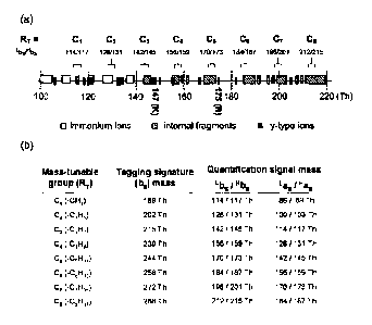
FIG. 2 is a schematic diagram showing a type of amino acid side chains
available as a
mass-tunable group (RT) for MBIT strategy, in which (a) shows the amino acid
side
chains available as a mass-tunable group (RT) for MBIT strategy and pairs of
quantitation signal mass in case of using the same amino acid, with
distribution of
possible fragment ions having mass range of 220 Th or below in tandem mass
spectra in
case of mass spectrometry of peptides, and (b) shows eight different mass-
tunable
groups (used in the present invention) with no significant interference with
possible low
mass fragments at the mass range of 220 Th or below.
FIG. 3 is a diagram showing the quantitation signal of MBIT in case of using
alkyl
groups as a mass-tunable group (RT) for MBIT strategy, in which (a) shows
possible low
mass fragments at the mass range of 220 Th or below in tandem mass spectra,
and (b)
shows the intrinsic tagging signature and quantitation signal mass of each
MBIT,
depending on the type of alkyl group that is used as a mass-tunable group.
FIG. 4 is a diagram showing experimental procedures for relative and absolute
quantitation of protein performed by using MBIT, in which (a) shows an
experimental
procedure for relative quantitation of the unknown amount of the same protein
produced
under the different conditions, and (b) shows an experimental procedure for
absolute
quantitation of the unknown amount of the identified protein.
FIG. 5 is a diagram showing the tandem mass spectra of the set of MBITs having
the
same property but differential signal mass, and showing the simultaneous
multiplexed
quantification methods for three or more samples using two or more sets of
MBITs.
FIG. 6 is a schematic diagram showing the process of synthesis of MBIT
reagents using
3
CA 02689686 2009-11-26
(a) the solid-phase synthesis and (b) the solution-phase synthesis.
FIG. 7 is a schematic diagram showing experimental method for the formation of
active
ester of the MBIT reagent and coupling of the formed active esters of MBIT
with target
peptides.
FIG. 8 is the results of MALDI mass spectrometry of peptide mixtures of
angiotensin II
and leucine enkephalin linked with eight pairs of N-acetylated dipeptide MBIT
reagents
[Ac-Xxx-Ala Xxx having a mass-tunable group is (a) alanine, (b) serine, (c)
valine, (d)
glutamine, (e) histidine, (f) phenylalanine, (g) arginine, and (h) tyrosine].
FIG. 9 is the results of MALDI tandem mass spectrometry of angiotensin II
([MAG(1)+H]+) each linked with eightdifferent pairs of N-acetylated dipeptide
MBIT
reagents as described in FIG. 8 [Ac-Xxx-Ala Xxx having a mass-tunable group is
(a)
alanine, (b) serine, (c) valine, (d) glutamine, (e) histidine, (f)
phenylalanine, (g) arginine,
and (h) tyrosine].
FIG. 10 is a diagram showing the quantitation signal mass window of FIG. 9, in
which
distribution of possible fragment ions at 220Th or below by tandem mass
spectrometry
of peptides is also shown. In the N-acetylated dipeptide MBIT reagent (Ac-Xxx-
Ala),
when Xxx having a mass-tunable group is(a) alanine, (b) serine, (c) valine,
(d) glutamine,
(e) histidine, (f) phenylalanine, (g) arginine, and (h) tyrosine, the results
are shown.
FIG. 11 is a diagram showing the results of tandem mass spectrometry of
leucine
enkephalin ion ([MLE(1)+H]+, herein H+is attached) detected after coupling
with MBIT.
In the N-acetylated dipeptide MBIT reagent (Ac-Xxx-Ala), when Xxx having a
mass-tunable group is basic (a) histidine and (b) arginine, the results are
shown.
FIG. 12 is a diagram showing the ratio of quantitation signal (xbs, X=H or L)
intensity
according to the mass-tunable group of N-acetylated dipeptide MBIT reagent,
and
quantitation signal intensity of fragment ions (xasor xbs-NH3) relative to
total sum of all
fragment ion intensities. Error bars stand for standard deviations from eight
repeated
experiments.
FIG. 13 is a diagram showing standard quantitation curve obtained by tandem
mass
spectrometry of the mixtures of different ratio of MBIT reagent-linked
angiotensin II. In
the N-acetylated dipeptide MBIT reagent (Ac-Xxx-Ala), when Xxx having a
mass-tunable group is (a) alanine, (b) serine, (c) valine, (d) glutamine, (e)
histidine, (f)
phenylalanine, (g) arginine, and (h) tyrosine, the results are shown. Error
bars stand for
standard deviations from eight repeated experiments.
FIG. 14 is a diagram showing standard quantitation curve obtained by tandem
mass
spectrometry of the mixtures of different ratio of MBIT reagent-linked leucine
enkephalin. In the N-acetylated dipeptide MBIT reagent (Ac-Xxx-Ala), when Xxx
having a mass-tunable group is basic (a)histidine and (b)arginine, the results
are shown.
FIG. 15 is the results showing the detection limit of quantitation signal of
the
N-acetylated dipeptide MBIT-labeled analyte.
FIG. 16 is a diagram showing the results of liquid chromatography and tandem
mass
spectrometry of peptides, produced by enzymatic hydrolysis of the same amount
of BSA
(Bovine Serum Albumin) using trypsin, tagged with a pair of N-acetylated
dipeptide
MBIT reagents, and mixed with each other. The results show the quantitation of
the
peptide having a YLYEIAR sequence. In FIG. 16, (a) shows the result of liquid
chromatography of eightdifferent pairs of MBIT-tagged YLYEIAR peptides. Also,
FIG.
16 is a diagram showing the result of MALDI tandem mass spectrometry of each
fraction detected from chromatography of pairs of MBIT-linked YLYEIAR in case
that
a mass-tunable group is (b) alanine, (c) serine, (d) valine, (e) glutamine,
(f) histidine, (g)
4
CA 02689686 2009-11-26
phenylalanine, (h) arginine, and (i) tyrosine side chains. From the result of
quantitation
analysis, the mean and standard deviations are given.
FIG. 17 is the results of MALDI mass spectrometry of angiotensin II linked
with
sevenpairs of alkyl group MBIT reagents. The MALDI mass spectra of MBIT
reagents
having a mass-tunable group (RT= C,,) of (a) ethyl (C2), (b) propyl (C3), (c)
butyl (C4),
(d) pentyl (CO, (e) hexyl (CO, (f) heptyl (C,), and (g) octyl (C8) are shown.
(Xn is
N-acetylated amino acid or N-acyl-Ala amino acid having a mass-tunable group
of Cn).
FIG. 18 is the results of MALDI tandem mass spectrometry of angiotensin II
linked with
sevenpairs of alkyl group MBIT reagents, showing the results of tandem mass
spectrometry of the mixtures of HMBIT-linked peptide and LMBIT-linked peptide
(a
mixing ratio of 1:1), and showing the collision-induced dissociation (CID)
spectra of
angiotensin II linked with MBITs having a mass-tunable group (RT = Cn) of (a)
ethyl
(C2), (b) propyl (C3), (c) butyl (C4), (d) pentyl (C5), (e) hexyl (CO, (f)
heptyl (C7), and
(g) octyl (C8). (Xn is N-acetylated amino acid or N-acyl-Ala amino acid having
a
mass-tunable group of Cn).
FIG. 19 is a diagram showing the ratio of quantitation signal intensity
according to the
alkyl mass-tunable group of each MBIT reagent relative to the total sum of all
fragment
ion intensities.
FIG. 20 is a diagram showing comparison of quantitation linearity in various
alkyl group
MBITs, in which LMBIT-linked angiotensin II and HMBIT-linked angiotensin II
are
mixed in a various mixing ratio, and experimental ratios and expected ratios
are used to
obtain quantitation linearity.
FIG. 21 is the results showing the detection limit of quantitation signal from
alkyl group
MBIT-labeled analyte. LMBIT- and HMBIT-labeled angiotensin II were mixed in a
ratio of 2:1, and then concentration was continuously diluted two-fold. Tandem
mass
spectrometry was performed to show the quantitation signal mass (bs) window.
When
the mass-tunable group (RT= Cn) is(a) ethyl (C2), (b) butyl (C4), (c)
pentyl(C5), (d) hexyl
(Co), (e) heptyl (C7), and (f) octyl(C8), the detection limit of quantitation
signal is shown.
FIG. 22 is a diagram showing quantitation of hemmaglutinin (HA)-Hsc82 protein
obtained from four different physiological states by using alkyl group MBIT
reagents.
Expression conditions of HA-Hsc82 protein are shown in (a), and HA-Hsc82
proteins
expressed under the conditions, purified from cell lysates, separated by gel
electrophoresis, and visualized by Sypro Ruby staining, as shown in (b). Gel
bands of
HA-Hsc82 proteins of four conditions were excised, enzymatically hydrolyzed
withtrypsin, and then conjugated to the alkyl group MBIT reagents as shown in
(c). (Xn
is N-acetylated amino acid or N-acyl-Ala amino acid having a mass-tunable
group of
CO.
FIG. 23 is a diagram showing the results of mass spectrometry of six different
types of
analytes of FIG. 22(c) that have been mixed in equal amounts and purified by
ZipTip.
Each analyte was linked with MBIT reagents having a mass-tunable group (RT=
Cn) of
hexyl (triangle), heptyl (square), and octyl (circle). Of the observed
peptides, five
peptides were used for tandem mass spectrometry. (Xn is N-acetylated amino
acid or
N-acyl-Ala amino acid having a mass-tunable group of Cn).
FIG. 24 is a diagram showing comparison of the quantitation results between
gel
imaging system and MALDI tandem mass spectrometry of alkyl group MBIT-linked
analyte. The relative amounts of Hsc82 proteins obtained from
fourphysiological states
can be simultaneously quantitated using three pairs of alkyl group MBIT
reagents.
FIG. 25 is the results of de novo sequencing from MALDI tandem mass
spectrometry of
CA 02689686 2009-11-26
fivetypes of analytes that were labeled with MBIT having a mass-tunable group
(RT =
C") of (a) hexyl (CO, (b) heptyl(C7), and (c) octyl (C8). Underlined amino
acids mean
that their sequences are verified. Amino acids marked with star represent MBIT-
labeled
amino acids.
[Best Model
The present invention provides variable mass labeling reagents, represented by
the
following Formula 1.
[Formula 1]
O RT O O R5 O
Ra-'-N'' -j"-Uner RT-"N-'_Y'-( Unkar
0 Ra or 0 Re
Wherein Rs and RB are each straight or branched chain C1-C18 alkyl; at least
one of Rs
and RB contains one or more deuterium atoms; RTis a mass-tunable group; and
Linker is
a reactive linker that induces the reaction with an analyte.
As used herein, the term "reactive linker"means an active ester, which becomes
a
leaving group by nucleophilic attack of amine. The amine is a primary amine.
In
addition, the reactive linker may be selected from the group consisting of
N-hydroxysuccinimidyl group, N-hydroxysulfosuccinimidyl group,
benzotriazol-1-yloxyl group, pentahalobenzyl group, 4-nitrophenyl group, and
2-nitrophenyl group. In an embodiment of the present invention, N-
hydroxysuccinimidyl
group was used as a linker.
As used herein, the term "mass-tunable group"means a group that binds with an
analyte
and functions to prevent the quantitation signal from overlapping with other
fragments
in tandem mass spectra by tuning the mass of N-acylated amino acid fragments.
The
quantitation signal mass window can be tuned by changing RT. The mass-tunable
group
is a side chain of natural or artificial amino acid residues.
The side chain of the natural amino acid in the mass-tunable group may be the
side chain
of alanine(Ala), serine(Ser), histidine(His), valine(Val), glutamine(Gln),
phenylalanine(Phe), arginine(Arg), or tyrosine(Tyr).
Further, the mass-tunable group may be straight or branched chain C2-C18
alkyl, and
straight or branched chain alkyl such as ethyl, propyl, butyl, pentyl, hexyl,
heptyl, and
octyl to embed similar or identical chemical properties.
The Rs and RB contain deuterium atoms, which allows quantitation analysis
based on
mass difference of the isotopes. Therefore, the Rs and RB are each straight or
branched
chain C 1-C 18 alkyl, and at least one of Rs and RB contains one or more
deuterium atoms.
It is preferable that the Rs and RB are methyl or methyl containing one or
more
deuterium atoms. The Rs and RB are composed of alkyl having the same number of
carbon atoms, but different number of deuterium atoms. In this regard, it is
preferable
that the Rs and RB are each CH3 and CD3 or CD3 and CH3. That is, in the
compound, if
Rs is CH3, RB is CD3, or if RB is CH3, Rs is CD3.
The Formula 1 represents an N-acylated dipeptide having isotopes and a C-
terminal
6
CA 02689686 2009-11-26
amine-reactive linker as a living group by nucleophilic attack. In addition,
the
dipeptide is a deuterated dipeptide.
Further, the present invention provides a set of variable mass labeling
reagents,
comprising two or more variable mass labeling reagents represented by Formula
1.
The set of variable mass labeling reagents consists of a pair of two different
compounds
represented by Formula 1. Since a pair of compounds contains a specific number
of
deuterium atoms in Rs and RB, the mass of each sample can be distinguished due
to the
mass difference of the isotopes in the resulting tandem mass spectra, thus
allowing
protein quantification by the comparison of their relative intensities. In
this regard, it is
preferable that each of Rs and RB in two variable mass labeling reagents
contains a
different number of deuterium atoms, and the two variable mass labeling
reagents
contain the same number of deuterium atoms.
If Rs contains deuterium atoms more than RB in compound 1, RBcontains
deuterium
atoms more than Rsin compound 2. Consequently, the total mass of compound 1
and 2
are the same as each other. In an embodiment of the present invention, a pair
of the
compound having each CH3 and CD3 in Rs and RB and the compound having each
CD3and CH3 in Rs and RB was synthesized.
Further, the present invention provides a multiplexed set of variable mass
labeling
reagents, comprising two or more sets of variable mass labeling reagents.
Further, the present invention provides a mixture comprising an analyte
labeled with the
variable mass labeling reagent, a salt thereof or a hydrate thereof. In an
embodiment of
the present invention, the amine-reactive linker functions as a leaving group
to link the
compound with an analyte.
In this connection, the analyte may be a protein, a carbohydrate or a lipid.
Further, the
analyte may be a peptide. Furthermore, the analyte may be a nucleic acid or a
derivative
thereof, or the analyte may be a steroid.
Further, the present invention provides an analysis method for simultaneous
peptide
sequencing and protein quantitation, comprising the steps of:
coupling an analyte with the set of variable mass labeling reagents; and
quantitating the analyte by fragmentation of the variable mass labeling
reagent-linked
analyte.
In this connection, the fragmentation for quantitation is performed by tandem
mass
spectrometry.
The tandem mass spectrometry is characterized in that the quantitation signal
mass
window is shifted by changing the mass-tunable group of the labeling reagent.
The quantitation signal is one or more fragment ions selected from the group
consisting
of bs ion, as ion, (bs - NH3) ion, ys ion, and internal fragment ions
containing RB.
If the mass-tunable group is a natural amino acid side chain, the quantitation
signal and
the tagging signature are as follows.
In the case where the mass-tunable group is a methyl group, the quantitation
signals (bs)
appear at 114 and 117 Th, the other quantitation signals (as) appear at 86 and
89 Th, and
the tagging signature appears at 188 Th.
In the case where the mass-tunable group is the side chain of serine, the
quantitation
signals (bs) appear at 130 and 133 Th, the other quantitation signals (as)
appear at 102
7
CA 02689686 2009-11-26
and 105 Th, and the tagging signature (bo) appears at 204 Th.
In the case where the mass-tunable group is the side chain of valine, the
quantitation
signals (bs) appear at 142 and 145 Th, the other quantitation signals (as)
appear at 114
and 117 Th, and the tagging signature (bo) appears at 216 Th.
In the case where the mass-tunable group is the side chain of glutamine, the
quantitation
signals (bs) appear at 171 and 174 Th, the other quantitation signals (as)
appear at 143
and 146 Th, and the tagging signature (bo) appears at 245 Th.
In the case where the mass-tunable group is the side chain of histidine, the
quantitation
signals (bs) appear at 180 and 183 Th, the other quantitation signals (as)
appear at 152
and 155 Th, and the tagging signature (bo) appears at 254 Th.
In the case where the mass-tunable group is the side chain of phenylalanine,
the
quantitation signals (bs) appear at 190 and 193 Th, the other quantitation
signals (as)
appear at 162 and 165 Th, and the tagging signature (bo) appears at 264 Th.
In the case where the mass-tunable group is the side chain of arginine, the
quantitation
signals (bs) appear at 199 and 202 Th, the other quantitation signals (bs -
NH3) appear at
182 and 185 Th, and the tagging signature (bo) appears at 273 Th.
In the case where the mass-tunable group is the side chain of tyrosine, the
quantitation
signals (bs) appear at 206 and 209 Th, the other quantitation signals (as)
appear at 178
and 181 Th, and the tagging signature (bo) appears at 280 Th.
If the mass-tunable group is an artificial amino acid side chain, the
quantitation signal
and the tagging signature are as follows.
In the case where the mass-tunable group is an ethyl group, the quantitation
signals (bs)
appear at 128 and 131 Th, the other quantitation signals (as) appear at 100
and 103 Th,
and the tagging signature (bo) appears at 202 Th.
In the case where the mass-tunable group is a straight or branched chain
propyl group,
the quantitation signals (bs) appear at 142 and 145 Th, the other quantitation
signals (as)
appear at 114 and 117 Th, and the tagging signature (bo) appears at 216 Th.
In the case where the mass-tunable group is a straight or branched chain butyl
group, the
quantitation signals (bs) appear at 156 and 159 Th, the other quantitation
signals (as)
appear at 128 and 131 Th, and the tagging signature (bo) appears at 230 Th.
In the case where the mass-tunable group is a straight or branched chain
pentyl group,
the quantitation signals (bs) appear at 170 and 173 Th, the other quantitation
signals (as)
appear at 142 and 145 Th, and the tagging signature (bo) appears at 244 Th.
In the case where the mass-tunable group is a straight or branched chain hexyl
group, the
quantitation signals (bs) appear at 184 and 187 Th, the other quantitation
signals (as)
appear at 156 and 159 Th, and the tagging signature (bo) appears at 258 Th.
In the case where the mass-tunable group is a straight or branched chain
heptyl group,
the quantitation signals (bs) appear at 198 and 201 Th, the other quantitation
signals (as)
appear at 170 and 173 Th, and the tagging signature (bo) appears at 272 Th.
In the case where the mass-tunable group is a straight or branched chain octyl
group, the
quantitation signals (bs) appear at 212 and 215 Th, the other quantitation
signals (as)
appear at 184 and 187 Th, and the tagging signature (bo) appears at 286 Th.
Further, the present invention provides an analysis method for simultaneous
peptide
sequencing and protein quantitation, characterized in that the multiplexed set
of variable
mass labeling reagents is linked to different analytes, followed by
fragmentation and
quantitation of the analyte.
8
CA 02689686 2009-11-26
Further, the present invention provides an analysis method for multiplexed
protein
quantitation, in which one sample and other different samples are separately
quantitated
by differential quantitation signal mass depending on the mass-tunable group,
during
quantitation process of coupling of the analyte with the multiplexed set of
variable mass
labeling reagents according to the present invention.
Hereinbelow, the present invention will be described in detail with reference
to the
accompanying drawings.
FIG. 1 is a schematic diagram showing the basic concept of MBIT reagent and
strategy,
in which (a) shows the structure of MBIT reagent, (b) shows the labeling
process by
coupling MBIT reagent to primary amines, (c) shows the expected fragment ions
of
MBIT-linked peptides by tandem mass spectrometry, and (d) shows the tandem
mass
spectra.
As shown in FIG. 1, the compound 1 according to the present invention is, not
theoretically limited to, an N-acylated dipeptide with a C-terminal amine-
reactive linker,
and its functions are as described in FIG. 1(a).
The compounds are able to bind with the analyte by conjugation with primary
amines of
target peptides, as depicted in FIG. 1(b). In a pair of MBITs, each MBIT has
the same
formula, except for the deuterated part, and is conveniently expressed as
HMBIT and
LMBIT (H: heavy and L: light), in which HMBIT has deuterated Rs and LMBIT has
deuterated RB. The total masses of LMBIT and HMBIT-linked analytes are the
same with
each other. However, of the fragments in tandem mass spectra, the fragments
containing only any one of Rs or RB have differential signal mass from each
other
depending on LMBIT and HMBIT, and appear at different regions of spectra, as
bs ions
shown in FIG. 1(c-d). The relative intensitiesof the peaks can be quantitated
as the
relative amounts of the MBIT-linked analytes. On the contrary, the fragments
containing
both or none of Rs and RB have constant signal mass, irrespective of LMBIT and
HMBIT,
and bo ions as well as bsions are detected in the spectra. The bo ions
constantly appear in
the spectra, irrespective of LMBIT and HMBIT, and serve as the tagging
signature for
MBIT conjugation.
FIG. 2 is a schematic diagram showing a type of amino acid side chains
available as a
mass-tunable group (RT) for MBIT strategy, in which (a) shows the amino acid
side
chains available as a mass-tunable group (RT) for MBIT strategy and pairs of
quantitation signal mass in case of using the same amino acid, with
distribution of
possible fragment ions having mass range of 220 Th or below in tandem mass
spectra in
case of mass spectrometry of peptides, and (b) shows eight different mass-
tunable
groups (used in the present invention) with no significant interference with
possible low
mass fragments at the mass range of 220Th or below.
The quantitation peak is shifted by changing the mass-tunable group (RT), and
as shown
in FIG. 2, alanine(Ala), serine(Ser), histidine(His), valine(Val),
glutamine(Gln),
phenylalanine(Phe), arginine(Arg), and tyrosine(Tyr) side chains afford the
signals at
114/117 Th, 130/133 Th, 180/183 Th, 142/145 Th, 171/174 Th, 190/193 Th,
199/202 Th,
and 206/209 Th, respectively. The above described mass-tunable groups showed
little
overlap with other fragment ions generated during tandem mass spectrometry. In
addition to the above described mass-tunable groups, as shown in FIG. 2,
threonine(Thr),
9
CA 02689686 2009-11-26
cysteine(Cys), leucine(Leu), isoleucine(Ile), asparagine(Asn), aspartic
acid(Asp),
glutamic acid(Glu), or methionine(Met) can be also used as a mass-tunable
group. In the
embodiment of the present invention, eightdifferent amino acid side chains of
alanine(Ala), serine(Ser), valine(Val), glutamine(Gln), histidine(His),
phenylalanine(Phe), arginine(Arg), and tyrosine(Tyr) were used, as shown in
FIG. 2b.
FIG. 3 is a diagram showing the quantitation signal of MBIT having alkyl
groups as a
mass-tunable group (RT) for MBIT strategy, in which (a) shows possible low
mass
fragments at the mass range of 220 Th or below in tandem mass spectra, and (b)
shows
the intrinsic tagging signature and quantitation signal mass of each MBIT,
depending on
the type of alkyl group that is used as a mass-tunable group.
The quantitation signal (bs) is shifted by changing the mass-tunable group
(RT), and as
shown in FIG. 3, methyl (C1), ethyl (C2), straight or branched chain propyl
(C3), straight
or branched chain butyl (C4), straight or branched chain pentyl (C5), straight
or branched
chain hexyl (C6), straight or branched chain heptyl (C7), and straight or
branched chain
octyl (C8) afford the signals at 114/117 Th, 128/131 Th, 142/145 Th, 156/159
Th,
170/173 Th, 184/187 Th, 198/201 Th, and 212/215 Th, respectively. When the
mass-tunable group is methyl, ethyl, straight or branched chain propyl,
straight or
branched chain butyl, straight or branched chain pentyl, straight or branched
chain hexyl,
straight or branched chain heptyl, and straight or branched chain octyl, their
as ions
deduced from the neutral CO-loss of bsare detected at 86/89 Th, 100/103 Th,
114/117
Th, 128/131 Th, 142/145 Th, 156/159 Th, 170/173 Th, and 184/187 Th,
respectively.
When the mass-tunable group is methyl, ethyl, straight or branched chain
propyl,
straight or branched chain butyl, straight or branched chain pentyl, straight
or branched
chain hexyl, straight or branched chain heptyl, and straight or branched chain
octyl, the
intrinsic tagging signature (bo) ions of each MBIT appear at 188 Th, 202 Th,
216 Th,
230 Th, 244 Th, 258 Th, 272 Th, and 286 Th, respectively.
In an aspect of the present invention, the present invention relates to a
compound
represented by the following Formula 2 and the compound-linked analyte.
[Formula 2]
O RT 4 O Rs O
0 Rs or 0 RO
wherein Rs and RB are straight or branched chain C1-C18 alkyl having one or
more
deuterium atoms, and RT is a mass-tunable group. In the present invention, the
Rs and
RB are alkyl having the same number of carbon atoms, but different number of
deuterium atoms. In the embodiment of the present invention, if Rs is CH3, RB
is CD3, or
if RB is CH3, Rs is CD3. In the embodiment of the present invention, for the
sake of
convenience, the mass-tunable group RTmay be selected from the group
consisting of
natural or artificial amino acid side chains having the same or similar
property. The
compound represented by Formula 2 can be converted to the compound of Formula
1
with the use of a proper activating reagent. Examples of the activating
reagent may
include a combination of
N-hydroxysuccinimide(NHS)/1-ethyl-3-(3-dimethylaminopropyl)carbodiimide (EDC),
a
combination of 1-benzotriazol(HOBt)/N,N 0 -diisopropylcarboimide(DIC),
(benzotriazol- I -yloxyl)tris(dimethylamino)phosphonium
hexafluorophosphate(BOP),
and a combination of NHS/EDC was used in the embodiment of the present
invention.
CA 02689686 2009-11-26
FIG. 4 is a diagram showing experimental procedures for relative and absolute
quantitation of protein performed by using MBIT, in which (a) shows an
experimental
procedure for relative quantitation of the unknown amount of the same protein
produced
under the different conditions, and (b) shows an experimental procedure for
absolute
quantitation of the unknown amount of the identified protein.
The MBIT compound is utilized for simultaneous peptide sequencing and protein
quantification, as described in FIG. 4. The MBIT compound can be employed in
both
relative and absolute quantitation of protein, as shown in FIG. 4(a) and 4(b).
The 2-plex relative quantitation is performed by the procedure as shown in
FIG. 4(a).
Theproteins of two samples (unknown amount) are subjected to enzymatic
digestion,
respectively. The peptides from Sample 1 and the peptides from Sample 2 are
labeled
with HMBIT and LMBIT, respectively. Subsequently, they are mixed and separated
by
chromatography, followed by tandem mass spectrometry for simultaneous peptide
sequencing and protein quantitation.
As shown in FIG. 4(b), the absolute quantitation can be accomplished, when
peptides or
proteins of known amounts are used to perform the procedures as in the above
relative
quantitation.
FIG. 5 is a diagram showing the tandem mass spectra of the set of MBITs having
the
same property but differential signal mass, and showing the simultaneous
multiplexed
quantification methods for three or more samples using two or more sets of
MBITs.
The set of MBITs show differential quantitation signal mass but similar
quantitation
signal intensity by tuning the property of mass-tunable group, allowing the
multiplexed
quantification.
First, for multiplexed quantification, the protein samples produced under
different
conditions and environments are subjected to enzymatic digestion to prepare
peptides.
The first multiplexed quantification is performed as follows. Of the prepared
peptides,
aliquots of one digested peptide that is obtained under one condition are
prepared in the
same number of comparative samples, and each of them is linked with HMBIT (or
LMBIT) variable mass labeling reagents having differential signal mass. The
comparative peptides are linked with LMBIT (or HMBIT) variable mass labeling
reagents having differential signal mass.
The second multiplexed quantification is performed as follows. Each pre ared
peptide
are divided into two aliquots, and mixed with either HMBIT(n-1) and MBIT(n) or
LMBIT(n-1) and HMBIT(n). All of the labeled peptides are mixed and separated
by
chromatography. The isobaric parent ions of each labeled peptide are analyzed
for
sequencing and quantitation by tandem mass spectrometry, allowing the
multiplexed
quantification.
With regard to the first multiplexed quantification method, the result
accuracy can be
improved by statistical combinations of the analysis results, which are
obtained by
repeating the analysis with various MBITs for each comparative sample or by
selecting a
sample under different conditions as a control. The second multiplexed
quantification
method is advantageous over the first method, in the case where the relative
amount is
not easily analyzed by one process, because of a large difference in relative
amounts.
[Mode for Invention]
Hereinafter, the variable mass labeling reagentsand analysis methods for
simultaneous
11
CA 02689686 2009-11-26
peptide sequencing and protein quantitation using the same according to the
present
invention will be described in detail with reference to examples and the
accompanying
drawings. However, the present invention should not be construed as being
limited to
examples set forth herein, and it will be apparent to those skilled in the art
that various
modifications and changes may be made thereto without departing from the scope
and
spirit of the invention.
The following experiments were separately carried out, concerning that the
mass-tunable
group is alanine (Ala), serine (Ser), histidine (His), valine (Val), glutamine
(Gln),
phenylalanine (Phe), arginine (Arg), or tyrosine (Tyr) side chains, and the
mass-tunable
group is ethyl (C2), propyl (C3), butyl (C4), pentyl (C5), hexyl (Co), heptyl
(C7), or octyl
(C8).
MBIT having the mass-tunable group of ethyl (C2), propyl (C3), butyl (C4),
pentyl (C5),
hexyl (CO, he~tyl (C7), or octyl (C8) has a dipeptide structure, conveniently
expressed
by HXõ-Ala or Xõ-Ala (H: heavy and L: light).
1. Synthesis of an Acid Form of MBITs
An acid form of MBIT reagents (xMBIT-OH, X = L or H) was synthesized by the
standard solid-phase peptide synthesis or solution-phase organic synthesis.
The standard
solid-phase peptide synthesis can be used for the preparation of all types of
MBITs,
where the mass-tunable group is an amino acid side chain and the corresponding
mass-tunable group is a natural amino acid side chain such as alanine(Ala),
serine(Ser),
histidine(His), valine(Val), glutamine(Gln), phenylalanine(Phe),
arginine(Arg), and
tyrosine (Tyr), or the mass-tunable group is an N-acyl group or amino acid
side chain
and the corresponding mass-tunable group is ethyl (C2), propyl (C3), butyl
(C4), pentyl
(C5), hexyl (C6), heptyl (C7), or octyl (CO. The solution-phase organic
synthesis can be
used for the preparation of the acid form of MBIT reagents, where the mass-
tunable
group is an amino acid side chain, and the corresponding mass-tunable group is
hexyl
(C6), heptyl (CA or octyl (C8).
FIG. 6 is a schematic diagram showing the process of synthesis of N-
acylateddipeptide
MBIT reagents using (a) the solid-phase synthesis and (b) the solution-phase
synthesis.
(a) Solid-phase peptide synthesis
Materials
Anhydrous N,N-dimethylformamide (DMF), piperidine, dichloromethane (DCM, HPLC
grade), trifluoroacetic acid (TFA, HPLC grade), thioanisol (TA, >99.5 %),
ethanedithiol
(EDT, >99.5%), anhydrous acetic acid, propionic acid, butyric acid, pentanoic
acid,
hexanoic acid, heptanoic acid, octanoic acid, nonanoic acid, and N-Fmoc-
alanine were
purchased from Sigma-Aldrich (St.Louis, MO). Acetic acid-d3 and
N-Fmoc-alanine-3,3,3-d3 arepurchased from CDN isotope (Toronto, Canada).
2-Clorotrityl resin was purchased from Merck. N,NO-diisopropylcarboimide
(DIC),
1-benzotriazol, and other N-Fmoc-protected amino acids were purchased from
Advanced ChemTech (Louisville, KY).
Synthesis
1) Step 1
N-Fmoc-alanine or N-Fmoc-alanine-3,3,3-d3 (75 mg) was dissolved in dehydrated
DCM
solution (1 mL), and completely dissolved by addition of DMF (100 pL). The
12
CA 02689686 2009-11-26
prepared N-Fmoc amino acid solution and DIPEA (170 pL) were mixed with
2-chlorotrityl resin (0.1 g) contained in a flame-dried vial, and the mixture
was
mildly stirred for 2-4 hrs. The resin was added to a polypropylene cartridge
adapted for peptide synthesis (total volume: 5 mL), and rinsed with a mixed
solution of
DCM/methanol/DIPEA (17/2/1, v/v/v) three times. Thereafter, the resin was
washed
with DCMthree times, and washed with DMF twice, Then, the resin was
additionally
washed with DCM twice, the solution was removed therefrom, and completely
dried
under reduced pressure.
2) Step 2
Approximately 3 mL of DMF was added to the dried resin that was prepared in
Step 1,
and stirred for 2-3 min. The process of removal of DMF was repeated five
times, and the
resin was sufficiently soaked in DMF. A 25% piperidine solution (about 3 mL)
in DMF
was added to the resin, and stirred for 5 min to remove the solution. Then,
the 25%
piperidine solution (about 3 mL) was additionally added to the resin, and
stirred for 15
min to remove the solution. Subsequently, the resin was washed with DMF three
times,
withmethanol three times, and with DMF three times.
3) Step 3
The MBIT reagent having a mass-tunable group of amino acid side chain was
synthesized as follows.
N-Fmoc-amino acid (0.6 M, 1 mL) (one of alanine, serine, valine, glutamine,
histidine,
phenylalanine, arginine, and tyrosine) in DMF was added to the resin prepared
in Step 2.
Each 1 mL of 0.6 M 1-benzotriazol and DIC in DMF was added thereto, and
stirred for 2
hrs and 30 min. After removing the mixed solution, the resin was sufficiently
washed
with DMF three times, with methanol three times, and with DMF three times.
The MBIT reagent having a mass-tunable group of acyl group was synthesized as
follows.
Each 1 mL of 0.6 M N-Fmoc-alanine-do (or N-Fmoc-alanine-3,3,3-d3), 1 -
benzotriazol,
and DIC in DMF was added to the alanine-d3(or alanine-do)-conjugated resin
prepared in
Step 2, and slowly stirred for 2 hrs and 30 min. After removing the mixed
solution, the
resin was sufficiently washed with DMF three times, with methanol three times,
and
with DMF three times.
4) Step 4
Approximately 3 mL of 25% piperidine in DMF was added to the resin prepared in
Step
3, and stirred for 5 min. After removing the solution, 25% piperidine solution
(3 mL) in
DMF was added to the resin, and stirred for 15 min. Then, the resin was
sufficiently
washed with DMF three times, with methanol three times, and with DMF three
times.
5) Step 5
The MBIT reagent having a mass-tunable group of amino acid side chain was
synthesized as follows.
Acetic acid-do or acetic acid-d3 (0.6 M, I mL) in DMF was added to the resin
prepared
in Step 4. If the resin was treated with N-Fmoc-alanine-do, acetic acid-d3 was
used. If the
resin was treated with N-Fmoc-alanine-3,3,3-d3, acetic acid-do was used. In
addition,
each 1 mL of 0.6 M 1-benzotriazol and DIC in DMF was added to the resin, and
slowly
13
CA 02689686 2009-11-26
stirred for 2 hrs and 30 min. After removing the mixed solution, the resin was
sufficiently washed with DMF three times, with methanol three times, with DMF
three
times, and with methanol three times. Subsequently, the resin was completely
dried
under reduced pressure, and transferred to a vial.
The MBIT reagent having a mass-tunable group of N-acyl group was synthesized
as
follows.
Each 1 mL of 0.6 M carboxylic acid (propionic acid, butyric acid, pentanoic
acid,
hexanoic acid, heptanoic acid, octanoic acid, or nonanoic acid), 1-
benzotriazol, and DIC
in DMF was added to the resin prepared in Step 4, and slowly stirred for 2 hrs
and 30
min. After removing the mixed solution, the resin was sufficiently washed with
DMF
three times, with methanol three times, with DMF three times, and with
methanol three
times. Subsequently, the resin was completely dried under reduced pressure,
and
transferred to a vial.
6) Step 6
A mixed solution (2 mL) of TFA/benzene/TA/distilled water/EDT (16.5/1/1/1/0.5,
v/v/v/v) was added to the resin prepared in Step 5, and stirred for 3 hrs.
During the
process, the synthesized acid form of MBIT reagent was cleaved from the resin.
The
resin was filtered out, and the remaining solution was collected and dried to
a
volume of 200 pL or less by nitrogen. Cold ether was added to the solution to
precipitate a white powder (an acid form of MBIT reagent). The precipitated
product was washed with cold ether three or four times, and completely dried
under
reduced pressure.
(b) Solution-phase organic synthesis
Materials
2-amino-4-pentenoic acid, anhydrous acetic acid (Ac20-do), Boc-l-alanine-do,
TFA,
4-octene, 5-decene, 1-heptene, and Grubbs ^ s catalyst (2nd generation) were
purchased
from Sigma-Aldrich (St. Louis, MO), and per-deuteratedanhydrous acetic acid
(Ac20-d6) were purchased from CDN Isotopes (Quebec, Canada).
Synthesis
1) Step 1
2-Amino-4-pentenoic acid (2 mmol) was dissolved in water (pH 9-10, 4 mL), and
anhydrous acetic acid-do or anhydrous acetic acid-d3 (4.0 mmol) was added
thereto at
0 C. 8 M NaOH was added thereto, and its pH was adjusted to 10. The reaction
mixture
was stirred at 0 C for 4 hrs. A concentrated hydrochloric acid solution was
added to the
solution to adjust the pH to 2 or less. The resultant was dissolved in
methanol, purified
and dried to recover solid 2-acetamido-4-pentenoic acid.
2) Step 2
Benzyl bromide was added to N-Boc-protected alanine to give N-Boc-alanine
benzyl
ester, and then Boc was removed by addition of TFA to prepare alanine benzyl
ester. 1.5
mL of I M NaOH and di-tertiary-butyl bicarbonate (1.1 mmol) were added to 0.33
M
1-alanine-d3(1 mmol) in a mixture of dioxane and water (2/1, v/v), and then
stirred at
room temperature for 6 hrs. After evaporating dioxane, the mixture was cooled
with ice,
and a saturated solution of KHSO4was added to the mixture to adjust the pH to
2-3. The
14
CA 02689686 2009-11-26
organic product was extracted using 10 mL of ethyl acetate (EA) three times,
and dried
over anhydrous Na2SO4. The resultant was purified by silica gel chromatography
to give
N-Boc-dl-alanine-d3 (0.14 g, 0.74 mmol). 0.5 mmol of N-Boc-dl-alanine-do or
N-Boc-dl-alanine-d3was dissolved in anhydrous acetone (5 mL), and potassium
carbonate (0.75 mmol) and benzyl bromide (0.55 mmol) were added thereto. After
refluxing for 5 hrs, the reaction product was cooled to room temperature,
concentrated,
and then dissolved in chloroform (10 mL). The organic layer was washed with a
concentrated aqueous solution of sodium carbonate (30 mL), and dried over
Na2SO4,
followed by silica gel chromatography to give the white solid N-Boc-dl-alanine-
do
benzyl ester or N-Boc-dl-alanine-d3 benzyl ester. N-Boc-dl-alanine-do benzyl
ester or
N-Boc-dl-alanine-d3 benzyl ester (0.98 mmol) was dissolved in DCM(10 mL), 8
mmol
TFA was added thereto at 0 C, and stirred for 1 hr. The solvent was removed
under
reduced pressure, and the residue was dried under high vacuum. The oily
product
(alanine-do benzyl ester or alanine-d3benzyl ester) was stored in anhydrous
THE (2 mL).
3) Step 3
A BOP reagent (1.01 mmol) was added to alanine-do benzyl ester or alanine-
d3benzyl
ester. (0.55 mmol) in THE (5 mL), prepared in Step 2, and stirred at room
temperature
for 30 min. DIPEA (3.36 mmol) was added thereto at 0 C, and stirred at room
temperature for 15 min. Then, 2-acet-d3-amido-4-pentenoic acid or
2-acet-do-amido-4-pentenoic acid in anhydrous THF, prepared in Step 1 was
added
thereto, and then stirred at room temperature overnight. After evaporating the
solvent,
the residue was dissolved in EA. The organic layer was washed with water. The
residual
oily product was purified by silica gel flash chromatography to give colorless
solid,
benzyl 2-(2-acetamido-4-penteneamido)propanate.
4) Step 4
Benzyl 2-(2-acetamido-4-penteneamido)propanate prepared in Step 3, alkene (4-
octene,
5-decene, or 1-heptene), and Grubbs Ll s catalyst were added to DCM, and
refluxed at
40 C for 24 hrs. After removing the catalyst and solvent, the resultant was
purified by
silica gel chromatography. The reaction product was mixed with 20 mol%
Pd(OH)2in
anhydrous methanol, and then stirred under H2pressure of 1 atm at room
temperature
overnight. After filtering out the catalyst, the resultant was concentrated
under vacuum,
followed by recrystallization using a mixture of methanol and ether (1:1, v/v)
to give an
acid form of MBIT reagent.
2. Coupling of MBIT Reagent with Target Peptide
Materials
Anhydrous acetonitrile (ACN, HPLC grade), anhydrous DMF, hydroxylamine
hydrochloride, trifluoroacetic acid (TFA, HPLC grade), alpha-cyano-4-
hydroxycinnamic
acid (HCCA), and N-hydroxysuccinimide (NHS) were purchased from Sigma-Aldrich
(St.Louis, MO). I -Ethyl- 3 -(3 -dimethylaminopropyl)carbodiimide(EDC) was
purchased
from Pierce (Rockford, I1). Bovine serum albumin (BSA) was purchased from
Calbiochem (San Diego, CA).
Preparation of Active Ester of MBIT Reagent and Coupling with Model Reptide
FIG. 7 is a schematic diagram showing experimental method for the formation of
active
ester of the MBIT reagent and coupling of the formed active esters of MBIT
with target
CA 02689686 2009-11-26
peptides.
The preparation method of succinimidyl ester (OSu) of MBIT reagent and
coupling with
model peptides are depicted in FIG. 7. xMBIT-OH (X=L or H), EDC, and NHS were
dissolved in DMF to a final concentration of 60, 35, 40 mM, respectively, and
stirred at
room temperature for 45 min. The prepared xMBIT-OSu solution was used for
coupling
with an analyte without additional purification.
Angiotensin II (DRVYIHPF) or leucine enkephalin (YGGFL) was used as a model
peptide. When the experiment was performed using N-acetylated dipeptide MBIT
reagents having the mass-tunable group of a natural amino acid side chain such
as
alanine(Ala), serine(Ser), histidine(His), valine(Val), glutamine(Gln),
phenylalanine(Phe), arginine(Arg), or tyrosine(Tyr), a model peptide mixture
of
angiotensin II and leucine enkephalin (molar ratio of 1:1) was used. When the
experiment was performed using MBIT reagents having the mass-tunable group of
ethyl
(C2), propyl (C3), butyl (C4), pentyl (C5), hexyl (C6), heptyl (C7), or octyl
(C8),
angiotensin II was only used as a model peptide.
The model peptide or peptide mixture was dissolved in 50 mM sodium bicarbonate
(NaHCO3) buffer to a concentration of 0.4 mM. 10 pLof the model peptide
solution was mixed with 10 pLof the prepared LMBIT-OSu or HMBIT-OSu
solution, and stirred at room temperature for 5 hrs. Then, 10 pL of
hydroxylamine solution (80 mM in 100 mM NaHCO3) was added thereto, and
stirred for 5 hrs or longer to reverse side reactions and to inactivate excess
MBIT-OSu reagents. The reaction was terminated with 5 pl of 10% TFA.
Conjugation of MBITs to Tryptic Peptides of BSA
MBIT reagents having the mass-tunable group of a natural amino acid side chain
such as
alanine(Ala), serine(Ser), histidine(His), valine(Val), glutamine(Gln),
phenylalanine(Phe), arginine(Arg), or tyrosine(Tyr) was used to perform the
conjugation
to tryptic peptides of BSA.
BSA dissolved in 100 mM sodium bicarbonate buffer (pH 8.1) (0.6 mg/mL)
was mixed with modified trypsin dissolved in 0.1 % acetic acid (0.1 pg/pL) at
a
weight ratio of 60:1 and incubated at 38 C for 12 hrs. Tryptic peptides were
divided into two aliquots of 16 pL and mixed with either HMBIT-OSu or
LMBIT-OSu solution (14 pL), and stirred for 30 min. Additionally, 6 pL of
HMBIT-OSu or LMBIT-OSu solution was added, and stirred for 30 min-2 hrs.
Then, 10 pL of 100 mM hydroxylamine was added, and stirred for 4 hrs or longer
to reverse side reactions. The residual xMBIT-Osu was removed. The reaction
was
terminated with 10 pL of 10% TFA.
Conjugation of MBITs to Tryptic Peptides of Hsc82p
MBIT reagents having a mass-tunable group (RT = Cõ) of hexyl (C6), heptyl (CO,
or
octyl (C8) were used to perform the conjugation to tryptic peptides of Hsc82.
An N-terminal hemagglutinin (HA)-tagged Hsc82 protein was obtained from
fourphysiological states. HA-Hsc82 protein expression conditions were divided
into four
groups by combinations of the presence of Hsp82 protein that is one of the
Hsp90 family
together with Hsc82 and yeast growth temperature, as shown in FIG. 22(a). The
norm 30
represents that yeast having both Hsp82 and Hsc82 proteins was cultured at 30
C, the
norm 39 represents that yeast having both Hsp82 and Hsc82 proteins was
cultured at
39 C for heat induction, the del 30 represents that yeast deficient for Hsp82
protein was
16
CA 02689686 2012-04-12
cultured at 30 C, and the del 39 represents that yeast deficient for Hsp82
protein was
cultured at 39 C for heat induction. HA-Hsc82 proteins expressed under the
conditions
were isolated from cell lysates, purified using anti-HA matrix (clone 3F10,
Roche), and
separated by SDS-polyacrylamide gel. The expressed HA-Hsc82 proteins were
visualized by Sypro Ruby staining (Molecular Probes, Eugene, OR), and
quantified
using a VersaDocTM 5000 MP gel imaging system (Bio-Rad, Hercules, CA).
To obtain Hsc82 peptides, each sample was digested with trypsin as follows.
Protein
bands were excised from the gel and incubated in 100 mM NaHCO3 buffer for 20
min.
After removing the buffer, the gels were cut into small pieces, and ACN was
added
thereto to remove water. 0.66 tg of trypsin in 50 mM NaHCO3 buffer was added
to
each sample, and incubated at 37 C for 20 hrs. Tryptic peptides were extracted
by
swelling gel pieces with a mixed solution of distilled water and ACN, and
dried.
Distilled water (35 L) was added to each dried sample. Aliquots (4 L) from
each
sample solution were mixed with t AMBIT-OSu or LMBIT-OSu solution (4 L), and
stirred for 5 hrs. At this time, norm 39 and LX6-Ala, del 30 and LX7-Ala, del
39 and
LX8-Ala, norm 30 and HX6-Ala, HX7-Ala, and 1'X8-Ala were reacted with each
other.
Then, hydroxylamine solution (80 mM, 4 L) was added, and stirred for 5 hrs or
longer
to reverse side reactions and to inactivate excess MBIT-OSu reagents. The
reaction was
terminated with 2 l of 10% TFA.
MALDI Sample Preparation of MBIT-Model Peptide
A solution of xMBIT-linked model peptide was diluted 500 times in 0.1% TFA for
MALDI analysis. LMBIT and HMBIT-model peptides were mixed in sevendifferent
ratios ([L]/[H] = 1/1, 2.3/1, 4/1, 6.3/1, 9/1, 12.3/1, 16/1). Each sample was
mixed with a
matrix solution (5 mg/mL HCCA in 50/50/0.1 H2O/ACN/TFA) in a volume ratio of
1:1.
The sample/matrix mixture (1 L) was loaded on a MALDI target plate. The total
amount of model peptides, angiotensin II and leucine enkephalin, per spot was
250 fmol.
LC-MALDI Sample Preparation of MBIT-Linked Tryptic
Peptides of BSA and Hsc82p
HMBIT or MMBIT-linked tryptic peptides were mixed in a ratio of 1:1, and an
aliquot
(6.4 L) was injected into a Reverse-Phase Nano-Liquid Chromatography (RP-nano-
LC)
system (LC Packings, Sunnyvale, CA) equipped with a PepMapTM column (100-
pore,
3-m particle diameter, 75-m i.d., 150-mm length). LC was run for 60 min with
the flow
rate of 0.3 L/min using a two solvent gradient: H20/ACN/TFA = 95/5/0.1
(solvent A)
and ACN/TFA = 100/0.1 (solvent B). The [A]/[B] gradient was started from
100/0,
changed to 30/70 between 0 and 20 min and to 0/100 for 20-40 min, maintained
at
0/100 between 40 and 45 min, and immediately dropped at 45 min and kept at
100/0
between 45 and 60 min. The eluted peptides were collected in every 25 sec on a
single
MALDI spot with a matrix solution using a ProbotTM microfraction collector
(Dionex,
Sunnyvale, CA). Each sample was eluted over total 144 MALDI spots in 60 min.
MALDI-MS and MS/MS
To analyze the samples applied to the MALDI targets, a 4700
ProteomicsAnalyzer(Applied Biosystems, Foster City, CA) was employed in a
positive
mode at the mass range of 500-2500 Th. At each MALDI spot, the time-of-
flight(TOF)
17
CA 02689686 2009-11-26
mass spectra were obtained by accumulating 1000 single laser-shot spectra.
xMBIT-linked model peptide ions were detected at different m/z values
according to the
mass-tunable group RT, and xMBIT-linked model peptides were selected as parent
ions
for tandem mass spectrometry. xMBIT-linked tryptic peptides of BSA were
detected at
different elution time.
For tandem mass spectrometry, CID was performed under 1.3 x 10⁻⁶ torr of air.
The CID
spectra were obtained by summing 2000 single laser-shot spectra. The baseline
of the
CID mass spectra was corrected using ABI-4700 DataExplore software (HApplied
Biosystems, Foster City, CA). After baseline correction, the heights of Lbs
and bs ions
were used for relative quantitation. Each CID spectrum was analyzed using
PEAKS 4.5
(Bioinformatics Solutions Inc., Canada) to perform de novo sequencing.
3. Experimental Results on MBIT
(a) Mass-tunable group of natural amino acid residue, including alanine(Ala),
serine(Ser), histidine(His), valine(Val), glutamine(Gln), phenylalanine(Phe),
arginine(Arg), and tyrosine(Tyr)
- Confirmation of N-acetylated dipeptide MBITs
In order to confirm N-acetylated dipeptide MBITs, angiotensin 11 (1045.5 Da)
was
labeled with each MBIT reagent to detect signal mass of [MAG(I)+H]+ ion (FIG.
8), and
to perform tandem mass spectrometry (FIG. 9). LMBIT and HMBIT-linked
angiotensin II
appeared at the same mass. When the mass-tunable group wasalanine, serine,
valine,
glutamine, histidine, phenylalanine, arginine, and tyrosine side chains,
[MAG(I)+H]+ions
were detected at 1233.6 Th, 1249.6 Th, 1261.7 Th, 1290.7 Th, 1299.7 Th, 1309.7
Th,
1318.7 Th, and 1325.7 Th, respectively. When the mass-tunable group
wasalanine,
serine, valine, glutamine, histidine, phenylalanine, arginine, and tyrosine
side chains, the
tagging signature and quantitation signal mass appeared at 188 Th (bo), 114 Th
(Lbs),
and 117 Th (Hbs), 204 Th (bo), 130 Th (Lbs), and 133 Th (Hbs), 216 Th (bo),
142 Th (Lbs),
and 145 Th (Hbs), 245 Th (bo), 171 Th (Lbs), and 174 Th (Hbs), 254 Th (bo),
180 Th (Lbs),
and 183 Th (Hbs), 264 Th (bo), 190 Th (Lbs), and 193 Th (Hbs), 273 Th (bo),
199 Th (Lbs),
and 202 Th (Hbs), and 280 Th (bo), 206 Th (Lbs), and 209 Th (Hbs),
respectively. The
results indicated that N-acetylated dipeptide MBIT reagents were favorably
synthesized
using natural amino acid side chains.
- Tandem Mass Spectrometry of N-acetylated dipeptide MBIT-linked Model
Peptides
FIG. 8 is the results of MALDI mass spectrometry of peptide mixture of
angiotensin II
and leucine enkephalin linked with eight pairs of N-acetylated dipeptide MBIT
reagents,
in which (a-h) show MALDI-TOF mass spectra of model peptides linked with
eightpairs
of MBIT reagents having eight different mass-tunable groups RT shown in FIG.
2(b). As
shown in FIG. 8, XX of [Mxx(n)+H]+ represents the type of peptide (AG =
angiotensin
II, LE = leucine enkephalin), and n represents the number of MBIT reagent
linked to
peptide. In the N-acetylated dipeptide MBIT reagent (N-acetyl-Xxx-Ala, or Ac-
XA),
when Xxx (or X) having a mass-tunable group is(a) alanine, (b) serine, (c)
valine, (d)
glutamine, (e) histidine, (f) phenylalanine, (g) arginine, and (h) tyrosine,
each
MALDI-TOF spectrum is shown. When a mass-tunable group wasalanine, serine,
valine,
glutamine, histidine, phenylalanine, arginine, and tyrosine, [MAG(I)+H]+ions
corresponding to angiotensin II were detected at 1233.6 Th, 1249.6 Th, 1261.7
Th,
1290.7 Th, 1299.7 Th, 1309.7 Th, 1318.7 Th, and 1325.7 Th, respectively. In
addition,
when a mass-tunable group was histidine and arginine, [MLE(1)+H]+ions
corresponding
18
CA 02689686 2009-11-26
to leucine enkephalin were detected at 809.5 Th and 828.5 Th, respectively.
The mass
values increased by coupling each MBIT reagent with model peptide were
identical to
the theoretically expected mass values increased by each MBIT reagent, which
indicated
that each MBIT reagent was successfully synthesized.
Leucine enkephalin was detected only after labeling with MBITs having basic
mass-tunable group (RT). All of MBIT-linked angiotensin II ([MAG(I)+H]+) were
detected in MALDI spectra, irrespective of the type of mass-tunable group RT.
[MAG(2)+H]+ suggesting that side reactions occurredin tyrosine side chain of
angiotensin
II was detected, but the intensity was weaker than that of [MAG(I)+H]+. As
shown in
FIG. 8(e), unreacted angiotensin II ([MAG(0)+H]+) was strongly detected only
when the
mass-tunable group RT wasa histidine side chain (Ac-HA MBIT), which could be
easily
prevented by improving the purity of reagent during synthesis and purification
process
of Ac-HA MBIT. From the relative intensities shown in FIG. 8, it was inferred
that
except for Ac-HA MBIT, coupling of MBITs with peptides proceeded completely.
Unlike angiotensin II, leucine enkephalin has no basic amino acid in its
peptide
sequence, thus it is not easily detected in MALDI mass spectra. As shown in
FIG. 8(e)
and (g), however, when Ac-HA and Ac-RA MBITs having a basic mass-tunable group
RT were linked to leucine enkephalin, strong signals were detected in MALDI
mass
spectra, which indicatedthat MBIT reagents having basic mass-tunable group
increased
the ionization yield of peptides that werenot easily detected in the known
MALDI mass
spectra, so as to allow their detection in MALDI mass spectra.
FIG. 9 is the results of MALDI tandem mass spectrometry of angiotensin II
([MAG(1)+H]+) each linked with eight different pairs of N-acetylated dipeptide
MBIT
reagents, in which with respect to each pair of MBIT reagent, HMBIT-linked
peptide and
LMBIT-linked peptide were mixed in a mixing ratio of 1:1 to perform tandem
mass
spectrometry. In FIG. 9(a-h), CID spectra of angiotensin II linked with MBIT
reagents
having different amino acid residues are shown, in which each CID spectrum
shows
angiotensin II linked with Ac-AA, Ac-SA, Ac-VA, Ac-QA, Ac-HA, Ac-FA, Ac-RA, or
Ac-YA MBIT, and each MBIT reagent has a [L]/[H] ratio of 1/1. Since MBIT
reagent
was linked to the N-terminal primary amine, y-type fragment ions were detected
at the
same m/z values, irrespective of the types of MBIT reagents. On the contrary,
a- or
b-type fragmentions were detected at the different m/z values, according to
the type of
mass-tunable group. Except for Ac-RA MBIT in FIG. 9(g), other sevendifferent
MBITs
displayed similar fragment ion distribution in CID spectra. It can be seen
that Ac-RA
MBIT has strong basic arginine side chain to affect the fragment ion
distribution. The
tagging signature (bo) and quantitation signal mass xbs ion pair (X = L or H)
appeared at
the different m/z values according to the type of MBITs. Ac-AA MBIT displayed
the
tagging signature ion and quantitation signal ion pair at 188 Th (bo), 114 Th
(Lbs), 117
Th (Hbs), Ac-SA MBIT at 204 Th (bo), 130 Th (Lbs), 133 Th (Hbs), Ac-VA MBIT at
216
Th (bo), 142 Th (Lbs), 145 Th (Hbs), Ac-QA MBIT at 245 Th ~bo), 171 Th (Lbs),
174 Th
(Hbs), Ac-HA MBIT at 254 Th (bo), 180 Th (Lbs), 183 Th (bs), Ac-FA MBIT at 264
Th (bo), 190 Th (Lbs), 193 Th (Hbs), Ac-RA MBIT at 273 Th (bo, 199 Th (Lbs),
202 Th
(Hbs), and Ac-YA MBIT at 280 Th (bo), 206 Th (Lbs), 209 Th ( bs), which agreed
with
the values expected in FIG. 2(b), indicating successful synthesis of N-
acetylated
dipeptide MBIT reagents.
xbs ion pair may be additionally dissociated by surplus energy during CID. As
shown in
FIG. 9, xbs-NH3 deduced from the neutral NH3-loss in arginine side chain of Ac-
RA
MBIT were detected at 182, 185 Th. Of other seven different MBITs, Ac-AA MBIT
19
CA 02689686 2009-11-26
displayed their Xas ions (28 Da loss) that were deduced from the neutral CO-
loss of xbs
at 86 Th (Las) and 89 Th (Has), Ac-SA MBIT at 102 Th (Las) and 105 Th (Has),
Ac-VA
MBIT at 114 Th (Las) and 117 Th (Has), Ac-QA MBIT at 143 Th (Las) and 146 Th
(Has),
Ac-HA MBIT at 152 Th (Las) and 155 Th (Has), Ac-FA MBIT at 162 Th (Las) and
165
Th (Has), and Ac-YA MBIT at 178 Th (Las) and 181 Th (Has).
FIG. 10 is a diagram showing quantitation signal xbs of each type of MBITs. As
shown
in Fig. 10, the [ bs]/[Hbs] ratio was found to be almost equal to the [L]/[H]
ratio of 1/l.
Ac-AA MBIT showed unknown chemical noise, which was presumably derived from
peptide, near 114 and 117 Th where xbs pair appeared. Ac-SA MBIT showed
relatively
weak signals, and its signal intensity ratio was not equal to the ratio of
1/1. However,
other six different MBITs showed little chemical noise, and theirsignal
intensity ratios
were almost equal to the ratio of 1/1.
FIG. 11 is the result of CID spectra of MBIT-linked leucine enkephalin, in
which (a) is
the result of Ac-HA-linked leucine enkephalin, and (b) is the result of Ac-RA-
linked
leucine enkephalin.Like the CID results of MBIT-linked angiotensin II as
described
above, y-type ions were detected at the same region, irrespective of the types
of MBIT
reagents, but a- or b-type ions were detected at the different regions,
according to the
type of mass-tunable group. In addition, since Ac-RA-linked leucine enkephalin
has
N-terminal arginine side chain, the neutral NH3-loss was detected in a- and b-
type ions.
Ac-HA- and Ac-RA-linked leucine enkephalins showed a great difference in
fragment
ion distribution, respectively, indicating that physical and chemical
properties of target
peptide could be tuned depending on the type of MBITs, and the mass-tunable
group RT
provided the tunability on quantitation signal mass and property of analyte.
FIG. 12 is a diagram showing the ratio of quantitation signal intensity of
each MBIT
reagent to total sum of all fragment ions intensities. For accurate
quantitation, the
intensity of quantitation signal xbs ion should be strong, and additional
dissociation of
the quantitation signal ion should not occur. MBIT reagents having the mass-
tunable
group of glutamine or histidine side chain showed the strongest quantitation
signals, and
the intensity of additional fragment ion was weak, relative to the
quantitation signal
mass. When the mass-tunable group was a histidine side chain, quantitation
signals were
amplified five-fold or more than alanine side chain due to its strongest xbs
ion intensity.
When the mass-tunable roup was a glutamine side chain, Xas ion generated by
additional dissociation of bs showed the weakest intensity. These results
indicated that
MBIT having mass-tunable group of histidine or glutamine side chain achieved
best
performances in quantitation analysis of peptide and protein.
FIG. 13 is a diagram showing quantitation linearity in various MBITs, in which
LMBIT-linked angiotensin II and HMBIT-linked angiotensin II were mixed in a
various
mixing ratio as described above, and experimental ratios and expected ratios
wereused to
obtain quantitation linearity. It was found that except for Ac-SA MBIT, seven
different
MBITs showed excellent linearity in quantitation analysis of angiotensin II.
In particular,
Ac-QA MBIT having the mass-tunable group of glutamine side chain and Ac-HA
MBIT
having the mass-tunable group of histidine side chain showed the least
standard
deviation in observed ratios (within 20% of measured value) and excellent
linearity,
resulting from strong quantitation signal intensities of Ac-QA and Ac-HA
MBITs. The
results indicated that Ac-QA and Ac-HA MBITs showed excellent performance in
quantitation analysis of peptide and protein. Ac-SA MBITs showed poor
performance
and no linearity,because the quantitation signal intensity in CID of Ac-SA
MBIT-linked
angiotensin II was weaker compared to those of other MBITs, and unexpected
chemical
CA 02689686 2009-11-26
noise was detected at 130 and133 Th. The chemical noise was the same as that
detected
in angiotensin II labeled with no MBIT.
FIG. 14 is a diagram showing quantitation linearity of leucine enkephalin,
resulting from
Ac-HA MBIT- or Ac-RA MBIT-linked leucine enkephalin. Like the results of
angiotensin II in FIG. 13, experimental ratios and expected ratios showed good
quantitation linearity.
FIG. 15 is the results showing the detection limit of quantitation signal from
N-acetylated dipeptide MBIT-labeled analyte. LMBIT- and HMBIT-labeled
angiotensin II were mixed in a ratio of 3:1, and then tandem mass spectrometry
was
performed to show the quantitation signal mass (bs) window. When Xxx having
mass-tunable group is (a) valine, (b) glutamine, (c) histidine, (d)
phenylalanine, (e)
arginine, and (f) tyrosine in N-acetylated dipeptide MBIT reagents (Ac-Xxx-
Ala), the
detection limit of quantitation signal is shown.
250 fmol of the sample was loaded on a MALDI spot, and two-fold serial
dilution was
performed to observe the quantitation signal-to-noise ratio. It was found that
a detection
limit reachedabout 4-8 fmol. The detection limit corresponds to the detection
limit of
MALDI mass spectrometry. Thus, it can be expected that detection limit of MBIT
reagents can be improved by using better equipment.
FIG. 16 is a diagram showing the results of liquid chromatography and tandem
mass
spectrometry of peptides, produced by enzymatic hydrolysis of the same amount
of BSA
(Bovine serum albumin) using trypsin, tagged with a pair of N-acetylated
dipeptide
MBIT reagents, and mixed with each other. The results show the quantitation of
peptide
having a YLYEIAR sequence. In FIG. 16, (a) shows the result of liquid
chromatography
of eight different pairs of MBIT-tagged YLYEIAR peptides. Also, FIG. 16 is a
diagram
showing the result of MALDI tandem mass spectrometry of each fraction detected
from
chromatography of pairs of MBIT-linked YLYEIARs in case that mass-tunable
group is
(b) alanine, (c) serine, (d) valine, (e) glutamine, (f) histidine, (g)
phenylalanine, (h)
arginine, and (i) tyrosine side chains. From the result of quantitation
analysis, the mean
and standard deviations are given. Since liquid chromatography is generally
used for
protein quantitation and sequencing, HMBIT- and LMBIT-linked peptides should
be
eluted at the same time in chromatography for favorable performance of MBIT in
protein quantitation and sequencing. Each fraction of HMBIT- and LMBIT-linked
peptides was found to have a constant mixing ratio, indicating that those
peptides eluted
at the same time in chromatography.
(b) Mass-tunable group of ethyl(C2), propyl(C3), butyl(C4), pentyl(CS),
hexyl(C6),
heptyl(C7), or octyl(C8)
- Confirmation of alkyl group MBITs
In order to confirm alkyl group MBITs, angiotensin 11 (1045.5 Da) was labeled
with
each MBIT reagent to detect signal mass of [MAG(1)+H]+ion (FIG. 17), and to
perform
tandem mass spectrometry (FIG. 18). LMBIT and HMBIT-linked angiotensin II
appeared
at the same mass. When the mass-tunable group was ethyl, propyl, butyl,
pentyl, hexyl,
heptyl, and octyl, [MAG(1)+H]+ions were detected at 1247.7 Th, 1261.7 Th,
1275.7 Th,
1289.7 Th, 1303.7 Th, 1317.7 Th, and 1331.8 Th, respectively. When the mass-
tunable
group wasethyl, propyl, butyl, pentyl, hexyl, heptyl, and octyl, the tagging
signature and
quantitation signal mass appeared at 202 Th (bo), 128 Th (Lbs), and 131 Th
(Hbs), 216 Th
(bo), 142 Th (Lbs), and 145 Th (Hbs), 230 Th (bo), 156 Th (Lbs), and 159 Th
(bs), 244
Th (bo), 170 Th (Lbs), and 173 Th (Hbs), 258 Th (bo), 184 Th (Lbs), and 187 Th
(Hbs),
91
CA 02689686 2009-11-26
272 Th (bo), 198 Th (Lbs), and 201 Th (Hbs), and 286 Th (bo), 212 Th (Lbs),
and 215 Th
(Hbs), respectively. The results indicated that alkyl group MBIT reagents
having
mass-tunable group of ethyl (C2), propyl (C3), butyl (C4), pentyl (C5), hexyl
(C6), heptyl
(CA or octyl (C8) were favorably synthesized.
- Tandem Mass Spectrometry of alkyl group MBIT-linked Model peptides
In order to confirm the reactivity of alkyl group MBIT reagents with peptides,
angiotensin II (1045.5 Da) was linked with each MBIT reagent to perform mass
spectrometry. FIG. 17 is the results of MALDI-mass spectrometry of angiotensin
II
linked with seven pairs of alkyl group MBIT reagents. The MALDI mass spectra
of
MBIT reagents having a mass-tunable group (RT = Cn) of (a) ethyl (C2), (b)
propyl (C3),
(c) butyl (C4), (d) pentyl (CO, (e) hexyl (C6), (f) heptyl (C7), and (g) octyl
(C8) are
shown. As shown in FIG. 17, when a mass-tunable group wasethyl, propyl, butyl,
pentyl,
hexyl, and heptyl, and octyl, signals were detected at 1247.7 Th, 1261.7 Th,
1275.7 Th,
1289.7 Th, 1303.7 Th, 1317.7 Th, and 1331.8 Th, respectively. Further, tagging
signature and quantitation signal mass of each analyte were also analyzed by
tandem
mass spectrometry. Unreacted peptides or peptides linked with two or more
MBITs were
not observed, and angiotensin II linked with only one MBIT was observed,
indicating
successful coupling.
Further, to confirm the quantitation signal mass of the corresponding MBIT
reagent,
angiotensin II ions coupled with sevendifferent MBIT reagents were subjected
to
MALDI tandem mass spectrometry. FIG. 18 is the results of tandem mass
spectrometry
of the mixtures of HMBIT-linked peptide and LMBIT-linked peptide (a mixing
ratio of
1:1). FIG. 18(a-g) shows CID spectra of angiotensin II-linked with MBITs
having
amass-tunable group (RT = Cn) of ethyl (C2), propyl (C3), butyl (C4), pentyl
(C5), hexyl
(C6), heptyl (C7), and octyl (CO, respectively. As expected from the values of
FIG. 3(b),
when the mass-tunable group wasethyl, the tagging signature and quantitation
signal
mass appeared at 202 Th (bo), 128 Th (Lbs), and 131 Th (Hbs), propyl at 216 Th
(bo), 142
Th (Lbs), and 145 Th (Hbs), butyl at 230 Th (bo), 156 Th (Lbs), and 159 Th
(Hbs), pentyl
at 244 Th (bo), 170 Th (Lbs), and 173 Th (Hbs), hexyl at 258 Th (bo), 184 Th
(Lbs), and
187 Th (Hbs), heptyl at 272 Th (bo), 198 Th (Lbs), and 201 Th (Hbs), and octyl
at 286 Th
(bo), 212 Th (Lbs), and 215 Th (Hbs). Since MBIT reagent was linked to the N-
terminal
primary amine, fragment y-type ions having C-terminal were detected at the
same m/z
values, irrespective of the types of MBIT reagents. In addition, all MBITs
displayed
similar fragment ion distribution in CID spectra. It can be seen that the
length difference
of each mass-tunable group does not affect the fragment ion distribution. The
results
indicate successful synthesis of alkyl group MBIT reagents and coupling with
model
peptides.
FIG. 19 is a diagram showing the ratio of quantitation signal intensity
according to the
alkyl mass-tunable group of each MBIT reagent relative to total sum of all
fragment ion
intensities. When the mass-tunable group was propyl to octyl, the relative
intensityof xbs
was 3.8%. When the mass-tunable group wasmethyl and ethyl, the relative
intensity of
xbs wasl.9% and 2.7%, respectively, which was lower than those of other MBITs.
The
intensity of xas became stronger, as the length of mass-tunable group got
longer.
FIG. 20 is a diagram showing comparison of quantitation linearity in various
alkyl group
MBITs, in which LMBIT-linked angiotensin II and HMBIT-linked angiotensin II
were
mixed in a various mixing ratio, and experimental ratios and expected ratios
were used
to obtain quantitation linearity.
22
CA 02689686 2009-11-26
FIG. 20(a-g) shows the results of quantitation analysis of the MBIT
quantitation signals,
Xas (white circle) and xbs (black circle), when the mass-tunable group is
ethyl, propyl,
butyl, pentyl, hexyl, heptyl, and octyl. The dotted lines denote the results
of experiments
using Xas, and the solid lines denote the results of experiments using Xbs. It
was found
that all MBITs used in the present invention showed excellent linearity in
quantitation
analysis of angiotensin II. The quantitation analysis using Xas showed the
excellent
linearity, similar to that of Xbs, indicating that as as well as xbs could be
used for
quantitation analysis.
FIG. 21 is the results showing the detection limit of quantitation signal from
alkyl group
MBIT-labeled analyte. LMBIT- and HMBIT-labeled angiotensin II were mixed in a
ratio
of 2:1, and then concentration was continuously diluted two-fold. Tandem mass
spectrometry was performed to show the quantitation signal mass (bs) window.
When
the mass-tunable group (RT = Cn) is (a) ethyl (C2), (b) butyl (C4), (c) pentyl
(C5), (d)
hexyl (CO, (e) heptyl (C7), and (f) octyl (C8), the detection limit of
quantitation signal is
shown.
250 fmol of the sample was loaded on a MALDI spot, and two-fold serial
dilution was
performed to observe the quantitation signal-to-noise ratio. It was found that
all
samples had the detection limit of about 5 fmol. The detection limit
corresponds to the
detection limit of MALDI mass spectrometry. Thus, it can be expected that
detection
limit of MBIT reagents can be improved by using better equipment.
FIG. 22 is a diagram showing quantitation of HA-Hsc82 protein obtained from
four
different physiological states and MBIT reagents used in each sample.
Expression
conditions of HA-Hsc82 protein are shown in (a). The norm 30 represents that
yeast
having both Hsp82 and Hsc82 proteins was cultured at 30 C, the norm 39
represents that
yeast having both Hsp82 and Hsc82 proteins was cultured at 39 C, the del 30
represents
that yeast deficient for Hsp82 protein was cultured at 30 C, and the del 39
represents
that yeast deficient for Hsp82 protein was cultured at 39 C. HA-Hsc82 proteins
expressed under those conditions were purified from cell lysates and then
separated by
gel electrophoresis. The expressed HA-Hsc82 proteins were visualized by Sypro
Ruby staining, as shown in (b). According to the quantification result using a
gel imaging system, norm 30 was 3.49 pg, norm 39 5.74 pg, del 30 2.93 pg,
and del 39 4.90 pg. Protein bands of HA-Hsc82 proteins expressed under
fourdifferent conditions were excised from the gel. After trypsin digestion,
the peptides
were coupled to MBIT reagents as shown in (c). At this time, norm 39 and LX6-
Ala were
reacted with each other, del 30 and LX7-Ala, del 39 and LX8-Ala, norm 30 and
HX6-Ala,
HX7-Ala, and HX8-Ala (Xõ is N-acetylated amino acid or N-acyl-Ala amino acid
having a
mass-tunable group of Ca). The 1:1 mixtures of LMBIT and HMBIT were
quantitated.
When the mass-tunable group was hexyl, heptyl, and octyl, the expected ratios
were 1.64,
0.84, and 1.40, respectively (norm 30 : norm 39 : del 30 : del 39 = 1 : 1.64 :
0.84: 1.40).
FIG. 23 is a diagram showing the results of mass spectrometry of six different
types of
analytes of FIG. 22(c) that were mixed in the same amount and purified by
ZipTip. Each
analyte was linked with MBIT reagents having a mass-tunable group of hexyl
(triangle),
heptyl (square), and octyl (circle). In mass spectrum, the identical analytes
were
separated depending on mass difference of MBITs (14 Da). Of the observed
peptides,
five peptides were used for tandem mass spectrometry (VLEIR, EIFLR, LLDAPAAIR,
QLETEPDLFIR, GVVDSEDLPLNLSR).
FIG. 24 is a diagram showing comparison of the quantification results between
gel
imaging system and MALDI tandem mass spectrometry of alkyl group MBIT-linked
93
CA 02689686 2009-11-26
analytes. The results from the alkyl group MBIT having a mass-tunable group of
hexyl
(norm 39 / norm 30) gave a mean value of 1.65, which wasO.8% higher than that
of gel
imaging system. The results (del 30 / norm 30) from using alkyl group MBIT
having a
mass-tunable group of heptyl gave a mean value of 0.85, which was 1.1% higher
than
that of gel imaging system. In addition, the results (del 39 / norm 30) from
the alkyl
group MBIT having a mass-tunable group of octyl gave a mean value of 1.46,
which
was4.0% higher than that of gel imaging system. It can be seen that the
results are
similar to those of gel imaging system. The relative amounts of Hsc82 proteins
that were
obtained from four physiological states could be simultaneously quantitated
using three
pairs of alkyl group MBIT reagents (norm 30 : norm 39 : del 30 : del 39 = 1 :
1.65
0.85 : 1.46).
FIG. 25 is the results of de novo sequencing from MALDI tandem mass
spectrometry of
five types of analytes that were labeled with MBIT having a mass-tunable group
of
hexyl, heptyl, and octyl. Underlined amino acids mean that their sequences
were verified.
Amino acids marked with star represent MBIT-labeled amino acids. Having the
same
composition, isoleucine is expressed as leucine.