Note : Les descriptions sont présentées dans la langue officielle dans laquelle elles ont été soumises.
CA 02696054 2010-02-09
WO 2009/064979 PCT/US2008/083547
METHOD AND APPARATUS FOR PROCESSING
A PULSATILE BIOMETRIC SIGNAL
FIELD OF THE INVENTION
[0001] The invention relates generally to a method and apparatus for
processing a
pulsatile biometric signal. Specifically, the invention relates to pulse
oximetry signal processing.
BACKGROUND OF THE INVENTION
[0002] Pulse oximetry is a non-invasive diagnostic procedure for measuring the
level of
oxygen saturation in a patient's arterial blood. Pulse oximetry is based on
the principle of
passing light energy from at least two wavelengths to a light-absorptive
physiologic medium,
acquiring the reflected (or transmitted) emitted light in response to the
light absorption, and
calculating the oxygen saturation level from the acquired signals. By
analyzing the color and/or
changes in the reflected light, pulse oximeters are able to determine the
heart beat (e.g., pulse
rate) and/or level of oxygen in a patient. Pulse oximeters can include two
components: a sensor
attached to a patient's skin for acquiring signals and a processing unit for
processing the acquired
signals in order to determine the arterial blood oxygen saturation and pulse
rate.
[0003] Conventional Reflective Pulse Oximetry (RPO) systems compute the level
of
oxygen saturation (e.g., Sp02) by determining a ratio of the ACRED/DCRED to
ACIR/DCIR, where
ACRED is the AC component (e.g., the pulsating part) of the red wavelength
detected light signal,
DCRED is the DC component (e.g., the average) of the red wavelength detected
light signal, ACIR
is the AC component (e.g., the pulsating part) of the infrared wavelength
detected light signal
and DCIR is the DC component (e.g., the average) of the infrared wavelength
detected light
signal. The DC component of a signal can be substantially greater than the AC
component of a
signal. For example, on a scale of 1 to 1,000,000, an AC component of a signal
can be on the
order of 1,000 units riding on top of a DC component of a signal on the order
of 800,000 units.
[0004] A detector, such as a detector unit comprising a photodetector with a
preamplifier
unit, can only process a certain amount of light before it becomes saturated.
Once a detector unit
is saturated, it can no longer convert the photons from the light into
electrical signals. Saturation
can occur when a DC component of a signal is too high. An AC component of a
signal can be
hard to detect when the AC component is smaller as compared to the
substantially larger DC
component of the signal (e.g., dynamic range limitations). Increasing a level
of an illumination
CA 02696054 2010-02-09
WO 2009/064979 PCT/US2008/083547
-2-
of the light may not necessarily boost the AC component of the signal because
increasing the
level of illumination can lead to saturation of the detector by the DC
component of the signal.
Conventional RPO systems reduce the level of illumination to avoid detector
saturation when
encountered with a signal having a large DC component. Reducing the level of
illumination,
however, can make the AC component of the signal even smaller or can prevent
the signal from
penetrating deep enough to extract relevant data from the arteries, making it
difficult to
accurately determine AC/DC ratios.
SUMMARY OF THE INVENTION
[0005] Avoiding detector unit saturation can be difficult with living subjects
having a
high amount of pigmentation (e.g., subjects with dark skin) because the AC/DC
ratio of a signal
from the light reflected from the subjects can be much lower than normal.
Avoiding saturation
can also be difficult when there is a relatively high amount of biological
tissue (e.g., fat, fluid,
muscle, etc.) that contains a limited amount of arterial vessels. In that
situation, the DC
component can be larger than normal, and the AC component can be even smaller
(e.g., 20 on a
scale of 1 to 1,000,000). Avoiding saturation can also be difficult when the
tissue under the
sensor swells with fluid (e.g., edema which is associated with bum patients).
The fluid and
tissue can reflect more light, increasing the DC component of the signal,
while decreasing an AC
arterial signal content (e.g., AC component of the signal). Therefore, it can
be difficult to avoid
detector unit saturation while still having enough of an AC component (e.g.,
the pulsating part)
of the signal to calculate accurately the pulse rate and the oxygen saturation
in a patient having a
high amount of pigmentation, high amount of biological tissue, edema, or any
combination
thereof. Techniques to avoid detector unit saturation can be beneficial if
used in pulse oximetry
systems used on patients having edema and can also allow for the placement of
a sensor on a part
of the body having a greater amount of pigmentation (e.g., on the forehead,
ear or nose of the
living subject) as compared to another part of the body (e.g., finger), on a
part of the body having
a high amount of biological tissue.
[0006] In one aspect, the invention features a method for processing a
pulsatile signal of
light reflected from a living subject. The method can include the step of
activating a light source
to transmit light to the living subject during an accumulation time. The
method can also include
detecting at least one sample of the light reflected from the living subject
using a detector unit
and determining if the at least one sample of light approaches a saturation
level of the detector
CA 02696054 2010-02-09
WO 2009/064979 PCT/US2008/083547
-3-
unit. The method can include adjusting the accumulation time to prevent
saturation of the
detector unit if it has been determined (e.g., in the determining step) that
the at least one sample
of light has approached a saturation level of the detector unit.
[0007] In another aspect, a method for processing a pulsatile signal of light
reflected
from a living subject can include activating at least a first light source and
a second light source
simultaneously to transmit light to a living subject during a first time
period. The method can
include detecting a first set of signals from light reflected from the living
subject using a detector
unit and filtering out a DC component of the first set of signals (e.g.,
analog signal) to extract an
AC component of the first set of signals. The method can also include
processing the AC
component to identify critical timing information (e.g., a rising portion of
the AC component)
that can be used to calculate a level of oxygen saturation of the living
subject.
[0008] In yet another aspect, a method for processing a pulsatile biometric
signal can
include detecting a first set of signals from a living source, where the first
set of signals can
include a pulsatile waveform and a constant component. The method can also
include extracting
the pulsatile waveform from the first set of signals and processing the
pulsatile waveform to
generate critical timing information. In some embodiments, the method can also
include
selecting from a second set of signals based on the critical timing
information to calculate a
biometric measurement. The second set of signals can include both the
pulsatile waveform and
the constant component.
[0009] In other examples, any of the aspects above, or any apparatus or method
described
herein, can include one or more of the following features.
[00010] In some embodiments, a step of activating a light source can include
transmitting
light in at least one of a red frequency, infrared frequency, or any
combination thereof. In some
embodiments, a method for processing a pulsatile signal of light reflected
from a living subject
includes transmitting light in at least one of a red frequency, infrared
frequency, second infrared
frequency, or any combination thereof. In some embodiments, activating a light
source to
transmit light to a living subject during an accumulation period includes
transmitting the light at
a predetermined power level.
[00011] In some embodiments, determining whether at least one sample of light
approaches a saturation level of a detector unit includes determining if the
at least one sample of
light has reached a threshold value. An accumulation time can adjusted to
prevent saturation of a
CA 02696054 2010-02-09
WO 2009/064979 PCT/US2008/083547 -4-
unit by lowering the accumulation time when at least one sample of light
reaches a
detector
threshold value. In some embodiments, the threshold value is about 70% to
about 85% of a
saturation level of a detector unit. A step of lowering an accumulation time
can include
activating a light source (e.g., one or more LEDs in a pulse oximetry system)
for a shorter period
of time (e.g., shortening an activation period of the light source). In some
embodiments, an
accumulation time is adjusted by lowering the accumulation time until the
accumulation time
reaches a minimum value. In some embodiments, an accumulation time is 400 ms
(e.g., initial
accumulation time) and the minimum value for the activation time is 200 ms. A
power level of
the transmitted light (e.g., activated light source) can be adjusted to avoid
saturation of a detector
unit if the accumulation time has been lowered to a minimum value.
[00012] In some embodiments, a method for processing a pulsatile signal of
light reflected
from a living subject includes calculating the level of oxygen saturation of
the living subject
based on at least one sample of light (e.g., red light source, infrared light
source, second infrared
light source, or any combination thereof) reflected from the living subject
during an
accumulation period.
[00013] In some embodiments, a detector unit detects a first set of signals
from light
reflected from the living subject by outputting a first set of analog signals
from light reflected
from the living subject.
[00014] In some embodiments, at least a red light source and an infrared light
source can
be activated simultaneously to transmit light to a living subject during a
first time period. In
some embodiments, one or more of a red light source, first infrared light
source, second infrared
light source or any combination thereof can be activated individually or
simultaneously during a
first time period.
[00015] In some embodiments, a method for processing a pulsatile signal of
light reflected
from a living subject can include activating a first light source to transmit
light to a living subject
during a second time period. The method can include detecting a second set of
signals from the
light reflected from the first light source during the second time period. The
method can also
include calculating the level of oxygen saturation of the living subject based
on signals selected
from the second set of signals. The signals selected from the second set of
signals can include
those signals corresponding to the rising portion of the AC component. The
rising portion of the
CA 02696054 2010-02-09
WO 2009/064979 PCT/US2008/083547
-5-
AC
component can be determined by processing an AC component of signals detected
during a
first time period.
[00016] In some embodiments, a set of signals (e.g., a first set of signals
detected during a
first time period when a first and second light source is simultaneously
activated) can be filtered
using at least one of a high pass filter or a low pass filter to filter out a
DC component of the
signals. In some embodiments, a first set of signals (e.g., analog signals)
are filtered using a
band pass filter to filter out the DC component of the signal and extract an
AC component. In
some embodiments, an AC component of a signal can be processed to determine a
heart rate of
the living subject.
[00017] In some embodiments, critical timing information is at least one of a
heart rate of
a living subject or a rising portion of the pulsatile waveform.
[00018] Other aspects and advantages of the invention can become apparent from
the
following drawings and description, all of which illustrate the principles of
the invention, by way
of example only.
BRIEF DESCRIPTION OF THE DRAWINGS
[00019] The advantages of the invention described above, together with further
advantages, may be better understood by referring to the following description
taken in
conjunction with the accompanying drawings. The drawings are not necessarily
to scale,
emphasis instead generally being placed upon illustrating the principles of
the invention.
[00020] Figure 1 is a schematic of a pulse oximetry system, according to an
illustrative
embodiment.
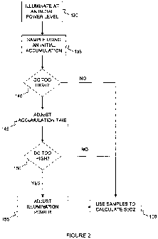
[00021] Figure 2 depicts a process for detecting and processing pulsatile
biometric signals
of light, according to an illustrative embodiment of the invention.
[00022] Figure 3 depicts a process for detecting and processing pulsatile
biometric signals,
according to another illustrative embodiment of the invention.
[00023] Figure 4 is a block diagram of a circuit for detecting and processing
pulsatile
biometric signals, according to an illustrative embodiment of the invention.
~
CA 02696054 2010-02-09
WO 2009/064979 PCT/US2008/083547
-6-
DETAILED DESCRIPTION OF THE INVENTION
[00024] While the illustrative embodiments disclosed herein are described in
the context
of pulse oximetry, the illustrative embodiments can be applied in other
contexts that relate to
signal processing of a pulsatile biometric signal. The illustrative
embodiments can be applied to,
for example, a reflective pulse oximetry system or a transmission pulse
oximetry system.
[00025] Figure 1 is a schematic of a pulse oximetry system 100. A pulse
oximetry system
can detect samples using both red and infrared (IR) light at a plurality of
times per second. The
system can include a plurality of light sources 105A and 105B (e.g., a source
of red light, a
source of infrared light, etc.) that can be activated to transmit light for a
period of time (e.g., an
accumulation time). A detector unit 110 (e.g., photodetector and a
preamplifier unit) can detect
(e.g., sense or acquire) light reflected 115 (e.g., which can include the
reflected red light or
infrared light) from the living subject 120 and can convert the detected light
(e.g., the photons
from the reflected light) into an electric signal 125. The signal 125 can be
processed to
determine, for example, the heart rate of the subject and/or the level of
oxygen saturation of the
living subject 120.
[00026] For each sample detected by a detector unit 110 (e.g., photons of
light detected by
a photodetector), the detector unit 110 can accumulate charge for a fixed
interval of time, where
the accumulated charge is proportional to the amount of light detected. In
some embodiments,
the amount of time that the detector unit 110 accumulates charge is
proportional to the amount of
time that the light source 105A or 105B transmits the light. Therefore, the
sampling rate of the
detector can be governed by the amount of time that the light source 105A or
105B transmits
light (e.g., accumulation time). The output of the detector unit 110 can be
then provided to an
analog to digital converter, which can convert the detector output into a
digital word (e.g., signal
125) that represents the amount of light received (e.g., reflected light 115),
which can be
processed. The system 100 can compute a level of oxygen saturation (e.g.,
Sp02) of the living
subject 120 by determining a ratio of ACRED/DCRED to ACIR/DCIR where ACRED is
the value of
the AC component (e.g., the pulsating part) of the reflected red light, DCRED
is the value of the
DC component (e.g., the average) of the reflected red light, ACIR is the value
of the AC
component (e.g., the pulsating part) of the reflected infrared light and DCIR
is the value of the
DC component (e.g., the average) of the reflected infrared light.
CA 02696054 2010-02-09
WO 2009/064979 PCT/US2008/083547
-7-
[00027] Figure 2 depicts a process for detecting and processing pulsatile
signals of light,
according to an illustrative embodiment of the invention. The process can be
applied to, for
example, a pulse oximetry system that can be reflectance pulse oximeter or a
transmission pulse
oximeter. The system can activate (e.g., illuminate) a light source (e.g., a
LED) to transmit light
to a living subject during an accumulation time. The system can use an initial
power level (e.g.,
predetermined power level) for the light source (step 130). In some
embodiments, the system
includes a plurality of light sources (e.g., a red light source, an infrared
light source, or any
combination thereof etc) which can be activated to transmit light to the
living subject. The light
source can be initially activated/operated at a maximum power level intensity
of an LED source.
The system can detect at least one sample of the light reflected from a living
subject (e.g., the
patient's body) during an initial accumulation time using a detector unit
(e.g., photodetector and
an analog to digital converter circuit) (step 135). The signal (e.g., the
light reflected from the
patient, detected by the photodetector, outputted as an electrical analog
signal which can be
converted as a digital signal) includes a sum of an AC component and a DC
component. The
samples (e.g., or corresponding signals) of light can be processed to
determined if the sample
approaches a saturation level of the detector unit (step 140). If the sample
approaches a
saturation level of the detector, the system can adjust an accumulation time
(e.g., the duration of
each sample or the time that the light source is illuminated/activated) to
maintain the light source
at the initial power level without approaching saturation of the detector unit
(step 145). The
signal can approach a saturation level of the detector unit if a DC component
of the signal is too
high. If adjusting an accumulation time does not prevent the signal from
approaching a
saturation level of the detector unit (step 150), the system can continue to
adjust the
accumulation time until a minimum accumulation time value is reached (e.g.,
200 ms). If the
system has adjusted an accumulation time to the minimum value, the system can
avoid detector
saturation by reducing the power to the light source in steps (step 155). The
sample(s) detected
by the detector unit can be used to calculate, for example, an oxygen
saturation level of a living
subject (step 160).
[00028] A detector unit becomes saturated when the ADC converter (e.g., analog
to digital
converter circuit) of the detector unit reaches its maximum value. In some
embodiments, the
saturation level of a detector unit is 1,000,000 units. If a detector unit
becomes saturated (e.
g=,
the detector is no longer able to convert more units of photons of light into
a digital signal), the
detector unit outputs a constant value which represents the maximum value that
the electric
circuit is capable of providing. For example, if the saturation level of a
detector is a
ft
CA 02696054 2010-02-09
WO 2009/064979 PCT/US2008/083547
-8-
number of units and the light detected exceeds this value, the detector will
only
predetermined
output a constant value. The constant value can become the DC component of the
signal but a
saturated detector unit can not detect an AC component of the signal,
therefore, a pulse rate and
oxygen saturation level Sp02 can not be calculated.
[00029] The accumulation time can be the length of time that the light source
is activated
and illuminates/transmits light to the subject or the conversion time of the
detector signal. To
adjust an accumulation time, the system can vary the length of time that the
light source
illuminates/transmits light. In some embodiments, the system adjusts an
accumulation time by
adjusting a sampling time of the detector (e.g., adjusting the conversion time
of the detector
signal).
[00030] By way of example, a pulse oximeter system can activate light source
to be
illuminated (e.g., step 130) at a maximum power level and detect samples
during an initial
accumulation time (step 135). The accumulation time can be the time that the
light source is
activated (e.g., the period of time that the light source transmits light).
The initial accumulation
time can be predetermined (e.g., 400 micro seconds). The system can process
the signal(s) to
determine if the corresponding sample(s) detected by the detector (e.g., the
integrated units of
light or total units of light acquired during the accumulation time)
approaches a saturation level
of the detector unit (step 140). The system can determine if a signal
approaches a saturation
level of the detector unit by determining if the sample reaches a threshold
value. The threshold
value can establish a triggering event prompting the system to adjust the
accumulation time (step
145) to avoid detector unit saturation. In some embodiments, if the signal
received by the
detector reaches a threshold value, the system responds by lowering the
accumulation time of the
detector. The triggering threshold value can be 70% to 85% of the saturation
level of the
detector. The system can adjust the accumulation time (step 145) to lower the
accumulation time
by shortening activation time of the light source (e.g., the amount of time
that the light source
transmits/illuminates light to the living subject). The system can continue to
adjust the
accumulation time (e.g., continue to lower the accumulation time) to avoid
saturation until a
minimum accumulation time value (e.g., 200 micro seconds) has been reached. By
adjusting the
accumulation time instead of adjusting a power level of the light source, the
system can avoid
saturation of the detector unit while simultaneously operating the light
source at a desired power
level (e.g., a maximum power intensity).
CA 02696054 2010-02-09
WO 2009/064979 PCT/US2008/083547
-9-
[00031] The light source can include an LED and an electronic driver for the
LED. As
more light is transmitted to a living subject, the deeper the light penetrates
in the tissue of a living subject. Transmitting more light (e.g., transmitting
light at a higher power level) can
allow the system to collect information from as much tissue volume as
possible, thereby
increasing accuracy of the measurements. In some embodiments, the system
lowers an
accumulation time so that the system can operate at a maximum power intensity
level of the light
source, thereby increasing accuracy of the measurements.
[00032] As described in Figure 2, the accumulation time can be
adjusted/lowered to avoid
saturation of the detector (e.g., step 145). However, a minimum value for the
accumulation time
can be set so that the accuracy of the signal does not suffer. An accumulation
time should be
long enough to reduce the electronic noise to alter the accuracy (signal to
noise ratio). If a noise
level of a device is constant, a greater accumulation time results in a signal
that is more valid
(e.g., a better/greater signal to noise ratio). The minimum accumulation time
value of a system
can be defined according to the noise level of the system. By setting a
minimum accumulation
time value (e.g., the lowest possible accumulation time the system can handle
before accuracy of
the signal begins to suffer), the system can start off at a maximum power
intensity level for the
light source and incrementally lower the accumulation time to obtain the
optimum working
condition while avoiding saturation of the detector unit. If the accumulation
time has been
lowered to a minimum accumulation time value and lowering it further would
cause the accuracy
of the signal to suffer, the system can avoid detector saturation by lowering
an intensity of the
light source (e.g., a power level of the transmitted light) (e.g., step 155).
[00033] Figure 3 depicts a process for detecting and processing a pulsatile
biometric
signal, according to another illustrative embodiment of the invention. The
embodiments as
described herein can be used to process a signal that includes a pulsatile
waveform (e.g.,
pulsatile biometric signal) that is swamped by a background signal or noise
(e.g., a signal
comprising an AC component and a DC component where the DC component can be
substantially larger than the AC component). The pulsatile waveform can be
separately
extracted to obtain, for example, critical timing information. The critical
timing information can
be used to process signals that include both the pulsatile waveform and the
background
signal/noise. For example, a method for processing a pulsatile biometric
signal can include
detecting a first set of signals from a living source, where the first set of
signals include a
pulsatile waveform and a constant component. The pulsatile waveform can be
extracted from
CA 02696054 2010-02-09
WO 2009/064979 PCT/US2008/083547
-10-
the first set of signals and processed to generate critical timing
information. The method can
also include, for example, selecting from a second set of signals based on the
critical timing information (e.g., heart rate of a living subject or a rising
portion of the pulsatile waveform) to
calculate a biometric measurement. The second set of signals can include both
the pulsatile
waveform and the constant component.
[00034] A pulse oximetry system can include a plurality of light sources
(e.g., red light
source, a first infrared light source, a second infrared light source, etc.)
that can be individually
or simultaneously activated to transmit light. In some embodiments, the
plurality of light
sources can be activated at different points in a cycle or at different time
periods. A detector can
detect samples (e.g., light reflected from the patient's body) of reflected
light (step 165) during a
first cycle, and another set of signals from a second cycle, etc. The detector
can output analog
signals from the detected samples of light that can be converted to digital
signals. The signals
detected during the different cycles can be processed in two ways. A first set
of signals (e.g.,
analog signals) can be filtered to remove the DC component of the signal (step
170) and extract
the AC component to obtain data relating to a pulsatile waveform (step 175).
In some
embodiments, the pulsatile waveform is a cardiac/arterial waveform of a
patient/subject and the
data is a heart rate and/or the location of the rising portion of an arterial
waveform. The data
relating to the pulsatile waveform can be used to process the data from
another set of signals that
include both the AC component and the DC component. The data relating to the
pulsatile
waveform can be used to identify which one(s) of the signals from the second
set of signals
should be used (step 180) to determine, for example, a biometric measurement
of a subject such
as the level of oxygen saturation (step 185). The set of signals used to
detect/extract the AC
component of the signal can be detected in different cycle than the set of
signals where the AC
component and DC component have been maintained and are used to calculate a
biometric
measurement of a subject.
[00035] In a pulse oximetry system, information from different wavelengths of
light (e.g.,
a red light "R", infrared light "IRl", and a second infrared light "IR2") are
used to calculate an
oxygen level of a living subject. The pulse oximetry system can include a red
light source, a first
infrared light source and a second infrared light source. The light sources
can be
activated/operated separately or any combinations the individual light sources
can be activated
simultaneously (e.g., R, IRI, IR2, R+IRI, R+IR2, R+IRI+IR2, etc.) during
different time
periods/cycles. During each time period/cycle, a detector can detect reflected
light from the
CA 02696054 2010-02-09
WO 2009/064979 PCT/US2008/083547
-11-
living subject and output corresponding signals (e.g., sets of signals
corresponding to each time
period/cycle). Light sources can be activated every cycle, every second cycle,
every third cycle
or every fourth cycle. For example, a first light source (e.g., the red light
source) can be
activated during a first time period/cycle, a second light source (e.g., a
first infrared light source)
can be activated during a second time period/cycle, a third light source
(e.g., a second infrared
light source) can be activated during a third time period/cycle, and any
combination of the light
sources can be activated simultaneously during a fourth time period/cycle
(e.g., red light source
+ first infrared light source, red light source + second infrared light
source, red light source +
first infrared light source + second infrared light source, etc.).
[00036] In conventional oximeters, the signal from the reflected light
including the AC
component and DC component is detected and converted to a digital form by the
detector unit
(e.g., the analog to digital converter (ADC) of the detector), and digital
signal processing (e.g.,
digital filtering) is used to separate the DC component and AC component.
However, by
detecting an AC signal using a set of signals (e.g., signals detected at a
point in the cycle when
all the light sources are activated) to calculate critical timing information,
a pulse oximetry
system can generate greater accuracy of oxygen level measurements. The
critical timing
information from the AC component of the signal can be used to process signals
acquired during
other cycles to calculate, for example, oxygen level measurements. For
example, signals from
the other cycles (e.g., where only one or some of the plurality of light
sources is on) can be used
to calculate, for example, an oxygen level of a patient. Signals from the
other cycles can include,
for example, R, IRI, IR2, R+IRl, R+IR2, where R is the activated red light
source, IRl is an
activated first infrared light source, and IR2 is the activated second
infrared light source. These =
signals can be processed to better filter the noise (e.g., as we know already
where the real pulses
are), resulting in greater accuracy of oxygen level measurements.
IS
[00037] In some embodiments, the AC component is calculated/extracted using
sets of
signals (e.g., analog signals) of the reflected light detected in the
cycle/time period when the
plurality of light sources are simultaneously activated (step 190). Activating
the plurality of light
sources simultaneously can result in increased power. For example, where the
light sources are
simultaneously activated every fourth cycle, the extraction of the AC
component (e.g., filtering
to remove the DC component) can be done on sets of signals sampled for every
fourth cycle.
The system can execute a sample and hold operation to coincide with the
desired cycle (e.g., the
cycle when the light sources are simultaneously activated), and filter the
output (e.g., analog
CA 02696054 2010-02-09
WO 2009/064979 PCT/US2008/083547
-12-
signal) of the sample and hold to remove the DC component and extract out the
AC component
(e.g., step 170). Extracting out the AC component can include detecting the AC
signal to
eliminate preserving the ratio of the AC component of the signal to the
background signal/DC
component of the outputted signal.
[00038] To extract out the AC component of a signal (e.g., an analog signal),
the signal
can be filtered (e.g., by an analog filter) while it is in analog form (step
195 and step 200). The
signal can be filtered to remove the DC component and only the AC component of
the reflected
light can be converted into a digital signal (e.g., where there is no need for
high dynamic range
from the ADC). Using an analog filter to filter out the DC component of the
reflected light can
result in an AC component that is stronger relative to the regular form (e.g.,
where AC
component and the DC component has been maintained). It can be easier to
filter out the DC
component of a reflected light using an analog filter (e.g., filtering out the
DC component while
the signal is in analog form). The system may not acquire wavelength
information, but the
increased power in the signal results in a larger AC component of the signal
(e.g., pulsating part
(AC) of the signal). A larger AC signal results in a better/greater signal to
noise ratio. Critical
timing information, such as the pulse rate or the location of the pulse, can
be detected more
easily with a larger AC signal.
[00039] The system can filter out the DC component of the signal using an
analog filter
(e.g., an analog filter with a 6db at the pass points) while it is in analog
form. The analog filter
can be a bandpass filter (e.g., high and low pass filters) (step 200).
Filtering out the DC
component (step 200) can help extract the pulsatile waveform (e.g., the AC
component of the
signal). In some embodiments, the system is a pulse oximetry system and the
pulsatile
waveform is an arterial waveform. The system can use high pass filters and low
pass filters to
isolate/extract the specific pulsatile waveform. For example, if a typical
heart rate has a
frequency of approximately 30-300 beats per min, the system can use high pass
filters and low
pass filters to filter out portions of the signal that do not fall within the
specified frequency
range, therefore filtering out the portion of the signal that is not related
to the arterial blood flow.
In some embodiments, the high pass filter is used to filter out frequencies
below about 0.7 Hz
and the low pass filter is used to filter out frequencies above about 5.5 Hz,
to extract a signal
having frequencies within the range of about 0.7 Hz to about 5.5 Hz.
[00040] The system can use the filters (step 195 and step 200) to generate a
large and
precise arterial waveform, (e.g., instead of a very small AC component of the
signal) which can
CA 02696054 2010-02-09
WO 2009/064979 PCT/US2008/083547
-13-
be processed to determine the heart rate of a patient and determine critical
timing information,
such as the heart rate and/or locating the rising portion of the periodic
signal (step 175). The step
of processing a signal to determine the heart rate and/or locate the rising
portion of the signal can
be implemented using an algorithm such as the artificial waveform templates
described in U.S.
patent application 11/536,058 entitled "Signal Processing for Pulse Oximetry"
filed on
September 28, 2006, which is incorporated herein by reference in its entirety.
[00041] In some embodiments, the system blue filters (step 195) the analog
signal
outputted from the sensor of the detector. A pulse oximetry system can use a
blue (step 195) to
filter out the blue light to isolate/extract the pulsatile waveform (e.g., the
arterial waveform). In
some embodiments, the system uses a blue filter to filter out the portion of
the signal that
substantially corresponds to non-moving, non-arterial or venous blood and
extract out the portion
of the signal corresponding to arterial blood, which can be used to calculate
the level of oxygen
saturation.
[00042] Data relating to critical timing information, such as the heart rate
and location of
the rising portion (e.g., identified from the extracted AC component/AC signal
in step 175), can
be sent to the digital portion of the system (step 205). The critical timing
information can be
used to select/identify the signals (step 180) to be processed or analyzed to
calculate an oxygen
level in a patient. For example, signals that correspond to a rising portion
of an arterial
waveform (e.g., the AC component) can be selected and used to calculate, for
example, an
oxygen saturation level (step 185). The AC to DC relationship of these signals
has been
maintained (e.g., the signal includes both the DC component and AC component).
In some
embodiments, the signals corresponding to the cycle where all the light
sources are activated are
used to calculate critical timing information while the signals from the other
cycles (e.g., where
only one/some but not all of the light sources are activated) are used to
calculate biometric
measurements, such as, oxygen saturation level of a patient. For example, if
all the light sources
are activated every fourth cycle, the signals from the first, second and third
cycles can be used to
calculate biometric measurements. The critical timing information can be used
to enhance
accuracy of the biometric measurements (e.g., Sp02) by determining which
signals from the
other cycles should be used in calculating biometric measurements.
[00043] By way of example, a method for processing a pulsatile signal of light
reflected
from a living subject can include simultaneously activating at least a first
light source and a
second light source (e.g., red light source and infrared light source) to
transmit light to a living
CA 02696054 2010-02-09
WO 2009/064979 PCT/US2008/083547
-14-
subject during a first time period. The method can include detecting a first
set of signals from
light reflected from the living subject using a detector unit (e.g., step 190)
and filtering out a DC
component of the first set of signals (e.g., using at least one of a high
pass, low pass, or band
pass filter) to extract an AC component of the first set of signals (e.g.,
step 170). Detecting a
first set of signals can include outputting a first set of analog signals from
light reflected from the
living subject. The method can also include processing the AC component to
identify a rising
portion of the AC component (e.g., step 175) used to calculate a level of
oxygen saturation of the
living subject (e.g., steps 180 and 185). The AC component can also be
processed to determine a
heart rate of the living subject (e.g., step 175). The method can also include
activating the first
light source to transmit light to a living subject during a second time period
and detecting a
second set of signals from the light reflected from the first light source
during the second time
period. The level of oxygen saturation of the living subject can be calculated
based on signals
selected from the second set of signals corresponding to the rising portion of
the AC component
(e.g., step 180).
[00044] Figure 4 is a block diagram of a circuit for detecting and processing
pulsatile
biometric signals, according to an illustrative embodiment of the invention.
The circuit can
include a sensor 210A or 210B for detecting samples of a biometric pulsatile
signal (e.g., a
photodetector that detects photons from reflected light from a patient). The
circuit can generate
signals (e.g., analog signals) based on the samples detected by the sensor
210A or 210B. The
signals can include both an AC component (e.g., a pulsatile waveform) and a DC
component.
The circuit can include a preamplifier 215 that can amplify the signals from
the sensor 210B.
The circuit can include a first module 220 and a second module 225. The first
module 220 can
filter a selected set of the signals from the samples detected from the sensor
210B to filter out a
DC component and isolate/extract an AC component of the signal. The AC
component of the
signal can be used to determine critical timing information (e.g., determine
heart rate and/or
identify the rising portion of an arterial waveform). The second module 225 of
the circuit can
process the signals from the samples detected by the sensor 210A or 210B,
(e.g., to calculate an
oxygen saturation level), which still have the AC component and the DC
component of the
signals preserved, using critical timing information generated from the first
module 220. The
circuit can also include analog to digital converters 250, 255 or 260.
[00045] In some embodiments, a preamplifier 215 filters the detector signal
before digital
conversion by converters 250 or 255. From the premplifier 215, the circuit can
include two
CA 02696054 2010-02-09
WO 2009/064979 PCT/US2008/083547
-15-
paths. A first path can go to an analog to digital converter 255, where the
digital signal
generated includes an AC component and a DC component which is later used to
calculate, for
example, an oxygen saturation level of a patient. The second path can be used
to filter out a DC component of the signal, resulting in an AC component of an
analog signal that can be used to
obtain critical timing information, such as pulse rate calculations and a
rising portion of a
pulsatile waveform.
[00046] In some embodiments, only some of the signals from sensor 210B are
used to ff
detect/extract the AC component by the first module 220. For example, a
plurality of light
sources in a pulse oximetery system may not be simultaneously activated during
every cycle.
Any one or any combination of the plurality of light sources can be activated
at different time
periods in a sequence. In some embodiments, the analog filters 235 and 245
filter a continuous
analog signal during one time period/cycle in the sequence. In some
embodiments, the AC
signal is only detected (e.g., the DC component filtered out) and the pulse
rate is calculated on
signals during the cycles/time periods/sequence when more than one light
source is activated
simultaneously. A controller 229 can control a sample and hold unit 230 to
operate on the
analog signal from the detector only during the cycle/time period where all or
more than one of
the light sources are simultaneously activated (e.g., cycle when a red light
source, a first infrared
light source and a second infrared light source is activated simultaneously).
[00047] In some embodiments, the plurality of light sources (e.g., red light
source, first
infrared light source, second infrared light source, etc.) in a pulse oximetry
system are
simultaneously activated every fourth cycle and a sample and hold can operate
on the analog
signal every fourth cycle. The analog signal from this fourth cycle can be
processed by the first
module 220 and filtered to remove the DC component, amplified using amplifier
240 and
converted to a digital form by converter 260 (e.g., A2D-1). The AC component
of the signal can
be used to calculate, for example, the pulse rate/location of a living
subject. The analog signals
from the other cycles (e.g., R, IRI, IR2) which still include the AC component
and the DC
component can be used to calculate, for example, an oxygen saturation level of
a living subject.
The analog signals from these other cycles (e.g., where more than one LED is
not activated) can
be converted to a digital form (e.g., with converter 255). In some
embodiments, the signal from
each light source during each "on period" (e.g., the period of time when each
LED is
individually activated) is converted to a digital form by converters 255 and
260 in the second
module 225. These digital signals reflect information from each individual
light source and
CA 02696054 2010-02-09
WO 2009/064979 PCT/US2008/083547
-16-
reflect information from the different wavelengths of light (e.g., red, first
infrared, second
infrared, etc.) which can be used to calculate the oxygen (e.g., Sp02) level.
[00048] By way of example, a red light source, first infrared light source and
second light
infrared source can be activated according to the following cycles/sequences:
R, IRI, IR2,
IRI+IR2+R, R, IR1, IR2, etc. where R is the red light source, IR1 is a first
infrared light source,
and IR2 is a second infrared light source. In some embodiments, only the
signals from the
detector during the cycle where all the light sources are simultaneously
activated are filtered
analog filters 235 and 245 (e.g., every fourth cycle/time period/sequence). To
reconstruct an
analog signal from the fourth sequence, a controller 229 can send a "sample
signal" to the
sample and hold unit 230 during the fourth sequence to direct the sample and
hold unit 230 to
sample the detector output during this time period. The output of the sample
and hold unit 230
can remain with this value (e.g., hold) during the other sequences (e.g.,
first sequence where red
light is activated, the second sequence when the first infrared source is
activated and the third
sequence when the second infrared source is activated) when the other light
sources are
individually activated. The result is the sample and hold 230 can output an
analog signal that
corresponds to the time periods of the sequence where all the light sources
are activated (e.g.,
fourth sequence). This analog signal can be now processed by the filters 235
and 245 and
amplifier 235 of the second module 220 and converted into a digital form by
converter 250.
[00049] The selected set of analog signals can be sent to a first filter 235
in the first
module 220, which can be a high pass filter. In some embodiments, the high
pass filter allows
signals of a frequency greater than approximately 0.7 Hz to pass, filtering
out signals having a
frequency lower than approximately 0.7 Hz. The circuit can also include an
amplifier 240 that
amplifies the filtered signal and correct for any offsets. The electronics
from the circuit (e.g.,
amplification from preamp 215 and filtering from filter 235) can add another
DC component to
the signal (e.g., offsets or voltage bias) that can be removed using amplifier
240 with an inverted
input that adjusts the DC voltage to the level needed to eliminate the DC
component that is
removed by the first module 220. The circuit can also include a second filter
245. In some
embodiments the second filter 245 is a low pass filter that filters out
signals having a frequency
greater than approximately 5.5. Hz. The signal outputted/generated by the
first module of the
circuit, therefore, is the AC component of the signal (e.g., the pulsatile
waveform resulting from
the DC component having been filtered out) that is converted into a digital
signal by converter
250. In some embodiments, the resulting AC component of the signal has been
amplified and
CA 02696054 2010-02-09
WO 2009/064979 PCT/US2008/083547
-17-
filtered to generate a large and precise arterial waveform. The AC component
of the signal
(e.g., the pulsatile waveform) can be used to generate critical timing
information (e.g., rising
portion of an arterial waveform). [00050] The second module 225 of the circuit
processes the analog signals from the
samples detected by the sensor 210A or 210B which can be converted into
digital signals using
converters 255 and 260. These signals still have the AC component and DC
component
maintained/preserved. These signals can be the signals from, for example, the
cycles where the
all the light sources are not simultaneously activated. In some embodiments,
the signals
processed by the second module are not the signals that are filtered and
processed by the first
module to generate an AC only component. In some embodiments, the second
module includes
an inverter 265. An amplifier (e.g., preamp 215) can act as a converter and an
inverter 265 can
invert the input signal from the sensor 210B and ADC converter 255. The
signals from the
sensor 210A or 210B can be processed by the circuit (e.g., second module 225)
to determine, for
example, a heart rate and/or oxygen saturation level of a subject. The
critical timing information
from the AC component of the signal generated by the first module 220 can be
used to select
which of the signals (e.g., signals from converter 255 or 260) from the second
module 225 will
be processed, for example, to calculate a level of oxygen saturation in a
subject.
[00051] In some embodiments, the system has two sensors 210A and 210B (e.g.,
photodetectors) that detects light reflected from different locations, which
can enhance the
accuracy of the measurements (e.g., measurement of level of oxygen
saturation). In this
embodiment, the system includes a sensor near 210B and sensor far 210A that
detects reflected
light at different depths/distances to ensure that readings are not distorted
by, for example, a vein
located close to one of the sensors.
[00052] While the invention has been particularly shown and described with
reference to
specific illustrative embodiments, it should be understood that various
changes in form and detail
may be made without departing from the spirit and scope of the invention.