Note : Les descriptions sont présentées dans la langue officielle dans laquelle elles ont été soumises.
CA 02696055 2012-03-19
1
METHOD AND APPARATUS FOR PROSTHETIC VALVE REMOVAL
Field of Invention
[0002) The present invention relates generally to removal of a previously
implanted cardiovascular valve, and more particularly to method and apparatus
for
facilitating removal of a percutaneously implantable valve (PIV) without open-
heart
surgery.
Background of the Invention
[0003] The demographics of patients suffering valvular disease are broad
and the
treatment modalities for each are complex. Historically, patients younger than
65
years of age have received mechanical valves, while older patients have
received
bioprosthetic valves. A new demographic of prosthetic valve recipients has
emerged
recently, namely, the old, sick, inoperable patient who previously would not
be a
candidate for surgical implantation of a prosthetic valve. These patients are
now
candidates for a relatively new type of prosthetic valve, i.e., the
percutaneously
implantable valve (PIV). The PIN is configured like an endovascular stent,
except
with a tissue valve sewn in the lumen. Like the endovascular stent, the PIV is
balloon
expandable or self-expanding, and is delivered by way of a catheter to the
operative
site, where it is deployed and the delivery system removed. The principal
advantage
of a PIV is that it avoids open-heart surgery. The old, sick patients who
would
otherwise not survive open heart surgery, can now benefit from the PINT.
[0004] Because of a number of design constraints, PIV's are expected to be
less
durable and are likely to wear out sooner than conventional, surgically
implantable
valves. Although PIVs are intended for the old, sick patients who have a
relatively
short life expectancy, there may be instances in which the patient outlives
the
functional lifespan of the PIV. Therefore, when the PIV ceases to function, it
must be
replaced.
[0005] One potential solution to replacement of a PIV is to insert a new
PIV inside
the pre-existing PIV. In the field of interventional cardiology, this
replacement
CA 02696055 2010-02-09
WO 2009/026272 PCT/US2008/073565
2
process is referred to as "restenting." Restenting a PIV invariably leads to a
reduction
of effective orifice area of the prosthetic valve, since the old metal cage
and worn-out
calcified leaflets remain in place and the new PIV is smaller than the pre-
existing PIV
in order to allow it to be inserted into the remaining lumen. Depending on the
original
size of the first PIV, and the degree of calcification and obstruction,
restenting with
another PIV may not lead to an effective orifice area that is compatible with
good
cardiac function.
[0006] As indicated above, there may be instances where an old, worn-
out PIV
will need to be replaced. Currently, the only means of replacing an old, worn-
out,
fibrosed PIV is through open heart surgery. Since the patient likely received
the PIV
because they were not a candidate for open-heart surgery and implantation of a
conventional bioprosthesis, the patient is unlikely to be a candidate for open
heart
surgery to replace a worn or failed PIV. Therefore, non-surgical removal of
the
existing PIV is a preferred option.
[0007] In view of the issues discussed above, the concept of a system
for the
removal of an old and/or failed PIV becomes very desirable. The present
invention
provides a method and apparatus for non-surgical removal of a PIV, and
includes a set
of tools comprising a valve holding tool, a cutting tool and a valve removal
tool that
facilitate removal of the PIV through the apex of the heart.
Summary of the Invention
[0008] In accordance with the present invention, there is provided a
holding tool
for facilitating removal of an implantable cardiovascular valve, the holding
tool
comprising: a first sliding member; a second sliding member moveable relative
to the
first sliding member; and a first articulating joint member connected to the
first and
second sliding members, said articulating joint member moveable between a
collapsed
position and an expanded position, wherein movement of the second sliding
member
relative to the first sliding member moves the first articulating joint member
between
the collapsed and expanded positions.
100091 In accordance with another aspect of the present invention,
there is
provided a cutting tool for facilitating removal of an implantable
cardiovascular valve,
the cutting tool comprising: a shaft having a longitudinal axis; and a cutting
arm
CA 02696055 2010-02-09
WO 2009/026272 PCT/US2008/073565
3
extending from the hollow shaft, wherein said cutting arm includes cutting
means for
cutting tissue.
[0010] In accordance with still another aspect of the present
invention, there is
provided a valve removal tool for facilitating removal of an implantable
cardiovascular valve from a heart, the valve removal tool comprising: a body;
capture
means mounted to the body and moveable between a collapsed position and an
expanded position, for capturing the implantable cardiovascular valve; and an
actuator
for actuating movement of the capture means between the collapsed and expanded
positions.
[0011] In accordance with yet another aspect of the present invention,
there is
provided a method for removing an implantable cardiovascular valve from a
heart, the
method comprising: holding the cardiovascular valve using a valve holding
tool;
separating the cardiovascular valve from fibrotic tissue that accumulates
adjacent to
the cardiovascular valve; and removing the cardiovascular valve from the heart
using a
valve removal tool, said step of removing including: capturing the
cardiovascular
valve, and extracting the cardiovascular valve from the heart.
[0012] An advantage of the present invention is the provision of
apparatus for
facilitating removal of a percutaneously implantable valve (PIV) from a heart.
[0013] Another advantage of the present invention is the provision of a
valve
holding tool, a cutting tool and a valve removal tool for facilitating removal
of a
percutaneously implantable valve (PIV) from a heart.
[0014] A still further advantage of the present invention is the
provision of a
method for facilitating removal of a percutaneously implantable valve (PIV)
from a
heart.
[0015] These and other advantages will become apparent from the
following
description taken together with the accompanying drawings and the appended
claims.
Brief Description of the Drawings
[0016] The invention may take physical form in certain parts and
arrangement of
parts, an embodiment of which will be described in detail in the specification
and
illustrated in the accompanying drawings which form a part hereof, and
wherein:
[0017] FIG. 1 is a perspective view of a typical PIV shown
schematically;
CA 02696055 2010-02-09
WO 2009/026272 PCT/US2008/073565
4
[0018] FIG. 2 is a schematic diagram showing a PIV deployed inside a
native
aortic valve;
[0019] FIG. 3 is a bottom perspective view (inflow aspect) of an aortic
root of a
heart, including native aortic valve leaflets;
[0020] FIG. 4 is a partial cut-away view of an aortic root of a heart
with a PIV
inserted between the native aortic valve leaflets.
[0021] FIG. 5 is a perspective view of a valve holding tool of the
present
invention, according to a first embodiment, wherein the valve holding tool is
shown in
a collapsed position;
[0022] FIG. 6A is a plan view of the articulating joint member of the
valve
holding tool of FIG. 5, wherein the valve holding tool is shown in a collapsed
position;
[0023] FIG. 6B is a plan view of the articulating joint member of the
valve holding
tool of FIG. 5, wherein the valve holding tool is shown in an expanded
position;
[0024] FIG. 7 is a perspective view of a valve holding tool of the
present
invention, according to a second embodiment, wherein the valve holding tool is
shown
in an expanded position;
[0025] FIG. 8 is a plan view of the articulating joint member of the
valve holding
tool of FIG. 7, wherein the valve holding tool is shown in a collapsed
position;
[0026] FIG. 9 is a perspective view of the valve holding tool of FIG. 7
in the
expanded position and engaged with a PIV;
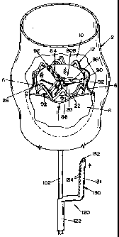
[0027] FIG. 10 is a perspective view of a cutting tool of the present
invention,
wherein the cutting tool is shown mounted over a stem portion of the valve
holding
tool shown in FIG. 7;
[0028] FIG. 11 is a perspective view showing the valve holding tool of
FIG. 7
engaged with a PIV located inside an aortic valve, and a cutting tool mounted
over the
stem portion of the valve holding tool;
[0029] FIG. 12 is a perspective view showing the valve holding tool of
FIG. 7
engaged with a PIV located inside an aortic valve, and a cutting tool mounted
over the
stem portion of the valve holding tool, the cutting tool having a cutting arm
located
between the PIV and the native aortic valve leaflets;
CA 02696055 2010-02-09
WO 2009/026272 PCT/US2008/073565
[0030] FIG. 13 is a top plan view of the aortic valve shown in FIG. 12,
wherein
the cutting arm of the cutting tool is located adjacent to the PIV, the
cutting tool
burning a channel adjacent to the metal cage of the NV;
[0031] FIG. 14 is a top plan view of the aortic valve shown in FIG. 12,
wherein
the cutting arm of the cutting tool is located adjacent to the PIV, the
cutting tool
burning a generally annular-shaped recess along the periphery of the PIV metal
cage;
[0032] FIG. 15 is a schematic diagram showing use of a valve removal
tool of the
present invention for extracting the PIV from the heart, wherein the valve
removal tool
is inserted into the heart through the apex after removal of the cutting tool,
said
removal tool facilitating collapse and extraction of the PIV;
[0033] FIG. 16 is a partial cross-sectional view of the valve removal
tool of FIG.
15, shown with articulating arms in an expanded (open) position;
[0034] FIG. 17 is a perspective view showing the valve holding tool of
FIG. 7
engaged with a PIV located inside the aortic valve, and the valve removal tool
of
FIGS. 15 and 16 mounted over a stem portion of the valve holding tool, shown
with
articulating arms in an expanded (open) position;
[0035] FIG. 18 is a perspective view of the valve removal tool, shown
with
articulating arms in a partially collapsed position for capturing the PIV;
[0036] FIG. 19 is a perspective view of the valve removal tool, shown
with
articulating arms in a collapsed (closed) position, thereby capturing the PIV;
[0037] FIG. 20 is a removal tool of the present invention, according to
an
alternative embodiment, wherein a wire mesh basket is substituted for
articulating
arms, the removal tool shown in an expanded (open) position; and
[0038] FIG. 21 is the removal tool of FIG. 19 shown in a collapsed
(closed)
position.
Detailed Description of the Invention
[0039] Referring now to the drawings wherein the showings are for the
purposes
of illustrating an embodiment of the invention only and not for the purposes
of
limiting same, FIG. 1 shows a typical PIV 10 that may be removed in connection
with
the present invention. PIV 10 is generally comprised of a flexible,
expandable, tubular
member 12, a tubular liner 22 and a plurality of leaflets 26. As illustrated,
tubular
member 12 is a mesh cylinder or metal cage formed of intersecting wire
sections 14
CA 02696055 2010-02-09
WO 2009/026272 PCT/US2008/073565
6
that define a plurality of openings 16. Tubular member 12 is radially
expandable to
contact with tissue, as shown in FIG. 2. Liner 22 is formed of tissue or a
fabric, such
as a woven polyester (e.g., polyethylene terepthalate). Leaflets 26 are
typically
formed from pericardial tissue, such as bovine or equine pericardium.
Alternatively,
leaflets 26 may be formed of synthetic materials. It should be appreciated
that PIV 10
shown in FIG. 1 is exemplary of a typical PIV, and is not intended in any way
to limit
the scope of the present invention. In this respect, it is contemplated that
the method
and apparatus of the present invention are suitable for use in connection with
implantable cardiovascular valves of a wide variety of configurations.
[0040] FIG. 2 shows a portion of a heart, including aortic root 2,
mitral valve 7
and left ventricle 8. PIV 10 of FIG. 1 is shown deployed inside a native
aortic valve 4,
wherein PIV 10 is inserted between native valve leaflets 6. FIG. 3 is a bottom
perspective view (inflow aspect) of aortic root 2 without PIV 10. In FIG. 4,
aortic root
2 is shown in detail with PIV 10 installed between aortic valve leaflets 6. It
should be
noted that fibrotic tissue (not shown) will accumulate around PIV 10 during
the years
following implantation.
[0041] Referring now to FIGS. 5, 6A and 6B, there is shown a valve
holding tool
40 of the present invention, according to a first embodiment. Holding tool 40
is
generally comprised of a first sliding member in the form of an outer tubular
body 42,
a second sliding member in the form of an inner rod 52, and an articulating
joint
member 80 that is pivotally connected with tubular body 42 and inner rod 52.
Tubular
body 42 and inner rod 52 form a stem portion of holding tool 40. Tubular body
42
defines a cylindrical recess dimensioned to receive rod 52 and has an outer
surface
dimensioned to receive a detachable handle 60. One end of rod 52 is connected
with
tubular body 42 by articulating joint member 80, while the other end of rod 52
is
adapted to receive a detachable handle 70.
[0042] With reference to FIG. 5, detachable handles 60 and 70 facilitate
longitudinally movement of rod 52 relative to tubular body 42 for moving
articulating
joint member 80 between collapsed and expanded positions, as will be described
below. Notches 54 may be respectively formed in tubular body 42 and rod 52 to
provide flat surfaces suitable for secure attachment of handles 60 and 70.
[0043] Handle 60 includes a pair of pivotally connected arms 62a and
62b. In the
illustrated embodiment, a set screw 64 is provided that moves arms 62a and 62b
CA 02696055 2010-02-09
WO 2009/026272 PCT/US2008/073565
7
towards each other when tightened, and moves arms 62a and 62b away from each
other when loosened. Accordingly, arms 62a and 62b are moved towards each
other to
capture tubular body 42 between arms 62a and 62b, and thereby detachably
engage
handle 60 with tubular body 42. Handle 70 includes a recess 72 that defines a
pair of
fingers 74a, 74b. Rod 52 is captured between fingers 74a and 74b to attach
handle 70
to rod 52.
100441 It should be appreciated that handles 60 and 70 are exemplary
embodiments of suitable detachable handles for use in connection with holding
tool
40, and that the handles may take other suitable forms. Moreover, handle 60
may be
substituted for handle 70, and vice versa. Handles 60 and 70 are configured to
be
detachable to allow other tools (e.g., cutting and valve removal tools) to
conveniently
slide over the stem portion of holding tool 40, as will be described below.
[0045] Articulating joint member 80 is comprised of a plurality of
articulating legs
84. Each articulating leg 84 includes first and second leg sections 86 and 88
that are
pivotally connected to each other at a hub member 90. First leg section 86 is
pivotally
connected at one end with tubular body 42 and second leg section 88 is
pivotally
connected at one end with rod 52. Each hub member 90 includes a projection 92
dimensioned to engage with tubular member 12 of PIV 10. In the illustrated
embodiment, projection 92 takes the form of an outward extending hook 92. It
is
contemplated that projection 92 may take other suitable forms.
[0046] As rod 52 is moved relative to tubular body 42, articulating
joint member
80 moves between a collapsed position (FIGS. 5 and 6A) and an expanded
position
(FIG. 6B). In the expanded position, projections 92 can grasp wire sections 14
of
tubular member 12 and/or hook onto liner 22, thereby engaging holding tool 40
with
PTV 10.
10047] It should be appreciated that the angular geometry of
articulating joint
member 80 allows projections 92 to exert significant outward force against
tubular
member 12 and/or liner 22 of PIV 10, when articulating joint member 80 is
moved to
the expanded position. Accordingly, a surgeon removing PIV 10 can conveniently
grasp holding tool 40 with one hand, thereby stabilizing the heart and PW 10,
while
manipulating cutting tool 120 around PTV 10, as will be described below.
[0048] FIGS. 7 and 8 illustrate a holding tool 40A of the present
invention,
according to a second embodiment. Holding tool 40A includes a first sliding
member
CA 02696055 2010-02-09
WO 2009/026272 PCT/US2008/073565
8
in the form of an outer tubular body 102, a second sliding member in the form
of an
inner tubular body 104, a third sliding member in the form of an inner tubular
body
106, a fourth sliding member in the form of an inner rod 108, and a pair of
articulating
joint members 80A and 80B. hi this embodiment, outer tubular body 102, inner
tubular body 104, inner tubular body 106, and inner rod 108 form a stem
portion of
holding tool 40A, wherein inner tubular body 104 extends through outer tubular
body
102, inner tubular body 106 extends through inner tubular body 104, and inner
rod 108
extends through inner tubular body 106.
[00491 Articulating joint members 80A and 80B are essentially the same
as
articulating joint member 80 described above. Thus, like components are given
the
same reference numbers. Articulating joint member 80A is pivotally connected
with
tubular body 102 and inner tubular body 104. Similarly, articulating joint
member
8013 is pivotally connected with inner tubular body 106 and inner rod 108.
Notches
54A dimensioned to receive detachable handles are respectively formed in outer
tubular body 102, inner tubular body 104, inner tubular body 106, and inner
rod 108.
The detachable handles may take the form of handles 60 or 70 described above.
10050] As inner tubular body 104 is moved relative to tubular body 102,
articulating joint member 80A moves between a collapsed position (FIG. 8) and
an
expanded position (FIG. 7). Likewise, as inner rod 108 is moved relative to
inner
tubular body 106, articulating joint member 80B moves between a collapsed
position
(FIG. 8) and an expanded position (FIG. 7). In the expanded position,
projections 92
of articulating joint members 80A, 80B grasp wire sections 14 of tubular
member 12
and/or hook onto liner 22, thereby engaging holding tool 40A with PIV 10. FIG.
9
illustrates holding tool 40A in engagement with PIV 10.
[00511 It should be appreciated that holding tools 40, 40A not only
serve the
function of holding PIV 10, but also act as a guide to locate the cutting and
valve
removal tools relative to PIV 10.
10052] Referring now to FIGS. 10 and 11, there is shown a cutting tool
120
according to the present invention. In the figures, cutting tool 120 is shown
mounted
over the stem portion of holding tool 40A. It should be appreciated that
holding tool
40 may be substituted for holding tool 40A. Cutting tool 120 is generally
comprised
of a hollow shaft 122, a handle portion 126 extending from a first end of
shaft 122,
and an L-shaped cutting arm 130 extending from a second end of shaft 122.
CA 02696055 2010-02-09
WO 2009/026272 PCT/US2008/073565
9
[0053] Shaft 122 includes a cylindrical recess dimensioned to receive
the stem
portion of holding tool 40A. In this respect, shaft 122 is slidable over the
stem portion
of holding tool 40A, when all handles are detached therefrom. Handle portion
126
provides a surface for gripping and maneuvering cutting tool 120.
[0054] Arm 130 includes an elongated portion 131 that is generally
parallel to the
longitudinal axis of shaft 122. A plurality of axially-mounted fiber optic
guides 132
and a plurality of transverse-mounted fiber optic guides 134 are mounted to
elongated
portion 131 of arm 130. Internal channels (not shown), formed within handle
portion
126, shaft 122 and arm 130, are dimensioned to receive fiber optic cable 142.
Fiber
optic cable 142 connects fiber optic guides 132, 134 to a source of laser
energy (not
shown). Accordingly, laser energy is transmitted to fiber optic guides 132,
134 via
fiber optic cable 142. Fiber optic guides 132 emit laser beams in a direction
generally
parallel to the longitudinal axis of shaft 122, while fiber optic guides 134
emit laser
beams in a direction transverse to the longitudinal axis of shaft 122.
Accordingly,
fiber optic guides 132 are appropriately positioned to cut (i.e., burn) a
channel
adjacent to PIV 10 (FIGS. 12 and 13), and fiber optic guides 134 are
appropriately
positioned to cut (i.e., burn) a generally annular recess around the periphery
of PIV 10
(FIG. 14).
[00551 In FIGS. 10-14, cutting tool 120 is shown in conjunction with
holding tool
40A for the purpose of illustrating operation of cutting tool 120. However, it
should
be appreciated that holding tool 40 may be substituted for holding tool 40A.
[0056] It is contemplated that other suitable cutting means may be
substituted for
the laser-based cutting means comprised of fiber optic guides, fiber optic
cable and a
laser energy source. For example, the cutting tool may include cutting means
in the
form of a mechanical cutting device, such as a conventional mechanical
oscillating
cutting blade, or an electrosurgical cutting device. A conventional
electrosurgical
cutting device includes electrode(s) for applying a high frequency, high
voltage to
tissue. It is further contemplated that the cutting tool may include a
combination of
different types of cutting means.
[0057] The operation of cutting tool 120 will now be described detail
with
reference to FIGS. 11-14. After holding tool 40A is properly engaged with PIV
10 (as
described above), handles 60 and 70 are removed from holding tool 40A. Cutting
tool
120 is then mounted over the stem portion of holding tool 40A, as shown in
FIG. 11.
CA 02696055 2010-02-09
WO 2009/026272 PCT/US2008/073565
Cutting tool 120 is slid along the stem portion while fiber optic guides 132
are
energized to emit laser beams in an axial direction. Accordingly, a channel is
burned
adjacent to PIV 10, as shown in FIGS. 12 and 13. Thereafter, cutting tool 120
is
rotated circumferentially while fiber optic guides 134 are energized to emit
laser
beams in a transverse direction. Accordingly, a generally annular recess is
formed
around the periphery of PIV 10, as shown in FIG. 14. Handle portion 126 is
used to
move and rotate cutting tool 120 relative to PIV 10. The cutting of the
channel and a
complete annular recess using cutting tool 120 is necessary to separate PIV 10
from
fibrotic tissue that accumulates adjacent to PIV 10. After PIV 10 is separated
from
fibrotic tissue, cutting tool 120 is removed by dismounting it from the stem
portion of
holding tool 40A. PIV 10 is stabilized by grasping the stem portion of holding
tool
40. Handles 60, 70 may be re-attached to the stem portion after mounting
cutting tool
120.
100581 FIG. 15 schematically illustrates a valve removal tool 150,
according to a
first embodiment. After removal of cutting tool 120, valve removal tool 150 is
slid
over the stem portion of holding tool 40A and inserted into the heart through
the apex.
Operation of removal tool 150 will be described in detail below.
[0059] Removal tool 150 will now be described in detail with reference
to FIG.
16. Removal tool 150 resembles a trocar, and is generally comprised of a
hollow
cylindrical body 152, a plurality of articulating arms 180, a cylindrical
inner sleeve
202, a plurality of links 212 for connecting arms 180 to inner sleeve 202, and
an
actuator 170 for controlling movement of arms 180.
[0060] Inner sleeve 202 is located inside a cylindrical recess 153 of
cylindrical
body 152. Axial movement of inner sleeve 202 within cylindrical body 152
results in
movement of arms 180 between a collapsed (closed) position (FIGS. 15 and 19)
and
an expanded (open) position (FIG. 16). Inner sleeve 202 is connected with arms
180
via links 212. The first end 214 of link 212 has a ball hinge that is
dimensioned to be
received by a generally spherical cavity 204 formed in inner sleeve 202. The
second
end 216 of link 212 is pivotally connected to arm 180. Link 212 extends
through a
slot 166 in cylindrical body 152 to connect with inner sleeve 202. Inner
sleeve 202
also includes a slot 205 and a pin 206. Pin 206 extends across slot 205 to
operatively
connect inner sleeve 202 with actuator 170. A generally cylindrical recess 203
is
defined by inner sleeve 202.
CA 02696055 2010-02-09
WO 2009/026272 PCT/US2008/073565
11
[0061] A bracket member 154 extends outward from the outer surface of
cylindrical body 152. Bracket member 154 supports actuator 170 that is
pivotally
attached to bracket member 154 by a pivot pin 156. Actuator 170 includes
fingers 172
that extend through a slot 158 formed in body 152. Fingers 172 capture pin 206
of
inner sleeve 202. Rotation of actuator 170 causes axial movement of inner
sleeve 202,
thereby moving arms 180 between the collapsed and expanded position. In the
illustrated embodiment, actuator 170 resembles a scissors handle.
[0062] Each arm 180 includes a curved elongated section 182, and an
inward
facing conical portion 184. A curved notch 186 is formed at the distal end of
conical
portion 184. When arms 180 are in the collapsed position, curved notches 186
define
an opening 188. Opening 188 and cylindrical recesses 153, 203 have diameters
dimensioned to receive the stem portion of holding tools 40, 40A (see FIG.
15). Each
arm 180 also includes a slot 196 dimensioned to receive a portion of link 212.
[0063] The operation of removal tool 150 will now be described with
reference to
FIGS. 15 and 17-19. Arms 180 are moved to a collapsed position and removal
tool
150 is mounted over the stem portion of holding tool 40A. Removal tool 150 is
inserted into the heart through the apex (FIG. 15) and moved toward PIV 10. As
removal tool 150 approaches PIV 10, arms 180 are moved to the expanded
position
(FIG. 17). Removal tool 150 is then moved to a position relative to PIV 10
such that
arms 180 can capture PIV 10 as arms 180 are moved towards collapsed position,
as
shown in FIG. 18. As arms 180 move to the collapsed position they exert a
force on
tubular member 12 of PIV 10, thereby causing tubular member 12 to collapse. In
the
illustrated embodiment, PIV 10 is fully captured within arms 180 when arms 180
are
in the fully collapsed position shown in FIG. 19. The PIV 10 is then removed
from
the heart by simultaneously withdrawing both holding tool 40A and removal tool
150
from the heart, as illustrated in FIG. 19.
[0064] Referring now to FIGS. 20 and 21, there is shown a removal tool
150A of
the present invention, according to a second embodiment. Removal tool 150A
includes some of the same components as removal tool 150, and such components
are
labeled with the same reference numbers.
[0065] Removal tool 150A is generally comprised of a cylindrical body
152A, a
cylindrical inner sleeve 202A located within a cylindrical recess 153A defined
by
cylindrical body 152A, and a conically-shaped wire mesh basket 220. A pivoting
arm
CA 02696055 2010-02-09
WO 2009/026272 PCT/US2008/073565
12
226 extends outward from one end cylindrical body 152A. Inner sleeve 202A
defines
a cylindrical recess 203A.
[0066] Wire mesh basket 220 is mounted to one end of cylindrical body
152A.
Wire mesh basket 220 includes a wire cable 222 that extends through a hole
formed in
pivoting arm 226 and connects with inner sleeve 202A. Basket 220 is
dimensioned to
receive Ply 10 when basket 220 is in an expanded (open) position, as shown in
FIG.
20.
[0067] A bracket member 154 extends outward from the outer surface of
cylindrical body 152A. Bracket member 154 supports actuator 170 that is
pivotally
attached to bracket member 154 by a pivot pin 156. Actuator 170 includes
fingers 172
that extend through a slot 158 formed in body 152A. Fingers 172 capture pin
206 of
inner sleeve 202A. Rotation of actuator 170 causes axial movement of inner
sleeve
202A, thereby causing movement of wire cable 222. Application of tension to
wire
cable 222 moves wire mesh basket 220 from an expanded (open) position (FIG.
20) to
a collapsed (closed) position (FIG. 21).
[0068] Removal tool 150A operates in a similar manner as removal tool
150 to
extract Ply 10 from a heart. In this respect, removal tool 150A is adapted to
be
mounted over the stem portion of a holding tool, and located proximate to a
PIV 10.
Wire mesh basket 220 is moved between an expanded position and a collapsed
position to capture and extract PTV 10.
[0069] The foregoing description is a specific embodiment of the present
invention. It should be appreciated that this embodiment is described for
purposes of
illustration only, and that numerous alterations and modifications may be
practiced by
those skilled in the art without departing from the spirit and scope of the
invention.
For instance, it is contemplated by the inventor that the present invention
may find
utility with implantable cardiovascular valves other than PIVs. It is intended
that all
such modifications and alterations be included insofar as they come within the
scope
of the invention as claimed or the equivalents thereof.