Note : Les descriptions sont présentées dans la langue officielle dans laquelle elles ont été soumises.
CA 02696201 2010-02-10
WO 2009/045255 PCT/US2008/010034
PATENT
RR-013 PCT
SYSTEMS AND METHODS FOR HARVESTING, STORING, AND
IMPLANTING HAIR GRAFTS
Field of the Invention
The present invention relates generally to devices, systems and
methods for storing objects used in cosmetic and dermatological procedures,
and it is especially useful for storing hair grafts or hair follicles.
Backizround of the Invention
Various cosmetic and dermatological procedures exist where there is a
need to collect and store biological units, for example, for future
examination,
or processing, or reuse. Hair transplantation procedures are among those well-
known procedures, and typically involve harvesting donor hair grafts from the
donor areas of the patient's body, most commonly scalp, and implanting them
in a bald area (recipient area).
The follicular units may be classified, or "typed," based on the number
of hairs in the unit and identified in shorthand as an "F1" for a single hair
follicular unit, an "F2" for a two hair follicular unit and so on for
follicular
units with 3-5 hairs.
Various procedures for hair transplantation have been previously
disclosed, including both manual and mechanized to certain degrees of
automation. In one well-known manual process, a linear portion of the scalp
is removed from a donor area by dissection with a scalpel down into the fatty
subcutaneous tissue. The strip is dissected (under a microscope) into the
component follicular units, which are then implanted into a recipient area in
respective puncture holes made by a needle. Forceps are typically used to
grasp and place the follicular unit grafts into the needle puncture locations,
although other instruments and methods are known for doing so.
CA 02696201 2010-02-10
WO 2009/045255 PCT/US2008/010034
-2-
U.S. Patent No. 6,585,746 discloses an automated hair transplantation
system utilizing a robot and various tools maneuverable by the robot to
harvest
and implant hair grafts.
During manual, semi-automatic, or robotically-assisted procedures for
hair transplantation, it is usually desirable to collect and retain harvested
follicular units or grafts in some storage device prior to their implantation.
Similarly, in other cosmetic and dermatological procedures that require
removal of the biological objects or tissue, it may be desirable to collect
and
store such objects before they are processed, reused or re-implanted. Often
these storage devices consist of a container for bulk hair grafts, from which
a
technician plucks individual grafts for implant. While attempts were made to
design some storage devices or cartridges for containing hair follicles for
use
in manual hair transplantation procedures, there is a clear need for an
improved storage device with an improved design and which could be used in
manual, partially or fully automated, or robotically-assisted systems and
procedures.
Summary of the Invention
In accordance with a general aspect of one of the inventions disclosed
herein, a storage device or a cartridge for holding biological units, for
example, follicular units, is provided. The storage device includes a body
having a first face and a second face and defining a plurality of receptacles
therein for holding biological units. The receptacles each pass through the
body from the first face to the second face and include a first opening at the
first face of the body and at least one second opening at the second face of
the
body. A permissive medium covers the second openings of the plurality of
receptacles. The permissive medium facilitates movement of a biological unit
to or from the receptacles, and may be, for example, meshes, screens, paper,
elastomeric materials such as silicone, various resealable materials, etc. The
permissive medium prevents biological units from passing into the first
opening and exiting the storage device from the second opening, while at the
same time allowing passage of air and/or liquids.
CA 02696201 2010-02-10
WO 2009/045255 PCT/US2008/010034
-3-
In one embodiment the storage device body is substantially cylindrical
or disk-shaped and has a thickness dimension along the direction of the
cylindrical axis, wherein the receptacles are arrayed in a pattern and through
the thickness dimension of the body. The pattern may include at least one
circular array of receptacles along a circumference of the storage device,
however the pattern may have other configurations, including random, if
desired. Alternatively, the body is substantially rectilinear having a
thickness
dimension and the receptacles are arrayed in a pattern through the thickness
dimension of the body. For instance, the receptacles may be arrayed in a
close-packed matrix. A pressure relief structure on the receptacles may limit
the maximum suction created therewithin.
The permissive medium may comprise a cover attached to the body
that extends at least partly over the second face and over the second
openings.
In one embodiment, each portion of the permissive medium that covers the
second openings of the receptacles is rendered unusable once punctured such
that the storage device is a single-use device, and is preferably disposable.
In
another embodiment each portion of the permissive medium that covers the
second openings of the receptacles is resealable once punctured such that the
storage device may be reused more than once.
In one useful embodiment, the biological unit is a hair graft, and the
receptacles are sized to closely receive the hair graft. Furthermore, the
storage
device may be configured to be removably received in a robotic hair
transplantation system. Indeed, the storage device may be configured to be
removably received in one or more of a hand-held, partially automated, and
fully automated device or system. At least one receptacle of the plurality of
receptacles may contain a biological unit preservation solution, and the
storage
device may be configured to allow for cooling of the biological unit once it
is
held in a receptacle of the plurality of receptacles.
Another aspect of the invention is a device for transplanting follicular
units (FUs) into tissue comprising a robotic system having a robotic arm and a
control mechanism, the device also employs a storage cartridge. An
implanting tool having a lumen therethrough connects to and is manipulated
CA 02696201 2010-02-10
WO 2009/045255 PCT/US2008/010034
-4-
by the robotic arm. The device includes the cartridge having a plurality of
receptacles each adapted to retain an FU. The control mechanism
automatically aligns the selected cartridge receptacle with the lumen of the
implanting tool and urges the FU from the selected receptacle through the
lumen of the implanting tool into the tissue. An obturator positioned to pass
through the selected cartridge receptacle may be directed by the control
mechanism to urge the FU from the selected receptacle. Alternatively, the
control mechanism may initiate a pressure differential through the selected
cartridge receptacle to urge the FU from the selected receptacle. In addition,
a
follicular unit removal tool having a lumen therethrough, may be connected to
and manipulated by the robotic arm to position the removal tool over an FU
located on a body surface, wherein the control mechanism is adapted to align
the lumen of the removal tool with a selected cartridge receptacle and urge
the
FU through the removal tool into the selected cartridge receptacle.
A still further aspect of the present invention is automated process for
shuttling a biological unit such as a follicular unit into and from a storage
device having a plurality of receptacles. The process includes at least:
acquiring a biological unit into a first tool using substantially automated
process; urging the biological unit from the first tool into a selected
receptacle
of the storage device using a substantially automated process; capturing the
biological unit in the selected receptacle; and displacing the biological unit
from the selected storage device receptacle into the said first tool or a
different
tool using a substantially automated process.
In the process, the first tool preferably defines a lumen therethrough
and the step of urging the biological unit through the first tool comprises
applying a pressure differential to the first tool lumen. Structure may be
provided along a path in which the biological unit travels from the first tool
to
the storage device receptacle to reduce the pressure differential along a
portion
thereof and thereby reduce the speed of the biological unit along the path.
For
instance, a parallel flow path outside of the path may be provided which
terminates just before the path reaches the storage device. In one embodiment
the process includes applying a source of suction to a proximal side of the
selected receptacle, and providing a pressure relief channel on the proximal
CA 02696201 2010-02-10
WO 2009/045255 PCT/US2008/010034
-5 -
side of each receptacle for limiting the maximum suction created within the
receptacle to less than a suction magnitude of the source of suction.
Alternatively, the first tool may define a lumen therethrough and the
step of urging the biological unit through the first tool comprises pushing
the
biological unit through the lumen of the first tool using mechanical means.
The process may further include cooling the biological unit while it is stored
in the receptacle of the storage device, and preserving the biological unit
while
it is stored in the receptacle of the storage device with a preservation
solution.
Preferably, at least some steps of the method are computer-controlled, and at
least some steps of the method may be performed by a robot.
In the process, the step of displacing the biological unit from the
storage device receptacle may comprise pushing the biological unit from the
receptacle using an obturator that passes into the receptacle. In one
embodiment, the storage device has a body with a thickness and the
receptacles passing through the body between a first opening and a second
opening on corresponding first and second ends of the receptacles. Further, a
permissive medium covers the second ends of the receptacles, wherein
displacing the biological unit from the storage device receptacle comprises
pushing the biological unit from the receptacle using an obturator that enters
the receptacle through the permissive medium. Alternatively, urging the
biological unit through the removal tool comprises applying a pressure
differential to the removal tool lumen by reducing the pressure at the second
end of the receptacle relative to the first end. For instance, the pressure at
the
second end of the receptacle is reduced relative to the first end by applying
a
low pressure source to the second end through the permissive medium, such as
by introducing a probe into the receptacle second end through the permissive
medium, the probe providing a source of low pressure.
In the process, the step of acquiring may include removing the
biological unit from one location on a body surface into the first tool,
wherein
the first tool is a removal tool, and further implanting the biological unit
from
the same removal tool or a different tool into another location on the body
surface. In one embodiment, the removal tool or said different tool is an
implanting tool, the implanting tool defining a lumen therethrough and the
CA 02696201 2010-02-10
WO 2009/045255 PCT/US2008/010034
-6-
step of displacing comprises mechanical pushing of the biological unit into
the
implanting tool lumen. The process may further including the steps of
disengaging the removal tool from the storage device receptacle, and engaging
an implanting tool with the storage device receptacle. The process is
especially
useful when the biological unit is a hair follicular unit.
According to another aspect of the present invention systems and
methods for managing biological units (for example, inspecting, classifying,
or
storing) is provided. In one exemplary embodiment, the system includes a
cartridge having a plurality of receptacles each sized to receive a biological
unit. An inspection device is operably connected to the cartridge, and the
system also includes a mechanism for transferring a biological unit past the
inspection device and into one of the plurality of cartridge receptacles.
Finally, a processor is provided for receiving signals from the inspection
device, and performing one or more of registering passage of the biological
unit into one of the plurality of cartridge receptacles, counting biological
units,
and classifying biological units. The processor is further adapted to record
the
results of one or more of the operations of registering, counting and
classifying for later recollection and selective retrieval from the
corresponding
cartridge receptacle.
The mechanism that transfers the biological units may comprise an
open channel having a pressure differential therein through which the
biological units pass. In one embodiment, the inspection device comprises a
light source and light detector for registering passage of the biological
unit. In
a second embodiment, the inspection device comprises a camera for recording
an image of the biological unit as it passes the inspection device. The system
may further utilize a strobe light arranged to periodically illuminate an
imaging point under focus of the camera. A tracking system located upstream
of the imaging point adjacent a channel through which the biological units
pass may register passage of a biological unit and signal the strobe light to
fire. For instance, the tracking system may include light source/detector
pairs.
A method of the present invention for managing biological units
comprises transferring a biological unit past an inspection device and into
one
of a plurality of receptacles of a cartridge, each receptacle being sized to
CA 02696201 2010-02-10
WO 2009/045255 PCT/US2008/010034
-7-
receive the biological unit, processing signals received from the inspection
device and performing one or more of registering passage of the biological
units, counting the biological units and/or classifying the biological units.
The
method further includes recording the results of one or more of the operations
of registering, counting and classifying for later recollection and selective
retrieval from the corresponding cartridge receptacle. The step of
transferring
may involve urging the biological unit through an open channel using a
pressure differential. A camera may be used to image each biological unit,
and a tracking system for anticipating the position of each biological unit
and
firing a strobe light may be incorporated.
A system for managing biological units in accordance with another
aspect of the present invention includes an inspection device for inspecting a
biological unit, a mechanism for transferring a biological unit through the
inspection device, and a processor for receiving signals from the inspection
device. The processor may also register passage of the biological unit through
the inspection device, counts biological units, and/or classifies biological
units. The processor is further adapted to record the results of one or more
of
the operations of registering, counting and classifying. Desirably, the
biological unit is a hair follicular unit. The system further may comprise a
cartridge having a plurality of receptacles each sized to receive a biological
unit, wherein the mechanism for transferring transfers the biological unit
past
the inspection device and into one of the plurality of cartridge receptacles.
The system is adapted for later recollection and selective retrieval of the
registered, counted or classified biological unit from the corresponding
cartridge receptacle. The mechanism for transferring may be an open channel
having a pressure differential therein through which the biological unit
passes.
The aforementioned inspection device may comprise a light source and
light detector for registering passage of the biological unit. Alternatively,
the
inspection device comprises a camera for recording an image of the biological
unit as it passes the inspection device. A strobe light may be arranged to
periodically illuminate an imaging point under focus of the camera. The
biological unit may pass through a channel and a tracking system may be
provided adjacent the channel upstream of the imaging point, the tracking
CA 02696201 2010-02-10
WO 2009/045255 PCT/US2008/010034
-8-
system is adapted to register passage of a biological unit and signal the
strobe
light to fire. The tracking system may comprise spaced apart light
source/detector pairs connected to relay biological unit position information
and the processor may be programmed to calculate when to signal the strobe
light to fire.
Additional method for managing biological units of the present
invention includes steps of: transferring a biological unit through an
inspection
device; processing signals received from the inspection device, and
performing one or more of the operations of registering passage of the
biological unit through the inspection device, counting biological units, and
classifying biological units; and recording the results of one or more of the
operations of registering, counting and classifying.
In the aforementioned method, the biological unit is desirably a
follicular unit, and the method further includes using the inspected
follicular
unit in a hair transplantation procedure. The method may comprise
transferring the biological unit past the inspection device and into one of a
plurality of cartridge receptacles, each receptacle sized to receive the
biological unit. The method may also include selectively retrieving the
biological unit from the corresponding cartridge receptacle based on the
recorded results.
Another system of the present invention for managing follicular units
comprises a removal tool for removing follicular units from a body surface
and transferring each along a pathway from one location to another. The
system further includes an inspection device located along the pathway
automatically inspects each follicular unit that passes thereby and a
processor
for receiving signals from the inspection device and registering passage of
the
follicular unit. The processor may count the number of follicular units that
pass by the inspection device, classify each follicular unit that passes by
the
inspection device, and/or show on a display the classification of each
follicular
unit as it passes by the inspection device. The removal tool may be connected
to a robotic arm and adapted to be manipulated by the robotic arm.
A still further method for managing biological units of the present
invention comprises removing a follicular units from a body surface and
CA 02696201 2010-02-10
WO 2009/045255 PCT/US2008/010034
-9-
transferring it along a pathway in a removal tool from one location to
another,
automatically inspecting the follicular unit that passes along the pathway,
and/or processing signals received from the inspection and registering passage
of the follicular unit. The method may include automatically counting the
number of follicular units inspected, automatically classifying each
follicular
unit inspected, and/or automatically displaying the classification of each
follicular unit classified. The method may further include aligning the
pathway with a selected receptacle of a storage cartridge and urging the
follicular unit into the selected cartridge receptacle.
A further aspect of the invention provides an automated process for
removing from a body surface and storing biological units. The process
comprises acquiring a biological unit from the body surface into a removal
tool; urging the biological unit through the removal tool into a receptacle of
a
cartridge; classifying the acquired biological unit; and recording the
classification of the biological unit and the location of the corresponding
cartridge receptacle for later recollection and selective retrieval.
Desirably,
the automated process is robotically-assisted.
Other and further objects and advantages of the invention will become
apparent from the following detailed description when read in view of the
accompanying figures.
Brief Description of the Drawings
Features and advantages of the present invention will become
appreciated as the same become better understood with reference to the
specification, claims, and appended drawings wherein:
FIGURE 1 is a perspective assembled view of an exemplary cartridge
shuttle subsystem of the present invention for harvesting and implanting
biological units, such as follicular units;
FIGURE 2 is a perspective exploded view of the cartridge shuttle
subsystem of FIGURE 1;
CA 02696201 2010-02-10
WO 2009/045255 PCT/US2008/010034
-10-
FIGURE 3 is a side elevational view of the cartridge shuttle subsystem
of FIGURE 1;
FIGURE 4 is a sectional view through the cartridge shuttle subsystem
taken along line 4-4 of FIGURE 3 and showing a number of flow ports
therethrough;
FIGURE 5 is a schematic diagram of a portion of one embodiment of a
cartridge shuttle subsystem with an exemplary follicular unit inspection
device;
FIGURES 6A and 6B are side elevational and sectional view through
an exemplary cartridge shuttle subsystem with the inspection device of
FIGURE 5;
FIGURES 7A-7E are various perspective, elevational, and sectional
views of an exemplary rectilinear cartridge for use with the cartridge shuttle
subsystem of FIGURE 1;
FIGURE 8 is a cutaway perspective view of the exemplary cartridge
shuttle subsystem shown in a follicular unit harvesting mode;
FIGURE 9 is a cutaway perspective view of the exemplary cartridge
shuttle subsystem shown in a follicular unit implant mode;
FIGURE 10 is a schematic perspective view of an exemplary robotic
biological unit harvesting and implanting system of the present invention;
FIGURES 11-13 are perspective, side and bottom plan views,
respectively of a head assembly of the system of FIGURE 10;
FIGURES 14A-14D are elevational, and sectional views of an
exemplary disk-shaped cartridge for storing hair grafts according to the
present invention;
FIGURE 15 is a perspective view of the exemplary disk-shaped
cartridge prior to loading into a harvesting/implanting system of the present
invention;
FIGURE 16 is a perspective view of the prepared cartridge of FIGURE
15 in proximity to certain harvesting/implanting system components with
which it directly interacts;
FIGURE 17 is a perspective sectional view of the components of
FIGURE 16 showing exemplary pressure differential reduction structure in a
CA 02696201 2010-02-10
WO 2009/045255 PCT/US2008/010034
-11-
passage from a harvesting tool to the cartridge to slow down the velocity of a
biological unit traveling therethrough;
FIGURE 18 is a partial perspective view of a proximal side of a
cartridge of the present invention showing exemplary pressure relief structure
at one of the receptacle openings;
FIGURE 19 is a perspective sectional view of one edge of an
alternative cartridge of the present invention showing a suction probe
extending through a permissive medium into a receptacle;
FIGURE 20 is a perspective view of the exemplary embodiment of the
follicular unit shuttle components according to the present invention;
FIGURE 21 is a side elevational view of the shuttle components;
FIGURES 22A-22D are sectional views through the shuttle
components taken along line 22-22 of FIGURE 20, and showing a sequence of
operation of those components of the one exemplary embodiment of the
system for harvesting a follicular unit; and
FIGURES 23A-23B are sectional views through the shuttle
components taken along line 22-22 of FIGURE 20, and showing a sequence of
operation of those components of one exemplary embodiment of the system
for implanting a follicular unit.
Detailed Description of the Preferred Embodiments
In the following Detailed Description, reference is made to the
accompanying drawings, in which are shown by way of illustration specific
embodiments in which the invention may be practiced. In this regard,
directional terms such as "top," "bottom," "front," "back," "distal,"
"proximal," etc., are used with reference to the orientation of the Figure(s)
being described. Because components or embodiments of the present
invention can be positioned in a number of different orientations, the
directional terminology is used for purposes of illustration and is in no way
limiting. It is to be understood that other embodiments may be utilized and
structural or logical changes may be made without departing from the scope of
the present invention. The following Detailed Description, therefore, is not
to
CA 02696201 2010-02-10
WO 2009/045255 PCT/US2008/010034
-12-
be taken in a limiting sense, and the scope of the present invention is
defined
by the appended claims.
The adjective "automated" with reference to a system or process as a
whole means that some part or all of a particular system or step in the
process
involves an autonomous mechanism or function; i.e., that mechanism or
function does not require manual actuation. Ultimately, one or more steps in
the procedure may be automated, or autonomous, with some parts requiring
manual input. This definition encompasses an automated system that requires
only an operator to depress an ON switch or schedule the operation, and also a
system in which hand held tools are used but some mechanism of the system
functions autonomously, i.e., without human input, to perform a function.
Some of the automated processes described herein may also be robotically-
assisted or computer/software/machine-instruction controlled. The devices and
methods of the present invention are useful in manual procedures and systems,
as well as in automated procedures and system, and they are especially useful
in the robotically-assisted systems and procedures. In contrast, the adverb
"automatically" when referred to use of a particular component of a system or
a particular step in a process means that such step is accomplished
autonomously, i.e., without real-time manual assistance.
The term "tool" as used in harvesting (or removal) tool with reference
to a hair transplantation procedure refers to any number of tools or end
effectors that are capable of removing or harvesting FUs from a body surface.
Likewise, a "tool" as used in implanting tool with reference to a hair
transplantation procedure refers to any number of tools or end effectors that
are capable of implanting/inserting FUs to a body surface. In this sense, a
body surface can be attached to the body or be a flap of skin removed from the
body. Such tools may have many different forms and configurations. In some
embodiments, the tool comprises a hollow tubular shaft. The distal end of
removal tools (for example, punches, coring devices, cutting and/or trimming
devices, needles), are typically sharpened, to cut and extract the tissue
(e.g.,
hair follicle). Implanting tools may also be sharpened so as to perform
puncture and delivery of the FU in one operation. However, the puncture may
be formed by another tool, with the implanting tool being relatively blunt and
CA 02696201 2010-02-10
WO 2009/045255 PCT/US2008/010034
-13-
used just for delivery of the FU. It also should be noted that the harvesting
and implanting tools could be the same or different instrument, depending on
the procedure and objects to be removed or harvested.
The present invention utilizes a storage device into which harvested
biological units are placed. The storage device includes receptacles for
receiving the biological units and may be immediately reused to present the
biological units for implantation, or may be retained for a period for later
use.
In one preferred embodiment, the storage device comprises a body having a
thickness dimension and the receptacles extend through the body along the
thickness dimension. The storage device may be alternately referenced herein
as a cartridge or storage cartridge. It should be understood that the
exemplary
storage devices (e.g., a cartridge for hair follicles) of the present
invention are
especially suited for use with a robotic system or computer-controlled system.
However, certain principles of the storage devices also provide improvements
that could be used with manual, other automated or partially automated
systems and devices.
"Biological units" includes discrete units used in cosmetic and
dermatological procedures, for example, various tissue, including that
extracted for biopsies or grafting, skin units, etc. One example of the
biological units particularly useful with the present invention are hair
grafts, or
follicles, or "follicular unit(s)."
The present invention discloses an entire system, a shuttle subsystem
component thereof, and the storage device useful for harvesting and
implanting biological units. As mentioned above, the term biological units
encompasses a number of things, though the present invention is particularly
useful in robotic hair transplantation, to provide an automated system and a
storage device for harvesting and implanting follicular units (FUs). As such,
the term follicular units (or FUs) will be used herein simply as an example
for
purposes of describing some embodiments of the present invention with the
understanding that it represents more broadly biological units. An exemplary
shuttle subsystem will be described first, and an entire system second, and it
should be understood that the robotic principles and mechanism that are
CA 02696201 2010-02-10
WO 2009/045255 PCT/US2008/010034
-14-
described with respect to the entire system could be adapted to utilize the
earlier-described subsystem.
According to the one aspect of the present invention, FIGURES 1-9
illustrate various components and process steps for an exemplary cartridge
shuttle subsystem 20. This shuttle subsystem "shuttles" or transfers hair
grafts
from the body surface and/or harvesting tool into the cartridge and back from
the cartridge into an implanting tool. With reference to FIGURES 1 and 2, the
subsystem 20 comprises a block-like base member 22 having a distal end 24
and a proximal end 26. A longitudinal direction will be defined extending
from the distal end 24 to the proximal end 26, while the transverse direction
extends perpendicular thereto in a horizontal plane. The base member 22
defines a transverse channel 28 that receives therein a rectilinear cartridge
30.
The cartridge 30 slides transversely within the channel 28 either manually or
automatically, for example, under the control of a robotic manipulator (not
shown).
A tool 32 extends distally from the distal end 24 of the base member
22. The illustrated exemplary tool 32 for convenience and simplicity
schematically represents both an FU removal/harvesting tool and an
implanting tool, which can be interchangeably coupled to the base member 22.
Typically, the harvesting tool includes a coring distal end, and both a
harvesting tool and an implanting tool typically define therein a lumen or
throughbore. An elongated rod-like obturator 34 projects proximally from the
proximal end 26 of the base member 22. The use of the obturator 34 will be
described below. The cartridge shuttle subsystem 20 further preferably
includes an inspection device 36. The inspection device 36 may be used in
variety of ways. It could simply register the passing of the harvested FU from
the harvesting tool into the cartridge, or from the cartridge back into the
implanting tool to verify that an FU was successfully removed and transferred
into the cartridge. Furthermore, the inspection device 36 could be used for
purposes of counting the number of FUs that are transferred; or in more
advanced systems, it may also allow for assessment or classification of, for
example, the size and/or character of harvested follicular units.
CA 02696201 2010-02-10
WO 2009/045255 PCT/US2008/010034
-15-
With reference now to FIGURE 4, the base member 22 defines a
longitudinal channel extending entirely through the body substantially along
the midline thereof. On the distal end 24 the channel receives the tool 32
such
as in an interference fit or with mutual threading. Just proximal to the tool
32,
a transparent inspection tube 40 defines a throughbore that provides a
continuation of the longitudinal channel across a gap 42 in the base member
22. As seen in FIGURE 2, the inspection device 36 includes a pair of
vertically-oriented fingers 43 that extend into the gap 42 and flank the
inspection tube 40. The fingers 43 contain sensors, such as light detectors,
or
cameras, etc., for inspecting and/or registering the FUs that travel through
or
pause in the inspection tube 40.
In one very simple and useful mode of operation, the inspection device
36 comprises an LED transmitter and receiver combination in the fingers 43.
When an FU traverses the inspection tube 40, the LED light beam is
interrupted, which is sensed by the receiver. This signals to the system that
an
FU has passed. If during one harvesting step no FU is sensed, the system
processor records the absence of an FU in that particular receptacle of the
cartridge, and the receptacle, for example, may then be skipped in a
subsequent implant sequence using that cartridge.
Another exemplary means of inspecting FUs is an imaging system that
acquires an image of any one follicular unit and utilizes image processing to
assess, for example, the type, size of the FU and/or number of follicles
therewithin, as well as to count them. Various ways to inspect and classify
FUs are disclosed in two PCT applications directed to systems and methods
for classifying and counting FUs, PCT/US07/76726 and PCT/US07/76728,
both filed on August 24, 2007. These applications are expressly incorporated
herein by reference. As will be explained below, in yet another aspect of the
present invention, the storage device or cartridge of the present invention
could be used in conjunction with the systems described in these two PCT
applications to provide information about the type of hair follicle unit
located
in selected receptacles in the cartridge 30. Various means of classifying
could
be used depending on the biological unit. For example, hair could be
CA 02696201 2010-02-10
WO 2009/045255 PCT/US2008/010034
-16-
classified based on whether it is a multiple or single hair unit, while for
other
biological units the scheme could be their size, shape, chemistry, etc.
In one preferred embodiment, the system of the present invention
includes a processor for receiving signals from the inspection device 36 and
the mechanism for transferring a biological unit into a known cartridge
receptacle. The processor classifies the biological unit and records the
classification for later recollection and selective retrieval from the
corresponding cartridge receptacle. For instance, the inspection device 36
may include an image acquisition device, and the processor is an image
processor configured for processing an image obtained by the image
acquisition device. As described in PCT/US07/76726 and PCT/US07/76728,
the image processor may be configured for counting and/or classifying the FU,
including for example, calculating a contour of the segmented image of the
FU, calculating an outline profile of the segmented image which disregards
concavities in the calculated contour of the segmented image of the FU,
determining the number of defects in the outline profile, and finally
classifying
the FU at least partially based on the number of determined defects.
Alternatively, the image processor may be configured for recording or
registering the FU.
Of course, various image acquisition devices could be used,
represented by the inspection device 36, such as those described in
PCT/US07/76726 and PCT/US07/76728. For example, the image acquisition
device may be one or more cameras, such as any commercially available
cameras. Or, the image acquisition device could be a video recording device
(such as a camcorder). While it is preferred that the image acquisition device
be a digital device, it is not necessary. It could be, for example, an analog
TV
camera that acquires an initial image which is then digitized into a digital
image. The image processor may comprise any device programmed and
configured to perform the method of registering, counting, and/or classifying
a
biological unit (e.g., an FU). One non-limiting example of a suitable image
processor is any type of personal computer ("PC"). Alternatively, the image
processor may comprise an Application Specific Integrated Circuit (ASIC) or
Field Programmable Gate Array (FPGA). The image processor may be
CA 02696201 2010-02-10
WO 2009/045255 PCT/US2008/010034
-17-
programmed with software configured to perform the methods of the present
invention.
To obtain better images of the follicular units to allow for inspection,
assessment and registering of the FUs, the present invention also provides
components for tracking or monitoring the position of the FU as it "shuttles"
or transfers from the body surface and/or harvesting tool into, for example, a
storage cartridge, such as through the exemplary shuttle subsystem 20.
Alternatively, FUs may be imaged and inspected as they pass through the
inspection device prior to their implantation without any use of a storage
cartridge. In such applications, the inspected biological units, such as FUs,
may be classified and then immediately implanted into a desired location
based on the results of the inspection and/or classification. Various
embodiments, including those with or without the use of a storage cartridge
may be employed using similar techniques, as described below. In general, an
inspection device assesses each FU (or a sampling of FUs) as it moves through
the shuttle subsystem 20. In this respect, a mechanism is used to transfer the
FUs (or more generally biological units) past the inspection device and, for
example, into one of the plurality of cartridge receptacles in those
embodiments where the cartridge is used. It should be understood that the
mechanism for transferring the FUs may be the open channel with a pressure
differential, as described above, or another such mover like a conveyor, pick
and place, or similar expedient.
By anticipating/calculating the position of the FU as it moves through
the subsystem, or by stopping the FU's motion in the field of view of the
camera, a strobe light may be fired at a point and time where a camera can
obtain a clear image. Generally, an imaging device (e.g. camera) may make
an image of an FU as it travels to or from the cartridge. In order to do it, a
tracking system located adjacent the channel through the shuttle subsystem 20
upstream of an imaging point may be used to detect the FU's presence in the
shuttling system and measure its velocity. The tracking system in conjunction
with a processor/controller may use the velocity information to strobe a lamp
at the instant the FU is within the field of view of the imaging device. The
strobe may freeze the motion of the FU for observation and inspection.
CA 02696201 2010-02-10
WO 2009/045255 PCT/US2008/010034
-18-
Alternatively, the FU can stop its motion in the field of view of the camera
and
have its image recorded. For instance, FIGURE 5 schematically shows
components for tracking/registering the movement of a biological unit such as
an FU in the systems of the present invention. For reference purposes, these
components may be incorporated into the subsystem 20, and therefore like
elements will be given like numbers. Indeed, FIGURES 6A and 6B are side
elevational and sectional view through a modified cartridge shuttle subsystem
20' with the addition of FU tracking components. This technique for
monitoring, registering, inspection, and assessment of an FU of interest may
also be useful in a handheld device for harvesting FUs, and illustration in
the
automated subsystem 20' should not be considered limiting.
In exemplary FIGURE 5, an FU is shown traveling at a velocity v
through the lumen of a harvesting tool 32 that projects distally from the
distal
end of the base member 22. As described above, the lumen of the tool 32
leads into a longitudinal channel extending through the base member 22 and
past an imaging point 44 aligned with an inspection device 36. The inspection
device 36 in the illustrated example includes a camera C, a strobe S, and a
beam splitter BS. The strobe S is shown aligned with the camera axis,
although it may also be misaligned. Prior to reaching the imaging point 44,
the FU passes between a first or upstream checkpoint comprising a first light
source L, and a first light detector D1, and a second or downstream checkpoint
including a second light source L2 and a second light detector D2. The first
and second checkpoints register passage of the FU when it breaks the
continuity of light transmission between the respective source and detector. A
processor/controller receives inputs from these sensors and sends outputs to
each of the various instruments, and includes a memory.
The processor/controller may be adapted to receive signals from the
inspection device, and performing one or more of the additional operations,
including but not limited to registering passage of the biological unit
through
the inspection device and into one of the plurality of cartridge receptacles
in
those embodiments including the cartridge, counting biological units, and
classifying biological units. Further, the processor/controller may record the
results of one or more of the operations of registering, counting and
CA 02696201 2010-02-10
WO 2009/045255 PCT/US2008/010034
-19-
classifying for later recollection and selective retrieval from the
corresponding
cartridge receptacle.
Although systems and methods of the present invention are considered
particularly useful for effectively managing/processing a plurality of
biological units in sequence, such as by classifying and/or storing them in
select receptacles in a cartridge, various concepts described herein are also
applicable for more manual one-by-one biological unit management. For
instance, an inspection device may be coupled to a manual or partially
automated hand-held biological unit removal tool for real-time assessment of
each biological unit. For example, such a follicular unit removal tool may
incorporate an inspection device that displays to the user the type of FU
(e.g.,
Fl, F2, etc.). The user can then easily detennine the subsequent action, such
as by implanting the FU in the appropriate location, or expelling the FU into
a
container holding those types of follicular units. In addition, such a tool
and
inspection device could be coupled to the processor/controller which keeps
track of the number of different types of FUs that have been removed. In
other words, although the various systems described herein are extremely
useful for automated or robotic biological or follicular unit
removal/management/implantation, they are also useful and desirable in
conjunction with hand-held or other manual non-robotic tools.
With reference again to FIGURE 5, the distance between the first
checkpoint and the second checkpoint is indicated as Pl, while the distance
between the second checkpoint and the imaging point 44 is indicated as 12.
The time Otl that the FU takes to travel between checkpoints is recorded, and
the velocity v of the FU can be calculated using the formula:
v = Pl/Ot,
Subsequently, the time At2 that the FU takes to travel between the
second checkpoint and the imaging point 44 can be calculated using the
formula:
Ot2 = P2/v
The controller then triggers the strobe S at a time At2 after the FU
passes the second or downstream checkpoint, and instructs the camera C to
CA 02696201 2010-02-10
WO 2009/045255 PCT/US2008/010034
-20-
take a picture of the imaging point 44. With proper response times, the FU
will be centered at the imaging point 44, or at least within the camera's
field of
vision 44', at the time the strobe S fires and the camera C takes a picture.
This
system thus ensures that a clear image of each FU can be obtained.
Information from the images of the FUs can then be used for multiple
purposes, including without limitation: registering passage of each FU through
the shuttle system, counting the number of FUs, including those sent to the
cartridge, classifying and sorting FUs, such as based on its size, character,
the
number of hairs therein (e.g., "Fl" for a single hair follicular unit, "F2"
for a
two hair follicular unit, and so on), or keeping track and recording
information
on the type of an FU contained in each receptacle of the cartridge. Because of
the relatively constant suction and therefore velocity v of the FUs passing
through the system, the time deltas between the checkpoints and the imaging
point 44 also remain fairly constant, which further ensures success of the
image collection by eliminating transients. Moreover, an operator may sample
the images and adjust the timing slightly if the FUs are not precisely
centered
at the imaging point 44.
FIGURES 6A and 6B illustrate a modified shuttle subsystem 20' with
the addition of FU tracking components. As provided in the original
embodiment of FIGURES 1 and 2, the subsystem 20' comprises a block-like
base member 22 having a distal end 24 and a proximal end 26. A longitudinal
direction extends from the distal end 24 to the proximal end 26, while the
transverse direction extends perpendicular thereto in a horizontal plane. A
transverse channel in the base member 22 receives a rectilinear cartridge 30
that slides either manually or automatically, for example, under the control
of
a robotic manipulator (not shown). An exemplary removal/harvesting tool 32
extends distally from the distal end 24 of the base member 22.
FIGURE 6B shows the subsystem 20' in horizontal cross-section, and
as in FIGURE 4 shows a gap 42 in the base member 22 within which is
positioned the inspection device 36. It should be noted that the inspection
device 36 schematically represents the assembly of the camera C, a strobe S,
and a beam splitter BS as described above with respect to FIGURE 5. The
base member 22 also receives first and second checkpoints 45a, 45b within
CA 02696201 2010-02-10
WO 2009/045255 PCT/US2008/010034
-21-
corresponding spaces 46. As described above, the checkpoints 45a, 45b each
desirably include a light source and light detector positioned to sense
longitudinal passage of a FU through the subsystem 20'. As in the schematic
of FIGURE 5, the upstream and downstream checkpoints 45a, 45b are spaced
apart a distance Pl, while the downstream checkpoint 45b is spaced from the
imaging point 44 a distance P2.
The cartridge 30 includes a plurality of receptacles for receiving FUs.
As each FU is inspected and identified by the device 36, the controller can
manipulate the cartridge 32 to place particular FUs within particular
receptacles, or simply catalog the contents of the receptacles of the
cartridge.
For example, all of the F1 FUs may be placed in one select group of
receptacles, while the F2 and larger FUs will be placed in the rest of the
receptacles.
Still with reference to FIGURE 4, the longitudinal channel through the
middle of the base member 22 continues across the transverse channel 28 (as
seen in Fig. 2) through one of a number of receptacles formed in the cartridge
30. A tubular sleeve 47 defines a throughbore that forms a distal section of
the
longitudinal channel. The tubular sleeve 47 fits within a bore formed in the
base member 22 and is secured therein with a cover 48 that is bolted to the
base member. The obturator 34 is shown closely fit within the throughbore of
the tubular sleeve 47. A plurality of 0-rings or seals 49 shown in FIGURE 4
prevent fluid leakage from the various sections of the channel and the
exterior.
The base member 22 defines a plurality of transverse fluid ports that
intersect the longitudinal channel. Specifically, a pair of proximal ports
50a,
50b extend from opposite sides of the base member 22 and converge in the
middle, registering with a pair of side ports 51 (see FIGURE 2) in the tubular
sleeve 47. A pair of distal ports 52a, 52b extend from opposite sides of the
base member 22 and converge in the middle in fluid communication with a
small section of the longitudinal channel between the tool 32 and the
inspection tube 40. The ports 50, 52 receive connectors (not shown) of
sources of fluid or vacuum. Therefore, as will be explained below in the
description of use of the subsystem 20, differential pressures may be created
along the longitudinal channel. Preferably, the fluid used to pressurize the
CA 02696201 2010-02-10
WO 2009/045255 PCT/US2008/010034
-22-
subsystem 20 is saline, although air may also be used. For hair follicles,
saline
is preferred to help maintain hydration of the FUs during the harvesting
and/or
implant procedure. However, any other preservation solutions are equally
useful in the present invention. Use of a preservation solution is
advantageous
as each receptacle desirably contains some of the preservation solution after
being filled with an FU. Moreover, to better preserve FUs, the preservation
solution may be cooled or chilled as desired.
According to another aspect of the present invention, a storage device
or cartridge 30 is provided. Such cartridge preferably has a high density of
holes or receptacles to store FUs (or other appropriate biological objects) in
small spaces. Moreover, such cartridge preferably permits storage of FUs
under a controlled environment, for example, keeping them sterile, or moist,
or at a desired cool temperature. The shape or configuration of the storage
device or cartridge 30 may take many forms, and neither the rectilinear or
later-described cylindrical or disk-like shapes are necessary or limiting, and
can vary according to the intended application. The storage devices of the
present invention are advantageously configured to define a plurality of
receptacles for receiving the biological units. Such storage devices can be
manipulated to register each receptacle, for example, sequentially with
harvesting and/or implanting tools. Preferably the storage devices are small
enough to be easily exchanged within the overall subsystem or system, and
easily sterilized if they are to be reused. Alternatively, the storage devices
may have certain features which prevent reuse, and thus they are disposable.
FIGURES 7A-7E illustrate a number of views of one exemplary
rectilinear cartridge 30 for use with the subsystem 20. FIGURE 7A shows a
distal face (or first face) 60 while FIGURE 7B shows a proximal face (or
second face) 62. The faces 60, 62 are planar and rectilinear, and lie in
parallel.
Some non-limiting examples of the rectilinear cartridges are those shaped like
squares or rectangles. A thickness dimension t shown in FIGURE 7B extends
perpendicular to the faces 60, 62. A plurality of receptacles 64 extend
entirely
through the cartridge 30 from the distal face 60 to the proximal face 62. Each
of the receptacles 64 defines a first opening 66 at the distal/first face 60,
and a
pair of second openings 68 at the proximal/second face 62. The precise
CA 02696201 2010-02-10
WO 2009/045255 PCT/US2008/010034
-23-
exemplary shape of the receptacles 64 of this embodiment is seen in cross-
section in FIGURES 7D and 7E, and may be easily formed, for example, by
drilling one large hole from the distal face 60, and two smaller holes from
the
proximal face 62. The cartridge 30 further defines a plurality of indexing
notches 70 along an upper edge on the proximal side. The notches 70 can be
used to displace the cartridge 30, or as location indicators for each of the
receptacles 64. In addition to the receptacles 64, a central bore 72 having a
constant diameter extends between the faces 60, 62. As will be described
below, the bore 72 permits passage of the obturator 34 through the cartridge
30.
FIGURES 8 and 9 illustrate two stages in a process for first harvesting
and then implanting an FU. The FU is shown in FIGURE 8 in proximity to
the tool 32. In the exemplary embodiment, the tool 32 is designed for
removing the FU from a body surface, and may include a sharp distal tip or
any other structure for grasping and removing the FU. Notice transverse
placement of the cartridge 30 so that a first or any other desired receptacle
64
on one end registers with the longitudinal channel. This places the receptacle
64 in fluid communication with the longitudinal channel, and with the ports
50, 52. The operator or an automated system manipulates the shuttle
subsystem 20 so that the FU enters a lumen 74 of the tool 32. For example,
the operator may cause the tool 32 to plunge into a body surface around the
FU.
Once the FU is within the lumen 74, the tool 32 is retracted from the
body surface and a pressure differential applied through the lumen 74 to cause
the FU to translate in a proximal direction toward the receptacle 64. The
pressure differential along the longitudinal channel is controlled by the
relative
pressures of fluid at the ports 50, 52. As the FU passes the inspection tube
40,
the inspection device 36 registers, counts, and/or classifies it. Desirably
the
FU continues at a constant rate through the longitudinal channel into the
receptacle 64. Alternatively, the FU may be caused to pause in or slow down
through the inspection tube 40 so that the inspection device 36 obtains a
sufficient image for classification purposes.
CA 02696201 2010-02-10
WO 2009/045255 PCT/US2008/010034
-24-
In various embodiments, the FU monitoring components seen in
FIGURES 5 and 6A-6B (or other alternative components designed to achieve
similar result or function) may be used to monitor the position of the FUs
passing through the longitudinal channel, and in particular when each FU
passes the inspection device 36 that includes an imaging device, such as a
camera. An image of each FU may be analyzed in real-time to determine, for
example, the character, count, size, and other characteristics of the FU,
which
is then further processed accordingly, such as by aligning the cartridge 30
along the transverse channel 28 to position a selected receptacle to receive
the
FU.
The end of travel of the FU is at the proximal end of the first receptacle
64. By virtue of the pair of second openings 68 on the proximal end of the
receptacle, fluid passes therethrough but the FU does not. The speed of the
FU as it approaches the receptacles 64 may be reduced by providing parallel
flow channels (not shown), or through various structural means, some of
which are described in reference to another embodiment below.
At this stage, the FU has been stored in the receptacle 64. The system
or operator then indexes the cartridge 30 along the transverse channel 28 to
position one of the other receptacles into registration with the longitudinal
channel, and the process of harvesting an FU is repeated. When all or any of
the desired receptacles 64 contain FUs, the cartridge 30 may be removed until
it is ready for use in implanting the FUs back into a body surface of a
recipient. Indeed, a plurality of cartridges 30 may be filled before the
implant
procedure. Or, the FUs stored in each cartridge may be immediately
implanted without changing the cartridge.
It should be noted that the linear pattern of receptacles 64 in the
cartridge 30 is exemplary only, and any number of receptacle patterns may be
utilized. Of course, if the receptacles 64 are not aligned linearly then the
cartridge will have to be displaced in at least two directions to register
each
receptacle with the longitudinal channel through the subsystem 20. Also, a
close-packed matrix that does not consist of regularly spaced perpendicular
rows may be used.
CA 02696201 2010-02-10
WO 2009/045255 PCT/US2008/010034
-25-
In a first step in the implant procedure, a cartridge 30 that has
receptacles filled with FUs is positioned within the base member 22 and one
of the receptacles 64 registers with the longitudinal channel. Shuttling of
the
hair follicles from the cartridge to the implant tool could be accomplished
using various approaches. In some embodiments, a pressure differential in the
distal direction may urge the FU out of that receptacle and into either the
inspection tube 40 or implant tool 32. For example, the vacuum tube 136
described above that creates a pressure differential urging the FU in the
proximal direction can also be used to reverse the pressure differential to
proper the FU distally. The reader will understand that the implant tool 32
may
be the same as the harvesting tool previously used, but is typically
configured
differently, thus requiring a change out.
In other embodiments, the FU may be pushed from the cartridge into
the implant tool using, for example, a mechanical device such as obturator, as
shown in FIGURE 9. FIGURE 9 illustrates the cartridge 30 having been
transversely displaced to register the bore 72 with the longitudinal channel.
At
this stage, the operator or system causes the distal end of the implanting
tool
32 to enter the body surface of the recipient. To accomplish this, the distal
end of the implanting tool 32 may be sharpened, or the tool may be introduced
into a previously formed puncture or incision. The operator or system then
translates the obturator 34 entirely through the longitudinal channel, thus
pushing the FU out of the tool 32. In other alternative embodiments, a
combination of mechanical pushing and pressure differential could be used to
expel FU from the cartridge.
FIGURE 10 is a schematic perspective view of an exemplary robotic
biological unit harvesting and implant system 100 according to another aspect
of the present invention. The system 100 includes a robotic arm 102 having a
head assembly 104 mounted for rotation on a down tube 106 of the robotic
arm. Various arrows are shown to illustrate the movement capabilities of the
system 100. Furthermore, as will be seen below, motors and other such
movement devices incorporated in the head assembly 104 enable fine
movements of an operating tip 108 in multiple directions.
CA 02696201 2010-02-10
WO 2009/045255 PCT/US2008/010034
-26-
The operating tip 108 is shown positioned over a body surface 110, in
this case a strip of tissue having hair follicles thereon. A personal computer
112 acting, for example, through a robotic control 114 controls the various
movement devices of the robotic arm 102 and head assembly 104. The control
system or mechanism 114 may be operatively coupled to the robotic arm and
configured to control the motion of the robotic arm, including the motion
based on the images or data acquired by any image acquisition device that
could be used with the system. Alternatively, all processing and controls of
all
movements of all the tools, including harvesting and implanting tools, the
robotic arm and any other moveable parts of the assembly, and those based on
the images or data acquired by the image acquisition device, may be
incorporated in one processing and control system, such as 114. An operator
monitors conditions and provides instructions through a monitor 115,
keyboard 116, and mouse 118. A magnified image of the body surface 110
can be seen on the monitor 115.
FIGURES 11-13 are perspective and elevational views of the head
assembly 104 of the system 100. The side view of FIGURE 12 shows the
body surface 110 in proximity to the operating tip 108. A bank of LEDs 120
illuminates the body surface 110 so that an imaging device, which is a pair of
cameras 122 in the illustrated embodiment, obtains a clear picture for
transmission back to the monitor 115. A plurality of circuit boards 124
mounted on the left side of the head assembly, as looking from the operating
tip 108, provides real-time control of the various subsystems thereon. Various
components are mounted for rotation or linear translation relative to the down
tube 106 of the robotic arm 102. Stepper motors, hydraulic cylinders, and the
like may be used, and will not be described in great detail herein.
FIGURES 11-13 illustrate a cylindrical cartridge 130 mounted in the
head assembly 104 of the illustrated embodiment for indexed rotation on an
axis that is parallel to the axis of a shaft 132 leading to the operating tip
108.
The shaft 132 mounts on the head assembly in a manner that permits linear
translation along its axis. A proximal end 134 of the shaft 132 projects
toward
a distal side of the cartridge 130. As this particular illustrated embodiment
uses a vacuum subsystem, it is shown on the proximal side of the cartridge
CA 02696201 2010-02-10
WO 2009/045255 PCT/US2008/010034
-27-
that a vacuum tube 136 mounts to the head assembly 104 in a manner that
permits linear translation of a distal tip thereof.
FIGURES 14A-14D are various views of an exemplary disk-shaped
cartridge 130 according to another aspect of the present invention. The
cartridge 130 comprises a body 140 defining a plurality of receptacles 142
therein. The body 140 defines an outer cylindrical surface 144, a flat
circular
distal face (or first face) 146, and a flat circular proximal face (or second
face)
148. The axial distance between the faces 146, 148 defines a thickness t
dimension, as seen in FIGURE 14D. The receptacles 140 extend axially
through the body 140 from a first opening 150 at the distal face 146 to a
second opening 152 at the proximal face 148. Each of the receptacles 142 is
analogous to the receptacles 64 for the previously described cartridge shuttle
subsystem 20 of FIGURES 1-9.
FIGURE 14A shows the distal side of the cartridge 130 illustrating the
distal/first face 146 and the plurality of first openings 150 for the
corresponding receptacles 142. The receptacles 142 are arrayed in a circular
pattern about the axis of the cartridge 130. In the illustrated embodiment,
the
circular pattern of receptacles 142 is located close to the periphery of the
cartridge (along the circumference), which maximizes the number of
receptacles because of a minimum required spacing therebetween. It should
be understood that multiple circular or non-circular patterns of receptacles
may be provided through the cartridge. As will be described below, the
circular pattern of receptacles 142 aligns with the axis of the shaft 132 seen
in
FIGURE 13. Indexing or rotating the cartridge 130, therefore, causes select
ones of the receptacles 142 to register with the proximal end 134 of the shaft
132. Of course, the receptacles 142 could be arranged in numerous ways with
a corresponding change in the required movement of the cartridge 130 relative
to the shaft 132 (or visa versa). For instance, the receptacles 142 could be
arranged in a pattern of at least one circular array of receptacles along a
circumference of the storage device as shown, or the pattern may include a
second (or third, etc.) circular array of receptacles concentrically arranged
at a
smaller radius than the first one. For that matter, the pattern could be
random,
or in certain order, have aligned rows or circles, or other arrangements. The
CA 02696201 2010-02-10
WO 2009/045255 PCT/US2008/010034
-28-
pattern is limited only by the potential movement of the cartridge 130
relative
to the shaft 132 (or visa versa).
The cartridge 130 further includes a cover 160 on its proximal/second
side that extends over the second openings 152 of the receptacles 142. The
cover 160 is made of a permissive medium as described below to permit
access via a number of means into the receptacle.
The term "permissive medium" refers to any number of materials that
could be used with a storage cartridge according to one aspect of the present
invention to facilitate movement of a biological unit to or from the cartridge
receptacles. Examples of such mediums include meshes, screens, paper,
elastomeric materials such as silicone, various contiguous polymers, various
resealable materials that allow creation of a slit or puncture that closes or
reseals on its own, etc. Any of the above-listed exemplary materials may
cover the back (or proximal) side of the cartridge to prevent FUs from passing
through and exiting the cartridge, while at the same time allowing air and/or
liquids to pass through. The permissive mediums useful in the present
invention are used to cover an opening of a receptacle of a cartridge for
storing, for example, follicular units (FUs), and must possess sufficient
structural integrity to block passage of an FU that is propelled into the
receptacle with some velocity. On the other hand, the permissive medium is
preferably either porous to permit air or fluid to pass freely therethrough
(air/fluid permeable), or is susceptible to puncture with a tool.
One subset of permissive medium is an air and/or fluid permeable
medium. If the cover 160 is permeable by air or fluids then a pressure
differential through the receptacles can be established across the cover
material. For instance, a mesh that permits passage of saline is a permeable
medium, and therefore a permissive medium. However, another subset of
permissive medium, a "puncturable medium," refers to any number of
materials that can relatively easily be punctured or pierced to create an
opening, at least temporarily. Note that a "puncturable medium" may be air'
and/or fluid permeable, as in a mesh, or not. If the cover 160 is fluid
impermeable yet puncturable, then for example a pressure differential through
the receptacles can be established using a probe that extends into the
CA 02696201 2010-02-10
WO 2009/045255 PCT/US2008/010034
-29-
receptacle through the cover. Even metal may be a puncturable medium if it is
very thin, as in a foil, or arranged in a fine mesh or screen. The puncture is
accomplished using an obturator (or rod, or needle, typically metallic) so
that
an opening can be formed to allow for air/fluid passage. The opening may be
either permanently formed or temporarily formed in case of resealable
medium. Creation of an opening, as mentioned enables introduction of a
suction probe for those embodiments and/or steps of the procedure that use
pressure differential or vacuum to pull FUs into receptacles of the cartridge.
Alternatively, openings may be formed to permit introduction of saline or
other known preserving solution to the receptacles.
Examples of puncturable mediums include meshes/screens (e.g., of
polymer), medical filter materials that are air-permeable but not fluid
permeable, and silicone rubbers. Desirably, the puncturable medium is non-
fraying, meaning that the puncture does not result in particles being shed or
severed therefrom. Such particles could degrade or contaminate the biological
unit in the receptacle.
An exemplary permissive medium may be medical grade silicone
rubber poly-dimethylsiloxane (PDMS) which can be punctured by an
obturator sized to fit through the receptacles 142. As seen in FIGURE 14D,
the cover 160 may be attached to the body 140 of the cartridge 130 by virtue
of a retaining ring 162. Although not shown, the retaining ring 162 may be
fastened to the body 140 with screws or bolts, or glue may be used for a more
permanent attachment. Alternatively, the use of the retaining ring is not
necessary, and cover 160 could be directly attached (either permanently or
removably) to the body of the cartridge, for example by gluing, fusing,
clipping, etc. Two different ways for clamping the cover 160 to the body 140
are shown below with respect to FIGURES 17 and 19.
Desirably, an amount of saline or other known preserving solution is
placed in each receptacle 142 of the cartridge 130 so that hair follicles or
other
biological objects remain hydrated or maintain a cool temperature during the
storage. One way to accomplish this is to utilize a permissive medium for the
cover 160 that wicks the preservative fluid against the second openings 152
and therefore transfers it to each receptacle due to surface tension effects
(i.e.,
CA 02696201 2010-02-10
WO 2009/045255 PCT/US2008/010034
-30-
capillary action). Another way is to directly insert a drop of fluid into each
receptacle 142.
The cartridge 130 (or 30 in the earlier embodiment) is not limited to
use with the robotic system of FIGURE 10, but also has utility in other
procedures. As mentioned, various kinds of biological units may be managed
using the cartridge of the present invention. One example is a process where a
plurality of biopsy samples are taken and stored for later analysis. The
system
desirably matches the donor location with the receptacles, and then can
deliver
any one biopsy sample as needed with knowledge of the area of the body from
which it came. Additionally, as mentioned previously, the cartridge may be
utilized in a primarily manual system, without the aid of the robotic system
described herein. To clarify, the cartridge of the present invention may be
utilized in a manual or primarily manual system where harvesting and/or
implanting is accomplished by a person (such as, physician or trained
technician) using, for example, a hand-held device, even though such device
or a procedure may be automated to various degrees.
FIGURE 15 is a perspective view of the disk-shaped cartridge 130
prior to loading into a harvesting/implanting system 100. The proximal side
of the cartridge is illustrated and it should be noted that the cover 160 is
not
shown so as to expose the receptacles 142.
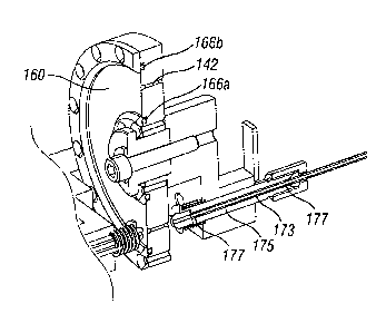
FIGURE 16 is a perspective view of a cartridge 130 mounted in
proximity to certain of the harvesting/implanting system 100 components with
which it directly interacts. The cartridge 130 is oriented the same way as in
FIGURE 15, with the proximal side to the left, though in this operational view
the permissive medium cover 160 occludes the receptacles 142. FIGURE 16
also illustrates an alternative configuration for mounting the cover 160. With
reference also to FIGURE 17, inner and outer mounting rings 164a, 164b bolt
to the body of the cartridge 130 and hold the retaining rings 166a, 166b
within
grooves (not numbered) in the proximal face of the cartridge 130. The
retaining rings 166a, 166b in turn capture and frictionally hold inner and
outer
circular edges of the cover 160, which is made of the material that flexes or
can be pressed into the grooves. For instance, the cover 160 may be made of a
CA 02696201 2010-02-10
WO 2009/045255 PCT/US2008/010034
-31-
silicone (PDMS) elastomer in the shape of the annulus with the inner and
outer edges thereof retained in the grooves by the rings 164, 166.
FIGURE 16 shows the vacuum tube 136 passing through a frame
member 168 mounted on the head assembly 104 (FIGURE 13). A spring
member 170 is seen at the terminal end of the vacuum tube 136, which helps
ensure good suction contact between the tube and the proximal face of the
cartridge 130 and reduces the need for precise relative positioning
tolerances.
Again, the head assembly 104 includes a movement mechanism (not shown)
for translating the vacuum tube 136 toward and away from the cartridge 130,
as indicated by the double-headed arrow.
The frame member 168 may also provide a platform for mounting a
piercing device 172 adjacent the terminal end of the vacuum tube 136. The
piercing device 172 comprises a thin rod, desirably pointed or sharpened,
which lies at the same radial distance from the rotational axis of the
cartridge
130 as the vacuum tube 136. In other words, the piercing device 172 aligns
with the circular pattern of receptacles 142. The piercing device 172 may be
utilized to pre-puncture holes or slits through the cover 160 for each of the
receptacles. The piercing device 172 may be utilized if the terminal end of
the
vacuum tube 136 comprises a thin probe for entering the receptacles, and the
probe is insufficiently sharp to cleanly form its own hole. Such an
embodiment will be described below in reference to FIGURE 19. In an
alternative configuration, the terminal end of the vacuum tube 136 may be
pointed to perform the puncture rather than using a separate piercing
instrument.
FIGURE 16 also illustrates a housing 174 within which may be located
a rotational prime mover, such as a stepper motor, for rotating or indexing
the
cartridge 130. Precise rotational movement-of the cartridge 130 registers each
of the receptacles 142 in turn relative to the vacuum tube 136. Software
controlling the movement mechanism enables any one of the receptacles 142
to be registered as desired. For example, as mentioned above an inspection
system for assessing the characteristic of each FU may be incorporated into
the harvesting procedure, and an understanding of what type of FU is in each
receptacle 142 enables the system to implant smaller FUs in one location,
CA 02696201 2010-02-10
WO 2009/045255 PCT/US2008/010034
-32-
while larger FUs are implanted in another location. Numerous such implant
schemes are known in the art and will not be described further herein. The
reader will also note that relative radial translation of the vacuum tube 136
and
cartridge 130 may be incorporated into the system to facilitate registration
of
the vacuum tube 136 with a second circular array of receptacles 142 (not
shown) concentrically arranged with respect to the illustrated array.
FIGURE 17 is an enlarged sectional view of the distal components of
FIGURE 16, andillustrates exemplary pressure differential reduction structure
desirably provided to slow down the velocity of a biological unit traveling
toward the cartridge 130. In one method of use of the system, a source of
suction is applied to a proximal side of a selected receptacle 142 during the
harvesting process, which creates a pressure differential in the proximal
direction thus urging an FU into the receptacle. The magnitude of the pressure
differential typically remains constant during travel of the FU along the path
from the harvesting tool to the receptacle 142, because the suction required
to
pull the FU from the body surface is relatively high, which results in the FU
moving a high rate of speed toward the receptacle. This high velocity
movement potentially may cause damage to the FU when it halts at the
proximal end of the cartridge receptacle, such as when it impacts the cover
160. Because of the short travel duration, however, it would be difficult
though not impossible to provide a system which steps down the magnitude of
the suction halfway between the harvesting tool and the receptacle.
Rather than incorporating a sophisticated (i.e., expensive) dual-stage
pressure step-down system, structure may be provided along a path in which
the biological unit travels from the removal tool to the storage device
receptacle to reduce the pressure differential along a portion thereof and
thereby reduce the speed of the FU along the path. For instance, the structure
may comprise a parallel flow path outside of the main path which terminates
just before the path reaches the cartridge. In FIGURE 17, the FU travel path
from the operating tip 108 through the distal shaft 132 passes along a main
tube 173 surrounded by an outer tube 175. An annular space is created
between the tubes 173, 175. Two sets of bypass holes 177 spaced apart
substantially the length of the shaft 132 enable flow to enter the annular
space.
CA 02696201 2010-02-10
WO 2009/045255 PCT/US2008/010034
-33-
This reduces the pressure within the main tube 173 and therefore slows down
travel of the FU in that area.
Now with reference to FIGURE 18, an exemplary pressure relief
structure for the cartridge receptacles 142 is shown. When a source of suction
is applied to a proximal side of a selected receptacle 142 during the
harvesting
process, the pressure differential continues for short period after the FU
reaches the receptacle. During that time, it is desirable to mitigate the
dehydrating effects of the suction on the FU, for instance if the FU
completely
includes the receptacle on its proximal side. In FIGURE 18, one of the
receptacles 142 shown is provided with pressure relief channels 174 to limit
the maximum suction created within the receptacle to less than a magnitude of
the source of suction. Stated another way, the vacuum tube 136 comprises a
source of suction which, if sealed around the proximal end of the receptacle
142, would apply a magnitude of suction to the receptacle. Without the
pressure relief channels 174, the FU within the receptacle would be exposed to
the maximum suction. By providing alternative paths for fluid or air into the
vacuum tube 136, the maximum suction to which the FU is exposed is less
than the magnitude of the source of suction.
The pressure relief channels 174 are shown as small notches in a cross
pattern at the second opening or proximal end of the receptacle 142. Of
course, other configurations can be used, including only one notch.
Additionally, the pressure relief channels 174 are only shown in one
receptacle
for example purposes, and desirably are provided for all the receptacles.
FIGURE 19 illustrates an alternative method for applying a source of
suction (creating a pressure differential) to the receptacles 142. Namely, a
suction probe 176 extends from the distal end of the vacuum tube 136. The
suction probe 176 features a blunt, closed end 177 and at least one side port
178 near the closed end. The outer diameter of the suction probe 176 is
sufficiently smaller than the inner diameter of the receptacle 142 so as to
provide a gap and permit air or fluid to flow around the probe and into the
side
port 178.
The suction probe 176 extends through the permissive medium cover
160 into the receptacle 142. In this embodiment, the cover 160 is optionally
CA 02696201 2010-02-10
WO 2009/045255 PCT/US2008/010034
-34-
retained against the proximal face of the cartridge 130 by a plate 179 that is
in
turn secured by an outer ring 181. A circular array of tapered openings (not
numbered) in the plate 179 register with each of the receptacles 142, which
facilitates introduction of the probe 176 to the receptacle. Suction probe 176
may be used with any of the permissive mediums, but is especially useful with
a cover 160 that is relatively impermissible to fluids. Pulling suction
directly
through such a material would be impractical, and therefore the probe 176
must be inserted through the cover 160 into the receptacle. Desirably the
cover 160 is made of a material that self-seals after removal of the probe
176;
for example, silicone (PDMS) elastomer.
The probe 176 may be pushed directly through the cover 160, but
given its blunt shape pre-formed slits or holes are desirably provided. For
example, the piercing tube 172 illustrated in FIGURE 16 may be used to form
such holes. A particularly useful piercing tube 172 is shaped in the form of a
Huber needle which is designed so as not to core a hole, but instead just form
a slit which is self-sealing. Alternatively, the terminal end of the suction
probe 176 may be more pointed, such as in the shape of a Huber needle, so as
to form the hole itself, but consideration should be given to the effect on
the
FU when it contacts the sharpened probe.
FIGURES 20 and 21 are perspective and side views isolating follicular
unit shuttle components from the overall head assembly 104. These
components can also be seen incorporated in head assembly 104 in FIGURES
11-13. To describe important operational interactions of the system, or
relative movement between the distal end of the system including operating tip
108, the cartridge 130, and the proximal end of the system including vacuum
tube 136 will be described. FIGURES 22A-22D illustrate a sequence for
harvesting an FU, while FIGURES 23A-23B describe a sequence for
implanting an FU. It should be understood that these operations may be
carried out sequentially by first filling the cartridge 130, and then emptying
it
without detaching from the system. However, depending on the number of
receptacles 142 in the cartridge 130, several cartridges may be required and
the harvesting operation is accomplished first before the implant procedure.
For instance, a cartridge may hold 125-500 FUs, while a typical procedure
CA 02696201 2010-02-10
WO 2009/045255 PCT/US2008/010034
-35-
requires up to 2000 FUs. The number of receptacles is of course variable and
a single cartridge may hold all that would be needed.
FIGURES 22A-22D are sectional views through the shuttle
components taken along line 22-22 of FIGURE 20, showing a sequence of
operation for harvesting or removing a follicular unit from a body surface 110
and shuttling it to the cartridge 130. In FIGURE 22A, the shaft 132 is shown
after having been displaced to the left within a cylinder 180. This action
causes a harvesting/removal tool 182 to punch into the body surface 110 to a
predetermined depth. The depth of penetration of the tool 182 is determined
with knowledge of the distance between the tool and the body surface prior to
displacement of the shaft 132.
In FIGURE 22B, a follicular unit FU is shown positioned within a
lumen of the removal tool 182. Also, the assembly of the shaft 132 and
cylinder 180 is shown translated to the right so that the proximal end 134
contacts the cartridge 130. The throughbore of the shaft 132 registers with
one
of the receptacles 142 in the cartridge 130.
FIGURE 22C illustrates a step wherein the FU shuttles from the
removal tool 182 through the shaft 132 and into the cartridge receptacle 142.
This may be accomplished using a pressure differential created, for example,
by suction through the vacuum tube 136. This step of shuttling the FU in
FIGURE 22C desirably utilizes the velocity reducing bypass structure
described above with respect to FIGURE 17, as well as the pressure reduction
channels described with respect to FIGURE 18. Alternatively, a mechanical
shuttle may be utilized to transfer the FU into the receptacle 142. For
instance, a rod or obturator (not shown) may be provided that extends through
the shaft 132 and pushes the FU until it deposits into the receptacle 142.
Finally, FIGURE 22D shows the assembly including the shaft 132
moving away from the cartridge 130 into distal direction, and the assembly
including the vacuum tube 136 moving away from the cartridge 130. At this
point, the cartridge 130 is free to rotate and index another receptacle into
registration with the distal and proximal components of the shuttle subsystem.
Now with reference to FIGURE 23A, the shuttle subsystem
components are shown essentially in the positions from FIGURE 22D,
CA 02696201 2010-02-10
WO 2009/045255 PCT/US2008/010034
-36-
although instead of a harvesting/removal tool, the distal end of the shaft 132
features an implant tool 184. A receptacle 142 having an FU registers with the
lumen of the shaft 132. On the proximal side, the vacuum tube 136 has been
removed and replaced with a solid rod-like obturator 186. The obturator 186
translates linearly along its axis through the receptacle 142 and through the
entire length of the shaft 132, and may be propelled in a number of ways
including by contact with a plurality of wheels 188.
FIGURE 23B shows the three components brought together and the
obturator 186 passed though the receptacle 142 and entire shaft 132 until the
FU reaches the implant tool 184. The implant tool 184 may be used to deposit
the FU into a preformed incision or create a new incision in a body surface.
The reader should understand that shuttling of the hair follicles from
the cartridge to the implant tool could be accomplished using several
alternative approaches. One example is to push the FUs from the cartridge all
the way through the distal end of the implant tool using the obturator as
described. Another option is to use a pressure differential to urge the FU out
of each receptacle and into the implant tool. For example, the vacuum tube
136 described above that creates a pressure differential urging the FU in the
proximal direction can also be used to reverse the pressure differential to
propel the FU distally. Or, a combination of physical pushing and pressure
could be used.
The foregoing illustrated and described embodiments of the invention
are susceptible to various modifications and alternative forms, and it should
be
understood that the invention generally, as well as the specific embodiments
described herein, are not limited to the particular forms or methods
disclosed,
and that many other embodiments are possible within the spirit and the scope
of the present invention. Moreover, although individual features of one
embodiment of the invention may be discussed herein or shown in the
drawings of the one embodiment and not in other embodiments, it should be
apparent that individual features of one embodiment may be combined with
one or more features of another embodiment or features from a plurality of
embodiments.
CA 02696201 2010-02-10
WO 2009/045255 PCT/US2008/010034
-37-
While the invention has been described in its preferred embodiments, it
is to be understood that the words which have been used are words of
description and not of limitation. Therefore, changes may be made within the
appended claims without departing from the true scope of the invention.