Note : Les descriptions sont présentées dans la langue officielle dans laquelle elles ont été soumises.
CA 02698883 2010-03-03
WO 2009/031033 PCT/IB2008/002872
LIQUID CORE PHOTONIC CRYSTAL FIBER BIOSENSORS USING SURFACE
ENHANCED RAMAN SCATTERING AND METHODS FOR THEIR USE
The present application claims priority to U.S. Provisional Patent Application
Serial Number 60/967,555 entitled "Liquid Core Photonic Crystal Fiber
Biosensors Using
Surface Enhanced Raman Scattering And Methods For Their Use", filed September
4,
2007, and U.S. Provisional Patent Application Serial Number 61/192,632
entitled "Liquid
Core Photonic Crystal Fiber Biosensors Using Surface Enhanced Raman Scattering
And
Methods For Their Use", filed September 19, 2008, which are herein
incorporated by
reference in their entirety for all purposes.
This invention was made partly using funds from the United States' National }
Science Foundation grant number ECS-0401206 and ARP/UARC grant number NAS2-
03144-TO.030.3MM.DGU-06. The United States Federal Government has certain
rights
to this invention.
Technical Field
[001] The present invention relates to crystal fibers having a coating of
particles
comprising metallic nanoparticles having useful properties. The invention
further relates
to methods of using the crystal fibers for detecting chemical and biological
analytes, and
in use in optical communications.
Background Art
[002] During the 1980s Raman Scattering in fibers was demonstrated by Lin,
Stolen,
and other co-workers of AT&T Bell Laboratories in Holmdel, New Jersey using
Raman
lasers operating between 0.3 to 2.0 m. In the early years of the Raman fiber
before
extensive work had begun, no one perceived that a Raman fiber could be pumped
by a
practical semiconductor laser-based source or that an efficient CW-pumped
Raman Fiber
Laser was possible. However, with the development of Cladding-pumped Fiber
Lasers
and Fiber Bragg Gratings, diode-laser-based CW Raman Fiber Lasers have been
made
1
CONFIRMATION COPY
CA 02698883 2010-03-03
WO 2009/031033 PCT/IB2008/002872
efficient, emitting at various wavelengths throughout the infrared spectrum a
reality. (See
van Gisbergen et al. (1996) Chem. Phys. Lett. 259: 599-604.)
[003] Raman spectroscopy is a powerful optical technique for detecting and
analyzing
molecules. Its principle is based on detecting light scattered off a molecule
that is shifted
in energy with respect to the incident light. The shift, called Raman shift,
is characteristic
of individual molecules, reflecting their vibrational frequencies that are
like fingerprints of
molecules. As a result, the key advantage of Raman spectroscopy is its
molecular
specificity while its main limitation is the small signal due to low quantum
yield of Raman
scattering. One way to enhance the Raman signal is to tune the excitation
wavelength to
be on resonance with an electronic transition, so called resonance Raman
scattering. This
can usually produce an enhancement on the order of 102_103 fold.
[004] Another technique to enhance Raman scattering is surface enhancement by
roughened metal surfaces, notably silver and gold, that provides an
enhancement factor on
the order of 106-108. This is termed surface enhanced Raman scattering (SERS).
Similar or somewhat larger enhancement factors (_108-1010) have been observed
for
metal, mostly silver or gold, nanoparticles.
[005] In the last few years, it has been shown that an even larger enhancement
1010-
1015) is possible for aggregates of metal nanoparticles (MNPs), silver and
gold. The
largest enhancement factor of 1014_ 1015 has been reported for rhodamine 6G
(R6G) on
single silver nanoparticle aggregates. This huge enhancement is thought to be
mainly due
to significant enhancement of the local electromagnetic field of the
nanoparticle aggregate
that strongly absorbs the incident excitation light for the Raman scattering
process. With
such large enhancement, many important molecules that are difficult to detect
with Raman
normally can now be easily detected. This opens many interesting and new
opportunities
for detecting and analyzing molecules using SERS with extremely high
sensitivity and
molecular specificity.
[006] SERS can also be developed into a molecular imaging technique for
biomedical
and other applications. Existing Raman imaging equipment should be usable for
SERS
2
CA 02698883 2010-03-03
WO 2009/031033 PCT/IB2008/002872
imaging. SERS will provide a much-enhanced signal and thereby significantly
shortened
data acquisition time, making the technique practically useful for medical or
other
commercial and industrial applications including chip inspection or chemical
monitoring.
SERS is also useful for detecting other cancer biomarkers that can interact or
bind to the
MNP surface. For example, Sutphen et al. have recently shown that
lysophospholipids
(LPL) are potential biomarkers of ovarian cancer (Sutphen et al. (2004) Cancer
Epidemiol.
Biomarker Prev. 13: 1185-1191).
[007] Photonic crystal fibers have been developed that can detect, identify,
and
quantify ultra small quantities of analytes in air and aqueous samples. In one
example of
the prior art, Du and Sukhishvili disclose a sensor comprising a photonic
crystal fiber
having an air hole cladding with functionalized air holes (Du and Sukhishvili,
US
Publication Number US 2007/0020144 Al, published 25 January, 2007). The
photonic
crystal fiber disclosed by Du and Sukhishvili comprises a solid core photonic
crystal fiber;
of note, Du and Sukhishvili described that "(c)omparison of FIG. 18 with FIG.
16 shows
that the Ag nanoparticles 82 are present at a much lower density than the Ag
nanoparticles
74 of the previous experiment. It is also apparent that the Ag nanoparticles
82 are much
larger than the nanoparticles 74, and would, therefore, be less suitable for
enhancement of
SERS spectra" and that "(t)he moderate signals detected from adsorbed Rh6G
(rhodamine
6G) in no salt aqueous solution were highly prone to fast photodegradation,
and in a
typical experiment, a SERS signal was not detectable after a 1 minute exposure
of the
substrate to 532 nm 10 mW laser radiation" (Du and Sukhishvili, paragraphs 59
and 52,
respectively).
[008] Others have also disclosed photonic crystal fibers, for example Konorov
et al.
(2005, Optics Express, 13: 3454-3459) and Konorov et al. (2006, Optics Lett.,
31: 1911-
1913). Konorov et al. (2005) disclose multicore hollow photonic crystal fibers
of fused
silica or soft glasses having inner diameters of the hollow core of about 2.5
m and 3 m
or about 3 m and 3.5 m, respectively. Konorov et al. (2006) disclose hollow
photonic
crystal fibers with inner diameters of between about 8.6 m and 9.5 m and an
outer
diameter of 84 m. Yan et al. also disclosed a novel hollow core photonic
crystal fiber
3
CA 02698883 2010-03-03
WO 2009/031033 PCT/IB2008/002872
surface-enhanced Raman probe in Yan et al., (2006) (Yan et al. (2006) Appl.
Phys. Lett.
89:204101).
[009] The original single multimode SERS fiber probe was demonstrated in 1991
by
Mullen et al. (Mullen and Carron (1991) Anal. Chem. 63: 2196). In the
following years,
studies involving different kinds of fiber tips were tested, such as flat,
angled and tapered
fibers (Viets and W. Hill (2000) J. Raman Spec. 31: 625; Viets and Hill (2001)
J. Phys.
Chem. B 105: 6330; and Viets and Hill (1998) Sens. Actuators B-Chem. 51: 92).
Although they were easy to implement, the small number of SERS substrate
particles in
the active region limited the sensitivity of these sensors. In order to
involve more particles
in the SERS activity, hollow core photonic crystal fiber (HCPCF) and liquid
core photonic
crystal fiber (LCPCF) were tested recently (see Zhu et al. (2006) Opt. Exp.
14: 3541; Yan
et al. (2006) Appl. Phys. Lett. 89: 204101; and Zhang et al. (2007) Appl.
Phys. Lett. 90,
193504. High sensitivity, and low fiber SERS background show a promising
future of
PCF sensors. However, the wavelength sensitive nature of HCPCFs limits the
application
of a HCPCF to a single excitation wavelength and the cost of PCFs is still
high. While
normal fibers are lower in cost, their sensitivities are somewhat limited,
often due to the
background Raman scattering from the fiber itself. Therefore, it is highly
desired to
improve the detection sensitivity of SERS sensors based on conventional
fibers. Fiber
SERS sensors with high sensitivity, remote sensing capability, and low cost
will find
potential applications in medical, environmental, food detection, and toxin
identification.
[0010] For many practical applications, for example SERS and optical fibers,
it is
highly desirable to narrow the distribution of size/shape of nanoparticle
aggregates. For
SERS in particular, the incident light has to be on resonance with the
substrate absorption.
Only those nanoparticle aggregates that have resonance absorption of the
incident light are
expected to be SERS active. It is thus extremely beneficial to have a narrow
size/shape
distribution and thereby narrow optical absorption.
[0011] Fluorescent nanoparticles (quantum dots (QDs) such as semiconductor
quantum
dots, SQDs) have been used recently as fluorescent biological markers and have
been
found to be extremely effective. They offer advantages including higher
stability, stronger
4
CA 02698883 2010-03-03
WO 2009/031033 PCT/IB2008/002872
fluorescence, tunability of color, and possibility of optical encoding based
on different
sized or colored SQDs.
[0012] Metal nanoparticles have been recognized for their unique optical
properties that
could be exploited in optoelectronic devices. Nanoparticle systems composed of
gold, for
example, have distinct optical properties that make them amenable to study by
Raman
scattering. The Raman spectrum of the adsorbed species is significantly
enhanced by 10
to 15 orders of magnitude when the metal nanoparticles have aggregated,
leading to
enhanced electromagnetic field effects near the surface that increases the
Raman scattering
intensity. The greater sensitivity found in the surface enhanced Raman
scattering (SERS)
of metal nanoparticle aggregates facilitates the detection and analysis of a
whole host of
molecules that were previously difficult to study.
[0013] Wang et al. disclose a method of using SQDs (dye-conjugated CdTe
nanoparticles, CT-NPs) to detect interactive binding between Ag-CT-NPs and Ab-
CT-NPs
(Wang et al. (2002) NanoLett. 2: 817-822). The interactions were determined by
differential quenching or enhancement fluorescence activity of two different
sized SQDs
(red or green) measured during the analysis.
[0014] The chemical methods used historically for the production of gold
nanoparticle
aggregates (GNAs) results in a wide distribution of aggregate size. This
distribution leads
to a broadened absorption spectrum. Accordingly, researchers have attempted to
narrow
the lineshape of the spectral peak due to the aggregates by homogenizing the
size of the
GNAs after they have been produced. By eliminating certain ranges of aggregate
size,
absorption spectrum peaks should narrow appreciably and concomitantly increase
in
intensity, resulting in more sensitive detection. Previous attempts to select
for a narrow
size range of aggregates have employed mechanical techniques such as passing a
solution
of aggregates through a filter. For example, Emory & Nie have employed size-
selective
fractionation using membrane filters to select for optically active silver
nanoparticles
(Emory and Nie, (1997) J. Phys. Chem. B, 102: 493-497).
CA 02698883 2010-03-03
WO 2009/031033 PCT/IB2008/002872
[0015] The use of SERS for analyte detection of biomolecules has been
previously
studied. U.S. Pat. No. 6,699,724 to West et al. describes a chemical sensing
device and
method (nanoshell-modified ELISA technique) based on the enzyme-linked
immunoadsorbant assay (ELISA). The chemical sensing device can comprise a core
comprising gold sulfide and a surface capable of inducing surface enhanced
Raman
scattering (SERS). In much of the patent disclosure, the nanoparticle is
disclosed as
having a silica core and a gold shell. The patent discloses that an
enhancement of
600,000-fold (6 x 10⁵) in the Raman signal using conjugated mercaptoaniline
was
observed.
[0016] In the nanoshell-modified ELISA technique, antibodies are directly
bound to the
metal nanoshells. Raman spectra are taken of the antibody-nanoshell conjugates
before
and after the addition of a sample containing a possible antigen, and binding
of antigen to
antibody is expected to cause a detectable shift in the spectra.
[0017] The conjugation of quantum dots to antibodies used for ultrasensitive
nonisotopic detection for use in biological assays has also been studied. U.S.
Pat. No.
6,468,808 B 1 to Nie et al. disclosed an antibody is conjugated to a water-
soluble quantum
dot. The binding of the quantum dot-antibody conjugate to a targeted protein
will result in
agglutination, which can be detected using an epi-fluorescence microscope. In
addition,
Nie et al. described a system in which a quantum dot is attached to one end of
an
oligonucleotide and a quenching moiety is attached to the other. The preferred
quenching
moiety in the Nie patent is a nonfluorescent organic chromophore such as 4-[4'-
dimethylaminophenylazo]benzoic acid (DABCYL).
[0018] Raman amplifiers are also expected to be used globally as a key device
in next-
generation optical communications, for example, in wavelength-division-
multiplexing
(WDM) transmission systems. Raman scattering occurs when an atom absorbs a
photon
and another photon of a different energy is released. The energy difference
excites the
atom and causes it to release a photon with low energy; therefore, more light
energy is
transferred to the photons in the light path.
6
CA 02698883 2010-03-03
WO 2009/031033 PCT/IB2008/002872
[0019] There is therefore a need in the art for use in the biomedical
analytical industries
and the optical communications industries to provide more sensitive
compositions and
devices that are inexpensive to manufacture and easy to use.
Disclosure of the Invention
[0020] The invention provides a photonic crystal fiber, methods for
manufacture and/or
fabrication of said a photonic crystal fiber, and methods for using the
photonic crystal
fiber. The photonic crystal fiber is used as a sensor for any analyte and is
many times
more sensitive than sensors in current use, an unexpected property. The
photonic crystal
fiber is used to measure the surface enhanced Raman scattering (SERS)
resulting from
interactions between the components of the photonic crystal fiber and the
analyte of
interest.
[0021] In one embodiment, the invention provides a photonic crystal fiber
having
improved sensitivity for detecting and/or sending a chemical, the fiber
comprising a
proximal end, a distal end, the ends defining a lumen, an outer surface, and
an inner
surface. In one embodiment, the inner surface further comprises, in part, a
metallic
nanoparticle composition. In an alternative embodiment, the outer surface
further
comprises, in part, a metallic nanoparticle composition. In one preferred
embodiment the
photonic crystal fiber has a cylindrical shape and an approximately circular
cross-section.
In another preferred embodiment the photonic crystal fiber is flexible. In one
preferred
embodiment the lumen of the fiber further comprises a liquid and/or a gas. In
another
preferred embodiment the lumen of the fiber comprises a solid composition. In
one
embodiment of the photonic crystal fiber, the metallic nanoparticle
composition comprises
a double substrate sandwich structure. In an alternative embodiment, the
metallic
nanoparticle composition comprises a single layer. In another alternative
embodiment, the
metallic nanoparticle composition comprises a plurality of layers.
[0022] In one embodiment the sensitivity is enhanced by at least 10 times. In
another
embodiment, the sensitivity is enhanced by at least 25 times. In another
embodiment the
sensitivity is enhanced by at least 50 times. In another embodiment, the
sensitivity is
enhanced by at least 75 times. In another embodiment the sensitivity is
enhanced by at
7
CA 02698883 2010-03-03
WO 2009/031033 PCT/IB2008/002872
least 100 times. In another embodiment, the sensitivity is enhanced by at
least 200 times.
The sensitivity can be enhanced by, for example, up to 10 times, up to 15
times, up to 20
times, up to 25 times, up to 30 times, up to 35 times, up to 40 times, up to
45 times, up to
50 times,up to 55 times, up to 60 times, up to 65 times, up to 70 times, up to
75 times, up
to 80 times, up to 85 times, up to 90 times, up to 95 times, up to 100 times,
up to 150
times, up to 200 times, up to 250 times, up to 300 times, up to 350 times or
more, or any
similar level thereabouts.
[0023] In an alternative embodiment, the photonic crystal fiber further
comprises a
plurality of lumens and wherein each end of the fiber comprises a plurality of
apertures to
each lumen.
[0024] In one alternative preferred embodiment the photonic crystal fiber is
solid.
[0025] In another embodiment, the metallic nanoparticle composition comprises
a
metal, wherein the metal is selected from the group consisting of can be gold,
silver,
platinum, copper, aluminum, palladium, cadmium, iridium, and rhodium. In a
more
preferred embodiment the metal is silver. In a most preferred embodiment, the
metallic
nanoparticle composition comprises silver citrate.
[0026] In one embodiment, the cross-section of the photonic crystal fiber has
dimensions of about between 0.1 m and 100 m. For example, the cross-section
of the
photonic crystal fiber can be about 0.1 m, 0.2 m, 0.3 m, 0.4 m, 0.5 m,
0.6 m, 0.7
m, 0.8 m, 0.9 m, 1 m, 2 m, 3 m, 4 m, 5 m, 6 m, 7 m, 8 m, 9 m, 10
m, 12
m, 13 m, 14 m, 15 m, 16 m, 17 gm, 18 m, 19 m, 20 m, 25 gm, 30 m, 35
gm,
40 m, 45 m, 50 m, 55 m, 60 m, 65 m, 70 m, 75 m, 80 m, 85 m, 90 m,
95
m, 100 m, or any dimension therebetween. In another embodiment, the length of
the
photonic crystal fiber has dimensions of about between 0.5 cm and 100 cm. For
example,
the length of the photonic crystal fiber can be about, 0.5 cm, 0.6 cm, 0.7 cm,
0.8 cm, 0.9
cm, 1cm,2cm,3cm,4cm,5cm,6cm,7cm,8cm,9cm, 10 cm, 12 cm, 13 cm, 14 cm,
15 cm, 16 cm, 17 cm, 18 cm, 19 cm, 20 cm, 25 cm, 30 cm, 35 cm, 40 cm, 45 cm,
50 cm,
8
CA 02698883 2010-03-03
WO 2009/031033 PCT/IB2008/002872
55 cm, 60 cm, 65 cm, 70 cm, 75 cm, 80 cm, 85 cm, 90 cm, 95 cm, 100 cm, or any
dimension therebetween. In another embodiment, the cross-section of the lumen
of the
photonic crystal fiber has dimensions of about between 0.1 m and 100 m. For
example,
the cross-section of the lumen of the photonic crystal fiber can be about 0.1
m, 0.2 m,
0.3 m, 0.4 m, 0.5 m, 0.6 m, 0.7 m, 0.8 m, 0.9 m, 1 m, 2 m, 3 m, 4
m, 5 m,
6 m, 7 m, 8 m, 9 gm, 10 m, 12 m, 13 m, 14 m, 15 m, 16 m, 17 m, 18
m, 19
m, 20 .m, 25 m, 30 m, 35 m, 40 m, 45 m, 50 m, 55 m, 60 m, 65 m, 70
m,
75 m, 80 m, 85 m, 90 m, 95 m, 100 m, or any dimension therebetween.
[0027] The photonic crystal fiber is particularly useful for sensing and
measuring the
quantities of an analyte. The photonic crystal fiber disclosed herein is an
improvement
over the prior art in that the presence of a fluid or liquid detection in the
lumen of the
photonic crystal fiber results in an unexpectedly superior enhancement factor
of the SERS
signal from the photonic crystal fiber and a test sample comprising the
analyte of interest.
In a preferred embodiment the analyte is a biological composition. The
biological
composition can be, for example, a protein, a peptide, a polyketide, an
antibody, an
antigen, a nucleic acid, a peptide nucleic acid, a sugar, a lipid, a
glycophosphoinositol, and
a lipopolysaccharide. In another alternative embodiment the analyte can be an
explosive,
a chemical and/or biological warfare agent, a toxin, a virus particle, and a
biological cell.
[0028] In yet a further embodiment, the photonic crystal fiber comprises a
support. In a
preferred embodiment, the support comprises a medium that is permeable to an
analyte of
interest. In one embodiment the support can be a gel, a solid, or a liquid.
The support can
comprise a synthetic composition, such as, but not limited to a polymer, a
block co-
polymer, a random copolymer, a carbon composite material, a metal composite
material,
or the like. Alternatively, the support can comprise a biological compound,
such as, but
not limited to, a starch composition, a cellulose composition, a collagen
composition, a
latex composition, a protein, a polypeptide, a carbohydrate, a sugar, a
mixture thereof, or
the like. In another alternative, the support can be a liquid or a gel-phase
composition,
such as, but not limited to, an aqueous composition, an alcohol composition, a
hydrogel, a
mixture thereof, or the like. The support can be in the form of a matrix, a
crystalline
9
CA 02698883 2010-03-03
WO 2009/031033 PCT/IB2008/002872
structure, a cross-linked polymer, a porous composition, or the like. Such
structures,
materials, and compositions are well known to those of skill in the art.
[0029] In another preferred embodiment, the photonic crystal fiber has a
surface
wherein the surface can induce surface enhanced Raman scattering (SERS).
[0030] In still another preferred embodiment, the photonic crystal fiber
further
comprises at least one detecting molecule, wherein the detecting molecule is
bound to the
surface or support. In a more preferred embodiment the detecting molecule is
selected
from the group consisting of proteins, peptides, antibodies, antigens, nucleic
acids, peptide
nucleic acids, sugars, lipids, glycophosphoinositols, and lipopolysaccharides.
[0031 ] In a yet more preferred embodiment the detecting molecule is an
antibody. In
another preferred embodiment, the detecting molecule is an antigen.
[0032] In another embodiment, the invention provides a photonic crystal fiber
further
comprising at least one semiconductor quantum dot. In a preferred embodiment
the
semiconductor quantum dot further comprises a linker molecule, the linker
molecule
selected from the group consisting of a thiol group, a sulfide group, a
phosphate group, a
sulfate group, a cyano group, a piperidine group, an Fmoc group, and a Boc
group.
[0033] In a still further embodiment, the invention provides a photonic
crystal fiber
comprising at least one semiconductor quantum dot wherein the semiconductor
quantum
dot further comprises a detecting molecule, wherein the detecting molecule is
bound to the
semiconductor quantum dot. In a more preferred embodiment, the detecting
molecule is
selected from the group consisting of proteins, peptides, antibodies,
antigens, nucleic
acids, peptide nucleic acids, sugars, lipids, glycophosphoinositols, and
lipopolysaccharides.
[0034] In a more preferred embodiment, the detecting molecule is an antibody.
In the
alternative, a more preferred embodiment comprises a chemical sensing device
wherein
the detecting molecule is an antigen.
CA 02698883 2010-03-03
WO 2009/031033 PCT/IB2008/002872
[0035] The invention further provides a method for sensing an analyte in a
test sample,
the method comprising the steps of: (i) providing the photonic crystal fiber
disclosed
herein; (ii) providing a test sample; (iii) immersing the photonic crystal
fiber in the test
sample; (iv) irradiating the photonic crystal fiber and the test sample with
an excitation
light, the excitation light having a wavelength in the visible to near infra-
red (near-IR)
portion of the spectrum, such as, for example, from between about 600 nm to
about 1,400
nm, from between about 620 to about 1,000 nm, from between about 650 to about
950 nm,
from between about 700 nm to about 900 nm, from between about 750 nm to about
880
nm, or from between about 770 nm to about 800 nm; (v) measuring the Raman
spectrum
of a photonic crystal fiber and a control sample, thereby determining the
background
Raman spectrum; (vi) detecting the surface enhanced Raman scattering (SERS)
signal
emitted from the photonic crystal fiber and the test sample; (vii) measuring
the Raman
spectrum of the photonic crystal fiber and the test sample, thereby
determining the analyte
Raman spectrum; subtracting the background Raman spectrum from the analyte
Raman
spectrum, thereby determining the quantity of the analyte in the sample;
(viii) determining
the enhancement factor of the SERS signal from the control sample; (ix)
determining the
enhancement factor of the SERS signal from the test sample; wherein the
enhancement
factor of the SERS signal from the test sample is at least 100-fold compared
with a SERS
signal from the control sample, the method resulting in sensing the analyte.
In a preferred
embodiment, the analyte is a biological composition. In a more preferred
embodiment, the
biological corimposition is selected from the group consisting of a protein, a
peptide, a
polyketide, an antibody, an antigen, a nucleic acid, a peptide nucleic acid, a
sugar, a lipid,
a glycophosphoinositol, and a lipopolysaccharide. In an alternative more
preferred
embodiment, the analyte is selected from the group consisting of an explosive,
a chemical
warfare agent, a biological warfare agent, a toxin, a virus particle, and a
biological cell.
[0036] The invention further provides a method for measuring the quantity of
an analyte
in a test sample, the method comprising the steps of: (i) providing the
photonic crystal
fiber disclosed herein; (ii) providing a test sample; (iii) immersing the
photonic crystal
fiber in the test sample; (iv) irradiating the photonic crystal fiber and the
test sample with
an excitation light, the excitation light having a wavelength in the visible
to near infra-red
11
CA 02698883 2010-03-03
WO 2009/031033 PCT/IB2008/002872
(near-IR) portion of the spectrum, such as, for example, from between about
600 nm to
about 1,400 nm, from between about 620 to about 1,000 nm, from between about
650 to
about 950 nm, from between about 700 nm to about 900 nm, from between about
750 nm
to about 880 nm, or from between about 770 nm to about 800 nm; (v) measuring
the
Raman spectrum of a photonic crystal fiber and a control sample, thereby
determining the
background Raman spectrum; (vi) detecting the surface enhanced Raman
scattering
(SERS) signal emitted from the photonic crystal fiber and the test sample;
(vii) measuring
the Raman spectrum of the photonic crystal fiber and the test sample, thereby
determining
the analyte Raman spectrum; subtracting the background Raman spectrum from the
analyte Raman spectrum, thereby determining the quantity of the analyte in the
sample;
(viii) determining the enhancement factor of the SERS signal from the control
sample; (ix)
determining the enhancement factor of the SERS signal from the test sample;
wherein the
enhancement factor of the SERS signal from the test sample is at least 100-
fold compared
with a SERS signal from the control sample, the method resulting in measuring
the
quantity of the analyte. In a preferred embodiment, the analyte is a
biological
composition. In a more preferred embodiment, the biological composition is
selected
from the group consisting of a protein, a peptide, a polyketide, an antibody,
an antigen, a
nucleic acid, a peptide nucleic acid, a sugar, a lipid, a
glycophosphoinositol, and a
lipopolysaccharide. In an alternative more preferred embodiment, the analyte
is selected
from the group consisting of an explosive, a chemical warfare agent, a
biological warfare
agent, a toxin, a virus particle, and a biological cell. In one preferred
embodiment the
wavelength of the excitation light is about 633 nm. In another alternative
preferred
embodiment the wavelength of the excitation light is about 785 nm.
[0037] Another embodiment of the invention provides a method for detecting an
analyte
in a sample using a photonic crystal fiber, the method comprising the steps
of: i)
providing a sample; ii) providing a semiconductor quantum dot comprising a
linker
molecule (LM-SQD); iii) conjugating the analyte in the sample with the LM-SQD
thereby
producing an analyte-LM-SQD conjugate; iv) providing a photonic crystal fiber
comprising a plurality of particles, each particle comprising: a shell having
at least one
surface and a lumen and wherein the shell comprises a sulfur-oxygen molecular
species,
and the shell surface further comprising a detecting molecule; v) incubating
the analyte-
12
CA 02698883 2010-03-03
WO 2009/031033 PCT/IB2008/002872
LM-SQD conjugate with the photonic crystal fiber for a predetermined time
period; and
vi) measuring the extent of binding between the analyte-LM-SQD conjugate and
the
photonic crystal fiber; thereby detecting the analyte in the sample.
[0038] In a yet additional embodiment, the invention provides an optical
communications device comprising a photonic crystal fiber, a plurality of
particles, each
particle comprising: a shell having at least one surface and a lumen.
[0039] In a more preferred embodiment the optical communications device
comprises a
fiber, wherein the fiber is selected from the group consisting of ceramics,
glasses,
polymers, and metal-polymer composites. In another preferred embodiment the
chemical
sensor is disposed upon a surface of the fiber.
[0040] The invention also provides a process for fabricating a photonic
crystal fiber, the
method comprising the steps of (i) mixing AgNO3 with ethanol; (ii) stirring
the AgNO3 in
ethanol until the AgNO3 is dissolved in the ethanol; (iii) adding three molar
equivalents of
hexanethiol to the solution; (iv) adding toluene to the solution; (v) reducing
the solution
using a ten-fold molar excess of NaBH4 dissolved in nanopure water; (vi)
washing the
solution at least three times with nanopure water thereby removing inorganic
impurities;
(vii) collecting the toluene phase; (viii) evaporating the toluene phase,
thereby causing
metallic nanoparticles to come out of solution; (ix) collecting the metallic
nanoparticles;
(x) dissolving the metallic nanoparticles in methanol; (xi) evaporating the
methanol; (xii)
collecting the metallic nanoparticles; re-dissolving the metallic
narioparticles in methanol;
(xiii) providing a crystal fiber, the crystal fiber having a proximal end and
a distal end;
(xiv) dipping the distal end of the crystal fiber into the metallic
nanoparticle solution; (xv)
removing the distal end of the fiber from the metallic nanoparticle solution;
(xvi) washing
the distal end of the crystal fiber with ethanol; (xvi) drying the distal end
of the crystal
fiber using a gas; (xvii) irradiating the crystal fiber with ultra-violet
radiation; (xviii)
repeating steps (xiv) through (xvii) at least once; the process thereby
fabricating a
photonic crystal fiber. In one preferred embodiment the metallic nanoparticles
are
hexanethiolate-protected silver (AgC6) nanoparticles.
13
CA 02698883 2010-03-03
WO 2009/031033 PCT/IB2008/002872
Brief Description of the Drawings
[0041] Figure 1 a shows the transmission spectrum of the Air-6-800 photonic
crystal
fiber.
[0042] Figure lb shows a micrograph of the cross section of a hollow core
photonic
crystal fiber (HCPCF).
[0043] Figur~ lc shows the probing tip of a HCPCF after post-fabrication
processing.
[0044] Figure ld is an enlarged view of Figure lc.
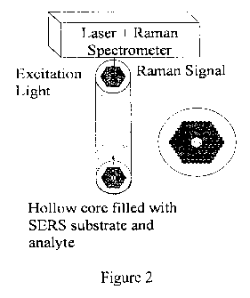
[0045] Figure 2 is a schematic of a liquid core photonic crystal fiber (LCPCF)
SERS
sensor and its cross-sectional view. The spectrometer above the surface
contains a CCD
detector, a monochromator, and electronics for data collection.
[0046] Figure 3 shows representative spectra of a hollow core photonic crystal
fiber.
[0047] Figure 3a, curve A: Background Raman spectrum of the HCPCF. Curve B:
rhodamine 6G (R6G) Raman spectrum obtained using a HCPCF SERS probe without
the
post-fabrication processing; the HCPCF was dipped into the nanoparticle/R6G
solution,
Curve C: Subtraction of curve A from curve B showing the net R6G Raman signal.
[0048] Figure 3b shows a human insulin SERS spectrum obtained using a LCPCF
SERS probe after the post-fabrication processing. The fiber background has
been
subtracted from the observed spectrum.
[0049] Figure 3c is a comparison of SERS intensities between tryptophan
obtained from
the post-processed LCPCF SERS probe and that obtained directly from a dried
nanoparticle/analyte film.
[0050] Figure 4 shows some of the confined modes of a photonic crystal fiber
(PCF)
when the hollow core is empty (upper plate) or filled with liquid (lower
plate).
14
CA 02698883 2010-03-03
WO 2009/031033 PCT/IB2008/002872
[0051] Figure 5 is a schematic of the tip coated multimode fiber sensor.
[0052] Figure 6 is a TEM micrograph of Ag-C6SH nanoparticles. The inset shows
a
size histogram, illustrating an average core size for the fiber of 4.9 2.1
nm.
[0053] Figure 7 shows SERS spectra of R6G molecules at various concentrations
using
different detection methods (TCMMF, MMF in sample solution, and direct
detection).
The concentrations of the R6G molecules are as follows: Figures 7a, 10-5 M;
7b, 10-6 M;
7c, 10-7 M; 7d, 101'M; and 7e, 10-9 M.
[0054] Figure 7f illustrates data from Figures 7a-e showing a plot of SERS
intensity
versus R6G concentration using the peak 1514.3 cm"' as an example for three
detection
methods (TCMMF, MMF in sample solution, and direct detection).
Mode(s) for Carrying Out the Invention
[0055] The embodiments disclosed in this document are illustrative and
exemplary and
are not meant to limit the invention. Other embodiments can be utilized and
structural
changes can be made without departing from the scope of the claims of the
present
invention.
[0056] As used herein and in the appended claims, the singular forms "a,"
"an," and
"the" include plural reference unless the context clearly dictates otherwise.
Thus, for
example, a reference to "a particle" includes a plurality of such particles,
and a reference
to "a surface" is a reference to one or more surfaces and equivalents thereof,
and so forth.
[0057] The invention provides a photonic crystal fiber and methods for
fabricating a
hollow photonic crystal fiber (HCPCF) and a liquid core photonic crystal fiber
(LCPCF)
and demonstrates using the SERS sensor for in vitro molecular detection.
CA 02698883 2010-03-03
WO 2009/031033 PCT/IB2008/002872
[0058] In another embodiment the invention provides a crystal fiber having a
configuration based on a double-substrate "sandwich" structure (DSSS) that is
designed to
enhance the SERS activity using two substrates simultaneously.
Liquid Core Photonic Crystal Fiber Sensor Based on Surface Enhanced Raman
Scattering
[0059] Surface enhanced Raman scattering (SERS) sensors based on optical
fibers have
attracted significant interest in molecule sensing. On one hand, SERS offers
rich
molecular information while amplifying the signal by orders of magnitude (-
109). On the
other hand, the flexibility of optical fibers makes it an ideal SERS platform
for practical
applications. Previously, fibers with different configurations such as a flat,
angled, or
tapered tip were tested as SERS platforms. The main limitation has been the
small
number of SERS substrate particles on the active fiber region, requiring high
laser
intensities and/or long integration times to attain reasonable SERS spectra.
[0060] To overcome this hurdle, several types of photonic crystal fibers were
suggested
and tested. Previously, SERS was reported with the gold nanoparticles and
analyte coated
(dried) on the inner surface of the air holes of a hollow core photonic
crystal fiber
(HCPCF) with the excitation light coupled into the opposite end. Although the
active
sensing area was significantly increased, the HCPCF SERS sensor performed well
when
the nanoparticles/analyte dried along the light path. If the HCPCF were dipped
directly
into the sample solution, the central hole, along with the surrounding
cladding holes,
would all be filled with solution, leading to a reduction of the refractive
index contrast
inside and outside the holes, therefore, losing the photonic bandgap. This
would in turn
result in the loss of light confinement and limit in vivo and in vitro
applications of as a
HCPCF SERS sensor. The cladding holes of the HCPCF (model Air-6-800 or model
HC-
633-01 fibers, for example; other suitable fibers may also be used) were
alternatively
sealed using a fusion splicer. Heat from the two electric tips of the fusion
splicer sealed
the cladding holes leaving the central core of the fiber open.
[0061] The invention provides methods, system, and apparatus to fabricate an
ultra-
sensitive chemical and biological sensor based on surface enhanced Raman
scattering and
a novel liquid core photonic crystal fiber (LCPCF). Surface enhanced Raman
scattering
16
CA 02698883 2010-03-03
WO 2009/031033 PCT/IB2008/002872
provides the fingerprint of the analyte molecules and enlarges or amplifies
the signal by up
to at least 1015 times that of regular Raman signals. The sensor can be used
for in vivo and
in vitro detection and sensing if a flexible LCPCF probe is used. With this
novel fiber
architecture, LCPCF achieved a much greater interaction volume compared with a
regular
solid core multimode fiber, due, in part, to both the photonic bandgap guiding
and the
index guiding mechanisms; hence, a highly improved sensitivity with an
additional
enhancement of at least one hundred times.
A Double Substrate "Sandwich" Structure for Fiber Surface Enhanced Raman
Scattering
Detection
[0062] The invention provides methods, systems, and apparatus to fabricate an
ultra-
sensitive chemical and biological sensor based on a novel liquid core photonic
crystal
fiber (LCPCF) with silver nanoparticles (SNPs) coated on the inner wall of the
fiber core
and surface enhanced Raman scattering (SERS). Surface enhanced Raman
scattering
provides the fingerprint of the analyte molecules and enlarges its signals by
up to 1015
times that of regular Raman signals and the flexible LCPCF probe makes the
sensor
applicable for in vivo and in vitro detection. At the same time, the SNPs on
the inner wall
can induce extra stronger electromagnetic field enhancement due to the
"sandwich"
structure, which can result in higher sensitivity. The analyte molecules are
sandwiched
between two SNPs. One is coated on the inner wall and another is in the
solution with the
molecules absorbed on it. As the simulation shows, the electromagnetic field
can be
stronger between two closely placed SNPs, thereby indicating that the stronger
electromagnetic field can result in higher SERS signal. This novel fiber
architecture
comprising an inner wall coated LCPCF achieved a much greater sensitivity up
to at least
ten times better than the uncoated LCPCF model (regular solid core multimode
fiber).
[0063] In one embodiment, a configuration based on a double-substrate
"sandwich"
structure (DSSS) was designed to enhance the SERS activity using two
substrates
simultaneously. One simple approach to achieve this was to coat one SERS
substrate, for
example, silver nanoparticles (SNPs), on the tip of a multimode fiber (MMF)
and mix
second substrate in solution with the target analyte molecules. Upon dipping
the coated
fiber probe into the solution, randomly formed structures of the two
substrates sandwich
17
CA 02698883 2010-03-03
WO 2009/031033 PCT/IB2008/002872
the analyte molecules in between. While this approach does not generate
controllable
sandwich structures, it is easy to implement. Perfect "sandwich" structures
would be
expected to show stronger enhancement than such random structures.
[0064] As shown in Xu and Kall's simulation (Xu and Kall, 2002), the
electromagnetic
field between two closely spaced silver nanoparticles was substantially
enhanced by an
order of 10' ' in hot nanojunctions. (See Xu and Kall (2002) Phys. Rev. Lett.
89: 246802;
Xu et al. (2000) Phys. Rev. E 62: 4138). Based on this huge enhancement,
"sandwich"
structures have the potential to reach greatly improved SERS sensitivity when
the analyte
molecules are placed between two metal substrate nanostructures.
[0065] There are different approaches to implement such a "sandwich"
structure. One
simple scheme is shown in Figure 5 based on a tip coated multimode fiber
(TCMMF).
The excitation light for SERS is focused into the MMF from one end and well ,
confined in
the fiber during the propagation to the far end of the fiber where most light
will be
absorbed by the SERS substrate, SNPs, coated onto the fiber tip and form a
strong field
around the tip. The sample solution is a mixture of the analyte molecules, for
example,
R6G, and SNPs with the molecules adsorbed on the nanoparticle surface. When
the
coated tip dips into the solution, the SNPs and analyte molecules in the
solution interact
and bind to the SNPs coated on the fiber tip. Statistically, some of the
molecules are
sandwiched in the junction between the two SNPs substrates, where the
electromagnetic
field is further enhanced leading to stronger SERS signals. The SERS signal
from the
sample propagates back from the MMF and photons are detected by the Raman
spectrometer.
[0066] SERS can also be developed into a molecular imaging technique for
biomedical
and other applications. Exciting Raman imaging equipment may be usable for
SERS
imaging. SERS can provide an enhanced signal and thereby significantly
shortened data
acquisition time, making the technique practically useful for medical or other
commercial
and industrial applications including, but not limited to, chip inspection or
chemical
monitoring.
18
CA 02698883 2010-03-03
WO 2009/031033 PCT/IB2008/002872
SERS for Raman aniplifier in optical communications
[0067] Raman amplifiers have been used to amplify signal in optical
communications.
SERS can provide more amplification than normal Raman amplifiers. By coating
nanoparticle compositions onto or into glass or polymer fibers, Raman
scattering from the
glass or polymer matrix can be used to amplify optical signal with the proper
wavelength.
Detection of Specific Compounds Using Fibers
[0068] The nanoparticle compositions can be used to detect specific compounds
that
may be at very low levels in a sample. Such a sample can be blood, urine,
saliva, lung
lavage, gastric fluid, lymphatic fluid, any other body fluid, or the like. In
addition, the
sample can be a sample of water or other aqueous medium, such as water from a
spring, a
stream, a river, a pond, a lake, a sea, or an ocean. The sample can be a
geological sample
such as from a geothermal spring, a lava evaporate or exudate, a hydrocarbon,
or from an
abyssal trench; a plant sample such as from the xylem or phloem of a stalk or
trunk; a
sample from a fluid in a man-made structure such as concrete, cement,
aggregate, or the
like; a sample of fluid from a piece of machinery such as an engine, motor,
compressor, or
the like.
[0069] The nanoparticle composition can be conjugated with antibody, the
antibody
having been synthesized to bind a specific compound. Such a specific compound
can be a
protein, a fatty acid, a carbohydrate, an organic compound based upon a
benzene ring
structure, an organic compound based upon a short chain hydrocarbon, a medium
chain
hydrocarbon or a long chain hydrocarbon. The specific compound can be modified
with a
reactive group. Such reactive groups are well known to those of skill in the
art and can
include phosphate groups, methyl groups, hydroxyl groups, sulphate groups,
acetyl
groups, or the like.
[0070] The resulting substrate surface can have a surface area that is up to
at least about
8,000-fold larger than the distal end surface of the original fiber. The
diameter of the fiber
can be from between about 0.01 m to about 10 m. In one alternative, the
diameter is
19
CA 02698883 2010-03-03
WO 2009/031033 PCT/IB2008/002872
from between about 0.1 m to about 1 m. In another alternative, the diameter
is between
about 0.2 m to about 8 m.
[0071] The nanoparticle composition coating is applied and incorporated onto
the
substrate surface and light is directed longitudinally through the fiber. The
light can be
coherent and/or non-coherent. The light interacts with the nanoparticle
aggregate-
antibody conjugate complex and a resulting SERS profile can be compared with a
SERS
profile from the nanoparticle aggregate-antibody conjugate complex that is
bound with a
known amount of specific compound. The SERS radiation is detected using a
photon
detector suitably disposed to detect the SERS radiation. The detector can be
disposed at or
near the substrate surface of the fiber at the distal end or distal section of
the fiber, at or
near the proximal end or proximal section of the fiber, or at another position
as disclosed
herein.
[0072] The fiber can have one or more such substrate surfaces. In the case of
two
substrate surfaces, the second substrate surface can reflect the SERS signal
from the first
substrate surface to the detector longitudinally along the length of the
fiber, resulting in a
markedly improved amplification of the SERS signal. Similarly, the first
substrate surface
can reflect a SERS signal from the second substrate surface to the detector.
[0073] In another alternative, at least one additional fiber can be positioned
in proximity
to the distal end or distal section of the fiber. The end of the additional
fiber can have the
same shape as the shape of the distal end or distal section of the fiber, such
that SERS
radiation emitted from the fiber is conducted through the additional fiber to
a detector.
Two additional fibers can be used in parallel where there are two new
substrate surfaces
on the fiber.
[0074] The fiber can additionally have a non-uniform diameter, for example,
the distal
end having a cross-section perpendicular to the longitudinal plane that is
larger in
magnitude than a cross-section of the proximal end. Such a shape can further
increase the
amount of SERS radiation produced by a photon source.
CA 02698883 2010-03-03
WO 2009/031033 PCT/IB2008/002872
[0075] The fiber can be made using glass, ceramics, or the like; or a
polymeric
compound such as cyclic olefin polymer (COP), polysulfone (for example, UDEL
and
RADEL resins), fluorinated terpolymers (such as those synthesized from
tertafluoroethylene, hexafluoropropylene, and vinylidene fluoride),
polycarbonate,
polyacrylate, polystryrene, or the like.
[0076] The SERS radiation can be further enhanced approximately 4-5-fold if an
electrical field of a few Volts per centimeter (V/cm) is applied across the
fiber,
approximately perpendicular to the substrate surface. The potential difference
can be
maintained through an electrically conducting solution. The electrically
conducting
solution can be aqueous or non-aqueous but should not quench SERS radiation to
the
extent that the SERS enhancement due to the electrical field is quenched by
the
electrically conducting solution.
[0077] In one embodiment, a method to fabricate the LCPCF has been developed.
The
LCPCF sensor based on SERS has been demonstrated in the detection of molecules
including R6G, human insulin, and tryptophan. With all the holes in a HCPCF
filled with
liquid samples, only the R6G SERS signal could be detected. However, using the
LCPCF
with only the hollow core filled with liquid samples, both human insulin and
tryptophan
SERS signals were easily detected besides R6G. This is attributed to
confinement of both
light and sample in the central core of the LCPCF and thereby increased
interaction
volume. Comparison between SERS signals measured with an LCPCF and by directly
focusing the excitation light on a sample dried on a crystal substrate has
indicated an
enhancement factor of 100 for LCPCF. Theoretical analysis has verified the
light
confinement in an LCPCF.
[0078] In another embodiment, a unique double substrate sandwich structure
based on
TCMMF has been developed as a highly sensitive SERS probe. This probe is
tested using
R6G molecules and the sensitivity has been found to be 10 times better than
that using a
single SNPs substrate in solution. Concentration as low as 10-9 M can be
readily detected
using this probe, which is not possible using one of the two single substrates
alone. The
improvement of SERS sensitivity is attributed to the extremely large
electromagnetic
21
CA 02698883 2010-03-03
WO 2009/031033 PCT/IB2008/002872
enhancement between SNPs. These experiments demonstrate the potential of using
such a
"sandwich" configuration for chemical and biological sensing and detection
applications.
Examples
[0079] The invention will be more readily understood by reference to the
following
examples, which are included merely for purposes of illustration of certain
aspects and
embodiments of the present invention and not as limitations.
Example I: Synthesis of Liquid Core Photonic Crystal Fiber Sensor
[0080] Here we describe an exemplary method developed to fabricate a liquid
core
photonic crystal fiber (LCPCF) and demonstrate the potential of using the
LCPCF SERS
sensor for in vitro molecular detection. The LCPCF was fabricated by sealing
the cladding
holes of a hollow core photonic crystal fiber (HCPCF) while leaving the
central core
channel open to the outside, then dipping the processed tip into a solution of
silver
nanoparticles/analyte to fill the core by the capillary action. The HCPCF was
purchased
from Newport (Photonic Crystal Fiber, Model Air-6-800) (Newport Corporation,
Irvine,
CA). The fiber possessed a good band gap for the excitation wavelength (785
nm) that
made it suitable for biomolecular sensing applications (see Figure la). The
HCPCF was
cut into segments of -10 cm in length, with both ends cleaved carefully
(Figure lb). The
cladding holes were sealed by exposing 2-3 mm of one tip of the well cleaved
HCPCF
into a high temperature flare (-1000 C) for 3-5 seconds. For a piece of well
processed
HCPCF, one could see that only the surrounding cladding holes were closed and
the
central hollow core was still left open, as desired (Figure lc). After
annealing, the
processed fiber tip (probing tip) was cooled down for about 5 min then dipped
into the
solution containing both the SERS substrate and the analyte for 5 seconds to
allow the
solution to fill the hollow cores by -1 cm v.ia capillary action, therefore,
only the central
hole is filled with the liquid sample making it a LCPCF. The fiber was then
lifted out and
mounted on the microscope with the measuring tip under the objective focus.
[0081] As shown in Figure 2, the excitation light was coupled in from the
unprocessed
end (measuring tip) of the LCPCF and was well confined in the core during the
22
CA 02698883 2010-03-03
WO 2009/031033 PCT/IB2008/002872
propagation. After interacting with the nanoparticles/analytes solution, the
SERS signal
from the sample propagated back to the measuring tip and was then collected
through the
objective lens into the Raman spectrometer. Sample measurements were obtained
using a
785 nm diode laser coupled into the fiber through a Renishaw micro-Raman
spectrometer
with a Leica microscope and 50X objective lens. Ideally, the excitation beam
should
propagate in the core of the fiber. However, the beam's elliptical shape and
size of -200
m2 was much larger than the radius of the fiber core (a=3 m).
[0082] Before using the HCPCF for measuring SERS spectrum of molecules, its
Raman
spectrum was obtained and presented as the inset in Figure 3a, curve A. The
spectrum is
the same as that of a conventional silica fiber with solid core.
[0083] Silver nanoparticles, used as the SERS substrate, were synthesized
using a
citrate reducing agent. The UV-Vis of the nanoparticles has broad plasmon band
in the
420 nm region indicates the presence of mainly individual silver nanoparticle
that have a
broad size/shape distribution and the TEM images verified that the size of the
nanoparticle
varies between 40 and 60 nm. Silver nitrate and sodium citrate were both
purchased from
Fisher Scientific. R6G, human insulin and tryptophan solutions (Sigma-Aldrich,
St Louis,
MO) were prepared and then mixed with the nanoparticles to test the LCPCF SERS
probe's sensitivity. The final concentrations of the samples were -1 0-4-10-5
M. Samples
with similar concentration has been detected before by other researchers,
however,
difference types of SERS substrate, laser excitation wavelength and power were
used,
which makes the quantitative comparison more difficult and unavailable.
[0084] Before the post-fabrication processing, a sample of R6G solution was
used to
test the HCPCF SERS sensor's performance. The observed SERS of R6G is shown in
Fig. 3(a), Curve B. As shown on Figure 3a, curve C is a difference spectrum of
curve B
and curve A obtained by using the subtraction function provided by Renishaw
(Renishaw
PLC, Wotton-under-Edge, Gloucestershire, United Kingdom), showing the net R6G
Raman signal. Similar experiments were conducted for human insulin and
tryptophan
solutions using the unprocessed HCPCFs. However, no SERS signals were detected
23
CA 02698883 2010-03-03
WO 2009/031033 PCT/IB2008/002872
through the probe, even at higher concentrations. This is because with both
the hollow
core and the cladding holes were filled with solution, the photonic bandgap
disappeared at
the excitation laser wavelength due to the reduced refractive index contrast
inside and
outside the holes.
[0085] With a processed LCPCF, SERS measurements were conducted for human
insulin and tryptophan again. The SERS signals presented in Figures 3b and 3c
were
collected with the 785 nm laser at 3 mW and a scanning period of 20 s. The
insulin SERS
signal measured through the LCPCF, Figure 3b, matches almost all
characteristic peaks of
the reference signal reported in literature.
[0086] The SERS signal of the silver nanoparticles/tryptophan solution
measured
through LCPCF is shown in Figure 3c, curve B. For comparison, a SERS signal
from a
100 l drop of the same solution dried on a crystal substrate was obtained.
The effective
size of the dried film was about 2000 m2, however, the laser spot size was
around 200
mz, meaning only 1/10 of the molecules in the dried film were involved in the
detection.
However, in the PCF, the volume of center core was about 0.3 l (r=3 m and 1
cm of the
central core is filled with solution). Therefore, were the molecules in the
probed dry film
area was 30 times that in the fiber. The Raman signal of the dried silver
nanoparticles/tryptophan film is also shown in Figure 3c, curve A. Clearly all
the
characteristic peaks match well. It is worth noticing that the magnitude of
the SERS
signal from the film sample is only 3 times that of the solution sample,
obtained by using
the curve fitting software provided by Renishaw, even though it was exposed to
a laser
power 10 times as strong and contained 30 times as many molecules. This gives
an
estimated enhancement factor -100, introduced by the LCPCF. This enhancement
is
believed to result from better light confinement in the fiber core and large
interaction
volume between the analytes and light.
[0087] To ensure that a LCPCF can guide the laser light inside the fiber core,
we
studied the modes of a PCF with its hollow core filled with liquid. A
theoretical analysis
of the fiber, modes was carried out for the HCPCF used in our experiments
using the MIT
24
CA 02698883 2010-03-03
WO 2009/031033 PCT/IB2008/002872
photonic-bands (MPB) code. The PCF core had a diameter of 6 m, and the
cladding air
holes, which were arranged in a triangular lattice with a 1.6 m pitch, had an
average
diameter of 1.5 m. Figure 4 shows some of the confined modes when the hollow
core is
empty or filled with liquid, respectively. The results show that when the
hollow core is
filled with liquid, the confinement actually becomes better, due to both the
index guiding
and the photonic bandgap guiding. Therefore, the theoretical simulation
suggests that a
LCPCF can improve the performance of the HCPCF SERS probe making it an ideal
probe
for sensing liquid samples.
[0088] In conclusion, a method to fabricate the LCPCF has been developed. The
LCPCF sensor based on SERS has been demonstrated in the detection of molecules
including R6G, human insulin, and tryptophan. With all the holes in a HCPCF
filled with
liquid samples, only the R6G SERS signal could be detected. However, using the
LCPCF
with only the hollow core filled with liquid samples, both human insulin and
tryptophan
SERS signals were easily detected besides R6G. This is attributed to
confinement of both
light and sample in the central core of the LCPCF and thereby increased
interaction
volume. Comparison between SERS signals measured with an LCPCF and by directly
focusing the excitation light on a sample dried on a crystal substrate has
indicated an
enhancement factor of 100 for LCPCF. Theoretical analysis has verified the
light
confinement in an LCPCF.
Example II: Synthesis of Double Substrate "Sandwich" Structure for Substrate
and/or Fiber
[0089] The light source was a 633 nm diode laser inside the Renishaw micro-
Raman
spectrometer with a Leica microscope and 50x objective. The multi-mode fiber
(MMF)
used as a SERS probe was purchased from Newport (Model F-MLD-500) (Newport
Corporation, Irvine, CA). The SNPs coated on the tip passivated with
hexanethiol were
prepared by using a modified Brust method (Brust et al. (1994) J. Chem. Soc.-
Chem.
Comm. 801: 1994). Typically, 170 mg of AgNO3 was dissolved in 5 ml of ethanol
and
kept under constant magnetic stirring. To that mixture, 3 molar equivalents of
hexanethiol
was added dropwise followed by an addition of 80 ml of toluene. The solution
was
CA 02698883 2010-03-03
WO 2009/031033 PCT/IB2008/002872
subsequently reduced with a ten-fold molar excess of NaBH4 in 10 ml of
nanopure water.
The reduction was allowed to proceed overnight. Afterward, the solution was
washed
several times with nanopure water to remove any inorganic impurities and the
toluene
phase was collected and was placed under rotary evaporation. The particles
were further
purified with methanol and the resulting purified hexanethiolate-protected
silver (AgC6)
nanoparticles were collected on a glass frit. In order to determine the core
size of the
particles, transmission electron microscopy was used (National Center for
Electron
Microscopy, Lawrence Berkeley National Labs). The samples were (-1 mg/ml)
dropcast
onto a 200 mesh carbon grid. Figure 6a shows a TEM micrograph of the Ag-C6SH.
The
average core diameter is 4.9 2.1 nm. UV-visible spectroscopic measurements
of the
resulting particles in tetrahydrofuran solvent exhibited an intense absorption
peak at 425
nm, characteristic of the surface plasmon resonance of SNPs.
[0090] The coating of the fibers was based on a simple dipping procedure. A
concentrated solution of the silver nanoparticles (10 mg/ml) was prepared. The
end of the
fiber, with its protection jacket removed, was then dipped into the solution
and left in the
solution for 5 minutes. After dipping, the end of the fiber coated with the
silver particles
was washed with copious amounts of ethanol and then dried with a gentle stream
of ultra-
high purity nitrogen. The fiber was then placed in a UVO chamber for ten
minutes to
remove the organic component from the particles. The dipping procedure was
repeated to
form a multilayer of particles on the surface of the fiber optic fiber.
[0091] The SNPs used in the solution were prepared by using a different
synthetic
protocol from Lee and Meisel (Lee and Meisel (1982) J. Phys. Chem. 86: 3391).
Briefly,
silver nitrate was used as the metal precursor and sodium citrate as the
reducing agent.
The formation of the SNPs was monitored by UV-vis spectroscopy using a HP
8452A
spectrometer with 2 nm resolution, and the corresponding surface plasmon
absorption in
the aqueous solution was observed at 406 nm. The core diameter of these SNPs
was
found to be 25 nm by observation under a transmission electron microscope
(TEM, Model
JEOL JEM 1200EX). Compared to the AgC6 particles organic solvent,
nanoparticles
made by the Lee and Meisel method in aqueous solution have larger average
diameter but
show a blue shift in the plasmon peak. The reason for this seemingly
contradictory data is
26
CA 02698883 2010-03-03
WO 2009/031033 PCT/IB2008/002872
that the peak position depends not only on particle size but also on the media
or the
solvent. The larger refractive index of dielectric constant of the organic
solvent causes a
substantial red-shift of the plamson peak compared to that of water.
[0092] The sample solution in this study was prepared for various
concentrations of
R6G molecules (10-5 M- 10-9 M) and sodium chloride (NaCI, 10mM) was added to
induce
aggregate formation. Starting with aqueous R6G solution (10-4M), SNPs was
added to
dilute the R6G solutions. 30 l of the R6G solution and 270 l of the SNPs
colloid were
mixed and therefore we obtained 300 l sample with a concentration of 10-5 M
of R6G
molecules. Then 30 l of the resulting solution was added to 270 l of the
SNPs colloid
again to obtain a sample solution with an R6G concentration of 10-6 M.
Solutions of
various concentrations from 10-' M to 10-9 M, respectively, were prepared
using the similar
method. The solutions were incubated for about 10 minutes at room temperature
and then
activated with 15 gl NaCl solution. Raman tests were performed about 20
minutes after
the introduction of salt.
[0093] Four different configurations were tested to compare the performance of
the
TCMMF sensors with other approaches, for various concentrations: 1) detection
with the
TCMMF probe dipped in the mixed sample solution; 2) direct detection of the
SERS
signal in the sample solution; 3) detection with an uncoated MMF as the probe
dipped in
the mixed sample solution; 4) detection with the TCMMF probe dipped in the
aqueous
R6G solution.
[0094] The lowest detectable concentration with the fourth configuration was
around
10-' M_ 104 M, which was much higher than the other three methods, therefore,
was not
included in the following comparison.
[0095] Figure 7a, 7b, 7c, 7d, and 7e compare results obtained with the first
three
methods for various concentrations. For each concentration, the output power
from the
laser diode was 3.2 mW, and at the far end of an ordinary MMF, the power was
around 3.0
mW, indicating a 93.75% coupling efficiency. Whereas at the far end of a
TCMMF, the
27
CA 02698883 2010-03-03
WO 2009/031033 PCT/IB2008/002872
power was 1.0 mW, indicating that most of the light was absorbed by the SNPs
coated at
the tip and the field was confined well around the tip. The lowest detectable
concentration
with the last approach was around 10-3 M- 10-4 M, which was much higher than
the other
three methods and did not considered in this comparison. Taking the peak at
1514.3 cm-'
as an example, the SERS intensity versus R6G concentration was shown in Figure
7f.
[0096] Based on quantitative comparison of the SERS results, the lowest
detectable
concentration using the MMF probe, direct solution detection, and the TCMMF
probe
were 10-6 M, 10"g M and 10-9 M, respectively. For the same concentration of
R6G, the
signal intensity from the TCMMF probe was consistently much higher than that
from the
MMF probe or direct solution detection, as well as the simple sum of the
signals from
MMF plus the direct solution detection. This indicates stronger SERS activity
with the
TCMMF due most likely to stronger electromagnetic enhancement as a result of
the
unique "sandwich" structure. Such sandwich structures formed by SNPs on the
fiber
probe with SNPs in solution are expected to exhibit stronger SERS due to
stronger
electromagnetic enhancement as compared to each substrate alone since some of
the R6G
analyte molecules are at junctions of SNPs. Under the same given conditions,
the
TCMMF experimental setup can be easily reproducible as for the practical
usage. These
results show that sandwich structures are indeed promising for improving SERS
detection.
[0097] In conclusion, a unique double substrate sandwich structure based on
TCMMF
has been developed as a highly sensitive SERS probe. This probe is tested
using R6G
molecules and the sensitivity has been found to be 10 times better than that
using a single
SNPs substrate in solution. Concentration as low as 10-9 M can be readily
detected using
this probe, which is not possible using one of the two single substrates
alone. The
improvement of SERS sensitivity is attributed to the extremely large
electromagnetic
enhancement between SNPs. These experiments demonstrate the potential of using
such a
"sandwich" configuration for chemical and biological sensing and detection
applications.
[0098] Those skilled in the art will appreciate that various adaptations and
modifications of the just-described embodiments can be configured without
departing
from the scope and spirit of the invention. Other suitable techniques and
methods known
28
CA 02698883 2010-03-03
WO 2009/031033 PCT/IB2008/002872
in the art can be applied in numerous specific modalities by one skilled in
the art and in
light of the description of the present invention described herein. Therefore,
it is to be
understood that the invention can be practiced other than as specifically
described herein.
The above description is intended to be illustrative, and not restrictive.
Many other
embodiments will be apparent to those of skill in the art upon reviewing the
above
description. The scope of the invention should, therefore; be determined with
reference to
the appended claims, along with the full scope of equivalents to which such
claims are
entitled.
29