Note : Les descriptions sont présentées dans la langue officielle dans laquelle elles ont été soumises.
CA 02702507 2010-04-13
WO 2009/055105 PCT/US2008/070032
APPARATUS FOR PLACING MEDICAL IMPLANTS
Cross-Reference to Related Applications
[1001] This application claims priority to U.S. Provisional Patent Application
Serial No.
60/981,159, entitled "Apparatus for Placing Medical Implants," filed October
19, 2007,
which is incorporated herein by reference in its entirety. This application is
a continuation of
U.S. Nonprovisional Patent Application Serial No. 12/143,408, entitled
"Apparatus for
Placing Medical Implants," filed June 20, 2008, which is incorporated herein
by reference in
its entirety.
Background
[1002] The invention relates generally to medical devices and procedures, and
more
particularly to medical devices for placing sutures, implants and/or grafts
within bodily
tissue.
[1003] Known suturing devices can be used in surgical procedures to anchor
grafts,
anchor implants and/or approximate bodily tissue. For example, some known
suturing
devices can be used to apply sutures to approximate, ligate and/or fixate
bodily tissue during
an endoscopic procedure. For example, some known suturing devices can be used
in
minimally invasive procedures for repair of various pelvic dysfunctions,
including
hysteroceles, cystoceles, rectoceles and vaginal vault prolapse.
[1004] Some known suturing devices are used to place sutures that include a
thin filament
with a needle at the end for piercing bodily tissue. Because of the small size
of the filament
and the needle, such sutures can be difficult and/or hazardous to use in
conjunction with
known suturing devices. Moreover, some known suturing devices are not
configured to place
alternate suture designs, such as, for example, needle-less sutures, implant
anchors and/or
sutures configured to dilate bodily tissue.
[1005] Thus, a need exists for a suturing device for anchoring grafts and/or
implants.
Additionally a need exists for a suturing device that can be used with
different types of
sutures and/or implants.
1
CA 02702507 2010-04-13
WO 2009/055105 PCT/US2008/070032
Summary
[1006] Medical devices for anchoring grafts and/or implants are described
herein. In
some embodiments, an apparatus includes a carrier configured to be movably
disposed within
a channel defined by an elongate member. The carrier includes a proximal end
portion and a
distal end portion. The proximal end portion is configured to be coupled to an
actuator. The
distal end portion includes a protrusion and an engagement surface. The
protrusion has a tip
configured to pierce bodily tissue. The protrusion is configured to be
received within a
lumen defined by a connecting portion of an implant, such as, for example, a
pelvic floor
implant, such that the tip extends through the lumen defined by the connecting
portion of the
implant. The engagement surface is configured to engage a portion of the
connecting portion
of the implant to limit movement of the connecting portion of the implant
relative to the
protrusion.
Brief Description of the Drawings
[1007] FIGS. 1 and 2 are schematic illustrations of a medical device according
to an
embodiment of the invention in a first configuration and a second
configuration, respectively.
[1008] FIG. 3 is a perspective view of a medical device according to an
embodiment of
the invention in a first configuration.
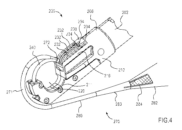
[1009] FIG. 4 is a perspective view of a portion of the medical device shown
in FIG. 3 in
a second configuration coupled to an implant according to an embodiment of the
invention.
[1010] FIG. 5 is a side view of a portion of the medical device shown in FIG.
3.
[1011] FIG. 6 is a side view of the implant shown in FIG. 4.
[1012] FIGS. 7 - 10 are cross-sectional side views of the portion of the
medical device
and the implant shown in FIG. 4 in a first configuration, a third
configuration, the second
configuration and a fourth configuration, respectively.
[1013] FIG. 11 is a side view of a carrier according to an embodiment of the
invention.
[1014] FIG. 12 is a side view of an implant according to an embodiment of the
invention.
2
CA 02702507 2010-04-13
WO 2009/055105 PCT/US2008/070032
[1015] FIG. 13 is a cross-sectional view of the carrier shown in FIG. 11
disposed within
the implant shown in FIG. 12.
[1016] FIG. 14 is a side view of a carrier according to an embodiment of the
invention.
[1017] FIG. 15 is a cross-sectional view of the carrier shown in FIG. 14
disposed within
an implant according to an embodiment of the invention.
[1018] FIG. 16 is a side view of a carrier according to an embodiment of the
invention.
[1019] FIG. 17 is a cross-sectional view of a portion of an implant according
to an
embodiment of the invention.
[1020] FIG. 18 is a cross-sectional view of the carrier shown in FIG. 16
disposed within
the portion of the implant shown in FIG. 17.
[1021] FIG. 19 is a perspective view of a distal end portion of a carrier
according to an
embodiment of the invention.
[1022] FIG. 20 is a perspective view of a portion of an implant according to
an
embodiment of the invention.
[1023] FIG. 21 is a perspective view of the distal end portion of the carrier
shown in FIG.
19 disposed within the portion of the implant shown in FIG. 20.
Detailed Description
[1024] In some embodiments, an apparatus includes a carrier configured to be
movably
disposed within a channel defined by an elongate member. The carrier includes
a proximal
end portion and a distal end portion. The proximal end portion is configured
to be coupled to
an actuator. The distal end portion includes a protrusion and an engagement
surface. The
protrusion has a tip configured to pierce bodily tissue. The protrusion is
configured to be
received within a lumen defined by a connecting portion of an implant, such
as, for example,
a pelvic floor implant, such that the tip extends through the lumen defined by
the connecting
portion of the implant. The engagement surface is configured to engage a
portion of the
connecting portion of the implant to limit movement of the connecting portion
of the implant
relative to the protrusion. For example, in some embodiments, the engagement
surface is
configured to limit movement of the connecting portion of the implant
proximally relative to
3
CA 02702507 2010-04-13
WO 2009/055105 PCT/US2008/070032
the protrusion while allowing movement of the connecting portion of the
implant distally
relative to the protrusion.
[1025] In some embodiments, an apparatus includes an elongate member, an
actuator and
a carrier. The elongate member has a proximal end portion and a distal end
portion. The
actuator is coupled to the proximal end portion of the elongate member. The
carrier is
movably coupled to the distal end portion of the elongate member. In some
embodiments,
for example, the distal end portion of the elongate member can define a
channel within which
the distal end portion of the carrier is movably disposed. The carrier
includes a proximal end
portion and a distal end portion. The proximal end portion is configured to be
coupled to the
actuator, which can be, for example, mechanical rod. The distal end portion is
configured to
be received by and disposed within a lumen defined by a connecting portion of
an implant.
The distal end portion of the carrier includes an engagement surface
configured to engage the
connecting portion of the implant to limit movement of the connecting portion
of the implant
relative to the distal end portion of the carrier.
[1026] In some embodiments, an apparatus includes an elongate member and a
carrier.
The elongate member has a distal end portion. The carrier is movably coupled
to the
elongate member for movement between a first position and a second position.
In the second
position, the distal end portion of the carrier is extended from the distal
end portion of the
elongate member. The distal end portion of the carrier is configured to be
received within
lumen defined by a connecting portion of an implant. The distal end portion of
the carrier
includes an engagement surface configured to engage the connecting portion of
the implant to
retain the distal end portion of the carrier within the lumen defined by the
connecting portion
of the implant when the carrier is moved between the first position and the
second position.
[1027] In some embodiments, an apparatus includes an implant configured to
support an
anatomical structure, such as, for example, a pelvic floor implant. The
implant has a
connector configured to removably engage a carrier of an implant delivery
device such that
the connector is moved distally with the carrier when the carrier is moved
distally. The
connector includes an inner surface and a retention surface. The inner surface
defines a
lumen configured to receive the carrier of the implant delivery device. The
retention surface
is configured to engage a retaining portion of the delivery device such that
movement of the
connector is limited when the carrier is moved proximally.
4
CA 02702507 2010-04-13
WO 2009/055105 PCT/US2008/070032
[1028] In some embodiments, a kit includes an implant delivery device and an
implant.
The implant delivery device has a carrier movably coupled to a distal end
portion of the
implant delivery device. The carrier includes a proximal end portion
configured to be
coupled to an actuator, and a distal end portion including an engagement
surface. The
implant is configured to support an anatomical structure and has a connecting
portion
defining a lumen configured to receive the carrier of the implant delivery
device. The
engagement surface of the carrier is configured to engage the connecting
portion of the
implant to limit movement of the connecting portion of the implant relative to
the distal end
portion of the carrier.
[1029] As used in this specification and the appended claims, the words
"proximal" and
"distal" refer to the direction closer to and away from, respectively, an
operator (e.g.,
surgeon, physician, nurse, technician, etc.) who would insert a medical device
into the
patient, with the tip-end (i.e., distal end) of the device inserted inside a
patient's body first.
Thus, for example, the end of a medical device first inserted inside a
patient's body is the
distal end, while the end of the medical device to last enter the patient's
body or the end of
the medical device extending from the patient's body during a procedure is the
proximal end.
Moreover, the movement of the distal end of a medical device within the body
can, in certain
situations, be considered as movement in the distal direction even though the
distal end of the
medical device may be moving towards the operator of the medical device. For
example, the
movement of the distal end of a medical device along a curved path within the
patient's body
can be considered as distal movement.
[1030] FIGS. 1 and 2 are schematic illustrations of a suturing device 100 and
a portion of
an implant 170 according to an embodiment of the invention in a first
configuration and a
second configuration, respectively. The suturing device 100 includes an
elongate member
102, a carrier 140 and an actuator 155. The elongate member 102 has a proximal
end portion
104 and a distal end portion 106 and defines a channel 108 therein. The
carrier 140 is
disposed within the channel 108 such that the carrier 140 can move along a
longitudinal axis
AL of the channel 108 between a first position (FIG. 1) and a second position
(FIG. 2), as
indicated by the arrow AA in FIG. 2. The longitudinal axis AL can, for
example, pass
lengthwise (e.g., from the proximal end portion 104 to the distal end portion
106) through the
centroid of the elongate member 102 and/or the channel 108 (e.g., the
longitudinal axis AL
can be a centroidal axis of the elongate member 102 and/or the channel 108).
When the
CA 02702507 2010-04-13
WO 2009/055105 PCT/US2008/070032
carrier 140 is in the first position, the suturing device 100 is said to be in
its first
configuration. Similarly, when the carrier 140 is in the second position, the
suturing device
100 is said to be in its second configuration.
[1031] The carrier 140 has a proximal end portion 142 and a distal end portion
144. The
proximal end portion 142 of the carrier 140 is coupled to the actuator 155 by
a coupler 156.
The distal end portion 144 of the carrier 140 includes a protrusion 145 and an
engagement
surface 147. The protrusion 145 has a tip 146 configured to pierce bodily
tissue T, which can
be, for example, pelvic tissue (e.g., a sacrospinous ligament, a tendineus
arch of levator
muscle and/or an iliococcygeus muscle). As shown, the protrusion 145 can be
received by
and disposed within a lumen 173 defined by a connecting portion 172 of the
implant 170 such
that the distal portion of the tip 146 extends through the lumen 173. Said
another way, the
protrusion 145 can be received within the lumen 173 such that the distal
portion of the tip 146
is disposed outside of the lumen 173 defined by the connecting portion 172 of
the implant
170. Moreover, the protrusion 145 can be received within the lumen 173 such
that the
engagement surface 147 of the carrier 140 contacts and/or engages a portion of
the
connecting portion 172 of the implant 170.
[1032] In this manner, the engagement surface 147 of the carrier 140 can limit
movement
of the connecting portion 172 of the implant 170 relative to the carrier 140.
For example, in
some embodiments, the engagement surface 147 of the carrier 140 can limit
movement of the
connecting portion 172 of the implant 170 proximally relative to the carrier
140 while
allowing movement of the connecting portion 172 of the implant 170 distally
relative to the
carrier 140. In other embodiments, the engagement surface 147 of the carrier
140 can limit
movement of the connecting portion 172 of the implant 170 both proximally and
distally
relative to the carrier 140.
[1033] The suturing device 100 can be used to place at least a portion of the
implant 170
within the bodily tissue T, as described below. The implant 170 can be loaded
onto the
suturing device 100 when the suturing device 100 is in the first configuration
(i.e., when the
carrier 140 is in its first, or retracted, position, as shown in FIG. 1). To
load the implant 170,
the connecting portion 172 of the implant is disposed at the distal end
portion 106 of the
elongate member 102 such that the protrusion 145 of the carrier 140 is
received within the
lumen 173 defined by the connecting portion 172. Although the tip 146 is shown
as being
disposed distally outside of the lumen 173 when the suturing device 100 is in
the first
6
CA 02702507 2010-04-13
WO 2009/055105 PCT/US2008/070032
configuration, in other embodiments, the carrier 140 can be disposed within
the channel 108
of the elongate member 102 such that the tip 146 is disposed within the lumen
173 (i.e., the
tip 146 does not extend outside of the lumen 173) when the suturing device 100
is in the first
configuration. In yet other embodiments, the carrier 140 can be disposed
within the channel
108 of the elongate member 102 such that the tip 146 is disposed entirely
within the in
channel 108 (i.e., the tip 146 is not disposed within the lumen 173) when the
suturing device
100 is in the first configuration. Said another way, in some embodiments, the
connecting
portion 172 can contact and/or engage the distal end portion 106 of the
elongate member 102
when the suturing device 100 is in the first configuration.
[1034] Similarly, although the engagement surface 147 of the carrier 140 is
shown as
being in contact with the connecting portion 172 of the implant 170 when the
suturing device
100 is in the first configuration, in other embodiments, the engagement
surface 147 can be
spaced apart from the connecting portion 172 of the implant 170 when the
suturing device
100 is in the first configuration. For example, in some embodiments, the
connecting portion
172 of the implant can be in contact with distal end portion 106 of the
elongate member 102
when the suturing device 100 is in the first configuration.
[1035] The suturing device 100 can be disposed within the body until the
connecting
portion 172 of the implant 170 and/or the distal end portion 106 of the
elongate member 102
is in contact with and/or adjacent to the bodily tissue T. The suturing device
100 can be
disposed within the body by any suitable means, such as, for example, via an
endoscope.
When the suturing device 100 is suitably disposed within the body, the
actuator 155 can be
used to move the carrier 140 within the channel 108 of the elongate member 102
from its first
position (FIG. 1) to its second position (FIG. 2), as indicated by the arrow
AA in FIG. 2. In
this manner, the suturing device 100 can be moved from its first configuration
to its second
configuration. The actuator 155 can be any suitable actuator, such as, for
example, a
mechanical actuator, an electronic actuator, a hydraulic actuator, a pneumatic
actuator or the
like. Similarly, the coupler 156 can be any suitable member for operatively
coupling the
actuator 155 to the proximal end portion 104 of the carrier 140. In some
embodiments, for
example, the coupler 156 can be a mechanical linkage.
[1036] When the carrier 140 moves from its first position (FIG. 1) to its
second position
(FIG. 2), the tip 146 of the carrier 140 pierces the bodily tissue T, thereby
defining a
passageway within the tissue T for receiving at least a portion of the implant
170. Moreover,
7
CA 02702507 2010-04-13
WO 2009/055105 PCT/US2008/070032
when the carrier 140 is moved from its first position to its second position,
the engagement
surface 147 of the carrier 140 contacts a portion of the connecting portion
172 of the implant
170, such that the connecting portion 172 moves distally with the carrier 140
and into the
passageway defined within the bodily tissue T. Said another way, when the
carrier 140 is
moved from its first position to its second position, the engagement surface
147 of the carrier
140 contacts a portion of the connecting portion 172 of the implant 170 such
that movement
of the connecting portion 172 of the implant 170 proximally relative to the
carrier 140 is
limited. In this manner, the passageway within the tissue T can be defined and
the implant
170 can be disposed within the passageway in one operation.
[1037] When the connecting portion 172 is secured within the bodily tissue T,
the
actuator 155 can be used to move the carrier 140 within the channel 108 of the
elongate
member 102 from its second position back to its first position. Accordingly,
the distal end
144 of the carrier 140 is moved distally relative to the connecting portion
172 of the implant
170, thereby leaving the implant 170 disposed within the tissue T. The
suturing device 100
can then be removed from the body.
[1038] FIGS. 3 - 10 illustrate a suturing device 200 and an implant 270
according to an
embodiment of the invention. Although FIGS. 3 - 10 show the same embodiment,
certain
reference numerals and/or features are omitted in some of the figures for
clarity. FIG. 3 is a
perspective view of the suturing device 200 in a first (i.e., retracted)
configuration. FIG. 4 is
a perspective view of the distal portion of the suturing device 200 in a
second (i.e., extended)
configuration coupled to a distal portion 271 of an implant 270. FIG. 5 is a
side view of the
carrier 240 of the suturing device 200. FIG. 6 is a side view of the implant
270. FIGS. 7 - 10
are cross-sectional side views of the suturing device and the implant 270 in
the first
configuration (i.e., a retracted configuration with the implant 270 loaded), a
third
configuration (i.e., a partially extended configuration), the second
configuration (i.e., the
extended configuration) and a fourth configuration (i.e., a retracted
configuration after
placement of the implant 270), respectively.
[1039] The suturing device 200 includes an elongate member 202, a carrier 240
(see e.g.,
FIG. 5), a handle 260 and actuator 255. The elongate member 202 has a proximal
end
portion 204 and a distal end portion 206 and defines a channel 208 (see FIGS.
7 - 10). As
described in more detail herein, the carrier 240 is movably disposed within a
curved portion
224 of the channel 208.
8
CA 02702507 2010-04-13
WO 2009/055105 PCT/US2008/070032
[1040] As shown in FIG. 3, the handle 260 is coupled to the proximal end
portion 204 of
the elongate member 202. A retaining ring 262 is movably disposed about the
proximal end
portion 204 of the elongate member 202 adjacent the handle 260. The retaining
ring 262 can
include openings, slots and/or any other suitable mechanism for releasably
retaining a portion
of the implant 270 when the suturing device 200 is in use. Examples of a
suitable retaining
ring 262 are shown and described in U.S. Patent Application No. 11/435,708
entitled "Tying
Knots," filed May 16, 2006, which is incorporated herein by reference in its
entirety.
[1041] The actuator 255 is slidably disposed within the handle 260 such that a
portion of
the actuator 255 is disposed within the channel 208 and is in contact with a
flexible coupling
rod 256 (see e.g., FIGS. 7 - 10). The coupling rod 256 is in contact with a
proximal end
portion 242 of the carrier 240. Accordingly, as discussed in more detail
herein, the actuator
255 can be used to move the carrier 240 within the curved portion 224 of the
channel 208. In
some embodiments, the suturing device 200 can include a biasing member (not
shown in
FIGS. 3 - 10) configured to bias the actuator 255, the coupling rod 256 and/or
the carrier 240
in a predetermined position within the channel 208. For example, in some
embodiments a
spring can be disposed between the actuator and a portion of the handle (e.g.,
the spring can
be located either inside of the handle 260 or outside of the handle 260) to
urge the actuator in
its proximal position (i.e., the position as shown in FIG. 3). In this manner,
the suturing
device 200 can be maintained and/or biased in its first (i.e., retracted)
configuration.
[1042] The distal end portion 206 of the elongate member 202 includes a
delivery head
210 having a first surface 212, a second surface 216 and a curved portion 211
between the
first surface 212 and the second surface 216. The first surface 212 is spaced
apart from the
second surface 216 such that an opening 220 is defined between the first
surface 212 and the
second surface 216, bounded by the curved portion 211. The first surface 212
defines an
opening 214 in communication with the curved portion 224 of the channel 208
(see e.g. FIG.
10). The second surface 216 defines an opening 218 about which a retainer 230
is disposed.
As shown in FIG. 4, the retainer 230, which is also referred to as a "catch,"
defines three
openings 232 each having an enlarged portion 234. The portions of the retainer
230 defining
the three openings 232 can be referred to as "ribs."
[1043] The carrier 240 is a tubular member (i.e., the carrier 240 has a
circular cross-
sectional area) and has a proximal end portion 242 and a distal end portion
244, and defines a
center line CL. The carrier 240 is curved such that the center line CL defines
a radius of
9
CA 02702507 2010-04-13
WO 2009/055105 PCT/US2008/070032
curvature R1. As shown in FIGS. 7 - 10, the proximal end portion 242 of the
carrier 240 is in
contact with and/or coupled to the connecting rod 256. In this manner, the
carrier 240 is
operatively coupled to the actuator 255 such that when the actuator 255 moves
relative to the
handle 260, the carrier 240 moves within the curved portion 224 of the channel
208.
[1044] The distal end portion 244 of the carrier 240 includes a protrusion 245
and an
engagement surface 247. The protrusion 245 is substantially solid (i.e., is
devoid of openings
therein) and has a tip 246 configured to pierce bodily tissue T (see e.g.,
FIGS. 7 - 10). The
bodily tissue T can be, for example, pelvic tissue (e.g., a sacrospinous
ligament, a tendineus
arch of levator muscle and/or an iliococcygeus muscle). The engagement surface
247 is
substantially normal to the center line CL of the carrier 240. Said another
way, the center
line CL of the carrier 240 and a line defined to include a portion of the
engagement surface
247 intersect at an angle of approximately 90 degrees. For example, when the
engagement
surface 247 is planar, a plane defined by the engagement surface 247
intersects the center line
CL of the carrier at an angle of approximately 90 degrees.
[1045] As shown in FIG. 5, the diameter D2 of the protrusion 245 is less than
the
diameter Di of the remaining portions of the carrier 240. The size of the
engagement surface
247 is associated with the difference in the diameter Di of the carrier 240
and the diameter D2
of the protrusion 245. Said another way, the engagement surface 247 is the
shoulder or step
between the diameter Di of the carrier 240 and the diameter D2 of the
protrusion 245. In
some embodiments, the diameter D2 of the protrusion 245 is approximately three
quarters the
diameter Di of the carrier 240. In other embodiments, the diameter D2 of the
protrusion 245
is approximately half the diameter Di of the carrier 240. In yet other
embodiments, the
diameter D2 of the protrusion 245 is approximately one quarter the diameter Di
of the carrier
240. In some embodiments, for example, the diameter Di of the carrier can be
between 10
and 12 mm. In other embodiments, the diameter Di of the carrier can be between
12 and 15
mm.
[1046] As shown in FIGS. 4 and 6, the implant 270 includes a distal portion
271, a dilator
280, a sleeve 282 and a strap 284. The distal portion 271 of the implant 270
includes a
connector 272 and defines an opening 285. The connector 272 includes an outer
surface 274,
an inner surface 275 and a retention surface 276. The inner surface 275
defines a lumen 273
therethrough. As described in more detail herein, the protrusion 245 of the
carrier 240 can be
disposed through the opening 285 and within the lumen 273 defined by the inner
surface 275
CA 02702507 2010-04-13
WO 2009/055105 PCT/US2008/070032
of the connector 272 such that the tip 246 extends through the lumen 273 (see
e.g. FIGS. 8
and 9).
[1047] Moreover, the protrusion 245 can be received within the lumen 273 such
that the
engagement surface 247 of the carrier 240 contacts and/or engages a portion of
the retention
surface 276 of the connector 272. In this manner, the engagement surface 247
of the carrier
240 can selectively limit movement of the connector 272 of the implant 270
relative to the
carrier 240. Said another way, when the carrier 240 moves as indicated by the
arrow CC in
FIG. 8, the engagement surface 247 of the carrier 240 contacts and/or engages
the retention
surface 276 of the connector 272 such that the connector 272 moves with the
carrier 240.
When the carrier 240 moves as indicated by the arrow EE in FIG. 10, the
engagement surface
247 of the carrier 240 becomes disengaged from the retention surface 276 of
the connector
272 such that the carrier 240 moves relative to the connector 272.
[1048] The suturing device 200 can be used to place the implant 270 within the
bodily
tissue T, as described below with reference to FIGS. 7 - 10. As shown in FIG.
7, the
connector 272 of implant 270 can be placed or "loaded" onto the suturing
device 200 when
the suturing device 200 is in the first configuration. When the suturing
device 200 is in the
first configuration, the tip 246 of the protrusion is disposed within the
curved portion 224 of
the channel 208. Accordingly, to load the implant 270, the connecting portion
272 of the
implant is disposed against the delivery head 210 such that at least a portion
of the retention
surface 276 of the connector 272 contacts at least a portion of the first
surface 212 of the
delivery head 210.
[1049] In some embodiments, the connector 272 can be removably coupled to the
first
surface 212 of the delivery head 210. In this manner, the implant 270 can
remain loaded onto
the suturing device 200 prior to use (e.g., when the suturing device is placed
on a surgical
tray or the like). In some embodiments, for example, the connector 272 can
include a
retaining mechanism, such as a tab or protrusion (not shown in FIGS. 7 - 10)
that is received
within a mating recess or opening defined by the first surface 212 to
removably couple the
connector 272 to the delivery head 210. In other embodiments, a portion of the
distal portion
271 of the implant can be received within an opening or notch defined by the
delivery head to
removably couple the implant 270 to the delivery head.
11
CA 02702507 2010-04-13
WO 2009/055105 PCT/US2008/070032
[1050] The suturing device 200 and the implant 270 can be disposed within the
body such
that a portion of the bodily tissue T is within the opening 220 defined
between the first
surface 212 and the second surface 216 of the delivery head 210. The suturing
device 200
can be disposed within the body by any suitable means, such as, for example,
via an
endoscope. When the suturing device 200 is suitably disposed within the body,
the actuator
255 can be moved distally relative to the handle 260, thereby causing the
flexible coupling
rod 256 to move within the channel 208 as indicated by the arrow BB in FIG. 8.
The
translation of the coupling rod 256 within the channel 208 causes the carrier
240 to move
within the curved portion 224 of the channel 208 as indicated by the arrow CC
in FIG. 8. In
this manner, the suturing device 200 can be moved from the retracted
configuration to the
partially extended configuration.
[1051] When the suturing device 200 is in the partially extended
configuration, as shown
in FIG. 8, the distal end portion 244 of the carrier 240 is disposed through
the opening 214
defined by the first surface 212 of the delivery head 210 and outside of the
curved portion
224 of the channel 208. The distal end portion 244 of the carrier 240 is
further disposed
within the opening 285 defined by the distal portion 271 of the implant such
that the
protrusion 245 is received within the lumen 273 defined by the inner surface
275 of the
connector 272. As shown, when the suturing device 200 is in the partially
extended and
extended configurations, the tip 246 extends through the lumen 273 (i.e., the
distal portion of
the tip 246 is disposed outside of the lumen 273). Moreover, the engagement
surface 247 of
the carrier 240 contacts the retention surface 276 of the connector 272 such
that the connector
272 moves with the carrier 240 when the carrier 240 moves in the direction as
indicated by
the arrow CC in FIG. 8.
[1052] As shown in FIG. 8, when the carrier 240 moves in the direction as
indicated by
the arrow CC, the tip 246 of the protrusion can pierce the tissue T to define
a passageway
within the tissue T through which a portion of the implant 270 (e.g., the
distal portion 271,
the dilator 280 and/or the strap 284) can be placed. The continued movement of
the carrier
240 causes the connector 272 to be inserted through and/or enlarge the
passageway through
the tissue T. As shown, the outer surface 274 of the connector 272 is tapered
to help the
connector further define and/or enlarge the passageway through the tissue.
[1053] Continued movement of the actuator 255 causes carrier 240 to move until
the tip
246 of the carrier 240 and/or the connector 272 of the implant 270 contacts
the retainer 230.
12
CA 02702507 2010-04-13
WO 2009/055105 PCT/US2008/070032
The openings 232 defined by the retainer 230 are smaller than the widest
portion of the
connector 272 thereby defining an interference fit between the connector 272
and the retainer
230. For example, in the illustrated embodiment, the widest portion of the
connector 272 is
the diameter of the retention surface 276. Thus, diameter of the retention
surface 276 forms
an interference fit with the retainer 230 when disposed within the openings
232 defined by
the retainer. Accordingly, continued movement of the actuator 255 causes the
portions of the
retainer 230 (e.g., the "ribs") defining the openings 232 to deflect thereby
allowing the
connector 272 to be disposed through one of the openings 232. In this manner,
the suturing
device 200 can be placed into the extended configuration, as shown in FIG. 9.
[1054] When the suturing device 200 is in the extended configuration, the
distal end
portion 244 of the carrier 240 and the connector 272 of the implant 270 are
disposed through
one of the openings 232 defined by the retainer 230 and into the opening 218
defined by the
second surface 216 of the delivery head 210. Said another way, the connector
272 is
disposed within the "catch" (i.e., the retainer 230). Moreover, the distal end
portion 271 of
the implant 270 is disposed within the passageway through the tissue T. After
the suturing
device 200 has been placed in the extended configuration, the actuator 255 can
be moved
proximally relative to the handle 260, thereby causing the coupling rod 256 to
move within
the channel 208 as indicated by the arrow DD in FIG. 10. The translation of
the coupling rod
256 within the channel 208 causes the carrier 240 to move within the curved
portion 224 of
the channel 208 as indicated by the arrow EE in FIG. 10. In this manner, the
suturing device
200 can be moved from the extended configuration back to the retracted
configuration. In
some embodiments, for example, the suturing device 200 can be moved from the
extended
configuration back to the retracted configuration by a biasing member (not
shown in FIGS. 3
-10).
[1055] Because the retainer 230 defines an interference fit between the
connector 272 and
the retainer 230 when the carrier 240 moves from the extended configuration to
the retracted
configuration, the connector 272 is retained within the opening 218. Said
another way, the
distal end 244 of the carrier 240 is moved relative to the connector 272 of
the implant 270,
thereby leaving the connector 272 within the opening 218. Said yet another
way, when the
carrier 240 moves from the extended configuration (FIG. 9) to the retracted
configuration
(FIG. 10), the engagement surface 247 of the carrier 240 is spaced apart from
the retention
surface 276 of the connector 272 such that the connector 272 does not move
with the carrier
13
CA 02702507 2010-04-13
WO 2009/055105 PCT/US2008/070032
240 when the carrier 240 moves in the direction as indicated by the arrow EE
in FIG. 10.
Accordingly, the carrier 240 moves in the direction as indicated by the arrow
EE back
through the passageway defined through the tissue T such that only the distal
end portion 271
of the implant 270 is within the tissue.
[1056] The suturing device 200 can then be moved within the body such that the
bodily
tissue T is no longer within the opening 220. Because the connector 272 is
retained within
the opening 218, movement of the suturing device 200 causes the implant 270 to
move within
the tissue T. In this manner, the implant 270 can be positioned and/or
tensioned within the
tissue T, as desired. When the implant 270 is suitably positioned within the
tissue T, the
connector 272 can be removed from the retainer 230 via the enlarged portion
234 of the
opening 232.
[1057] The strap 284, which can also be referred to as an arm, a support
member and/or
an implant member, is configured to engage bodily tissue T to retain the
implant 270 in its
desired position and/or support an anatomical structure when disposed within
the body. In
some embodiments, for example, the strap 284 can be constructed from a mesh
material
configured to promote tissue in-growth to enhance the anchoring of the implant
270.
Similarly, in some embodiments, the strap 284 can include roughened and/or
jagged edges to
enhance the anchoring of the implant 270 within the tissue T. In some
embodiments, for
example, the strap 284 can include protrusions or "tangs" along an edge of the
strap 284 to
enhance the anchoring of the implant 270 within the tissue T.
[1058] The sleeve 282 is coupled to an end of the dilator 280 and houses or
encloses the
strap 284. The sleeve 282 is constructed from a smooth material and provides
for a smooth
transition of the strap 284 through the tissue T during insertion of the
implant assembly 270.
For example, in some embodiments, the sleeve 282 can help prevent the strap
284 from
prematurely engaging the tissue T during delivery. Similarly, the dilator 280
is constructed
from a smooth material and has a tapered portion 283. In this manner, when the
implant 270
is being placed within the tissue T, the dilator can enlarge the passageway
within the tissue T
so that the sleeve 282 and the strap 284 can be positioned within the tissue
as desired.
[1059] When the implant 270 is properly positioned and tensioned within the
tissue T as
described above, the sleeve 282 and/or the dilator 280 can be removed from the
strap 284
14
CA 02702507 2010-04-13
WO 2009/055105 PCT/US2008/070032
(not shown in FIG. 10). In this manner, the strap 284 can engage the
surrounding tissue T to
secure the implant 270 in position.
[1060] The components of the suturing device 200 can be constructed from any
suitable
biocompatible material. For example, in some embodiments, the elongate member
202, the
delivery head 210, the actuator 255 and/or the handle 260 can be constructed
from any
suitable medical grade plastic, such as polypropylene, polycarbonate or glass-
filled
polycarbonate. In some embodiments, the carrier 240 can be constructed a metal
alloy, such
as stainless steel. In some embodiments, the coupling rod 256 can be
constructed from a
flexible material, such as stainless steel, Nitinol or the like.
[1061] Similarly, the components of the suturing device 200 can be constructed
using any
suitable manufacturing process or combination of manufacturing processes. For
example, in
some embodiments, the elongate member 202 can be constructed from a medical
grade
plastic using an extrusion process. In other embodiments, the elongate member
202 can be
constructed from a medical grade plastic using a molding process. In some
embodiments, for
example, the delivery head 210 can be constructed separately and/or using a
different process
from the elongate member 202. In such embodiments, the delivery head 210 can
be coupled
to the elongate member 202 using fasteners (e.g., screws, rivets or the like),
an adhesive
bond, a weld or the like. In other embodiments, the delivery head 210 and the
elongate
member 202 can be monolithically formed. Similarly, in some embodiments, the
tip 246 of
the carrier 240 can be constructed separately from the carrier 240 and can be
coupled to the
distal end portion 244 of the carrier 240 by an adhesive, a weld or the like.
[1062] The components of the implant 270 can be constructed from any suitable
biocompatible material. In some embodiments, for example, the implant 270 can
constructed
from synthetic materials, such as, for example, nylon, silicone, polyethylene,
polyester,
polyimide, polyurethane, polypropylene, fluoropolymers or the like. In other
embodiments,
the implant 270 can constructed from natural materials, such as, for example
materials
derived from human and/or animal tissue. In yet other embodiments, the implant
270 can be
constructed from a combination of synthetic and natural materials.
[1063] Although the implant 270 has been shown and described above without
being
associated with any specific anatomical structures, in some embodiments, the
implant 270
can be associated with the repair of various pelvic dysfunctions. For example,
in some
CA 02702507 2010-04-13
WO 2009/055105 PCT/US2008/070032
embodiments, the implant can 270 can be an implant configured to be delivered
into a pelvic
region to repair a prolapsed uterus. Similarly, the implant 270 can be placed
within the tissue
T by any suitable method, For example, in some embodiments, portions of the
implant 270
can be placed into the body through an obturator (e.g., using a transobturator
approach).
Moreover, the implants can be placed within the body using any suitable
surgical approach
(e.g., a retro-pubic, supra pubic, or pre-pubic approach).
[1064] Although the carrier 240 is shown and described as including a
protrusion 245
having a tip 246 configured to pierce bodily tissue, in other embodiments, a
carrier can be
devoid of a protrusion and/or a tissue piercing tip. For example, in FIG. 11
shows a carrier
340 according to an embodiment of the invention that can be disposed within
any suitable
suturing device of the type shown and described herein. The carrier 340 has a
proximal end
portion 342 and a distal end portion 344, and defines a center line CL. The
carrier 340 is
curved such that the center line CL defines a radius of curvature R2. As
previously described,
the proximal end portion 342 of the carrier 340 can be operatively coupled to
an actuator (not
shown in FIG. 11) such that the carrier 340 can be moved within an elongated
member (not
shown in FIG. 11), in a similar manner as that described above.
[1065] The distal end portion 344 of the carrier 340 includes a protrusion 345
having an
engagement surface 347 disposed at the distal end thereof. The protrusion 345
differs from
the protrusion 245 shown and described above, in that the protrusion 345 has a
blunt end (i.e.,
the engagement surface 347) and is not configured to pierce bodily tissue. The
engagement
surface 347 is substantially normal to the center line CL of the carrier 340.
Said another way,
the center line CL of the carrier 340 and a line defined to include a portion
of the engagement
surface 347 intersect at an angle of approximately 90 degrees. For example,
when the
engagement surface 347 is planar, a plane defined by the engagement surface
347 intersects
the center line CL of the carrier 340 at an angle of approximately 90 degrees.
[1066] FIGS. 12 and 13 show an implant 370 according to an embodiment of the
invention that can be placed within the body using a suturing device including
the carrier 340.
The implant 370 includes a distal portion 371, a dilator 380, a sleeve 382 and
a strap 384.
The distal portion 371 of the implant 370 includes a connecting portion 372.
The connecting
portion 372 includes an outer surface 374, an inner surface 375 and an opening
385. The
outer surface 374 defines a tip 377 configured to pierce bodily tissue. The
outer surface 374
also defines a shoulder surface 379. The inner surface 375 includes a
retention surface 376
16
CA 02702507 2010-04-13
WO 2009/055105 PCT/US2008/070032
and defines a lumen 373. The lumen 373 terminates at the retention surface
376. Said
another way, the lumen 373 is a "blind hole" (i.e., the tip 377 does not
define a passageway
into the lumen 373). The dilator 380, sleeve 382 and strap 384 are similar to
those described
above with reference to FIG. 6, and are therefore not discussed in detail.
[1067] As shown in FIG. 13, the distal end portion 344 of the carrier 340 can
be disposed
within the opening 385 such that the protrusion 345 is received within the
lumen 373 defined
by the inner surface 375 of the connecting portion 372. Moreover, the
protrusion 345 can be
received within the lumen 373 such that the engagement surface 347 of the
carrier 340
contacts and/or engages a portion of the retention surface 376 of the
connector 372. In this
manner, the engagement surface 347 of the carrier 340 can selectively limit
movement of the
connector 372 of the implant 370 relative to the carrier 340. Said another
way, when the
carrier 340 moves distally, the engagement surface 347 of the carrier 340
contacts and/or
engages the retention surface 376 of the connecting portion 372 such that the
implant 370
moves with the carrier 340 when the carrier 340 moves distally. When the
carrier 340 moves
proximally, however, the engagement surface 347 of the carrier 340 becomes
disengaged
from the retention surface 376 of the connecting portion 372 such that the
carrier 340 can
move proximally relative to the implant 370.
[1068] In some embodiments, when the carrier 340 moves distally the connecting
portion
372 of the implant can be disposed within a retainer (not shown in FIGS. 11 -
13) similar to
the retainer 230 shown and described above. Accordingly, when the carrier 340
moves
proximally, the shoulder surface 379 can engage a portion of the retainer to
maintain the
position of the connecting portion 372 within the retainer.
[1069] Although the engagement surfaces of the carriers shown and described
are planar
surfaces substantially normal to the center line of the carrier, in other
embodiments, an
engagement surface can be angularly offset from the center line by an angle
other than 90
degrees. Moreover, in other embodiments, an engagement surface can be a non-
planar (i.e.,
curved) surface. For example, FIGS. 14 and 15 show a carrier 440 and according
to an
embodiment of the invention that can be disposed within any suitable suturing
device of the
type shown and described herein. The carrier 440 has a proximal end portion
442 and a distal
end portion 444, and defines a center line CL. The carrier 440 is curved such
that the center
line CL defines a radius of curvature R3. As previously described, the
proximal end portion
442 of the carrier 440 can be operatively coupled to an actuator (not shown in
FIGS. 14 and
17
CA 02702507 2010-04-13
WO 2009/055105 PCT/US2008/070032
15) such that the carrier 440 can be moved within an elongated member (not
shown in FIGS.
14 and 15), as described above.
[1070] The distal end portion 444 of the carrier 440 includes a protrusion 445
having a
tapered engagement surface 447. The protrusion 445 also defines a tip 446
configured to
pierce bodily tissue, as described above. As shown in FIG. 15, the engagement
surface 447 is
angularly offset from the center line CL of the carrier 440 by an acute angle
0 (e.g., the taper
angle). Moreover, because the engagement surface 447 includes the conical
portion of the
protrusion 445, the engagement surface 447 has a curved shape.
[1071] FIG. 15 shows a connector 472 of an implant 470 disposed about the
protrusion
445 of the carrier 440. The connector 472 includes an outer surface 474 and an
inner surface
475. The outer surface 474 defines a shoulder surface 479. The inner surface
475 defines a
lumen 473 that extends through the connector 472. As shown, the protrusion 445
of the
carrier 440 can be disposed within the lumen 473 such that that the engagement
surface 447
contacts and/or engages a portion of the inner surface 475 of the connector
472. The lumen
473 can be sized such that the tip 446 is disposed outside of the lumen 473.
In this manner,
when the carrier 440 moves, tip 446 can pierce the targeted bodily tissue to
define a
passageway therethrough. Moreover, the engagement surface 447 of the carrier
440 can
selectively limit movement of the connector 472 relative to the carrier 440.
Said another
way, when the carrier 440 moves distally, the engagement surface 447 of the
carrier 440
contacts and/or engages the inner surface 475 of the connector 472 such that
the implant (not
shown in FIGS. 14 and 15) moves distally with the carrier 440.
[1072] When the carrier 440 moves proximally, however, the engagement surface
447 of
the carrier 440 becomes disengaged from the inner surface 475 of the connector
472 such that
the carrier 440 can move proximally relative to the inner surface 475 of the
implant. For
example, in some embodiments, the connector 472 of the implant 470 can be
disposed within
a retainer (not shown in FIGS. 11 - 13) similar to the retainer 230 shown and
described
above. Accordingly, when the carrier 440 moves proximally, the shoulder
surface 479 of the
connector 472 can engage a portion of the retainer to maintain the position of
the connecting
portion 472 within the retainer.
[1073] Although the engagement surface 447 is shown in FIG. 15 as being
angularly
offset from the center line CL of the carrier 440 by a non-zero angle 0, in
some
18
CA 02702507 2010-04-13
WO 2009/055105 PCT/US2008/070032
embodiments, the engagement surface 447 of the carrier 440 can be
substantially parallel to
the center line CL of the carrier 440. Said another way, in some embodiments,
the
engagement surface 447 of the carrier 440 can be coaxial with the center line
CL of the
carrier 440. In some such embodiments, for example, the engagement surface of
the carrier
can be configured to form an interference fit with the inner surface of the
connector to
selectively limit distal and/or proximal movement of the connector relative to
the carrier
and/or retain the connector to the carrier.
[1074] Although the carriers are shown and described above as including a
single,
continuous retention surface, in other embodiments, a carrier can include
multiple surfaces.
For example, FIGS. 16 and 18 show a carrier 540 and according to an embodiment
of the
invention that can be disposed within any suitable suturing device of the type
shown and
described herein. The carrier 540 has a proximal end portion 542 and a distal
end portion
544, and defines a center line CL. The carrier 540 is curved such that the
center line CL
defines a radius of curvature R4. As previously described, the proximal end
portion 542 of
the carrier 540 can be operatively coupled to an actuator (not shown in FIGS.
16 and 18) such
that the carrier 540 can be moved within an elongated member (not shown in
FIGS. 16 and
18), as described above.
[1075] The distal end portion 544 of the carrier 540 includes a first
engagement surface
547A and a second engagement surface 547B opposite the first engagement
surface 547A.
The first engagement surface 547A and the second engagement surface 547B
collectively
define an annular opening or groove 548. The distal end portion 544 of the
carrier 540 also
includes a tip 546 configured to pierce bodily tissue, as described above.
[1076] FIG. 17 shows a connector 572 of an implant 570 configured to be
disposed about
the distal end portion 544 of the carrier 540. The connector 572 includes an
outer surface 574
and an inner surface 575. The inner surface 575 defines a lumen 573 that
extends through the
connector 572. The inner surface 575 includes a first retention surface 576A
and a second
retention surface 576B opposite the first retention surface 576A. The first
retention surface
576A and the second retention surface 576B collectively define an annular
protrusion 578.
The proximal end of the connector 572 includes a shoulder surface 579
configured to engage
a retainer (not shown in FIGS. 16 - 18), as described below.
19
CA 02702507 2010-04-13
WO 2009/055105 PCT/US2008/070032
[1077] As shown in FIG. 18, the distal end portion 544 of the carrier 540 can
be disposed
within the lumen 573 such that that the protrusion 578 of the connector 572 is
received within
the groove 548 of the carrier 540. In some embodiments, the protrusion 578 and
the groove
548 can be sized such that they collectively form an interference or "snap"
fit between the
carrier 540 and the connector 572. Said another way, in some embodiments, the
protrusion
578 of the connector 572 matingly engages the groove 548 of the carrier 540 to
removably
couple the connector 572 to the carrier 540.
[1078] When the protrusion 578 of the connector 572 is received within the
groove 548
of the carrier 540, the first engagement surface 547A contacts and/or engages
the first
retention surface 576A and the second engagement surface 547B contacts and/or
engages the
second retention surface 576B. In this manner, the connector 572 can be
retained in position
about the carrier 540. Accordingly, when the carrier 540 moves, the first
engagement surface
547A of the carrier 540 can selectively limit the proximal movement of the
connector 572
relative to the carrier 540. Similarly, when the carrier 540 moves, the second
engagement
surface 547B of the carrier 540 can selectively limit the distal movement of
the connector
572 relative to the carrier 540. When the connector is retained within the
delivery head of the
suturing device (not shown in FIGS. 16 - 18) as described above, the
protrusion 578 and the
groove 548 can be sized such that the force imparted by the retainer (not
shown in FIG. 16 -
18) on the shoulder surface 579 is sufficient to move the protrusion 578 from
the groove 548,
thereby releasing the connector 572 from the distal end portion 544 of the
carrier 540.
[1079] FIG. 19 shows a distal end portion 644 of a carrier and according to an
embodiment of the invention that can be disposed within any suitable suturing
device of the
type shown and described herein. As described above, the carrier defines a
center line CL.
In some embodiments, the carrier can be curved such that the center line CL
defines a radius
of curvature, as described above. The distal end portion 644 of the carrier
includes a tip 646
and defines a retention slot 649. The tip 646 is configured to pierce bodily
tissue, as
described above. The slot 649 includes an opening 650 at the distal end of the
tip 646 and is
bounded by a surface 647 disposed proximally from the distal end of the tip
646. The slot 649
is substantially parallel to and coaxial with the center line CL.
[1080] FIG. 20 shows a distal end portion 671 of an implant configured to be
disposed
about the distal end portion 644 of the carrier according to an embodiment of
the invention.
The distal end portion 671 of the implant includes a connector 672. The
connector 672
CA 02702507 2010-04-13
WO 2009/055105 PCT/US2008/070032
includes a tapered outer surface 674, an inner surface 675 and a retention
member 690. As
described above, the inner surface 675 defines a lumen 673 that extends
through the
connector 672. The outer surface 674 includes a shoulder surface 679
configured to engage a
retainer (not shown in FIGS. 16 - 18).
[1081] The retention member 690 includes a first end portion 691, a second end
portion
692 and a central portion 693. The first end portion 691 and the second end
portion 692 are
each coupled to the outer surface 674 of the connector 672 such that the
retention member
690 forms a loop at the distal end of the connector 672. The retention member
690 can be
coupled to the outer surface 674 in any suitable fashion. In some embodiments,
for example,
the retention member 690 can be coupled to the outer surface 674 by an
adhesive, by a
thermal bond, by a weld or the like. In other embodiments, the connector 672
and the
retention member 690 can be monolithically formed. Similarly, the retention
member 690
can be constructed from any suitable material, such as, for example, a
flexible polymer, a
rigid polymer, a metallic alloy or the like.
[1082] As shown in FIG. 21, the distal end portion 644 of the carrier can be
disposed
within the lumen 673 such that that the retention member 690 of the connector
672 is
disposed through the opening 650 and is received within the slot 649. In this
manner, the
central portion 693 of the retention member 690 can contact and/or engage the
surface 647 to
selectively maintain a position of the connector 672 relative to the distal
end portion 644 of
the carrier. Said another way, when the carrier moves distally, the surface
647 of the carrier
contacts and/or engages the central portion 693 of the retention member 690
such that the
distal portion 671 of the implant moves distally with the carrier.
[1083] When the carrier moves proximally, however, the carrier can move
relative to the
retention member 690 of the implant. Accordingly, the retention member 690 can
move
outside of the slot 649 through the opening 650. For example, in some
embodiments, the
connector 672 can be disposed within a retainer (not shown in FIG. 21) similar
to the retainer
230 shown and described above. Accordingly, when the carrier moves proximally,
the
shoulder surface 679 of the connector 672 can engage a portion of the retainer
to maintain the
position of the connecting portion 672 within the retainer. The proximal
movement of the
carrier thereby causes the connector 672 to become disposed apart from the
distal end portion
644 of the carrier.
21
CA 02702507 2010-04-13
WO 2009/055105 PCT/US2008/070032
[1084] While various embodiments of the invention have been described above,
it should
be understood that they have been presented by way of example only, and not
limitation.
Where methods described above indicate certain events occurring in certain
order, the
ordering of certain events may be modified. Additionally, certain of the
events may be
performed concurrently in a parallel process when possible, as well as
performed sequentially
as described above. Thus, the breadth and scope of the invention should not be
limited by
any of the above-described embodiments. While the invention has been
particularly shown
and described with reference to specific embodiments thereof, it will be
understood that
various changes in form and details may be made.
[1085] For example, although the carrier 240 is shown and described above as
being
configured to move along a circular path as shown in FIGS. 7 - 10, in some
embodiments a
carrier can be configured to move along a path having any suitable shape. For
example, in
some embodiments, a carrier can be configured to move along a path having a
non-circular
curved shape (e.g., a path having multiple radii of curvature) In some
embodiments, a carrier
can be configured to move along a path having a shape that is curved in three
dimensions
(e.g., a "corkscrew" shaped path).
[1086] Although the carrier 240 is shown and described as having a circular
cross-
sectional area, in other embodiments, the carrier can have any suitable cross-
sectional shape.
For example, in some embodiments, a carrier can have an oval shape. In other
embodiments,
a carrier can have a rectangular shape.
[1087] Although the carrier 240 is shown and described herein as including a
tip 246
configured to pierce tissue, in some embodiments, a carrier can be configured
to include
multiple tips that can be interchangeably coupled to the distal end portion of
the carrier. In
this manner, for example, a user can select a tip having a desired size and/or
shape. For
example, in some embodiments, a carrier can include a first tip configured to
pierce bodily
tissue of a first type (e.g., tissue composed primarily of tendons) and a
second tip configured
to pierce bodily tissue of a second type (e.g., tissue composed primarily of
muscle).
[1088] Although the retainers shown and described above define multiple
openings, in
some embodiments, a retainer can define a single opening. Similarly, in some
embodiments,
a retainer can define an opening having any suitable shape and/or size such
that the retainer
can retain a portion of an implant, as described herein.
22
CA 02702507 2010-04-13
WO 2009/055105 PCT/US2008/070032
[1089] Although the retainers shown and described above include ribs that
define
multiple openings and deflect to allow a connector of an implant to pass
therethrough, in
some embodiments, a retainer can include ribs that are substantially rigid. In
such
embodiments, for example, the connector of the implant can be configured to
deflect to all
the connector to pass through the openings defined by the ribs such that the
connector can be
retained within the retainer.
[1090] Although the connectors and/or connecting portions of the implants are
shown and
described as including a shoulder surface configured to "catch" or be retained
within a
retainer, in some embodiments, an implant can include a connecting portion
configured to be
retained within the bodily tissue. For example, in some embodiments, an
implant can include
a connecting portion having roughened and/or jagged edges to anchor the
implant within the
tissue T. In some embodiments, for example, a connecting portion can include
protrusions or
"tangs" along its outer edge to enhance the anchoring of the connecting
portion within the
bodily tissue.
[1091] Although various embodiments have been described as having particular
features
and/or combinations of components, other embodiments are possible having a
combination of
any features and/or components from any of embodiments as discussed above. For
example,
one such embodiment includes a carrier being devoid of a tissue-piercing tip
(see e.g., carrier
340) and a having a outer surface that defines an annular groove configured to
form an
interference fit with a connecting portion of an implant (see e.g., carrier
540).
[1092] In some embodiments, an apparatus includes a carrier configured to be
movably
disposed within a channel defined by an elongate member. The carrier includes
a proximal
end portion and a distal end portion. The proximal end portion is configured
to be coupled to
an actuator. The distal end portion includes a protrusion and an engagement
surface. The
protrusion has a tip configured to pierce bodily tissue. The protrusion is
configured to be
received within a lumen defined by a connecting portion of an implant, such
as, for example,
a pelvic floor implant, such that the tip extends through the lumen defined by
the connecting
portion of the implant. The engagement surface is configured to engage a
portion of the
connecting portion of the implant to limit movement of the connecting portion
of the implant
relative to the protrusion.
23
CA 02702507 2010-04-13
WO 2009/055105 PCT/US2008/070032
[1093] In some embodiments, for example, the engagement surface is configured
to limit
movement of the connecting portion of the implant proximally relative to the
protrusion
while allowing movement of the connecting portion of the implant distally
relative to the
protrusion.
[1094] In some embodiments, at least the distal end portion of the carrier is
arcuate.
Similarly stated, in some embodiments, a center line of the distal end portion
of the carrier
defines a radius of curvature.
[1095] In some embodiments, the distal end portion of the carrier is devoid of
an opening.
[1096] In some embodiments, a diameter of the protrusion is smaller than a
diameter of
the distal end portion of the carrier. For example, in some embodiments, the
diameter of the
protrusion is approximately one half of a diameter of the distal end portion
of the carrier.
[1097] In some embodiments, the engagement surface is further configured to
engage a
portion of the connecting portion of the implant to retain the protrusion
within the lumen
defined by the connecting portion of the implant.
[1098] In some embodiments, the engagement surface is substantially normal to
a center
line of the distal end portion of the carrier.
[1099] In some embodiments, the engagement surface is configured to form an
interference fit with the portion of the connecting portion of the implant.
For example, in
some embodiments, the engagement surface is configured to matingly engage with
a
corresponding surface of the portion of the connecting portion of the implant.
[1100] In some embodiments, the connecting portion of the implant includes at
least one
of a suture, a tapered connecting ring, a dilator, an implant strap, an
implant sleeve or a soft
tissue anchor.
[1101] In some embodiments, an apparatus includes an elongate member, an
actuator and
a carrier. The elongate member has a proximal end portion and a distal end
portion. The
actuator is coupled to the proximal end portion of the elongate member. The
carrier is
movably coupled to the distal end portion of the elongate member. In some
embodiments,
for example, the distal end portion of the elongate member can define a
channel within which
the distal end portion of the carrier is movably disposed. The carrier
includes a proximal end
24
CA 02702507 2010-04-13
WO 2009/055105 PCT/US2008/070032
portion and a distal end portion. The proximal end portion is configured to be
coupled to the
actuator, which can be, for example, mechanical rod. The distal end portion is
configured to
be received by and disposed within a lumen defined by a connecting portion of
an implant.
The distal end portion of the carrier includes an engagement surface
configured to engage the
connecting portion of the implant to limit movement of the connecting portion
of the implant
relative to the distal end portion of the carrier.
[1102] In some embodiments, for example, the engagement surface is configured
to limit
movement of the connecting portion of the implant proximally relative to the
distal end
portion of the carrier and allow movement of the connecting portion of the
implant distally
relative to the distal end portion of the carrier.
[1103] In some embodiments, the engagement surface is configured to engage the
connecting portion of the implant such that the connecting portion of the
implant is moved
distally when the carrier is moved distally relative to the distal end portion
of the elongate
member.
[1104] In some embodiments, the distal end portion of the carrier includes a
protrusion
configured to be received within the lumen defined by the connecting portion
of the implant.
[1105] For example, in some embodiments, the distal end portion of the carrier
includes a
protrusion having a tip configured to pierce bodily tissue. The protrusion is
configured to be
received within the lumen defined by the connecting portion such that the tip
extends through
the lumen to an area outside of the connecting portion.
[1106] In some embodiments, the distal end portion of the carrier is
substantially solid.
[1107] In some embodiments, the engagement surface is further configured to
engage a
portion of the connecting portion of the implant to retain the protrusion
within the lumen
defined by the connecting portion of the implant.
[1108] In some embodiments, the engagement surface is substantially normal to
a center
line of the distal end portion of the carrier.
[1109] In some embodiments, a portion of the distal end portion of the carrier
is
configured to form an interference fit with the connecting portion of the
implant.
CA 02702507 2010-04-13
WO 2009/055105 PCT/US2008/070032
[1110] In some embodiments, the connecting portion of the implant includes at
least one
of a suture, a tapered connecting ring, a dilator, an implant strap, an
implant sleeve or a soft
tissue anchor.
[1111] In some embodiments, the distal end portion of the elongate member
defines a
channel and an opening between the channel and an area outside of the elongate
member.
The carrier is disposed within the channel. The opening of the distal end
portion of the
elongate member is configured to receive a portion of the implant.
[1112] In some embodiments, the distal end portion of the elongate member
includes a
retainer configured to receive a portion of the distal end portion of the
carrier when the carrier
is moved relative to the distal end portion of the elongate member.
[1113] In some embodiments, the engagement surface is configured to engage the
connecting portion of the implant such that the connecting portion of the
implant is moved
distally when the carrier is moved distally relative to the distal end portion
of the elongate
member. The distal end portion of the elongate member includes a retainer
configured to
receive a portion of the distal end portion of the carrier and the connecting
portion of the
implant when the carrier is moved distally relative to the distal end portion
of the elongate
member. The retainer configured to retain the connecting portion of the
implant when the
carrier is moved proximally relative to the distal end portion of the elongate
member.
[1114] In some embodiments, an apparatus includes an elongate member and a
carrier.
The elongate member has a distal end portion. The carrier is movably coupled
to the
elongate member for movement between a first position and a second position.
In the second
position, the distal end portion of the carrier is extended from the distal
end portion of the
elongate member. The distal end portion of the carrier is configured to be
received within
lumen defined by a connecting portion of an implant. The distal end portion of
the carrier
includes an engagement surface configured to engage the connecting portion of
the implant to
retain the distal end portion of the carrier within the lumen defined by the
connecting portion
of the implant when the carrier is moved between the first position and the
second position.
[1115] In some embodiments, the distal end portion of the elongate member
defines a
channel. The distal end portion of the carrier is disposed entirely within the
channel when the
carrier is in the first position. The distal end portion of the carrier is
disposed outside of the
channel when the carrier is in the second position.
26
CA 02702507 2010-04-13
WO 2009/055105 PCT/US2008/070032
[1116] In some embodiments, the distal end portion of the carrier includes a
protrusion
configured to be received within the lumen defined by the connecting portion
of the implant.
[1117] In some embodiments, the distal end portion of the carrier includes a
protrusion
having a tip configured to pierce bodily tissue. The protrusion is configured
to be received
within the lumen defined by the connecting portion such that the tip extends
through the
lumen to an area outside of the connecting portion of the implant.
[1118] In some embodiments, the engagement surface is substantially normal to
a center
line of the distal end portion of the carrier.
[1119] In some embodiments, the engagement surface is configured to matingly
engage
with a corresponding surface of the connecting portion of the implant.
[1120] In some embodiments, the distal end portion of the elongate member
includes a
retainer configured to receive a portion of the distal end portion of the
carrier when the carrier
is in the second position.
[1121] In some embodiments, the engagement surface is configured to retain the
distal
end portion of the carrier within the lumen defined by the connecting portion
of the implant
such that the connecting portion of the implant is moved distally when the
carrier is moved
between the first position and the second position. The distal end portion of
the elongate
member includes a retainer defining an opening configured to receive and
retain the
connecting portion of the implant when the carrier is in the second position.
[1122] In some embodiments, an apparatus includes an implant configured to
support an
anatomical structure, such as, for example, a pelvic floor implant. The
implant has a
connector configured to removably engage a carrier of an implant delivery
device such that
the connector is moved distally with the carrier when the carrier is moved
distally. The
connector includes an inner surface and a retention surface. The inner surface
defines a
lumen configured to receive the carrier of the implant delivery device. The
retention surface
is configured to engage a retaining portion of the delivery device such that
movement of the
connector is limited when the carrier is moved proximally.
27
CA 02702507 2010-04-13
WO 2009/055105 PCT/US2008/070032
[1123] In some embodiments, the connector further includes an engagement
surface
configured to engage the carrier of the implant delivery device to limit
movement of the
connector of the implant proximally relative to the carrier.
[1124] In some embodiments, the connector further includes an engagement
surface
configured to engage the carrier of the implant delivery device to limit
movement of the
connector of the implant relative to the carrier. The engagement surface is
spaced apart from
the retention surface.
[1125] In some embodiments, the retention surface has a size greater than a
diameter of
the carrier of the implant delivery device.
[1126] In some embodiments, the lumen defined by the inner surface of the
connector
extends through the connector.
[1127] In some embodiments, the connector further includes a tip configured to
pierce
bodily tissue.
[1128] In some embodiments, the connector is coupled to the implant via at
least one of a
suture, a dilator, an implant strap or an implant sleeve.
[1129] In some embodiments, a kit includes an implant delivery device and an
implant.
The implant delivery device has a carrier movably coupled to a distal end
portion of the
implant delivery device. The carrier includes a proximal end portion
configured to be
coupled to an actuator, and a distal end portion including an engagement
surface. The
implant is configured to support an anatomical structure and has a connecting
portion
defining a lumen configured to receive the carrier of the implant delivery
device. The
engagement surface of the carrier is configured to engage the connecting
portion of the
implant to limit movement of the connecting portion of the implant relative to
the distal end
portion of the carrier.
[1130] In some embodiments, the engagement surface is configured to limit
movement of
the connecting portion of the implant proximally relative to the distal end
portion of the
carrier. The engagement surface is configured to allow movement of the
connecting portion
of the implant distally relative to the distal end portion of the carrier.
28
CA 02702507 2010-04-13
WO 2009/055105 PCT/US2008/070032
[1131] In some embodiments, the engagement surface is configured to engage the
connecting portion of the implant such that the connecting portion of the
implant is moved
distally when the carrier is moved distally relative to the distal end portion
of the implant
delivery device.
[1132] In some embodiments, the distal end portion of the carrier includes a
protrusion
having a tip configured to pierce bodily tissue. The protrusion is configured
to be received
within the lumen defined by the connecting portion of the implant such that
the tip extends
through the lumen to an area outside of the connecting portion.
[1133] In some embodiments, the distal end portion of the carrier is
substantially solid.
29