Note : Les descriptions sont présentées dans la langue officielle dans laquelle elles ont été soumises.
CA 02703129 2013-08-19
-1-
MINIMALLY INVASIVE REPAIR OF A VALVE LEAFLET IN A BEATING HEART
FIELD OF THE INVENTION
The present invention relates to minimally invasive delivery of a suture. More
particularly, the present invention relates to attaching artificial chordae
tendineae to a flailing or
prolapsing leaflet in a beating heart.
BACKGROUND OF THE INVENTION
Various types of surgical procedures are currently performed to investigate,
diagnose,
and treat diseases of the heart and the great vessels of the thorax. Such
procedures include repair
and replacement of mitral, aortic, and other heart valves, repair of atrial
and ventricular septal
defects, pulmonary thrombectomy, treatment of aneurysms, electrophysiological
mapping and
ablation of the myocardium, and other procedures in which interventional
devices are introduced
into the interior of the heart or a great vessel.
Using current techniques, many of these procedures require a gross
thoracotomy, usually
in the form of a median sternotomy, to gain access into the patient's thoracic
cavity. A saw or
other cutting instrument is used to cut the sternum longitudinally, allowing
two opposing halves
of the anterior or ventral portion of the rib cage to be spread apart. A large
opening into the
thoracic cavity is thus created, through which the surgical team may directly
visualize and
operate upon the heart and other thoracic contents.
Surgical intervention within the heart generally requires isolation of the
heart and
coronary blood vessels from the remainder of the arterial system, and arrest
of cardiac function.
Usually, the heart is isolated from the arterial system by introducing an
external aortic cross-
clamp through a sternotomy and applying it to the aorta between the
brachiocephalic artery and
the coronary ostia. Cardioplegic fluid is then injected into the coronary
arteries, either directly
into the coronary ostia or through a puncture in the aortic root, so as to
arrest cardiac function.
CA 02703129 2010-04-16
WO 2009/052528
PCT/US2008/080560
-2-
In some cases, cardioplegic fluid is injected into the coronary sinus for
retrograde perfusion of
the myocardium. The patient is placed on cardiopulmonary bypass to maintain
peripheral
circulation of oxygenated blood.
Of particular interest to the present invention are intracardiac procedures
for surgical
treatment of heart valves, especially the mitral and aortic valves. According
to recent estimates,
more than 79,000 patients are diagnosed with aortic and mitral valve disease
in U.S. hospitals
each year. More than 49,000 mitral valve or aortic valve replacement
procedures are performed
annually in the U.S., along with a significant number of heart valve repair
procedures.
Various surgical techniques may be used to repair a diseased or damaged valve,
including
annuloplasty (contracting the valve annulus), quadrangular resection
(narrowing the valve
leaflets), commissurotomy (cutting the valve commissures to separate the valve
leaflets),
shortening mitral or tricuspid valve chordae tendonae, reattachment of severed
mitral or tricuspid
valve chordae tendonae or papillary muscle tissue, and decalcification of
valve and annulus
tissue. Alternatively, the valve may be replaced by excising the valve
leaflets of the natural
valve and securing a replacement valve in the valve position, usually by
suturing the replacement
valve to the natural valve annulus. Various types of replacement valves are in
current use,
including mechanical and biological prostheses, homografts, and allografts.
The mitral valve, located between the left atrium and left ventricle of the
heart, is most
easily reached through the wall of the left atrium, which normally resides on
the posterior side of
the heart, opposite the side of the heart that is exposed by a median
stemotomy. Therefore, to
access the mitral valve via a stemotomy, the heart is rotated to bring the
left atrium into a
position accessible through the stemotomy. An opening, or atriotomy, is then
made in the left
atrium, anterior to the right pulmonary veins. The atriotomy is retracted by
means of sutures or a
retraction device, exposing the mitral valve directly posterior to the
atriotomy. One of the
aforementioned techniques may then be used to repair or replace the valve.
An alternative technique for mitral valve access may be used when a median
stemotomy
and/or rotational manipulation of the heart are/is undesirable. In this
technique, a large incision
is made in the right lateral side of the chest, usually in the region of the
fifth intercostal space.
One or more ribs may be removed from the patient, and other ribs near the
incision are retracted
outward to create a large opening onto the thoracic cavity. The left atrium is
then exposed on the
posterior side of the heat, and an atriotomy is formed in the wall of the left
atrium, through
which the mitral valve may be accessed for repair or replacement.
CA 02703129 2010-04-16
WO 2009/052528
PCT/US2008/080560
-3-
The mitral and tricuspid valves inside the human heart include an orifice
(annulus), two
(for the mitral) or three (for the tricuspid) leaflets and a subvalvular
apparatus. The subvalvular
apparatus includes multiple chordae tendineae, which connect the mobile valve
leaflets to
muscular structures (papillary muscles) inside the ventricles. Rupture or
elongation of the
chordae tendineae result in partial or generalized leaflet prolapse, which
causes mitral (or
tricuspid) valve regurgitation. A commonly used technique to surgically
correct mitral valve
regurgitation is the implantation of artificial chordae (usually 4-0 or 5-0
Gore-Tex sutures)
between the prolapsing segment of the valve and the papillary muscle. This
operation is
generally carried out through a median stemotomy and requires cardiopulmonary
bypass with
aortic cross-clamp and cardioplegic arrest of the heart.
Using such open-chest techniques, the large opening provided by a median
stemotomy or
right thoracotomy enables the surgeon to see the mitral valve directly through
the left atriotomy,
and to position his or her hands within the thoracic cavity in close proximity
to the exterior of the
heart for manipulation of surgical instruments, removal of excised tissue,
and/or introduction of a
replacement valve through the atriotomy for attachment within the heart.
However, these
invasive open-chest procedures produce a high degree of trauma, a significant
risk of
complications, an extended hospital stay, and a painful recovery period for
the patient.
Moreover, while heart valve surgery produces beneficial results for many
patients, numerous
others who might benefit from such surgery are unable or unwilling to undergo
the trauma and
risks of current techniques.
One alternative to open heart surgery is a robotically guided,
thoracoscopically assisted
cardiotomy procedure marketed under the tradename of the DaVinci system.
Instead of
requiring a stemotomy, the DaVinci system uses a minimally invasive approach
guided by
camera visualization and robotic techniques. Unfortunately, the DaVinci
system is not
approved for mitral valve repair procedures on a beating heart. Thus, the use
of the DaVinci
system for mitral valve repair still requires a cardiopulmonary bypass with
aortic cross-clamp
and cardioplegic arrest of the heart.
While there are other laparoscopic and minimally invasive surgical techniques
and tools
that have been developed, none of these devices are useable for the unique
requirements of
mitral valve repair on a beating heart. Suturing devices like the SuperstichTM
vascular suturing
device or the Gore suture passer are designed to permit manual placement of
sutures as part of
a surgical procedure, but are not designed for use on a beating heart. While
certain annuloplasty
techniques and instruments that can suture an annuloplasty ring as part of
vascular repair or heart
CA 02703129 2010-04-16
WO 2009/052528
PCT/US2008/080560
-4-
bypass surgery may be used in conjunction with a beating heart, these
annuloplasty procedures
do not involve the capture or retention of a constantly moving leaflet.
Consequently, the design
and use of annuloplasty techniques and instruments are of little help in
solving the problems of
developing instruments and techniques for minimally invasive thoracoscopic
repair of heart
valves.
Recently, a technique has been developed for minimally invasive thoracoscopic
repair of
heart valves while the heart is still beating. Intl Pub. No. WO 2006/078694 A2
to Speziali
discloses a thoracoscopic heart valve repair method and apparatus. Instead of
requiring open
heart surgery on a stopped heart, the thorascopic heart valve repair methods
and apparatus taught
by Speziali utilize fiber optic technology in conjunction with transesophageal
echocardiography
(TEE) as a visualization technique during a minimally invasive surgical
procedure that can be
utilized on a beating heart. U.S. Publication No. 2008/0228223 to Alkhatib
also discloses a
similar apparatus for attaching a prosthetic tether between a leaflet of a
patient's heart valve and
another portion of the patient's heart to help prevent prolapse of the leaflet
and/or to otherwise
improve leaflet function.
While the Speziali invention represents a significant advance over open heart
techniques
for heart valve repair, it would be advantageous to further improve upon this
new technique.
SUMMARY OF THE INVENTION
Embodiments of the present invention are generally directed to apparatus and
methods
for minimally invasive surgical procedures. Although embodiments of the
present invention
disclosed herein may be adapted or used for any number of purposes, the
present invention can
generally be used to repair mitral valve leaflets by delivering an implanting
one or more sutures
to function as artificial chordae tenindae.
In an embodiment, a device for repairing a valve leaflet in a beating heart of
a patient
comprises a handle assembly, a capture assembly, and a needle head. The handle
assembly
includes a shaft extending from a distal end of the handle adapted to be
extended into a chest
cavity of the patient and an actuator mechanism positioned proximate a
proximal end of the
handle assembly. The shaft has a diameter and a generally circular cross-
section along a
longitudinal axis at a distal portion of the shaft that is adapted to pass
through an incision in the
beating heart. The capture assembly extends from the distal portion of the
shaft and is adapted to
be positioned within the beating heart. The capture assembly has a distal
portion including a
clamping mechanism adapted to grasp and release the valve leaflet and a
proximal portion
CA 02703129 2010-04-16
WO 2009/052528
PCT/US2008/080560
-5-
operably connected to the shaft. The distal portion of the capture assembly
has a maximum
diameter of an asymmetric cross section transverse to the longitudinal axis of
the capture
assembly that is greater than the diameter of the shaft. One of a first
clamping jaw or a second
clamping jaw of the clamping mechanism is selectively positionable along a
longitudinal axis of
the capture assembly in response to actuation of the actuator mechanism to
create a space
between interior surfaces of the first clamping jaw and the second clamping
jaw having an
asymmetric perimeter. The needle head is slidably positionable within the
capture assembly to
engage a suture at least partially carried by the capture assembly in response
to selective
activation of a needle by the actuator mechanism as the needle penetrates the
valve leaflet. The
area of the interior surfaces is increased relative to an interior surface
area of a circular clamping
jaw having a diameter equal to the diameter of the shaft. The capture assembly
is rotatable
within the heart with reduced blood loss relative to blood loss of rotation of
the asymmetric
perimeter of the first clamping jaw and the second clamping jaw in the
incision of the heart.
In further embodiments the first clamping jaw and the second clamping jaw may
be
separable along a bifurcation plane. The bifurcation plane may form a
bifurcation angle with the
longitudinal axis of the capture assembly. The bifurcation angle may be
between approximately
forty-five degrees and ninety degrees, or between approximately fifty-five
degrees and
approximately sixty-five degrees. The area of the interior surfaces may be
increased relative to
the interior surface area of a circular clamping jaw having a diameter equal
to the diameter of the
shaft by between 20% and 100%, or between 30% and 50%. The diameter of the
shaft may be
less than 12 mm, or less than 9 mm. The space between interior surfaces of the
first clamping
jaw and the second clamping jaw of the distal tip portion, when positioned in
an open position,
may provides a distance along the longitudinal axis of the capture assembly
between interior
surfaces of the first clamping jaw and the second clamping jaw of between 1
and 5 cm, or
between 2 and 3 cm. The capture assembly may be configured to penetrate the
valve leaflet with
the needle head from a distance of between approximately one millimeter and
approximately
four millimeters from a leading edge of the valve leaflet. The distal portion
of the shaft may be
isodiametric and the proximal portion of the capture assembly may include a
tapered region
having cross sections that transition from a substantially circular cross
section of the distal
portion of the shaft to the asymmetric perimeter of the first clamping jaw and
the second
clamping jaw. The distal portion of the capture assembly may have a generally
oblong
asymmetric egg three dimensional shape, with the bifurcation angle being
approximately 60
CA 02703129 2010-04-16
WO 2009/052528
PCT/US2008/080560
-6-
degrees and the asymmetric perimeter of the first clamping jaw and the second
clamping jaw
being generally loaf shaped cross section.
In an embodiment, a device for repairing a valve leaflet in a beating heart of
a patient
comprises a shaft, a handle, a capture assembly, and a needle. The shaft has a
proximal end
outside the patient and a distal end adapted for insertion into the beating
heart of the patient. The
handle has an actuator operably connected to the proximal end of the shaft.
The capture
assembly is adapted to penetrate the beating heart, operably coupled to the
distal end of the shaft,
and includes a clamping mechanism, bifurcated tip, adapted to grasp the valve
leaflet in response
to selective actuation of the actuator. The needle is slidably positionable
within the capture
[0 assembly to penetrate the valve leaflet. The shaft is generally
isodiametric. The capture
assembly has a cross-sectional perimeter that is asymmetric at the bifurcated
tip. A maximum
diameter of the cross sectional perimeter at the bifurcated tip is greater
than a diameter of a
portion of the shaft adapted to be positioned proximate a wall of the beating
heart.
In further embodiments, the handle may include a first actuator extending
generally
5 outwardly the handle for lateral operation and a second actuator
generally axially along the
handle for inline operation. The first actuator may be operably connected to
the needle assembly
and the second actuator may be operably connected to the capture assembly. The
handle may
define a first and second spaced-apart aperture and the actuator may define a
third aperture. The
first, second, and third apertures may be adapted to receive fingers of an
operator. The handle
ZO and the actuator may also be adapted for robotic control. The robotic
control may be performed
by a multi-axis control system. The capture assembly may include a pivot joint
operably
controllable by the multi-axis control system. The capture assembly may be
pivotable about at
least two axes of rotation.
In an embodiment, device for repairing a valve leaflet in a beating heart of a
patient
)..5 comprises a handle assembly, a capture assembly, a needle head, and a
capture confirmation
system. The handle assembly includes a shaft extending from a distal end of
the handle adapted
to be extended into a chest cavity of the patient and an actuator mechanism
positioned proximate
a proximal end of the handle assembly. The capture assembly extends from the
distal portion of
the shaft and is adapted to be positioned within the beating heart. The
capture assembly has a
30 distal portion including a clamping mechanism adapted to grasp and
release the valve leaflet.
The capture assembly also has a proximal portion operably connected to the
shaft. A first
clamping jaw of the clamping mechanism is selectively positionable along a
longitudinal axis of
the capture assembly in response to actuation of the actuator mechanism to
create a space
CA 02703129 2010-04-16
WO 2009/052528
PCT/US2008/080560
-7-
between interior surfaces of the first clamping jaw and a second clamping jaw.
The clamping
mechanism has an asymmetric perimeter. The capture assembly further includes a
plurality of
pairs of fiber optic fibers. Each pair of fibers has a transmission fiber and
a return fiber
terminated on a distal end at an interior surface of the clamping mechanism
where the fiber
extends through the shaft and out of the handle assembly to a proximal end
beyond the handle
assembly. The needle head is slidably positionable within the capture assembly
to engage a
suture at least partially carried by the capture assembly in response to
selective activation of a
needle by the actuator mechanism as the needle penetrates the valve leaflet.
The capture
confirmation system verifies capture of the valve leaflet in the space between
the interior
surfaces of the first clamping jaw and the second clamping jaw. The capture
confirmation
system includes a housing separate from the handle assembly and at least one
lense. The
housing separate from the handle assembly contains a battery powered optical
light source in
optical communication with a proximal end of each transmission fiber. The at
least one lens is
visible from an exterior surface of the housing and in optical communication
with a proximal end
of each return fiber to display light received from the space between the
interior surfaces of the
first clamping jaw and the second clamping jaw corresponding to the distal end
of each return
fiber as an indication of whether there is capture of the valve leaflet by the
capture assembly.
In further embodiments, the capture confirmation system may provide a binary
indication
of whether the valve leaflet is grasped between the interior surfaces of the
first clamping jaw and
the second clamping jaw by displaying a first color when a surface of the
valve leaflet confronts
the fiber optic pairs at the interior surfaces and a second color when the
valve leaflet does not
confront the fiber optic pairs at the interior surfaces. The first color may
be indicative of blood
and the second color is indicative of valve leaflet. The optical light source
may be a light-
emitting diode (LED) and the proximal end of the transmission fiber may be
positioned less than
approximately 0.5 cm from the LED, or between approximately 0.1 cm and
approximately 0.2
cm from the LED. Each lens may have a thickness of between approximately 0.2
cm and
approximately 0.5 cm, or between approximately 0.3 cm and approximately 0.35
cm. The
proximal end of each return fiber may be positioned within approximately .3 cm
of the
corresponding lens, or between approximately .15 cm and approximately .2 cm of
the
corresponding lens. The shaft may define a needle lumen adapted to receive the
needle and the
pairs of fiber optic fibers may be canied by the shaft outside the needle
lumen. Each pair of
fiber optic fibers may include at least approximately 1 m of length external
to the handle such
CA 02703129 2010-04-16
WO 2009/052528
PCT/US2008/080560
-8-
that the housing of the capture confirmation system is positionable proximal a
separate patient
display apparatus after insertion of the device into the chest cavity of the
patient.
In an embodiment, a device for repairing a valve leaflet in a beating heart of
a patient
comprises a handles assembly, a capture assembly, and a needle head. The
handle assembly
includes a shaft extending from a distal end of the handle and is adapted to
be extended into a
chest cavity of the patient. The shaft includes a first channel adapted to
receive a suture and a
second channel adapted to receive a needle. The handle assembly also includes
an actuator
mechanism and a suture retention mechanism. The capture assembly extends from
a distal
portion of the shaft and is adapted to be positioned within the beating heart.
The capture
assembly has a distal portion including a clamping mechanism adapted to grasp
and release the
valve leaflet and a proximal portion operably connected to the shaft. The
needle head is slidably
positionable within the capture assembly to engage the suture at least
partially carried by the
capture assembly in response to selective activation of the needle by the
actuator mechanism as
the needle penetrates the valve leaflet. The suture retention mechanism
selectively tensions the
suture across a path of travel of the needle through the needle head prior to
engagement by the
needle.
In further embodiments, the capture assembly may define a needle detent and
the suture
may be substantially taught across the needle detent. The needle head may
present a hook
adapted to receive the suture.
In an embodiment, a device for repairing a valve leaflet in a beating heart of
a patient
may comprise a handle assembly, a capture assembly, and a needle head. The
handle assembly
includes a shaft and an actuator mechanism. The shaft extends from a distal
end of the handle
and is adapted to be extended into a chest cavity of the patient. The actuator
mechanism is
positioned proximate a proximal end of the handle assembly. The capture
assembly extends
from the distal portion of the shaft and is adapted to be positioned within
the beating heart. The
capture assembly has a distal portion and a proximal portion. The distal
portion includes a
clamping mechanism adapted to grasp and release the valve leaflet. The
proximal portion is
operably connected to the shaft. A first clamping jaw of the clamping
mechanism is selectively
positionable along a longitudinal axis of the capture assembly in response to
actuation of the
actuator mechanism to create a space between interior surfaces of the first
clamping jaw and a
second clamping jaw. The needle head is slidably positionable within the
capture assembly to
engage a suture at least partially carried by the capture assembly in response
to selective
activation of a needle by the actuator mechanism as the needle penetrates the
valve leaflet. At
CA 02703129 2010-04-16
WO 2009/052528
PCT/US2008/080560
-9-
least one of the handle assembly or the capture assembly includes a biasing
member adapted to
bias at least one of the first clamping jaw and the second clamping jaw with
respect to one
another such that selective actuation of the actuator mechanism overcomes the
biasing member
before the space is created or closed between the interior surfaces of the
first clamping jaw and
the second clamping jaw.
In further embodiments, the first clamping jaw and the second clamping jaw may
be
biased toward a closed position. The biasing member may exert a force of
between
approximately one pound per square-inch and approximately ten pounds per
square-inch, or
approximately five pounds per square-inch.
In an embodiment, a method of repairing a valve leaflet in a beating heart of
a patient
includes using any of the embodiments of the devices described heretofore.
In an embodiment, a method of providing instruments and instructions for
repairing a
valve leaflet comprises any of the embodiments of the described heretofore and
providing
instructions for operating any of the embodiments described heretofore to
repair the valve leaflet.
In an embodiment of the invention, a valve repair device with a replaceable
suture
cartridge for repair of a valve leaflet in a beating heart of a patient
comprises a valve repair
device and a replaceable suture cartridge. The valve repair device includes a
main shaft, a
handle, a capture assembly, and a needle head. The main shaft has a proximal
end outside the
patient and a distal end adapted for insertion into the beating heart of the
patient. The handle has
an actuator operably connected to the proximal end of the main shaft. The
capture assembly is
operably coupled to the distal end of the main shaft and includes one portion
of a jaw assembly
adapted to grasp the valve leaflet in response to selective actuation of the
actuator. The needle
head is slidably positionable within the capture assembly to penetrate the
valve leaflet. The
replaceable suture cartridge includes a secondary shaft having a distal
portion and a proximal
portion. The distal portion includes a second portion of the jaw assembly
integrally couplable to
the capture assembly. The proximal portion is releasably couplable to the
handle and the
actuator. The secondary shaft is adapted to slidingly engage structure defined
along the main
shaft such that the actuator is actuatable to selectively position the second
portion of the jaw
assembly along a longitudinal axis the capture assembly. The replaceable
suture cartridge
includes structure defining a channel within which a suture is carried, the
suture having a loop
portion presented proximate the jaw assembly when the replaceable suture
cartridge is engaged
with the valve repair device.
CA 02703129 2015-04-13
-10-
In further embodiments, the replaceable suture cartridge further may include a
means for
retaining the suture. The secondary shaft may define a proximally located
suture channel adapted
to receive the suture and the replaceable suture cartridge may further include
a biasing member
adapted forceably retain a portion of the suture within the suture channel.
The needle head may
be slidably positionable within the channel to engage the suture at a fully
extended position. The
suture retention system may be adapted to release the suture from the biasing
member when the
needle head reaches the fully extended position. The handle may include a
release button and the
replaceable suture cartridge may be configured such that actuation of the
release button causes the
secondary shaft to disengage from the handle. The loop portion of the suture
may be adapted for
the formation of a girth knot or an Alfieri stitch. The distal portion of the
secondary shaft may
include a first channel adapted to receive the loop portion and a second
channel adapted to
receive the needle head when actuated to an extended position. The second
channel may
interface with the first channel to present the loop portion to the needle
head in the extended
position.
In an embodiment, a plurality of the replaceable suture cartridges and the
valve repair
device may be provided together as a kit.
In an embodiment, a method includes using any of the embodiments of the valve
repair
device and the replaceable suture cartridge as described heretofore as part of
a valve repair
operation.
In an embodiment, a method includes providing any of the embodiments of the
valve
repair device and the replaceable suture cartridges as described heretofore
and providing
instructions for using the replaceable suture cartridge together with the
valve repair device to
perform a valve repair operation.
In further embodiments, the device can be used in conjunction with external
transesophageal echocardiography (TEE) to visualize a valve leaflet to verify
leaflet capture. In
various embodiments, the device can provide assistance in performing repair of
heart valves
through a midline sternotomy during cardiopulmonary by-pass thoracotomy
modalities, including
anterolateral thoracotomy, in addition to minimally invasive procedures.
Furthermore, in a broad aspect, the present invention provides a device for
repairing a
valve leaflet in a beating heart of a patient, comprising: a handle assembly
including a shaft
extending from a distal end of the handle configured to be extended into a
chest cavity of the
patient and an actuator mechanism positioned proximate a proximal end of the
handle assembly,
the shaft having a maximum diameter defining a generally circular cross-
section along a
CA 02703129 2015-04-13
-10a-
longitudinal axis at a distal portion of the shaft to pass through an incision
in the beating heart; a
capture assembly extending from the distal portion of the shaft and
configured_to be positioned
within the beating heart, the capture assembly including a clamping mechanism
having a first
clamping jaw and second clamping jaw having interior surfaces to grasp and
release the valve
leaflet, the capture assembly having a maximum diameter of an asymmetric cross
section
transverse to a longitudinal axis of the capture assembly that is greater than
the maximum
diameter of the shaft with the interior surfaces of the first clamping jaw and
second clamping jaw
capable of contacting one another along a diameter greater than the maximum
diameter of the
shaft, wherein one of the first clamping jaw or the second clamping jaw of the
clamping
mechanism is selectively positionable along the longitudinal axis of the
capture assembly in
response to actuation of the actuator mechanism to create a space between the
interior surfaces of
the first clamping jaw and the second clamping jaw having an asymmetric
perimeter; and a
needle head slidably positionable within the capture assembly to engage a
suture at least partially
carried by the capture assembly in response to selective activation of a
needle by the actuator
mechanism as the needle penetrates the valve leaflet, such that an area of the
interior surfaces
used to grasp the valve leaflet is greater than an interior surface area of
the generally circular
cross-section of the shaft at the maximum diameter and the capture assembly is
rotatable within
the heart with the generally circular cross-section of the shaft positioned in
the incision of the
heart.
In another broad aspect, the present invention provides a device for repairing
a valve
leaflet in a beating heart of a patient, comprising: a shaft having a proximal
end outside the
patient and a distal end for insertion into the beating heart of the patient,
the shaft including at
least two cooperating portions that are longitudinally slidable relative to
each other, the two
cooperating portions at least partially overlapping such that at least a
portion of a generally
circular outer perimeter of the shaft is formed by outer surfaces of each of
the cooperating
portions that are longitudinally aligned with each other; a handle with an
actuator operably
connected to the proximal end of the shaft; a capture assembly configured to
penetrate the beating
heart, the capture assembly being operably coupled to the distal end of the
shaft and including a
bifurcated tip to grasp the valve leaflet via longitudinal movement of the
bifurcated tip in
response to selective actuation of the actuator, and wherein each portion of
the bifurcated tip is
unitarily formed with at least one of the portions of the shaft; and a needle
slidably positionable
within the capture assembly to penetrate the valve leaflet; wherein the shaft
is generally
isodiametric and the capture assembly has a cross sectional perimeter that is
asymmetric at the
CA 02703129 2015-04-13
-10b-
bifurcated tip and a maximum diameter of the cross sectional perimeter at the
bifurcated tip that is
greater than a diameter of a portion of the shaft configured to be positioned
proximate a wall of
the beating heart and the portions of the bifurcated tip are capable of
contacting one another along
a diameter greater than the diameter of the shaft.
In another broad aspect, the present invention provides a device for repairing
a valve
leaflet in a beating heart of a patient, comprising: a handle assembly
including a shaft extending
from a distal end of the handle configured to be extended into a chest cavity
of the patient and an
actuator mechanism positioned proximate a proximal end of the handle assembly
the shaft
including at least two cooperating portions that are longitudinally
displaceable relative to each
other, the two cooperating portions at least partially overlapping such that
at least a portion of a
generally circular outer perimeter of the shaft is formed by outer surfaces of
each of the
cooperating portions that are longitudinally aligned with each other; a
capture assembly extending
from a distal portion of the shaft and configured to be positioned within the
beating heart, the
capture assembly including a clamping mechanism having a first clamping jaw
and a second
clamping jaw having interior surfaces to grasp and release the valve leaflet,
the first clamping jaw
and the second clamping jaw each unitarily formed with at least one of the
portions of the shaft,
wherein one of the first clamping jaw or the second clamping jaw of the
clamping mechanism is
selectively positionable along a longitudinal axis of the capture assembly in
response to actuation
of the actuator mechanism to create a space between interior surfaces of the
first clamping jaw
and the second clamping jaw having an asymmetric perimeter, with the interior
surfaces of the
first clamping jaw and second clamping jaw capable of contacting one another
along a diameter
greater than the maximum diameter of the shaft; the capture assembly further
including a plurality
of pairs of fiber optic fibers, each pair of fibers having a transmission
fiber and a return fiber
terminated on a distal end at an interior surface of the clamping mechanism
where the fiber
extends through the shaft and out of the handle assembly to a proximal end
beyond the handle
assembly; and a needle head slidably positionable within the capture assembly
to engage a suture
at least partially carried by the capture assembly in response to selective
activation of a needle by
the actuator mechanism as the needle penetrates the valve leaflet, and a
capture confirmation
system that verifies capture of the valve leaflet in the space between the
interior surfaces of the
first clamping jaw and the second clamping jaw, the capture confirmation
system including a
housing separate from the handle assembly that contains a battery powered
optical light source in
optical communication with a proximal end of each transmission fiber and at
least one lens
visible from an exterior surface of the housing and in optical communication
with a proximal end
CA 02703129 2015-04-13
-10c-
of each return fiber to display light received from the space between the
interior surfaces of the
first clamping jaw and the second clamping jaw corresponding to the distal end
of each return
fiber as an indication of whether there is capture of the valve leaflet by the
capture assembly.
In another broad aspect, the present invention provides a device for repairing
a valve
leaflet in a beating heart of a patient, comprising: a handle assembly
including a shaft extending
from a distal end of the handle and configured to be extended into a chest
cavity of the patient,
the shaft including a first channel extending longitudinally along a length of
the shaft to receive a
suture and a second channel to receive a needle, the handle assembly including
an actuator
mechanism and a suture retention mechanism; a capture assembly extending from
a distal portion
of the shaft and configured to be positioned within the beating heart, the
capture assembly having
a clamping mechanism to grasp and release the valve leaflet; and a needle head
slidably
positionable within the capture assembly to engage the suture at least
partially carried by the
capture assembly in response to selective activation of the needle by the
actuator mechanism as
the needle penetrates the valve leaflet, wherein the suture retention
mechanism selectively
tensions the suture transverse to and across a path of travel of the needle in
a position to be
engaged by the needle prior to engagement by the needle.
In another broad aspect, the present invention provides a device for repairing
a valve
leaflet in a beating heart of a patient, comprising: a handle assembly
including a shaft extending
from a distal end of the handle configured to be extended into a chest cavity
of the patient and an
actuator mechanism positioned proximate a proximal end of the handle assembly;
a capture
assembly extending from a distal portion of the shaft and configured to be
positioned within the
beating heart, the capture assembly having a clamping mechanism including a
first clamping jaw
and a second clamping jaw to grasp and release the valve leaflet the shaft,
wherein one of the first
clamping jaw or the second clamping jaw of the clamping mechanism is
selectively positionable
along a longitudinal axis of the capture assembly in response to actuation of
the actuator
mechanism to create a space between interior surfaces of the first clamping
jaw and the second
clamping jaw; and a needle head slidably positionable within the capture
assembly to engage a
suture at least partially carried by the capture assembly in response to
selective activation of a
needle by the actuator mechanism as the needle penetrates the valve leaflet,
wherein at least one
of the handle assembly or the capture assembly includes a biasing member
extending
longitudinally within a slot in the shaft and a pin in the slot extending
transverse to the biasing
member, the biasing member biased against the pin to bias at least one of the
first clamping jaw
and the second clamping jaw with respect to one another such that selective
actuation of the
CA 02703129 2015-04-13
-10d-
actuator mechanism overcomes the biasing member before the space is created or
closed between
the interior surfaces of the first clamping jaw and the second clamping jaw.
Throughout the specification, any references to such relative terms as top and
bottom,
and the like are intended for convenience of description and are not intended
to limit the present
invention or its components to any one positional or spatial orientation. It
will be further
understood that various dimensions of the components in the attached figures
may vary
CA 02703129 2010-04-16
WO 2009/052528
PCT/US2008/080560
-11 -
dep ending upon specific applications and intended use of the invention
without departing from
the scope of the invention.
BRIEF DESCRIPTION OF THE DRAWINGS
The embodiments of the present invention may be more completely understood in
consideration of the following detailed description of various embodiments in
connection with
the accompanying drawings, in which:
FIG. 1A is a perspective view of a device for delivering and manipulating a
suture in a
beating heart, according to an embodiment of the present invention;
FIG. 2 is a front/top perspective view of the handheld suture deployment
device depicted
in FIG. 1A;
FIG. 3 is a front/top perspective view of the handheld suture deployment
device depicted
in FIG. 1A;
FIG. 4A is a front/top perspective view of the distal tip of the handheld
suture
deployment device depicted in FIG. 1A;
FIG. 4B is a front/top perspective view of the distal tip of the handheld
suture
deployment device depicted in FIG. 2;
FIG. 4C is a side elevation view of the distal tip of the handheld suture
deployment
device depicted in FIG. 2;
FIG. 4D is a rear/side perspective view of the open distal tip of the handheld
suture
deployment device depicted in FIG. 2;
FIG. 4E is a front/side perspective view of the open distal tip of the
handheld suture
deployment device depicted in FIG. 2;
FIG. 4F is a front/bottom perspective view of the upper clamp jaw and shaft of
the
handheld suture deployment device depicted in FIG. 2;
FIG. 4G is a front/side perspective view of the open distal tip of the
handheld suture
deployment device depicted in FIG. 2;
FIG. 4H is a side elevation view of the open distal tip of the handheld suture
deployment
device depicted in FIG. 2;
FIG. 41 is a rear/top perspective view of the open distal tip of the handheld
suture
deployment device depicted in FIG. 2;
FIG. 4J is a rear/top perspective view of the open distal tip of the handheld
suture
deployment device depicted in FIG. 2;
CA 02703129 2010-04-16
WO 2009/052528
PCT/US2008/080560
-12-
FIG. 5 is a front/top perspective view of the pre-loaded suture cartridge
depicted in FIG.
2;
FIG. 5A (view A cartridge in phantom) is a front/top perspective view of the
distal end of
a pre-loaded suture cartridge;
FIG. 5C (view A, rotated, cartridge in phantom) is a rear/top perspective view
of the
distal end of a pre-loaded suture cartridge;
FIG. 5D (view A, rotated) is a rear/top perspective view of the distal end of
a pre-loaded
suture cartridge;
FIG. 5B (view B) is a front/top perspective view of the proximal end of a pre-
loaded
suture cartridge;
FIG. 5E (view B, cartridge in phantom) is a front/top perspective view of the
proximal
end of a pre-loaded suture cartridge;
FIG. 6 is a front/top perspective view of the operating room loaded cartridge
depicted in
FIG. 3;
FIG. 6A (rotated) is a rear/top perspective view of the distal end of a
operating room
loaded cartridge;
FIG. 7 is a front/top perspective view of the needle assembly depicted in FIG.
1A;
FIG. 7A is a front/top perspective view of the distal end of a needle
assembly;
FIG. 8 is a rear/top perspective view of an extended needle within the open
distal tip of
the handheld suture deployment device depicted in FIG. 1A;
FIG. 8A is a front/top perspective view of the proximal end of the handheld
suture
deployment device depicted in FIG. lA with the needle assembly in the start
position;
FIG. 8B is a front/top perspective view of the proximal end of the handheld
suture
deployment device depicted in FIG. I A with the needle assembly in the start
position;
FIG. 8C is a front/top perspective view of the proximal end of the handheld
suture
deployment device depicted in FIG. 1A with the needle assembly in the fully
advanced position;
FIG. 8D is a front/top perspective view of the proximal end of the handheld
suture
deployment device depicted in FIG. lA with a retracted needle assembly;
FIG. 9 is a front/top perspective view of the proximal end of the handheld
suture
deployment device depicted in FIG. 1A (with certain parts omitted for
clarity);
FIG. 10 is a rear/bottom perspective view of the proximal end of the handheld
suture
deployment device depicted in FIG. 1A;
FIG. 11 is a front/top perspective view of the plunger assembly depicted in
FIG. 8A;
CA 02703129 2010-04-16
WO 2009/052528
PCT/US2008/080560
-13-
FIG. 12 is a front/top perspective view depicting fiber optic cable assembly
depicted in
FIG. lA and leaflet capture verification monitor depicted in FIG. 1A;
FIG. 13 is an exploded front/top perspective view of the fiber optic cable
assembly
depicted in FIG. 12;
FIG. 14 is a front/bottom perspective view of a mitral valve leaflet in need
of repair, and
the distal end of the handheld suture deployment device depicted in FIG. 1A;
FIG. 15 is a front/bottom perspective view of a mitral valve leaflet in need
of repair, and
the distal end of the handheld suture deployment device depicted in FIG. 1A;
FIG. 16 is a front/bottom perspective view of a mitral valve leaflet in need
of repair
captured by the clamp of the handheld suture deployment device depicted in
FIG. 1A;
FIG. 17 is a front perspective view of the leaflet capture verification
monitor depicted in
FIG. 1A;
FIG. 18 is a front perspective view of the leaflet capture verification
monitor depicted in
FIG. 1A;
FIG. 19 is a side/bottom perspective view of a mitral valve leaflet captured
by the clamp
of the handheld suture deployment device depicted in FIG. 1A;
FIG. 20 is a side/bottom perspective view of a mitral valve leaflet captured
by the clamp
of the handheld suture deployment device depicted in FIG. 1A;
FIG. 21 is a side/bottom perspective view of a mitral valve leaflet captured
by the clamp
of the handheld suture deployment device depicted in FIG. 1A, with the clamp
shown in
phantom;
FIG. 22 is a side/bottom perspective view of a mitral valve leaflet captured
by the clamp
of the handheld suture deployment device depicted in FIG. 1A, with the clamp
shown in
phantom;
FIG. 23 is a side/bottom perspective view of a mitral valve leaflet in need of
repair
captured by the clamp of the handheld suture deployment device depicted in
FIG. 1A, with the
clamp shown in phantom;
FIG. 24 is a side/bottom perspective view of a mitral valve leaflet in need of
repair
captured by the clamp of the handheld suture deployment device depicted in
FIG. 1A, with the
clamp shown in phantom;
FIG. 25 is a side/bottom perspective view of a mitral valve leaflet in need of
repair
captured by the clamp of the handheld suture deployment device depicted in
FIG. 1A, with the
clamp shown in phantom;
CA 02703129 2010-04-16
WO 2009/052528
PCT/US2008/080560
-14-
FIG. 26 is a side/bottom perspective view of a mitral valve leaflet in need of
repair
captured by the clamp of the handheld suture deployment device depicted in
FIG. 1A, with the
clamp shown in phantom;
FIG. 27 is a side/bottom perspective view of a mitral valve leaflet in need of
repair
captured by the clamp of the handheld suture deployment device depicted in
FIG. 1A, with the
clamp shown in phantom;
FIG. 28 is a top/rear perspective view of the handheld suture deployment
device depicted
in FIG. 1A;
FIG. 29 is a top/rear perspective view of the handheld suture deployment
device depicted
in FIG. 1A;
FIG. 30 is a top/rear perspective view of the handheld suture deployment
device depicted
in FIG. 1A and the needle assembly depicted in FIG. 1A partially retracted
from the handheld
suture deployment device;
FIG. 31 is a top/rear perspective view of the handheld suture deployment
device depicted
in FIG. 1A, the needle assembly depicted in FIG. 1A retracted from the
handheld suture
deployment device, and the suture depicted in FIG. 1A;
FIG. 32 is a top/rear perspective view of the handheld suture deployment
device depicted
in FIG. 1A, the needle assembly depicted in FIG. 1A retracted from the
handheld suture
deployment device, and the suture depicted in FIG. 1A;
FIG. 33 is a front/bottom perspective view of a mitral valve leaflet in need
of repair, and
the distal end of the handheld suture deployment device depicted in FIG. 1A
partially retracted
from the heart chamber;
FIG. 34 is a front/bottom perspective view of a mitral valve leaflet in need
of repair, and
the distal end of the handheld suture deployment device depicted in FIG. 1A
partially retracted
from the heart chamber;
FIG. 35 is a front/bottom perspective view of a mitral valve leaflet in need
of repair, and
the distal end of the handheld suture deployment device depicted in FIG. 1A
partially retracted
from the heart chamber;
FIG. 36 is an perspective view of the loop and non-loop ends of the suture
depicted in
FIG. 1A;
FIG. 37 is an perspective view of the loop and non-loop ends of the suture
depicted in
FIG. 1A;
CA 02703129 2010-04-16
WO 2009/052528
PCT/US2008/080560
-15-
FIG. 38 is an perspective view of the loop and non-loop ends of the suture
depicted in
FIG. 1A;
FIG. 39 is a front/bottom perspective view of a mitral valve leaflet in need
of repair, and
a loose girth hitch on the leaflet;
FIG. 40 is a front/bottom perspective view of a mitral valve leaflet in need
of repair, and
a loose girth hitch on the leaflet;
FIG. 41 is a front/bottom perspective view of a mitral valve leaflet in need
of repair, and
an adjusted girth hitch on the leaflet;
FIG. 42 is screen capture of the display of an external transesophageal
echocardiography
showing a reduction in MR;
FIG. 43 is a schematic top plan view of a mitral valve;
FIG. 44 is a cross-sectional view of a heart;
FIG. 45A is a cross-sectional view of a heart with a normal mitral valve;
FIG. 45B is a partial cross-sectional view of a heart with an abnormal mitral
valve;
FIG. 46 is an perspective partial cut-away front view of apical access of a
heart with
insets showing the mitral valve leaflets and chordae tendonae;
FIG. 47 is a view of a surgeon tensioning a suture and of a suture securing a
leaflet;
FIG. 48 is a view of a suture securing a leaflet;
FIG. 49 is a series of side elevation views of the open distal tip of the
handheld suture
deployment device depicted in FIG. 2 capturing a leaflet, and two front
perspective views of the
leaflet capture verification monitor depicted in FIG. 1A;
FIG. 50 is a top/rear perspective view of the open distal tip of the handheld
suture
deployment device depicted in FIG. 2;
FIG. 51 is a top/front perspective view of the open distal tip of the handheld
suture
deployment device depicted in FIG. 2;
FIG. 52 is a top plan view of the open distal tip of the handheld suture
deployment device
depicted in FIG. 2;
FIG. 53 is a front perspective view of the open distal tip of the handheld
suture
deployment device depicted in FIG. 2.
FIG. 54 is a top plan view and a side elevation view of the suture cartridge
depicted in
FIG. 1A; and
FIG. 55 is a side elevation view and a front/bottom perspective view of the
shaft depicted
in FIG.1A.
CA 02703129 2010-04-16
WO 2009/052528
PCT/US2008/080560
-16-
While the present invention is amenable to various modifications and
alternative forms,
specifics thereof have been shown by way of example in the drawings and will
be described in
detail. It should be understood, however, that the intention is not to limit
the present invention to
the particular embodiments described. On the contrary, the intention is to
cover all
modifications, equivalents, and alternatives falling within the spirit and
scope of the present
invention.
DETAILED DESCRIPTION OF THE DRAWINGS
Certain embodiments of the present invention are directed to apparatus,
systems and
methods for performing thoracotomy modalities to repair heart valves in either
a beating heart or
a heart during cardiopulmonary by-pass; or thoracoscopic repair of heart
valves in a beating
heart. A device that can be used for these purposes is depicted generally with
reference numeral
100.
Although the methods and apparatuses of the present invention can be used for
any
number of treatments requiring the delivery and manipulation of a suture, the
present invention,
according to certain embodiments, is generally intended for use in treating a
heart condition
known as mitral valve regurgitation (MR). Mitral valve regurgitation, which is
also commonly
referred to as mitral insufficiency or mitral incompetence, is a condition
characterized by failure
of the mitral valve to close properly. When the mitral valve does not close
tightly, blood is
allowed to flow backward in relation to its normal flow path within the heart.
As many as one in
five people over fifty-five years of age have some degree of mitral valve
regurgitation.
As depicted in Figures 44-45, the heart has four chambers. The two upper
chambers,
called the left and right atria, receive blood. The two lower chambers, called
the left and right
ventricles, pump blood. Four valves aid in directing blood flow through the
heart's chambers.
These heart valves open and close, allowing blood to flow in only one
direction.
A mitral valve is depicted illustratively in Figures 43-45. Situated between
the left atrium
and left ventricle, the mitral valve consists of two flaps of tissue, or
leaflets. The mitral valve
annulus forms a ring around the valve leaflets, thereby connecting the
leaflets to the heart
muscle. Papillary muscles are located at the base of the left ventricle.
Anchoring the mitral
valve leaflets to the papillary muscles are tendon-like cords called chordae
tendineae. Normal
chordae tendineae prevent the leaflets from prolapsing, or inverting, into the
left atrium, as
depicted in Figure 45A.
CA 02703129 2010-04-16
WO 2009/052528
PCT/US2008/080560
-17-
Under normal cardiac conditions, the left atrium contracts and forces blood
through the
mitral valve and into the left ventricle. As the left ventricle contracts,
hemodynamic pressure
forces the mitral valve shut and blood is pumped through the aortic valve into
the aorta. For the
mitral valve to shut properly, the valvular edges of the valve leaflets must
form a non-prolapsing
seal that prevents the backflow of blood during left ventricular contraction.
A properly functioning mitral valve opens and closes fully. When the mitral
valve fails
to fully close, as depicted in Figure 45B, blood from the left ventricle is
able to flow backward
into the left atrium instead of flowing forward into the aorta. This backflow
of blood through the
heart valve is called regurgitation. The regurgitation of blood through the
heart due to the failure
of the mitral valve to close properly is the condition known as mitral valve
regurgitation. A
common symptom of mitral valve regurgitation is congestion of blood within the
lungs.
When blood regurgitates from the left ventricle into the left atrium, such as
due to MR,
less blood is pumped into the aorta and throughout the body. In an attempt to
pump adequate
blood to meet the blood needs of the body, the left ventricle tends to
increase in size over time to
compensate for this reduced blood flow. Ventricular enlargement, in turn,
often leads to
compromised contractions of the heart, however, thereby exacerbating the
congestion of blood
within the lungs. If left untreated, severe MR can eventually lead to serious
cardiac arrhythmia
and/or congestive heart failure (CHF).
Mitral valve regurgitation can be caused by any number of conditions,
including mitral
valve prolapse (a condition in which the leaflets and chordae tendineae of the
mitral valve are
weakened resulting in prolapse of the valve leaflets, improper closure of the
mitral valve, and the
backflow of blood within the heart with each contraction of the left
ventricle), damaged chords
(wherein the chordae tendineae become stretched or ruptured, causing
substantial leakage
through the mitral valve), rheumatic fever (the infection can cause the valve
leaflets to thicken,
limiting the valve's ability to open, or cause scarring of the leaflets,
leading to regurgitation),
endocarditis (an infection inside the heart), deterioration of the mitral
valve with age, prior heart
attack (causing damage to the area of the heart muscle that supports the
mitral valve), and a
variety of congenital heart defects. Normally, mitral valve regurgitation does
not pose a serious
health threat. As MR becomes exacerbated over time, however, the condition can
become more
severe, resulting in life-threatening complications, including atrial
fibrillation (an irregular heart
rhythm in which the atria beat chaotically and rapidly, causing blood clots to
develop and break
loose and potentially result in a stroke), heart arrhythmias, and congestive
heart failure
CA 02703129 2010-04-16
WO 2009/052528
PCT/US2008/080560
-18-
(occurring when the heart becomes unable to pump sufficient blood to meet the
body's needs due
to the strain on the right side of the heart caused by fluid and pressure
build-up in the lungs).
According to certain embodiments, the present invention generally reduces the
need to
treat mitral valve regurgitation in most individuals with a stemotomy and
cardiopulmonary
bypass surgery. Specifically, the present invention can provide a minimally
invasive treatment
of MR. This treatment significantly decreases trauma to surgical patients by
facilitating
transapical access of a beating heart via a lateral thoracotomy, as depicted
in Figure 46, in a
manner that eliminates certain surgical steps normally required to complete
mitral valve repair
procedure by stemotomy.
Transapical access to a heart includes all entry points that are within
approximately the
bottom third of the heart. As used in this patent application, transapical
access to a heart includes
all directions of entry and points of entry, as well as all angles of entry at
each entry point.
According to certain embodiments, the present invention is compatible with,
and directed
to percutaneous access to the heart. According to other embodiments, the
present invention is
compatible with, and directed to other access points to a heart.
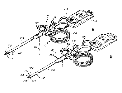
Referring to Figure 1B, device 100 may include handle assembly 300, capture
assembly
302, and needle 138 according to an embodiment of the present invention.
Handle assembly 300
generally has distal end 304 and proximal end 306. Handle assembly includes
shaft 308 and
actuator 309. Shaft 308 extends from distal end 304 of handle assembly 300 and
is generally
adapted to be extended into the chest cavity of a patient. Actuator 309 is
positioned proximate
proximal end 306. Capture assembly 302 generally has distal portion 310 and
proximal portion
312. Distal portion 310 includes clamping mechanism 314 formed by first
clamping jaw 316
and second clamping jaw 318. In an embodiment, clamping mechanism 314 is
adapted to grasp
and release a valve leaflet. In a further embodiment, first clamping jaw 316
or second clamping
jaw 318 is selectively positionable along a longitudinal axis of capture
assembly 302 in response
to actuation of actuator mechanism 314 to create a space between the interior
surfaces (not
shown) of the first and second clamping jaws 316, 318.
Referring to Figure 1A, device 100 can deliver and manipulate a suture in a
beating heart
and generally includes a handheld suture deployment device 118, and capture
confirmation
system 101, according to an embodiment of the invention. The handheld suture
deployment
device 118 generally includes a suture cartridge 102, a shaft 104, a handle
106, and a needle
assembly 116. Capture confirmation system 101 generally includes fiber optic
cable assembly
108, and leaflet capture verification (LCV) monitor 110. Although device 100
can be used for
CA 02703129 2010-04-16
WO 2009/052528
PCT/US2008/080560
-19-
any number of purposes without departing from the spirit or scope of the
present invention, the
aforementioned platform of components, as is described hereinafter in further
detail, enable the
extending of a shaft through the chest cavity and into a beating heart chamber
to capture a valve
leaflet of a valve needing repair, and to further provide a needle to operably
penetrate the
captured valve leaflet and draw a suture therethrough
Suture cartridge 102 may be pre-loaded suture cartridge 120 or operating room-
loaded
cartridges 122. Referring to Figure 5, pre-loaded suture cartridge 120 can
include a tapered
lower clamp jaw 124, a suture 112, a suture retention system 130, a handle
interface 174, a
channel 131, and a groove on the clamp surface 162a. Suture cartridge 120 has
proximal 198
and distal 196 ends. The lower clamp jaw 124 is located at the distal end 196
of suture cartridge
120. The handle interface 174 is located at the proximal end 198 of suture
cartridge 120. Channel
131 is provided with a pair of openings, a first opening which is located on
the top surface, and a
second opening which is located on the bottom surface of suture cartridge 120.
Channel 131 runs
vertically through suture cartridge 120, and is located near the proximal end
198 of suture
cartridge 120, such that channel 131 and handle interface 174 are located
generally adjacent to
one another. Intermediate channel 131 and lower clamp jaw 124 is a cartridge
shaft 176.
Referring to Figures 4A-4E, 4G-4J and Figure 5, lower clamp jaw, or distal tip
portion,
124 is provided on the distal end of suture cartridge 120 according to an
embodiment of the
invention. For example, lower clamp jaw 124 and upper clamp jaw 128 may work
cooperatively
to form a low profile, tapered tip grasping device. Lower clamp jaw 124
generally includes a
low profile tip 180, a lumen 182, a groove 162, a lower clamp surface 126 and
two channels 163.
Lumen 182 extends from the distal end to the proximal end of lower clamp jaw
124, parallel to
the axis of cartridge shaft 176. Lumen 182 can be substantially straight, with
an inner diameter
adapted to receive needle end 146. Groove 162 can be either groove 162a or
groove 162b.
According to an embodiment of this invention, groove 162a is disposed on lower
clamp
surface 126, and is located laterally along surface 126, as depicted in Figure
4D. The depth and
width of groove 162a is generally equal to, or greater than, the diameter of
suture 112.
According to an embodiment, groove 162b is disposed on the upper surface of
lower
clamp surface 126, as depicted in Figures 4G-4J. The depth and width of groove
162b is
generally equal to, or greater than, the diameter of suture 112. For
embodiment of this invention
where groove 162 is groove 162b, cutout 161 is provided, as depicted in
Figures 4G, 41, and 4J.
Cutout 161 is generally a groove that is parallel with, and has a width that
is generally at least
equal to the diameter of lumen 182. The distal end of cutout 161 joins with
groove 162b and the
CA 02703129 2010-04-16
WO 2009/052528
PCT/US2008/080560
-20-
proximal end extends to surface 126. The depth of cutout 161 extends from the
surface of lower
clamp jaw 124 to the centerline of lumen 182.
According to an embodiment, a lower clamp surface 126 is defined by the
generally
planar canted surface of lower clamp jaw 124. Clamping plane 129 is the planar
distal face of
upper clamp jaw 128. Clamp 114 is in a closed position when lower clamp
surface 126 contacts
clamping plane 129. Lower clamp surface 126 has a surface finish generally
suitable for
retaining a grasped valve leaflet. Suitable surface finishes include a
striated or textured surface
finish. As depicted in Figures 4D, 4I-4J, and 5, a suitable surface finish may
include a series of
groves and ridges.
According to an embodiment, the proximal opening of lumen 182 is located to
intersect
groove 162a, as depicted in Figure 4D and view A of Figure 5. According to
another
embodiment, the proximal opening of lumen 182 is located to intersect groove
162b, as depicted
in Figures 41 and 4J.
According to an embodiment, the low profile tip 180 is generally smooth in
shape and
surface finish, and is generally free of sharp edges or points. The low
profile tip 180 is
sufficiently large so that when needle assembly 116 is in a fully extended
position, needle end
146 does not protrude from the distal opening of lumen 182.
According to an embodiment, cartridge shaft 176 is provided with a cross-
sectional
profile that is compatible to be slidably retained within cartridge channel
172. Cartridge shaft
176 is relatively wide, in comparison to the diameter of shaft 104, as
depicted in Figures 50-53.
In an embodiment, the width of cartridge shaft 176 is approximately 65% of the
diameter of shaft
104. In another embodiment, the width of cartridge shaft 176 is between
approximately 65% and
approximately 100% of the diameter of shaft 104. In another embodiment, the
width of cartridge
shaft 176 is less than approximately 65% of the diameter of shaft 104. A wide
cartridge shaft 176
can prevent body tissue from entering clamp 114 from the bottom and presenting
a false capture
by capture confirmation system 101.
According to an embodiment, groove 178 is longitudinally disposed along the
centerline
of the top surface of shaft 176. The depth of groove 178 is generally equal
to, or greater than,
the diameter of suture 112. The cross-sectional area is generally sufficient
to simultaneously
encompass the cross-sectional area of two sutures 112.
According to certain embodiments of this invention, channels 163 are provided
along a
portion of the proximal surface of lower clamp jaw 124, as depicted in Figure
5A. The depth of
channels 163 is generally equal to, or greater than, the diameter of suture
112. As depicted in
CA 02703129 2010-04-16
WO 2009/052528
PCT/US2008/080560
-21-
Figure 5A (view A cartridge in phantom), channels 163 also form a combined
cavity that extends
generally from the bottom surface of lower clamp jaw 124 to the distal top
surface of cartridge
shaft 176. The proximal ends of channels 163 open to groove 178, and the
proximal end of
groove 178 opens to channel 131, thus providing a continuous path for suture
112.
According to an embodiment of this invention, suture 112 is fed through the
suture
cartridge 120, as depicted in Figure 5. The length of suture 112 is generally
divided into two
halves, with the mid-point of the suture length generally located within
groove 162a. Suture 112
runs along the entire length of groove 162a and channels 163. Suture 112 is
also located within
groove 178 and channel 131. The two free ends of suture 112 extend through
channel 131.
The suture retention system 130 may generally include a J-shaped flat spring
located
near the proximal end of suture cartridge 120. The straight portion of the "J"
is generally
parallel with, and located near, the top surface of suture cartridge 120. The
curved portion of the
"J" generally descends into channel 131. The suture retention system 130 is
positioned such that
the curved portion of the "J" forms an interference fit with the distal wall
of channel 131. The
suture retention system 130 acts to retain suture 112 in place within suture
cartridge 102 by
applying a frictional force on the portion of suture 112 that passes through
channel 131. The
frictional force generally acts to retain suture 112 as fed within suture
cartridge 102. Suture
retention system 130 can release suture 112 once needle 138 has been advanced
to a fully
extended position, as depicted in Figure 8C.
According to an embodiment, handle interface 174 is located on the proximal
end 198 of
suture cartridge 120. Handle interface 174 is provided with suitable structure
for being releasably
retained within handle 106. Handle interface 174 may also be provided with
suitable structure for
being releasably retained within plunger assembly 152. Suitable structure may
include, for
example, latches, screws, friction fit attachments, and the like.
As depicted in Figures 5B, 9 and 10, a cavity located on the lower surface of
handle
interface 174 is provided. This cavity mates with a catch mechanism located on
the lower surface
of suture cartridge interface 184. Thus, handle interface 174 is releasably
retained to suture
cartridge interface 184, within the housing of handle 106, due to the catch
mechanism mating
with the cavity. Retention of handle interface 174 can be released through
operation of release
button 160.
According to an embodiment, operating room loaded cartridges 122 are
substantially
similar in form fit and function to pre-loaded suture cartridges 120, except
that operating room
loaded cartridges 122 are not provided with a suture 112.
CA 02703129 2010-04-16
WO 2009/052528
PCT/US2008/080560
-22-
According to certain embodiments of the invention, shaft 104 has a distal end
and a
proximal end, as depicted in Figure 1A. Shaft 104 generally includes lumen
134, upper clamp
jaw 128, cartridge channel 172 and at least one fiber optic bundle 136. In one
embodiment, shaft
104 includes two or more fiber optic bundles 136. In an embodiment, shaft 104
includes four
fiber optic bundles 136.
Shaft 104 generally has a diameter that is approximately 6.5 millimeters. The
diameter
can be greater or less than approximately 6.5 millimeters, however, without
departing from the
spirit or scope of the present invention. Upper clamp jaw, or proximal tip
portion, 128 is located
at the distal end of shaft 104, and handle 106 is located at the proximal end.
Referring to Figure
4F, cartridge channel 172 defines an opening at the distal end of shaft 104.
Cartridge channel
172 may be a keyed channel that runs for substantially the full length of
shaft 104, and is
substantially axially parallel to shaft 104. As a result of its profile, which
generally includes two
shoulders, cartridge channel 172 acts to retain suture cartridge 102.
In one embodiment, shaft 104 generally has a diameter that is less than 12
millimeters. In
another embodiment, shaft 104 generally has a diameter that is less than 9
millimeters.
In one embodiment, shaft 104 generally has a tapered region 200 at the distal
end of shaft
104 and a substantially uniform region extending proximally from the tapered
region, as depicted
in Figure 1A. The uniform region being substantially uniformly cylindrical and
the tapered
region transitioning from a substantially circular end to a substantially
oblong end. In one
embodiment, tapered region 200 is between approximately one centimeter and ten
centimeters in
length. In another embodiment, tapered region 200 is between approximately two
centimeters
and five centimeters in length. In another embodiment, tapered region 200 is
between
approximately four centimeters and five centimeters in length.
In one embodiment, tapered region 200 has a substantially uniform top-to-
bottom height
that is between approximately one quarter of one centimeter and two
centimeters. In another
embodiment, tapered region 200 has a substantially uniform top-to-bottom
height that is between
approximately one-half-of-one centimeter and one and one-quarter-of-one
centimeters. In
another embodiment, tapered region 200 has a substantially uniform top-to-
bottom height that is
approximately 0.81 centimeters.
In one embodiment, the uniform region of shaft 104 has a substantially
circular cross-
section, and the substantially oblong end of tapered region 200 has a side-to-
side width that is
less than the diameter of the uniform region. In another embodiment, the side-
to-side width of
CA 02703129 2010-04-16
WO 2009/052528
PCT/US2008/080560
-23-
the oblong end of tapered region 200 is approximately between approximately
twenty-five
millimeters and two and one-half millimeters less than the diameter of the
uniform region.
Lumen 134 is substantially axially parallel with both shaft 104 and cartridge
channel 172,
according to certain embodiments of the invention. Lumen 134 defines an
opening 135 on the
planar distal surface of upper clamp jaw 128 and a proximal opening in handle
106. Lumen 134
is generally substantially straight. The inner diameter of lumen 134 is
generally appropriately
sized to accommodate needle assembly 116 when inserted alone, and needle
assembly 116 when
extracted with a captured suture 112. Lumen 134 is substantially co-axial with
lumen 182
According to certain embodiments of the invention, fiber optic bundles 136 are
positioned within shaft 104. Each fiber optic bundle 136 generally includes
two fiber optic
strands. Each fiber optic bundle 136 functionally terminated at clamping plane
129, such that a
light input to one of the fiber optic strands results in a reflected, or
refracted optical signal that is
detectable by the other fiber optic strand within a fiber optic bundle 136.
Such a reflected or
refracted optical signal may correspond to the nature and color of any
material that is present at,
or in proximity to, clamping plane 129. Fiber optic bundles 136 are operably
connected through
fiber optic cable assembly 108 to the leaflet capture verification (LCV)
monitor 110.
As depicted in Figures 4A--4E and 4G-4J, lower clamp jaw 124 and upper clamp
jaw 128
work cooperatively to form clamp, or bifurcated tip, 114. According to certain
embodiments of
the invention, clamp 114 which is generally bifurcated, low-profile, and
tapered so as to perform
any number of grasping functions.
Through the actuation of plunger assembly 152, lower clamp jaw 124 can be
extended
distally from upper clamp jaw 128, and can be retracted. When lower clamp jaw
124 is fully
retracted, clamp 114 is in a closed position. In the closed position, lower
clamp surface 126
contacts clamping plane 129. In the closed position, the outer surfaces of
upper clamp jaw 128
and the outer surfaces of lower clamp jaw 124 are substantially coextensive.
In a closed position,
the outside surfaces of lower clamp jaw 124 and upper clamp jaw 128 form a
substantially
smooth surface such that no snagging, rough or sharp edges or overlaps are
formed. When lower
clamp jaw 124 is extended, clamp 114 is in an open position. In an open
position, lower clamp
jaw 124, and upper clamp jaw 128 can be positioned around a piece of tissue,
such as a mitral
valve leaflet. Through the relative movement of lower clamp jaw 124, clamp 114
is operable to
capture a valve leaflet, and needle 138 can penetrate the captured valve
leaflet via lumens 134,
182.
CA 02703129 2010-04-16
WO 2009/052528
PCT/US2008/080560
-24-
According to certain embodiments of the invention, clamp 114 presents an
oversized
leaflet capture area compared to the cross-sectional area of shaft 104.
In a closed position, the outside surfaces of lower clamp jaw 124 and upper
clamp jaw
128 form a substantially smooth surface, according to certain embodiments of
the invention. This
smooth surface can facilitate the insertion of clamp 114 into a tissue opening
that is smaller than
the clamp's cross-sectional area due to the elasticity of tissue over short
periods of time. For the
embodiments of the invention depicted in Figures 4A-4E and 8, the shaft
diameter is
approximately 85% of the maximum diameter of clamp 114. By employing this
ratio of clamp-
to-shaft diameters, body tissues can be stretched within their elastic limits,
which permits an
oversized leaflet capture area within clamp 114 as compared to the cross-
sectional area of shaft
104.
An oversized leaflet capture area, as compared to the shaft's 104 cross-
sectional area, is
presented due to the clamping angle 0, according to certain embodiments of the
invention.
Clamping angle 0 is the angle that clamping plane 129 makes with a horizontal
plane through the
centerline of shaft 104 as indicated by 0 on Figure 4C. For the embodiments of
the invention
depicted in Figure 4C, clamping angle 0 is approximately 120 degrees. In other
embodiments of
the invention, clamping angle 0 is approximately between 115 degrees and 125
degrees. In other
embodiments of the invention, clamping angle 0 is approximately between 90
degrees and 135
degrees. In still other embodiments of the invention, clamping angle 0 is
approximately between
135 degrees and 155 degrees. A clamping angle that is greater than 90 degrees
may result in a
leaflet capture area of clamp 114 that is larger, relative to shaft's 104
cross-sectional area, than
would be possible were the clamping angle 90 degrees. For a clamping angle
that is
approximately 120 degrees, the leaflet capture area of clamp 114 will be
approximately 30% to
40% larger than if the clamping angle were 90 degrees.
In an embodiment of the present invention, a canted tip with increased clamp
travel
improves leaflet capture. In another embodiment of the present invention, an
exchangeable
cartridge improves the simplicity and reliability of suture deployment. In
another embodiment
of the present invention, a suture deployment and manipulator mechanism is
integrated with a
visualization and verification system to deploy sutures within a suture zone
of a valve leaflet.
According to certain embodiments of the invention, clamp 114 is a low profile
tapered tip
grasping device. The shape of the tapered tip facilitates leaflet capture by
providing a large
surface area for leaflet capture, relative to the diameter of the shaft. In
one embodiment, the
surface area for leaflet capture is between 30% and 50% greater than the cross-
sectional area of
CA 02703129 2010-04-16
WO 2009/052528
PCT/US2008/080560
-25-
the shaft 104. In another embodiment, the surface area for leaflet capture is
between 20% and
100% greater than the cross-sectional area of the shaft 104.
According to certain embodiments of the invention, clamp 114 is a low profile
canted tip
grasping device. Clamp 114 can be canted in any number of directions.
Generally, however, the
canted tip is canted up, as depicted in Figures 54-55. A large surface area of
the canted tip,
relative to the diameter of the shaft, facilitates leaflet capture.
A large leaflet capture area can provide a surgeon with certain advantages as
compared to
a smaller leaflet capture area. These advantages include improved ability to
capture a leaflet that
may be damaged or enlarged and a leaflet capture that is more stable. Greater
stability in turn can
provide a surgeon enhanced control of a captured leaflet.
According to an embodiment of the invention, the maximum linear travel of
lower clamp
jaw 124 in relation to upper clamp jaw 128 is between approximately one and
five centimeters.
According to another embodiment of the invention, the maximum linear travel of
lower clamp
jaw 124 in relation to upper clamp jaw 128 is between approximately two and
three centimeters.
According to certain embodiments of the invention, handle 106 is formed to be
manipulated by an operator. Operator may be, for example, a surgeon, or the
controllable device-
interfacing end of a robotic system. In one embodiment, handle 106 is adapted
to be grasped by
the index and middle finger of a surgeon. Shaft 104 extends from the distal
end of handle 106,
and plunger assembly 152 is retained in the proximal end. As depicted in
Figure 9, structure is
provided within handle 106 to retain plunger assembly 152 such that plunger
assembly 152 is
permitted to engage with suture cartridge 102, and to translate in both the
distal and proximal
directions. Suitable structure for retaining plunger assembly 152 within
handle 106 include, for
example, a pin and shackle arrangement, a retaining collar, a boss within a
groove, and the like.
As depicted in Figures 9 and 11, a pin and retaining shackle arrangement is
employed, with the
pin biased against spring 158 within slot 132 of plunger shaft 156, in order
to permit translational
movement of plunger assembly 152. Release button 160 is located on the bottom
surface of
handle 106, as depicted in Figure 10. Release button 160 transfers an
operator's input to the
retaining structure of handle interface 174 in order to uncouple suture
cartridge 102 from plunger
assembly 152. A track may also be provided on the top surface of handle 106
that accepts needle
carriage 144. Markings are provided on the top surface of the handle, adjacent
to the track, to aid
an operator in positioning needle carriage 144.
As depicted in Figures 9 and 11, plunger assembly 152 generally includes
plunger thumb
handle 154, plunger shaft 156, suture cartridge interface 184 and spring 158,
according to certain
CA 02703129 2010-04-16
WO 2009/052528
PCT/US2008/080560
-26-
embodiments of the invention. Plunger thumb handle 154 is formed to be grasped
by the thumb
of an operator and is provided on the proximal end of plunger assembly 152.
Suture cartridge
interface 184 is provided on the distal end of plunger assembly 152 and is
formed to engage and
releasably retain suture cartridges 102. Intermediate suture cartridge
interface 184 and plunger
thumb handle 154 is plunger shaft 156. Slot 132 is located along a portion of
the length of
plunger shaft 156. Spring 158 is located within slot 132 of plunger shaft 156,
and in cooperation
with a pin and retaining structure within handle 106, serves to bias plunger
assembly 152 to a
proximal position relative to handle 106. As a result of the releasable
retention between suture
cartridge interface 184 and suture cartridges 102, the biasing action of
spring 158 is translated to
suture cartridge 102. This biasing action favors retention of clamp 114 in a
closed or grasping
position. Biasing of plunger 152 in this manner facilitates slow and
incremental clamp extension
and contraction.
In one embodiment, spring 158 favors retention of clamp 114 in a closed or
grasping
position with a force in the range of approximately zero pounds per inch of
travel to twenty
pounds per inch of travel. In one embodiment, spring 158 favors retention of
clamp 114 in a
closed or grasping position with a force of approximately five pounds per inch
of travel.
As illustrated in Figure 7, certain embodiments of needle assembly 116
generally include
needle 138, needle handle 140, and needle head, or needle end, 146. Needle 138
is formed from
304 stainless steel wire or other suitable material, is generally circular in
shape, and has a distal
end and a proximal end. Needle end 146 is provided on the distal end of needle
138 and needle
handle 140 is provided on the proximal end of needle 138. Needle end 146 is
flattened and a
notch 148 is provided to create hook 150. Notch 148 is equal to, or greater
than, the diameter of
suture 112. Needle handle 140 generally includes finger tabs 142, and needle
carriage 144.
Needle carriage 144 is permitted to travel along a track that is provided
within the top housing of
handle 106. Such travel permits needle 138 from moving from a starting
position (needle end
146 is within upper clamp jaw 128, as depicted in Figure 51) to a fully
extended position (needle
hook 150 within lumen 182). Needle carriage 144 is also permitted to travel in
a proximal
direction along the track, such proximal travel extending to a position where
needle carriage 144
disengages from the track, and needle assembly 116 is removed from the
handheld device 118.
Markings provided adjacent to the track aid an operator in selecting the
correct position of the
needle carriage 144 in order to achieve a desired position of needle 138. A
detent is also
provided to aid in locating the starting position of needle assembly 138.
Finger tabs 142 fan out
from the centerline of needle assembly 116 and in so doing, act to prevent
needle carriage 144
CA 02703129 2010-04-16
WO 2009/052528
PCT/US2008/080560
-27-
from being inadvertently displaced. In order for an operator to displace
needle carriage 144, an
operator must first grasp and press finger tabs 142 together, and then needle
carriage 144 can be
displaced along the track. In one embodiment, a biasing member opposes the
movement of
needle carriage 144 to a distal position.
According to certain embodiments of the invention, fiber optic cable assembly
108
generally includes fiber optic cable 166 and strain relief 164. Fiber optic
cable 166 generally
includes four (4) fiber optic bundles 136 that run from the distal surface of
upper clamp jaw 128
to the leaflet capture verification (LCV) monitor 110. The four (4) fiber
optic bundles 136 are
bundled together within fiber optic cable 166 and are jacketed with a medical
grade PVC cover,
or other suitable covering material. Strain relief 164 is provided at the
interface between fiber
optic cable 166 and leaflet capture verification (LCV) monitor 110 as depicted
in Figure 12.
According to certain embodiments of the invention, fiber optic cable assembly
108 is at
least two-hundred-and-twenty centimeters long. For these embodiments, in an
operating room
setting, LCV monitor 110 can be placed outside of the sterile field, which
results in the option to
package device 100 in such a manner that LCV monitor 110 need not be
sterilized.
According to another embodiment, a fiber optic connector (not depicted) can be
used to
operably connect fiber optic cable assembly 108 to LCV monitor 110. The use of
such a
connector permits the sterilization and sterile packaging of the handheld
device 118 and fiber
optic cable assembly 108, while the LCV monitor 110 can be separately packaged
in an
unsterilized condition. In an operating room setting, handheld device 118 and
fiber optic cable
assembly 108 can be introduced into the sterile field, while LCV monitor 110
can be placed
outside of the sterile field, within surgical line-of-sight of a TEE monitor,
and the fiber optic
connector used to operably connect LCV monitor 110 and fiber optic cable
assembly 108.
As depicted in Figures 12 and 13, leaflet capture verification (LCV) monitor
110
generally includes power button 168, four (4) LED displays 170, housing 186,
circuit board 188,
and an internal power supply 190, according to certain embodiments of the
invention. Housing
186 includes an integrated loop which is adapted to be securely clipped or
hung such that the
LED displays 170 of LCV monitor 110 can be placed within surgical line-of-
sight of a TEE
monitor. Disposed on circuit board 188 is internal power supply 190, power
button 168 and a
light source, such as an LED. For other embodiments, more than one light
source can be used.
Circuit board 188, internal power supply 190, power button 168 and the light
source are all
operably connected in a manner familiar to those who are skilled in the art.
Activation of power
button 168 results in the light source being turned on/off. Four sets of fiber
optic bundles 136
CA 02703129 2010-04-16
WO 2009/052528
PCT/US2008/080560
-28-
enter housing 186 via fiber optic cable 166 and strain relief 164. Each fiber
optic bundle 136
generally includes two fiber optic strands. For each fiber optic bundle 136,
one of the fiber optic
strands is operably connected to the light source, while the other fiber optic
strand is operably
connected to one of the four (4) LED displays 170. Power button 168, the four
(4) LED displays
170, circuit board 188, the an internal power supply 190, and the light
source(s) are all contained
within housing 186. The four (4) LED displays 170 are visible to an operator
from outside of
housing 186, and power button 168 is operable from outside of housing 186.
In operation, device 100 can be used to attach a suture within the suture
target zone 194
of a valve leaflet, as depicted in Figure 43. To accomplish this, the device
100 may employ a
visualization and verification system. The visualization and verification
system integrates
external transesophageal echocardiography (TEE) to visualize a valve leaflet
in multiple axes
and fiber optics to verify leaflet capture. In an embodiment, suture target
zone 194 is generally
two millimeters from the leading (prolapsing) edge of the leaflet. In another
embodiment, suture
target zone 194 is one millimeter wide and has a centerline that is located
two millimeters from
the leading (prolapsing) edge of the leaflet. In another embodiment, suture
target zone 194 is one
millimeter wide and has a centerline that is located three millimeters from
the leading
(prolapsing) edge of the leaflet. In another embodiment, suture target zone
194 is one millimeter
wide and has a centerline that is located four millimeters from the leading
(prolapsing) edge of
the leaflet. In another embodiment, suture target zone 194 is greater than one
millimeter wide
and has a centerline that is located between two millimeters and five
millimeters from the
leading (prolapsing) edge of the leaflet. In another embodiment, suture target
zone 194 is less
than one millimeter wide and has a centerline that is located between two
millimeters and five
millimeters from the leading (prolapsing) edge of the leaflet. In one
embodiment, the fiber optics
include a leaflet capture verification (LCV) monitor 110 and a fiber optic
cable assembly 108, as
depicted in Figures lA and 12.
Referring to Figures 4E and 4F, in an embodiment, fiber optic bundles 136
terminate at
upper clamp jaw 128 in a configuration that surrounds lumen opening 135. In
another
embodiment, fiber optic bundles 136 terminate at upper clamp jaw 128 in a
configuration that is
near lumen opening 135. Those skilled in the art will realize that many
variations in the
configuration of the placement of the terminations of fiber optic bundles 136
at clamping plane
129 are possible in order to meet the spirit and scope of the present
invention. The identification
of certain configurations is not intended to exclude others which are not
identified, but are
provided as examples of possible configurations.
CA 02703129 2010-04-16
WO 2009/052528
PCT/US2008/080560
-29-
Fiber optic bundles 136 are operably connected through fiber optic cable
assembly 108 to
the leaflet capture verification (LCV) monitor 110, according to certain
embodiments of the
invention. When a valve leaflet has been grasped in clamp 114, the LCV monitor
110 displays a
light transmission that corresponds to the configuration of fiber optic
bundles 136 at clamping
plane 129, and which identifies whether the valve leaflet is properly captured
in clamp 114.
According to certain embodiments, the present invention can be used with
robotic multi-
axis control and manipulation of the device. Proximal control of the
instrument can be achieved
with a system interface comprised of the necessary electrical and electro-
mechanical
interconnects to actuate the mechanical operations of the instrument.
According to an
embodiment, the distal tip of the device can have a rigid shaft. According to
another
embodiment, the distal end of the device can have an articulating, multiple
axis tip for
orientation of the clamp and suture delivery.
According to certain embodiments of the invention, the movable tip typically
remains in
the closed position during thoracoscopic insertion and manipulation of the
handheld device 118.
As desired by an operator, plunger 152 can be manipulated to separate the two
portions of the
moveable tip, as depicted in, for example, Figures 4D-4E, 40-4J and 49.
According to certain embodiments of the invention, clamp 114 is biased to a
closed
position through the use of spring 158, or other biasing member. A clamp that
is biased closed
aids in leaflet capture verification as it can provide a surgeon with a
distinctive tactile feedback
when a leaflet has been captured, as compared to when the result is a failed
or partial leaflet
capture.
In practice, certain embodiments of the present invention can be used to
attached a suture
to the suture zone of a valve leaflet in a beating heart, as depicted in
Figures 14-42 and 46-48.
In one embodiment, the apex of the left ventricle is accessed. Such access can
be obtained by
thoracotomy or other suitable surgical technique. Shaft 104 of the handheld
suture deployment
device 118 is then inserted through the apex of the heart into the left
ventricle using
transesophageal echocardiography (TEE) to guide the surgeon. A purse string
suture at the site of
left ventricular apical access can be used to control blood loss.
As depicted in Figures 14-16, while the heart is beating, the movable tip of
the platform
is used to guide the capture of a flailing leaflet as clamp 114 is closed. A
surgeon can use
external transesophageal echocardiography to guide the placement of the
movable tip relative to
a target leaflet. Through further use of transesophageal echocardiography, as
well as the tactile
feel of plunger 152, and LCV monitor 110, a surgeon can verify leaflet
capture.
CA 02703129 2010-04-16
WO 2009/052528
PCT/US2008/080560
-30-
Once the leaflet is captured, a surgeon can verify capture by examining the
leaflet capture
verification (LCV) monitor 110 to assure leaflet tissue is present. In an
embodiment, the four
LED displays 170 of the LCV monitor 110 present red when blood is present at
clamping plane
129, as depicted in Figure 17, while a display of four white lights indicates
that the tissue has
been fully captured by the movable tip, as depicted in Figure 18.
In one embodiment, an operator can penetrate the leaflet with needle 138 and
retrieve
secured suture 112 from the lower clamp jaw 124 by engaging needle assembly
116. First,
needle 138 is advanced by guiding the needle assembly carriage 144 forward, or
toward the
distal end of the platform as depicted in Figures 19-24 (the movable tip is
illustrated in phantom
in Figures 21-24 so that the advancement of needle 138 can be visibly
depicted). Once needle
116 is fully advanced, the needle assembly is rotated to engage suture 112 as
depicted in Figures
25-27 (the movable tip is illustrated in phantom in Figures 25-27 so that the
rotation of needle
138 can be visibly depicted). The suture loop is retrieved by retracting
(movement is in the
proximal direction) the needle assembly entirely from handheld device 118 as
depicted in
Figures 28-32. The handheld device 118 can then be extracted from the
ventricle while
maintaining control of both ends of the suture as depicted in Figures 33-35.
In another embodiment, no rotation of needle 138 is necessary. A surgeon
advances
needle 138 by guiding the needle assembly carriage 144 forward, or toward the
distal end of the
platform as depicted in Figure 8C. Once needle 116 is fully advanced, needle
hook 150 engages
with suture 112, as depicted in Figure 8, when needle assembly carriage 144 is
retracted as
depicted in Figure 8D. Needle hook 150 can advance past suture 112 without
dislodging suture
112 from groove 162 because suture retention system 130 acts to retain suture
112 as threaded
on and within suture cartridge 102. Suture retention system 130 releases
suture 112 once needle
138 has been is fully advanced.
In embodiments of the invention that have cutout 161, handheld device 118 can
be
extracted with clamp 114 in a closed position. This is because cutout 161
petmits suture 112 to
be clear of clamp 144 after the suture loop is retrieved from handheld device
118. Extracting
handheld device 118 with clamp 114 in a closed position facilitates the
extraction.
In one embodiment, the non-loop end of the suture 112 is passed through the
loop to
create a girth hitch on the leaflet as depicted in Figures 36-41 and 47-48.
The girth hitch
provides for distributed stress on the leaflet with two suture legs and avoids
the need for a knot at
the site of leaflet capture.
CA 02703129 2010-04-16
WO 2009/052528
PCT/US2008/080560
-31-
In one embodiment, a surgeon can thread one of the free ends of the suture 112
into an
operating-room loaded cartridge 122 and repeat the capture process on an
adjacent (non-flailing)
leaflet to create leaflet plication or what is commonly known as the Alfieri
stitch.
In other embodiments, the handheld device 118 can be adapted to form different
types of
knots or stitches that can be used for mitral valve repair. This can be
accomplished through
changes to one or more of: the relative location of the needle within the
shaft; the relative
orientation of the suture within the distal tip; the configuration of the
suture within the distal tip;
the relative orientation of the needle hook; the addition of one or more
needle ends to the needle
assembly; and the relative locations of multiple needle ends within the shaft.
At this stage, the surgeon can visualize the function of the mitral valve
leaflet using TEE
as depicted in Figure 42. An operator can then incrementally adjust the
tension on the suture,
while monitoring the corresponding mitral valve regurgitation through the use
of TEE, to allow
for ideal coaptation of the mitral valve leaflets and consequently a reduction
or elimination of
MR. If the competency of the mitral valve is satisfactory, the suture can be
secured to a suitable
location. Suitable locations for this purpose can include the epicardium, a
papillary muscle and
other like locations. Securing the suture can be accomplished using a standard
surgical knot and
pledget.
In one embodiment of the present invention, the process can be repeated by
removing
exchangeable cartridge 102 from the handheld device 118 and replacing it with
a pre-loaded
suture cartridge 120. In another embodiment, the process can be repeated by
removing
exchangeable cartridge 102 from the handheld device 118 and threading a suture
112 into
operating room loaded cartridge 122 which can then be installed into handheld
device 118.